Embed Size (px)
Citation preview
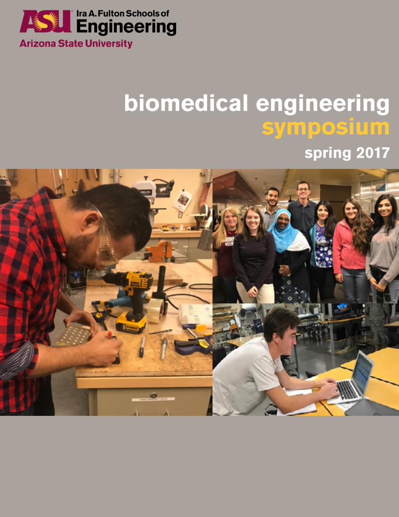
biomedical engineering symposium
spring 2017
4 Welcome5 Acknowledgements
bme capstone projects
6 1: Glucogoals: an affordable method to measure glucose in developing countries6 2: Optogenetic photomask system for spatiotemporal control of stem cell differentiation6 3: Nobleguard: prevention of soft spinal tissue tears7 4: Retrac: hydrocephalus specialized surgical retractor7 5: Biomimetic lumbar spine testing model8 6: Pedgun: the custom spinal screwdriver for spinal fixation8 7: Expandable intervertebral implant for lumbar spinal fusion surgery8 8: Stegospine9 9: Spine-x: retractable anchoring pedicle screw9 10: Stepplus: a parkinsonian gait feedback device9 11: C-port: a detachable hemodialysis catheter10 12: Non-invasive periodontal pocket measuring device10 13: H.O.P.E. – head orthostatic & posture evaluation11 14: The fishbone prosthetic: a lightweight press-fit myoelectric prosthetic made with nitinol actuators and a thermal relief socket11 15: Bio-tac: point-of-care monitoring to improve organ transplant patient health12 16: Development of a rapid diagnostic tool for detecting navajo neurohepatopathy12 17: Automated rehabilitative technology (A.R.T.)12 18: Nucleic acid transport in resource limited settings13 19: Troxie – tracking oximetry for pediatric use13 20: Alcohol detection monitor for breast milk13 21: Kinderfeed14 22: Orthocool: cooling cervical orthoses14 23: Mantis – adaptive athletic ice climbing prosthetic15 24: Ocutrack15 25: Pancreatic cancer diagnostic sensor15 26: Regulation of amniotic fluid via vesicoamniotic shunting for bladder outlet obstruction in utero16 27: Vital check: home care wearable system for detection of internal bleeding of geriatrics experiencing blunt trauma from injury-related falls16 28: Syringe solutions: a self-stirring syringe pump for long term drug infusions17 29: 3d tracking-assisted functional region mapping tool for awake neurosurgery17 30: Rethro – a dynamic splint for prevention & correction of hand deformities17 31: Hydrate Meter18 32: Wireless sensor network (WSN) for health care monitoring and emergency alarm system18 33: Oxysaurus pediatric oximeter18 34: Trauma in the ER: a continuous lactate flow system19 35: Trauma-gel: traumatic would clotting device19 36: Illumilimb19 37: Neural stem cell delivery device20 38: Vendia dx- continuous wound monitoring for venous leg ulcers20 39: Rise: the assistive lift shower chair21 40: Recyclable and sterile surgical template for grafting surgeries

21 41: Locking cap for posterior segmental fixation of the spine21 42: Multiple sclerosis autoinjector stabilizer22 43: Gastrislow – device to slow bolus flow rate into small intestine22 44: S.R.S stability rating sensor. (formerly C.P.S concussion prevention sensor)
masters applied projects page
23 1: Spinal rod strain comparisons: ASTMF1717 vs. flexibility; worst-case vs. cadaveric model23 2: Sleep apnea detection and alarming device for infants23 3: Quantitatively assessing levels of oxygen at the neural interface using MR techniques; a feasibility study24 4: Piezoelectric breast cancer marker24 5 Sensitivity of lyapunov exponents calculated using marker and accelerometer data24 6 Effect of flow on nanoparticle adhesion in drug delivery25 7: Developing a matlab program to control the motionstim 8 electrostimulation device25 8: Locomotor adaptation on implicit visual feedback distortion and split-belt treadmill26 9: Soft robotics third arm26 10: Distributed platform for real-time analysis of neural activity26 11: Characterization of the anterior longitudinal ligament for computer modeling the human spine27 12: Interpreting carotid artery scans usingw single shot detection27 13: Passive patient authentication for foreseeable use and misuse of wearable devices27 14: Optimization of models for experimentation to assess gaseous gradients for metabolic study applications with contrast agents for MR technologies 28 15: Changes in compensatory stepping and handrail grasping during balance disturbances28 16: Computer modeling of human brain surface micromotions as a result of respiration28 17: Surgical navigation for implanting pedicle screws during lumbar spinal fusion29 18 The impact of faculty development workshops on shifting faculty teaching beliefs and classroom practice toward student centeredness29 19: Understanding the kinematics of transferred training between functionally-different upper extremity tasks using Cyber Glove technology29 20: Predictive engineering - modeling availability for medical devices30 21 Flexible pressure sensor for measuring intracranial Pressure (ICP)30 22: Real time control and stimulation testbed for hand prosthetics30 23: An interactive GUI for computing IVIM parameters from diffusion weighted MR images and signal intensities from dynamic contrast enhanced images31 24: TearScan EC31 25: Evaluation of approaches to implantation of microelectronics for advanced neuroprosthetics31 26: The development of a wearable device that continuously monitors blood alcohol content through the measurement of ethanol levels in sweat32 27: Requirements management configuration for design spine curriculum32 28: Assessment of walking speed and external weight on local dynamic stability32 29: EMG processing of pathological activity related to spasticity33 30 Wireless patency sensing stent33 31 Investigation: how gene expression affected by its adjacent nucleotide sequence
On behalf of the students, staff, faculty and affiliated colleagues of the School of Biological and Health Systems Engineering, one of the six schools in the Ira A. Fulton Schools of Engineering at Arizona State University (ASU) and the Harrington Bioengineering Program along with our clinical and industrial partners, it is our pleasure to once again welcome you to our annual design symposium. Proudly displayed before you in this annual symposium are the collective creative outcomes developed by our biomedical engineering senior capstone designers and masters applied project candidates that exemplify this culminating event. It is a testament to the wide range of expertise provided by our dedicated mentors and professional staff who, year in and year out, support the next generation of biomedical engineering scientists and designers who are expected to solve the pressing global grand challenges in health care. In addition, with an intensifying culture of innovation continuing to emerge at ASU and within the greater Arizona community, the growing entrepreneurial spirit will continue to provide unprecedented opportunities to our biomedical engineering students who will have acquired the skill sets to become the next generation of health care technology leaders in the 21st Century. Our ability to build our entrepreneurial capacity and engage in new global partnerships is better than ever. Please come join us in this exciting and rewarding journey!
Marco Santello, PhDDirector, SBHSEHarrington Endowed Chair & Professor
Vincent Pizziconi, PhDFounder & Director SBHSE Design Studio
biomedical engineering capstone design & masters applied project instructors
James Abbas, associate professorMichael Caplan, associate professor
biomedical engineering faculty advisory design group
Jeffrey Kleim, associate professor Jeffrey La Belle, assistant professorVincent Pizziconi, associate professorBarbara Smith, assistant professorMichael VanAuker, lecturer
Jeffrey Kleim, associate professorVincent Pizziconi, associate professor
Casey Ankeny, lecturerJerry Coursen, lecturerMichael Caplan, associate professorEmma Frow, assistant professorAntonio Garcia, professorKarmella Haynes, assistant professor
capstone teaching assistantsAnngela Adams, MS candidateVlad Voziyanov, PhD candidate
facilitatorsKirsten Jefferys, MS candidateWilliam Langenback, MS candidateElizabeth Lopez, MS candidateBlake Woods, MS candidate
design studio staffMichael Sobrado, lab coordinator sr.
SBHSE faculty and mentorsJames Abbas, associate professorCasey Ankeny, lecturerDavid Brafman, assistant professorChris Buneo, associate professorMichael Caplan, associate professorJerry Coursen, lecturerMo Ebrahimkhani, assistant professorDavid Frakes, associate professor Emma Frow, assistant professorAntonio Garcia, professorBradley Greger, associate professorLeland Hartwell, nobel laureate and professorKarmella Haynes, assistant professorSteve Helms Tillery, associate professor Claire Honeycutt, assistant professorTony Hu, associate professorSamira Kiani, assistant professorJeff Kleim, associate professorVikram Kodibagkar, assistant professorJeffrey La Belle, assistant professorThurmon Lockhart, professorSteve Massia, associate professor
Jit Muthuswamy, associate professorMehdi Nikkhah, assistant professorScott Parazynski, university explorer, professor of practice Vincent Pizziconi, associate professorRosalind Sadleir, assistant professorMarco Santello, director, professorSydney Schaefer, assistant professor Barbara Smith, assistant professorSarah Stabenfeldt, assistant professorJamie Tyler, associate professorMichael VanAuker, lecturerBrent Vernon, associate professorXiao Wang, associate professor
SBHSE advising staff
Laura Hawes, academic success specialistJessica Meeker, academic success specialist, sr.Keli Palmer Greenhagen, assistant director, academic servicesElizabeth Tripodi, academic success specialist Robbie Runk, academic success specialistKellie Spear, student services coordinator associate
SBHSE staffTammera Cameron, business operations specialist, sr.Mark DiStasi, associate director of developmentDebbi Howard, business relations coordinatorJessica Jensen, business operations specialistJayson Johnson, research advancement adminSean Jones, research advancement admin, sr.Alana La Belle, laboratory managerElaine Miller, executive administrative support specialistSolo Pyon, systems support analystMichael Sobrado, lab coordinator, sr.Tomi St John, business operations manager
bme capstone projects
1: GLUCOCOALS: AN AFFORDABLE METHOD TO MEASURE GLUCOSE IN
DEVELOPING COUNTRIES
Nicole Fisk, Kaleia Kramer, Fionnuala McPeake, Paige Stokes
mentors: Dr. Michael Caplan – SBHSE | Dr. Michael Goryll - ECEE | Dr. Jan Snyder
Diabetes is the fastest growing chronic disease in the world. The recommended amount of testing for a diabetic is between 4-8 times per day, and the standard method for measuring glucose is the finger-stick method using a glucometer. The dilemma is that the average price per test strip is 25 cents, for the Luo community in Bondo, Kenya the average salary is about $2.00 per day; this creates a cost constraint for testing with an ideal value of $0.01 per test. The current price of test strips isn’t feasible and therefore, many diabetics in this community don’t test their blood sugar more than once a week. GlucoGoals aims to provide cost-effective, sustainable solutions to diabetes management in developing countries. Our device is focused around measuring glucose concentrations using polarized filters and using the resulting optical rotation of glucose to determine the concentration of a sample. This will allow the device to be cheap and reusable. Costs will be reduced further by ensuring that potential solutions and materials are reusable and accessible within Kenya. The unit manufacturing cost will be roughly $14.00, since the device is reusable, if a user tests 4 times per day for a year, the price per test will be roughly one cent. Due to the existence of predicate glucose monitoring device and associated risk associated with testing blood, our polarimeter will be a class 2 device. The device has the potential for patentability since polarimetry is predominantly used in the food industry. The unique use for this method and the resulting device design are novel. In addition to this, the device design is as simple as possible to allow local Kenyans to manufacture the device. Since Kenya has very little governing over their medical device development and manufacturing there will be no reimbursement for these devices.
2: OPTOGENETIC PHOTOMASK FOR SPATIOTEMPORAL CONTROL
OF STEM CELL DIFFERENTIATION
Vincent Avila, Emigdio Esquivel, Suyen Go, Austin Tielke
mentor: Dr. Mo Ebrahimkhani – SBHSE
Being able to generate human tissues from stem cells for disease modelling and pharmaceutical screening would speed the process of drug development and its journey from its experimental phase to its actual implementation in patients. There are several tools used for stem cell differentiation, but they lack parameters for spatiotemporal control. Utilizing optogenetic technology, our team has designed an optogenetic photomask system research tool to add spatial and temporal control. This biomedical research tool targets tissue engineering and regenerative medicine markets in the medical device and diagnostics industry. Projected customers include research institutes and facilities, academia, and R&D departments of regenerative medicine companies. In 2015, global tissue engineering and regenerative medicine markets were valued at $23.2 billion and $4.254 billion, respectively. Over the next decade, tissue engineering is expected to exceed $94.7 billion and regenerative medicine is expected to exceed $30.237 billion. The Optogenetic Photomask System was interfaced with a photoactivatable transcription system. This transcription system employs a photolyase to stimulate gene activation within stem cell populations upon exposure to blue light (465nm). The photomask we developed provides spatial control, while the light source can program temporal control to the millisecond. The system was optimized for a 24-well cell plate and was tested for cell culture compatibility with regards to its interaction with a sterile environment and a biological incubator. While the photomask design was a simple star for proof-of-concept, we look forward to designing patterns which are morphologically and physiologically relevant in the future. Currently, the manufacturing cost and for one unit is $520.58 and the sales cost is projected to be $2500.00 initially, with no potential for reimbursement from Medicare/Medicaid. Grant and research funding are potential methods of payment for this device once the team has patented its components.
3: NOBLEGUARD: PREVENTION OF SOFT SPINAL TISSUE TEARS
Nima Afzalian, Shayan Naeini, Josh Sarbolandi
mentors: Dr. James Abbas – SBHSE | Dr. Michael Bohl – BNI
Incidental durotomy, or dural tears, is a common complication associated with spinal surgery that leads to spinal fluid leakage, chronic back pain, and longer hospitalization time. According to the Journal of Medical Devices, this complication arises in roughly 16% of U.S. spine operations a year. Current surgical instruments, such as the Kerrison Ronguer, remove sections
of bone from the spinal column but frequently cause spinal tears. There is a clinical need for the develop of a device that performs the same function as the Kerrison Rongeur, but limits the incidence of dural tears. The NobleGuard design is composed of an intricate safety guard that would attach to current Kerrison Rongeurs and prevent the device from harming the underlying soft spinal tissue during an operation. Since the obstruction of a surgeon’s view plays a major role in creating dural tears, a suction component will allow surgeons to remove any bodily fluid without having to stop their operation minimizing the duration of the procedure. This is more accessible for the surgeon and reduces the overall operation time. The attachment is meant for one-time use; this will reduce cost and need for sterilization by the SPD department. The device is considered to be Class II and will be following the De Novo regulatory pathway due to lack of predicate devices in the market. The manufacturing cost of the device is $14.34 and will be sold for the low cost of $29.99 to healthcare facilities without needing any reimbursement processes. NobleGuard’s manufacturing process is fully considered for mass production and easy process of assembly. Upon completing numerous patented searches, the NobleGuard has the potential to be fully patentable due to the lack of prior designs for these attachment components. NobleGuard’s safety component through verification testing to be resistant to below 33 N with 95% confidence interval. The device was validated by having neurosurgeons at BNI complete a survey with a target goal of 95% satisfaction to be achieved from their feedback. The NobleGuard should decrease the risk of durotomy and ensure ease of operation for surgeons. The surgical instrument market is expected to grow to 15.7 billion over the next 5 years with the U.S making up roughly one-third of this growing market.
4: RETRAC: HYDROCEPHALUS SPECIALIZED SURGICAL RETRACTOR
Callaway Freeland, Diego Ibarra, Omar Al-Fayhani, Taylor Olvey
mentor: Dr. Michael Bohl – BNI
This new retractor consists of an ABS plastic and a thumb clamp component to cease the flow of fluids from a patient via a catheter. Tensile strength testing, electromagnetic testing, and sterilization testing experiments have verified that the retractor will function as the customers have required. The device is able to withstand 10.6N of force for an extended period of 6 hours, which exceeds the amount of force being applied during procedures that last roughly 30 minutes to an hour. The design is similar to current models with the addition of the clamp component as well as an alternate material, which increases the overall efficiency of the Hydrocephalus procedure. A change in material alone is not viable for patentability, so we looked further into the treatment of Hydrocephalus to improve our design. With the addition of a clamp to secure the loose catheter during the surgery, we are confident that this redesign can be patient. Our device will be labeled as a class 2 device under the FDA, which will require a 510(k) premarket notification that our device is as safe and effective the stainless steel counterparts. Our current design has shifted from a focus on the ability to 3D print to a viable design that can be mass produced with industrial plastic molds. With the price of ABS at this time and the shared cost of manufacturing thousands of retractors, each unit will be an estimated $10 to create. We want to charge $25 per unit will give us a profitable product and our customers a far cheaper alternative. Since our device will eventually be cleared as a Category B Class II device, we will be able to argue that our device is reasonable and necessary during the surgery to remove issues such as site contamination and inaccurate scans.
5: BIOMIMETIC LUMBAR SPINE TESTING MODEL
Emma Alina, Gonzalo Ahuactzin, Jordan Krause, Robel Okbe
mentors: Dr. Brian Kelly - BNI & Dr. Michael Bohl - BNI
In order to perform biomechanical tests on the spine to gain research information or validate biomedical instrumentation devices, a testing platform that mimics the properties of a human’s spine is required. Currently, the main options available to research labs are costly synthetic spine models or a bio-hazardous cadaver spine. The main objective of this project is to economically produce a lumbar spine model that mimics the human lumbar spine accurately enough for use in the testing situations mentioned above. The intended customers for this product will be research laboratories for biomechanical testing, doctors, nurses, orthopedic industries, and medical students for surgical training and anatomical demonstrations. The market segment for this product includes research laboratories, spinal surgical training facilities, medical schools, hospitals, and orthopedic companies. It is forecasted that 865 models will be sold annually. To develop a biomimetic lumbar spine model, our team has divided the project into four different problem areas. These problems include developing the vertebral body, the intervertebral disc, and the ligaments independently, and then assembling the components into a complete synthetic lumbar spine model to demonstrate the actuation properties of the human lumbar spine. Some of the most important product specifications that have been identified for the validation of our system are 9 ± 2.1 degrees of flexion, 2 ± 1 degrees of extension, 4.7 ± 2.4 degrees of lateral bending, 2.16 ± 10 N/m^2 of axial compression, and a pedicle screw pullout force of 1002 ± 500 N. These are common tests used in research labs, and successfully mimicking the human lumbar spine in these categories will demonstrate the success of our system.
6: PEDGUN: THE CUSTOM SPINAL SCREWDRIVER FOR SPINAL FIXATION
Caitlin Byrne, Lindsey Macias, Daylin Morgan, Alvaro Rascon
mentors: Dr. James Abbas - SBHSE | Dr. Michael Bohl - BNI
The PedGun is a custom spinal screwdriver created by S.P.I.N. Applications for spinal fixation surgeries. During this invasive surgery, problems can arise including: screw stripping, screw slipping, loosening of the threading, deviation of trajectory, and the dropping of screwdrivers into the patient’s body. The design team has adapted a hand powered, mechanical device that will allow the user to minimize deviation in trajectory and eliminate rotational motion of traditional screwdrivers. The design team applied best industry practices by using known orthopedic surgical instrument standards for optimizing their device. Concerns were raised during validation testing, leading the team to change the orientation of the beta prototype gear system and run a design of experiments around the handle. It was found that the farther away from the axis of rotation the user applies force on the handle, the moment increases. Due to this result, the current design includes a short, but comfortable handle and cylindrical casing. Inside of the casing, a gear rack moves horizontally in order to turn a custom gear made of both a spur and bevel gear. This then turns another bevel gear, the ratchet adapter, a locking mechanism, and a screwdriver head in order to drill into bone. The PedGun has the potential to be patentable based on the application and the structural differences of its predecessors. This is a Class I device for orthopedic manual surgical instruments, and is exempt from 510(K) premarket notification procedures. The device is not reimbursable since the company is marketing the device towards hospitals and not individuals. As the PedGun was designed, the S.P.I.N. Applications team considered the manufacturability of the components used to complete the device. For each unit, it would cost $131.26 to manufacture, making the overall retail price of the PedGun $1,181.99.
7: EXPANDABLE INTERVERTEBRAL IMPLANT FOR LUMBAR SPINAL
FUSION SURGERY
Nathan Flath, Cody Gates, Veda Inamdar
mentors: Dr. Randall Hlubek - BNI | Dr. Jit Muthuswamy - SBHSE
Our device is a class II interbody fusion disc implant targeted primarily at reducing the rate of spinal collapse that is associated with similar devices for lumbar spinal fusion surgery. When collapsed, our device will be 8x40x2mm allowing it to be implemented through a Posterior and Transforaminal approach. Once inserted into the disc space, the multi-link segments will expand laterally outward (similar to that of a stent) and vertical expansion will be induced through the insertion of secondary expansion rods. This expansion places the device primarily on the dense cortical bone and allows it to conform to as much space as possible. This method will also leave an opening to allow the surgeon to insert a large quantity of bone graft which will further assist in the spinal fusion process. Current products fail to address this need - leaving them prone to collapse and migration. Our final product will be made from PEEK (for the body of the device), and Nitinol (for the metal expansion rod). Utilization of PEEK material qualifies this device for reimbursement under Medicare and Medicaid laws. The approximated manufacturing cost of our device will be $300, given large scale production utilizing injection molding. Compressive testing has also been performed on our gamma prototype to determine if our device will sufficiently distribute load while validating our simulation for our device. We are currently pursuing a provisional patent with an ASU patent law student. Future applications and testing for this design is to rescale the design in order to be applicable in thoracic and cervical levels as well as test the potential of using different sized expansion rods to correct for lordosis.
8: STEGOSPINE
Andrew Cable, Teo Ferrari, Aaron Frisby, Sairah Furness
mentor: Dr. Michael Bohl – BNI
A laminectomy is a component of spinal decompression and fusion surgeries in which a portion of the vertebra is removed and its attached muscles severed, giving access to the spinal cord and dura mater. This leaves a dead space in the spine prone to fluid buildup and infections, as well as loss of posture and strength from the muscular damage.. The StegoSpine attaches to the vertebrae that have undergone the laminectomy, filling the dead space while discouraging fluid buildup and infection, while also protecting the dura from injury. The device is outfitted with a set of 4mm wide holes through the vertical plate that allow for a surgeon to suture the muscle tissue back onto a point of leverage, helping to maintain strength and posture. Additionally, the implant has screw points sized for M3 titanium screws on the base of the implant to allow for the surgeon to attach the device to vertebra. These screw points are arranged in two rows of three on either side of the implant, allowing for connection at two ranges of width in the dead space, with the outer set being removed if unnecessary for a given procedure. The implant is currently being modeled using 3D printed ABS, but we plan to ultimately move into 3D printable biocompatible ceramics, preferably silicate bioceramics, which are converted by the body into living bone tissue over time. We currently project our device
will cost $286.69 per unit, with a selling cost of around $500.00. The device is a Class II device under FDA standards, and will likely be reimbursable for spinal fusions under precedent for reimbursing bone grafts. We are currently pursuing a provisional patent in partnership with the ASU School of Law, and our legal consultant who is confident we differ significantly from prior art.
9: SPINE-X: RETRACTABLE ANCHORING PEDICLE SCREW
Eyerusalem Assefa, Angie Chan, Jossel Disengi, Mariama Salifu
mentors: Dr. Brian Kelly - BNI | Dr. Michael Bohl - BNI
Of the estimated 10 million people in the US suffering from back pain, the majority face spinal problems including degenerative diseases, which may require spinal fusion surgery. The surgery utilizes pedicle screws that are inserted into the pedicle bone and aligned to a fixation rod along the spine. Unfortunately, pedicle screws often experience extrusion or loosening from the spine. Spine-X is a modified pedicle screw with a retractable anchoring component that improves stability and eliminates chances of extrusion, loosening, toggling, and other adverse effects. It consists of a hollowed screw that houses the nitinol anchoring rod which is partially split in half lengthwise. When force is exerted downwards on the anchoring rod, it splits through two opposing tunnels that are angled downwards and deploys the anchoring component out of the shaft. This concept is modeled accurately by a titanium iliac screw that is hollowed out to fit a nitinol anchoring rod. Spine-X was verified with pull-out, modified pull-out, and toggling tests, which proved that the anchoring component increased stability by 35%, exceeding our top specifications. Spine-X is currently not patented, but it is an excellent contender for patentability: its anchoring component is much simpler to manufacture and use than most of its competitors with extremely intricate designs. The expected regulatory pathway requires a Premarket Notification 510k, filed under Class II with the device segment of Surgical and Medical Instrument Manufacturing. It is expected to be covered by Medicare and Medicaid, as it is required for spinal fusion surgery. Spinal implants and instrumentation market is projected to exceed $19.54 billion by 2024. Since we expect 0.1% of the market share, we expect to sell 310 units per month upon the launch of Spine-X with a selling price of $599 targeting neurosurgeons, orthopedic surgeons, and insurance companies.
10: STEPPLUS: A PARKINSONIAN GAIT FEEDBACK DEVICE
Brandon Bartels, Arianna Moreno, Maria Jose Quezada, Haley Sivertson
mentors: Dr. James Abbas - SBHSE | Dr. Narayanan Krishnamurthi - CONHI
According to the National Institute of Neurological Disorders and Stroke (NINDS), Parkinson’s disease (PD) affects one million Americans, with 60,000 diagnosed each year. Symptoms associated with PD, such as asymmetric gait, freezing of gait, and shortened step length, increase PD individuals’ susceptibility to falls and ultimately hinders their quality of life. Current rehabilitation techniques intend to improve gait overall. Yet, there are no sufficient at-home rehabilitation strategies to prevent the progression of these symptoms, especially shortened step length. StepPlus is a wearable Class II medical device that provides auditory and haptic feedback through a smartphone application. It measures and tracks step length in real-time, encourages users to actively make changes in their gait, and saves the user’s progress in a short, concise report to their neurologist or physical therapist for reimbursement purposes and modification of treatments. An inertial measurement unit (IMU) placed on the lower extremity will be used to determine acceleration and angular velocity to provide information about the position of the foot during the gait cycle. The magnetometer monitors the trajectory of each foot to decipher step length from stride length; the patentability of this distinction is being explored. Manufacturing is attained at approximately $100, with a $300 sales cost. Combining these aspects through design controls, the device provides step length feedback through the smartphone application with clear contrast for steps of varying lengths to help user’s regain independence when walking. Additionally, by quantifying the individual’s step length progress, StepPlus bridges the gap between clinicians and physical therapists to attain more specific and beneficial therapy. StepPlus aims to prevent falls in PD populations and create opportunity on the forefront of clinical and research gait analysis.
11: C-PORT: A DETACHABLE HEMODIALYSIS CATHETER
Elizabeth Burkett, Dominick Cocciola, Danielle Eldred
mentors: Dr. Michael Caplan - SBHSE | Tammy DeLozier - AKDHC | Dr. Vincent Pizziconi - SBHSE
As of 2015, over 14 percent of the U.S. population suffered from Chronic Kidney Disease, and 468,000 of these patients required dialysis, according to the NIH. The current standard to combat kidney disease is to utilize a constant blood access port so the patient can undergo dialysis, which replaces the function of the kidneys. C-Port is redesigning the central venous catheter, which is a type of blood access for renal failure patients in need of dialysis. Even though patients are able to live with these implanted devices, central venous catheters have around an 85 percent infection rate within or around
the catheter; because of this, the U.S. spends over $1 Billion each year on complications with central venous catheters and the facilities that supply the treatment, in accordance with the NIH. C-Port have been manufacturing a class three medical device, however if the device is proven to be safe and effective it can be marketed through a 510(k) due to a FDA panel meeting in 2013. Our team designed a detachable hemodialysis catheter that reduced infection outside the catheter, improved of quality of life for patients with the implanted device and reduced overall costs. We have achieved these goals by creating a dual lumen catheter that is equipped with detachable and disposable dialysis ports. When the patient is dialyzing, the detachable ports connect to the exterior connector of the device that extends from the chest. When this process is over, the two lumen will be detached, disposed of, and the access ports will be sealed off with an airtight cap to prevent outer microbial penetration. Our device was tested under a series of design of experiments that included flow rate, leakage, and wear ability of the device. The results obtained from the results were compared to the gold standard catheter on the market to ensure accuracy of our device. The gold standard catheter does not have any detachable or disposable pieces and if the device needs replaced a surgery is needed to replace the device our device will allow the potential for no replacement surgery. This innovative hemodialysis catheter has a lifespan of two years in the patient with minimal to no maintenance and will be covered fully under Medicare, as aforementioned. C-Port is creating a revolutionary medical device that aims to lead the kidney disease treatment field to a new forefront with innovative ideas and simplistic beliefs.
12: NON-INVASIVE PERIODONTAL POCKET MEASURING DEVICE
Jesus Calderon, Travis Tibbs
mentor: Dr. Vikram Kodibagkar – SBHSE
Periodontal disease affects 47 percent of Americans over the age of 32 and periodontal pocket depth is a key factor in diagnosis. Currently, dental professionals use sharp, metal probes which offers ambiguous measurements, requires two employees to probe and often causes periodontal tissue bleeding, which is indicative of periodontal disease. Dentists, dental hygienists and the dental offices they work for want a device that measures with greater speed, accuracy, and automation. The Non-Invasive Periodontal Pocket Measuring Device (NIPPMD) will use near-infrared light to photograph the periodontal pocket. This image will undergo edge detection processing in an external program. The probing and measuring process has taken place in under ten minutes with one hygienist. This device will also display periodontal pocket depths in 0.5 mm increments. Secondary design efforts focused on the imaging of hairline tooth fractures, early cavity development, and calculus buildup. These key factors also contribute to periodontal health. A device that measures these factors will not only be innovative but necessary in the future of diagnosing a patient’s periodontal health. Current prototyping efforts have proven near infrared light to be the best wavelength to photograph the periodontal pockets. Many images have been produced showing the efficiency and accuracy of the device. Prototype experiment efforts have also verified the NIPPMD to be accurate to at least 0.5 millimeters. The NIPPMD has been used by a dental professional for validation in its early prototype stages. By using best industry practices and through its design for manufacturing, the unit production costs for the NIPPMD will be around $70 per unit. Like most intraoral dental devices, the NIPPMD will be a class two medical device. It will not be reimbursable through insurance but with its innovative concept, it will be patented at a future date.
13: H.O.P.E. – HEAD ORTHOSTATIC & POSTURE EVALUATION
Christopher Cusick, Ashley Martin, Rachel Wylde
mentor: Dr. Richard Herman – SBHSE
One of the most common forms of muscular disabilities is cerebral palsy (CP), which is an extremely prevalent neurological disorder with no known cure, affecting about 500,000 children in the United States. In the case of CP, there is an impairment of motor function due to abnormal development of the brain, affecting muscle control, motor skills, muscle coordination, posture, and balance. Currently there are no devices that provides objective feedback for physical therapists to aid them in their treatment and diagnosis for children who lack head control. H.O.P.E. is a rehabilitative tool that provides quantitative feedback to the physical therapists to facilitate patient diagnosis and treatment. H.O.P.E. is designed for the use of children, ages 1-5, during the critical developmental stages of growth. The device is a wheelchair wearable item that securely acts as a comfortable headrest, which includes two feedback systems to provide two distinct types of data. Force sensitive resistors are aligned to determine force applied and the location of the force applied. The accelerometer cap functions to calculate the child’s head angle to determine the efficacy of current posture treatment. In turn, this enables physical therapists to better their assessment, treatment and track the child’s progress over time. Due to it’s unique design, and lack of competitors in the market, our device is patentable. H.O.P.E. is expected to cost $179.10 and will be sold for $250. Reimbursement pathways for our device include social security benefits for disabled children and waivers filled out for reimbursement through Medicaid. Since HOPE is an assessment device, it’s classified with the Food and Drug Administration as a class I device, and will not require a 510K. Through verification, H.O.P.E. has outputted accurate and reliable readings with 99.99% confidence, and has been noted to output data in a meaningful way for physical therapists.
14: THE FISHBONE PROSTHETIC: A LIGHTWEIGHT PRESS-FIT
MYOELECTRIC PROSTHETIC MADE WITH NITINOL ACTUATORS
AND A THERMAL RELIEF SOCKET
Kandace Donaldson, Sarah McBryan, Hayden McIver, Fred Sebastian
mentor: Dr. Jeffrey La Belle – SBHSE
According to recent estimations, there are approximately 41,000 patients with upper-limb deficiencies in the United States. The Fishbone Prosthetic is a myoelectric press-fit hand and forearm prosthetic made with advanced manufacturing techniques. The device uses four key technologies to create an affordable and lightweight prosthetic to increase patient comfort and compliance: a press-fit frame and hand design, a porous socket used for heat dissipation, and nitinol actuators for increased biomimicry. The patent-pending press-fit design of the mechanical frame utilizes interlocking two-dimensional pieces to eliminate the need for adhesives and extra hardware. Extensive analysis was done to ensure maximum compressive and torsional strengths while optimizing weight and interstitial free space within the device. This allows for embedded actuation and electrical components as well as customizable weight distribution within the device. Current market socket designs do not allow for airflow to the residual limb, resulting in extreme temperatures, moisture accumulation, and bacterial growth. The Fishbone socket is made from vacuum formed HDPE sheets with uniform perforations for increased airflow and heat dissipation to increase patient comfort. Nitinol wires are used in a novel staggered array configuration to maximize force output of the artificial press-fit hand. This shape metal alloy mimics the movement of muscles, lowering the learning curve of control for new prosthetic users while being lightweight and less bulky than analog motors. The nitinol arrays are actuated and controlled standard EMG sensors. Each component of the Fishbone Prosthetic can be modularly applied and used interchangeably with current market devices for patient point-of-care limbs. The current MSRP for the Fishbone device is approximately $5,000, making it an attractive alternative to $100,000+ myoelectrics for both insurance companies and users. It is a Class II medical device eligible for 510(k) FDA approval, which similar devices have obtained within two years of filing. We anticipate a similar timeframe for approval using similar standards and quality controls.
15: BIO-TAC: POINT-OF-CARE MONITORING TO IMPROVE ORGAN
TRANSPLANT PATIENT HEALTH
Meera Doshi, Minh Pham, Nitish Peela, Swaroon Sridhar
mentors: Dr. Jeffrey La Belle – SBHSE | Dr. David E. Steidley - Mayo Clinic
Each year, 30,000 patients obtain solid organ transplants. These patients are prescribed immunosuppressant drugs to minimize risk of transplant rejection, but these drugs have nephrotoxic effects. Currently, the standard of care for these patients involves bimonthly blood draws, after which the levels of immunosuppressant drug, kidney damage, and immune activation are measured. Bio-Tac is a novel device proposing continuous, point-of-care, and simultaneous monitoring of tacrolimus (a commonly prescribed immunosuppressant), cystatin-c (an indicator of kidney health), and IL-10 (an indicator of immune health) to ensure a holistic and detailed understanding of patient health. Customer needs were identified and include speed of monitoring, connection to emergency medical records, availability to the patient, and accuracy. Initial prototyping leveraged electrochemical impedance spectroscopy, which measures the impedance generated from antibody-antigen binding and relates it to marker concentration. It contains numerous novelties over previous devices, indicating high potential for patentability. Furthermore, the incorporation of a bluetooth sensor allows for direct and immediate communication of results to healthcare providers. The device is classified as a class III device as it needs scientific comparison to the state-of-the-art ELISA testing prior to premarket approval. Thus, the prototype was validated compared to ELISA results based on real patient samples from the Mayo Clinic, which indicated that the accuracy was within 90% of the state-of-the-art. Because the device improves the quality of care at an insignificant fee ($1,500 annually) compared to organ transplant procedures (~$1,000,000), insurance companies will mandate the purchase of the device and fully reimburse patient costs through private insurance, Medicare, or Medicaid. With efficient procurement, supply chain, and outsourcing, the device has a unit production cost of $100, yielding over 1500% profit. Overall, the creation of 45,000 devices per year is expected, as well as the generation of $75 million in annual revenue after a three-year ramp-up period.
.
16: DEVELOPMENT OF A RAPID DIAGNOSTIC TOOL FOR DETECTING
NAVAJO NEUROHEPATOPATHY
Allison Marley, Courtney DuBois, and Meilin Ossanna
mentor: Dr. Michael Caplan – SBHSE
Navajo neurohepatopathy (NNH) is a genetic disorder caused by the R50Q mutation in the MPV17 gene. The disease affects 1-in-1600 Navajo babies and is characterized by brain damage and liver disease/failure. Upon exhibiting symptoms, children in the American Southwest region seek help from Phoenix Children’s Hospital (PCH). Currently, differential diagnosis and gene sequencing is the definitive method for identifying the R50Q mutation that causes NNH. While this process is conclusive, there are limitations, as it requires both money (>$700) and time (3-4 weeks). Thus, Tentacle Tech has developed a rapid diagnostic tool using Tentacle ProbeTM technology (TPTM) to accurately detect the R50Q mutation. TPTM can be used with real-time polymerase chain reaction (qPCR) to obtain both low-cost (~$150) and quick results (~10 hours) while maintaining efficacy. With the TPTM incorporated in qPCR, fluorescence can be measured, and the output can be used to deliver a NNH diagnosis. Tentacle Tech has worked to optimize, verify, and validate the NNH diagnostic test. Optimization experiments determined a 66°C annealing temperature is specific and sensitive for the designed primers. Optimization also revealed that a 55°C annealing temperature is optimal for the TPTM to produce a significant difference in target and non-target fluorescence. Finally, it was proven that a lower copy number of synthetic DNA produces optimal results. Experimentation for verification of the TPTM with target and non-target synthetic DNA demonstrated that the probe has 100% specificity and 100% sensitivity for synthetic DNA. Future work will continue to verify the efficacy of the TPTM by using positive and negative NNH patient samples from PCH. Thus far, clinicians at PCH have validated the results produced by the TPTM in synthetic DNA. Ultimately, the development of this diagnostic tool can help to improve the quality of care for Navajo children and decrease overall health care costs.
17: AUTOMATED REHABILITATIVE TECHNOLOGY (A.R.T.)
Matthew Gerveler, Michelle Sigona, Michael Spina, Shawn Womack
mentor: Dr. James Abbas - SBHSE
Individuals with Parkinson’s disease often experience an abnormal gait, characterized by small, shuffling steps, a difficult time beginning and ending ambulation, and episodes of frozen gait. Many Parkinson’s patients experience an issue often referred to as “freezing”, which involves the temporary, involuntary ability to move. This often occurs in the lower half of the body while momentum continues in the upper half, causing a fall that could lead to serious injury. About 60,000 Americans are diagnosed every year and about 68% of those experience falls. 6.1 million adults use assistive devices, particularly those over the age of 65, the average age of diagnosis being 62 years old for Parkinson’s. Parkinson’s patients and other limited-stability patients will benefit from our assistive device through its rehabilitation and assistance in preventing freezing. Ambulation assistance device distributors, speciality clinics, neurological branches and rehabilitation centers would be interested in our device, along with end users such as physical and occupational therapists, rehabilitation nurses, and orthopedics. Our device will belong to the “Rehabilitative Technology” and “Personal Mobility Device” markets. Walking aids are in a market worth $4.16 Billion USD and expected to grow 11% by 2020. Our chosen concept based off of evaluation of customer needs will focus and consist of a visual cue for step guidance, an IR sensor in communication with an audio cue, a collapsible and stable frame, and a push-down braking system to help patients with low finger dexterity. The device will be lightweight and compact, allowing for easy transportation, with a standard of being able to fit in the overhead bin on an airplane or backseat of a car. The device will also focus on ease of use, allowing any patient/customer to pick up and use the device without requiring complex training.
18: NUCLEIC ACID TRANSPORT IN RESOURCE LIMITED SETTINGS
Cameron Ghods, Nolan Kern, Jordan Nelson, Chris Stark
mentor: Dr. Barbara Smith – SBHSE
In the global effort to fight HIV and AIDS, many institutions conduct research to monitor the evolution of the virus and the response to treatment efforts. Rural research zones, particularly in sub-Saharan Africa, are often lacking in infrastructure and public resources. Research efforts in these resource-limited settings require a simple and, most importantly, less expensive blood transportation device that will preserve the viral nucleic acid with the same effectiveness as current methods. The primary customers of this product are the HIV/AIDS researchers that are attempting to obtain samples from distant, resource-limited areas with minimal budgets. The market segment for a nucleic acid transportation/preservation device falls under In-Vitro Diagnostic Substances, and further as a Research Use Only (RUO) device. According to the US census, this segment accounts for approximately 10% of the US medical device industry’s value of sale, with 200 firms employing 25,000 people. The RNA preservation device uses dried blood spots on absorbent paper that are then be vacuum sealed into a plastic bag containing a silica gel packet. This process removes water from the sample and isolates it from reactive elements in the air that are major contributors to viral RNA degradation. The goal for our device was to
preserve between 50-75% of the viral nucleic acids after enduring up to 1-2 months of storage and transportation at room temperature. The prototypes were verified in an accelerated aging protocol due to time constraints, then the amount of intact RNA was quantified by gel electrophoresis analysis in ImageJ. The current estimated unit manufacturing cost is under 50 cents, which can be greatly reduced through mass production.
19: TROXIE – TRACKING OXIMETRY FOR PEDIATRIC USE
Samantha Brenna, Chloe Houlihan, Maria Morrow, Courtney Van Bussum
mentor: Dr. Erica Forzani – SEMTE
6.2 million children suffer from asthma in the United States. In fact, over half of children who have an asthma attack each year miss school because of their asthma. Research has shown that pulse oximetry can help keep kids in school by reducing emergency department stays. Unfortunately, pulse oximetry technology has changed very little in the thirty years since it was invented. The need for a finger-based attachment means it is difficult and inefficient to use pulse oximeters outside of a physician’s office or hospital setting. Troxie aims to address this problem. Unlike current devices on the market, Troxie gets rid of the need for the cumbersome finger probe traditionally used to measure blood oxygen saturation. Instead, Troxie uses a wrist-based system to allow for accurate measurements in everyday environments, creating more freedom for the user. Troxie is a wrist-worn, wearable pulse oximeter made for children with respiratory illnesses, such as severe asthma. The device can be worn on the wrist, similar to a watch, in order to continuously monitor blood oxygen saturation levels. In this way, an alert can be sent out if a child’s blood oxygen levels begin to drop dangerously low. By preemptively indicating when levels are dropping, appropriate action can be taken to help the child before more severe incidents occur. Using a reflectance technique in combination with accelerometry, heart rate, and time-based components, the device will take readings during sleep, rest, exercise, and motion. This will provide continuous monitoring of the child, thus allowing the child the freedom to move and play, while giving parents ease of mind. Troxie’s mission is to help all children to thrive despite any respiratory condition by making pulse oximetry easy, useful, and convenient for both parent and child.
20: ALCOHOL DETECTION MONITOR FOR BREAST MILK
Courtney Willson, Eli Jacobson, Hillary Bratlien
mentors: Dr. Casey Ankeny – SBHSE | Dr. Jennie Bever - CONHI
Mothers are strongly encouraged to breastfeed for as long as possible due to the significant benefits that breastmilk has over the leading formulas. According to the Center for Disease Control and Prevention, it is recommended that mothers breastfeed for up to two years to maximize these benefits that breastmilk offers to the developing baby. However, mothers that are breastfeeding must carefully monitor what foods and beverages they consume because they metabolize into the breastmilk and are directly passed onto the baby. This means when a mother consumes alcohol, this could be passed on to the baby. This alcohol, when consumed at a level higher than 0.02% has a negative effect on a baby’s neurological and physiological development. With every mother’s body processing alcohol at different rates, there is no sure way for a mother to know the exact alcohol concentration in her breastmilk. BreastAssured has developed a class I wellness device to confidently determine how much alcohol is in a mother’s breastmilk. This device is a quantitative measurement device that is able to repeatedly measure various alcohol levels in breastmilk samples. Currently on the market there does not exist such a device. It works through the use of pressure gradients that change the liquid ethanol in the breastmilk sample into an ethanol vapor. Once in vapor form this ethanol is measured by an alcohol sensor, which is then processed and output as a percentage on an LCD screen. The device is sensitive enough to detect alcohol in a sample containing less than 0.02 percent alcohol with an accuracy of plus or minus 0.01 percent. It is held to the CSLI C60-A standard for blood alcohol content because there currently does not exist a relevant standard for ethanol content in breastmilk. With a retail price of $60 and a unit manufacturing cost of $35 per unit there stands to be a potential profit to be made upon the potential 1.84 million market size of mothers. Simply put, the device will quickly and accurately measure the alcohol concentration of a nursing mother’s breastmilk which will give her the reassurance that her baby is consuming safe milk.
21: KINDERFEED
Callie LaMarche, Sydney Lankford, Patrick McFarland, Juan Patterson
mentors: Dr. Casey Ankeny – SBHSE | Nancy Scherer – Department of Speech and Hearing Science
KinderFeed is a feeding bottle for infants and toddlers with severe feeding difficulties resulting from cleft palate, Down’s Syndrome, premature birth, and heart defects. Infants with these conditions can physically feed from traditional
bottles; however, due to their inability to create negative pressure, these infants burn more calories than they consume during feeding. KinderFeed proposes a bottle that will change the geometric distribution of the bottle, the squeezing site, and by allowing use of the entire hand rather than just two fingers, functioning much like a pipette. The bottle will not require a medical device pathway as it falls under similar regulatory paths as previous baby bottles. Additionally, the bottle will eliminate leaking and clogging at a cheaper price than that of the benchmark products with an expected unit production cost of $4. This cost is anticipated to decrease as the bottle is manufactured in bulk; many of the bottle parts can be created with injection molding or purchased as standard pieces in bulk. The bottle will hold 10 ounces of milk, the same capacity as a standard bottle; as a result, no refilling is required during each feeding session. KinderFeed will be marketed directly to caretakers of the infant for $15 a bottle and $3.50 for additional pieces, such as nipples and other related accessories. Currently, the bottle will be sold in the United States, which has a market size of about $200 million. Eventually, the team intends to make the bottle available in international markets as well at an affordable price.
22: ORTHOCOOL: COOLING CERVICAL ORTHOSES
Jeremy Becker, Alison Llave, Blossom Mendonca, Sahar Mohamed
mentors: Dr. Michael VanAuker – SBHSE & Dr. Joshua Catapano – BNI
According to the National Spinal Cord Injury Statistical Center (NSCISC), the number of cervical spine injuries amount to 48,000 per year with an additional 400,000 spinal fusions annually in the United States alone. Most these cases will require the patients to wear cervical collars for extended periods of time. However, the problem with cervical collars today is that they are highly uncomfortable, causing patient compliance issues. This is particularly true in areas of hotter climates where the patients proceed to remove the collars to cool down their necks, risking spinal misalignment. There are current cooling solutions, although, none of them are fully compatible with the use of cervical collars and their purpose of arresting spinal movement with a maximum angle of 35 degrees. The objective of this project then is to redesign the Class I medical device cervical collar so that it cools the patient while remaining lightweight, with a weight of less than 16 oz. The demographics of our customer base include patients, families of patients, insurance companies, surgeons, and physical therapists. We have been collaborating with these customer groups, partnering with Barrow Neurological Institute (BNI) at St. Joseph’s Hospital and Medical Center, to produce the optimal solution. The collar will comprise phase change material to promote heat dissipation and a surface temperature of at least 15°F less than ambient temperature. The manufacturing cost would amount to $15 including overhead charges, resulting in a $45 sales cost. The product will be sold to companies that manufacture the cervical collars so that they can sell their cervical collars with our pads. Next steps include patenting the idea due to the phase change material’s characteristic to be tuned to improve the product’s specifications.
23: MANTIS – ADAPTIVE ATHLETIC ICE CLIMBING PROSTHETIC
Allison Lind, Shelby Martin, Rene Reynolds
mentor: Dr. Stephen Helms Tillery - SBHSE
The Mechanical and Natural Tool for Ice Scaling (also known as MANTIS) is an adaptive athletic arm prosthetic that will be used to help upper arm amputees continue or start their love for ice climbing. There is no current product on the market that allows upper arm amputees to continue or start ice climbing. The concepts for this device mimic other adaptive athletic prosthetics, but mainly consist of an arm made of carbon fiber connected in the middle by an elbow joint, lined with titanium, with two springs to help the user hit into the ice. It is a unique and new design, and there is the possibility to patent the device. Because this device is a prosthetic device, and not implanted into the body, it is classified as a Class I device. Due to the device being a Class I device, the manufacturing and design regulations are not controlled by the FDA, but the team is still regulating it to design a device with integrity. Some of the key product specifications, that were verified through testing, include how much weight the harness holds (approximately 130 kg), the temperature range the device must function in (-10 to 50 C), and that the device needs to be lightweight (less than 2.5 kg). All of these specifications, and more, were confirmed using a combination of verification testing and three separate design of experiments. This device will be marketed for right around $1,000 and there is no possibility for reimbursement, because it is a recreational tool. The cost of the device is relatively high due to the fact that this is a custom built device that will be tailored to each user’s specific measurements.
24: OCUTRACK
Ryan Bridges, Wei Lu, Ceasar Udave
mentor: Dr. Javier Cardenas – BNI | Dr. Jit Muthuswamy – SBHSE
OcuTrack aims to assist in the assessment of risk of concussion. This device utilizes two micro-cameras, one accelerometer, and a single board computer, to track the user’s eye movements. The software of the system builds unique profiles for individual athletes to obtain baseline eye movement readings. It then compares eye movements at times post concussive impact to determine whether or not the athlete is displaying symptoms of concussion. The cost to produce the gamma prototype of OcuTrack came out to be around $192. By using industry manufacturing practices, OcuTrack would like to reduce the manufacturing cost to around $60 and lease the systems at $200 per system per year. OcuTrack will be filing a PMA (pre-market approval) application with the FDA as a Class II medical device, via the 510 (k) pathway using predicate nystagmus diagnosing devices such as Eye-Sync. Reimbursement for the OcuTrack system can be sought by filing under the medical billing code CPT 96120 which covers any neurophysiological exam administered by computer. OcuTrack is not seeking a patent since development is in the early phases and still has many improvements to be made such as the replacement of the CCTV cameras with CMOS camera sensors, the replacement of the Raspberry Pi with DSP chips, and making the system truly mobile. The OcuTrack was tested against the eye tracking system Tobii, the gold standard of competitors. In a simple t-test comparison for static eye location the OcuTrack had no statistical difference in y direction measurements but in the x direction OcuTrack had a statistically significant measurement with a P value of .1 and 95% confidence. This means that in the x direction the OcuTrack system failed the gold standard but in the y direction the standard was met.
25: PANCREATIC CANCER DIAGNOSTIC SENSOR
Akshara Malla, Keerthana Murali
mentor: Dr. Jeffrey LaBelle – SBHSE
Despite improvements in current diagnostic technologies, pancreatic cancer patients are diagnosed during stage 3 or stage 4 of the disease. About 9 percent of cases are diagnosed with the cancer limited to the pancreas. An electrochemical sensor, using the technique of electrochemical impedance spectroscopy (EIS), was created and tested for use in early pancreatic cancer detection and disease monitoring. EIS is a more sensitive, specific, time and cost effective method to quantify biomarkers in comparison to ELISA, western blot, and electrophoresis. The prototype consisted of a gold disk electrode immobilized with two pancreatic cancer specific antibodies, anti-QSOX1 and anti-CEA. The final marketed device includes disposable test strips multiplexed with anti-QSOX1 and anti-CEA and a separate sensor that quantifies the reaction that occurs within the sample and on the test strip. The prototype and device tests the concentrations of QSOX1 and CEA in peripheral blood samples and pancreatic cyst fluid. Verification tests of the sensor included the use of purified serum samples of QSOX1 and CEA to test the binding between the antigen and antibody pairs in addition to improving the specificity of the output impedance values. The final device will cost around $275 and will be sold to clinics and hospitals for physicians to use on their patients. Based on current sensors on the market, the device itself is expected to be paid for by health administrators, however, the test should be reimbursed by Medicare and Medicaid. The functioning of the device will mirror a self-monitoring glucose meter, however, due to the use of EIS, the sensor and test strip system will fall under Class II and require a premarket application to the FDA before it is commercially marketed and sold. The device has the potential to impact pancreatic cancer patient population, which was 53,070 individuals in 2016 alone.
26. REGULATION OF AMNIOTIC FLUID VIA VESICOAMNIOTIC SHUNTING
FOR BLADDER OUTLET OBSTRUCTION IN UTERO
Kirstin Peters, Niko Vlastos, Shaun Wootten
mentors: Sarah Stabenfeldt - SBHSE | Emmanuel Vlastos - SLU Med
Fetuses diagnosed with Bladder Outlet Obstruction (BOO) cannot properly urinate resulting in bladder swelling which prevents organ development and causes renal failure. According to Cardinal Glennon, one in every 4100 fetuses will be diagnosed with BOO translating into approximately 31,000 fetuses a year. NatEvac hopes to change the lives of fetuses diagnosed with BOO with regulating the amniotic fluid in the fetus’ bladder with its novel ball-magnet pressure regulated mechanism. The NatEvac device designed by Fetia will allow the bladder muscles to develop, as well as providing the fluid the fetus needs for drinking and lungs development.The valve is considered a Class III Implantable device according to the FDA. The unit manufacturing cost will be $118.00 while Fetia plans to sell NatEvac at $413.00. According to the Center of Medicare and Medicaid the medical device is eligible for reimbursement because it sustains life and will go through premarket approval (PMA). NatEvac
would be used by fetal surgeons to treat BOO in parallel with the Harrison Fetal Stent Set offered by Cook Medical. NatEvac was designed to be assembled in the z-axis which is the most efficient orientation for manufacturing and assembly. Fetia went for a simple and sophisticated design that is comprised of only four components: the skeleton, ferrous carbon-steel ball, ball catching screen, and 45N neodymium magnet. The customers are primarily physicians and hospitals that specialize in fetal care. The market size of Fetal and Neonate Care Equipment is predicted to reach 7.8 billion (USD) by the year 2020. NatEvac has potential for patentability because there is no other device like NatEvac on the market as of right now.
27: VITAL CHECK: HOME CARE WEARABLE SYSTEM FOR DETECTION
OF INTERNAL BLEEDING OF GERIATRICS EXPERIENCING BLUNT TRAUMA
FROM INJURY-RELATED FALLS
Naazaneen Maududi, Francis Taguinod, Sadaf Tahmasebi, Jason Yang
mentors: Dr. Vincent Pizziconi - SBHSE | Dr. Michael VanAuker - SBHSE
Based on CDC statistics, falls are the leading cause of fatal/nonfatal injuries among geriatric adults aged ≥65 years in the US. Each year, 2.8 million geriatrics are treated in the emergency room for fall-related injuries. A significant fraction of the elderly who fall experience blunt trauma and those who are on blood thinners are particularly susceptible to internal bleeding, a silent killer that virtually goes undetected. With estimates of more than 1 out of 4 elderlies who fall each taken together with the rapidly growing baby boomer population who, as a group, are turning 65 at a rate of 10,000 per month for practically every month for the next 20 years, the market for Vital Check is significant for the foreseeable future. This is especially true, from wearable diagnostic technology which is anticipated to experience a 35% increase in growth and $34 billion in sales in the US alone by 2020. In response to this critical unmet clinical need, the Vital Check team is developing a wearable, noninvasive diagnostic device that utilizes an embedded algorithm and selected biosensors (pulse oximetry & piezoelectric sensor) that, in integrated form, can discriminate among complex, dynamic physiological events associated with clinically relevant vital signs of internal bleeding. These include heart rate, blood pressure, stroke volume, and heart rate variability, among others. In final functional form, this Class II VC diagnostic device will wirelessly alert a first responder of a confirmed internal bleeding event when medical intervention is critically required, as well as, inform the responder of the clinical stage of internal bleeding along with the appropriate level of intervention to achieve the most effective treatment modality. VC is being developed under stringent FDA QSR/Design Control industry best practices to assure regulatory market clearance and early market entry. The relatively low cost to manufacture this lifesaving, wearable technology, estimated to be $85, assures that Vital Check will be affordable to serve all elderly who are prone to falls while saving 3.1 million lives and $34 billion health care dollars annually.
28: SYRINGE SOLUTIONS: A SELF-STIRRING SYRINGE PUMP FOR
LONG TERM DRUG INFUSIONS
Hannah Switzer, Casey Weinstein, Avery Witting
mentors: Dr. Douglas Faigel – Mayo Clinic | Dr. Brent Vernon – SBHSE
There is currently no existing product capable of actively mixing a non-neutrally buoyant drug solution during long-term infusions. A device that can address this need would extend and improve existing methods of targeted cancer drug delivery. The primary goal of the Self-Stirring Syringe Pump Project is to develop a product that mixes the contents of a syringe while simultaneously infusing the drug over an extended period. The device must keep a non-neutrally buoyant drug suspended within a solution as it is infused into a patient. The intended customer base includes healthcare professionals in both hospital and private practice settings who work with similar drugs. This device could be of particular interest to oncologists, anesthesiologists, and interventional radiologists. A prototype has been developed and has been proven capable of effectively stirring the syringe contents. It functions by inducing magnetic fields to cause rotation of a stir bar within the syringe body, resulting in a homogeneous suspension of microbeads within solution. The prototype consists of a stacked steel wafer stator with copper coiling that houses a standard 20 mL syringe. The stir rate is customizable, and it is intended that the output concentration falls within 5% of the desired concentration. The device is controlled using a user interface panel attached to the preexisting infusion pump that features an LCD screen, power switches, and knobs for adjusting the stir rate and infusion rate. Efficacy testing of the self-stirring syringe pump prototype has been performed using quantitative analysis through fluorescent spectroscopy and the analysis of microbead concentrations for given infusion volumes. Some of the primary customer needs include safety, efficacy, reusability, and ease of use. The cost will be comparable to similar products on the market, but will enable long term mixing, which other devices do not offer.
29: 3D TRACKING-ASSISTED FUNCTIONAL REGION MAPPING TOOL FOR AWAKE NEUROSURGERY
Barrett Anderies, Mohammad Mousa, John Tobey
mentors: Dr. Bernard Bendok – Mayo Clinic | Dr. Marco Santello - SBHSE
During some neurosurgical procedures, surgeons must precisely localize critical brain regions to avoid damaging these areas. This technique involves electrical interrogation of cortex and white matter tracts, observing patient response to stimulus, and physically marking identified regions with sterile paper tags. The paper tags serve as a visual reminder to the surgeon later in the procedure. This process is imprecise, time consuming, and the paper tags are prone to accidental displacement and provide limited spatial information. Lead users have vocalized the need for integrating the process with modern neuronavigational equipment and incorporating additional features. We address this need by digitizing the tagging process. After digital tagging of critical regions is complete, our system then provides continuous heads-up alerts about relative tool position to the operating surgeon, allowing the surgeon to operate without looking away from the surgical area while maintaining awareness of the location of critical areas. Based on similar surgical devices, a Class II device regulatory pathway and Medicare and Medicaid reimbursement pathways are anticipated. Controlled testing of prototype probes and registration techniques using a realistic phantom object created from a real MRI DICOM series has demonstrated sub-millimeter accuracy with for tools less than 200mm, which is typical for most surgical tools. Based on the number of glioma and epilepsy surgeries conducted each year, which are the types of surgery in which awake neurosurgery is most commonly used, the market for this device is estimated to be $765 million. The unit manufacturing cost, ~$41, was calculated for an injection molded body and sterile, disposable electronics, allowing for an ample profit margin, as the leading stimulator tip currently runs >$200. Finally, the device is compatible with existing neuronavigational systems, allowing the device to be sold as a stand-alone system or as an add-on for existing stereotactic systems.
30: RETHRO – A DYNAMIC SPLINT FOR PREVENTION & CORRECTION
OF HAND DEFORMITIES
Velia Francis, Nick Vale, Anthony Zlaket
mentors: Dr. Marco Santello, SBHSE | Jeffry Skiba, Biomedical Enginering Consultant
People who are affected by hand-deforming diseases not only experience reduced hand functionality and pain, but are often embarrassed by their hand’s aesthetics and do not feel comfortable to go in public. Therefore, these individuals need a product that straightens their fingers for aesthetic reasons, while concurrently returning their fingers to as close to normal, physiological use as possible. Rethro tackles preventative and rehabilitative orthopedics. The rehabilitative aspect focuses on individuals that suffer from arthritis and other diseases that cause hand deformities, while the preventative aspect primarily applies to at-risk individuals without hand deformities. Rethro’s preventative market size is estimated nearly 300 million, while the rehabilitative market’s size is around 40 million in the United States alone. After speaking with multiple physicians, patients, and industry professionals, Rethro takes form as a dynamic splint that physically and psychologically helps patients with hand deformities by straightening their fingers, and prevents patients who do not have hand deformities from developing them. A similar concept to straightening teeth with dental braces, this dynamic splint slowly moves a disease-suffering patient’s fingers back to a straight, neutral position with regular wear. This is accomplished by applying varying forces to each affected finger (including the thumb) towards the anterior direction of the hand, without allowing for hyperextension. Alternatively, the preventative aspect can be compared to dental retainers—keeping the user’s teeth (in our case, fingers) from moving or deforming. The first segment of Rethro is exempt from premarket applications whereas the rehabilitation phase requires a 501(k) submission and is an FDA class II device that has a potential reimbursement process. Both of these phases will be patentable.
31: HYDRATE METER
Carilee Farrell, Davis Matthews, Hannah Spehar, Casey Weigel
mentor: Dr. Antonio Garcia - SBHSE
Many people do not know they are dehydrated until it is too late and is already adversely affecting their bodies. Dehydration can cause muscle fatigue and even cause the patient to faint if it gets too severe. Per the Arizona Department of Health Services, over 2,000 people per year in Arizona are admitted to hospitals and emergency rooms with symptoms of dehydration. Dehydration in athletes is a currently a major problem, excessive amounts of water and ions are lost through vigorous activity. When dehydration occurs there’s a major decrease in athletic performance up to forty five percent with as small as two percent of the body’s water weight being lost [2]. The goal of Hydrate Meter is to create a product that will help determine the level of hydration of the subject to prevent the issues and symptoms of dehydration. In order to determine dehydration levels, we are going to test skin impedance, which will read the change in the interstitial fluid and give a value for hydration levels in humans. The main consumer market will be athletes and athletic trainers. The hydrate meter will initially be used in athletes, but with the popularity of wearable fitness devices and other health and wellness tracking devices, the hydrate meter device could be
extremely popular in amateur athletes as well. For market size reference, 21.4 million Fitbits were sold in 2015 per company reports. Currently there are no wearable devices on the market to actively track hydration levels in a small, durable device.
32: WIRELESS SENSOR NETWORK (WSN) FOR HEALTH CARE MONITORING
AND EMERGENCY ALARM SYSTEM
Abdulaziz Alamal, Ibrahim Almuteb and Rakan Aldossari
mentor: Abdulrahman Alshareef - CIDSE
The level of care a patient gets within hospital premises is of the best possible quality. The patients have the medical personnel at their back and call. They are constantly monitored both by nurses as well as machines specialized to monitor body’s vital functions. In case of any kind of emergency, the problem can be detected at once and taken care of in the timeliest manner. Compared to that when the patient is discharged, the level of car is drastically dropped as the patient is now on his own. The patients usually retort towards self-diagnosis once they are sent off to their homes. The need of this project arises from all the afore-mentioned short comings in the current health care system. This project is being undertaken to design, simulate and implement a wireless sensor network comprising of several independent nodes communicating wirelessly with a central control device. Each node consists of multiple biomedical sensors to obtain biometric information from the patient. The biometric data from the patient is checked for critical levels and a medical alarm to summon medical personnel or first aid is triggered, if an abnormality is detected. This project does exactly that by attaching sensor nodes to the patients’ bodies and collect different kinds of physical readings such as temperature, heart rate, blood oximetry, and nasal airflow (the sensors can be changed according to the patients’ personalized needs. The global industrial wireless sensor network market size is estimated to grow from $401.23 Million in 2013 to $944.92 Million by 2020, at a CAGR of 12.96% from 2014 to 2020. The project lies in the market segment of industry wireless network and healthcare organizations. Main customers of this project include healthcare organizations, wireless network developing companies and hospitals.
33: OXYSAURUS PEDIATRIC OXIMETER
Kinjal Ahir, Matthew Devera, Sharon Gooi
mentor: Dr. James Abbas – SBHSE
In low resource settings, affordable pulse oximeters made specifically for children under the age of five are not readily available. To overcome this, Team Oxysaurus is designing an affordable and easy to use pediatric oximeter. We have completed working beta prototypes of the different components of the oximeter, and developed a gamma prototype based on the results of the verification and validation of the beta prototypes. This gamma prototype is an oximeter meant to be attached to a child’s foot using straps or adhesive patches, using Bluetooth to transmit data. With time and resources to learn the necessary skills and customize the different components of the Oxysaurus, we believe that we will be able to successfully develop a market ready oximeter. Despite the team being unable to complete a market ready prototype, the team has prepared a preliminary Premarket Notification or 510(k) submission for a Class II device to be submitted to the Food and Drug Administration once the oximeter is completed and clinical trials are completed. The team believes that the device is not patentable, since it is using previously existing products and technology to build the prototype. In addition to that, the team has also researched and designed a business plan for the eventual marketing and distribution of the Oxysaurus, with the hope that the Oxysaurus will be priced for ideally $50 or below with the reduced price of the components ordered in bulk. The Oxysaurus will be reimbursable by Medicaid if it is used for patients with the conditions that might affect their oxygen saturation with documentation of the physician’s request. With the Oxysaurus, the team hopes to remove barriers for the treatment of respiratory diseases such as pneumonia in children of five years of age or younger in low resource settings.
34: TRAUMA IN THE ER: A CONTINUOUS LACTATE FLOW SYSTEM
Omar Eltohamy, Heather Feldman
mentor: Dr. Jeffrey LaBelle – SBSHE
Hospitals and trauma centers take blood lactate measurements in order to discern the amount of damage done to the human body after an accident or distressing situation. High lactate levels can indicate an array of health problems including heart failure and sepsis. According to the American College of Emergency Physicians, a lactate level above 4.0 mmol/L is associated with a 27% mortality rate. Currently, hospitals and trauma centers will take one discrete measurement over a course of 2-3 hours, which is an outdated measurement due to the sheer time it takes. As a result, our overall aim is to shorten this time period from hours to minutes, switching over from discrete to continuous measurements. This will be done through a flow system that will take readings from patient blood and using a much simpler and more efficient model than in current use,
as well as performing the job faster than the current gold standard. Specially, blood will flow through a microdialysis probe to isolate the lactate before flowing to an 3-electrode-housing unit that will read the blood lactate levels by using an amperometric method. Our main customer is Dr. Ayan Sen, one of the top trauma surgeons in the country, who wishes to use this device to help any patient that walks through the hospital doors with extensive damage. Currently, our main goal is to be able to create a working model that will be used by the Mayo Clinic and hospitals, and gauge our success based off of that. We would first create models for the local scene, focusing on both Mayo Clinic campuses, and slowly spread to local hospitals and trauma centers. Other goals include keeping our product affordable, and making sure that it is cheaper than a hospital standard catheter.
35: TRAUMA-GEL: TRAUMATIC WOULD CLOTTING DEVICE
Pete Akerele-Ale, Justin Dombrowski, Michael Tsarouhas
mentor: Dr. Brent Vernon - SBHSE
Traumatic wound clotting methods for deep wounds and lacerations to the thoracic and abdominal regions are outdated and rudimentary in comparison to the rapidly developing technology at the disposal of first responding medical personnel. The development of biomaterials for structural repair and drug release are becoming more predominate and can fill the gap in a market with a targeted customer group including EMT respondents and Military Personnel. Severe hemorrhaging from deep wounds can rapidly become fatal within ten minutes of the point of injury. In addition, 51% of combat injuries present some level of microbial presence in the wound site and therefore, the need for a device that can quickly and effectively treat a bacterial infection is high. Trauma-Gel’s solution is a fast-acting foaming gel that structurally prevents blood loss by applying pressure to the inside of wounds while also promoting the clotting cascade. Trauma-Gel sets itself apart from competitors with the additional release of a local anesthetic and antiseptic; producing a unique clotting device that reduces localized pain while also treating the risk of bacterial infection. Traditional hemostasis methods often involve wound dressings that are extremely difficult to remove and can create further bleeding and damage. Trauma-Gel will ease the transition from field dressing to hospital care by providing a gel that is removable via a cold saline bath. There are roughly 250,000 EMT/EMS personnel and 6,000 military medical personnel in the Army alone that would benefit from the development of blood loss prevention devices. Trauma gel seeks to improve the devices used by medical professionals dealing with traumatic wound care in hopes of improving the survivability rates of all patients who suffer from life threatening injuries.
36: ILLUMILIMB
Colby Mark, Dirk Marshall, Kenna Lum, Ross Talbert
mentor: Dr. Marco Santello - SBHSE
Trans-radial amputee patients utilizing myoelectric prosthetics often have difficulty activating solely the muscles used as the electrode connection points for the prosthetic, since inadvertent muscle activity can interfere with the electromyographic signal. IllumiLimb proposes a solution to this problem through a sleeve that fits over the residual limb with electrode pairs placed strategically around the sleeve to capture the electromyographic inputs from muscles around the limb. LED lights corresponding to each pair illuminate to indicate the degree to which each muscle site is activated. This device serves to inform these patients when they are activating incorrect muscle areas, as well as to train weak or atrophied muscle so that prosthetics can be successfully activated. As a therapeutic device, IllumiLimb will be used both by patients themselves as well as orthopedic therapists. There are approximately 6,300 trans-radial amputee patients in the US, with about 1,000 additional cases occurring annually, many of which use myoelectric prosthetic devices. It is these patients that compose the primary market for the device, as well as the orthopedic therapists that help to train these patients for their myoelectric prosthetics. The unit cost of the device is $70.36, and it will be sold for $140.72. As a wellness device, IllumiLimb will not be subject to FDA regulations, nor will it have the potential for reimbursement. The current devices on the market have only one to two pairs of electrodes that are not at fixed locations around the residual limb and contact must be maintained manually. Since no similar devices exist, IllumiLimb has the potential to be patentable. IllumiLimb provides a compact sleeve with fixed electrodes that can be used by patients for an extended period, thus serving to improve the use of their myoelectric prosthetics and, in turn, their lives.
37: NEURAL STEM CELL DELIVERY DEVICE
Cory Riecken, Wade Savage, Nicholas Walker
mentor: Dr. David Brafman - SBHSE
Stem cell therapies promise to improve recovery for an increasing number of diseases, from traumatic brain injuries (TBI) to neuropathological conditions such as Alzheimer’s disease (AD). A major problem with therapies currently in development is generally low and variable cell viability; part of this results from shear forces that mechanically destroy cells during injection. Our
team is developing an effective delivery system to optimize stem cell viability during injection, the Grey Matter Super-Syringe (GMSS). Primary customers will be hospitals as more therapies are developed and become standard measures in healthcare. Clinical trials and academic research are the target markets for this product. The GMSS has been optimized for use in sensitive tissues such as the central nervous system, and the platform can be easily adapted for any tissue type. Flow rate is a critical element for cell viability during injection. Existing research shows that optimal flow rate for most human stem cell lines is in the range of 1-300 μL/min through a typical hypodermic needle. The GMSS can modulate flow to achieve rates in that range. Initial testing for volume specificity shows that the relationship between the devices expected and real performance has an R2 value of 0.9985, and further testing will be done to analyze flow rate variance and resolution. Our team has established the relationships necessary to continue to validate our product and are confident that our device can perform its intended function in a clinical setting. This device could be used to study the effects of stem cell therapies for diseases like TBI, AD, or other conditions to help researchers control therapeutic conditions across patients. Eventually, the GMSS will enhance patients’ outcomes from stem cell therapeutics and increase their efficacy by improving the viability of the cells used in their treatment.
38: VENDIA DX- CONTINUOUS WOUND MONITORING FOR VENOUS LEG ULCERS
Hany Arafa, Robert Childers
mentor: Dr. Jennifer Blain Christen – ECEE
Per the Center for Disease Control and Prevention, there are an estimated 12 million Americans 65 years or older with diabetes. In conjunction with the exponential increase in elderly people, due to the baby boomer generation, there is a large and continually growing market. The creation of a disposable device to cost-effectively and accurately monitor a chronic wound or leg ulcer could help prevent the progression of an infection in a patient. The proposed solution to the problem is the development of a flexible disposable device that conforms to the shape of a chronic wound or ulcer. This device incorporates a compression sleeve to provide a positive pressure gradient as well as a lateral flow assay for the detection of antibodies in the wound interstitial fluid. This is done by wicking the fluid to a sensing membrane which has an embedded electrochemical sensor which detects antibody antigen binding. From there once a certain threshold is reached, the sensor provides a colorimetric change along with an electrical signal which is sent to a Bluetooth transmitter. Measurements from the sensor are then transmitted to a mobile phone where caregivers are notified if there are any potential detection events. In this stage of prototyping, the team is able to print antibodies on nitrocellulose with a high precision printer with limits of detection in the micrograms per milliliter range for anti-human IgG. Further experimentation will be performed for the assay which includes the design on an ELISA assay for the detection of prostaglandins. The proposed device would fall under a Class II combination device and needs a 510k submission for FDA approval. There are no competing devices on the market and at a proposed price of $35, this would make the device affordable for those who need it the most.
39: RISE: THE ASSISTIVE LIFT SHOWER CHAIR
Alex Baldwin, Akihiko Ishihara, Karthik Puncha, Andy Son
mentor: Dr. Herman - SBHSE
Customers with debilitating mobility impairment encounter the daily struggle of transitioning into shower chairs. Largely ignored by the medical industry, these people lack an affordable and cost-effective assistive lift shower chair. Our aim is to help patients with limited mobility to safely and easily transition in and out of their shower chairs. These patients include the millions of Americans diagnosed with ambulatory issues and other nervous system diseases. Our product will fit the role of a traditional shower chair, including a toilet seat for waste disposal and waterproofing for shower use, but with the additional benefit of variable height for safer transfer. This lift assistive shower chair will allow patients to change from a resting height of 12 inches to 18 inches using two hydraulic pistons actuated by a manual pump. A hydraulic system is cheaper and more reliable than other lift mechanisms, allowing for a more scalable product. Similar chairs have used a 510(k)-premarket notification as FDA class 1 devices, and is the expected regulatory path. The chair will be made of strong and durable PVC pipe material with housing compartments for the selector valve, hydraulic hose, and liquid reservoir. In order to ensure patient comfort, the chair will have bath cushioning, a headrest, and leg rests. Additionally, the chair also has caster wheels on the bottom to facilitate easy transfer from toilet to shower. The market segment for our device includes hospitals, clinics, and nursing homes. The market size for wheelchair users per the National Institute of Health is 2.2 million, and includes those who need wheelchairs for mobility and to perform day to day tasks.
40: RECYCLABLE AND STERILE SURGICAL TEMPLATE
FOR GRAFTING SURGERIES
Prerna Gupta, Sara Jerez, Megan Mendieta, Estefania Meza
mentors: Dr. David G. Lott, Mayo Medical School | Dr. Rosalind Sadleir - SBHSE
During reconstructive bone grafting surgeries, there is currently no accurate and efficient way for a surgeon to assess how much tissue is needed. Often, they estimate using trial and error to assess the amount of bone needed, however, there are times when their approximation is incorrect the surgical team either wastes tissue or does not have enough collected tissue to complete the procedure. We present a 3D printed bone flap template, sterilized by a low-temperature plasma device and transported in Tyrek material to partnering hospitals. The primary customers are the hospitals conducting reconstructive surgeries and surgeons performing grafting surgeries. This problem is general and widespread but the team is focusing on bone grafting surgeries to approach the problem with a degree of focus. The market segment is surgical appliances. The customers of the market include all hospitals in which bone flap surgeries occur including most major US hospitals as it is a common practice in regards to reconstructive surgery. Our proposed design is a Class I device; estimated manufacturing cost is $200 and the device would retail at $400. The approach is different enough to be patented, and by improving patient health outcomes could be reimbursable. CT data is used as the input data to an image analyzing software called 3D Slicer. The data is then converted to G-Code by an add-on software into the appropriate file format for 3D printing by fused deposition compliant with industry best practice. Once printed the template is treated with an acetone bath to smooth the surface. Preliminary testing indicates that this method doesn’t change the overall shape or mechanical properties of the print and it is still sterilizable. After this treatment, the print will be sterilized and packaged, ready for shipping.
41: LOCKING CAP FOR POSTERIOR SEGMENTAL FIXATION OF THE SPINE
Almoustafa Abbas, Cole Landeene, Marcus Sansoni
mentor: Dr. Michael Bohl - BNI
Per Asian Spine Journal, 40% of all spinal support system failures are caused by issues related to cap screws, which are known to loosen or completely back out over time. When this happens, patients are required to undergo a new surgery to fix the broken spinal support system. This costs the patients who are often older and have a much higher chance of surgical complications, a large amount of risk due to complications of a major surgery and the financial strain that goes along with it. Locking Cap (LC) is a Class II medical device that will use its rigidity, and meticulously tested design to guarantee that the cap screws will be locked into place, reducing the risk of having additional surgeries and saving the patient’s potentially thousands of dollars associated with readmittance and subsequent surgeries. LC is composed of Ultra-High-Molecular-Weight Polyethylene which provides structural strength while minimizing the final cost to the patient. The opposing clips on either side of the device snap over-and-onto the rod to keep the device secured in place. The center plug is designed to fit snuggly into the cap screw to add additional opposing forces in the form of frictional force. Our device is currently undergoing multiple tests to verify the design. These tests include; Vibration testing, Impact testing, and Ease of Installation testing. The results that we have found from Impact testing and Ease of Installation testing have all met the standards that we expected to see. Although there are 19 patents pending for devices similar to the goal of our project; our device is different enough to not conflict with prior patents and is eligible to be patented itself. Since the device will be implemented by the surgeons during spinal fixation surgery, the patients will not be required or eligible for health care reimbursement since it will not be available for their personal use. The device is designed using Solidworks and this digitized design can be easily processed for manufacturing. The estimated manufacturing cost is $4 per part including plastic casting/molding processing, tools, and polyethylene material. This is done with the intend to streamline the manufacturing process to minimize cost and reduce waste including materials, energy, and time. With simplicity in mind in all aspects of our device and processes, surgeons respond with positivity in particular to how easy our device is to use and made note that they look forward to seeing our device implemented in their practices in the future.
42: MULTIPLE SCLEROSIS AUTOINJECTOR STABILIZER
Alex Bugarin, John Sherman, Quintin Woods
mentor: Dr. James Abbas - SBHSE
Relapsing, Remitting Multiple Sclerosis (RRMS), an autoimmune disease that disrupts nervous system communication, requires disease modifying medication. This is delivered through subcutaneous injections in order to prevent further disease progression. 935,000 individuals currently utilize an autoinjector to deliver this essential medication. Tactility issues caused by Multiple Sclerosis and overall anxiety manifest into the inability to properly inject at the arm, resulting in reach and sense of direction struggles. Practice pads remain the current solution to encouraging proper injection technique. StableMate integrates into the
injection process in order to create a full and healthy experience. StableMate, an expected Class II medical device after the approval of a de novo 510(k), seeks to precisely confine and restrict the overall movement of autoinjectors in order to ensure proper medication delivery, decrease needle anxiety and enhance autonomy. It is composed of a neoprene armband assembly containing an internal plastic cuff structure with an extending cylindrical guiding adapter. Testing confirmed proper function including a less than 5-degree change in injection angle during use and two-minute maximum device use timeline. Incorporating sustainable efforts, the expected mass manufacturing cost is $10. The mechanical basis of StableMate will allow it to be easily and economically manufactured, resulting in an established sales cost of $40. StableMate is expected to be reimbursable by Medicare/Medicaid due to the added value and overall significant medical benefit. Focused on restricting movement and counteracting the tactility issues caused by Multiple Sclerosis, StableMate will greatly improve the quality of life experienced by those with RRMS. Due to the unmet need solved with the technology of StableMate along with the growing market size of those with RRMS, patentability is solidified for future obtainment. Individuals diagnosed with Relapsing, Remitting Multiple Sclerosis will be able to properly inject their preventative medication in an autonomous manner regardless of physical abilities.
43: GASTRISLOW – DEVICE TO SLOW BOLUS FLOW RATE INTO
SMALL INTESTINE
Sarah Beeman, Lorelei Gorr, Deven Govin
mentor: Dr. Vincent Pizziconi – SBHSE
Total gastrectomy (stomach removal) is performed on 430 people per year; the current gold standard for this surgery is to connect the esophagus to the small intestine, leaving little capacity to digest food. These surgeries are typically performed in middle-age adults to prevent an aggressive form of stomach cancer from spreading. Although relatively small, the market for this condition is expected to increase. The most prevalent side effects to gastrectomies are a limited diet and a grouping of symptoms called dumping syndrome that include nausea, dizziness, and abdominal pain among others. GastriSlow has been designed to help alleviate these symptoms by using two main features: a food crushing mechanism, where food particles are reduced to 3mm or less, and an auger that slowly rotates to control the flow rate of digested food prior to entering the lower GI set at 30mL/kg/day based on the flow rate of feeding tubes. The final design of the food crushing mechanism consists of a roller with alternating silicone flaps and ridges protruding from the roller surface that serve in trapping the food and mashing it against the inner wall of the device. The external volume has been designed not to exceed 2L and the internal volume is estimated at 900 mL based on the average adult human stomach. Because this device is implantable, it will be considered Class III by the FDA. The cost to manufacture one unit is about $2500 and we plan to sell GASTRISLOW for $8000. Since the device helps with a situation that is not life threatening, it is considered to be for quality of life and is not likely to be reimbursed by insurance. There is currently nothing else like this on the market and we did not find any patents for closely related devices.
44: S.R.S STABILITY RATING SENSOR. (FORMERLY C.P.S CONCUSSION
PREVENTION SENSOR)
Essang Akpan, Aaron Blank, Neema Jamali
mentors: Dr. Rahul Soangra – SBHSE | Dr. Thurmon Lockhart – SBHSE
Concussions are a pervasive issue in high-contact sports. This issue has the potential to cause great detriment to players’ quality of life if left undiagnosed. There are over 5 million athletes in the United States alone that would benefit from a stability rating device. Athletes are interested in a new, improved method to detect concussions. Our class II device targets athletes who participate in football, rugby, and action sports such as motocross and skiing. Our device will be marketed to parents and trainers alongside athletes. To present the proper medical specialists with the conditions surrounding concussions, our device will use gait and impact analysis to rate player stability. Based on preset parameters, our $150 device will compare players’ gait post impact to pre-impact gait analysis data. If the players gait is abnormal, or cranial impact exceeds the device’s threshold of 100 g’s, our device will wirelessly notify the medical trainers, coaches, or parents of the players’ decreased stability. This notification will inform the users that a player should seek further medical attention. To achieve our specifications, our device uses a triple axis accelerometer sensor capable of accurately measuring up to 400 g’s of force. The sensor communicates with a Arduino Nano. A rechargeable LiPO battery powers the sensor system, and is capable of holding a charge for over 50 hours. This Arduino Nano is equipped with Bluetooth, and wirelessly communicates with a Matlab application while being manufactured with the best industry practices due to our closely documented and followed design controls. The accompanying application will present data to the user in a user-friendly output. Based on this data, the user can assess if the player using the stability rating system will need further medical attention. Our sensor will not be reimbursed by Medicare/Medicaid without further clinical trials, but given the market for current sensors, does have a high potential for patenting.
masters applied projects
1: SPINAL ROD STRAIN COMPARISONS: ASTMF1717 VS. FLEXIBILITY; WORST-CASE VS. CADAVERIC MODEL
Vanessa Barker
mentors: Dr. James Abbas, SBHSE | Dr. Brian Kelly, BNI | Dr. Brent Vernon, SBHSE
Spinal fusion surgery is a popular treatment for spinal injuries and complications that involves joining two or more vertebrae in the affected area. A popular approach to spinal fusion is the use of a posterior pedicle screw-rod construct to stabilize the spine and allow for bony growth. While this approach can greatly improve quality of life for spinal patients, failure of these rods can be catastrophic, potentially resulting in serious injury and necessitating further surgery. The goal of this project was to analyze the loads undergone by spinal rods in a variety of testing conditions to determine the reliability of current testing standards and make recommendations to improve these standards. The American Society for Testing and Materials (ASTM) standard F1717 is typically used for assessing spinal rods. However, this standard is limited in the fact that it only tests compression bending, excluding all other types of spinal motion. Additionally, the ASTM standard provides a “worst-case scenario” model; that is, it assumes all of the force will be supported by the rod construct and provides no representation of support via the passive spinal column itself. In reality, the load would be shared between the screw-rod construct and the spinal column with the addition of an interbody device if necessary. In order to better understand all of the forces at play on these rods, spinal rods were instrumented with strain gages and tested in a variety of conditions. Rods were first placed in a construct as described by the ASTMF1717 standard and subjected to the prescribed compression-bending tests. This construct was also subjected to pure moment flexibility tests of flexion, extension, and lateral bending as a means of assessing the impact of other types of spinal motion on the rods. Additionally, rods were placed in rod-screw constructs on cadaveric spinal column specimen and the same range of tests were performed. Strain gage data was analyzed to determine the loads undergone by the rods in response to the different test methods in order to assess whether current standards can accurately represent in vivo conditions and what improvements could be made to these standards.
2: SLEEP APNEA DETECTION AND ALARMING DEVICE FOR INFANTS
Khadijah Bazaid
mentors: Dr. Jit Muthuswamy, SBHSE | Dr. Jose Maria Reynaldo Apollo Arquiza, SBHSE | Dr. Michael VanAuker, SBHSE
Sleep apnea is one of the most common disorders in the infant. It is a temporary cessation of breathing for ten seconds or more during sleep. Exceeding ten seconds will pose a significant risk to the infant’s health and life. In this case external assistance such as oxygen mask should be provide to the infant by his/her guardians. The goal of this project is to develop a wearable device that will detect the oxygen level in the blood and alarm the guardians if the oxygen level is below the normal. The device will measure the oxygen saturation (SpO2) by using reflectance oximetry sensor. The oximetry sensor consists of red LED, infrared LED, and photodiode (detector), all on the same side. The two LEDs will emit a light that will go through the tissues and blood vessels passing through the skin until it meets the bone. It will reflect off bone back the same way it came through the blood vessels and tissues until it hit the detector. Once the photodiode detects a low oxygen level, it will send an alarm signal along with the recorded data via Bluetooth to a smart phone. To ensure accuracy, my device’s results were compared with the transmittance pulse oximetry and matching results were obtained every time.
3: QUANTITATIVELY ASSESSING LEVELS OF OXYGEN AT THE NEURAL INTERFACE USING MR TECHNIQUES; A FEASIBILITY STUDY
James Beauchamp
mentors: Dr. Jerry Coursen, SBHSE | Dr. Vikram Kodibagkar, SBHSE | Dr. Jit Muthuswamy SBHSE
A greater understanding of the physiological response exhibited by brain tissue, following implantation of an electrode, has long been of interest to researchers. Specifically, determining how the brain responds to various electrode configurations and coatings is of high importance; One of the greatest hurdles in obtaining accurate intracortical measurements during chronic experimentation is often thought to be accounting for and determining the microenvironment surrounding the electrode. To allow for direct tissue oxygenation measurements and to facilitate future studies in neural engineering and the tissue microenvironment surrounding electrodes, the PISTOL MR technique is proposed as a means for quantitatively assessing tissue oxygenation at the neural interface. To evaluate the feasibility of the proposed technique, a series of sequential
phantom experiments were carried out utilizing Polydimethylsiloxane (PDMS) encased in silicone. Initial experiments focused on determination of a minimum diameter of silicone tubing and necessary volume of PDMS, with currently established protocols on a 7T preclinical MRI system. This showed an ability to faithfully measure oxygenation with a 1500 µm silicone tube and potential with a 600 µm tube. Following this determination, the resolution and ability of PDMS to differentially measure oxygen in response to the surrounding environment was established through artificially enforcing an oxygen gradient across a sample of PDMS. Further testing with an animal model established the ability of the PISTOL MR technique to successfully measure tissue oxygenation in the rodent brain. While further refinement is indeed necessary, utilizing PISTOL MR techniques for measuring tissue oxygenation immediately surrounding electrodes shows exceeding promise. With proper advancements, future studies will focus on obtaining tissue oxygenation measurements from electrodes coated in a matrix embedded with PDMS.
4: PIEZOELECTRIC BREAST CANCER MARKER
Nathaniel Bennett
mentors: Dr. Thurmon Lockhart, SBHSE | Dr. Jit Muthuswamy, SBHSE | Dr. Bruce Towe, SBHSE
Before being surgically removed, breast cancer is currently marked with an iodine-125 radioactive slug, which can then be found during surgery with a Geiger counter. Unfortunately, many medical professionals are uncomfortable with these markers, both because of the radiation they give off and the many hours of training necessary to be able to handle and manage them. To that end, a piezoelectric marker has been designed. This marker is the same size as the radioactive marker used and has the added benefit of being safe and easily handled. In order to test the new marker’s effectiveness, a breast tissue phantom was created having the same ultrasonic and electric properties as actual breast tissue. The work characterizes the ability of the technique to detect markers deeply placed within tissue where the signal is relatively weak. Markers are placed at set depths within the phantom and an ultrasound transducer used to interrogate the marker, creating an electrical signal detected by electrodes on the surface of the phantom. Results suggest that electrical signals can be usefully detected relatively deep within the breast tissue phantom. Thus appears the potential for piezoelectric markers to serve as a replacement for the current radioactive markers.
5: SENSITIVITY OF LYAPUNOV EXPONENTS CALCULATED USING MARKER
AND ACCELEROMETER DATA
Dale Franco L. Caagbay
mentors: Dr. James Abbas, SBHSE | Dr. Hyunglae Lee, SEMTE | Dr. Thurmon Lockhart, SBHSE
Lyapunov exponents are often used in dynamic gait analysis, as an indicator of how stable a subject is during their gait cycles. Data on 5 different subjects were collected during 6 different scenarios. The trials that were performed included 200 seconds of continuous walking with the GRAIL system at set normal walking pace and at self pace. Normal walking pace is defined as the comfortable set speed at which the subject normally walks with. Self pace is more variable as it changes speed during the trial, as the subject decides to speed up and slow down, due to normal walking habits. In addition to the two speeds, the subjects also walked on three different inclinations. The GRAIL was tilted to 0, 5, and -5 degrees for during both normal walking and self pace, in order to create a change in walking condition. Two forms of data collection were used to gather information on the subjects’ gait. The first type is kinematic data collected by placing 25 reflective markers on the body. The second type of data collected is accelerometer data, which was collected by 3 Xsense accelerometers placed on the shanks and the chest. The Lyapunov exponent was then calculated, and compared using the data obtained from the xiphoid marker, and the accelerator placed near the sternum. In order to quantify dynamic stability, these two data collection methods are the most commonly used. The purpose of this experiment is to determine whether calculations of Lyapunov Exponents from marker data or accelerometer data is more sensitive to change in walking condition. The results of the experiment will help determine which method is more suitable to be used when quantifying dynamic gait stability.
6: EFFECT OF FLOW ON NANOPARTICLE ADHESION IN DRUG DELIVERY
Ricki Ceton
mentors: Dr. Casey Ankeny, SBHSE | Dr. Sarah Stabenfeldt, SBHSE | Dr. Rachael Sirianni, BNI, SBHSE
Polymeric nanoparticle mediated drug delivery is a promising method to deliver therapeutic candidates for the treatment of neurological diseases. Advantages of using nanoparticles for drug delivery include the ability to deliver insoluble drugs, protection of encapsulants from degradation and clearance, and customizability such as tissue targeting. However, despite many efforts to produce central nervous system (CNS) targeted nanoparticles, truly efficient delivery of nanoparticles across
the blood-brain-barrier into the CNS have yet to be demonstrated. Previous work found a positive correlation between poly(lactic-co-glycolic acid) (PLGA) nanoparticle mediated drug-surrogate delivery and cerebral blood volume in different regions of the CNS. In addition, we have found that nanoparticles surface modified with cell-penetrating peptide TAT had an increased biophysical association with bEnd.3 cell surfaces compared to non-modified control PLGA nanoparticles in laminar flow conditions. However, this increased in association can be reduced by a high flow rate of nanoparticles at conditions present in normal cerebral blood flow. Based on this data, we hypothesized that nanoparticle mediated drug delivery can be enhanced by modulating cerebral blood flow. To address our hypothesis, we utilized carbogen (5% CO2, 95% O2) treatment to increase cerebral blood flow in mice during nanoparticle administration. Numerous reports have demonstrated that inhalation of 5 percent carbon dioxide is an effective way to increase blood flow in vivo. Therefore, mice were analyzed for surface modified TAT-nanoparticle delivery in the CNS after being exposed to carbogen inhalation immediately after tail vein injection of the nanoparticles. The increase in cerebral blood flow could allow for an increased amount of nanoparticles delivered to the CNS. Additionally, it is hypothesized that an ideal flow rate exists at which nanoparticle delivery will be the highest. This was analyzed in vitro, by flowing media containing TAT-nanoparticles, at different flow rates that can occur within the CNS, through a flow chamber with bEnd.3 cells. Live cell imaging allowed the analysis of the biophysical association of TAT-nanoparticles with the cell surface. Increasing nanoparticle delivery efficiency through increased blood flow using carbogen inhalation could be a simple addition to help treat various neurological diseases.
7: DEVELOPING A MATLAB PROGRAM TO CONTROL THE MOTIONSTIM 8 ELECTROSTIMULATION DEVICE
Candice Chen
mentors: Dr. James Abbas, SBHSE | Dr. Stephen Helms Tillery, SBHSE | Dr. Jit Muthuswamy, SBHSE
A program has been developed in Matlab for controlling the Motionstim 8, which is a neuruomuscular stimulation device that uses surface electrodes. Matlab is frequently used in research due to the relative ease of writing programs and conducting data analysis. While the Motionstim 8 is primarily intended as a therapy device, it is also able to function as a research instrument and is capable of being used for real-time feedback control. However, it does not have native support for Matlab, so we are creating a program capable of receiving sensor input and interfacing with the Motionstim 8 to modulate its output. Such a program would facilitate use of the Motionstim 8 for research by making it much easier to adjust device parameters and collect feedback data. The Matlab program will undergo testing to verify whether it can adequately and reliably communicate in real-time with the Motionstim 8. Next steps will include the development of a suite of feedback control modules for use in research studies.
8: LOCOMOTOR ADAPTATION ON IMPLICIT VISUAL FEEDBACK DISTORTION AND SPLIT-BELT TREADMILL
Pranathi Chunduru
mentors: Dr Claire Honeycutt, SBHSE | Dr Hyunglae Lee, SEMTE | Dr Thurmon Lockhart, SBHSE
Restoring functional and independent walking is the major goal in gait rehabilitation. Conventional gait therapy uses a treadmill training paradigm to provide safe, intensive, and task oriented rehabilitation to people with motor impairments such as stroke and Parkinson’s disease, but the goal of normalizing walking pattern particularly symmetry in walking, is not easily attainable. A split-belt treadmill training is one popular strategy to address this issue, which utilizes asymmetric belt speeds. More recently, our prior work illustrated that incorporating visual cues in the form of visual feedback distortion in treadmill training, entails an implicit or unconscious adaptive process in subjects’ spatial gait pattern. The purpose of our current study is to determine effectiveness of two paradigms to promote long term improvements in locomotor adaptation. Here we investigated the aftereffects following visual feedback distortion and split-belt treadmill distortion to construct a potential sensory feedback mechanism to re-establish inter-limb symmetry in gait rehabilitation. Fifteen healthy adult subjects participated in the experiment and were tested under two conditions: (1) in the visual feedback distortion paradigm, we implicitly distorted visual representation of the step symmetry so that the subjects perceived their step length being asymmetric during adaptation; (2) in split belt distortion paradigm, subjects were tested walking on treadmill with belts tied at different configuration with one belt moving at faster speed than another during adaptation. In both scenarios, the amount of adaptation and retention rate were evaluated to determine which modality could have positive impact in correcting gait asymmetry. The primary findings illustrated greater retention during implicit visual distortion studies thereby highlighting the dominance of visually triggered locomotion over other sensory feedback mechanisms (such as proprioception). Based on these results, the incorporation of visual and proprioceptive feedback can provide an effective rehabilitation therapy that can remediate spatial asymmetry and improve gait function.
9: SOFT ROBOTICS THIRD ARM
Alexandra Hoffmann
mentors: Dr. Panagiotis Polygerinos, TPS | Dr. Marco Santello, SBHSE | Dr. Sydney Schaefer, SBHSE
Cervical Spondylotic Myelopathy (CSM) is the most common cause of spinal dysfunction in the US. Patients experience shooting pain in arms as well as muscle weakness and loss of fine motor control that may affect one’s daily life. The Soft Robotics Third Arm is a wearable device that allows CSM patients to participate in activities of daily living. The arm is made up of a series of pneumatic actuators comprised of silicone. The actuators are held together by small 3D printed connectors that inflate and deflate to elongate and contract respectively, mimicking the actions of muscles. At around 3kg, the arm is lightweight enough to be easily supported on a sturdy frame and worn for an extended period of time. The positioning of the device relative to the human body allows for minimal extra muscle engagement with negligible loss of energy while standing. The design and construction of the arm is being evaluated using finite element modeling, material strength and fatigue testing, motion capture, and in the future, patient trials.
10: DISTRIBUTED PLATFORM FOR REAL-TIME ANALYSIS OF NEURAL ACTIVITY
Austin Jacobson
mentors: Dr. James Abbas, SBHSE | Dr. Stephen T. Foldes, Phoenix Children’s Hospital/BNI | Dr. Bradley Greger, SBHSE
Current systems used for the research and clinical evaluations of neural activity are highly specialized systems that are incredibly difficult to customize outside their intended purpose. Typically, these systems integrate neural recordings from a single source with a single stimulus trigger to synchronize evoked events, such as the presentation of a particular picture. These recording systems are usually built to address a specific research need without consideration for possible expansion. A system that could collect and process data from various recording modalities and integrate the presentation of a variety of stimuli, would be valuable for neuroscience research. We developed a modular computer communication infrastructure that could stream data between computers and be easily customized for many research and clinical projects. The individual modules of the project are responsible for adding functionality, getting rid of the singular purposed system. Instead of working from the ground up, this project was built upon a well-defined and reliable message protocol MQ Telemetry Transport which acts as the system’s backbone. The MQTT messaging broker acts as the system’s main control point and is responsible for all the communication between the individual modules allowing them to be passively added increasing the capabilities of the system without interfering with current functionality. An example experiment that can demonstrate the utility of this system would be to adjust data collection based on data being collected and processed. For example, a visual stimulus for evoking EEG activity can be initiated once the participant has relaxed their muscles below an EMG threshold to ensure a subject is relaxed. Our system can stream EMG data to a module for signal processing which sends a trigger to a separate stimulus presentation module all the while data is concurrently being collected and synchronized by a third module. The system’s top layer consisting of these custom modules, takes advantage of the versatile messaging allowing the system to be usable for various tasks ranging from simple neural recordings to brain computer interfaces. The three main modules developed consisted of a subject interface module, neural data recorder, and MQTT broker interface module. With these modules in place, we can now easily handle the passing of neural data between multiple systems and display brain signal feedback in real-time.
11: CHARACTERIZATION OF THE ANTERIOR LONGITUDINAL LIGAMENT FOR COMPUTER MODELING THE HUMAN SPINE
Kirsten Jefferys
mentors: Dr. James Abbas, SBHSE | Dr. Brian Kelly, BNI | Dr. Rosalind Sadlier, SBHSE
Finite element analysis (FEA) is commonly used and valuable computational tool used in spinal biomechanics. A major benefit to using finite element (FE) models is the capability of modeling pathophysiology of the spine, including disc degeneration and ligament failure. Spinal FE models also help predict biomechanical effects of spinal surgical, resulting in improved surgical outcome.
The best method for modeling spinal ligaments for FEA purposes remains inconsistent. Solid elements provide better anatomical representation, but are computationally expensive. Line elements (such as springs) are commonly used to model ligaments because they are a simple way for non-linear and linear representation in modeling and are computationally less arduous. However, the single attachment points may not distribute stress/strain as seen physiologically. Most often, line element ‘ligaments’ are attached from an inferior location of one structure (such as on a bone) to a superior location on the next structure. This is not truly representative of the anterior longitudinal ligament, which interfaces uninterrupted across vertebrae and intervertebral discs.
This is a sub-study of the anterior longitudinal ligament (ALL) to determine appropriate modeling of the ligament with simple

line-elements for future FEM spinal models. Simplified models consisting of two vertebrae and a line-element ALL. Multiple line-elements were placed to connect the vertebrae at variable attachment sites (i.e. midline to midline, some midline to superior). Material properties for bone and ALL were collected from literature. Stress-strain data (load versus displacement) was collected from a cadaveric spine tensile load to failure test, with the ALL ligament being the only attachment between two vertebrae. The models were calibrated and validated against experimental stress-strain data, and surface-strain patterns were generated per model. The model which closely predicts the surface strain patterns on the bone lateral to the ALL when compared to experimental strain data will be the best model for line-element representation of the ALL ligament.
12: INTERPRETING CAROTID ARTERY SCANS USING SINGLE SHOT DETECTION
Sriravali Kamthamraju
mentors: Dr. James Abbas, SBHSE | Dr. Jianming Liang, College of Health Solutions | Dr. Rosalind Sadleir, SBHSE
The world’s leading health organizations all have identified cardiovascular diseases to be the leading cause of death. However, cardiovascular diseases can be detected and dire consequences prevented if suitable diagnostic techniques are available. One such prevalent cardiovascular disease is atherosclerosis wherein there is a buildup of plaque on the arterial walls. This leads to narrowing down and hardening of arteries thus limiting the flow of blood to different organs of the body. Since arterial lining is an indicator of how far atherosclerosis has progressed ,ultrasonography is used to assess the extent to which the artery has sustained damage The primary objective of this project is to identify the region of interest which consists of the artery lining .This is achieved by training a single deep neural network to recognize the region of interest and represent that region with a bounding box. The single shot detection model gives scores for the objects present within the bounding boxes and alters the shape of the box to better suit the shape of the object the network is identifying. Compared to previous research, this project uses a more proper constraint to determine the correctness of each ROI prediction. Free-response receiver operating characteristic(FROC) and boxplot are used for the assessment and evaluation of this system. The results indicate that the system is reliable and robust.
13: PASSIVE PATIENT AUTHENTICATION FOR FORESEEABLE USE AND MISUSE OF WEARABLE DEVICES
Kara Karaniuk
mentors: Dr. Jeffrey La Belle, SBHSE | Dr. Heather Ross, SFIS | Dr. Vincent Pizziconi, SBHSE
Remote monitoring is becoming a more realistic and feasible method of patient monitoring. As consumer sports and fitness monitors become more common there is a push to introduce these devices into the clinical field. However, there are many barriers to entry for these products. Patient authentication is one of these important considerations. Current standard methods of patient authentication are not passive and could influence patient compliance. This project sought to implement a passive method of patient authentication for a wearable device that was developed in-house using biosignals that are easily measured from the device. Participants wore the device, which was capable of measuring electrocardiogram (ECG), galvanic skin response (GSR), activity, and temperature. Preliminary data analysis shows that trait analysis of an ECG can be an effective way of authenticating users, with about 86% accuracy. However, this method is not ideal for two major reasons. The first is that it must be characterized for changes in ECG that could be caused by factors such as stress or activity. This could be addressed by including additional factors in the authentication algorithm, such as activity level or GSR. The second reason is that the processing power required to characterize individual ECG waves is much greater than that required to measure simple biosignals such as HRV and GSR. Machine learning algorithms were then used to generate a patient authentication algorithm using solely HRV and GSR data. This method took into account changes in stress that were induced on the participant, as these have a characteristic effect on both biosignals. Ideally, this project will lay the groundwork for more complex algorithms that can be integrated into a wearable device and passively authenticate users.
14: OPTIMIZATION OF MODELS FOR EXPERIMENTATION TO ASSESS GASEOUS GRADIENTS FOR METABOLIC STUDY APPLICATIONS WITH CONTRAST AGENTS FOR MR TECHNIQUES
Jordan Kariniemi
mentors: Dr. Jerry Coursen, SBHSE | Dr. Vikram Kodibagkar, SBHSE | Dr. Jit Muthuswamy, SBHSE
Developing optimized models for experimental testing has always been a primary interest for researchers within all applications of study. Experimentation continually focuses on designing models that address repeatability, ease of use, and accuracy. One area of interest for model optimization abides within MRI studies. MR techniques require the use of a contrast agent for scanning visibility, with many current applications focusing on a Polydimethylsiloxane (PDMS) or hexamethyldisiloxane (HMDSO) contrast
agents. These agents have generally resided within the liquid phase. One technique to measure metabolic changes within a model, is to assess oxygenation levels using an oxygen gradient. Specifically, a PISTOL MR technique has been developed to measure oxygenation levels at a neural interface. To evaluate oxygenation gradients, several experiments using PDMS, within an Agar gel was performed. Preliminary experiments were designed to look at the feasibility of creating oxygen gradients from multiple 3D printed models and standard Eppendorf tubing. After selection of a specified model was complete, future studies looked at further improving significant design factors for establishing said oxygen gradients. One 3D model was additionally designed to allow for animal studies, to which an experiment was carried out using a rodent brain which was encased in an agar gel. Although further improvements are still recommended for continued experimental repeatability, the use of an ABS plastic model has been developed. This model has shown an accurate and consistent process in establishing oxygenation gradients. Additionally, the model will assist in early feasibility studies that aim to quantitatively assess metabolic changes within neural interfaces using MR techniques. Future studies could also allow for a temperature gradient to be established within a liquid system. 3D model applications for how temperature changes can affect contrast agents in MR techniques could be utilized.
15: CHANGES IN COMPENSATORY STEPPING AND HANDRAIL GRASPING DURING BALANCE DISTURBANCES
Anisha Ravi Kumar
mentors: Dr. James Abbas, SBHSE | Dr. Claire Honeycutt, SBHSE | Dr. Thurmon Lockhart, SBHSE
Each year one-fourth of Americans aged 65+ falls (U.S. Centers for Disease Control and Prevention), making falls the leading cause of injuries in older adults. Around 45% of falls occur while walking. Handrails are used as an effective assistive strategy to reduce the risk of falls. A light touch of a stable body is known to reduce Center of Pressure (COP) sway. The presence of Handrails improves balance by initiating a change in strategy by redistribution of joint movements and optimal control of balance. The primary objective of this study is to identify the changes in metrics in the presence of a handrail. The secondary objective is to evaluate the different handrail-assistive strategies individuals use to prevent falls. We will expose unimpaired young adults to 2 types (forward stepping and backward stepping) of treadmill perturbations with 2 levels of increasing intensities (small and large). Joint and Body kinematics and stability measures are calculated using reflective markers. Results are compiled to compare the changes in metrics during the presence of a handrail.
16: COMPUTER MODELING OF HUMAN BRAIN SURFACE MICROMOTIONS AS A RESULT OF RESPIRATION
David Kawalik
mentors: Dr. James Abbas, SBHSE | Dr. Jit Muthuswamy, SBHSE | Dr. Subramaniam Rajan, SSEBE
It is well known that the human body is not a static system. Due to constantly changing pressures from sources such as breathing and the beating heart, micromovements propagate throughout the entire body. What is not as well-known is the effect that these micromotions play in brain tissue on the long-term efficacy interfacing with stationary neural implants. Prior research has measured the magnitude of micromovements at the surface of brains in rats, but similar characterizations in humans have not yet been tested. This project attempts to explore these human brain micromotions through computer modeling. Utilizing finite element analysis software LS-DYNA, respiration is simulated in a full body male human model created by the Global Human Body Models Consortium. From this simulation, initial assessments are computed of the micromovements propagating through the surface of the human brain.
17: SURGICAL NAVIGATION FOR IMPLANTING PEDICLE SCREWS DURING LUMBAR SPINAL FUSION
William Langenbach
mentors: Dr. James Abbas, SBHSE | Dr. Brian Kelly, BNI | Dr. Jit Muthuswamy, SBHSE
Each year 100,000 pedicle screws are misplaced during spinal fusion. Misplaced screws can lead to postoperative complications including nerve damage, chronic pain, and in serious cases lower limb paralysis. There are systems that aim to improve pedicle screw placement accuracy, but widespread use has been limited due to the prohibitive cost of integrated surgical navigation systems. The aim of this project is to provide a low cost surgical navigation solution using MEMS inertial measurement units (IMUs) during the three main stages of pedicle screw implantation: Probe, Tap, and Insert. While previous work on this project has enabled users to probe the pedicle within 2 degrees of the optimum trajectory, this project aims to expand that capability to the Tap and Insert part of the procedure. These stages present unique challenges in extracting surgically relevant measurements from the sensor measurements. The standard usage of the tool will require more rigorous sensor fusion, coordinate system conversion, and vector projection than the existing system. The tool will work in conjunction with an Apple iPad to display the current trajectory of the tool to the surgeons in real time. Using off the shelf parts costing less than $100, this project can provide an affordable surgical navigation solution for pedicle screw implantation.
18: THE IMPACT OF FACULTY DEVELOPMENT WORKSHOPS ON SHIFTING FACULTY TEACHING BELIEFS AND CLASSROOM PRACTICE TOWARD STUDENT CENTEREDNESS
Elizabeth Lopez
mentors: Dr. Casey Ankeny, SBHSE | Dr. Michael Caplan, SBHSE | Dr. Stephen Krause, SEMTE | Dr. Lindy Mayled, ASU
JTFD (Just-in-Time-Teaching with Two Way Formative Feedback (JTF) to the Multiple Disciplinary program level) is an NSF-funded Improving Undergraduate Science Education (IUSE) project at a large southwestern university. This project aims to promote a shift in faculty beliefs and classroom practice towards student-centeredness through professional development workshops with pairs of faculty members from multiple engineering disciplines. During the 9-week training period, 8 JTFD faculty (4 disciplinary pairs) were trained in the JTFD pedagogy and evidence-based instructional strategies (EBIS). This project reports the impact of the faculty development program on the change in faculty beliefs and their classroom practice as well as student outcomes with respect to the following four factors: 1) quantification of the degree of classroom student centeredness through the Reformed Teaching Observational Protocol (RTOP) and measured average RTOP gains (%); 2) measured workshop participation rate (%) to examine how this metric influences average RTOP gains; and 3) impact on student outcomes before and after JTFD implementation. This impact was determined by comparing the grade ratio (i.e., AB/CDEW: A’s and B’s to C’s, D’s, E’s and course withdrawals) between two consecutive semesters for participating faculty. The average baseline RTOP measurement displayed a score of 36.6 out of 100. During this 9-week program, minimum and maximum average RTOP gain per JTFD Disciplinary Pair Leader were 33.6% and 64.6%, respectively, compared to baseline RTOP measurements. The result of the one-way ANOVA (p=0.0124) displays a statistically significant difference between low and high RTOP performing groups in workshop participation rate (%). With respect to student outcomes, three faculty members were observed for the same course and curriculum between two consecutive semesters before and after JTFD implementation. Two of the three JTFD faculty exhibited improved student outcome performance ratios (i.e., AB/CDEW: A’s and B’s to C’s, D’s, E’s and course withdrawals) from 0.94 and 1.00 to 1.92 and 2.20, respectively. Student outcomes from the third JTFD faculty member was maintained between the consecutive semesters. Preliminary results indicate that JTFD workshops promote a positive impact on both shifting faculty beliefs and classroom practice to student centeredness as well as student performance.
19: UNDERSTANDING THE KINEMATICS OF TRANSFERRED TRAINING BETWEEN FUNCTIONALLY- DIFFERENT UPPER EXTREMITY TASKS USING CYBERGLOVE TECHNOLOGY
Nathan Moore
mentors: Dr. Qiushi Fu, SBHSE | Dr. Marco Santello, SBHSE | Dr. Sydney Schaefer, SBHSE
Each year approximately 795,000 people in the United States suffer from a stroke. While the majority do survive, many are plagued with motor complications that hinder their ability to perform activities of daily living. Task-specific training has shown promising results as an effective approach to improving motor function after stroke however there is little evidence showing if such training can transfer to functional motor tasks not trained during rehabilitation. Schaefer and Lang (2012) showed that when participants trained on a simulated feeding task, they were able to improve in a separate simulated dressing task that was not trained on. These results suggest that the two tasks share some underlying control mechanisms however the only metric used in the experiment was time, therefore it is not yet certain what specific digit movements are transferred over. The purpose of this project is to determine if CyberGlove technology could be used as an effective tool to ascertain what specific control mechanisms are transferred between the two motor tasks. Using the experimental setup previously used by Schaefer and Lang (2012), the CyberGlove will be able to capture individual joint angles, thereby providing a more clear idea of any biomechanical similarities between the tasks.
20: PREDICTIVE ENGINEERING - MODELING AVAILABILITY FOR MEDICAL DEVICES
Elysar Mougharbel
mentors: Dr. Stephen Helms Tillery, SBHSE | Dr. Eric Maass, Medtronic and SBHSE | Naisargi Nandedkar, Medtronic
Predictive engineering tools can be used to help forecast the success of a device on the market, prior to the market release. Bayesian Belief Networks are an example of a predictive, graphical modeling tool that can be employed to assist with early risk management efforts. Prior work has been completed on designing Bayesian Belief Networks to predict probability metrics for device yield, field corrective actions, and manufacturing readiness. While Availability has not historically been used as a primary metric to define biomedical device success, it is a key consideration in other industries, especially, the telecommunications industry. Availability refers to the percent of the time a device is available for use with desired functionality. Planned maintenance, unplanned failures, and subsequent replacements and/or repairs each reduce Availability. There is some anecdotal evidence that Availability of medical devices relates to dissatisfaction and complaints from users in the medical community. Consequently, predicting and optimizing Availability for a new medical device could enable greater satisfaction, and fewer complaints, for new
medical device products. Availability modeling will provide opportunities for optimization and novel approaches to improve device availability at the levels of device design (i.e. redundancy in design to prevent or better control impact to availability), device usage (i.e. repairs, effective maintenance processes), and supplier relationships (i.e. repair and return processes). My Master’s Applied Project explores device Availability as a metric to assist in predicting product success in the medical device industry as it relates to customer complaints and employs a Bayesian Belief Network based approach to modeling device availability.
21: FLEXIBLE PRESSURE SENSOR FOR MEASURING INTRACRANIAL PRESSURE (ICP)
Jesse Munoz
mentors: Dr. James Abbas, SBHSE | Dr. Jennifer Blain Christen, ECEE and SBHSE | Dr. Michael Goryll, ECEE
Intracranial pressure (ICP) is the pressure inside the cranium, cerebral spinal fluid (CSF), and the brain tissue. If not promptly addressed, an elevation in ICP can cause serious neurological damage and even lead to death. Some of the common factors that can lead to an increase in ICP levels include head trauma, brain tumors, cerebral edema, stroke, and other incidents that cause swelling of the brain. Therefore, it is imperative that ICP can be easily and accurately measured to reduce the chances of devastating brain damage. The current gold standard of measuring ICP includes drilling a hole in the cranium and inserting a catheter through the brain tissue into one of the ventricles. The ultimate goal of this research is to investigate an alternative method by which ICP can be measured balancing invasiveness and accuracy. This study investigates the potential use of two flexible materials, Velostat and Polyvinylidene fluoride (PVDF), for measuring ICP. We aim to deposit the material on a flexible substrate to investigate a new approach to deployment of the sensor. In addition, we consider piezoelectric and piezoresistive sensor materials. Piezoelectric materials have the potential to obtain self-powered pressure readings, but they are more challenging to employ in sensing static values. These materials’ ability to accurately and consistently measure pressure were tested and compared to current standards for efficacy. In addition, the materials’ ability to be deformed and folded then opened to its original shape and still have the ability to record accurate readings will be tested. It is hypothesized that even after deformation, the Velostat and PVDF will successfully be able to measure pressure as accurate as the current methods. If successful, future work will include incorporating these materials into a custom sensor used to measure ICP in a minimally invasive procedure investigating novel sensor deployment methods.
22: REAL TIME CONTROL AND STIMULATION TESTBED FOR HAND PROSTHETICS
Roy Patchin
mentors: Dr. James Abbas, SBSHE | Dr. Chris Buneo, SBHSE | Dr. Stephen Helms Tillery, SBHSE
A testbed was developed to create training and testing tasks for a system that provides artificial neural stimulation for prosthetic hands. The testbed was developed using MatLab and C++ and uses the Grapevine stimulation system. The testbed works by measuring hand movements and translating them into a virtual display called Multi-Joint dynamics with Contact or MuJoCo. MuJoCo simulates the translated hand motions for virtual tasks. The MuJoCo hand is a model of a powered prosthesis. Data are collected from both the glove and MuJoCo and this allows for different types of tasks to be completed. These data are used to build the foundation for stimulation. The translation from hand to virtual uses a system of linear equations. The coefficients for the equations are found by looking at the minimums and maximums for the gloves bend sensor for each user. The testbed includes a calibration subsystem to perform that task. Each fingers proximal interphalangeal and metacarpophalangeal joints will have the angle measured as the finger moves toward the palm. Those angles are compared to the reported angles for the modeled hand. The Neural stimulation is based on pressure data sent from sensors in the tip of the fingers of the glove or MuJoCo. The pressure data are interpolated into bins and a probability curve sets the bin values. The bins contain binary data that determine if a biphasic stimulation pulse occurs. The amount of detected pressure determines the odds of a pulse happening. The stimulation section was built so that modulation of the pulse parameters can occur to achieve graded natural sensations.
23: AN INTERACTIVE GUI FOR COMPUTING IVIM PARAMETERS FROM DIFFUSION WEIGHTED MR IMAGES AND SIGNAL INTENSITIES FROM DYNAMIC CONTRAST ENHANCED IMAGES
Aditya Pendharkar
mentors: Dr. John Chang, MD Anderson Cancer Center | Dr. Vikram Kodibagkar, SBHSE | Dr. Jit Muthuswamy, SBHSE
Unlike other diagnostic imaging modalities perfusion imaging can provide specific information about tissue characteristics than just morphology. It has gained prominence as a diagnostic imaging tool in the evaluation of stroke, brain tumors and other pathological diseases. DCE (dynamic contrast enhanced) MR imaging and IVIM (intravoxel incoherent motion) are two such techniques used for perfusion imaging. The DCE however is problematic to use on patients with kidney dysfunction
due to the administration of an intravenous contrast agent. Whereas the IVIM (intravoxel incoherent motion) model offers a different and safer approach towards calculating perfusion parameters. This project aims to obtain a correlation between blood velocity(flow), in abdominal region, and the perfusion related parameters derived through the IVIM model.
24: TEARSCAN EC
Garrett Repp
mentors: Dr. Jerry Coursen, SBHSE | Dr. Jeffrey La Belle, SBHSE | Dr. Barbara Smith, SBHSE
The TearScan EC is a second-generation ocular diagnostics tool that differentiates Dry Eye Disease from eye allergies. Using electrochemical impedance spectroscopy, the concentration of biomarkers known to be associated with both Dry Eye and eye allergies, lactoferrin and IgE respectively, can be calculated and compared to a calibration curve. To accomplish this, custom biosensors containing the enzymes encapsulated in mesoporous carbon must be manufactured. Before the custom sensors were created, the reproducibility of the screen printer was determined by printing replicas of the commonly used Zensor and comparing the impedance variance. The impedance variance of the Zensor was found to be 0.31%, whereas the impedance variance of the replica was 2.22%. Due to the high variance of the replica, it was determined that the custom sensors would be manufactured professionally. This, however, still required a complete design of each component (carbon, Ag/AgCl, mesoporous carbon, and insulayer). This design was determined by the surface area of the Whatman 41 filter paper, which was the selected method of wicking tear fluid from the eye. Through experimenting with a microbalance, the optimal surface area of the filter paper was found to be 6.25mm^2, which was vital to the layout of the custom sensor. The outer casing of the TearScan EC was influenced by the feedback of three medical residents at St. Joseph’s Hospital in Phoenix, AZ. The feedback received stressed the importance of a design that would be both comfortable for the physician, and nonthreatening for the patient. SolidWorks, a common Computer Aided Design program, was used to create a virtual model of the device that could be quickly altered without sacrificing material. With a myriad of 3D Printer setting combinations, a design of experiments was conducted to determine the optimal parameters. It was discovered that the highest quality print is a result of the following settings: Speed = 25%, Temperature = 250 C, Infill = 60%, and Support Structures = 100%. These were the settings used to 3D print the physical prototype of the TearScan EC ocular diagnostics device.
25: EVALUATION OF APPROACHES TO IMPLANTATION OF MICROELECTRONICS FOR ADVANCED NEUROPROSTHETICS
Shantel Shaver
mentors: Dr. Ted Brandon, SBHSE | Dr. Jit Muthuswamy, SBHSE | Dr. Bruce Towe, SBHSE
New approaches to the design of advanced neuroprosthetics employ microelectronic implanted diodes that have shown potential for application in multichannel neurostimulation. Implantation of such small electronic devices into nerves has a history of challenges. The focus of this project is to address such challenges utilizing various methods for microelectronic implantations inside a frog’s sciatic nerve. Several parameters were tested for viability including needle characteristics, needle ejection mechanisms, diode time to failure, and biomaterials that would assist in injection; potentially doubling as an insulator for the diode.
Polyethylene glycol (PEG) was tested as a biomaterial suitable to support injection of diodes. PEG was chosen because it has been known to help in the repair of damaged nerves and this could be useful if there is residual damage caused by the injection alone. Additionally, PEG and other biomaterials used have been used to insulate electronics, which potentially could lead to better interfaces for the rf diode. The project tested the performance of injected devices under various conditions using a frog’s sciatic nerve model by way of analyzing CAP events and nerve histology. It was determined that microdevice injection into nerve was possible using extremely fine 27 and 30 gage syringe needles. Various needle bevel lengths, shapes and angles were tested for optimal performance. A diode injection approach was tested using a tungsten wire as a micro-push rod The work suggests the feasibility of syringe injection of microscale bioelectronic diodes using a biocompatible carrier fluid and that diodes may be sufficiently robust to withstand at least short term nerve implantation.
26: THE DEVELOPMENT OF A WEARABLE DEVICE THAT CONTINUOUSLY MONITORS BLOOD ALCOHOL CONTENT THROUGH THE MEASUREMENT OF ETHANOL LEVELS IN SWEAT
Pahul Singh
mentors: Dr. Jeffrey LaBelle, SBHSE | Dr. Heather Ross, ASU | Dr. Barbara Smith, SBHSE
Continuous real-time monitoring of blood alcohol content is a need that currently exists in the medical field. A wearable device that solves this issue would be a huge advancement in the field of alcohol monitoring and research, because it would
provide an improvement in quantifiable data allowing for more accurate research. Once alcohol is digested, it becomes present throughout the body particularly in blood, urine, breath, and sweat. Approximately 1% of alcohol that is ingested leaves the body through sweat, which is known as the transdermal alcohol content (TAC), and this TAC can be correlated to the blood alcohol level (BAC). The design of this device is based on the measurement of the TAC through electrochemical techniques such as cyclic voltammetry (CV) and amperometry. To test the system, a range of ethanol and PBS solutions were used to replicate a range of sweat levels after alcohol consumption. From the use of CV, a range of voltages (voltage sweep) was pumped through the samples and their correlating current values could be compared. From this, an optimal voltage value of -1V was determined because at this point the current values for each concentration of solution could be differentiated. Using this voltage value, amperometry was used to compare the current of the system for each concentration solution over time, showing the best delay at which to measure the current in the system. From this data, a correlation curve of current versus concentration could be developed. An amperometry circuit setup was created to measure the current from the sensor, and using this correlation curve a concentration can be determined. The sensor is on the bottom of the device, surrounded by an adhesive layer provide optimal connection with the skin. The housing is a 3D printed casing that carries the internal circuit including the battery and microprocessor, and the TAC data is transferred wireless to be viewed remotely in real-time.
27: REQUIREMENTS MANAGEMENT CONFIGURATION FOR DESIGN SPINE CURRICULUM
Josh Snyder
mentors: Emily Byrne, Medtronic | Dr. Michael Caplan, SBHSE | Dr. Jerry Coursen, SBHSE
The success of an engineering project is largely dependent on the execution of a good process. Gathering the voice of the customer, creating requirements, and tracing these through concept selection are critical steps to create relevant and quality products. Knowledge of requirement flow is increasingly stressed in industry, and is therefore a necessary skill to expose BME students to during product development labs. Cognition Cockpit is a configurable, web based requirements management tool that allows students to manage their own projects within a customized work space. In order to begin the teaching and adoption of Cognition Cockpit on ASU campus, templates were created to supplement curriculum in the courses BME 182, 282, and 382. First, a group of students creates a project from their respective class template. Document sections house tables where students can enter information relevant to their projects including voices, requirements, and design decisions. Set up and administration of Cockpit requires knowledge of coding using a proprietary language and advanced knowledge of requirements flow. Implementing Cognition Cockpit into the design spine curriculum allows students to obtain experience with requirements management using an industry relevant tool.
28: ASSESSMENT OF WALKING SPEED AND EXTERNAL WEIGHT ON LOCAL DYNAMIC STABILITY
David Tze
mentors: Dr. James Abbas, SBHSE | Dr. Hyunglae Lee, SEMTE | Dr. Thurmon Lockhart, SBHSE
Millions of people are admitted to the hospital for fall related incidents yearly. It is the sixth leading cause of death for elderly people and considerably impacts the economy. The government and hospitals spend millions of dollars each year establishing safety measures in workplaces and re-admitting patients who were prematurely discharged. Countless research studies have been conducted attempting to identify a marker that could classify people who are prone to falling, but no technique has been labeled as “the gold standard”. One of the leading techniques is using maximum finite time Lyapunov exponents to characterize a person’s stability. The purpose of this experiment is to further explore Lyapunov exponents by assessing how walking speed and excess external weight affects a person’s local dynamic stability. Subjects walked at a constant speed they determined as normal and a self-paced setting where the treadmill continuously adjusted to the person’s speed. Subjects also wore a 15kg military rucksack during the trials. Previous findings suggest walking speed affect local dynamic stability. This experiment will verify these results, illustrate how additional weight affects one’s stability, and suggest initial hypotheses on the stability of soldiers.
29: EMG PROCESSING OF PATHOLOGICAL ACTIVITY RELATED TO SPASTICITY
Zachary Winters
mentors: Dr. James Abbas, SBHSE | Dr. Stephen Foldes, BNI, PCH | Dr. Claire Honeycutt, SBHSE
Overactivity of muscle and reflexes, i.e. spasticity, greatly impairs mobility and independence in patients with cerebral palsy (CP). Quantifying this overactivity could help to better diagnose CP and to help evaluate treatments of spasticity. Our long-term goal is to evaluate electrophysiological indicators of pathological activity which contribute to spasticity. To start, we are interested in quantifying the differences in muscle control stability and spinal stretch reflexes of patients with CP compared to typically developing controls. This project worked towards developing a methodology for capturing and quantifying these activities.
We developed an electromyography (EMG) system and procedures for recording muscle activity during reflex and motor control tasks. A system was designed to stream EMG data in real-time to provide feedback of EMG levels to a subject or data quality to experimenters. This system included EMG signal processing methods such as low pass filtering and full-wave rectification. Furthermore, we developed data collection protocols for recording stretch reflexes and muscle control stability to account for factors that contribute to the variability in muscle activity and reflexes.
With these new tools, we will begin assessing stretch reflexes and motor control stability of patients with CP compared to control subjects. The results from this project could be used to categorize spasticity in cerebral palsy (CP) patients. This would be important because it would allow clinicians to better diagnose CP and help to evaluate possible treatments for this spasticity. This would also allow the clinicians to have a more standardized approach when evaluating and treating spasticity in CP patients, leading to improved care and treatment.
30: WIRELESS PATENCY SENSING STENT
Blake Woods
mentors: Dr. Jeffrey La Belle, SBHSE | Dr. Bruce Towe, SBHSE | Dr. Michael Vanauker, SBHSE
Cardiovascular stents are widely used tools designed to hold the lumen of a blood vessel open after angioplasty. In-stent restenosis and thrombosis have been the largest problems facing the stenting industry, and while developments in drug eluting stents (DES) and bioabsorbable vascular stents (BVS) have made significant progress in alleviating those concerns, tradeoffs occur with these technologies. This project aims to develop a proof of concept that would allow an implanted stent to act as a blood flow sensor so that physicians may quickly and easily establish patency within the stent. In a previous work, it was established that a stent may act as a pressure sensor, though device power and communication strategies were not explored. Furthermore, a single pressure measurement may be insufficient to provide the information that physicians need to make informed clinical decisions. This project explores a wireless hot film anemometer technique to acquire useful information about patency while minimally changing the stent composition.
31: INVESTIGATION: HOW GENE EXPRESSION AFFECTED BY ITS ADJACENT NUCLEOTIDE SEQUENCE
Song Yuan
mentors: Dr. David Brafman, SBHSE | Karmella Haynes, SBHSE | Dr. Xiao Wang, SBHSE
An mRNA is described to be polycistronic when it carries several open reading frames (ORFs). Each of ORFs could translated into a polypeptide and their coding sequence is grouped and regulated together in a regulatory region containing promoter and RBS. Polycistronic mRNA is very common in bacteria and archaea. Since those ORFs contain in one mRNA, their expression could be affected by adjacent sequence. In this project, a series of experiments would be conducted to study how gene expression is affected by its adjacent nucleotide sequence. Firstly, we would construct a reporter library (GFP) using different promoters and ribosome binding sites (RBS). By selecting a properly expressed reporter from the library, we will next generate several genetic circuits with variant genes locating 5’ upstream of the GFP gene transcribed by the same promoter. Drug dose response experiment would be carried on using genetic circuits we build to analyze the affection of adjacent sequence. Flow cytometry experiment would be applied to measure the fluorescence of GFP. Conclusion about how 5’ gene sequence affected downstream gene expression would be make based on the results of flow cytometer.
Elon Musk
“Engineering is the closest thing to magic that exists in the world”
thank youOn behalf of the SBHSE Design Studio we would like to thank BME alumni, industry partners and mentors. We hope to continue our partnership and collaborations, as well as stay connected to our seniors, to be part of what the future may hold for them!