Embed Size (px)
Citation preview
We already surveyed lipids and their “self-assembly” of bilayer andvesicle polymers, during the section on water. We’ll skip
carbohydrates and turn to: Proteins and nucleic acidsNote: Your textbook covers nucleic acids in much more detail than proteins, so we will
spend most time on proteins
Biopolymers
Chemical evolution: The primary problem
Theory for origin of life by chemical evolution must explain following:nuclei atoms molecules monomers polymers
It's the last step that is the problem, as we’ll see.But biological polymers are clearly special, with potential for multiple stages ofhierarchical structure. How is this possible? Carbon-based molecules can bend,twist, and fold, reversibly. Consider protein shown below. “Polypeptide” on farleft is already a polymer (amino acids are the monomer).
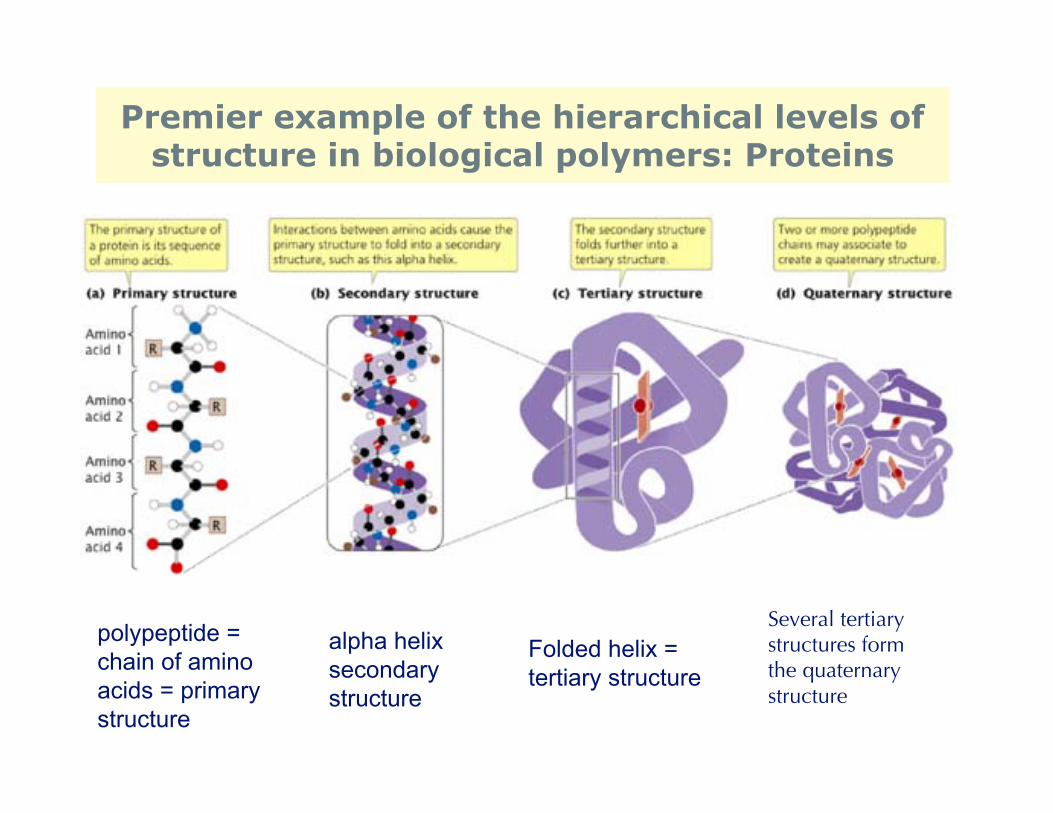
Premier example of the hierarchical levels ofstructure in biological polymers: Proteins
polypeptide =chain of amino acids = primarystructure
alpha helixsecondary structure
Folded helix = tertiary structure
Several tertiarystructures formthe quaternary structure
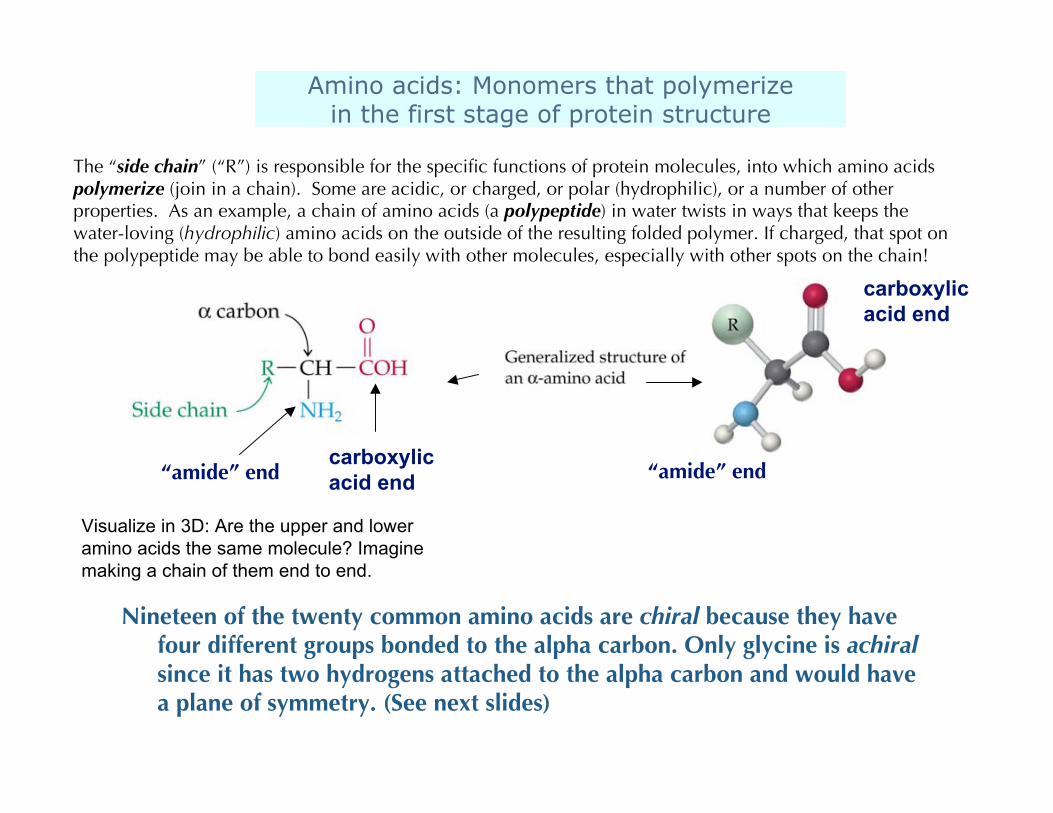
Amino acids: Monomers that polymerizein the first stage of protein structure
“amide” endcarboxylicacid end
Nineteen of the twenty common amino acids are chiral because they havefour different groups bonded to the alpha carbon. Only glycine is achiralsince it has two hydrogens attached to the alpha carbon and would havea plane of symmetry. (See next slides)
“amide” end
carboxylicacid end
The “side chain” (“R”) is responsible for the specific functions of protein molecules, into which amino acidspolymerize (join in a chain). Some are acidic, or charged, or polar (hydrophilic), or a number of otherproperties. As an example, a chain of amino acids (a polypeptide) in water twists in ways that keeps thewater-loving (hydrophilic) amino acids on the outside of the resulting folded polymer. If charged, that spot onthe polypeptide may be able to bond easily with other molecules, especially with other spots on the chain!
Visualize in 3D: Are the upper and loweramino acids the same molecule? Imaginemaking a chain of them end to end.
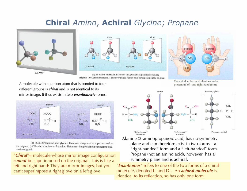
Chiral Amino, Achiral Glycine; Propane
Alanine (2-aminopropanoic acid) has no symmetryplane and can therefore exist in two forms—a“right-handed” form and a “left-handed” form.Propane (not an amino acid), however, has asymmetry plane and is achiral.
A molecule with a carbon atom that is bonded to fourdifferent groups is chiral and is not identical to itsmirror image. It thus exists in two enantiomeric forms.
“Chiral”= molecule whose mirror image configurationcannot be superimposed on the original. This is like aleft and right hand: They are mirror images, but youcan’t superimpose a right glove on a left glove.
“Enantiomer” refers to one of the two forms of a chiralmolecule, denoted L- and D-. An achiral molecule isidentical to its reflection, so has only one form.

Another way to see it: Can you draw a plane through the molecule that divides it into identical parts?Alanine has no symmetry plane and can therefore exist in two forms—a “right-handed” form and a “left-
handed” form. Propane (not an amino acid), however, has a symmetry plane and is achiral. The interestingmolecules for origin of life are chiral i.e. not symmetric.
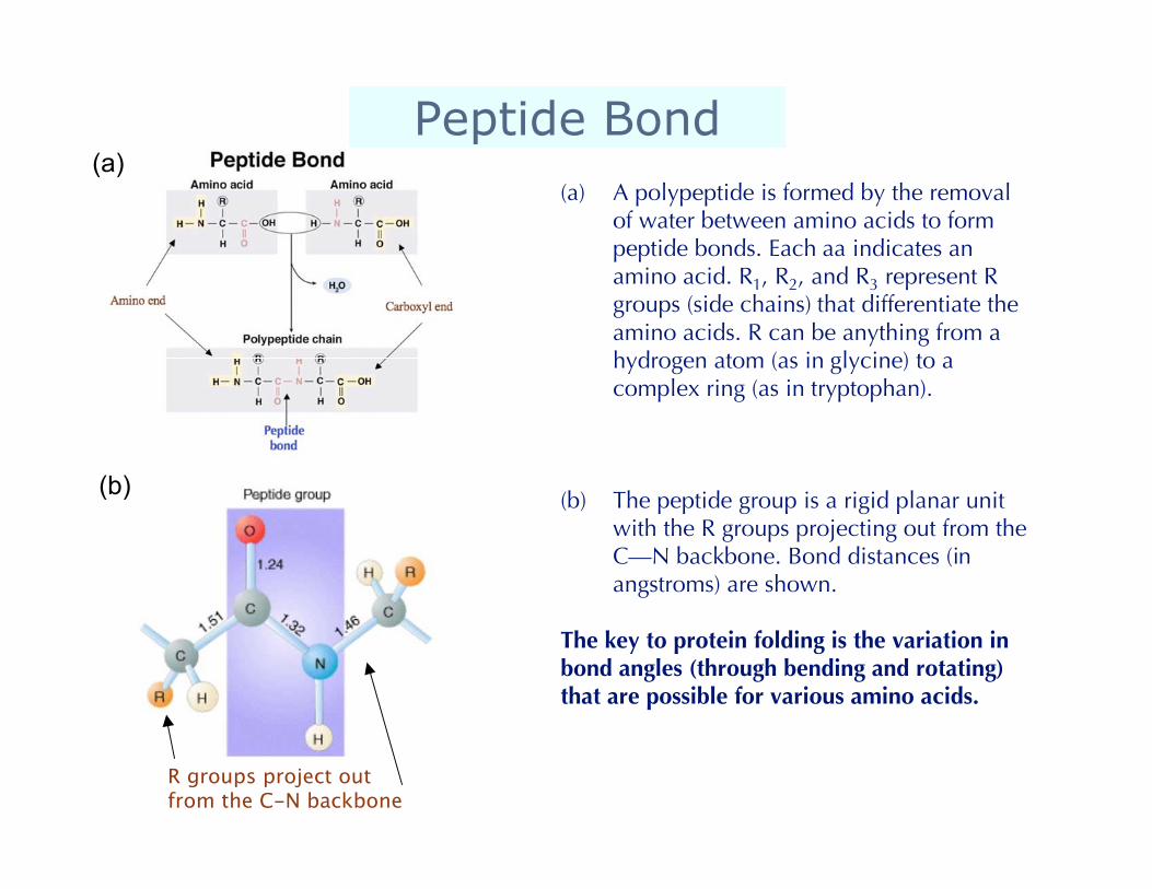
Peptide Bond(a) A polypeptide is formed by the removal
of water between amino acids to formpeptide bonds. Each aa indicates anamino acid. R1, R2, and R3 represent Rgroups (side chains) that differentiate theamino acids. R can be anything from ahydrogen atom (as in glycine) to acomplex ring (as in tryptophan).
(b) The peptide group is a rigid planar unitwith the R groups projecting out from theC—N backbone. Bond distances (inangstroms) are shown.
The key to protein folding is the variation inbond angles (through bending and rotating)that are possible for various amino acids.
(a)
(b)
R groups project out from the C-N backbone
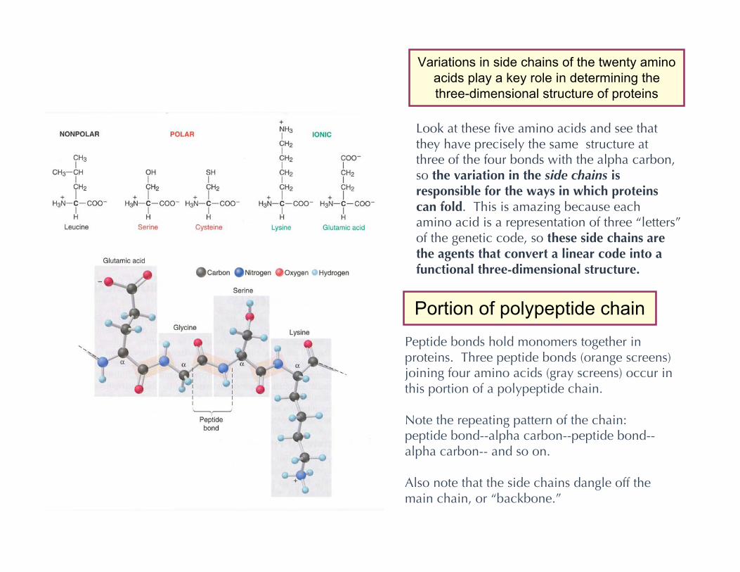
Portion of polypeptide chain
Peptide bonds hold monomers together inproteins. Three peptide bonds (orange screens)joining four amino acids (gray screens) occur inthis portion of a polypeptide chain.
Note the repeating pattern of the chain:peptide bond--alpha carbon--peptide bond--alpha carbon-- and so on.
Also note that the side chains dangle off themain chain, or “backbone.”
Look at these five amino acids and see thatthey have precisely the same structure atthree of the four bonds with the alpha carbon,so the variation in the side chains isresponsible for the ways in which proteinscan fold. This is amazing because eachamino acid is a representation of three “letters”of the genetic code, so these side chains arethe agents that convert a linear code into afunctional three-dimensional structure.
Variations in side chains of the twenty aminoacids play a key role in determining thethree-dimensional structure of proteins
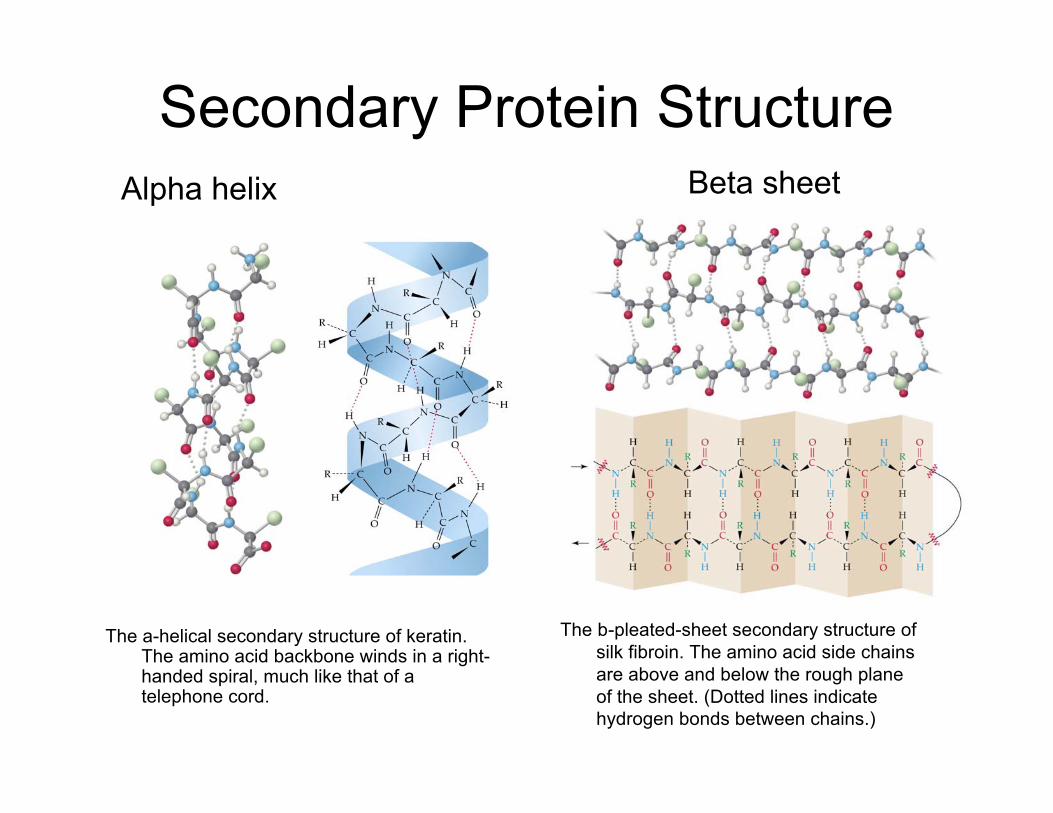
Secondary Protein Structure
The a-helical secondary structure of keratin.The amino acid backbone winds in a right-handed spiral, much like that of atelephone cord.
Alpha helix Beta sheet
The b-pleated-sheet secondary structure ofsilk fibroin. The amino acid side chainsare above and below the rough planeof the sheet. (Dotted lines indicatehydrogen bonds between chains.)
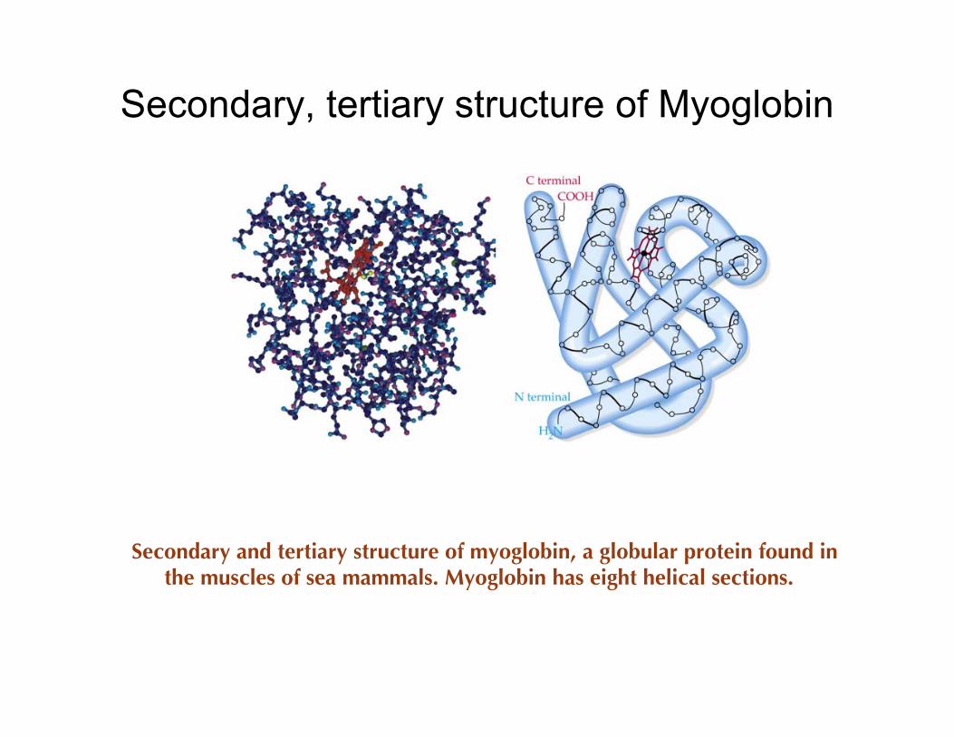
Secondary, tertiary structure of Myoglobin
Secondary and tertiary structure of myoglobin, a globular protein found inthe muscles of sea mammals. Myoglobin has eight helical sections.
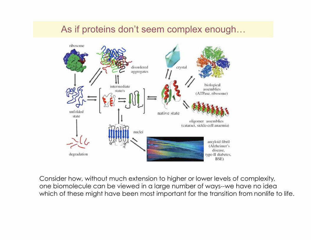
As if proteins don’t seem complex enough…
Consider how, without much extension to higher or lower levels of complexity, one biomolecule can be viewed in a large number of ways--we have no idea which of these might have been most important for the transition from nonlife to life.
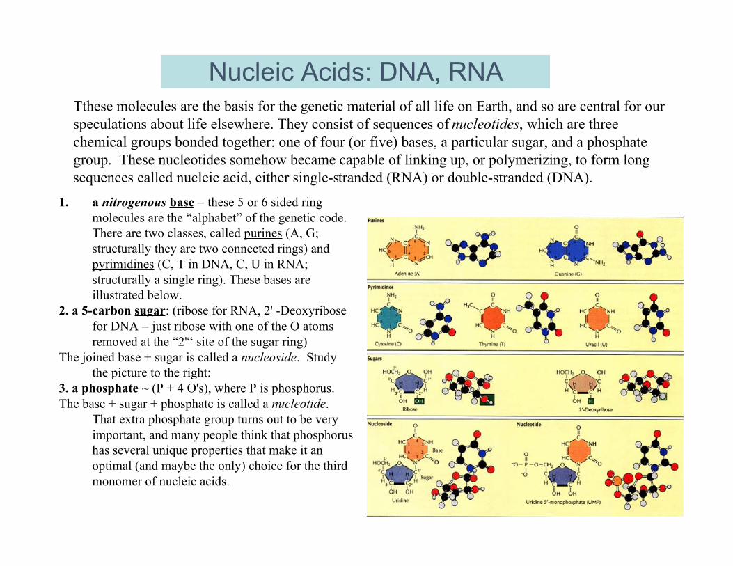
Nucleic Acids: DNA, RNATthese molecules are the basis for the genetic material of all life on Earth, and so are central for ourspeculations about life elsewhere. They consist of sequences of nucleotides, which are threechemical groups bonded together: one of four (or five) bases, a particular sugar, and a phosphategroup. These nucleotides somehow became capable of linking up, or polymerizing, to form longsequences called nucleic acid, either single-stranded (RNA) or double-stranded (DNA).
1. a nitrogenous base – these 5 or 6 sided ringmolecules are the “alphabet” of the genetic code.There are two classes, called purines (A, G;structurally they are two connected rings) andpyrimidines (C, T in DNA, C, U in RNA;structurally a single ring). These bases areillustrated below.
2. a 5-carbon sugar: (ribose for RNA, 2' -Deoxyribosefor DNA – just ribose with one of the O atomsremoved at the “2'“ site of the sugar ring)
The joined base + sugar is called a nucleoside. Studythe picture to the right:
3. a phosphate ~ (P + 4 O's), where P is phosphorus.The base + sugar + phosphate is called a nucleotide.
That extra phosphate group turns out to be veryimportant, and many people think that phosphorushas several unique properties that make it anoptimal (and maybe the only) choice for the thirdmonomer of nucleic acids.
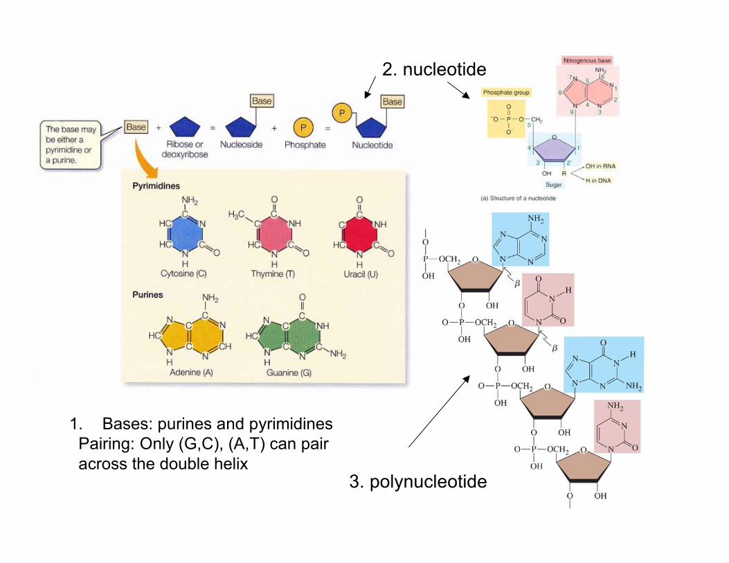
1. Bases: purines and pyrimidines Pairing: Only (G,C), (A,T) can pair across the double helix
2. nucleotide
3. polynucleotide
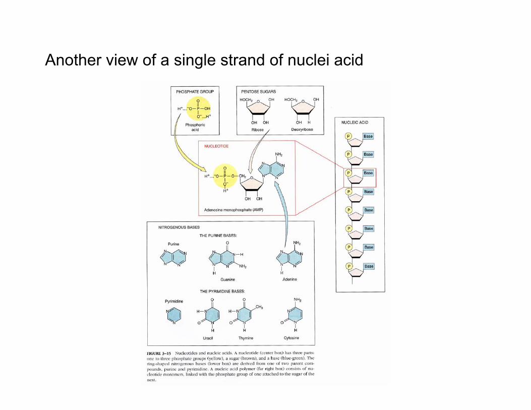
Another view of a single strand of nuclei acid
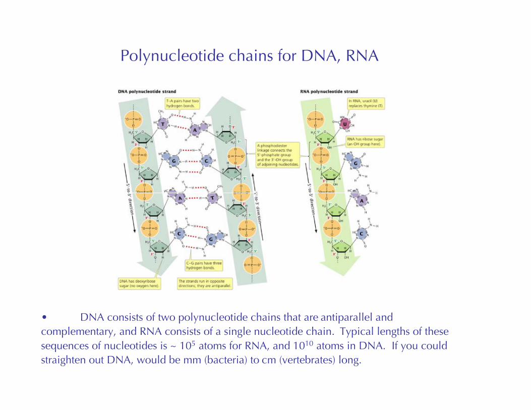
Polynucleotide chains for DNA, RNA
• DNA consists of two polynucleotide chains that are antiparallel andcomplementary, and RNA consists of a single nucleotide chain. Typical lengths of thesesequences of nucleotides is ~ 105 atoms for RNA, and 1010 atoms in DNA. If you couldstraighten out DNA, would be mm (bacteria) to cm (vertebrates) long.
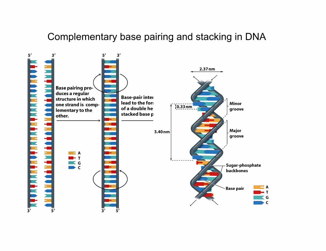
Complementary base pairing and stacking in DNA
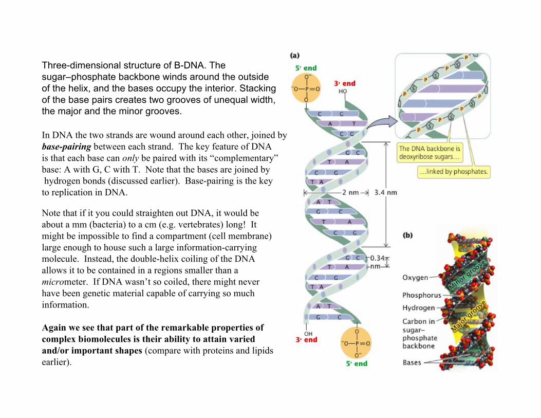
Three-dimensional structure of B-DNA. Thesugar–phosphate backbone winds around the outsideof the helix, and the bases occupy the interior. Stackingof the base pairs creates two grooves of unequal width,the major and the minor grooves.
Note that if it you could straighten out DNA, it would beabout a mm (bacteria) to a cm (e.g. vertebrates) long! Itmight be impossible to find a compartment (cell membrane)large enough to house such a large information-carryingmolecule. Instead, the double-helix coiling of the DNAallows it to be contained in a regions smaller than amicrometer. If DNA wasn’t so coiled, there might neverhave been genetic material capable of carrying so muchinformation.
Again we see that part of the remarkable properties ofcomplex biomolecules is their ability to attain variedand/or important shapes (compare with proteins and lipidsearlier).
In DNA the two strands are wound around each other, joined bybase-pairing between each strand. The key feature of DNAis that each base can only be paired with its “complementary”base: A with G, C with T. Note that the bases are joined by hydrogen bonds (discussed earlier). Base-pairing is the keyto replication in DNA.
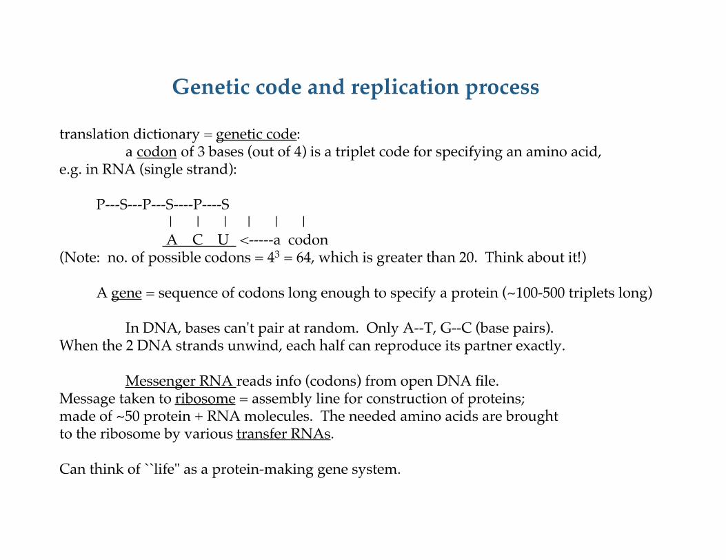
Genetic code and replication process
translation dictionary = genetic code:a codon of 3 bases (out of 4) is a triplet code for specifying an amino acid,
e.g. in RNA (single strand):
P---S---P---S----P----S | | | | | | A C U <-----a codon
(Note: no. of possible codons = 43 = 64, which is greater than 20. Think about it!)
A gene = sequence of codons long enough to specify a protein (~100-500 triplets long)
In DNA, bases can't pair at random. Only A--T, G--C (base pairs). When the 2 DNA strands unwind, each half can reproduce its partner exactly.
Messenger RNA reads info (codons) from open DNA file. Message taken to ribosome = assembly line for construction of proteins; made of ~50 protein + RNA molecules. The needed amino acids are brought to the ribosome by various transfer RNAs.
Can think of ``life" as a protein-making gene system.
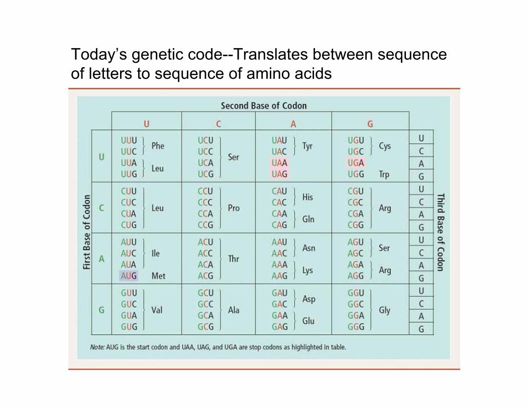
Today’s genetic code--Translates between sequenceof letters to sequence of amino acids
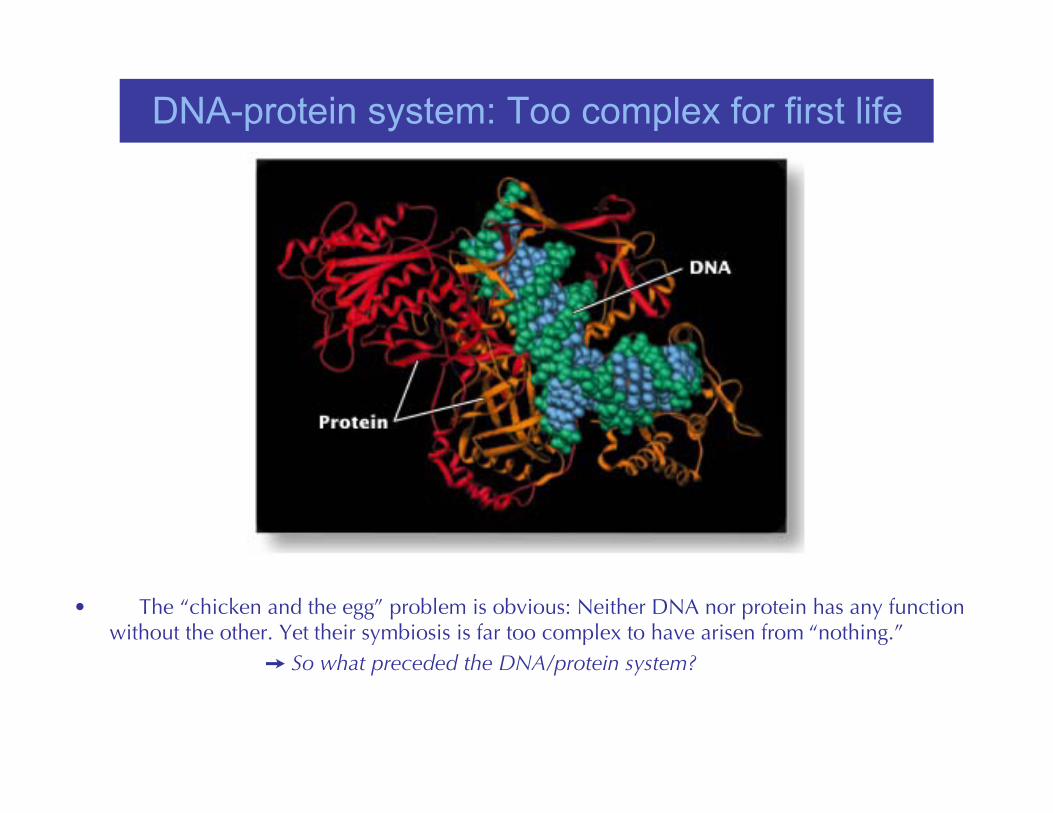
DNA-protein system: Too complex for first life
• The “chicken and the egg” problem is obvious: Neither DNA nor protein has any functionwithout the other. Yet their symbiosis is far too complex to have arisen from “nothing.”
So what preceded the DNA/protein system?
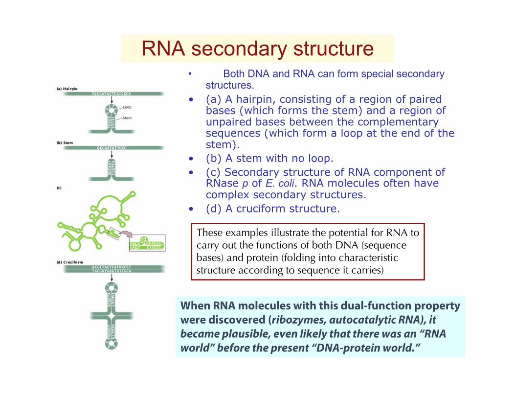
RNA secondary structure• Both DNA and RNA can form special secondary
structures.• (a) A hairpin, consisting of a region of paired
bases (which forms the stem) and a region ofunpaired bases between the complementarysequences (which form a loop at the end of thestem).
• (b) A stem with no loop.• (c) Secondary structure of RNA component of
RNase p of E. coli. RNA molecules often havecomplex secondary structures.
• (d) A cruciform structure.
These examples illustrate the potential for RNA tocarry out the functions of both DNA (sequencebases) and protein (folding into characteristic structure according to sequence it carries)
When RNA molecules with this dual-function propertywere discovered (ribozymes, autocatalytic RNA), it became plausible, even likely that there was an “RNAworld” before the present “DNA-protein world.”
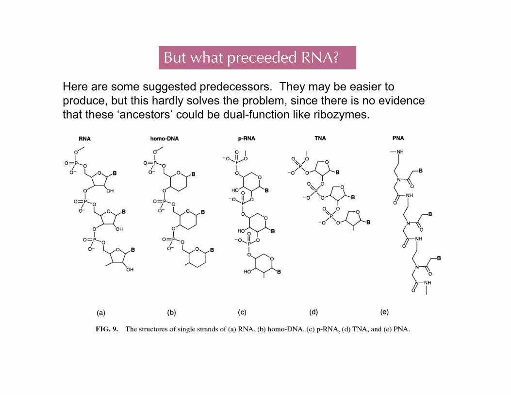
But what preceeded RNA?
Here are some suggested predecessors. They may be easier toproduce, but this hardly solves the problem, since there is no evidencethat these ‘ancestors’ could be dual-function like ribozymes.
“Naked gene” or “random replicator” theories(RNA world is an example)
1st need nucleosides (= base+ sugar)→heat sugar + bases + salts → suggests drying tidepoolsBut then must polymerize the nucleosides. Tough!
Spiegelmann: Qβ virus + enzyme + free nucleiotides→ “Spiegelman monster” (w/long RNA)
Eigen: enzyme + free nucleotides + salts → short RNA random replicator.
But both S. & E. started with proteins.
Orgel: RNA can form a double helix without any protein. But then stopped.
Cech et al.: self-catalytic RNA--RNA can cut up different RNAs, acting as an enzyme.It can also join short RNAs into longer chains.(Extremely influential result; gave rise to term “RNA World” )
1994: Joyce et al. made synthetic RNA that can copy itself (given the rightproteins).
1997: Two studies in Jan.21 Proc.Nat.Acad.Sci. claim experimental evidencerelated to enzymes that convert between RNA and DNA.
2001 RNA shown to catalyze its own replication without enzymes.