Embed Size (px)
Citation preview

Pure Appl. Chem., Vol. 76, No. 4, pp. 723–752, 2004.© 2004 IUPAC
723
Biosensors for environmental applications:Future development trends*
Sara Rodriguez-Mozaz1, Maria-Pilar Marco2, Maria J. Lopez de Alda1,and Damià Barceló1,‡
1Department of Environmental Chemistry, IIQAB-CSIC, C/Jordi Girona 18-26,08034 Barcelona, Spain; 2Department of Biological Organic Chemistry, IIQAB-CSIC, C/Jordi Girona 18-26, 08034 Barcelona, Spain
Abstract: Biosensors can be excellent analytical tools for monitoring programs working toimplement legislation. In this article, biosensors for environmental analysis and monitoringare extensively reviewed. Examples of biosensors for the most important families of envi-ronmental pollutants, including some commercial devices, are presented. Finally, futuretrends in biosensor development are discussed. In this context, bioelectronics, nanotechnol-ogy, miniaturization, and especially biotechnology seem to be growing areas that will have amarked influence on the development of new biosensing strategies in the next future.
INTRODUCTION
Public concern and legislation are nowadays demanding better environmental control. In addition, theincrease in the number of analytes to monitor requires more suitable analytical methods. For conven-tional “off-site” analysis, samples need to be sent to a laboratory for testing. Conventional methods areactually reaching the highest accuracy with low detection limits, but are expensive, time-consuming,and require the use of highly trained personnel [1]. The current tendency to carry out field monitoringhas driven the development of biosensors as new analytical tools able to provide fast, reliable, and sen-sitive measurements with lower cost; many of them aimed at on-site analysis. In this sense, biosensorswould not compete with official analytical methods, but they can be used both by regulatory authoritiesand by industry to provide enough information for routine testing and screening of samples [2].Biosensors are defined as analytical devices incorporating a biological material, a biologically derivedmaterial, or biomimic, intimately associated with or integrated within a physicochemical transducer ortransducing microsystem [3]. Biosensors should be distinguished from a bioassay or a bioanalytical sys-tem, which require additional processing steps, such as reagent addition [4] and where the assay designis permanently fixed in the construction of the device.
Biosensors are being developed for different applications, including environmental and bio-process control, quality control of food, agriculture, military, and, particularly, medical applications. Infact, most of the commercially available biosensor systems are applied in the clinical and pharmaceuti-cal markets. Accordingly, most research and development has been devoted to this area. In the food in-dustry, the detection of contaminants, verification of product content, monitoring of raw materials con-version, and product freshness [5] are areas of potential biosensor application. The beer industry hasalready identified ways for improving and controlling their products through the use of biosensors [6].Biosensors can be also a defense tool through the early detection of hazardous materials such as germs
*Plenary lecture presented at the Southern and Eastern Africa Network of Analytical Chemists (SEANAC), Gaborone, Botswana,7–10 July 2003. Other presentations are published in this issue, pp. 697–888.‡Corresponding author
or chemical warfares. The detection of illicit drugs and explosives, with airport security purposes, isalso a major area of research in biosensors [5]. For environmental control and monitoring, biosensorscan provide fast and specific data of contaminated sites. They offer other advantages over current ana-lytical methods such as the possibility of portability and working on-site and the ability of measuringpollutants in complex matrices with minimal sample preparation. On the other hand, biosensors offerthe possibility of determining not only specific chemicals, but also their biological effects, such as tox-icity or endocrine-disrupting effects, which are sample information of great interest.
BIOSENSOR CONFIGURATIONS
Biosensors are usually classified into various basic groups, according to the signal transduction and tothe biorecognition principles. On the basis of the transducing element, biosensors can be categorized aselectrochemical, optical, piezoelectric, and thermal sensors. The electrochemical biosensors, andamong them the amperometric and the potentiometric ones, are the best described in the literature; thosebased on optical principles are the next most commonly used transducers. In fact, most catalytic biosen-sors are based on electrochemical methods, whereas affinity biosensors have generally proved moreamenable to optical detection methods [7]. The various types of optical transducers exploit propertiessuch as simple light absorption, fluorescence/phosphorescence, bio/chemiluminescence, reflectance,Raman scattering, and refractive index [5]. Surface plasmon resonance (SPR) is another common trans-duction mechanism whose main advantage over most optical biosensors is that the analyte presence canbe determined directly, without the use of labeled molecules. Finally, cantilever biosensors are anemerging group of biosensors, which are based on the bending of silicon cantilevers caused by the ad-sorption of target molecules onto the cantilever surface, where receptor molecules are immobilized.
According to the biorecognition principle, biosensors are classified into immunochemical, enzy-matic, nonenzymatic receptor, whole-cell, and DNA biosensors. Immunosensors present the advantagesof sensitivity and selectivity inherent to the use of immunochemical interactions. Limitations are thetroubles derived from the regeneration of the immunosurface, and cross-reactivity, although a certaindegree of cross-reactivity is often desirable in order to determine different congeners of the same fam-ily. Enzymes are suitable to act as recognition elements because of their specificity and the wide rangeof enzymes available. In general, enzymatic biosensors are based on the selective inhibition of specificenzymes by different classes of compounds. The decrease of activity of the immobilized enzyme in thepresence of the target analyte is frequently used for its quantification. Biosensors based on natural re-ceptors can be built by integrating the specific receptor within a membrane and by coupling it to a trans-ducing device. These natural receptors are proteins of noncatalytic or nonimmunogenic origin, whichspan cell membranes and can specifically bind certain compounds. The binding signal is detected as astructural change or an associated enzyme activity. Whole cells of living organisms, such as bacteria,yeast, fungi, plant and animal cells, or even tissue slices have been used as the recognition componentby interrogating their general metabolic status. These biosensors are useful to determine whether a sub-stance is toxic to certain cells. Another application of whole-cell biosensors is the determination of the“biological oxygen demand” (BOD). In the case of DNA biosensors, two strategies are applied to de-tect pollutants: one is the hybridization detection of nucleic acid sequences from infectious micro-organisms, and the other one, the monitoring of small pollutants interacting with the immobilized DNAlayer (drugs, mutagenic pollutants, etc.) [8].
One key step in the development of biosensors is the immobilization of the biological componentat the transducer surface. The immobilization procures both the stabilization of the biomaterial and theproximity between the biomaterial and the transducer. The immobilization methods most generally em-ployed are: physical adsorption at a solid surface, cross-linking between molecules, covalent binding toa surface, and entrapment within a membrane, surfactant matrix, polymer or microcapsule [5]. In addi-tion to these conventional methods, sol-gel entrapment, Langmuir–Blodgett (LB) deposition, electro-polymerization, self-assembled biomembranes and bulk modification have been also used.
S. RODRIGUEZ-MOZAZ et al.
© 2004 IUPAC, Pure and Applied Chemistry 76, 723–752
724
ENVIRONMENTAL APPLICATIONS
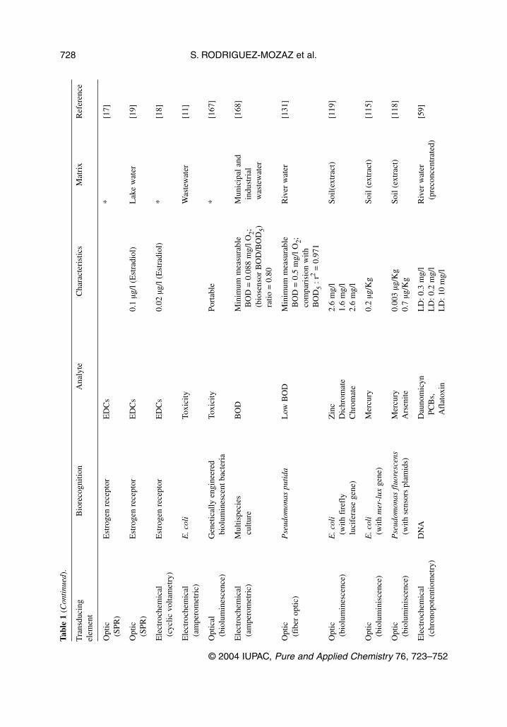
In the following section, we describe biosensors developed for environmental monitoring, consideringfirst, biosensors that measure an effect such as toxicity and endocrine effect biosensors and second,biosensors that detect a compound or a group of compounds based on the specific recognition of a bio-molecule. A wide range of compounds of environmental concern or under suspicion are being consid-ered for biosensor development. Table 1 lists examples of these reported biosensors for different envi-ronmental applications.
Toxicity
In environmental pollution monitoring, it is becoming a general opinion that chemical analysis by itselfdoes not provide sufficient information to assess the ecological risk of polluted waters and wastewaters[9]. In the European Union, along with more stringent demands for water treatment (Council Directive91/271/EEC), industrial and urban wastewater effluents shall reach certain limits of nontoxicity beforethe effluent can be discharged into the environment. Thus, much effort has been made during the lastyears to develop and use different bioassays and biosensors for toxicity evaluation of water samples [9].
Whole organisms are used to measure the potential biological impact (toxicity) of a water or soilsample. That is the case of the toxicity assays Microtox® (Azure, Bucks, UK), or ToxAlert® (Merck,Darmstadt, Germany). These systems are based on the use of luminescent bacteria, Vibrio fischeri, tomeasure toxicity from environmental samples. Bacterial bioluminescence has proved to be a convenientmeasure of cellular metabolism and, consequently, a reliable sensor for measuring the presence of toxicchemicals in aquatic samples. Some bioassay methods are integrated now in biosensors such as theCellsense®, which is an amperometric sensor that incorporates Escherichia coli bacterial cells for rapidecotoxicity analysis. It uses ferricyanine, a soluble electron mediator, to divert electrons from the res-piratory system of the immobilized bacteria of a suitable carbon electrode. The resulting current is, thus,a measure of bacterial respiratory activity, and the perturbation by pollutants can be detected as achange in the magnitude of the current. Cellsense has been applied to investigate the toxicity of3,5-dichlorophenol and other phenols in wastewater [10], for the determination of nonionic surfactantsand benzene sulfonate compounds [11], for the analysis of wastewater treatment works (WWTW) in-fluent and effluent [11], and for the toxicity testing of wastewaters and sewage sludge [12]. Moreover,Cellsense has been proposed as one of the newer rapid toxicity assessment methods within the directtoxicity assessment (DTA) demonstration program of the UK Environmental Agency [13].
Most environmental biosensors have focused on bacterial systems while eukariotic biosensors arerare; even more rare is the use of mammalian cells. The mammalian cell, which is more complex thanbacteria, can give a more sensitive response when compared to bacteria while also responding to the es-trogenic effects of chemicals [14]. A recombinant fluorescent Chinese Hamster Ovary cell line, utiliz-ing a fluorescent reporter system, was used to monitor various toxicants, especially endocrine-disrupt-ing compounds (EDCs), in diverse aqueous environments [14]. EDCs have been also analyzed by Guet al. [15] with a multichannel two-stage mini-bioreactor system using a genetically engineered bio-luminescent bacteria. The toxicity of various samples spiked with known endocrine-disrupting chemi-cals, and phenol was investigated.
Endocrine effect biosensors
Nowadays, there is an increasing concern regarding many environmental contaminants that produce ad-verse effects by interfering with endogenous hormone systems, the so-called EDCs. EDCs constitute aclass of substances not defined by chemical nature, but by biological effect and thus, taking advantage
© 2004 IUPAC, Pure and Applied Chemistry 76, 723–752
Biosensors for environmental applications 725
(Text continues on p. 730.)

S. RODRIGUEZ-MOZAZ et al.
© 2004 IUPAC, Pure and Applied Chemistry 76, 723–752
726Ta
ble
1B
iose
nsor
s fo
r en
viro
nmen
tal a
pplic
atio
ns.
Tra
nsdu
cing
Bio
reco
gniti
onA
naly
teC
hara
cter
istic
sM
atri
xR
efer
ence
elem
ent
elem
ent
Ele
ctro
chem
ical
Ant
ibod
ies
Atr
azin
eL
D: 1
µg/
l*
[2]
(am
pero
met
ric)
Opt
ical
Ant
ibod
ies
Sim
azin
eL
D: 0
.2 µ
g/l
Nat
ural
, gro
und,
[42]
(wav
egui
de S
PR)
and
surf
ace
wat
er
Opt
ical
Ant
ibod
ies
Pest
icid
es a
nd e
stro
neV
alid
ated
Riv
er w
ater
[39–
41,5
7]
Ele
ctro
chem
ical
Ant
ibod
ies
Surf
acta
nts
LD
: µg/
l ran
ge*
[75]
(alk
ylph
enol
s an
d th
eir
etho
xyla
tes)
Ele
ctro
chem
ical
Ant
ibod
ies
Est
radi
olL
D: 1
ng/
l*
[143
](a
mpe
rom
etri
c)
Ele
ctro
chem
ical
Ant
ibod
ies
Esc
heri
chia
col
i10
cel
ls/m
lD
rink
ing
wat
er[1
04]
(pot
entio
met
ric)
Opt
icA
ntib
odie
sSa
lmon
ella
ent
erid
itis
,10
6ce
lls/m
l*
[106
](S
PR)
Lyst
eria
mon
ocyt
ogen
es
Aco
ustic
Ant
ibod
ies
Salm
onel
la t
yphy
mur
ium
100
cells
/ml
*[1
07]
Opt
icE
nzym
eO
rgan
opho
spho
rous
LD
: 2 m
g/l
Wat
er[2
2](f
iber
opt
ic)
(AC
hE)
com
poun
ds
Ele
ctro
chem
ical
Enz
yme
Para
oxon
and
D
iscr
imin
atio
n W
aste
wat
er[1
54]
(am
pero
met
ric)
(AC
hE)
carb
ofur
an (
pest
icid
es)
betw
een
diff
eren
tA
ChE
inhi
bito
rsby
neu
ral n
etw
orks
LD
: 0.2
µg/
l
Ele
ctro
chem
ical
Enz
ymat
icPh
enol
sL
D: 0
.1 µ
g/l
Soil,
slu
dge,
[2]
(am
pero
met
ric)
(tyr
osin
ase)
“phe
nol i
ndex
”an
d w
ater
corr
elat
ion
(aft
er e
xtra
ctio
n)

© 2004 IUPAC, Pure and Applied Chemistry 76, 723–752
Biosensors for environmental applications 727
Ele
ctro
chem
ical
Enz
ymat
icPh
enol
sIn
-fie
ldW
aste
wat
er[1
65]
(cel
lobi
ose
dehy
drog
enas
em
easu
rem
ents
and
quin
opro
tein
-dep
ende
ntL
D: 0
.8 µ
g/l
gluc
ose
dehy
drog
enas
e)
Opt
icE
nzym
atic
Chl
orop
heno
lsL
D: 1
.4–1
975
µg/l
*[6
7]ch
emilu
min
esce
nce
(hor
sera
dish
per
oxid
ase)
(fib
er o
ptic
)
Ele
ctro
chem
ical
Enz
ymat
icIn
orga
nic
phos
phat
eL
D: 0
.57
mg/
l*
[2]
(am
pero
met
ric)
(tri
enzy
mat
ic c
onfi
gura
tion)
Ele
ctro
chem
ical
Enz
ymat
icE
. col
i10
3 –104
cells
/ml
Was
tew
ater
[109
](a
mpe
rom
etri
c)(t
yros
inas
e)
Ele
ctro
chem
ical
Phot
osys
tem
II
(PSI
I)D
iuro
nL
D: 0
.233
µg/
l*
[34]
(am
pero
met
ric)
Atr
azin
eL
D: 0
.431
µg/
lSi
maz
ine
LD
: 0.8
06 µ
g/l
Opt
icSc
enes
desm
ussu
bspi
catu
sA
traz
ine
and
endr
ine
1 or
0.1
µg/
l*
[166
](f
luor
esce
nce)
(alg
ae c
ells
)(h
erbi
cide
s)
Opt
icC
hlor
ella
vul
gari
sIs
opro
turo
n, d
iuro
n,0.
025
µg/l
*[3
3](f
luor
esce
nce)
(alg
ae c
ells
)si
maz
ine
0.5
µg/l
Ele
ctro
chem
ical
Pse
udom
onas
and
Surf
acta
nts
LD
: 0.2
5 m
g/l (
SDS)
*[7
1,72
](a
mpe
rom
etri
c)A
chro
mob
acte
r(b
eari
ng a
pla
smid
for
anio
nic
surf
acta
ntde
grad
atio
n)
Ele
ctro
chem
ical
Tric
hosp
oron
LA
SIn
situ
R
iver
wat
er[7
4](a
mpe
rom
etri
c)cu
tane
umm
easu
rem
ent
(O2
cons
umpt
ion
(LA
S de
grad
ing
LD
: 0.2
mg/
lm
easu
rem
ent)
bact
eria
)
Tabl
e 1
(Con
tinu
ed).
Tra
nsdu
cing
Bio
reco
gniti
onA
naly
teC
hara
cter
istic
sM
atri
xR
efer
ence
elem
ent
(con
tinu
es o
n ne
xt p
age)
S. RODRIGUEZ-MOZAZ et al.
© 2004 IUPAC, Pure and Applied Chemistry 76, 723–752
728
Opt
icE
stro
gen
rece
ptor
ED
Cs
*[1
7](S
PR)
Opt
icE
stro
gen
rece
ptor
ED
Cs
0.1
µg/l
(Est
radi
ol)
Lak
e w
ater
[19]
(SPR
)
Ele
ctro
chem
ical
Est
roge
n re
cept
orE
DC
s0.
02 µ
g/l (
Est
radi
ol)
*[1
8](c
yclic
vol
tam
etry
)
Ele
ctro
chem
ical
E. c
oli
Toxi
city
Was
tew
ater
[11]
(am
pero
met
ric)
Opt
ical
Gen
etic
ally
eng
inee
red
Toxi
city
Port
able
*[1
67]
(bio
lum
ines
cenc
e)bi
olum
ines
cent
bac
teri
a
Ele
ctro
chem
ical
Mul
tispe
cies
BO
DM
inim
um m
easu
rabl
eM
unic
ipal
and
[168
](a
mpe
rom
etri
c)cu
lture
BO
D =
0.0
88 m
g/l O
2;in
dust
rial
(b
iose
nsor
BO
D/B
OD
5)w
aste
wat
erra
tio =
0.8
0
Opt
icP
seud
omon
as p
utid
aL
ow B
OD
Min
imum
mea
sura
ble
Riv
er w
ater
[131
](f
iber
opt
ic)
BO
D =
0.5
mg/
l O2;
com
pari
sion
with
BO
D5
: r2
= 0
.971
Opt
icE
. col
iZ
inc
2.6
mg/
lSo
il(ex
trac
t)[1
19]
(bio
lum
ines
cenc
e)(w
ith f
iref
lyD
ichr
omat
e1.
6 m
g/l
luci
fera
se g
ene)
Chr
omat
e2.
6 m
g/l
Opt
icE
. col
iM
ercu
ry0.
2 µg
/Kg
Soil
(ext
ract
)[1
15]
(bio
lum
inis
cenc
e)(w
ith m
er-l
uxge
ne)
Opt
icP
seud
omon
as f
luor
esce
nsM
ercu
ry0.
003
µg/K
gSo
il (e
xtra
ct)
[118
](b
iolu
min
isce
nce)
(with
sen
sors
pla
mid
s)A
rsen
ite0.
7 µg
/Kg
Ele
ctro
chem
ical
DN
AD
auno
mic
ynL
D: 0
.3 m
g/l
Riv
er w
ater
[59]
(chr
onop
oten
tiom
etry
)PC
Bs,
L
D: 0
.2 m
g/l
(pre
conc
entr
ated
)A
flat
oxin
LD
: 10
mg/
l
Tabl
e 1
(Con
tinu
ed).
Tra
nsdu
cing
Bio
reco
gniti
onA
naly
teC
hara
cter
istic
sM
atri
xR
efer
ence
elem
ent

© 2004 IUPAC, Pure and Applied Chemistry 76, 723–752
Biosensors for environmental applications 729
Ele
ctro
chem
ical
DN
AC
hlam
ydia
tra
chom
atis
Prev
ious
PC
RR
iver
wat
er[5
9](c
hron
opot
entio
met
ry)
(hyb
ridi
zatio
n)(D
NA
)am
plif
icat
ion
(pre
conc
entr
ated
)L
D: 0
.2 m
g/l
Piez
oele
ctri
cD
NA
Aer
omon
as h
ydro
phil
aPr
evio
us D
NA
Min
eral
and
[108
](h
ybri
diza
tion)
extr
actio
n an
d PC
Rdr
inki
ng w
ater
ampl
ific
atio
n st
ep
*No
real
sam
ple.
Tabl
e 1
(Con
tinu
ed).
Tra
nsdu
cing
Bio
reco
gniti
onA
naly
teC
hara
cter
istic
sM
atri
xR
efer
ence
elem
ent
of this feature, “endocrine effect biosensors” have been developed. Steroid hormones induce differenteffects in mammalian cells after binding to specific intercellular receptors, which are ligand-dependenttranscription factors. Many endocrine disruptors are also believed to bind to the estrogen receptor (ER)as agonists or antagonists. Thus, the binding ability of the chemicals toward the ER would be a crucialfactor for screening or testing their potential environmental toxicity. Based on estrogen receptors, sev-eral biosensors have been developed which provide significant and useful information about estrogenicpotency of the sample. The advantage of receptor assays is that they are quite simple to perform andallow the identification of all endocrine disruptors that act through the corresponding receptor [16].These biosensors constitute a similar approach as that described above for toxicity biosensors, whereeffects as a general parameter and not a specific substance are monitored. By employing the commonlyutilized human estrogen receptor, the SPR biosensor BIAcore has been applied in the determination ofestrogens and xenoestrogens and in binding studies of target compounds [17–19]. In addition to opticalbiosensors, electrochemical [20] and piezoelectric [21] biosensors have been developed also based onestrogen receptors.
Biocides
The extensive use of pesticides for agricultural purposes is the cause of their widespread presence innatural waters. Concerns about their toxicity and persistence in the environment has led the EuropeanCommunity to set limits on the concentration of pesticides in different environmental waters: The di-rective 98/83/EC on the quality of water for human consumption has set a limit of 0.1 µg/l for individ-ual pesticides and of 0.5 µg/l for total pesticides. Although conventional techniques such HPLC/MS andGC/MS gives satisfactory analytical results for pesticide determination, new assays and sensors forcheaper and faster on-site analysis are being developed. Enzymatic sensors, based on the inhibition ofa selected enzyme, are the most extended biosensors used for the determination of these compounds.Based on the inhibition of acetyl cholinesterase (AChE) and colin oxidase, various biosensors have beendeveloped for the detection of organophosphorous and carbamate pesticides such as those described byChoi et al. [22], Vangelis et al. [23], and Andres et al. [24]. Although sensitive, biosensors based onAchE inhibition are not selective (since the AchE is inhibited by neurotoxins, which includeorganophosphorous pesticides (OPs), carbamate pesticides, and many other compounds) and cannot,therefore, be used for quantitation of either an individual or a class of pesticides. One approach to solvethe lack of specificity of AchE involves the genetic engineering of cholinesterase enzyme to obtain newspecific enzymes for desired analytes or families. Different expression systems for the production of re-combinant AChEs for biosensor applications were reviewed by Schulze et al. [25]. The organo-phosphorous hydrolase (OPH), on the other hand, is able to hydrolyze a number of OP pesticides suchas paraoxon and parathion, and chemical warfare agents such as sarin and soman. Hydrolysis of theseOP pesticides generates p-nitrophenol, which is an electroactive and chromophoric product. Thus, OPHcould be combined with an optical transducer to measure the absorbance of p-nitrophenol or with anamperometric transducer to monitor the oxidation or reduction current of this product [26]. With a dif-ferent approach, the capability of various pesticides such as cyanide [27], diethyldithiocarbamates [28],and hydrazines [29] to inhibit the enzyme tyrosinase was also reported. Similarly, diazinon and dichlor-vos were detected at limits around 5 µM and 75 µM, respectively, using a tyrosinase-based oxygen sen-sor [30]. Dithiocarbamate fungicides have been measured by their ability to inhibit the enzyme alde-hyde dehydrogenase (AlDH). Particularly, the detectability of the pesticide Maneb could be improvedto a level of 1.5 µg/l, by using a bienzymatic system based on the combination of AlDH and diaphorase[31]. Photosynthesis inhibition is an interesting indicator that rapidly reflects the toxic effect of certainpollutants. Taking advantage of this feature, some biosensors based on Photosystem II (PSII) have beenreported to be able to detect herbicides in the environment [32]. About 30 % of herbicides, includingphenylurea, triazine, and phenolic herbicides, inhibit photosynthetic electron flow by blocking the PSIIquinone-binding site and thus modify chlorophyll fluorescence [33]. An amperometric biosensor de-
S. RODRIGUEZ-MOZAZ et al.
© 2004 IUPAC, Pure and Applied Chemistry 76, 723–752
730

veloped by Koblizek et al. [34] exhibited, for example, selective sensitivity to phenylurea and triazineherbicides, whereas phenolic herbicides were not registered. Heavy metals were also able to inhibit theactivity of the PSII biosensor, but their effect is usually found at much higher concentrations than thosetypical for herbicides. Another PSII-based biosensor [33] allowed the detection of herbicides such asatrazine, simazine, isoproturon, and diuron at sub-µg/l concentration levels.
In a different approach, biosensors based on immunological assays have been developed. Wilmeret al. [35], determined 2,4-dichlorophenoxyacetic (2,4-D) acid in water by an amperometric im-munosensor. In this immunosensor, the enzyme alkaline phosphatase (AP) catalyzes the conversion ofPAPP to PAP allowing a limit of detection of 0.1 µg/l. Based on the evanescent wave (EW) transducingprinciple, atrazine was detected at concentrations around 0.1 µg/l [36,37] and cyclodiene insecticides inthe µg/l range [38]. Mallat et al. [39–41] applied the “River Analyzer” (RIANA) immunosensor in thedetermination of pesticides such as atrazine, simazine, isoproturon, 2,4-D, alachlor, and paraquat in nat-ural waters. Studies focused on the evaluation of matrix effects, interferences due to the presence ofcross-reactant substances and on the validation of the sensor. The system was used for monitoring ofatrazine, simazine, and alachlor in different Spanish and Portuguese regions. Based also on SPR prin-ciple, another immunosensor was used to measure triazine pesticides [42] achieving a detection limitfor simazine of 0.2 µg/l. Analyses carried out on surface and groundwater samples showed a good cor-relation with parallel chromatographic results. An immunosensor for the determination of Irgarol 1051was applied to the direct analysis of natural waters and water extracts [43]. It allowed the analysis ofsamples with an organic solvent content of up to 50 or 25 % of methanol and acetonitrile, respectively[44]. In spite of the lack of specificity and the interferences observed in liquid media, some applicationshave also been reported on the use of piezoelectric immunosensors for the determination of pesticidessuch as atrazine [45,46], 2,4-D [47], and parathion [48]. Recently, a label-free direct piezoelectric im-munosensor built on a flow-through cell was used for the determination of 2,4-D in water with a limitof detection around 0.2 µg/l [49].
Hormones
Endogenous hormones of human or animal origin have been reaching the environment for thousandsof years, even though to an increasing extent they are due to growing population and more intensivefarming. Besides endogenous hormones, exogenous sex steroids used as growth promoters in severalcountries have become a matter of concern not only because of the residues potentially found in meat,but also because environmental soil and water samples may be contaminated as result of the animalexcreta. These residues may have endocrine-disrupting activity in aquatic fauna or even terrestrial [50].Although very low concentrations (ng/l range) of hormones such as estradiol, estrone, and ethynil-estradiol have been found in water [51–54], their widespread use and their capability to induce re-sponses in fish at concentrations as low as ng/l or even pg/l level, have alerted scientists to the poten-tial dangerous consequences of their presence in the aquatic environment [55,56]. As an example,estrone, along with other organic pollutants (atrazine and isoproturon), was determined by Rodriguez-Mozaz et al. [57], with an optical immunosensor in real water samples. The European Union (EU) isfunding the development of new biosensing strategies for the control of hormone residues in an effortto improve food quality controls and to protect public health against the adverse effect of these sub-stances. As a model of example, in the framework of the EU project “Development of single and multi-analyte affinity sensors for rapid detection of androgen residues in live an post-mortem animals”(RADAR) (<http://intel.ucc.ie/>), a group of researchers are developing single and multi-analyte affin-ity sensors and receptor-based sensors for the rapid detection of androgens such as testosterone andmetabolites [58].
© 2004 IUPAC, Pure and Applied Chemistry 76, 723–752
Biosensors for environmental applications 731
PCBs
Polychlorinated biphenyls (PCBs) are ubiquitous environmental pollutants widely used as industrialchemicals, particularly as dielectric fluids in electrical transformers and capacitors. The high toxicity ofsome PCB congeners represents a risk for public health as these compounds are still present in the en-vironment, even though the production of PCBs has been banned in several countries many years ago.Different biosensor configurations have been designed to determine PCBs in the environment, and theseinclude the DNA biosensor with chronopotentiometric detection [59], and various immunosensors withfluorescence [60], SPR [61], and electrochemical [62] detection principles.
Dioxins
Apart from PCBs, other polychlorinated compounds of environmental concern are the dioxins, whichare released as by-products in a number of chemical processes involving chlorine. Thus, processes suchas the production of some pesticides, the manufacture of PVC plastics, the chlorine bleaching of pulpand paper and waste incineration generate dioxins. They are considered carcinogenic and are a poten-tial threat to human health [63], and more recently, they have been included in lists of potential EDCs[61]. Conventional dioxins analysis requires laborious multistep clean-up procedures that increase thecost of each analysis. A significant number of immunoassays for dioxins have been developed in an ef-fort to provide simplified and routine analysis [63–65]. The SPR biosensor developed by Shimomura etal. [61] for the determination of PCB (mentioned above) was also employed in the determination of thedioxin 2,3,7,8-TCDD. Similarly, another biosensor for detection of dioxin-like chemicals (poly-halogenated dioxins, furans, and biphenyls) based on a recombinant mouse hepatoma cell line was char-acterized and optimized by Pasini et al. [66].
Phenols
Phenolic compounds that appear in the environment originate from the paper and pulp industry and fromthe production of drugs, dyes, and antioxidants. Phenolic compounds, and especially chlorophenols, areimportant environmental pollutants because of their high toxicity and possible accumulation in the envi-ronment [67]. They are also considered as precursors of the dioxins [68]. Parellada et al. [2] developedan amperometric biosensor, with tyrosinase (a polyphenol oxidase with a relatively wide selectivity forphenolic compounds) immobilized in a higrogel on a graphite electrode, which correlated satisfactorilywith the official method for the determination of the phenol index in environmental samples.Chlorophenols have been also detected with a flow-injection chemiluminescence fiber optic biosensor[67], exploiting the ability of certain substituted phenols to enhance the chemiluminescence reaction ofluminol, catalyzed by horseradish peroxidases. Finally, a Lux-based biosensor was used to assess the tox-icity of a paper mill sludge being some metals (Cd and Cu) and pentachloro-phenol (PCP) [69].
Surfactants
Detergent products use surfactants as the basic “active” component. The anionic surfactants are themost widely used, while the cationic surfactants represent only 5 % of the total [70]. An amperometricbiosensor for detection of anionic surfactants was constructed with Pseudomonas rathonis T (bearing aplasmid for surfactant degradation) as a biological element. Oxygen consumption acted as an indicatorof cell metabolism and thus of the surfactant content of the sample. The limit of detection achieved forsodium dodecyl sulfate (SDS) was within 0.25–0.75 mg/l [71]. Taranova et al. [72] also studied the sen-sitivity and selectivity of biosensors based on bacterial strain Pseudomonas and Achromobacter andtheir ability to degrade the anionic surfactants. The degradation of surfactants by the bacteria caused adecrease in dissolved oxygen and a change in the oxygen electrode current. The microbial biosensor en-
S. RODRIGUEZ-MOZAZ et al.
© 2004 IUPAC, Pure and Applied Chemistry 76, 723–752
732
abled detection of surfactants with high selectivity, sensitivity, and reproducibility. The lower limitreached for SDS was near 0.25 µg/l.
Since the 1960s, a new formula with more biodegradable surfactants such as alkylbenzenesulphonates (ASs) was introduced. Linear alkylbenzene sulfonates (LASs) are abundantly used in theformulation of household detergents. Despite the efficient removal of LASs in wastewater treatmentplants, residues of LASs are still present in surface waters at the low µg/l range [73]. Even though LASsare not severely toxic, they contribute to the permeation of other pollutants into aquatic animals [74]. Acombination of two whole-cell biosensors was applied to river water samples for the determination ofanionic surfactants [74]. The first biosensor was based on the detection of the dissolved oxygen con-sumed in the degradation of LASs by immobilized LAS-degrading bacteria. On the contrary, the othersensor, which used T. cutaneum yeast, did not respond to LASs. Values for LAS concentrations derivedfrom the combined use of both biosensors correlated well with values determined using conventionalmethods.
Alkylphenol ethoxylates (APEs) belong to the group of nonionic surfactants, whose detectionhave gained more importance due to their endocrine-disrupting properties. APEs are used on a varietyof industrial applications, such as manufacturing of pulp and paper, metals, textiles, paints, resins, ad-hesives, latex, rubber, and plastics. The APEs have been shown to be estrogenic both in vivo and invitro. In wastewater treatment processes and in the environment, APEs degrade to alkylphenols (APs),which tend to be more toxic and show greater estrogenic activity. Rose et al. [75] described the devel-opment of a capillary-based immunoassay (CIA) for APEs and APs, utilizing glucose dehydrogenase(DH) as label.
Alkanes, aromatic compounds, and polycyclic aromatic hydrocarbons (PAHs)
Contamination of soils and surface and groundwater supplies with petroleum products is a serious envi-ronmental problem. Of particular concern for drinking water quality are water-soluble aromatic compo-nents (e.g., benzene, toluene, ethylbenzene, and xylenes) of petroleum products. Although many of thesecontaminants are readily biodegradable, they often persist in the environment [76]. A green fluorescentprotein-based Pseudomonas fluorescens strain biosensor was constructed and characterized for its po-tential to measure benzene, toluene, ethylbenzene, and related compounds in aqueous solutions. Thebiosensor is based on a plasmid carrying the toluene-benzene transcriptional activator [76]. Another mi-crobial whole-cell biosensor, using E. coli with the promoter luciferase luxAB gene, was developed forthe determination of water-dissolved linear alkanes by luminescence [77]. The biosensor was used to de-tect the bioavailable concentration of alkanes in heating oil-contaminated groundwater samples.
PAHs are carcinogenic compounds generally formed during incomplete combustion or pyrolysisof organic matter containing carbon and hydrogen. They are very abundant, ubiquitous, and recognizedcarcinogenic compounds. Amperometric biosensors for naphthalene found in contaminated soils, wereconstructed using Sphingomonas yanoikuyae B1 [78]. For benzo(a)pyrene (BaP) and related adducts, afiber optic fluoroimmunosensor has also been developed in which high sensitivity is achieved by laserexcitation and optical detection [79]. The laser radiation reaches the sensor probe and excites the BaPbound to the fiber optic probe.
Antibiotics
Medical substances have been released into the environment with very little attention until recently. Thepresence of antibiotics in the environment is worrying since they promote antibiotic resistance. The in-creasing use of antibiotics for therapeutic purposes or as growth promoters in dairy cattle and as feedadditives in fish farms or in livestock during the last five decades has caused a genetic selection of moreharmful bacteria, which is a matter of great concern [80]. The widespread administration of antibioticsraises significant food safety issues since antibiotic resistance can be transferred to humans on inges-
© 2004 IUPAC, Pure and Applied Chemistry 76, 723–752
Biosensors for environmental applications 733

tion of affected meat and milk products [81]. Therefore, most of the biosensors developed are aimed atdetermining them in biological or food samples. For example, a commercial biosensor BIACORE 3000was used to study the cross-reactivity between two sulfonamides (a group of antibiotics): sulfa-methazine and furosemide [82]. Sulfonamides sometimes cause allergic reactions, whereas their effectin the human inmunosystem is of high interest for their therapeutical application. Sulfamethazine hasbeen also determined with an optical immunosensor by Akkoyun et al. [83] in animal urine. Hansen andSorensen [84] presented three different reporter gene systems from V. fischeri, E. coli, and Aequoreavictoria all combined with a tetracycline inducible promoter in the development of three correspondingwhole-cell biosensors. They respond to low levels of tetracyclines by producing galactosidase, light orgreen fluorescent protein, respectively. In the field of food monitoring, different biosensors were ableto determine penicillin G [81] or tetracyclines [84], both in milk. More references of biosensors for an-tibiotic determination can be found in a review by Patel [4].
Toxins
Toxins are a very heterogeneous group capable of affecting different biochemical processes includingmembrane function, ion transport, transmitter release, and DNA and protein synthesis. In many cases,specific details of the site and mode of action of a toxin at the molecular level are not known. A num-ber of attempts have been made to detect toxins in environmental and clinical samples using receptorsensors [85]. The induction of reporter genes by a promoter, which responds to a wide variety of toxiccompounds, can be used to produce biosensors useful as first indicators of the presence of pollutants inthe environment. When the cells are exposed to toxic substances, there can be either a reduction (neg-ative signal) or an activation (positive) in reporter-protein production [86]. Toxin-sensitive cells that ex-press a reporter gene can be used, therefore, as a biosensor for the nonspecific detection of toxics, butnot for a specific toxin.
A great number of specific sensors for bacterial toxins and mycotoxins have been developed forfood and environmental control [87–91]. Thus, an integrated optical sensor has been reported for theanalysis of aflatoxin B in corn [92]. A light-addressable potentiometric immunosensor based on thecommercial device (Threshold®) for the analysis of saxitoxin and ricin has also been described. An im-pedance-based immunosensor has been prepared by using an ultrathin platinum film with an immobi-lized layer of antibodies against the staphylococcal enterotoxin B [93]. Various evanescent wave im-munosensors have also been reported to be capable of detecting botulin with very low limits of detection[94]. A rapid and sensitive immunosensor for the detection of the Clostridium botulinum toxin A hasalso been developed. This fiber optic-based biosensor utilizes the evanescent wave of a tapered opticalfiber where antibodies antitoxin A have been covalently immobilized at the distal end. The toxin couldbe detected by means of a rhodamine label, within a minute at concentrations as low as 5 ng/ml. Thereaction was highly specific, and no response was observed against tetanus toxin [95]. With a similarconfiguration, it has been described for the detection of the cholera toxin [96]. A portable fiber opticbiosensor for quantification of the staphylococcal enterotoxin B in diverse media has also been de-scribed [97].
Microorganisms
Bacteria, viruses, and other microorganisms are found widely in polluted, untreated, and treated waters,which implies a worldwide public health problem. In the case of untreated water systems, fecal col-iforms may enter rivers and streams through agricultural runoff and nonpoint source pollution [98] andvia domestic wastewater. Pathogenic compounds may reach humans by various routes, such as the useof these waters for recreation or sports, for the irrigation of fruit and vegetables and as drinking water.Therefore, surface waters may play an important role in the transmission of pathogens. Suitable moni-toring of the water supply for the presence of pathogens can assist in preventing disease from these
S. RODRIGUEZ-MOZAZ et al.
© 2004 IUPAC, Pure and Applied Chemistry 76, 723–752
734

sources [99]. Current methods for detection of pathogenic viruses, bacteria, protozoa, and helminthstend to be inaccurate, time-consuming, and expensive. As a result, indicator bacteria such asSalmonella, “total fecal coliforms”, and “total fecal streptococci” are commonly used to determine therisk of fecal contamination and the possible presence of pathogens in water and wastewaters [100].Other emerging pathogenic microorganisms, Campylobacter, Aeromonas, and Yersinia genera [101]and in recent years, also bacteriophages, have been proposed by several groups as indicators for the mi-crobial quality of water since they are more suitable to signal viral contamination of water sources[102]. On the other hand, monitoring of microorganisms is not only of environmental concern; from amilitary point of view, there are a number of pathogenic bacteria that can be considered possible bio-logical warfare agents [103].
Conventional analytical methods for microorganisms are based on colony-forming unit (CFU)count and require selective culture, biochemical, and serological characterization [104]. In most cases,they require also some enrichment steps. These methods are sensitive and selective, but time-consum-ing: days to weeks are needed to get a result. The increasing public concern over environment safetyhas led to a search for technologies capable of rapidly identifying contamination problems at source.Biosensors capable of detecting an organism quickly will be important in the environmental monitor-ing of pathogens in real time. The microbial content of a sample can be determined by monitoring mi-crobial metabolism. The transducer can either detect consumption of oxygen or the appearance/disap-pearance of an electrochemically active metabolite [103]. However, proliferation of nucleic acid andimmuno-based detection technologies has provided sensitive and specific detection systems for patho-genic bacteria and viruses. DNA detection may be more specific than immunologically based detection,and the sensitivity can improve thanks to its combination with polymerase chain reaction (PCR) meth-ods. Gene probes are already finding application in detection of disease-causing microorganisms inwater supplies, food, or in plants, animal or human tissues [103]. Immunological detection, on the otherhand, is faster and more robust than DNA detection. Moreover, it has the ability to detect not only con-taminating organisms, but also their biotoxins [105]. Koubaka et al. [106] reported detection ofSalmonella enteriditis and Listeria monocytogenes in real time using an SPR sensor based on antibod-ies immobilized on the gold sensor surface. Salmonella and Listeria were detected by the sensor at con-centrations down to 106 cell/ml. Recently, a number of piezoelectric biosensors formats have been de-veloped for the detection of several microbial contaminants [107]. Salmonella typhimurium detectionin liquid samples by an immunosensor based on the acoustic wave principle was reported by Pathiranaet al. [107]. Limits of detection were around 100 cells/ml in less than 2 min. In another example ofpiezoelectric biosensors, 23-mer biotinylated probes sequence, specific to Aeromonas hydrophila, wereimmobilized onto the surface of a streptavidin-coated gold surface of a quartz crystal [108]. This sen-sor was capable of detecting a PCR product amplified from a specific gene of A. hydrophila and of dis-tinguishing between samples that contained the gene and samples that did not. Ercole et al. [104] de-scribed a biosensor for the determination of E. coli in water samples by an immunochemicalpotentiometric alternating biosensor. The monitored change in the redox potential was due to the pro-duction of NH3 by a urease-E. coli antibody conjugate linked with the E. coli cells present in the water.Hasebe et al. [109] described an amperometric tyrosinase-based biosensor for the detection of E. coliin wastewater. The detection was based on tyrosinase-catalyzed oxidation of polyphenolic compounds,which are produced microbiologically from salicylic acid, and the subsequent signal amplification. Thesensor was capable of detecting 103–104 cells/ml after an enrichment step.
The commercialization of current research in biosensor technology will provide consumers withreal-time biosensors capable of maintaining sensitivity better than 100 CFU/ml. Industries such asCaliper Technologies, Cepheid, Nanogen, ACLARA BioSciences, MICROGEN Systems, and LawrenceLivermore Laboratories are developing microfabricated systems for detection and identification of spe-cific microbial agents [105]. Advances in antibody production and the recent emergence of phage-dis-played peptide biosensors offer increased possibilities for the rapid detection of pathogens [110,111], butstill require time-consuming preenrichment in order to detect low numbers of pathogens in water [112].
© 2004 IUPAC, Pure and Applied Chemistry 76, 723–752
Biosensors for environmental applications 735

Metals
The determination of traces of heavy metals such as Cu, Cd, Hg, and Zn in the environment is very im-portant because of their high toxicity, their increasing environmental levels (due to their use in indus-trial processes), and because metals can bioaccumulate in living organisms, especially in marine or-ganisms [113]. Metals are usually determined after digestion with strong acids. Common analyticaltechniques used are ion chromatography, inductively coupled plasma, and polarography [114]. Heavymetals can also be determined with ion-selective electrodes. However, these methods are not able to dis-tinguish between available (potentially hazardous) and nonavailable (potentially nonhazardous) frac-tions of metals to biological systems [115]. One advantage of the whole cell-sensors is their ability toreact only to the available fraction of metal ions. Recent progress has been made in the development ofbiosensors relying on intact bacterial cells to monitor toxic metals. Both nonspecific and specificbiosensors that utilize intact cells have been developed for this purpose [86]. Nonspecific microbialbiosensors, which have been used for several years, measure only general toxicity, such as the already-mentioned Cellsense. On the other hand, heavy metals are well known to inhibit the activity of enzymes,and application of this phenomenon to the determination of these hazardous toxic elements offers sev-eral advantages such as simplicity and sensitivity. Besides, in many cases, the inhibition effect is relatedto its biological toxicity [113]. Durrieu and Tran-Minh described a biosensor for the determination ofheavy metals based on inhibition of the AP present on the external membrane of Chlorella vulgarismicroalgae. The microalgae cells were immobilized on removable membranes placed in front of the tipof an optical fiber [114]. Krawczynski vel Krawczyk et al. [113] studied the inhibition effect of mer-cury and other heavy metal ions on urea hydrolysis catalyzed by urease immobilized in a polyvinylchloride (PVC) layer at the surface of a pH electrode in the form of potentiometric biosensor. Urease isthe most frequently applied enzyme for inhibition determination of mercury, as it is relatively cheap andeasily available. Because the method was not specific, it was applied for the determination of the totalinhibition effect caused by heavy metal ions in water samples. The application of another urease-basedoptical sensor for monitoring low levels of mercury has also been reported by Yerian and Ruzicka [116].Urease immobilized into Nafion film on the surface of an ion-sensitive field transistor (ISFET) was alsoused for heavy metal measurement by Volotovsky et al. [117]. In this approach, enzyme immobilizationinto a negatively charged polymer seemed to cause an increase in the inhibition effect of metals due tocation accumulation in the polymeric matrix. A combination of specific additives and selective re-washing techniques to make the urease-based biosensor sensitive only to mercury ions was proposed.
For the determination of a specific metal, recombinant bacterial sensors have been constructedand used. Specific biosensors, based on inducible promoters fused to reporter genes (e.g., those thatcode for bioluminescence proteins, such as luciferase), are more sensitive than both chemical analysismethods and nonspecific toxicity biosensors [118]. The light emitted can be detected by photometers,illuminometers, and charge-coupled devices [86]. Recombinant luminescent bacterial sensors wereused by Ivask et al. [119] for the determination of the bioavailable fraction of cadmium, zinc, mercury,and chromium in soil. In this work, two bacterial recombinant heavy metal sensors were constructedbased on two different receptor-reporter systems: one was inducible by Zn2+, Cd2+, and Hg2+, and theother by Cr(VI) and Cr(III). The bacterial sensors used were not perfectly specific to one heavy metal,but responded to some “nontarget” metals as well. In another example, the mer-lux gene fusion inE. coli was used to estimate bioavailable mercury in soil. The bioavailable fraction was defined here asbeing part of the water-leachable fraction. The mer-promoter was activated when Hg(II), present in thecytoplasm of the biosensor bacterium, binds to MerR, resulting in transcription of the lux genes andsubsequent light emission [115]. The luminescence-based bacterial sensor strains Pseudomonas fluo-rescens OS8 (pTPT11) and Pseudomonas fluorescens OS8 have also been used for mercury and arsen-ite detection, respectively, in soil extracts [118]. Soil samples were extracted with water, ammonium ac-etate, hydrogen peroxide, and nitric acid, and the results obtained were compared with those attainedwith traditional methods.
S. RODRIGUEZ-MOZAZ et al.
© 2004 IUPAC, Pure and Applied Chemistry 76, 723–752
736

Other biosensors have been designed, based on bioengineered proteins. In these cases, the bio-sensor monitors conformational changes caused by the binding of the metal ion to the engineered pro-tein [120]. Bontidean et al. [121] used mercuric ion-binding regulatory proteins as the biological partof the biosensor, MerR. The conformational change resulting from the binding of the metal ion to theprotein caused a change in the capacitance, which was proportional to the concentration of the metalions determined.
Inorganic phosphate
Inorganic phosphate found in surface waters is used as a measure of the degree of eutrophism. Traditionalmethods for its determination are chromatography, volumetric titration, or spectrophotometry. Therefore,the development of simple and fast biosensors represents an interesting alternative to them. Various en-zymatic phosphate biosensors for phosphate determination have appeared in the literature in recent years[122,123]. Parellada et al. [2] described a configuration based on the sequential action of three enzymesthat opens up a way to the construction of reagentless enzymatic phosphate sensors.
Nitrate
The increasing nitrate levels found in ground and surface waters are of concern because they can harmthe water environment. In line with this, urban wastewater treatment regulations aim to reduce pollu-tion, including nitrate pollution, from sewage treatment works and industry. A biosensor containing im-mobilized denitrifying bacteria was applied for the determination of NO3
– in tap water. Through the re-duction of NO3
– in a reaction chamber, N2O was formed and determined by a N2O microelectrode,which was the sensing element of the biosensor [124]. A microscale biosensor for nitrate/nitrite deter-mination was used for in-site monitoring in an activated sludge plant. The biosensor was based on thediffusion of nitrate/nitrite through a tip membrane into a dense mass of bacteria converting the ions intonitrous oxide with subsequent electrochemical detection [125].
Biochemical oxygen demand
Biochemical oxygen demand (BOD or BOD5) is defined as the oxygen required to neutralize organicwastes over 5 days at 20 °C, and is a parameter widely used to indicate the amount of biodegradable or-ganic material in water [126]. The conventional BOD test has certain benefits such as being a universalmethod of measuring most wastewater samples, and, furthermore, no expensive equipment is needed.It has, however, the limitation of being time-consuming, and consequently it is not suitable for onlineprocess monitoring. Thus, it is necessary to develop an alternative method that could circumvent theweakness of the conventional BOD test [127]. Fast determination of BOD could be achieved by biosen-sor-based methods. A common feature of these sensors is that they consist of a microbial film that canbiooxidize the organic substrate to be quantified, sandwiched between a porous cellulose membrane anda gas-permeable membrane as the biological recognition element. The response is usually a change inconcentration of dissolved oxygen or other phenomena such as light emission [127]. Most BOD sen-sors rely on measuring the bacterial respiration rate in close proximity to a transducer, commonly Clark-type (an amperometric sensor for measuring dissolved oxygen developed by Clark in 1956 [127]).
Some BOD sensors have been developed and marketed by various manufactures in both biofilmand bioreactor-type configurations. Most commercially available BOD sensors are flow-type systemsthat can be more easily automated, but generally require high maintenance to prevent fouling and clog-ging [128]. Instrument information about many BOD commercial biosensors was provided by Liu andMattiason [127]. Despite the good agreement between biosensor results and conventional BOD analy-sis, and despite the short response time of biosensors, current BOD biosensor systems still present a se-ries of limitations that restrict their industrial applicatios: the lack of standardization and legislation in
© 2004 IUPAC, Pure and Applied Chemistry 76, 723–752
Biosensors for environmental applications 737

most countries, complicated maintenance requirements, and insufficient resistance to various toxiccompounds such as heavy metal ions, CN– and phenol in the wastewater. It is possible to eliminate thetoxic effects of heavy metal ions by using a chelating agent that complex the ions, e.g., ethylene diaminetetra-acetate (EDTA) and sodium diethyl dithiocarbamate (DDTC) [129,130]. Prevention of contami-nation by other microbes is also important for a reliable biofilm-type BOD sensor [127].
Many BOD biosensors have been developed for the determination of high BOD values in indus-trial wastewater and not adapted to the measurement of low BOD values [126]. An optical fiber biosen-sor was developed for the evaluation of low BOD values in river waters by Chee et al. [131]. The im-mobilized Pseudomonas putida bacterium membrane was placed on the top of an optode, which waslinked to a photodiode that detected fluorescence signal. The response time was 15 min for chloride upto 1000 mg/l.
Bioremediation
Bioremediation is an application of the microbial capacity to transform complex organic molecules intosimpler inorganic constituents. Parameters such as nutrient availability, metal ions, pH, dissolved oxy-gen, and temperature, influence the growth of bacteria. Biosensors that can monitor these parameterswill help to better control the bioremediation process. Different molecular biosensors implemented tomonitor these parameters were reviewed by Purohit [132]. These biosensors use the luciferase expres-sion system. The biological component in this molecular biosensor is a recombinant plasmid. It has aspecific promoter, whose expression is sensitive to a target molecule.
Biosensors as detectors of separation methods
Although chemical separations can be highly efficient, some mixtures are too complicated to be sepa-rated by chromatography. At the same time, biosensors suffer often from lack of selectivity in the de-termination of certain compounds. The combination of these two methods can enable identification ofmolecules that are not detectable separately by either method [133]. Components are thus identified byboth functional recognition and separation retention time [134,135]. A review by Fishman et al. pre-sented examples and principles of combining chemical separation with biosensor detection using livingsystems, whole cells, membrane receptors, enzymes, and immunosensors [133].
COMMERCIAL BIOSENSORS
Despite the high number of biosensors under development and also the amount of research literature onthis area, few practical systems are currently enjoying market acceptance. The first successful com-mercial biosensor was the “glucose pen”, launched by Exactech in 1987. Nowadays, 90 % of sales comefrom glucose-detecting biosensors for medical applications [98]. However, in other areas such as infood, agriculture, military, veterinary, and the environment, there is a potential market still to be estab-lished. Many of the instrumentations developed for the medical diagnostics market could be adapted forenvironmental market, for example [1]. Even though commercial returns from environmental biosen-sors are substantially less than from medical diagnostics, public concern and government funding hasgenerated a major research effort [136] aimed at the application of biosensors to the measurement ofpollutants and other environmental hazards.
A list of commercially available biosensors for the determination of different pollutants is shownin Table 2. SPR biosensors constitute the most successful type in the commercial instruments for en-vironmental monitoring. The pioneers of SPR-based biosensing were Pharmacia Biosensor AB, nowBIACORE AB (Uppsala, Sweden), who launched the original BIAcore system in 1990. The company
S. RODRIGUEZ-MOZAZ et al.
© 2004 IUPAC, Pure and Applied Chemistry 76, 723–752
738
(Text continues on p. 741.)

© 2004 IUPAC, Pure and Applied Chemistry 76, 723–752
Biosensors for environmental applications 739Ta
ble
2C
omm
erci
ally
ava
ilabl
e bi
osen
sors
.
Inst
rum
ent
Bio
logi
cal
Tra
nsdu
ctio
nA
naly
teC
ompa
nyR
efer
ence
elem
ent
BIA
CO
RE
Bio
mol
ecul
arO
ptic
al (
SPR
)Su
lfon
amid
esB
iaco
re A
B (
Upp
sala
, Sw
eden
)[1
8,61
,82,
136,
169,
170]
inte
ract
ion
Path
ogen
s<
http
://w
ww
.bia
core
.com
>
IBIS
Bio
mol
ecul
arO
ptic
al (
SPR
)W
inds
or S
cien
tific
, Ltd
. (B
erks
, UK
)[1
71]
inte
ract
ion
<ht
tp://
ww
w.w
inds
or-l
td.c
o.uk
>
SPR
-CE
LL
IAW
hole
cel
ls o
rO
ptic
al (
SPR
)N
ippo
n L
aser
and
Ele
ctro
nics
Lab
mac
rom
olec
ules
<ht
tp://
ww
w.r
ikei
.com
>
Spre
eta
Bio
mol
ecul
arO
ptic
al (
SPR
)Te
xas
Inst
rum
ents
, Inc
. (D
alla
s, U
SA)
[172
]in
tera
ctio
n<
http
://w
ww
.ti.c
om>
BIO
S-1
Bio
mol
ecul
arO
ptic
al (
SPR
)A
rtif
icia
l Sen
sing
Ins
trum
ents
[1]
inte
ract
ion
(Zur
ich,
Sw
itzer
land
)–
Imm
unor
eage
ntO
ptic
al (
SPR
)Pa
thog
ens
Am
ersh
am I
nter
natio
nal
<ht
tp://
ww
w.a
mer
sham
.com
>–
Bio
mol
ecul
arO
ptic
al (
SPR
)X
anTe
c B
ioan
alyt
ics
Gm
bH (
Mün
ster
, Ger
man
y)in
tera
ctio
n<
http
://w
ww
.xan
tec.
com
>
Kin
omic
sB
iom
olec
ular
Opt
ical
(SP
R)
Bio
Tul
AG
(M
unic
h, G
erm
any)
Plas
moo
nTM
inte
ract
ion
<ht
tp://
ww
w.b
iotu
l.com
>
IASy
s pl
us T
MA
ntib
ody
orO
ptic
al (
SPR
)A
ffin
ity S
enso
rs (
UK
)[1
69]
othe
r ag
ent
evan
esce
nt w
ave
<ht
tp://
ww
w.a
ffin
ity-s
enso
rs.c
om>
RE
ME
DIO
SW
hole
cel
lsO
ptic
alTo
xici
tyR
emed
ios
(Abe
rdee
n, S
cotla
nd)
(bio
lum
inis
cenc
e)<
http
://w
ww
.rem
edio
s.uk
.com
>
Cel
lsen
seE
. col
iE
lect
roch
emic
alTo
xici
tyE
uroc
lon,
Ltd
. (Y
orks
hire
, UK
)[1
1,12
](a
mpe
rom
etri
c)<
http
://w
ww
.eur
oclo
ne.n
et/e
nvir
on/e
nv_c
ells
ns.h
tm>
ToxS
enT
MB
iom
olec
ular
Ele
ctro
chem
ical
Abt
ech
Scie
ntif
ic, I
nc. (
Yar
dley
, USA
)[1
73]
inte
ract
ion
<ht
tp://
ww
w.a
btec
hsci
.com
>
PZ 1
06A
ntib
odie
sPi
ezoe
lect
ric
Path
ogen
sU
nive
rsal
Sen
sors
(K
insa
le, I
R)
Imm
unob
iose
nsor
<ht
tp://
inte
l.ucc
.ie/s
enso
rs/u
nive
rsal
/>Sy
stem
(con
tinu
es o
n ne
xt p
age)
S. RODRIGUEZ-MOZAZ et al.
© 2004 IUPAC, Pure and Applied Chemistry 76, 723–752
740
AR
AS
BO
DB
OD
Dr
Bru
no L
ange
Gm
bH(D
uess
eldo
rf, G
erm
any)
<w
ww
.drl
ange
.com
>
NE
Ci’s
Nitr
ate
Nitr
ate
Am
pero
met
ric
Nitr
ate
Nitr
ate
Elim
inat
ion
Co.
, Inc
.[1
74]
Bio
sens
orre
duct
ase
(Mic
higa
n, U
SA)
(NaR
)<
http
://w
ww
.nitr
ate.
com
/>
Tabl
e 2
(Con
tinu
ed).
Inst
rum
ent
Bio
logi
cal
Tra
nsdu
ctio
nA
naly
teC
ompa
nyR
efer
ence
elem
ent

now has a large range of biosensors, which includes several generations of the original BIAcore (series1000, 2000, and 3000) as well as other configuration systems offering varying degrees of automationand parameter specifications [112]. The BIACORE 3000 biosensor, which is based on SPR principle,monitors biomolecular interactions as they proceed over time. The advantage of the method is that thereis no need for labeling the interactants [82]. Approximately 90 % of the 1998 and 1999 commercialbiosensor publications cite the use of BIACORE instruments [137]. Other SPR commercial biosensorsare provided by Windsor Scientific, Ltd. (Berks, UK), which markets the IBIS system; and by NipponLaser and Electronics Labs (Hokkaido, Japan), which markets CELLIA sensor. CELLIA systems canbe configured for either whole cells or macromolecules. Texas Instruments (Dallas, TX) integrates SPRdetector Spreeta that can be configured for industrial, environmental, and biological applications.BioTul AG (Munich, Germany) launched its Plasmon SPR instrument in August 1999 [137]. On theother hand, Affinity Sensors (Franklin, MA) manufactures the IAsys line of instruments, which useevanescent wave technology. In contrast, REMEDIOS, a whole cell-based biosensor, can be employedfor diagnosis of contaminated lands or soils. It detects the levels of any toxicity that affects the meta-bolic activity of the biosensor organisms. Under normal conditions, the biosensor gives out visible light.The light output is directly proportional to the metabolic activity. If the sample (water, soil, sediment,or sludge) has any available toxicity, the bioluminescence will decrease in direct proportion to the levelof toxicity. Biosensor organisms are selected as representative of bacterial strains found in the environ-ment as well as those that are involved in bioremediation processes.
FUTURE PERSPECTIVES
In spite of the past and current large amount of research in biosensor development, there is still a chal-lenge to create improved and more reliable devices. This section presents foreseeable future trends inbiosensor research activities.
Nanotechnology
Nature is full of intricate nanosystems, far more advanced than all of the man-made systems [138]. Asignificant effort is being made nowadays to either mimic or use such systems in the development ofnew applications and devices such as sensors and biosensors. In order to improve biosensing interfaces,design and analysis on the molecular level have been proposed. All are addressing biointerfaces in mo-lecular dimensions and thus can be summarized by the term “molecular nanotechnology” [139].Therefore, there is a trend toward the combination of physics and biology in the creation of new nano-structures. Nanotechnology comprises a group of emerging techniques from physics, chemistry, biol-ogy, engineering, and microelectronics that are capable of manipulating matter at nanoscale. This noveltechnology bridges the gap between materials science, coming from the micrometer range, and bio-chemistry/chemistry, where individual molecules are of major interest [140].
Inspired by nature, molecular self-assembly has been proposed for the synthesis of nanostructurescapable of performing unique functions. Self-assembly is the formation of organized, patterned struc-tures without external direction. Biomaterials, such as proteins, lipids, and nucleic acids, can self-as-semble [139,141,142]. Such nanostructures are applied for the development of amperometric immuno-sensors. Tiefenauer et al. [143] have developed a nanostructured gold film electrode with openingswhere the biotinilated antigen is bound to the capture molecule streptavidin (covalently immobilized onsilica). The close contact of the recognition and redox centers to the electrodes is designed to facilitateelectron transfer. Several research groups have begun to explore alternative strategies for the develop-ment of optical SPR biosensors based on the extraordinary optical properties of metal nanoparticles[144,145]. Metal nanoparticles that differ in size and composition can be designed as well, to scatterlight of different wavelengths according to their distinct SPR [146]. Cao et al. [147] showed howoligonucleotides and presumably other biomolecules (e.g., proteins) can be used to modify the surfaces
© 2004 IUPAC, Pure and Applied Chemistry 76, 723–752
Biosensors for environmental applications 741
of such particles, thereby imparting useful biorecognition properties to them. For example, Lazarides etal. [148] have developed gold colloidal nanoparticle aggregates that are linked by short pieces of DNA.These materials exhibit a color change from red to blue after DNA hybridization. This color change isdue to electromagnetic coupling between the gold nonspherical nanoparticles.
Lundstrom et al. [138] have proposed the use of pigment-containing cells (present in the skin ofcertain fishes and frogs) as biosensors. The cell membrane receptors, the G-protein coupled receptors,control the pigment particles aggregation, which is simple to measure. This natural nanosystem showslarge promise for new biosensing and bionanalytical systems since, through several biochemical mod-ifications, G-protein coupled receptors of almost any kind can be provided.
Multianalyte determination
Development of sensors capable of determining several analytes simultaneously can represent an inter-esting tool in environmental monitoring and screening [57]. This configuration allows the reduction intime and sample volume and other reagents required. Different technologies have been developed in re-cent years to produce multianalyte sensors. The development of large-scale biosensor arrays composedof highly miniaturized signal transducer elements, for example, enables the real-time parallel monitor-ing of multiple species and is an important driving force in biosensor research [144]. In clinical diag-nosis, simultaneous determination of multiple analytes has been a great challenge [149]. Multichannelbiosensors are required for direct detection in high-throughput screening systems in the search for newpharmaceuticals. An example of multichannel performance is the biosensor proposed by Berger et al.[150] using BIAcore system that demonstrates the feasibility of a multisensing device for monitoringfour separate immunoreactions simultaneously in real time. A planar array immunosensor equippedwith a charge-coupled device (CCD) as a detector and a diode laser as light source, has been also de-veloped and applied to either the determination of multiple compounds, such as virus, toxins, and bac-terial spores in a single sample analysis or a single analyte in multiple samples simultaneously[151–153].
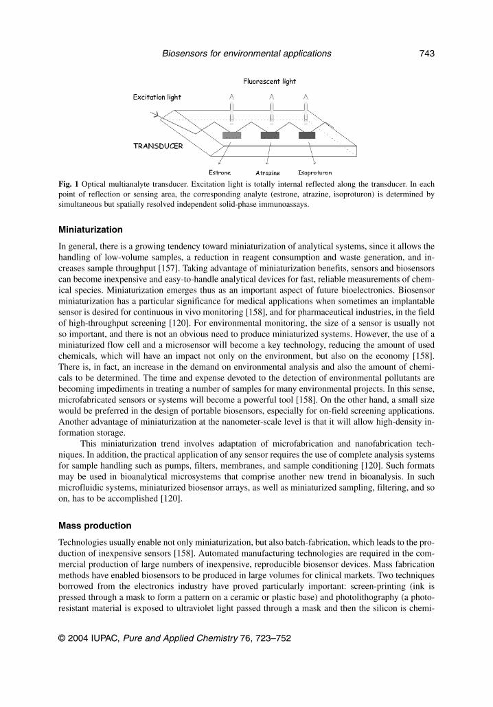
For environmental applications, a multianalyte enzyme biosensor based on a disposable, thick-film multielectrode was developed for the analyte discrimination of binary mixtures of the pesticidesparaoxon and carbofuran [154]. Other studies aimed at the development of multianalyte immunosen-sors, such as that described by Gonzalez-Martínez et al. [3], which developed a competitive captureassay for carbaryl, atrazine, and irgarol 1051 as target compounds. Mastichiadis et al. [155] have ap-plied recently an optical capillary immunosensor to the simultaneous determination of the pesticidesmesotrione, hexaconazole, paraquat, and diquat, by preparing an ordered array capillary of four distinctanalyte bands. After the optimal characterization of analytical conditions in deionized water, further ex-perimentation is required in order to apply the biosensor to natural water sample analysis. Barzen et al.[156] and Rodriguez-Mozaz et al. [57] have described the application of a multianalyte immunosensorbased on total internal reflection fluorescence (RIANA), to the simultaneous determination of differentanalytes in the same sample (see Fig. 1). Atrazine, isoproturon, and estrone were successfully deter-mined in natural water samples with this biosensor [57].
S. RODRIGUEZ-MOZAZ et al.
© 2004 IUPAC, Pure and Applied Chemistry 76, 723–752
742
Miniaturization
In general, there is a growing tendency toward miniaturization of analytical systems, since it allows thehandling of low-volume samples, a reduction in reagent consumption and waste generation, and in-creases sample throughput [157]. Taking advantage of miniaturization benefits, sensors and biosensorscan become inexpensive and easy-to-handle analytical devices for fast, reliable measurements of chem-ical species. Miniaturization emerges thus as an important aspect of future bioelectronics. Biosensorminiaturization has a particular significance for medical applications when sometimes an implantablesensor is desired for continuous in vivo monitoring [158], and for pharmaceutical industries, in the fieldof high-throughput screening [120]. For environmental monitoring, the size of a sensor is usually notso important, and there is not an obvious need to produce miniaturized systems. However, the use of aminiaturized flow cell and a microsensor will become a key technology, reducing the amount of usedchemicals, which will have an impact not only on the environment, but also on the economy [158].There is, in fact, an increase in the demand on environmental analysis and also the amount of chemi-cals to be determined. The time and expense devoted to the detection of environmental pollutants arebecoming impediments in treating a number of samples for many environmental projects. In this sense,microfabricated sensors or systems will become a powerful tool [158]. On the other hand, a small sizewould be preferred in the design of portable biosensors, especially for on-field screening applications.Another advantage of miniaturization at the nanometer-scale level is that it will allow high-density in-formation storage.
This miniaturization trend involves adaptation of microfabrication and nanofabrication tech-niques. In addition, the practical application of any sensor requires the use of complete analysis systemsfor sample handling such as pumps, filters, membranes, and sample conditioning [120]. Such formatsmay be used in bioanalytical microsystems that comprise another new trend in bioanalysis. In suchmicrofluidic systems, miniaturized biosensor arrays, as well as miniaturized sampling, filtering, and soon, has to be accomplished [120].
Mass production
Technologies usually enable not only miniaturization, but also batch-fabrication, which leads to the pro-duction of inexpensive sensors [158]. Automated manufacturing technologies are required in the com-mercial production of large numbers of inexpensive, reproducible biosensor devices. Mass fabricationmethods have enabled biosensors to be produced in large volumes for clinical markets. Two techniquesborrowed from the electronics industry have proved particularly important: screen-printing (ink ispressed through a mask to form a pattern on a ceramic or plastic base) and photolithography (a photo-resistant material is exposed to ultraviolet light passed through a mask and then the silicon is chemi-
© 2004 IUPAC, Pure and Applied Chemistry 76, 723–752
Biosensors for environmental applications 743
Fig. 1 Optical multianalyte transducer. Excitation light is totally internal reflected along the transducer. In eachpoint of reflection or sensing area, the corresponding analyte (estrone, atrazine, isoproturon) is determined bysimultaneous but spatially resolved independent solid-phase immunoassays.
cally etched). [7]. Silicon fabrication technology has been preferred as a means of mass production ofbiosensors. This is an area of intense research activity mainly by diagnostics companies. The use of sil-icon microfabrication for both electrochemical and optical sensors is expanding, and the capability ofon-chip electronic signal amplification and data processing are very attractive. Bookham Technology(Oxfordshire, UK) is using microelectronics technology based on silicon to produce optical chips forbiosensor application [1]. Other techniques such as air-brush and Cabro deposition have been adaptedfor biosensor fabrication, especially for thick-film biosensors. Thin-film sensors are generally producedby a variety of vapor deposition techniques, electrochemical methods, and more recently by the use ofLangmuir–Blodgett technology. These have been used in the production of sensors based on SPR [1].Laser desorption is particularly promising and offers the ability to “write” proteins to surface with veryhigh resolution (patterning) [159].
Network systems
The future integration of wireless communications systems will generate huge interest as this will in-evitably lead to the emergence of extensive networked multiple autonomous analytical stations in rivers,lakes, wells, or even water treatment plants. These units will provide high-quality information about keychemical parameters that determine the quality of our environment [157]. By means of this network sys-tem and by an intelligent remote surveillance, an active response will be possible. To accomplish this,there is a need for robust and unattended working analytical instrumentation. Each unit will be portable,plug-and-playable, user-friendly, and fully automated with little need for maintenance. The emergenceof these compact, self-sustaining, networked instruments will have enormous impact on all field-basedenvironmental measurements including biosensors. Gu et al. [15] have recently applied a novel earlywarning protocol to monitor the toxicity of the effluents of a water treatment plant. A toxicity earlywarning system is needed to prevent the outflow of water containing dissolved toxic chemicals [15] andis important to ensure compliance with EU directives.
Validation
Despite the practical advantages of biosensors, they must be able to provide results in real time, mustbe simple to use, portable, and cost-effective [4]. Acceptance of these methods is dependent upon sev-eral factors: they should be comparable to conventional analytical systems in terms of reliability, sensi-tivity, selectivity, specificity, and robustness. Therefore, biosensor measurements have to be verified bycomparing them with the results of chemical analysis [77], as shown in Fig 2. This verification processis known as validation. Method validation is the process of demonstrating that the combined proceduresof sample preparation (extraction, clean-up, etc.) and analysis will yield acceptably accurate, precise,and reproducible results for a known analyte in a specific matrix [160]. Thevenot et al. [161] recom-mend several analytical parameters needed to define in order to characterize biosensor performance.They include four sets of parameters: (i) calibration characteristics: sensitivity, working and linear con-centration range, limit of detection (LOD) and of quantification (LOQ); (ii) selectivity and reliability;(iii) steady-state, and transient response times and sample throughput; (iv) reproducibility, stability, andlifetime.
The overall commercial status and general acceptance of the technology will depend on the per-formance characteristics, sample throughput, associated costs, and acceptance by regulatory authorities,based on independent validation data generated using internationally recognized procedures (e.g.,AOAC, ISO, and IDF) [4].
S. RODRIGUEZ-MOZAZ et al.
© 2004 IUPAC, Pure and Applied Chemistry 76, 723–752
744
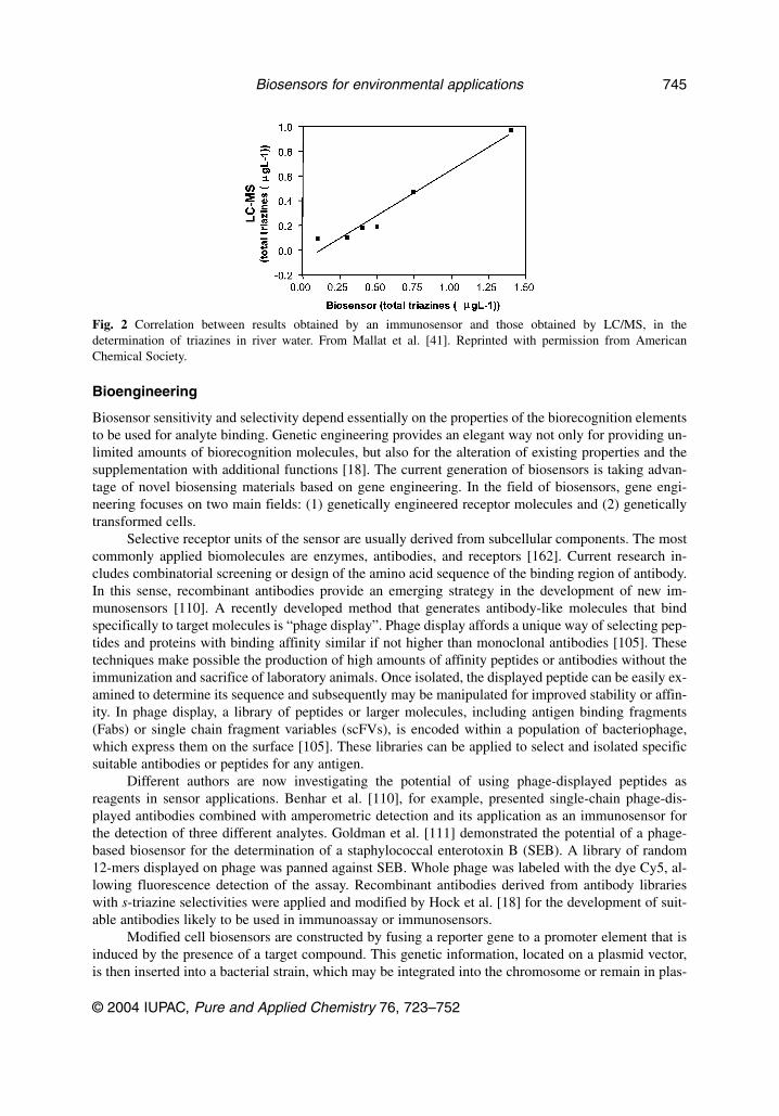
Bioengineering
Biosensor sensitivity and selectivity depend essentially on the properties of the biorecognition elementsto be used for analyte binding. Genetic engineering provides an elegant way not only for providing un-limited amounts of biorecognition molecules, but also for the alteration of existing properties and thesupplementation with additional functions [18]. The current generation of biosensors is taking advan-tage of novel biosensing materials based on gene engineering. In the field of biosensors, gene engi-neering focuses on two main fields: (1) genetically engineered receptor molecules and (2) geneticallytransformed cells.
Selective receptor units of the sensor are usually derived from subcellular components. The mostcommonly applied biomolecules are enzymes, antibodies, and receptors [162]. Current research in-cludes combinatorial screening or design of the amino acid sequence of the binding region of antibody.In this sense, recombinant antibodies provide an emerging strategy in the development of new im-munosensors [110]. A recently developed method that generates antibody-like molecules that bindspecifically to target molecules is “phage display”. Phage display affords a unique way of selecting pep-tides and proteins with binding affinity similar if not higher than monoclonal antibodies [105]. Thesetechniques make possible the production of high amounts of affinity peptides or antibodies without theimmunization and sacrifice of laboratory animals. Once isolated, the displayed peptide can be easily ex-amined to determine its sequence and subsequently may be manipulated for improved stability or affin-ity. In phage display, a library of peptides or larger molecules, including antigen binding fragments(Fabs) or single chain fragment variables (scFVs), is encoded within a population of bacteriophage,which express them on the surface [105]. These libraries can be applied to select and isolated specificsuitable antibodies or peptides for any antigen.
Different authors are now investigating the potential of using phage-displayed peptides asreagents in sensor applications. Benhar et al. [110], for example, presented single-chain phage-dis-played antibodies combined with amperometric detection and its application as an immunosensor forthe detection of three different analytes. Goldman et al. [111] demonstrated the potential of a phage-based biosensor for the determination of a staphylococcal enterotoxin B (SEB). A library of random12-mers displayed on phage was panned against SEB. Whole phage was labeled with the dye Cy5, al-lowing fluorescence detection of the assay. Recombinant antibodies derived from antibody librarieswith s-triazine selectivities were applied and modified by Hock et al. [18] for the development of suit-able antibodies likely to be used in immunoassay or immunosensors.
Modified cell biosensors are constructed by fusing a reporter gene to a promoter element that isinduced by the presence of a target compound. This genetic information, located on a plasmid vector,is then inserted into a bacterial strain, which may be integrated into the chromosome or remain in plas-
© 2004 IUPAC, Pure and Applied Chemistry 76, 723–752
Biosensors for environmental applications 745
Fig. 2 Correlation between results obtained by an immunosensor and those obtained by LC/MS, in thedetermination of triazines in river water. From Mallat et al. [41]. Reprinted with permission from AmericanChemical Society.

mid form. The engineered genetic fusion is then replicated along with the cell’s normal DNA [163]. Asreported above, in the case of bioluminescence sensors, the recombinant bacteria employed containsgene fusions between the regulatory region of the mer operon (merR) and bacterial luminescence genes,which respond to contaminants. Recombinant luminescent bacterial sensors were used by Ivask et al.[119] for the determination of cadmium, zinc, mercury, and chromium. When Hg(II), present in thecytoplasm of the biosensor bacterium, binds to merR, the mer promoter is activated, resulting in tran-scription of the lux genes and subsequent light emission. Three different whole-cell biosensors for thedetermination of mercury were made using E. coli by fusing the mercury-inducible promoter, Pmer, andits regulatory gene merR, with three different reporter genes producing light, β-galactosidase or greenfluorescent protein, respectively, as response to the presence of low levels of mercury [164]. The samegroup presented the choice of three different reporter gene systems combined with a tetracycline-in-ducible promoter in the development of three different biosensors for the analysis of tetracyclines [84]as reported above.
CONCLUSIONS
Despite the huge potential of biosensors, and the ever-increasing number of biosensors developed, com-mercially available biosensors are being applied to a restricted area of the potential market. In general,biosensors for environmental analysis have several limitations: sensitivity, response time, and lifetime,which should be improved for them to become a competitive analytical tool. The areas of developmentthat are expected to have an impact in biosensor technology are: immobilization techniques, nano-technology, miniaturization, and multisensor array determinations. However, a crucial aspect may bethe production of new sensing elements easy to synthesize and with the capability to broaden the spec-tra of selectivities that can be reached by a biosensor. At present, the preparation and production in largescales of biomolecules such as enzymes or antibodies need an investment of time and knowledge.Synthetic peptides and MIPs are contemplated as promising alternatives overcoming the above-men-tioned limitations. Unfortunately, the affinity accomplished by these synthetic receptors is still severalorders of magnitude below that of the antibodies. Improvement in the affinity, specificity, and mass pro-duction of the molecular recognition components may ultimately dictate the success or failure of de-tection technologies [2]. The possibility of tailor binding molecules with predefined properties, such asselectivity, affinity, and stability, is one of the major aims for biotechnology. The development of ad-vanced receptors will allow the analysis of complex real samples and in situ measurements resolvingthe responses from the analyte and from nonspecific background effects. Since scientific attention iscurrently being given to biotechnology, as this review has pointed out, the development of improvedmolecular recognition elements will be followed by a corresponding enhancement of the biosensor fea-tures.
From the above viewpoint, it is clear that the future of biosensors will rely on the success ofemerging sophisticated micro and nanotechnologies, biochemistry, chemistry, thin-film physics, andelectronics. To reach this goal, an important investment in research, expertise, and the necessary facili-ties is needed [5]. However, as the world becomes more concerned about the impact that environmen-tal contamination may cause on public health and the ecosystem, the demand for rapid detecting biosen-sors will only increase. Biosensors still need to achieve the confidence of potential users, having in mindthat the commercialization of new devices will always be the best indicator of the success of a biosen-sor technology.
The analysis of complex matrices and of analytes difficult to determine by the actual analyticalprocedures (i.e., highly polar compounds), are progressively being approached by biosensors. However,there is still a lack of alternative biosensing systems for an important bunch of emerging contaminantssuch as bisphenol A, phtalates, and polybrominated compounds (used as flame retardants), veterinaryand human medicines and personal care products (nutraceuticals, synthetic fragances, sun screenagents, etc.).
S. RODRIGUEZ-MOZAZ et al.
© 2004 IUPAC, Pure and Applied Chemistry 76, 723–752
746

ACKNOWLEDGMENTS
This work has been supported by AWACSS (EVK1-CT-2000-00045) and by RADAR (QLRT-2000-01670). Sara Rodriguez acknowledges Ministerio de Ciencia y Tecnología (Project PPQ 2000-3006-CE) and I3P Program (Itinerario integrado de inserción professional; co-financed by CSIC andEuropean Social Funds). María J. López de Alda acknowledges a Ramon y Cajal contract from theSpanish Ministry of Science and Technology.
REFERENCES
1. I. E. Tothill. Comput. Electron. Agric. 30, 205–218 (2001).2. J. Parellada, A. Narvaez, M. A. Lopez, E. Dominguez, J. J. Fernandez, V. Pavlov, I. Katakis. Anal.
Chim. Acta 362, 47–57 (1998).3. M. A. Gonzalez-Martinez, R. Puchades, A. Maquieira. TrAC, Trends Anal. Chem. 18, 204–218
(1999).4. P. D. Patel. TrAC, Trends Anal. Chem. 21, 96–115 (2002).5. A. F. Collings and F. Caruso. Rep. Prog. Phys. 60, 1397–1145 (1997).6. D. Meadows. Adv. Drug Delivery Rev. 21, 179–189 (1996).7. A. P. F. Turner. Science 5495, 1315–1317 (2000).8. J. Wang, G. Rivas, X. Cai, E. Palecek, P. Nielsen, H. Shiraishi, N. Dontha, D. Luo, C. Parrado,
M. Chicharro. Anal. Chim. Acta 347, 1–8 (1997).9. M. Castillo, M. C. Alonso, J. Riu, M. Reinke, G. Kloter, H. Dizer, B. Fischer, P. D. Hansen, D.
Barcelo. Anal. Chim. Acta 426, 265–277 (2001).10. M. R. Evans, G. M. Jordison, D. M. Rawson, J. G. Regerson. Pestic. Sci. 54, 447 (1998).11. M. Farre, O. Pasini, M. Carmen Alonso, M. Castillo, D. Barcelo. Anal. Chim. Acta 426, 155–165
(2001).12. M. Farre and D. Barcelo. TrAC, Trends Anal. Chem. 22, 299–310 (2003).13. Environmental Agency UK. Direct Toxicity Assessment, Newsletter, July (1999).14. E. J. Kim, J. E. Lee, M. B. Gu. Water Sci. Technol. 46, 51–56 (2002).15. M. B. Gu, E. J. Kim, J. Cho, P. D. Hansen. Environ. Monit. Assess. 70, 71–81 (2001).16. A. J. Oosterkamp, B. Hock, M. Seifert, H. Irth. TrAC, Trends Anal. Chem. 16, 544–553 (1997).17. M. Usami, K. Mitsunaga, Y. Ohno. J. Steroid Biochem. Molec. Biol. 81, 47–55 (2002).18. B. Hock, M. Seifert, K. Kramer. Biosens. Bioelectron. 17, 239–249 (2002).19. M. Seifert, S. Haindl, B. Hock. Anal. Chim. Acta 386, 191–199 (1999).20. M. Murata, M. Nakayama, K. H. Yakabe, K. Fukuma, Y. Katayama, M. Maeda. Anal. Sci. 17,
387–390 (2001).21. M. Zhihong, L. Xiaohui, F. Weiling. Anal. Commun. 36, 281–283 (1999).22. J.-W. Choi, Y.-K. Kim, I.-H. Lee, J. Min, W. H. Lee. Biosens. Bioelectron. 16, 937–943 (2001).23. V. G. Andreou and Y. D. Clonis. Biosens. Bioelectron. 17, 61–69 (2002).24. R. T. Andres and R. Narayanaswamy. Talanta 44, 1335–1352 (1997).25. H. Schulze, S. Vorlova, F. Villatte, T. T. Bachmann, R. D. Schmid. Biosens. Bioelectron. 18,
201–209 (2003).26. A. Mulchandani, W. Chen, P. Mulchandani, J. Wang, K. R. Rogers. Biosens. Bioelectron. 16,
225–230 (2001).27. M. H. Smith and G. A. Rechnitz. Anal. Chem. 65, 380 (1993).28. F. A. McArdle and K. C. Persaud. Analyst 118, 419 (1993).29. J. Wang and L. Chen. Anal. Chem. 67, 3824–3827 (1995).30. W. R. Everett and G. A. Rechnitz. Anal. Chem. 70, 807–810 (1998).31. T. Noguer and J.-L. Marty. Anal. Chim. Acta 347, 63–69 (1997).32. M. T. Giardi, M. Koblizek, J. Masojidek. Biosens. Bioelectron. 16, 1027–1033 (2001).
© 2004 IUPAC, Pure and Applied Chemistry 76, 723–752
Biosensors for environmental applications 747
33. C. Vedrine, J.-C. Leclerc, C. Durrieu, C. Tran-Minh. Biosens. Bioelectron. 18, 457–453 (2003).34. M. Koblizek, J. Maly, J. Masojidek, J. Komenda, T. Kuera, M. T. Giardi, A. K. Mattoo, R.
Pilloton. Biotechnol. Bioeng. 78, 110–116 (2002).35. M. Wilmer, D. Trau, R. Renneberg, F. Spener. Anal. Lett. 30, 515–525 (1997).36. E. F. Schipper, A. J. H. Bergevoet, R. P. H. Kooyman, J. Greve. Anal. Chim. Acta 341, 171–176
(1997).37. E. F. Schipper, S. Rauchalles, R. P. H. Kooyman, B. Hock, J. Greve. Anal. Chem. 70, 1192–1197
(1998).38. K. E. Brummel, J. Wright, M. E. Eldefrawi. J. Agr. Food Chem. 45, 3292–3298 (1997).39. E. Mallat, C. Barzen, R. Abuknesha, G. Gauglitz, D. Barcelo. Anal. Chim. Acta 427, 165–171
(2001).40. E. Mallat, C. Barzen, R. Abuknesha, G. Gauglitz, D. Barcelo. Anal. Chim. Acta 426, 209–216
(2001).41. E. Mallat, C. Barzen, A. Klotz, A. Brecht, G. Gauglitz, D. Barcelo. Environ. Sci. Technol. 33,
965–971 (1999).42. C. Mouvet, R. D. Harris, C. Maciag, B. J. Luff, J. S. Wilkinson, J. Piehler, A. Brecht, G. Gauglitz,
R. Abuknesha, G. Ismail. Anal. Chim. Acta 338, 109–117 (1997).43. M. A. Gonzalez-Martinez, R. Puchades, A. Maquieira. Anal. Chem. 73, 4326–4332 (2001).44. J. Peñalva, M.-A. Gonzalez-Martinez, R. Puchades, A. Maquieira, M.-P. Marco, D. Barceló. Anal.
Chim. Acta 387, 227–233 (1998).45. M. Minunni, P. Skladal, M. Mascini. Life Chem. Rep 11, 391–8 (1994).46. K. Yokoyama, K. Ikebukuro, E. Tamiya, I. Karube, N. Ichiki, Y. Arikawa. Anal. Chim. Acta 304,
139–45 (1995).47. M. Minunni, P. Skladal, M. Mascini. Anal. Lett. 27, 1475–87 (1994).48. J. Ngeh-Ngwainbi, P. H. Foley, S. S. Kuan, G. G. Guilbault. J. Am. Chem. Soc. 108, 5444–5447
(1986).49. J. Horacek and P. Skladal. Biosens. Bioelectron. 12, v–vi (1997).50. I. G. Lange, A. Daxenberger, B. Schiffer, H. Witters, D. Ibarreta, H. H. D. Meyer. Anal. Chim.
Acta 473, 27–37 (2002).51. D. G. J. Larsson, M. Adolfsson-Erici, J. Parkkonen, M. Pettersson, A. H. Berg, P.-E. Olsson, L.
Forlin. Aquat. Toxicol. 45, 91–97 (1999).52. T. A. Ternes, P. Kreckel, J. Mueller. Sci. Total Environ. 225, 91–99 (1999).53. A. C. Belfroid, A. Van der Horst, A. D. Vethaak, A. J. Schafer, G. B. J. Rijs, J. Wegener, W. P.
Cofino. Sci. Total Environ. 225, 101–108 (1999).54. Environmental Issues Series, Consultative Report, Environment Agency (1998).55. P.-D. Hansen, H. Dizer, B. Hock, A. Marx, J. Sherry, M. McMaster, C. Blaise. TrAC, Trends Anal.
Chem. 17, 448–451 (1998).56. C. E. Purdom, P. A. Hardiman, V. J. Bye, N. C. Eno, C. R. Tyler, J. P. Sumpter. Chem. Ecol. 8,
275–285 (1994).57. S. Rodriguez-Mozaz, S. Reder, M. Lopez de Alda, G. Gauglitz, D. Barceló. Biosens. Bioelectron.
19, 633–640 (2004).58. A. Boenke. Anal. Chim. Acta 473, 83–87 (2002).59. G. Marrazza, I. Chianella, M. Mascini. Anal. Chim. Acta 387, 297–307 (1999).60. C. Q. Zhao, N. A. Anis, K. R. Rogers, R. H. Kline, J. Wright, A. T. Eldefrawi, M. E. Eldefrawi.
J. Agric. Food. Chem. 2308–2315 (1995).61. M. Shimomura, Y. Nomura, W. Zhang, M. Sakino, K.-H. Lee, K. Ikebukuro, I. Karube. Anal.
Chim. Acta 434, 223–230 (2001).62. M. Del Carlo, I. Lionti, M. Taccini, A. Cagnini, M. Mascini. Anal. Chim. Acta 342, 189–197
(1997).63. N. Lee, C. K. Holtzapple, L. H. Stanker. J. Agric. Food Chem. 46, 3381–3388 (1998).
S. RODRIGUEZ-MOZAZ et al.
© 2004 IUPAC, Pure and Applied Chemistry 76, 723–752
748
64. M. Vanderlaan, L. H. Stanker, B. E. Watkins, P. Petrovic, S. Gorbach. Biotechnol. Adv. 14, 486(1996).
65. L. H. Stanker, B. Watkins, N. Rogers, M. Vanderlaan. Toxicology 45, 229–243 (1987).66. P. Pasini, G. Gentlilomi, M. Baraldini, M. Musiani, A. Roda. 12th International Symposium on
Bioluminescence and Chemiluminescence (2002).67. A. Degiuli and L. J. Blum. J. Med. Biochem. 4, 32–42 (2000).68. J. Angerer, T. Goen, G. Lehnert. Organohalogen Compd. 14, 163–167 (1993).69. G. Palmer, R. McFadzean, K. Killham, A. Sinclair, G. I. Paton. Chemosphere 36, 2683–2697
(1998).70. R. Badia and M. E. Diaz Garcia. Anal. Chim. Acta 371, 73–80 (1998).71. A. N. Reshetilov, I. N. Semenchuk, P. V. Iliasov, L. A. Taranova. Anal. Chim. Acta 347, 19–26
(1997).72. L. Taranova, I. Semenchuk, T. Manolov, P. Iliasov, A. Reshetilov. Biosens. Bioelectron. 17,
635–640 (2002).73. J. Waters and T. C. J. Feijtel. Chemosphere 30, 1939–1956 (1995).74. Y. Nomura, K. Ikebukuro, K. Yokoyama, T. Takeuchi, Y. Arikawa, S. Ohno, I. Karube. Biosens.
Bioelectron. 13, 1047–1053 (1998).75. A. Rose, C. Nistor, J. Emneus, D. Pfeiffer, U. Wollenberger. Biosens. Bioelectron. 17, 1033–1043
(2002).76. L. Stiner and L. J. Halverson. Appl. Environ. Microbiol. 68, 1962–1971 (2002).77. P. Sticher, M. C. Jaspers, K. Stemmler, H. Harms, A. J. Zehnder, J. R. van der Meer. Appl.
Environ. Microbiol. 63, 4053–4060 (1997).78. A. Koenig, C. Zaborosch, F. Spener. In Field Screening Europe, J. Gottlieb, H. Hotzl, K. Huck,
R. Niessner (Eds.), pp. 203–206, Kluwer Academic, Dordrecht (1997).79. J. P. Alarie, J. R. Bowyer, M. J. Sepaniak, A. M. Hoyt, T. Vo-dinh. Anal. Chim. Acta 236, 237–244
(1990).80. I. Coille, S. Reder, S. Bucher, G. Gauglitz. Biomol. Eng. 18, 273–280 (2002).81. S. J. Setford, R. M. Van Es, Y. J. Blankwater, S. Kroger. Anal. Chim. Acta 398, 13–22 (1999).82. A. Ahmad, A. Ramakrishnan, M. A. McLean, D. Li, M. T. Rock, A. Karim, A. P. Breau. 300,
177–184 (2002).83. A. Akkoyun, V. F. Kohen, U. Bilitewski. Sens. Actuators, B 70, 12–18 (2000).84. L. H. Hansen and S. J. Sorensen. FEMS Microbiol. Lett. 190, 273–278 (2000).85. S. Subrahmanyam, S. A. Piletsky, A. P. F. Turner. Anal. Chem. 74, 3942–3951 (2002).86. S. Ramanathan, M. Ensor, S. Daunert. Trends Biotechnol. 15, 500–506 (1997).87. G. G. Guilbault, D. E. T. Sibley, R. M. Carter, G. J. Lubrano. NATO ASI Ser., Ser. E 252, 377–385
(1993).88. R. M. Carter, G. J. Lubrano, G. G. Guilbault. Life Chem. Rep. 11, 271–277 (1994).89. J. B. Delehanty and F. S. Ligler. Anal. Chem. 74, 5681–5687 (2002).90. G. Palleschi, D. Compagnone, D. Moscone. VTT Symp. 177, 141–160 (1997).91. C. Tran Minh and P. C. Pandey. Bull. Electrochem. 8, 199–204 (1992).92. A. A. Boiarski, J. R. Busch, R. S. Brody, R. W. Ridgway, W. P. Altman, C. Golden. Proc. SPIE-
Int. Soc. Opt. Eng. 2686, 45–52 (1996).93. C. D. Feng, T. E. Nelson, S. Hardman, P. J. Hesketh, G. J. Maclay, S. M. Gendel, J. R. Stetter. In
The 8th International Conference on Solid-State Sensors and Actuators, and Eurosensors IX, pp.538–541, Stockholm (1995).
94. P. Kumar, J. T. Colston, J. P. Chambers, E. D. Rael, J. J. Valdes. Biosens. Bioelectron. 9, 57–63(1994).
95. R. A. Ogert, J. E. Brown, B. R. Singh, L. C. Shriver-Lake, F. S. Ligler. Anal. Biochem. 205,306–312 (1992).
© 2004 IUPAC, Pure and Applied Chemistry 76, 723–752
Biosensors for environmental applications 749
96. R. S. Marks, Z. M. Hale, M. M. Levine, C. R. Lowe, F. P. Payne. Proc. SPIE-Int. Soc. Opt. Eng.2131, 495–503 (1994).
97. L. A. Tempelman, K. D. King, G. P. Anderson, F. S. Ligler. Anal. Biochem. 233, 50–57 (1996).98. E. C. Alocilja and S. M. Radke. Biosens. Bioelectron. 18, 841–846 (2003).99. A. M. Nasser and S. D. Oman. Water Res. 33, 1748–1752 (1999).
100. S. Toze. Water Res. 33, 3545–3556 (1999).101. A. Pianetti, W. Baffone, F. Bruscolini, E. Barbieri, M. R. Biffi, L. Salvaggio, A. Albano. Water
Res. 32, 1515–1521 (1998).102. N. Contreras-Coll, F. Lucena, K. Mooijman, A. Havelaar, V. Pierzo, M. Boque, A. Gawler, C.
Holler, M. Lambiri, G. Mirolo. Water Res. 36, 4963–4974 (2002).103. D. Ivnitski, I. Abdel-Hamid, P. Atanasov, E. Wilkins. Biosens. Bioelectron. 14, 599–624 (1999).104. C. Ercole, M. D. Gallo, M. Pantalone, S. Santucci, L. Mosiello, C. Laconi, A. Lepidi. Sens.
Actuators B 83, 48–52 (2002).105. S. S. Iqbal, M. W. Mayo, J. G. Bruno, B. V. Bronk, C. A. Batt, J. P. Chambers. Biosens.
Bioelectron. 15, 549–578 (2000).106. V. Koubova, E. Brynda, L. Karasova, J. Skvor, J. Homola, J. Dostalek, P. Tobiska, J. Rosicky.
Sens. Actuators B 74, 100–105 (2001).107. S. T. Pathirana, J. Barbaree, B. A. Chin, M. G. Hartell, W. C. Neely, V. Vodyanoy. Biosens.
Bioelectron. 15, 135–141 (2000).108. S. Tombelli, M. Mascini, C. Sacco, A. P. F. Turner. Anal. Chim. Acta 418, 1–9 (2000).109. Y. Hasebe, K. Yokobori, K. Fukasawa, T. Kogure, S. Uchiyama. Anal. Chim. Acta 357, 51–54
(1997).110. I. Benhar, I. Eshkenazi, T. Neufeld, J. Opatowsky, S. Shaky, J. Rishpon. Talanta 55, 899–907
(2001).111. E. R. Goldman, M. P. Pazirandeh, J. M. Mauro, K. D. King, J. C. Frey, G. P. Anderson. J. Molec.
Recog. 13, 382–387 (2000).112. P. Leonard, S. Hearty, J. Brennan, L. Dunne, J. Quinn, T. Chakraborty, R. O’Kennedy. Enzyme
Microb. Technol. 32, 3–13 (2003).113. T. Krawczynski vel Krawczyk, M. Moszczynska, M. Trojanowicz. Biosens. Bioelectron. 15,
681–691 (2000).114. C. Durrieu and C. Tran-Minh. Ecotoxicol. Environ. Saf. 51, 206–209 (2002).115. L. D. Rasmussen, S. J. Sorensen, R. R. Turner, T. Barkay. Soil Biol. Biochem. 32, 639–646 (2000).116. T. D. Yerian, G. D. Christian, J. Ruzicka. Anal. Chim. Acta 204, 7–28 (1988).117. Y. J. Nam, N. Kim, V. Volotovsky. Sens. Actuators B 42, 233–237 (1997).118. T. Petanen and M. Romantschuk. Anal. Chim. Acta 456, 55–61 (2002).119. A. Ivask, M. Virta, A. Kahru. Soil Biol. Biochem. 34, 1439–1447 (2002).120. C. Ziegler and W. Gopel. Curr. Opin. Chem. Biol. 2, 585–591 (1998).121. I. Bontidean, J. R. Lloyd, J. L. Hobman, J. R. Wilson, E. Csoregi, B. Mattiasson, N. L. Brown. J.
Inorg. Biochem. 79, 225–229 (2000).122. J. Kulys, I. J. Higgins, J. V. Bannister. Biosens. Bioelectron. 7, 187–191 (1992).123. U. Wollenberger, F. Schubert, F. W. Scheller. Sens. Actuators, B 7, 412–415 (1992).124. L. H. Larsen, T. Kjær, N. P. Revsbech. Anal. Chem. 69, 3527–3531 (1997).125. L. H. Larsen, L. R. Damgaard, T. Kjaer, T. Stenstrom, A. Lynggaard-Jensen, N. P. Revsbech.
Water Res. 34, 2463–2468 (2000).126. A. Abdelghani, N. Jaffrezic-Renault. Sens. Actuators, B 74, 117–123 (2001).127. J. Liu and B. Mattiasson. Water Res. 36, 3786–3802 (2002).128. W. Bourgeois, J. E. Burgess, R. M. Stuetz. J. Chem. Technol. Biotechnol. 76, 337–348 (2001).129. Z. Qian and T. C. Tan. Water Res. 33, 2923–2928 (1999).130. F. Li and T. C. Tan. Biosens. Bioelectron. 9, 315–324 (1994).131. G.-J. Chee, Y. Nomura, K. Ikebukuro, I. Karube. Biosens. Bioelectron. 15, 371–376 (2000).
S. RODRIGUEZ-MOZAZ et al.
© 2004 IUPAC, Pure and Applied Chemistry 76, 723–752
750

132. H. J. Purohit. J. Cleaner Prod. 11, 293–301 (2003).133. H. A. Fishman, D. R. Greenwald, R. N. Zare. Annu. Rev. Biophys. Biomol. Struct. 27, 165–198
(1998).134. F. Ortega, E. Dominguez, E. Burestedt, J. Emneus, L. Gorton, G. Marko-Vargo. J. Chromatogr. A
675, 65–78 (1994).135. O. Adeyoju, E. Iwuoha, M. Smyth, D. Leech. Analyst 121, 1885–1889 (1996).136. S. Kroger, S. Piletsky, A. P. F. Turner. Mar. Pollut. Bull. 45, 24–34 (2002).137. R. L. Rich and D. G. Myszka. Curr. Opin. Biotechnol. 11, 54–61 (2000).138. I. Lundstrom and S. Svensson. Curr. Appl. Phys. 2, 17–21 (2002).139. L. Tiefenauer and R. Ros. Colloids Surf. B: Biointerfaces 23, 95–114 (2002).140. S. Kossek, C. Padeste, L. X. Tiefenauer, H. Siegenthaler. Biosens. Bioelectron. 13, 31–43 (1998).141. D. t. i. center.142. T. H. Wink, S. J. van Zuilen, A. Bult, W. P. van Bennekom. Analyst 122, 43–50 (1997).143. L. X. Tiefenauer, S. Kossek, C. Padeste, P. Thiebaud. Biosens. Bioelectron. 12, 213–223 (1997).144. A. J. Haes and R. P. Van Duyne. J. Am. Chem. Soc. 124, 10589–10595 (2002).145. M. D. Malinsky, K. L. Kelly, G. C. V. D. Schatz, P. Richard. J. Am. Chem. Soc. 123, 1471–1482
(2001).146. T. A. Taton, G. Lu, C. A. Mirkin. J. Am. Chem. Soc. 123, 5164–5165 (2001).147. Y. Cao, R. Jin, C. A. Mirkin. J. Am. Chem. Soc. 123, 7961–7962 (2001).148. A. A. Lazarides, K. Lance Kelly, T. R. Jensen, G. C. Schatz. J. Mol. Struct. 529, 59–63 (2000).149. K. Ramanathan and B. Danielsson. Biosens. Bioelectron. 16, 417–423 (2001).150. C. E. H. Berger, T. A. M. Beumer, R. P. H. Kooyman, J. Greve. Anal. Chem. 70, 703706 (1998).151. C. A. Rowe-Taitt, J. P. Golden, M. J. Feldstein, J. J. Cras, K. E. Hoffman, F. S. Ligler. Biosens.
Bioelectron. 14, 785–794 (2000).152. C. A. Rowe-Taitt, J. W. Hazzard, K. E. Hoffman, J. J. Cras, J. P. Golden, F. S. Ligler. Biosens.
Bioelectron. 15, 579–589 (2000).153. R. M. Wadkins, J. P. Golden, L. M. Pritsiolas, F. S. Ligler. Biosens. Bioelectron. 13, 407–415
(1998).154. T. T. Bachmann and R. D. Schmid. Anal. Chim. Acta 401, 95–103 (1999).155. C. Mastichiadis, S. E. Kakabakos, I. Christofidis, M. A. Koupparis, C. Willetts, K. Misiakos.
Anal. Chem. 74, 6064–6072 (2002).156. C. Barzen, A. Brecht, G. Gauglitz. Biosens. Bioelectron. 17, 289–295 (2002).157. M. Sequeira, M. Bowden, E. Minogue, D. Diamond. Talanta 56, 355–363 (2002).158. H. Suzuki. Mater. Sci. Eng. C 12, 55–61 (2000).159. <http://www.cranfield.ac.uk/biotech/chinap.htm>160. J. Antonio Gabaldon, A. Maquieira, R. Puchades. Crit. Rev. Food. Sci. Nutr. 39, 519–538 (1999).161. D. R. Thevenot, K. Toth, R. A. Durst, G. S. Wilson. Biosens. Bioelectron. 16, 121–131 (2001).162. B. Kuswandi, R. Andres, R. Narayanaswamy. Analyst 1469–1491 (2001).163. A. Keane, P. Phoenix, S. Ghoshal, P. C. K. Lau. J. Microbiol. Meth. 49, 103–119 (2002).164. L. H. Hansen and S. J. Sorensen. FEMS Microbiol. Lett. 193, 123–127 (2000).165. C. Nistor, A. Rose, M. Farre, L. Stoica, U. Wollenberger, T. Ruzgas, D. Pfeiffer, D. Barcelo, L.
Gorton, J. Emneus. Anal. Chim. Acta 456, 3–17 (2002).166. D. Frense, A. Muller, D. Beckmann. Sens. Actuators, B 51, 256–260 (1998).167. S. H. Choi and M. B. Gu. Biosens. Bioelectron. 17, 433–440 (2002).168. T. C. Tan and C. Wu. Sens. Actuators, B 54, 252–260 (1999).169. B. Catimel, J. Weinstock, M. Nerrie, T. Domagala, E. C. Nice. J. Chromatogr. A 869, 261–273
(2000).170. M. Murata, M. Nakayama, H. Irie, K. Yakabe, K. Fukuma, Y. Katayama, M. Maeda. Anal. Sci.
17, 387–390 (2001).
© 2004 IUPAC, Pure and Applied Chemistry 76, 723–752
Biosensors for environmental applications 751
171. T. Wink, J. de Beer, W. E. Hennink, A. Bult, W. P. van Bennekom. Anal. Chem. 71, 801–805(1999).
172. K. Kukanskis, J. Elkind, J. Melendez, T. Murphy, G. Miller, H. Garner. Anal. Biochem. 274, 7–17(1999).
173. A. Giuseppi-Elie, G. Wallace, T. Matsue. In Handbook of Conducting Polymers, T. A. Skotheim,R. L. Elsenbaumer, J. R. Reynolds (Eds.), Marcel Dekker, New York (1998).
174. S. A. Glazier, E. R. Campbell, W. H. Campbell. Anal. Chem. 70, 1511–1515 (1998).
S. RODRIGUEZ-MOZAZ et al.
© 2004 IUPAC, Pure and Applied Chemistry 76, 723–752
752