Embed Size (px)
Citation preview
Ciechańska D., Wietecha J., Kaźmierczak D., Kazimierczak J.; Biosynthesis of Modified Bacterial Cellulose in a Tubular Form.FIBRES & TEXTILES in Eastern Europe 2010, Vol. 18, No. 5 (82) pp. 98-104.
98
Biosynthesis of Modified Bacterial Cellulose in a Tubular Form
Danuta Ciechańska, Justyna Wietecha,
Dorota Kaźmierczak, Janusz Kazimierczak
Institute of Biopolymers and Chemical Fibresul. M. Skłodowskiej-Curie 19/279, 90-570 Łódź, Poland
E-mail: [email protected]
AbstractPresented herein is a method to prepare tubes of modified bacterial cellulose immediately in the course of biosynthesis conducted with Acetobacter sp. The biosynthesis was carried out under both static and dynamic conditions. The material of the tubes obtained was tested and assessed with respect to its structural, physical-mechanical and biological properties, as well as its chemical purity. The testing witnessed a high biocompatibility of the modified bacterial cellulose, essential for the material in blood vessel prostheses. It was found that the mechanical properties of the tubes can be improved by applying a polyester plaited carrier in the course of the dynamic synthesis of the modified cellulose.
Key words: modified cellulose, biosynthesis, vascular prostheses.
n IntroductionDefects of the cardiovascular system which require surgical repair are a seri-ous social problem, affecting about 0.5% of the population. About 20 000 cardio-vascular operations a year are carried out in Poland, resulting from atherosclerosis, injuries and aneurysms. Vessel prosthe-ses, made mainly of polyester woven or knitted fabrics, are employed in about 50% of the treatments. The percentage is higher in the US, reaching 75%.
Over the last 50 years, a wide array of synthetic polymers has been harnessed for the manufacture of vessel prostheses: poly-(vinyl alcohol), polyamide, poly-acrylonitrile, and polytetrafluoroethylene [1]. American, German and British com-panies lead the way in the manufacture of woven and knitted vessel prostheses made of poly-(ethylene terephtalate) and polytetrafluoroethylene, offering unsealed and sealed prostheses in which the sealant constitutes either bovine cross-linked gelatine or bovine collagen. Tricomed Co, the sole Polish producer, offers knitted vessel prostheses of poly-(ethylene terephtalate) in unsealed and sealed (porcine gelatin) version. Virtually all manufacturers of the vessels employ animal-derived substances as sealants.Three kinds of artery prostheses are used in clinical surgery nowadays [2]:n biological - autogenic (patient’s own
veins or arteries) and allogenic (e.g. modified umbilical cord veins), Al-logenic transplants are only used in extreme cases and after suitable modi-fication (treatment with DMSO and freezing)
n semi-biological – prostheses are built up of a non-resorbable synthetic ma-trix superficially impregnated with a resorbable polymer like gelatin deriv-atives, human albumin and collagen
n synthetic - from poly-(ethylene tere-phtalate) or expanded polytetrafluor-oethylene.
High quality demands are placed on vessel prostheses. For many years after the implantation the prosthesis material should resist degradation caused by the environmental conditions of the human body, mainly the temperature and pres-ence of enzymes and body fluids. No ge-notoxic action of the prosthesis upon the tissue surrounding the implant is allowed
nor an allergenic action upon the pa-tient’s organism. The material ought to be flexible and elastic, easing adjustment to the vessels repaired. Artificial implants, particularly those which come in direct contact with blood, must be haemocom-patible, resistant to internal stress caused by blood pressure, and impermeable to blood elements. The inner surface of a vessel prosthesis cannot cause the ad-hesion of haemocytes and ought to be covered with endothelium cells, while connecting tissue should cover the outer surface.
Experimental investigations have shown that implants of poly-(ethylene terephta-late) and expanded polytetrafluoroethyl-ene are close to a model solution, yet the prostheses available are encumbered by defects that limit their application. Ex-panded polytetrafluoroethylene (ePTFE), for example, with its suitable porosity and low thrombogenic action, lends it-self for the preparation of low diameter prostheses (below 8 mm). The material is, however, prone to degradation dur-ing radiation sterilisation, and its surface binds lipids dissolved in the blood, thus contributing to atherosclerosis. Although porous, the (ePTFE) prostheses are not ingrown with tissue. Moreover, these prostheses carry a high risk of bacterial infection [3]. Other disadvantages are a low elasticity, which makes connecting with the vessel repaired difficult, and bleeding, which may occur at the inter-connection. Furthermore, most semi-bio-logical prostheses exhibit a long healing time and run the risk of transmitting ani-mal – derived diseases.
The surface of all the synthetic vessel prostheses produced is haemostatically active, contributing to the forming of clots of varied thickness on the surface
99FIBRES & TEXTILES in Eastern Europe 2010, Vol. 18, No. 5 (82)
of the vessel walls. This phenomenon brought about the pre-operation sealing (pre-clotting) of macro-porous prosthe-ses [4]. The method exploits the clotting of blood occurring when the blood comes in contact with a thrombogenic surface, inherent to synthetic prostheses. This instance of the self-sealing of a prosthe-sis results in acquiring a surgical tight-ness provided by a fibrin layer contain-ing blood morphotic elements deposited on the inner surface. Unfortunately, this phenomenon also has its hazard – the surface clots formed make the surface highly thrombogenic [5], and an uneven out-pressing of the clots may cause un-controlled bleeding. The out-pressing does not warrant a complete removal of the clots, thus running the danger of an embolism.
The drawbacks of currently available commercial prostheses have encouraged scientists to investigate new, ever better materials. World-wide intensive research is under way to develop prostheses that would mimic the features of natural blood vessels.
These were the reasons why the authors undertook to investigate the possibility of preparing biological blood prostheses based upon an advanced material: modi-fied bacterial cellulose [6, 7]. The mate-rial can be obtained by modification of the nutrient medium in the microbiologi-cal synthesis of cellulose. The bacterial synthesis method adopted, the conditions of the biosynthesis and the efficiency of the bacteria strain are all factors influenc-ing not only the output of the process but also the physical-mechanical character-istic and related useful properties of the cellulose prepared [8, 9].
Bacterial cellulose is produced i.e. by bacteria of the Gluconacetobacter type. It is secreted from cells in the form of fibrils built up of cellulose chains. Unlike natural plant cellulose, the bacterial kind does not contain lignin, pectin and hemicellulose. Bacterial cellulose is highly water-absorp-tive and does not cause irritation nor al-lergic reactions, which make the material suitable for use as artificial skin in wound healing [10]. Thanks to its high chemical purity and outstanding physical-chemical features, it may also be used as an addi-tive in dietary food, in the manufacture of electro-acoustic and filtration membranes, as ultra-strong paper, and, in the form of mi-cro-fibre, as suspension of good covering, binding and thickening properties [11 - 15].
amongst the materials examined, bacterial cellulose demonstrated the least intensive and most delayed activation of the clotting cascade [29].
The objective of the research presented herein was the preparation of a method to produce prostheses of blood vessels based on bacterial cellulose modified with chi-tooligosaccharides. The results of earlier investigations into the synthesis of bacte-rial cellulose [30, 31] were a starting point for the present research, aimed at the prep-aration of tubes for implants.
The investigations can be subdivided into the following :n Biosynthesis of modified bacterial cel-
lulose using selected matrices n Assessment of the structure, morphol-
ogy and physical-mechanical properties of prototypes of vessel prostheses of modified bacterial cellulose.
n Assessment of the biological properties of the vessel prostheses obtained.
n Materials and methodsAn acetic strain of bacteria - Acetobacter sp. (CCM 2360) from the Czech Collec-tion of Microorganisms, Masaryk Uni-versity, Brno, Czech Republic was used in the synthesis of bacterial cellulose.
The nutrition medium Hestrin-Schramm was also used, composed of the following (on 1000 cm3): glucose 20.0 g, yeast ex-tract 5.0 g, soy peptone 5.0 g. di-sodium phosphate 2.7g, acetic acid 1.2 g, ethanol 20.0 cm3, and chitosan oligomers 2.0 g. The characteristic of the chitosan oli-gomer ChitoOligo-100 (by Amicogen, Korea) given by the manufacturer is shown in Table 1.
Assessment of structural propertiesGPC analysis was carried out accord-ing to the Turbak procedure [32, 33] us-ing Hewlett Packard HP 1050 apparatus equipped with a RI HP 1047A refracto-metric detector and set of columns with a rigid hydrophilic filling based on poly-meric gel. Samples were prepared for the analysis according to the modified Ekma-nis method [34].
Infrared spectrophotometric analysis was made with the use of FTIR Unicam appa-ratus with WinFIRST software of Matt-son Co, within a wave number range of 4000 - 650 cm-1.
Very positive is the fact that the shape de-sired can be given to products of bacterial cellulose in the course of its generation (in situ) [16]. Patents [17, 18] describe the in-stant forming of shapes, such as tubes of bacterial cellulose, in the course of bio-synthesis suited to replace blood vessels or other organs.
Bacterial cellulose modified with chitooli-gosaccharides combines the properties of both chitosan and cellulose, leading to an improvement in its biocompatibility and bioactivity. Incorporating chitooligosac-charide segments into the cellulose chain may provide conformity with the proper-ties of natural blood vessels where muco-polysaccharides display an integral mul-tifunctional system, which stands for the elasticity of the blood vessel and the form-ing of an athrombogenic surface [19]. Polyaminosaccharides and their derivatives enhance wound healing [20] by inducing fibroblasts to deliver interleukin -8, which is responsible for the migration and pro-liferation of cell blood vessel endothelium [21, 22].
Up to now, several uses of bacterial cel-lulose are known in human and veteri-nary medicine. The application of Biofill® and Gengiflex® bacterial cellulose in surgery and as implants in dentistry is de-scribed in [23 - 26].
Investigations into the biosynthesis of bac-terial cellulose by D. Klemm et. al. [27] confirmed the possibility of forming tubes which were next implanted as prostheses of the carotid artery in micro operations with rats. Based on histological prepara-tions, it was found that four weeks after the operation the inner surface of the im-plant had been entirely covered by a layer of endothelium cells, while on the outer surface the implant had been tightly cov-ered by connecting tissue.
Also reported is the biocompatibility of bacterial cellulose with live tissue and its impact upon the clotting of blood. It was found that subcutaneous implants in rats did not cause chronic inflammation for 1, 4 and 12 weeks after the operation. The fresh tissue generated growing over the implant revealed the presence of newly formed capillary blood vessels and synthesised collagen [28]. Comparison of the bacte-rial cellulose with commercial prostheses of poly-(ethylene terephtalate) (Dacron®) and ePTFE (GORE-TEX®), with regard to the interaction with blood shows that
FIBRES & TEXTILES in Eastern Europe 2010, Vol. 18, No. 5 (82)100
Microscopic images were taken with a scanning electron microscope - ESEM type Quanta 200 of FEI Co (USA).The water retention value (WRV) was measured according to Standard ISO 23714:2007.
Assessment of physical-mechanical and chemical propertiesThe physical-mechanical testing (wall thickness, peripheral and longitudinal te-nacity, and suture retention strength) of the prosthesis prototypes was carried out at the Laboratory of Metrology of IBChF
(certificate PCA AB 388) according to Standard ISO 7198:1998.
Chemical purity assessment of the pros-thesis prototypes was made at Tricomed Co according to procedures applied in the testing of the company’s own pros-theses. Tested were aqueous extracts of the material assessed, prepared in the fol-lowing way: mass proportion of the test sample to water - 1 : 10, temperature - 37 °C ± 1 °C, and the extraction time was 24 h. The chemical purity assessment in-cluded the following tests: 1. Visual assessment of the aqueous
extracts – in accordance with Polish Pharmacopeia VII: 2006.
2. pH of the aqueous extracts - in ac-cordance with Standard PN-EN ISO 3071:2007 - ‘Textiles, pH estimation of aqueous extracts’.
3. Permanganate value of the aqueous extracts - in accordance with Stand-ard PN-P-04896:1984 - ‘Methods of Chemical Testing. Knitted medical devices. Estimation of the permanga-nate value of aqueous extracts’.
4. Ultraviolet absorption of the aqueous extracts - in accordance with Stand-ard PN-P-04990:1989 - ‘Methods of Chemical Testing. Knitted medical devices. Estimation of the absorb-ance maximum of ultraviolet radia-tion’.
5. Content of chloride anions - in ac-cordance with Standard PN-P-04895:1984 - ‘Methods of Chemical Testing. Knitted medical devices. Estimation of the content of chloride anions’.
6. Content of ammonia ions - in accord-ance with Standard PN-P-04992:1989 - ‘Methods of textile testing. Textile dressing materials. Estimation of am-monia and ammonia salt content’.
7. Content of sulfate ions - in accord-ance with Standard PN-P-04781-04:1987 ‘ Methods of textile testing. Textile dressing materials. Estima-tion of sulfate content’.
8. Content of surfactants - in accord-ance with Standard PN-P-04781-14:1989 ‘Methods of textile testing. Textile dressing materials. Estima-tion of foaming agents’.
9. Content of heavy metal ions - in accordance with Standard PN-P-04991:1989 - ‘Methods of Chemi-cal Testing. Knitted medical devices. Estimation of wash off ions of heavy metals’.
10. Content of substances dissolvable in 2-propanol - in accordance with
Standard PN-P-04607:1983 - ‘Test-ing of textile raw materials and yarn. Estimation of a non-fibrous sub-stance’.
Testing of biological propertiesBioactivityThe prosthesis prototypes were subjected to the following tests:1. In vitro testing of cytotoxic action
according to Standard PN-EN ISO 10993-5:2001 by a direct contact method at the Dept. of Cell Culture at the Medical University, Wrocław, Poland.
2. In vitro assessment of haemocom-patibility based upon examination of haemolytic action using erythro-cytes and whole blood according to Standards PN-EN ISO 10993-1:2004 and PN-EN ISO 10993-4:2006 at the Dept. of Experimental Surgery and Testing of Biomaterials at the Medi-cal University, Wrocław, Poland.
3. In vivo assessment of skin sensitisa-tion using guinea pigs according to Standard PN-EN ISO 10993-10:2007 and OECD directives (OECD Guide-line for the Testing of Chemicals), method No 406 (Skin Sensitisation). The testing was carried out at the Laboratory of Medical and Veteri-nary Products at the Institute of Oc-cupational Medicine, Łódź, Poland.
4. In vivo assessment of Intradermal reactivity using rabbits according to Polish Pharmacopeia VIII, Europe-an Pharmacopeia Pharmacopeia VI and Standards PN-EN ISO 10993-10:2007 and PN-EN ISO 10993-12:2008. The testing was carried out at the Laboratory of Medical and Vet-erinary Products at the Institute of Oc-cupational Medicine, Łódź, Poland.
5. Local post-implant effect along with an in vivo assessment of histopathol-ogy using rats according to Standard PN-EN ISO 10993-6:2007. The test-ing was carried out at the Laboratory of Medical and Veterinary Products at the Institute of Occupational Medi-cine, Łódź, Poland. The Laboratory decided on the observation time and exact locality of the implant.
Prior to physical-chemical, chemical pu-rity and biocompatibility testing, all the samples of tubes of bacterial cellulose were sterilised by a fast electron beam at a dose of 28 kGy. The samples were packed in polyethylene film of the type used for the sterilisation of medical de-
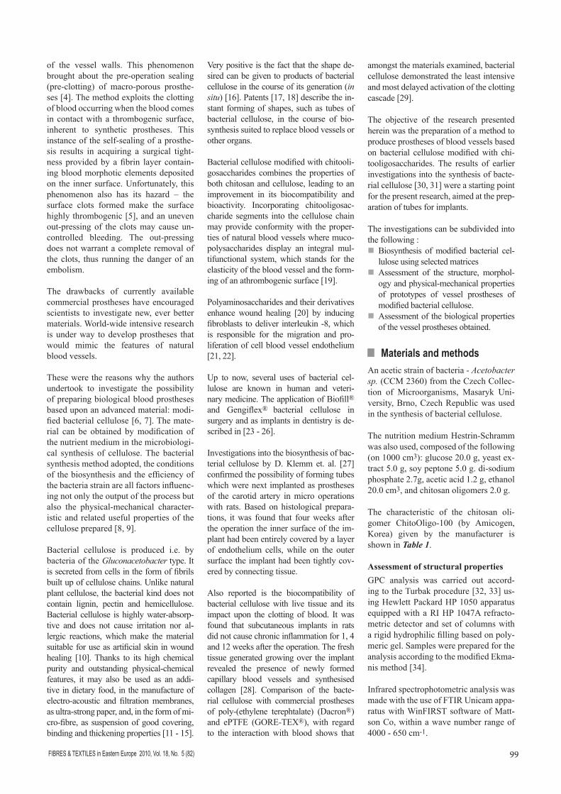
Figure 2. Prototypes of the vessel prostheses obtained from the culture in the silicone tube.
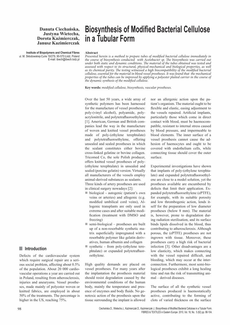
Figure 1. Culture on the inner surface of the silicone tube.
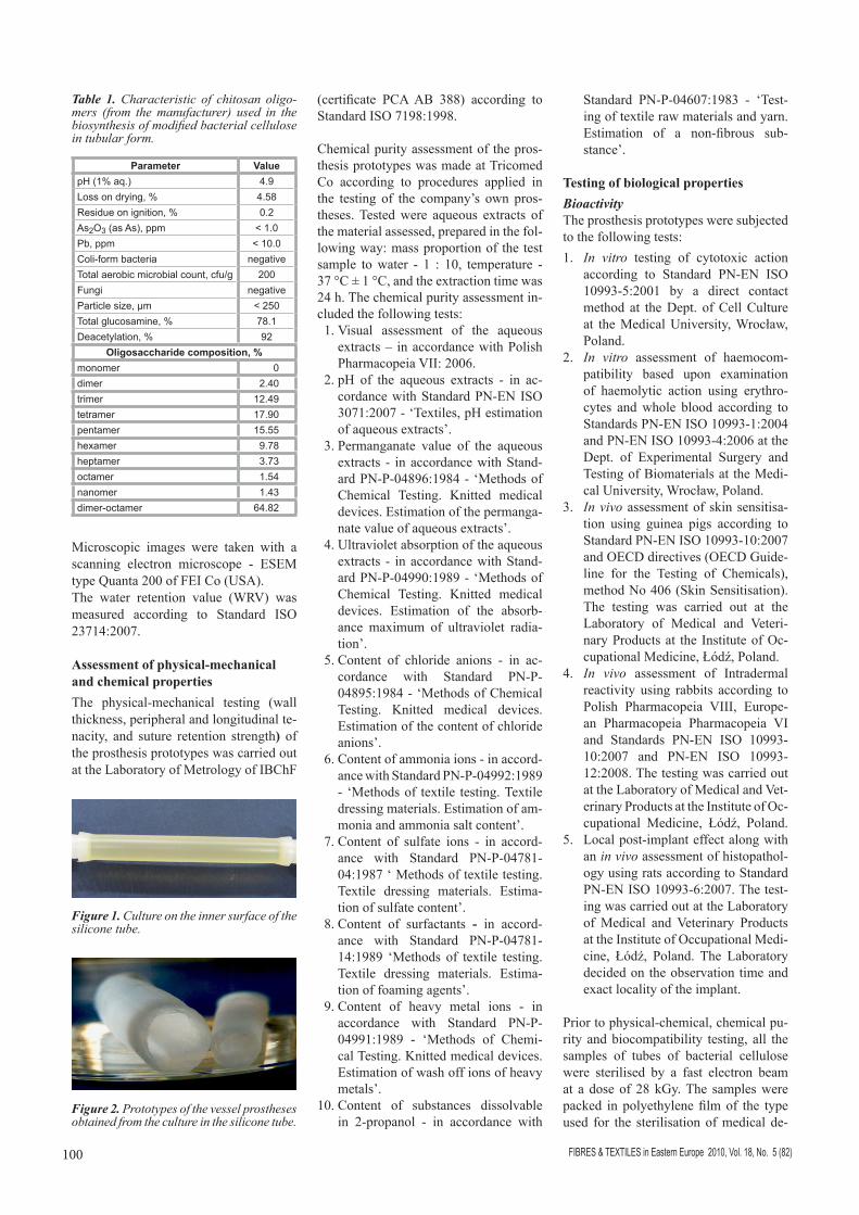
Table 1. Characteristic of chitosan oligo-mers (from the manufacturer) used in the biosynthesis of modified bacterial cellulose in tubular form.
Parameter Value pH (1% aq.) 4.9Loss on drying, % 4.58Residue on ignition, % 0.2As2O3 (as As), ppm < 1.0Pb, ppm < 10.0Coli-form bacteria negativeTotal aerobic microbial count, cfu/g 200Fungi negativeParticle size, µm < 250Total glucosamine, % 78.1Deacetylation, % 92
Oligosaccharide composition, %monomer 0dimer 2.40trimer 12.49tetramer 17.90pentamer 15.55hexamer 9.78heptamer 3.73octamer 1.54nanomer 1.43dimer-octamer 64.82

101FIBRES & TEXTILES in Eastern Europe 2010, Vol. 18, No. 5 (82)
Dynamic culture in a reactor equipped with a rotating shaft partly immersed in a nutrition medium
A reactor equipped with an 8 mm PTFE shaft was employed in the synthesis of the modified bacterial cellulose in a tube shape. The shaft, half-immersed in a nu-trition medium, was driven by an electric motor at 10 r.p.m (see Figure 3). Tubes were prepared with a surface density of about 10 g/m2. In another version a pleat-ed polyester vein reinforcement of the BR-1 type (by Tricomed Co) was used to improve the tenacity of the prosthesis prototypes. The vein reinforcement ap-plied on the PTFE shaft (see Figure 3) served as a carrier. Composite prosthe-ses were prepared in which the bacterial cellulose was deposited onto the poly-ester carrier (see Figure 4). The surface density of such tubes was also about 10 g/m2. The modification allowed to prepare tubes which, when taken out of water, maintained their shape, could be freeze-dried without deformation and, after wetting with glycerol, dried with-out disturbing the inside diameter of the tube. Table 3 shows selected physical-mechanical properties of the prosthesis
vices at Tricomed Co. The Unit of Ra-diation Sterilisation of Medical Devices and Implants at the Institute of Radiation Chemistry and Nuclear Technique in Warsaw carried out the sterilisation.
n ExperimentalThe biosynthesis of the modified bacte-rial cellulose was conducted in a static culture on the inner surface of a silicone tube to form the tubular shape of a pros-thesis. The silicone tube played the role of a bioreactor. Trials were also carried out in a dynamic culture in a bioreactor equipped with a rotating PTFE rod half-immersed in the culture medium. A car-rier made of PET was also used in the syntheses.
Once the bacterial cellulose synthesis had been completed, the tubes of modi-fied bacterial cellulose obtained were washed with distilled water to remove components of the culture medium (the conductance of the cellulose after wash-ing was < 20 µS), and then they were submerged in 1% aqueous NaOH and placed in a steam autoclave at 121 ºC for 15 min. Afterwards the tubes were
washed with water to a neutral pH and conductance below 20 µS.
n ResultsStatic culture on the inner surface of the silicone tubeThe biosynthesis was conducted with the use of silicone tubes as matrix (Fig-ures 1 and 2). The tubes were 10 and 16 mm in diameter. The culture proceed-ed in a warming chamber for 7 days with the forced air flow at 30 ºC.
The outcome of the culture were tubular prototypes of vessel prostheses with a surface density of about 10 g/m2. Their physical-mechanical and useful proper-ties are shown in Table 2. Tubes from the static culture prepared on silicone matrices are characterised by a peripheral tenacity in the range of 12 to 14 cN/mm, a longitudinal tenacity from about 450 to approx. 930 cN, a suture re-tention strength from about 40 to 50 cN, and by a favourable water permeabil-ity below 1 ml/min cm2 at a pressure of 120 mm Hg.
Table 2. Selected physical-mechanical and useful properties of tubes from the static culture.
Inner diameter of Peripheral strength/CV, Peripheral tenacity,
Longitudinal tenacity/CV,
Suture retention strength/CV,
Water permeability at a pressure of. 120 mm Hg,
the matrix, mm
relaxed tube,mm cN % cN/mm cN % cN % ml/min·cm2
10 6.6 290 13.6 14.5 934.0 9.3 51.1 5.5 0.8616 12.3 240 11.1 12.6 451.0 10.1 40.1 5.4 0.89
Figure 4. Prototype of a prosthesis obtained in a reactor with a rotating shaft and the polyester vein reinforcement applied - BR-1 (Tricomed SA).
Figure 3. Dynamic culture in a reactor with a rotating PTFE shaft and the polyester vein reinforcement applied - BR-1 (Tri-comed SA).
Table 3. Selected physical-mechanical and useful properties of the vessel prosthesis prototypes obtained in a reactor with a rotating shaft.
VariantInner diameter of relaxed tube,
Peripheral strength/CV,
Peripheral tenacity,
Longitudinal tenacity/CV,
Suture retention strength/CV,
Water permeability at a pressure of 120 mm Hg,
mm cN % cN/mm cN % cN % ml/min·cm2
Without a carrier 8.7 219 15.7 11.0 329.0 12.4 35.4 8.9 0.93With PET a carrier 8.6 297 7.8 14.9 9090.0 8.2 70.0 6.7 0.89
FIBRES & TEXTILES in Eastern Europe 2010, Vol. 18, No. 5 (82)102
prototypes obtained by the method de-scribed herein.
It was found that the growth of the modi-fied bacterial cellulose in a tubular form proceeds both with and without a syn-thetic carrier. Good adhesion is achieved between the carrier and cellulose. The carrier-modified tubes showed much bet-ter mechanical properties compared with those without a carrier: the peripheral tenacity increased by 35%; the longitu-dinal tenacity saw a 27-fold increase, and the suture retention strength increased by 98%. Both kinds of tubes of modi-fied bacterial cellulose are characterized by very low water permeability; a factor favourable in vessel prostheses.
Molecular, structural and morphology characteristicGel chromatography (GPC) was em-ployed to analyse the molecular, struc-
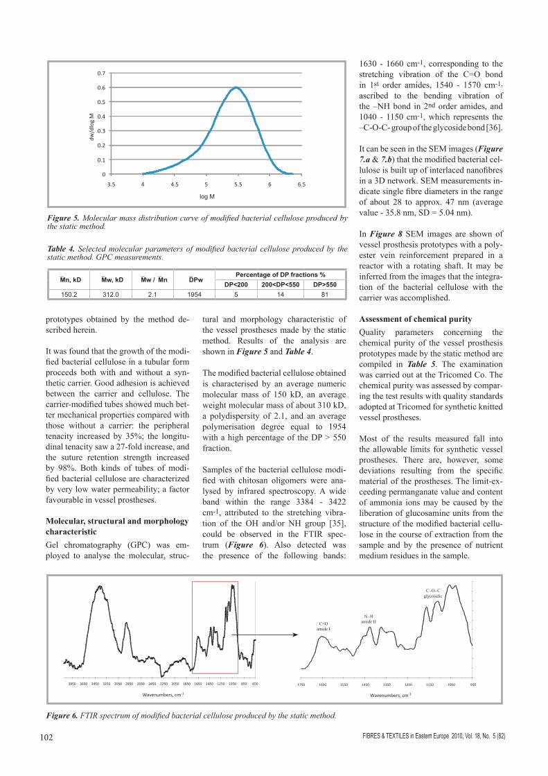
tural and morphology characteristic of the vessel prostheses made by the static method. Results of the analysis are shown in Figure 5 and Table 4.
The modified bacterial cellulose obtained is characterised by an average numeric molecular mass of 150 kD, an average weight molecular mass of about 310 kD, a polydispersity of 2.1, and an average polymerisation degree equal to 1954 with a high percentage of the DP > 550 fraction.
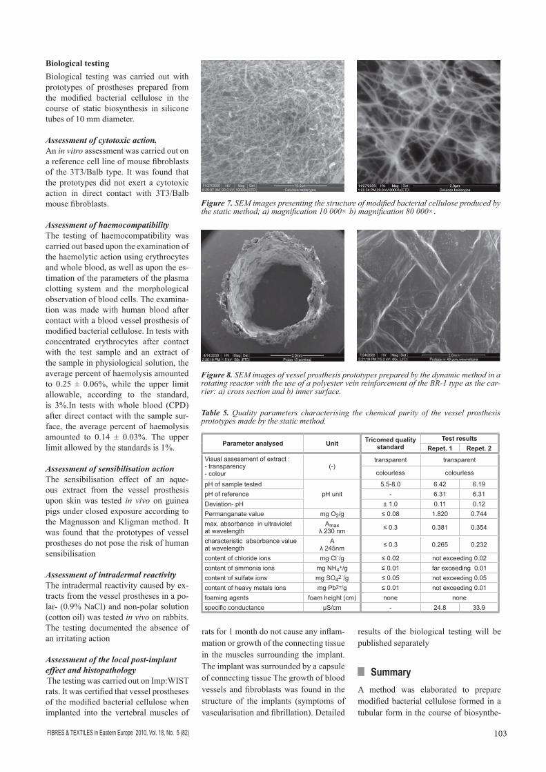
Samples of the bacterial cellulose modi-fied with chitosan oligomers were ana-lysed by infrared spectroscopy. A wide band within the range 3384 - 3422 cm-1, attributed to the stretching vibra-tion of the OH and/or NH group [35], could be observed in the FTIR spec-trum (Figure 6). Also detected was the presence of the following bands:
1630 - 1660 cm-1, corresponding to the stretching vibration of the C=O bond in 1st order amides, 1540 - 1570 cm-1,
ascribed to the bending vibration of the –NH bond in 2nd order amides, and 1040 - 1150 cm-1, which represents the –C-O-C- group of the glycoside bond [36].
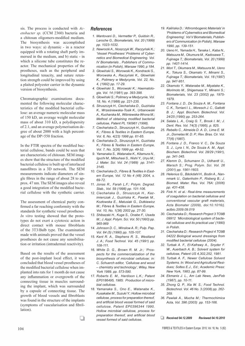
It can be seen in the SEM images (Figure 7.a & 7.b) that the modified bacterial cel-lulose is built up of interlaced nanofibres in a 3D network. SEM measurements in-dicate single fibre diameters in the range of about 28 to approx. 47 nm (average value - 35.8 nm, SD = 5.04 nm).
In Figure 8 SEM images are shown of vessel prosthesis prototypes with a poly-ester vein reinforcement prepared in a reactor with a rotating shaft. It may be inferred from the images that the integra-tion of the bacterial cellulose with the carrier was accomplished.
Assessment of chemical purityQuality parameters concerning the chemical purity of the vessel prosthesis prototypes made by the static method are compiled in Table 5. The examination was carried out at the Tricomed Co. The chemical purity was assessed by compar-ing the test results with quality standards adopted at Tricomed for synthetic knitted vessel prostheses.
Most of the results measured fall into the allowable limits for synthetic vessel prostheses. There are, however, some deviations resulting from the specific material of the prostheses. The limit-ex-ceeding permanganate value and content of ammonia ions may be caused by the liberation of glucosamine units from the structure of the modified bacterial cellu-lose in the course of extraction from the sample and by the presence of nutrient medium residues in the sample.
Figure 6. FTIR spectrum of modified bacterial cellulose produced by the static method.
Table 4. Selected molecular parameters of modified bacterial cellulose produced by the static method. GPC measurements.
Mn, kD Mw, kD Mw /Mn DPwPercentage of DP fractions %
DP<200 200<DP<550 DP>550150.2 312.0 2.1 1954 5 14 81
Figure 5. Molecular mass distribution curve of modified bacterial cellulose produced by the static method.
103FIBRES & TEXTILES in Eastern Europe 2010, Vol. 18, No. 5 (82)
Biological testingBiological testing was carried out with prototypes of prostheses prepared from the modifi ed bacterial cellulose in the course of static biosynthesis in silicone tubes of 10 mm diameter.
Assessment of cytotoxic action.An in vitro assessment was carried out on a reference cell line of mouse fi broblasts of the 3T3/Balb type. It was found that the prototypes did not exert a cytotoxic action in direct contact with 3T3/Balb mouse fi broblasts.
Assessment of haemocompatibilityThe testing of haemocompatibility was carried out based upon the examination of the haemolytic action using erythrocytes and whole blood, as well as upon the es-timation of the parameters of the plasma clotting system and the morphological observation of blood cells. The examina-tion was made with human blood after contact with a blood vessel prosthesis of modifi ed bacterial cellulose. In tests with concentrated erythrocytes after contact with the test sample and an extract of the sample in physiological solution, the average percent of haemolysis amounted to 0.25 ± 0.06%, while the upper limit allowable, according to the standard, is 3%.In tests with whole blood (CPD) after direct contact with the sample sur-face, the average percent of haemolysis amounted to 0.14 ± 0.03%. The upper limit allowed by the standards is 1%.
Assessment of sensibilisation actionThe sensibilisation effect of an aque-ous extract from the vessel prosthesis upon skin was tested in vivo on guinea pigs under closed exposure according to the Magnusson and Kligman method. It was found that the prototypes of vessel prostheses do not pose the risk of human sensibilisation
Assessment of intradermal reactivityThe intradermal reactivity caused by ex-tracts from the vessel prostheses in a po-lar- (0.9% NaCl) and non-polar solution (cotton oil) was tested in vivo on rabbits. The testing documented the absence of an irritating action
Assessment of the local post-implant effect and histopathology The testing was carried out on Imp:WIST rats. It was certifi ed that vessel prostheses of the modifi ed bacterial cellulose when implanted into the vertebral muscles of
rats for 1 month do not cause any infl am-mation or growth of the connecting tissue in the muscles surrounding the implant. The implant was surrounded by a capsule of connecting tissue The growth of blood vessels and fi broblasts was found in the structure of the implants (symptoms of vascularisation and fi brillation). Detailed
results of the biological testing will be published separately
n Summary A method was elaborated to prepare modifi ed bacterial cellulose formed in a tubular form in the course of biosynthe-
Figure 8. SEM images of vessel prosthesis prototypes prepared by the dynamic method in a rotating reactor with the use of a polyester vein reinforcement of the BR-1 type as the car-rier: a) cross section and b) inner surface.
Figure 7. SEM images presenting the structure of modifi ed bacterial cellulose produced by the static method; a) magnifi cation 10 000× b) magnifi cation 80 000×.
Table 5. Quality parameters characterising the chemical purity of the vessel prosthesis prototypes made by the static method.
Parameter analysed Unit Tricomed quality standard
Test results Repet. 1 Repet. 2
Visual assessment of extract :- transparency- colour
(-)transparent transparent
colourless colourless
pH of sample testedpH unit
5.5-8.0 6.42 6.19pH of reference - 6.31 6.31Deviation- pH ± 1.0 0.11 0.12Permanganate value mg O2/g ≤ 0.08 1.820 0.744max. absorbance in ultraviolet at wavelength
Amaxλ 230 nm ≤ 0.3 0.381 0.354
characteristic absorbance valueat wavelength
Aλ 245nm ≤ 0.3 0.265 0.232
content of chloride ions mg Clֿ/g ≤ 0.02 not exceeding 0.02content of ammonia ions mg NH4+/g ≤ 0.01 far exceeding 0.01content of sulfate ions mg SO42ֿ/g ≤ 0.05 not exceeding 0.05content of heavy metals ions mg Pb2+/g ≤ 0.01 not exceeding 0.01foaming agents foam height (cm) none nonespecifi c conductance µS/cm - 24.8 33.9
FIBRES & TEXTILES in Eastern Europe 2010, Vol. 18, No. 5 (82)104
sis. The process is conducted with Ac-etobacter sp. (CCM 2360) bacteria and a chitosan oligomers-modified medium. The biosynthesis was accomplished in two ways: a) dynamic - in a reactor equipped with a rotating shaft partly im-mersed in the medium, and b) static - in which a silicone tube constitutes the re-actor. The mechanical properties of the prostheses, such as the peripheral and longitudinal tenacity, and suture reten-tion strength could be improved by using a plaited polyester carrier in the dynamic version of biosynthesis.
Chromatographic examinations docu-mented the following molecular charac-teristics of the modified bacterial cellu-lose: an average numeric molecular mass of 150 kD, an average weight molecular mass of about 310 kD, a polydispersity of 2.1, and an average polymerisation de-gree of about 2000 with a high percent-age of the DP>550 fraction.
In the FTIR spectra of the modified bac-terial cellulose, bands could be seen that are characteristic of chitosan. SEM imag-es show that the structure of the modified bacterial cellulose is built up of interlaced nanofibres in a 3D network. The SEM measurements indicate diameters of sin-gle fibres in the range of about 28 to ap-prox. 47 nm. The SEM images also reveal a good integration of the modified bacte-rial cellulose with the synthetic carrier.
The assessment of chemical purity con-firmed a far reaching conformity with the standards for synthetic vessel prostheses In vitro testing showed that the proto-types do not exert a cytotoxic action in direct contact with mouse fibroblasts of the 3T3/Balb type. The examination made with animals proved that the vessel prostheses do not cause any sensibilisa-tion or irritation (intradermal reactivity).
Based on the results of the assessment of the post-implant local effect, it was concluded that blood vessel prostheses of the modified bacterial cellulose when im-planted into rats for 1 month do not cause any inflammation or overgrowth of the connecting tissue in muscles surround-ing the implant, which was surrounded by a capsule of connecting tissue. The growth of blood vessels and fibroblasts was found in the structure of the implants (symptoms of vascularisation and fibril-lation).
References 1. Mantovani D., Vermetter P., Guidoin R.,
Laroche G.; Biomaterials, Vol. 20 (1999) pp. 1023-1032.
2. Nawrocki A., Noszczyk W., Raczyński K.; ‘Vessel Prostheses’ Problems of Cyber-netics and Biomedical Engineering. Vol. lV Biomaterials , Publishers of Commu-nication (in Polish), Warsaw 1990, p 184.
3. Głowiński S., Worowski K., Kondracki S., Worowska A., Raczyński K., Głowiński K., Polimery w Medycynie, Vol. 22, No. 4, (1992) pp. 17-29.
4. Głowiński S., Worowski K.; Haematolo-gia, Vol. 14 (1981) pp. 393-398.
5. Głowiński S.; Polimery w Medycynie, Vol. 18, No. 4 (1988) pp. 221-235.
6. Struszczyk H., Ciechańska D., Guzińska K., Wrześniewska-Tosik K., Urbanowski A., Kucharska M., Wiśniewska-Wrona M.; Method of obtaining modified bacterial cellulose, Patent PL 190961 (1998).
7. Ciechańska D., Struszczyk H., Guzińska K.; Fibres & Textiles in Eastern Europe, Vol. 6, No. 4(23) 1998 pp. 61-65.
8. Ciechańska D., Struszczyk H., Guzińska K.; Fibres & Textiles in Eastern Europe, Vol. 7, No. 3(26) 1999 pp. 49-52.
9. Yamanaka S., Watanabe K., Kitamura N., Iguchi M., Mitsuhasi S., Nishi Y., Uryu M.; J. Mater. Sci. Vol. 24 (1989) pp. 3141-3145.
10. Ciechańska D.; Fibres & Textiles in East-ern Europe, Vol. 12 No 4 (48) 2004, s. 69-72.
11. Jonas R., Farah L.F.; Polym. Degrad. Stab., Vol. 59 (1998) pp. 101-106.
12. Ciechańska D., Struszczyk H., Kaz-imierczak J., Guzińska K., Pawlak M., Kozłowska E., Matusiak G., Dutkiewicz M.; Fibres & Textiles in Eastern Europe, Vol. 10, No. 1(36) 2002 pp. 27-30.
13. Shibazaki H., Kuga S., Onabe F., Usada M.; J. Appl. Polym. Sci. Vol. 50 (1993) pp. 965-969
14. Johnson D. C., Winslow A. R.; Pulp. Pap. Vol. 64 (5) (1990) pp. 105-107.
15. Kent R. A., Stephens R. S., Westland J. A.; Food Technol. Vol. 45 (1991) pp. 108-111.
16. White D. G., Brown R. M. Jr.; ‘Pros-pects for the commercialization of the biosynthesis of microbial cellulose; in: C. Schuerch editor, ‘Cellulose and wood – chemistry and technology’, Wiley, New York 1989, pp. 573-590.
17. Roberts E. M., Hardison L.K.; Patent EP0186495, 1985: Production of micro-bial cellulose.
18. Yamanaka S., Ono E., Watanabe K., Kusakabe M., Suzuki Y.; Hollow microbial cellulose, process for preparation thereof, and artificial blood vessel formed of said cellulose, Patent EP0396344 1990. Hollow microbial cellulose, process for preparation thereof, and artificial blood vessel formed of said cellulose.
19. Kalińska D.: ‘Athrombogenic Materials’ in ‘Problems of Cybernetics and Biomedical Engineering’. Vol V Biomaterials, Publish-ers of Communication (in Polish), Warsaw 1990, pp. 139-151.
20. Ueno H., Yamada H., Tanaka I., Kaba N., Matsuura M., Okumura M., Kadosawa T., Fujinaga T.; Biomaterials, Vol. 20 (1999) pp. 1407-1414.
21. Mori T., Okumara M., Matsuura M., Ueno K., Tokura S., Okamato Y., Minami S., Fujinaga T.; Biomaterials, Vol. 18 (1997) pp. 947-951.
22. Okamoto Y., Watanabe M., Miyatake K., Morimoto M., Shigemasa Y., Minami S.; Biomaterials, Vol. 23 (2002) pp. 1975-1979.
23. Fontana J. D.. De Souza A. M., Fontana C. K., Torriani I. L., Moresch J. C., Gallotti B. J.; Appl. Biochem. Biotechnol. Vol. 24/25 (1990) pp. 253-264.
24. Salata L. A., Craig G. T., Brook I. M.; J. Dent. Res. Vol. 74(3) (1995), p. 825.
25. Rebello C., Almeida D. A. D., Lima E. M. Jr., Dornelas M. D. P.; Rev. Bras. Cir. Vol. 77(6) (1987).
26. Fontana J. D., Franco V. C., De Souza S. J., Lyra I. N., De Souza A. M., Appl. Biochem. Biotechnol. Vol. 28/29 (1991), pp. 341-345.
27. Klemm D., Schumann D., Udhardt U., Marsch S.; Prog. Polym. Sci. Vol. 26 (2001) pp. 1561-1603.
28. Helenius G., Bäckdahl H., Bodin A., Nan-nmark U., Gatenholm P., Risberg B.; J. Biomed. Mater. Res. Vol. 76A: (2006) pp. 431-438.
29. Fink H. et al.: Real-time measurements of coagulation on bacterial cellulose and conventional vascular graft materials, Acta Biomater (2009), doi:10.1016/j.actbio.2009.09.019
30. Ciechańska D.; Research Project 3 TO9B 09912 ‘Microbiological system of bacte-rial cellulose and its practical use’ (1990) in Polish.
31. Ciechańska D.: Research Project 4 TO9B 04222 Biological wound dressings from modified bacterial cellulose (2004).
32. Turbak A. F., El-Kafrawy A., Snyder F. W., Auerbach A. B.; Solvent system for cellulose, Patent US 4,302,252, 1981.
33. Turbak A. F., Newer Cellulose Solvent Sys tems. In: Wood and Agricultural Resi-dues; Soltes E.J., Ed.; Academic Press: New York, 1983, pp. 87-99.
34. Ekmanis J. L., Am Lab News, Jan/Feb (1987), pp. 10-11.
35. Zhong Q. P., Xia W. S.; Food Technol. Biotechnol. Vol. 46 No. 3 (2008) pp. 262-269.
36. Pawlak A., Mucha M.; Thermochimica Acta, Vol. 396 (2003) pp. 153-166.
Received 04.12.2009 Reviewed 04.10.2010