Embed Size (px)
Citation preview
1
BIOTECHNOLOGICAL MODIFICATION OF STEROIDAL STRUCTURES
A THESIS SUBMITTED TO THE GRADUATE SCHOOL OF NATURAL AND APPLIED SCIENCES
OF MIDDLE EAST TECHNICAL UNIVERSITY
BY
UMUT ERKILIÇ
IN PARTIAL FULFILLMENT OF THE REQUIREMENTS FOR
THE DEGREE OF MASTER OF SCIENCE IN
BIOTECHNOLOGY
FEBRUARY 2008
2
Approval of the Thesis:
BIOTECHNOLOGICAL MODIFICATION OF STEROIDAL STRUCTURES
submitted by UMUT ERKILIÇ in partial fulfillment of the requirements for the
degree of Master in Biotechnology Department, Middle East Technical
University by,
Prof. Dr. Canan Özgen _____________________ Dean, Graduate School of Natural and Applied Sciences Prof. Dr. Gülay Özcengiz _____________________ Head of Department, Biotechnology Prof. Dr. Ayhan Sıtkı Demir Supervisor, Chemistry Department, METU _____________________ Prof. Dr. Zümrüt B. Ögel Co-Supervisor, Food Engineering Dept., METU _____________________ Examining Committee Members: Prof. Dr. Haluk Hamamcı _____________________ Chemistry Department, METU Prof. Dr. Ayhan Sıtkı Demir _____________________ Food Engineering Dept., METU Prof. Dr. Gülay Özcengiz _____________________ Biology Department, METU Prof. Dr. Pınar Çalık _____________________ Chemical Engineering Dept., METU Assoc. Prof. Dr. Nezire Saygılı _____________________ Faculty of Pharmacy, HU
Date: 05.02.2008
3
I hereby declare that all information in this document has been obtained and presented in accordance with academic rules and ethical conduct. I also declare that, as required by these rules and conduct, I have fully cited and referenced all material and results that are not original to this work.
Name, Last name : Umut ERKILIÇ Signature :
iii
4
ABSTRACT
BIOTECHNOLOGICAL MODIFICATION OF STEROIDAL STRUCTURES
Erkılıç, Umut
M.S., Department of Biotechnology
Supervisor: Prof. Dr. Ayhan Sıtkı Demir
Co-supervisor: Prof Dr. Zümrüt B. Ögel
February 2008, 71 pages
Steroids are important biological regulators existing in hormones which are used to
control metabolism of the body. There are widespread applications in the
pharmaceutical industry. Drugs of steroid nature - anti-inflammatory and anti-
allergic corticosteroids, diuretics, anabolics, androgens, gestagens, contraceptives,
antitumor medications, etc. - are now widely used in human and veterinary medicine.
Nowadays, biotechnological modifications of steroids are preferred over chemical
modifications as a green chemistry since they are more likely to be natural.
In this work four different Fusarium species were screened for bioconversion of
steroids into pharmaceutically important derivatives of steroids by reduction,
dehydrogenation, side-chain degradation etc. on A and D-rings containing many
active sites.
Fusarium spp. used in this work, namely Fusarium roseum OUT 4019, Fusarium
anguioides OUT 4017, Fusarium bulbigenum OUT 4115 and Fusarium solani OUT
iv
5
4021 are filamentous fungi, which belong to the class of Deuteromyces. They can
grow using simple carbohydrates and nitrogen sources.
4-androstene-3,17-dione conversion is used as a model system. Under same
environmental conditions it is found that whole cells of Fusarium roseum OUT 4019
can dehydrogenate at C-1 and C-2 producing androsta-1,4-diene-3,17-dione and also
reduce at C-17 in addition to dehydrogenate at C-1 and C-2 producing 17-hydroxy-
androsta-1,4-dien-3-one, Fusarium anguioides OUT 4017 can reduce at C-17
producing 17-hydroxy-androst-4-en-3-one, Fusarium solani OUT 4021 can reduce at
C-3 and C-17 producing androst-4-ene-3,17-diol at 25 C° and 160 rpm with
uncontrolled pH.
In these conversions, androsta-1,4-diene-3,17-dione, 17-hydroxy-androsta-1,4-dien-
3-one, 17-hydroxy-androst-4-en-3-one, androst-4-ene-3,17-diol were isolated with 54
%, 22 %, 26 %, 90 % yields, respectively.
In another study, bioconversion reactions of aromatic methyl ethers by Fusarium
roseum OUT 4019 were investigated and for some compounds, cleavage of methyl
ether was observed.
Keywords: 4-androstene-3,17-dione, Fusarium spp., C-3 and C-17 Steroidal Ketone
Reduction, Steroidal dehydrogenation, Ether Cleavage.
v
6
ÖZ
STEROĐT YAPILARIN BĐYOTEKNOLOJĐK MODĐFĐKASYONU
Erkılıç, Umut
Yüksek Lisans, Biyoteknoloji Bölümü
Tez Yöneticisi: Prof. Dr. Ayhan Sıtkı Demir
Yardımcı Tez Yöneticisi: Prof. Dr. Zümrüt B. Ögel
Şubat 2008, 71 sayfa
Steroitler vücut metabolizmasının denetiminde kullanılan hormonların yapısında
bulunan önemli biyolojik düzenleyicilerdir. Đlaç sanayinde yaygın uygulamaları
bulunmaktadır. Doğal steroit ilaçlar: anti-inflammatör ve anti-allerjik
kortikosteroitler, diüretikler, doğum kontrol ilaçları, tümör tedavi ilaçları, vücut
geliştiriciler v.b. birçok özelliklerinden dolayı tıp ve veterinerlikte yaygın olarak
kullanılmaktadır.
Günümüzde, steroitlerin biyoteknolojik modifikasyonları doğal olmaya daha yatkın
oldukları için bir yeşil kimya olarak kimyasal modifikasyonların yerine
kullanılmaktadır.
Bu çalışmada dört değişik mikroorganizma türü A ve D halkalarında birçok etkin
bölge bulunan steroitlerin önemli ilaç türevlerine indirgenme, dehidrojenasyon, yan
zincir parçalanması v.b. yöntemlerle dönüşümleri için taranmıştır.
Bu çalışmada kullanılan Fusarium roseum OUT 4019, Fusarium anguioides OUT
4017, Fusarium bulbigenum OUT 4115 ve Fusarium solani OUT 4021 türlerini
vi
7
içeren Fusarium cinsi Deuteromyces sınıfına ait olan filamentus fungi çeşididir ve
çok basit karbon ve azot kaynakları kullanarak kolayca çoğalabilmektedirler.
4-androsten-3,17-dion’un dönüştürülmesi model sistem olarak kullanılmıştır. Aynı
çevresel koşullardaki hücre türlerinden Fusarium roseum OUT 4019, C-1 ve C-2
bölgesinde dehidrojenasyon reaksiyonlarıyla androsta-1,4-dien-3,17-dion ürününü ve
de aynı bölgede çift bağ oluşumunun yanı sıra C-17’de ketonun indirgenmesi ile 17-
hidroksi-androsta-1,4-dien-3-on ürününü oluşturmuştur. Fusarium anguioides OUT
4017 C-17 ketonun indirgenmesi ile 17-hidroksi-androst-4-en-3-on ürününü,
Fusarium solani OUT 4021 ise C-3 and C-17 ketonların indirgenmesi ile androst-4-
en-3,17-diol ürünü 25 °C‘de ve 160 devir/dakika ve kontrolsüz pH ortamında
oluşturmuştur.
Bu dönüşüm tepkimelerinde, androsta-1,4-dien-3,17-dion, 17-hidroksi-androsta-1,4-
dien-3-on, 17-hidroksi-androst-4-en-3-on, androst-4-ene-3,17-diol ürünleri sırasıyla
54 %, 22 %, 26 %, 90 % verimlerde elde edilmiştir.
Diger çalışmada, aromatik halkaya bağlı metil eter içeren bileşiklerin Fusarium
roseum OUT 4019 ile biyodönüşümleri incelenmiş ve bazı örneklerde methy eter
kısmının koparılarak alkole dönüştürüldüğü saptanmıştır.
Anahtar kelimeler: 4-androsten–3,17-dion, Fusarium, C-3 ve C-17 Steroidal
Ketonların Đndirgenmesi, Steroidal Dehidrojenasyon, Eter Bağı Koparma.
vii

8
To my father and mother
viii
9
ACKNOWLEDGEMENT
I owe my special thanks and sincere appreciation to Prof. Dr. Ayhan Sıtkı Demir for
his invaluable guidance, supervision, encouragement, insight and understanding
throughout the research.
I would like to express my deepest thanks to my family for helping, supporting,
encouraging and loving me all through my life.
I would like to express my special thanks to my friends Peruze Ayhan, Ş. Betül
Sopacı, Hacı Eşiyok, Umut Demirtaş and Đlke Şimşek for their helps and friendships
during this research.
I am also indebted to Mrs. Fatoş Doğanel Polat and Ms. Seda Karayılan for their
technical support.
Finally, I would also like to thank all of the members of Ayhan Sıtkı Demir research
group for their help, friendship, and cooperation.
ix
10
TABLE OF CONTE NTS
ABSTRACT................................................................................................................iv
ÖZ................................................................................................................................vi
ACKNOWLEDGEMENT..........................................................................................ix
TABLE OF CONTENTS.............................................................................................x
LIST OF TABLES....................................................................................................xiii
LIST OF FIGURES...................................................................................................xiv
CHAPTER
1. INTRODUCTION....................................................................................................1
1.1. Bioconversions in organic chemistry................................................................1
1.1.1. Fungi mediated bioconversions................................................................3
1.1.2. Fusarium species in bioconversion reactions...........................................5
1.2. Steroids.............................................................................................................6
1.2.1. Important steroid types, their structures and biological activities............8
1.2.2 Chemical properties of steroids...............................................................13
1.2.3. Chemical conversion reactions with steroids..........................................13
1.2.4. Bioconversion reactions with steroid......................................................14
1.2.4.1. Hydroxylation.................................................................................17
1.2.4.2. ∆1, 2-Dehydrogenation of steroids...................................................20
1.2.4.3. Ester saponification and oxidation of hydroxyl groups of
steroids........................................................................................................................21
1.2.4.4. Reduction of keto groups of steroids..............................................22
1.2.4.5. Sterol side chain degradation of steroids........................................22
1.2.4.6. Other types of reactions in microbial transformation of steroids...23
1.3. Fungi..........................................................................................................25
1.3.1. Fusarium species................................................................................25
1.4. Aim of the work..............................................................................................27
x
11
2. RESULTS AND DISCUSSION.............................................................................28
2.1. STEROID BIOCONVERSIONS....................................................................28
2.1.1. Perspective of the work...........................................................................28
2.1.2. Screening of fungi...................................................................................31
2.1.3. Optimization studies for fungal bioconversion.......................................34
2.1.3.1. Effect of solvent..............................................................................35
2.1.3.2. Effect of glucose concentration.......................................................36
2.1.3.3. Effect of type of growth media.......................................................37
2.1.3.4. Effect of inoculum size...................................................................38
2.1.4. Fusarium mediated conversion of 4-androstene-3, 17-dione.................38
2.1.4.1. Bioonversion of 4-androstene-3,17-dione with F. roseum OUT
4019.............................................................................................................................39
2.1.4.2. Bioconversion of 4-androstene-3,17-dione with F. anguioides OUT
4017.............................................................................................................................41
2.1.4.3. Bioconversion of 4-androstene-3,17-dione with F. solani OUT
4021.............................................................................................................................43
2.2. DEMETHYLATION OF AROMATIC METHYL ETHERS........................45
2.2.1 Demethylation of aromatic methyl ethers by fungi.................................45
3. MATERIALS AND METHODS............................................................................48
3.1. Materials.........................................................................................................48
3.2. Agar preparations for fungal growth...............................................................49
3.2.1. Agar preparation for Fusarium roseum OUT 4019................................49
3.2.2. Agar preparation for Fusarium solani OUT 4021...................................49
3.2.3. Agar preparation for Fusarium anguioides OUT 4017...........................50
3.3. Broth preparations for fungal growth.............................................................50
3.3.1. Broth preparation for Fusarium roseum OUT 4019...............................50
3.3.2. Broth preparation for Fusarium solani OUT 4021..................................51
3.3.3. Broth preparation for Fusarium anguioides OUT 4017..........................51
3.4. Fungus preparation for experiments...............................................................51
3.5. Synthesis of 17-hydroxy-androsta-1,4-dien-3-one and androsta-1,4-diene-3,
17-dione from 4-androstene-3,17-dione with F. roseum OUT 4019..........................52
xi
12
3.6. Synthesis of 17-hydroxy-androst-4-en-3-one, from 4-androstene-3,17-dione
with F. anguioides 4017.............................................................................................53
3.7. Synthesis of androst-4-ene-3,17-diol from 4-androstene-3,17-dione with F.
solani 4021..................................................................................................................53
4. CONCLUSION.......................................................................................................55
REFERENCES...........................................................................................................58
APPENDICES
APPENDIX A........................................................................................................64
APPENDIX B........................................................................................................65
xii
13
LIST OF TABLES
TABLES
1. Wet and dry cell weights of Fusarium species after incubation.............................38
2. Rf values of starting material and product of Fusarium roseum............................40
3. Rf values of starting material and product of Fusarium anguioides......................42
4. Rf values of starting material and product of Fusarium solani..............................44
xiii
14
LIST OF FIGURES
FIGURES
1. Enzymatic reaction of progesterone into 11α-hydroxy-progestrone........................2
2. Common reactions of fungal bioconversion.............................................................4
3. Fusarium species catalyzed reactions.......................................................................5
4. Steroid skeleton structure..........................................................................................6
5. 5α (Allo) series, rings A/B trans...............................................................................6
6. 5β (Allo) series, rings A/B........................................................................................7
7. Cholesterol................................................................................................................8
8. Estradiol and Ethynylestradiol.................................................................................9
9. Progesterone............................................................................................................10
10. Testosterone..........................................................................................................10
11. Aldosterone...........................................................................................................11
12. Cortisol..................................................................................................................12
13. Reaction of 17-hydroxy-androst-1,4-dien-3-one with LiALD4 or
NaBD4.........................................................................................................................14
14. Hydroxylation positions on steroidal structure.....................................................17
15. Reactions on steroid skeleton by various fungi species........................................18
16. Reaction scheme of ∆1, 2-dehydrogenation of steroids.........................................20
17. The conversion catalyzed by Flavobacterium dehydrogenans.............................21
18. Reaction scheme of ketone reduction...................................................................22
19. Enzymatic hydrolysis of steroid epoxides...........................................................23
20. Reaction scheme of de-acetylation of 3β,17β-diacetoxy-5α-
androstane...................................................................................................................24
21. Scheme of purposed bioconversions of steroids by Fusarium species.................27
22. Scheme of purposed bioconversion of aromatic methyl ethers by
Fusarium roseum OUT 4019.....................................................................................27
23. Starting materials for the synthesis of steroid hormones......................................29
24. Reaction scheme for steroids to obtain a chiral center on A-ring.........................30
25. Reaction scheme for steroids to obtain a chiral center on D-ring.........................31
xiv
15
26. Molecular structure of 4-androstene-3,17-dione and 4-cholesten-3-
one...............................................................................................................................32
27. Possible sites for the steroid bioconversion by Fusarium species........................33
28. Reaction scheme of 4-androstene3,17-dione by Fusarium roseum......................39
29. TLC profile of the starting material and products of F. roseum...........................40
30. Reaction scheme of 4-androstene3,17-dione by Fusarium anguioides………....41
31. TLC profile of the starting material and product of F. anguioides.......................42
32. Reaction scheme of 4-androstene3,17-dione by Fusarium solani........................43
33. TLC profile of the starting material and product of F. solani..............................44
34. Scheme of proposed bioconversion of aromatic methyl ethers by Fusarium
roseum OUT 4019.......................................................................................................45
35. Methyl ether substituted compounds used in bioconversion................................46
36. Demethylation reactions performed by Fusarium roseum...................................47
37. All steroid conversions performed by Fusarium species......................................56
38. Bioconversion schemes of aromatic methyl ethers by Fusarium
roseum OUT 4019.......................................................................................................57
39. GC-MS spectra of androsta-1,4-diene-3,17-dione................................................64
40. 1H NMR spectrum of starting material, 4-androstene-3,17-dione........................65
41. 13C NMR spectrum of starting material, 4-androstene-3,17-dione.......................66
42. 1H NMR spectrum of androsta-1,4-diene-3,17 dione...........................................67
43. 1H NMR spectrum of 17-hydroxy-androsta-1, 4-dien-3-one................................68
44. 1H NMR spectrum of 17-hydroxy-androst-4-en-3-one.........................................69
45. 1H NMR spectrum of androst-4-ene-3,17-diol.....................................................70
46. 13C NMR spectrum of androst-4-ene-3,17-diol....................................................71
xv
16
CHAPTER 1
INTRODUCTION
1.1. Bioconversion in organic chemistry
Bioconversion is the conversion of chemicals into usable products or energy sources
by biological processes or agents, such as certain microorganisms or enzymes [1].
The development of scientific screening and isolation methods allows the selection
of desirable natural occurring or mutating microorganisms for specific purposes. The
diverse catalytic activities of microorganisms are being used more and more widely
to perform specific chemical reactions [2].
Apart from the biosynthesis and biodegradations, the enzymes of microorganisms
have been used for decades for biotransformations. These reactions are
transformations of individual educts in identifiable type reactions resulting in
metabolites with chemically defined structures. Biotransformations also represent a
useful tool for organic chemistry because of their main advantages: attack of non-
activated positions, reaction specificity, region-, stereo- and enantioselectivity and
the mild reaction conditions [3].
These advantages are obvious in the special modification of natural or chemically
synthesized compound by selective enzymatic reactions for the preparation of
pharmaceuticals, agrochemicals, food constituents, fine chemicals, even bulk
chemicals. The general targets are: the shortening of chemical multistep synthesis,
elimination of side reactions, preparation of optically active synthons, so-called
chirons, the imitation of mammalian metabolism, the preparation of “natural”
1
17
products instead of compounds identical to natural ones and using the ecological
profits: saving energy and decrease of environmental pollution compared to chemical
procedures [3].
Enantiopure compounds have undoubtedly gained a central role in the development
of chemical technology. This is most evident from the changes in the drug market,
where single-enantiomer drugs currently occupy the highest share. The request for
more efficient, more specifically targeted drugs will place a growing demand on
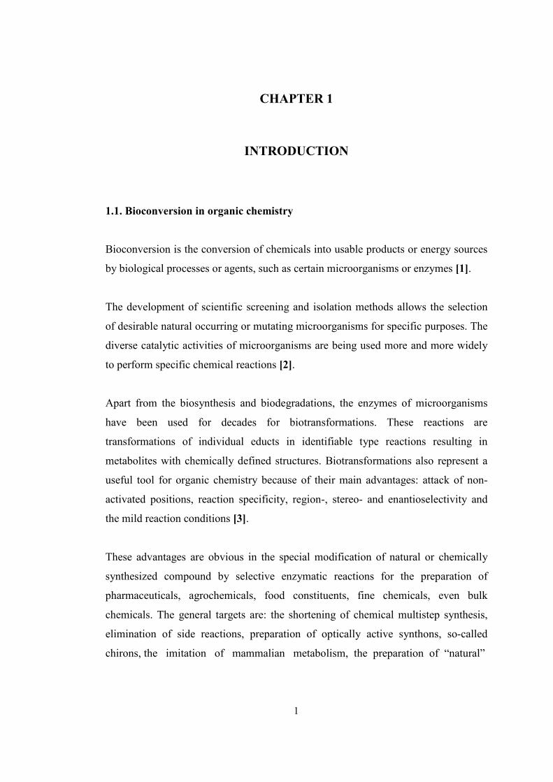
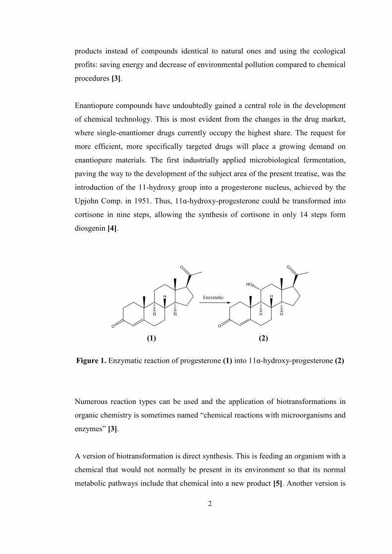
enantiopure materials. The first industrially applied microbiological fermentation,
paving the way to the development of the subject area of the present treatise, was the
introduction of the 11-hydroxy group into a progesterone nucleus, achieved by the
Upjohn Comp. in 1951. Thus, 11α-hydroxy-progesterone could be transformed into
cortisone in nine steps, allowing the synthesis of cortisone in only 14 steps form
diosgenin [4].
O
O
H
H
H
O
O
HO
H
H
H
Enzymatic
(1) (2)
Figure 1. Enzymatic reaction of progesterone (1) into 11α-hydroxy-progesterone (2)
Numerous reaction types can be used and the application of biotransformations in
organic chemistry is sometimes named “chemical reactions with microorganisms and
enzymes” [3].
A version of biotransformation is direct synthesis. This is feeding an organism with a
chemical that would not normally be present in its environment so that its normal
metabolic pathways include that chemical into a new product [5]. Another version is
2
18
indirect synthesis. In this method, chemical material isolated from the environment
of the organism is given to it so that its metabolism convert the material to the
desired product, or by using enzyme of it in a suitable condition imitating the
metabolism of the organism specific product is produced.
The most commonly used biotransformations involve acylases, esterases, lipases,
beta lactamases, penicillin acylase, peptidases, proteases, and steroid transforming
enzymes. These are always used in whole organisms, as many enzymes are involved
in each biotransformation. There are very wide ranges of microorganisms used in
biotechnology. Some are used for more than one thing and so crop up in several
biotechnological contexts. Some of the more commonly used organisms are
Aspergillus niger, Bacillus subtilis, Candida utilis, Clostridium acetobutylicum,
Corynebacterium glutamicum, Escherichia coli, Penicillium, Pischia pastoris,
Pseudomonas, Saccharomyces and Streptomycetes [5].
The areas of microbial biotechnology that are now receiving attention are:
application of the newer concepts of genetic engineering of microorganisms for their
improvement as transforming agents; solubility improvement for carrying out
biotransformation of substrates that are sparingly soluble in water; immobilization of
enzymes or whole cells in a suitable matrix for repetitive economic utilization of
enzymes; development of a continuous process for economic product recovery; and
manipulation of culture media for improvement in product yields [6].
1.1.1. Fungi mediated bioconversions
Fungal biotransformation of steroids is among the earliest examples of biocatalysis
for producing stereo- and site-specific products, including the commercially
important cytochrome P450- mediated steroid hydroxylation [7].
For many years fungi have been used for the hydroxylation of steroids since they
have an ability to catalyze reactions by enzyme systems with high regio- and
stereospecifity, which is known as a common feature of filamentous fungi [8].
3
19
Since 1950, the range of reactions that can be efficiently carried out by fungal
bioconversions has been expanded enormously, and now includes examples of
hydrolytic, oxidation, condensation, and reduction processes. Other studies have
focused on substrate groups such as steroids, alkaloids, sulfides, environmental
pollutants and bioactive compounds; on single bioconversion reactions such as
halogen metabolism, and alcohol dehydrogenase activity [9].
S
O
RO
3 1
42
O
O
2
3
1
4
1: Sulfur oxidation
2: Aromatic hydroxylation
3: Heteroatom dealkylation
4: Carbonyl reduction
1: Carbonyl hydroxylation
2: Carbonyl reduction
3: Baeyer-Villiger oxidation
followed by ester hydrolysis
4: Dehydrogenation
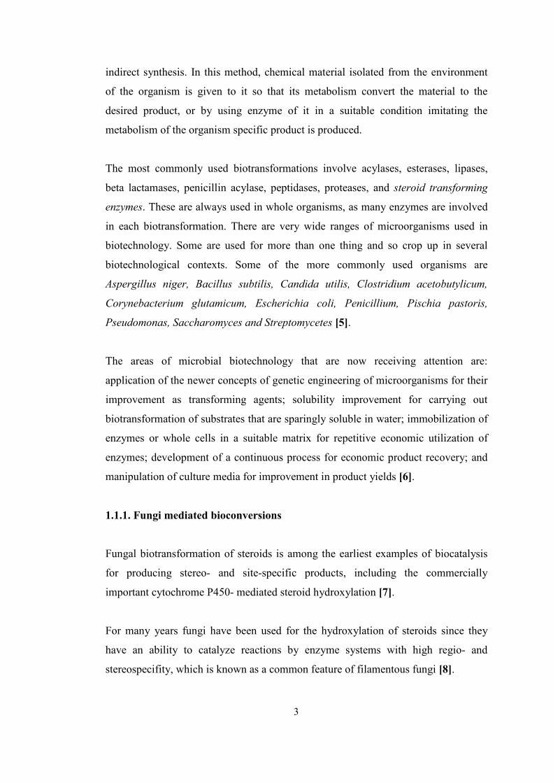
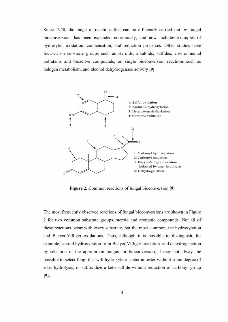
Figure 2. Common reactions of fungal bioconversion [9]
The most frequently observed reactions of fungal bioconversions are shown in Figure
2 for two common substrate groups, steroid and aromatic compounds. Not all of
these reactions occur with every substrate, but the most common, the hydroxylation
and Baeyer-Villiger oxidations. Thus, although it is possible to distinguish, for
example, steroid hydroxylation from Baeyer-Villiger oxidation and dehydrogenation
by selection of the appropriate fungus for bioconversion, it may not always be
possible to select fungi that will hydroxylate a steroid ester without some degree of
ester hydrolysis, or sulfoxidize a keto sulfide without reduction of carbonyl group
[9].
4
20
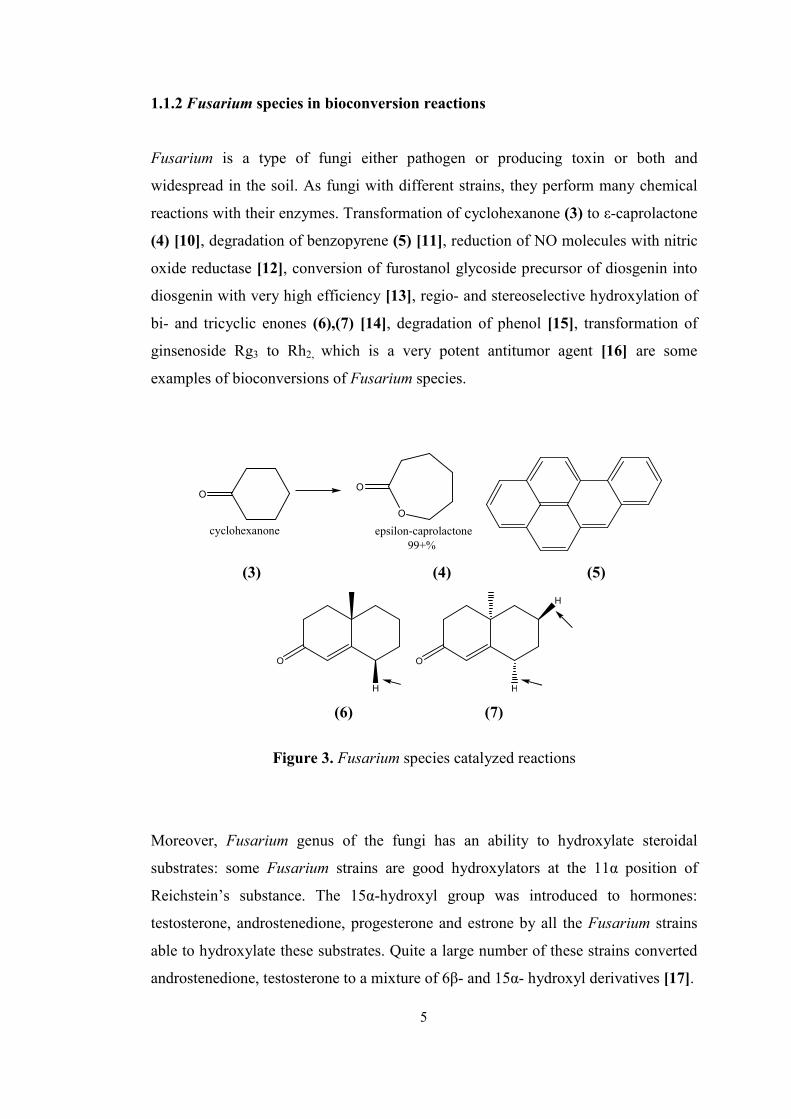
1.1.2 Fusarium species in bioconversion reactions
Fusarium is a type of fungi either pathogen or producing toxin or both and
widespread in the soil. As fungi with different strains, they perform many chemical
reactions with their enzymes. Transformation of cyclohexanone (3) to ε-caprolactone
(4) [10], degradation of benzopyrene (5) [11], reduction of NO molecules with nitric
oxide reductase [12], conversion of furostanol glycoside precursor of diosgenin into
diosgenin with very high efficiency [13], regio- and stereoselective hydroxylation of
bi- and tricyclic enones (6),(7) [14], degradation of phenol [15], transformation of
ginsenoside Rg3 to Rh2, which is a very potent antitumor agent [16] are some
examples of bioconversions of Fusarium species.
O
cyclohexanone
O
O
epsilon-caprolactone99+%
(3) (4) (5)
O
H
O
H
H
(6) (7)
Figure 3. Fusarium species catalyzed reactions
Moreover, Fusarium genus of the fungi has an ability to hydroxylate steroidal
substrates: some Fusarium strains are good hydroxylators at the 11α position of
Reichstein’s substance. The 15α-hydroxyl group was introduced to hormones:
testosterone, androstenedione, progesterone and estrone by all the Fusarium strains
able to hydroxylate these substrates. Quite a large number of these strains converted
androstenedione, testosterone to a mixture of 6β- and 15α- hydroxyl derivatives [17].
5
21
1.2. Steroids
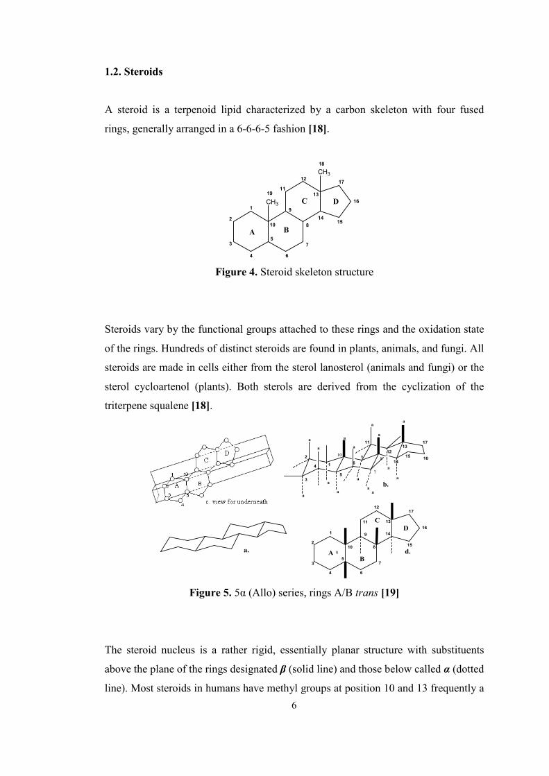
A steroid is a terpenoid lipid characterized by a carbon skeleton with four fused
rings, generally arranged in a 6-6-6-5 fashion [18].
2
3
4
5
6
1
7
8
9
10
11
12
13
1415
16
17
A B
C DCH3
18
CH3
19
Figure 4. Steroid skeleton structure
Steroids vary by the functional groups attached to these rings and the oxidation state
of the rings. Hundreds of distinct steroids are found in plants, animals, and fungi. All
steroids are made in cells either from the sterol lanosterol (animals and fungi) or the
sterol cycloartenol (plants). Both sterols are derived from the cyclization of the
triterpene squalene [18].
a.
1
2
3
4
5
6
7
8
9
10
11
12
13
14
15
16
17
A 1
D
B
C
d.
5
6
11
12
13
14
1516
17
a
a
a
a
a
a
a
a
ab.
1
2
3
4
aa
a
a
a
a
10
7
89
Figure 5. 5α (Allo) series, rings A/B trans [19]
The steroid nucleus is a rather rigid, essentially planar structure with substituents
above the plane of the rings designated β (solid line) and those below called α (dotted
line). Most steroids in humans have methyl groups at position 10 and 13 frequently a
6
22
side chain at position 17, which is β oriented, also most of them are non-polar [66].
The principal ones are cholestane (all 27 C’s), cholane (C’s 1-24), pregnane (C’s 1-
21), androstane (C’s 1-19) and oestrane (C’s 1-18) [21].
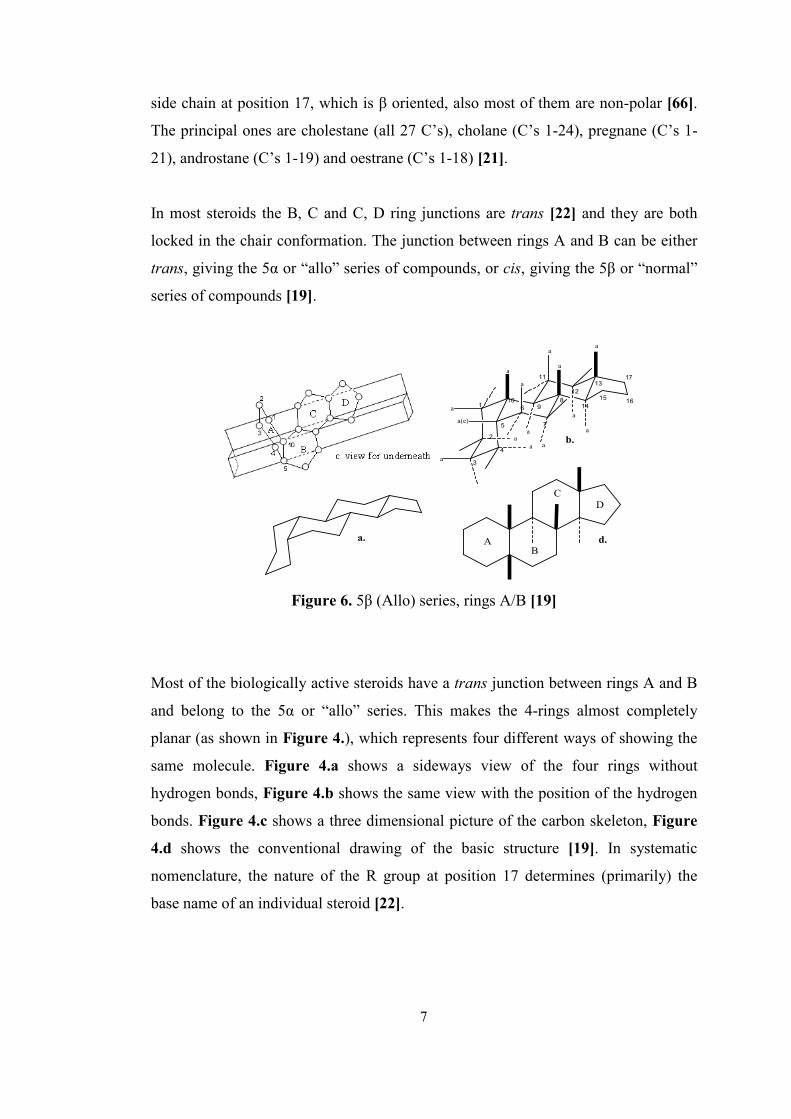
In most steroids the B, C and C, D ring junctions are trans [22] and they are both
locked in the chair conformation. The junction between rings A and B can be either
trans, giving the 5α or “allo” series of compounds, or cis, giving the 5β or “normal”
series of compounds [19].
a. A
D
B
C
d.
1
2
3
4
5
6
7
89
10
11
12
13
14
1516
17
aa
a
a(e)
a
a
a
a
a
a
a
a
a
a
b.
Figure 6. 5β (Allo) series, rings A/B [19]
Most of the biologically active steroids have a trans junction between rings A and B
and belong to the 5α or “allo” series. This makes the 4-rings almost completely
planar (as shown in Figure 4.), which represents four different ways of showing the
same molecule. Figure 4.a shows a sideways view of the four rings without
hydrogen bonds, Figure 4.b shows the same view with the position of the hydrogen
bonds. Figure 4.c shows a three dimensional picture of the carbon skeleton, Figure
4.d shows the conventional drawing of the basic structure [19]. In systematic
nomenclature, the nature of the R group at position 17 determines (primarily) the
base name of an individual steroid [22].
7
23
1.2.1. Important steroid types, their structures and biological activities
In the categories of steroids, three main groups exist: animal, plant and fungus
steroids. Animal steroids composed of vertebrate steroids containing steroid
hormones and cholesterol. For steroid hormones, they have mainly sex steroids
(androgens, estrogens and progestagens), corticosteroids (glucocorticoids,
mineralocorticoids), and also anabolic steroids [18].
Steroids occupy an important position among pharmaceutical preparations used for
treating and preventing diseases of various groups in endocrinology, oncology,
rheumatology, gynecology, etc. The relatively broad nomenclature of efficient
steroid drugs is continually expanding. Of note, several preparations administered for
life-saving indications have no non-steroid analogues [23].
Most steroid hormones are active in extremely minute amounts [19]. These small,
relatively similar molecules are able to have greatly differing effects because the
slight structural differences among them allow interactions with specific receptor
molecules [24]. They still play an important role in the drug industry. Drugs of
steroid nature - anti-inflammatory and anti-allergic corticosteroids, diuretics,
anabolics, androgens, gestagens, contraceptives, antitumor medications, etc. - are
now widely used in medicine and veterinary medicine [25]. The hormone
preparations make up the widest group of these drugs, which are mainly used as
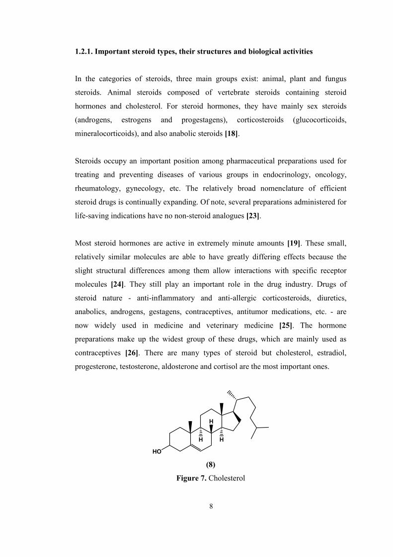
contraceptives [26]. There are many types of steroid but cholesterol, estradiol,
progesterone, testosterone, aldosterone and cortisol are the most important ones.
HO
H
H
H
(8)
Figure 7. Cholesterol
8
24
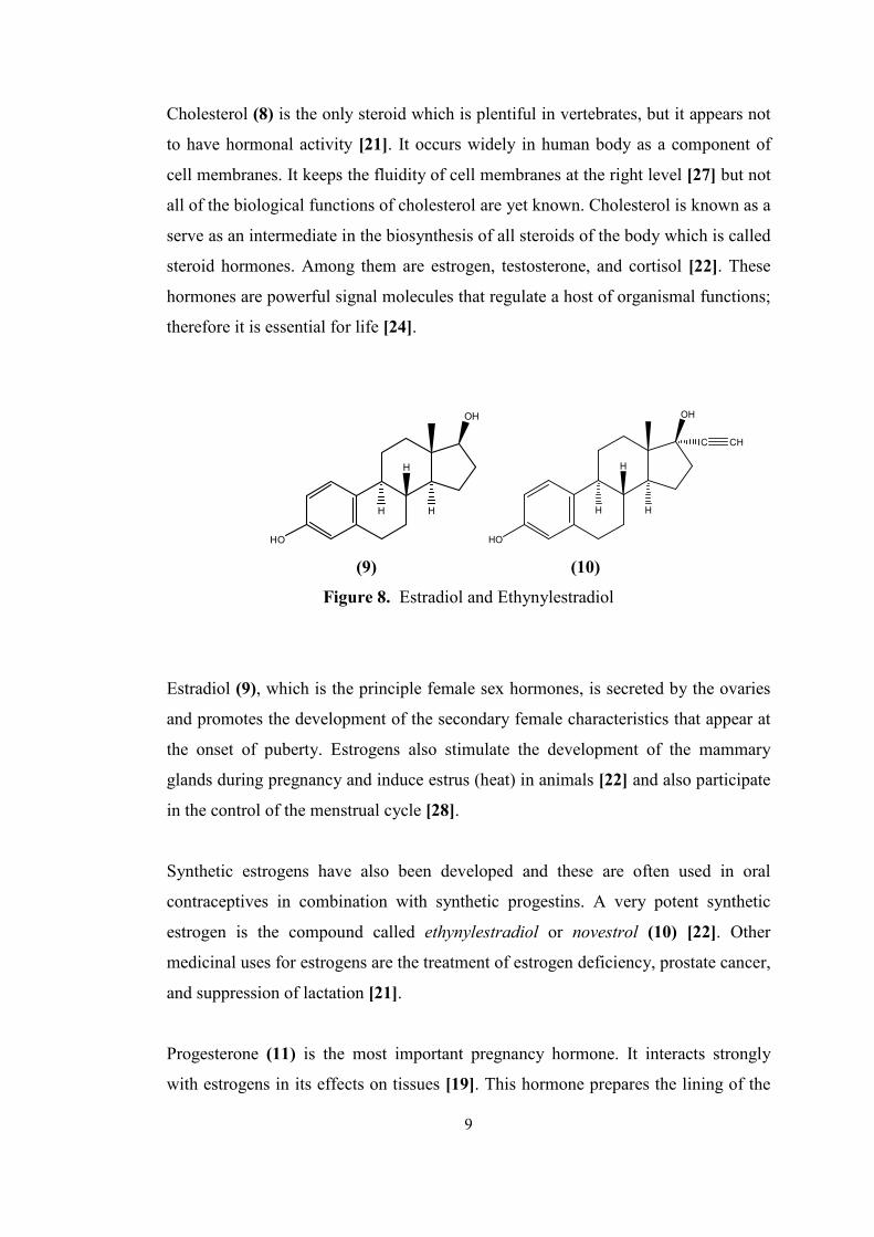
Cholesterol (8) is the only steroid which is plentiful in vertebrates, but it appears not
to have hormonal activity [21]. It occurs widely in human body as a component of
cell membranes. It keeps the fluidity of cell membranes at the right level [27] but not
all of the biological functions of cholesterol are yet known. Cholesterol is known as a
serve as an intermediate in the biosynthesis of all steroids of the body which is called
steroid hormones. Among them are estrogen, testosterone, and cortisol [22]. These
hormones are powerful signal molecules that regulate a host of organismal functions;
therefore it is essential for life [24].
HO
OH
H
H
H
OH
H
H
H
HO
C CH
(9) (10)
Figure 8. Estradiol and Ethynylestradiol
Estradiol (9), which is the principle female sex hormones, is secreted by the ovaries
and promotes the development of the secondary female characteristics that appear at
the onset of puberty. Estrogens also stimulate the development of the mammary
glands during pregnancy and induce estrus (heat) in animals [22] and also participate
in the control of the menstrual cycle [28].
Synthetic estrogens have also been developed and these are often used in oral
contraceptives in combination with synthetic progestins. A very potent synthetic
estrogen is the compound called ethynylestradiol or novestrol (10) [22]. Other
medicinal uses for estrogens are the treatment of estrogen deficiency, prostate cancer,
and suppression of lactation [21].
Progesterone (11) is the most important pregnancy hormone. It interacts strongly
with estrogens in its effects on tissues [19]. This hormone prepares the lining of the
9

25
uterus for implantation of the fertilized ovum, and continued progesterone secretion
is necessary for the completion of pregnancy [22].
O
O
H
H
H
(11)
Figure 9. Progesterone
Progesterone also suppresses ovulation, and it is the chemical agent that apparently
accounts for the fact that pregnant women do not conceive again while pregnant.
Therefore, with this function, synthetic progesterone could be used as oral
contraceptives. Progesterone, itself, requires very large doses to be effective in
suppressing ovulation when taken orally because it is degraded in the intestinal tract.
A number of such compounds have been developed and are now widely used [22].
O
OH
H
H
H
(12)
Figure 10. Testosterone
Testosterone (12), which is one of the male sex hormones secreted by the testes, is
the hormone that promotes the development of secondary male characteristics; the
growth of facial and body hair; the deepening of the voice; muscular development;
and the maturation of the male sex organs [22], and so a deficiency may lead to a low
sperm count and impotence [21]. 10
26
Androgens are used in the treatment of male sterility, impotency and female breast
and genital cancers [26]. The conversion of androgens to estrogens is catalyzed by
aromatase and so Numazawa et al. states that 2-methyleneandrostenedione is a
powerful inhibitor of aromatase which is useful in treating estrogen-dependent breast
cancer [29]. Anabolic drugs having low androgenicity are useful in the treatment of
underdeveloped children and for patients having debilitating diseases or in
convalescence [21]. In addition, synthetic testosterone analogs are used in medicine
to promote muscle and tissue growth, i.e. in patients with muscular atrophy [28].
Corticosteroids are divided into two main groups depending on the biological
activity. The mineralocorticoids affect the excretion of fluid and electrolytes. The
glucocorticoids affect intermediary metabolism and suppress inflammatory processes
[19].
O
OH
O
HO
H
H
H
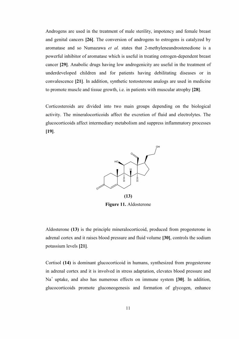
(13)
Figure 11. Aldosterone
Aldosterone (13) is the principle mineralocorticoid, produced from progesterone in
adrenal cortex and it raises blood pressure and fluid volume [30], controls the sodium
potassium levels [21].
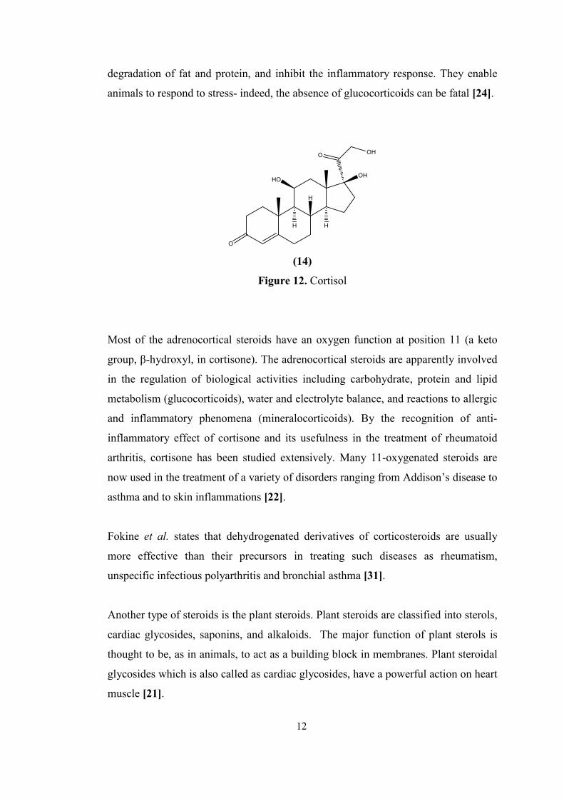
Cortisol (14) is dominant glucocorticoid in humans, synthesized from progesterone
in adrenal cortex and it is involved in stress adaptation, elevates blood pressure and
Na+ uptake, and also has numerous effects on immune system [30]. In addition,
glucocorticoids promote gluconeogenesis and formation of glycogen, enhance
11
27
degradation of fat and protein, and inhibit the inflammatory response. They enable
animals to respond to stress- indeed, the absence of glucocorticoids can be fatal [24].
O
OOH
OHHO
H
H
H
(14)
Figure 12. Cortisol
Most of the adrenocortical steroids have an oxygen function at position 11 (a keto
group, β-hydroxyl, in cortisone). The adrenocortical steroids are apparently involved
in the regulation of biological activities including carbohydrate, protein and lipid
metabolism (glucocorticoids), water and electrolyte balance, and reactions to allergic
and inflammatory phenomena (mineralocorticoids). By the recognition of anti-
inflammatory effect of cortisone and its usefulness in the treatment of rheumatoid
arthritis, cortisone has been studied extensively. Many 11-oxygenated steroids are
now used in the treatment of a variety of disorders ranging from Addison’s disease to
asthma and to skin inflammations [22].
Fokine et al. states that dehydrogenated derivatives of corticosteroids are usually
more effective than their precursors in treating such diseases as rheumatism,
unspecific infectious polyarthritis and bronchial asthma [31].
Another type of steroids is the plant steroids. Plant steroids are classified into sterols,
cardiac glycosides, saponins, and alkaloids. The major function of plant sterols is
thought to be, as in animals, to act as a building block in membranes. Plant steroidal
glycosides which is also called as cardiac glycosides, have a powerful action on heart
muscle [21].
12
28
1.2.2. Chemical properties of steroids
Steroids are generally stable, crystalline compounds with similar properties to those
of simpler analogues except that their reactions tend to be highly stereo- and
regio- selective. As an aspect of regioselectivity, the reactivities of steroidal
functional groups depend on their positions. The commonly encountered positions of
steroidal carbonyl groups are 3, 11, 17 and 20 and their reactivities towards
nucleophilic attack (e.g. hydride reduction, ketal formation, and Grignard addition)
are in the order 3 > 17 ≥ 20 > 11, which is also the order of increased steric
crowding. For stereoselectivity, many steroid reactions may yield two or more
products differing in the orientation (α or β) of the groups participating in the
reaction, and the predominant isomer may alter with the reaction conditions [21].
1.2.3. Chemical conversion reactions with steroids
Steroids undergo all of the reactions that we might expect of molecules containing
double bonds, hydroxyl groups, keto groups and so on. While stereochemistry of
steroid reactions is often quite complex, it is many times strongly influenced by the
steric hindrance presented at the β face of the molecule by the angular methyl groups.
Many reagents react preferentially at the relatively unhindered α face, especially
when the reaction takes place at a functional group very near an angular methyl
group and when the attacking reagent is bulky [22].
Two strategies, both requiring major chemical advances, were followed: the
conversion of a readily available steroids into desired hormones (partial synthesis),
and the total synthesis of steroidal hormones from simple chemicals [21]. In partial
synthesis, as the starting materials for the chemical preparation of the various
structures, it is basically the natural product deoxycholic acid, stigmasterol and
diosgenin [32], sapogenin [21] are used.
As a synthetic method, total synthesis is used in the production of steroid structures.
Because of their importance in medicinal use steroids have enjoyed a tremendous
13
29
amount of synthetic activity, and much of this is already incorporated into the basic
organic chemistry texts [26].
O
OHMe
Me
O
OHMe
Me H
D
LiAID4
or NaBD4
(15) (16)
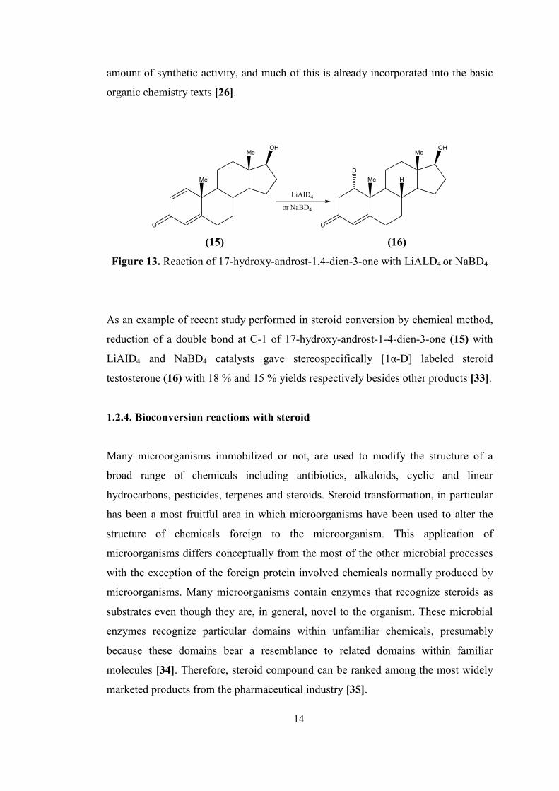
Figure 13. Reaction of 17-hydroxy-androst-1,4-dien-3-one with LiALD4 or NaBD4
As an example of recent study performed in steroid conversion by chemical method,
reduction of a double bond at C-1 of 17-hydroxy-androst-1-4-dien-3-one (15) with
LiAID4 and NaBD4 catalysts gave stereospecifically [1α-D] labeled steroid
testosterone (16) with 18 % and 15 % yields respectively besides other products [33].
1.2.4. Bioconversion reactions with steroid
Many microorganisms immobilized or not, are used to modify the structure of a
broad range of chemicals including antibiotics, alkaloids, cyclic and linear
hydrocarbons, pesticides, terpenes and steroids. Steroid transformation, in particular
has been a most fruitful area in which microorganisms have been used to alter the
structure of chemicals foreign to the microorganism. This application of
microorganisms differs conceptually from the most of the other microbial processes
with the exception of the foreign protein involved chemicals normally produced by
microorganisms. Many microorganisms contain enzymes that recognize steroids as
substrates even though they are, in general, novel to the organism. These microbial
enzymes recognize particular domains within unfamiliar chemicals, presumably
because these domains bear a resemblance to related domains within familiar
molecules [34]. Therefore, steroid compound can be ranked among the most widely
marketed products from the pharmaceutical industry [35].
14
30
The importance of microbial biotechnology in the production of steroid drugs and
hormones was realized for the first time in 1952 when Murray and Peterson patented
the process of 11α-hydroxylation of progesterone by a Rhizopus species [36].
Therefore, to make 11-hydroxy steroids were available in limited quantities. It had
been known that yeast could dehydrogenate and hydrogenate steroids since 1937.
However, the substrates used are foreign to the microorganisms and also natural
sterols, e.g. ergesterols, are resistant to transformations by means of microorganism.
After isolation of other Rhizopus species, 11-α hydroxylation product of steroids
were obtained with higher yields [37]. Previously, this product could only be
prepared starting from deoxy cholic acid on via 26 chemical steps [3].
Since then, microbial reactions for the transformation of steroids have proliferated,
and specific microbial transformation steps have been incorporated into numerous
partial syntheses of new steroids for evaluation as drugs and hormones. These
biotransformations have provided adequate tools for the large scale productions of
natural or modified steroid analogues. The latter are currently favored when
compared to their natural counterparts due to some therapeutic advantages, such as
an increased potency, longer half-lives in the blood stream, simpler delivery
methods, and reduced side effects. The preferential use of whole cells over enzymes
as biocatalysts for the production of these pharmaceutical derivatives mostly results
from the costs of the latter enzyme isolation, purification, and stabilization [36]. In
addition, these biotransformation approaches may involve the use of free or
immobilized cells or enzymes both in aq. and organic media (two phase system) [38].
Furthermore, the use of microbial models to mimic mammalian metabolism is well
known [36], the reactions of microorganisms performed on steroid compounds have
been valuable in the solution of specific problems in steroid metabolism in animals
and human body, and in conjunction with chemical syntheses, in the commercial
production of useful and complex steroids. Nowadays, in addition to the
hydroxylation reactions, at almost any position of steroid nucleus, many organisms
have been found performing many bioconversion reactions such as oxidation,
dehydrogenation, reduction, and side-chain degradation etc [37].
15
31
The oxidation and reduction reactions that microorganisms perform on steroid
provide particularly impressive examples of regio-selective and stereo-specific
biotransformations and also demonstrate the ability of enzymes to promote reactions
at inactivated centers in hydrocarbons [39].
In the production of specific steroids, a precursor steroidal compound is isolated
from some biological source such as ox bile, urine, or a plant. Once a precursor is
obtained, it is converted into the desired form by using a combination of
microbiological and chemical methods. If such reaction is chemically difficult,
expensive or otherwise impractical, and if a microbe can accomplish it readily and
inexpensively, the transformation is done with the aid of a microbe [40].
One of the main drawbacks in steroid biotransformation is the low solubility of the
substrates in water, which diminishes reaction rates and overall productivity [41].
Therefore, the utilization of high amounts of the steroidal substrates is one of the
important factors affecting the economy of the transformation process [42].
For the conversion of steroids, biotechnological methods involving whole cells or
enzymes of the microorganism are primarily preferred due to many advantages when
compared to chemical conversion methods.
Microorganisms have been widely applied for biotransformations of steroids in order
to prepare derivatives which are difficult to obtain in a different way. The following
three microbial steroid modifications are particularly important in modern
biotechnology: selective side chain cleavage of natural sterols, 1-dehydrogenation,
and 11-hydroxylation [51]. These biotransformations, mostly associated to chemical
synthesis steps, have provided adequate tools for the large scale production of natural
or modified steroid analogues. The manufactured steroid compounds have a wide
range of therapeutic purposes, namely, as anti-inflammatory, immunosuppressive,
progestational, diuretic, anabolic and contraceptive agents [35].
16
32
1.2.4.1. Hydroxylation
Microbial hydroxylations are among the most studied and useful transformations
since they can be achieved under mild conditions with high chemo-, regio- and
stereoselectivity [41]. This procedure also remains one of the most useful preparative
methods for the introduction of hydroxyl groups at sites of the steroid nucleus remote
from other functionality, and the value of microbial steroid hydroxylation in the
preparation of pharmacologically active steroids is well-established [43].
6
7
89
10
11
172
12
13
3
4
5
1 14
15
16
18
19C201
C21
AC
BD
α
β
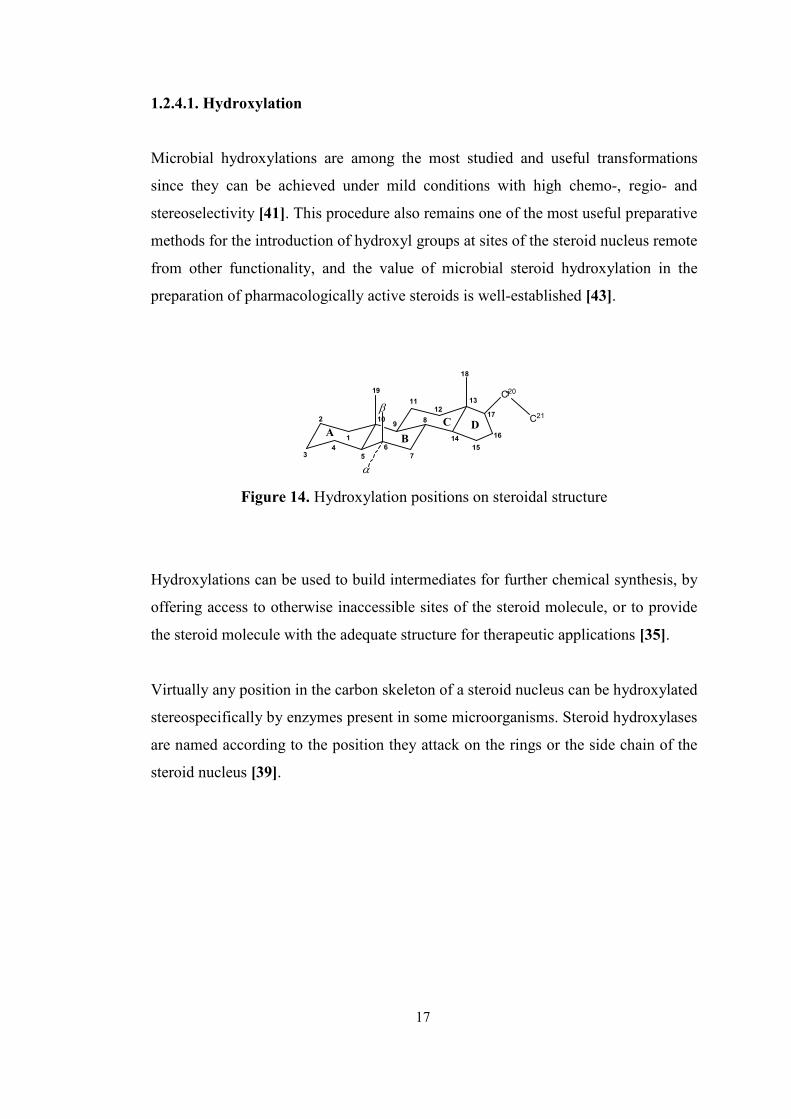
Figure 14. Hydroxylation positions on steroidal structure
Hydroxylations can be used to build intermediates for further chemical synthesis, by
offering access to otherwise inaccessible sites of the steroid molecule, or to provide
the steroid molecule with the adequate structure for therapeutic applications [35].
Virtually any position in the carbon skeleton of a steroid nucleus can be hydroxylated
stereospecifically by enzymes present in some microorganisms. Steroid hydroxylases
are named according to the position they attack on the rings or the side chain of the
steroid nucleus [39].
17
33
10
3
4
5
6
21
12
15
16
17
CH3
19
CH3
C
CH3
O
1 98
7
14
1312
11
1820
2
1β-position = Botryodiplodia malorum
2β-position = Gnomonia funicolata
6β-position = Rhizopus arrhizus
7α-position = Botryodiplodia theobromae IFO 6469
7β-position = Botryosphaeria obtusa CBS 38560
8β-position = Corynespora melonis CBS 16260
9α-position = Corynespora cassiicola ATCC 16718
12α-position = Cercospora kaki CBS 12839
14α-position = Mucor griseocyanus ATCC 1207 a
1β-position = Penicillium stolonifer CBS P102
15α-position = Aspergillus fumigatus
15β-position = Sepedonium ampullosporum
16α-position = Aspergillus niger NRRL 599
16β-position = Trichothecium roseum
17α-position = Corynespora cassicola IMI 56007
18 = Nigrospora sphaerica ATCC 12772
19 = Ophiobolus herpotrichus
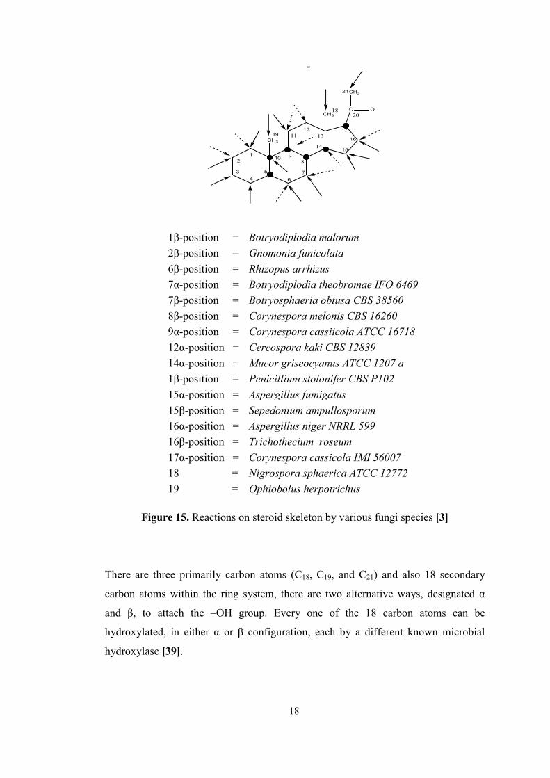
Figure 15. Reactions on steroid skeleton by various fungi species [3]
There are three primarily carbon atoms (C18, C19, and C21) and also 18 secondary
carbon atoms within the ring system, there are two alternative ways, designated α
and β, to attach the –OH group. Every one of the 18 carbon atoms can be
hydroxylated, in either α or β configuration, each by a different known microbial
hydroxylase [39].
18
34
Primary hydroxylations are 11α, 11β, 16α-hydroxylations performed by
microorganism. In mammals oxidation of C11 is an important transformation of the
cortical steroids leading to cortisol and cortisone [44]. Transformation of
progesterone by using spores of Aspergillus ochraceus at 11α- and also 6β, 11α-
positions. For 11α-hydroxylaiton of progesterone yield of above 50 % was observed.
However, it is stated that it can be increased up to 80-90 % by optimizing growth
conditions using fungal spores [45].
Sedlaczek et al. applied different method, fungal protoplast, in the transformation of
steroids. Using protoplast of Cunnighamella elegans, 11α- and 11β-hydroxylation on
cortexolone gave better results when compared to that of mycelium: the rate of
cortexolone transformation by protoplast is four times higher than that for mycelium
[46]. Transformation of 2-oxatestosterone gave successful results in the study of
Holland et al. by Aspergillus ochraceus with 82 % isolated yield of 11α-hydroxy-2-
oxatestosterone [43]. Furthermore, by another Aspergillus species, Aspergillus
ochraceus, hydroxylation of progesterone into 11α-hydroxyprogesterone
successfully achieved with approximately 90 % yield [47].
Berrie et al. reported that transformation of progesterone into 16α-
hydroxyporgesterone achieved with a 1:3.6 ratio to the main product by
Streptomyces reseochromogenes [35]. Transformation of 9α-fluorohydrocortisone
into 16α-hydroxylated product was also performed by Streptomyces species [32]. In
addition, it was reported that testosterone, deoxycorticosterone, estrone, estradiol,
cortisol, androst-4-ene-3,17-dione etc. were also hydroxylated at C-16α position with
Streptomyces spp. [37].
7α-hydroxy steroids might play a key role in the regulation of glucocorticoids action
and the immune process [48]. In the study of Cotillon et al., transformation of 3-
hydroxy steroid performed by the DHEA-induced 7α-hydroxylase of Fusarium
moniliforme. Dehydroepiandrosterone (DHEA) was converted to 7α-hydroxlated
product successfully with a yield of 98 % when compared to non-induced yield about
50 % [48].
19
35
Moreover, Botrytis cinera was found to be an efficient 7α-hydroxylator of steroidal
4-ene-3-ketones. The amount of hydroxy derivatives comprised about 26-82 % of
total metabolites for testosterone derivatives. 1-dehydrotestosterone was also
significantly hydroxylated at a 14α-position [49]. Testosterone derivatives were also
converted by Absidia glauca into products of 6β, 7α, 7β, 10β, 11α, and 12β or 15β
hydroxylation with reasonable yields [50].
Microbial hydroxylation of 2-oxotestosterone was performed by organisms that are
known to be efficient hydroxylators at 6β and 15β: at 15β position with 35 % yield
by Bacillus megaterium, 6β- hydroxy derivative of it with 59 % yield by Rhizopus
arrhizus, and also 14α-hydroxylation with 40 % yield by Curvularia lunata [43].
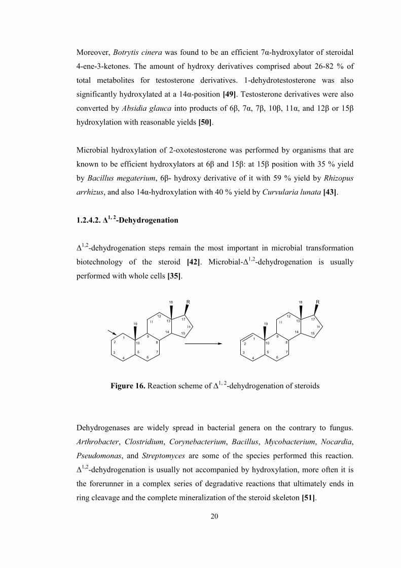
1.2.4.2. ∆1, 2-Dehydrogenation
∆1,2-dehydrogenation steps remain the most important in microbial transformation
biotechnology of the steroid [42]. Microbial-∆1,2-dehydrogenation is usually
performed with whole cells [35].
1
2
3
4
5
6
7
8
9
10
12
13
14 15
17
18
19
R
11
16
1
2
3
4
5
6
7
8
9
10
12
13
14 15
17
18
19
R
1116
Figure 16. Reaction scheme of ∆1, 2-dehydrogenation of steroids
Dehydrogenases are widely spread in bacterial genera on the contrary to fungus.
Arthrobacter, Clostridium, Corynebacterium, Bacillus, Mycobacterium, Nocardia,
Pseudomonas, and Streptomyces are some of the species performed this reaction.
∆1,2-dehydrogenation is usually not accompanied by hydroxylation, more often it is
the forerunner in a complex series of degradative reactions that ultimately ends in
ring cleavage and the complete mineralization of the steroid skeleton [51].
20
36
In the transformation of 3-oxosteroids, Bartmanska et al. performed mainly ∆1,2-
dehydrogenation which has been rarely observed in fungi cultures. In this
transformation (conversion on A-ring) cortexolone was converted into 1-
dehydrocortexolone by Trichoderma hamatum with 89 % yield. Besides this, 6α,
11α, 12β positions, ester bond hydrolysis, oxidation of 17α-hydroxly group, C17/C20
bond scission and ring D-lactonization. It was also stated that absence of 19-methyl
group led to lower yield in hydroxylation and 1-dehydrogenation [51].
Songtao et al. were used Arthrobacter simplex in the conversion of methyl-
testosterone into methandienone with a conversion more than 95 % in biphasic
system which was used to increase the solubility of product [52]. The same reaction
was performed with the same species for high concentration of 16-methyl-
Reichstein’s compound S-21 acetate (16MRSA) in microemulsion system with a
conversion of 98 % [53] and in the conversion of hydrocortisone to prednisolone at
high concentration by immobilized Corynebacterium simplex with a conversion of
80 % [54]. In another study, the same reaction was performed by Bacillus sphaericus
ATCC 13805, Bacillus sphaericus SRP III and Arthrobacter simplex 6946 and 87.6,
70.6, 88.3 % product yields obtained respectively in a two-liquid-phase system using
butyl acetate as an organic phase [55].
1.2.4.3. Ester saponification and oxidation of hydroxyl groups
O O
H
H
H
O
HO
H
H
H
O
esterase oxidase
isomerase
O
H
H
H
O
(17) (18)
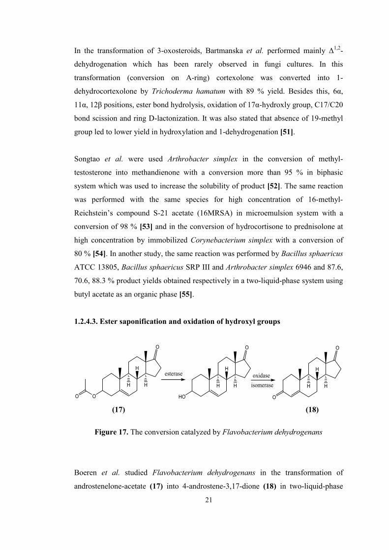
Figure 17. The conversion catalyzed by Flavobacterium dehydrogenans
Boeren et al. studied Flavobacterium dehydrogenans in the transformation of
androstenelone-acetate (17) into 4-androstene-3,17-dione (18) in two-liquid-phase
21
37
system with more than obtaining 98% conversion using octane as a second phase in
the media [56].
It was reported that Mycobacterium species was used for the conversion of
phytosterols, β-sitosterol into 4-androstene-3,17-dione (18) with a 90 % molar yields
[35].
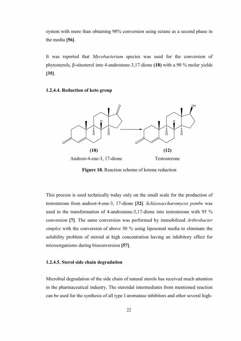
1.2.4.4. Reduction of keto group
O
OH
H
H
H
O
O
H
H
H
(18) (12)
Androst-4-ene-3, 17-dione Testosterone
Figure 18. Reaction scheme of ketone reduction
This process is used technically today only on the small scale for the production of
testosterone from androst-4-ene-3, 17-dione [32]. Schizosaccharomyces pombe was
used in the transformation of 4-androstene-3,17-dione into testosterone with 95 %
conversion [7]. The same conversion was performed by immobilized Arthrobacter
simplex with the conversion of above 50 % using liposomal media to eliminate the
solubility problem of steroid at high concentration having an inhibitory effect for
microorganisms during bioconversion [57].
1.2.4.5. Sterol side chain degradation
Microbial degradation of the side chain of natural sterols has received much attention
in the pharmaceutical industry. The steroidal intermediates from mentioned reaction
can be used for the synthesis of all type I aromatase inhibitors and other several high-
22
38
value steroidal drugs. The vast majority of microbial side-chain degradation reaction
is still based mainly on cholestane-based compounds of both animal and plant origin,
such as cholesterol and phytosterol mixtures, sitosterols [58].
In addition, the selective sterol side-chain cleavage is much more limited in both
bacteria and fungi being confined mainly to the various species of Mycobacterium
genus [58]. Moreover, the only way to produce AD on a commercial scale is
microbiological synthesis, which involves selective cleavage of the side chain of
animal (cholesterol) or plant (mainly sitosterol) β-sterols by mutant bacterial strains,
which belongs to Mycobacteria displaying a high oxidizing activity combined with
the ability to emulsify hydrophobic hydrocarbons in aqueous media [25].
Sripalakit et al. used different bacterial and fungal strains in the conversion of sterols
into various steroids: β-sitosterol was highly converted into total androstenones in
yield of 75.87 and 83.86 % by Mycobacterium spp. NRRL B-3683 and NRRL B-
3805, respectively. Almost equivalent of maximum 4-androstene-3,17-dione (AD)
and 1,4-androstadiene-3,17-dione (ADD) with total conversion of 81.83 % was
observed in mixed cultures of both strains. The molecular structure of natural sterol
affected on the productivity of AD and ADD using free strains of those [58].
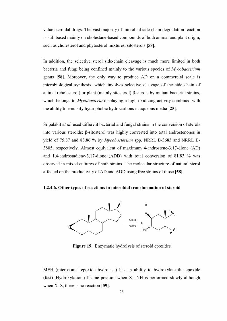
1.2.4.6. Other types of reactions in microbial transformation of steroid
R
X
X
HO
H
MEH
buffer
Figure 19. Enzymatic hydrolysis of steroid epoxides
MEH (microsomal epoxide hydrolase) has an ability to hydroxylate the epoxide
(fast) .Hydroxylation of same position when X= NH is performed slowly although
when X=S, there is no reaction [59]. 23
39
Bioconversion of 3-acetoxypregna-5,16-diene-20-one (16-DPA) to androsta-1,4-
diene-3,17-dione was performed by using mixed bacterial culture containing
Pseudomonas diminuta and Comamonas acidovorons. Using either sequential or co-
cultivation gave the same results. Although single culture was tried, there was no
conversion obtained [60].
In the study of Yazdi et al. it is stated that nandrolone decanoate was transformed
into products, estr-4-en-3,17-dione, 17β-hydroxyestr-4-en-3-one, 15α-hydroxyestr-4-
en-3,17-dione and 15α,17β-dihydroxyestr-4-en-3-one with ester hydrolysis,
oxidation and hydroxylation respectively by Acetomonium strictum [61].
Aspergillus terreus was used in the transformation of androstenedione into
testosterone and testololactone by means of 17-carbonyl reduction and Baeyer-
Villiger oxidation respectively [62].
OAc
AcO
H
OAc
HO
H
C.rugosa lipase
68 % yield[4]
(19) (20)
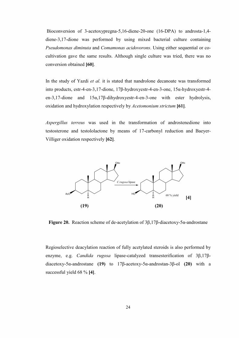
Figure 20. Reaction scheme of de-acetylation of 3β,17β-diacetoxy-5α-androstane
Regioselective deacylation reaction of fully acetylated steroids is also performed by
enzyme, e.g. Candida rugosa lipase-catalyzed transesterification of 3β,17β-
diacetoxy-5α-androstane (19) to 17β-acetoxy-5α-androstan-3β-ol (20) with a
successful yield 68 % [4].
24
40
1.3. Fungi
The fungi (mycophyta) are eukaryotes which may be derived from the colorless
representatives of the unicellular algae. Since they are not capable of forming
plastids, they are heterotrophic. They live as saprophytes or parasites in fresh water
(rarely in salt water) and on land. The saprophytes, and also many of the parasites,
can be cultivated in the laboratory [32]. The fungi embrace eukaryotic organisms
variously referred to as molds, mildews, rusts, smuts, yeasts, mushrooms, and
puffballs. Of the soil organism, the fungi as a group are the organotrophs primarily
responsible for the decomposition of organic residues [63].
Fungi are a unique group of organisms, different from all others in their behavior and
cellular organization. They also have enormous range of activities – as pathogens of
crop plants or humans, as decomposer organisms, as experimental “model
organisms” for investigating genetics and cell biology, and as producers of many
important metabolites. Thus, they have an enormous range of biochemical activities
that are exploited commercially – notably the production of antibiotics (e.g.
penicillins), steroids (for contraceptives), ciclosporins (used as immunosuppressants
in transplant surgery), and enzymes for many purposes such as food processing,
bioconversions in organic chemistry [20].
1.3.1. Fusarium species
Fusarium is a large genus of filamentous fungi widely distributed in soil and in
association with plants. Most species are harmless saprobes and are relatively
abundant members of the soil microbial community [65] and they are active in the
decomposition of cellulosic plant materials. Fusarium is one of the three major
fungal genera producing toxins [64]. The main toxins produced by these Fusarium
species are fumonisins and trichothecenes [65].
Fusarium is a type fungus which exists in a group of Fungi imperfecti, class of
Deuteromyces with the features of septate hyphae reproduced only by asexual
conida, if sexual stage found, usually ascomycete [63]. The character which defines
25
41
the genus Fusarium is the production of septate, fusiform to crescent shaped conidia,
termed macroconidia, with a foot shape basal cell and a more or less beaked apical
cell [64].
26
42
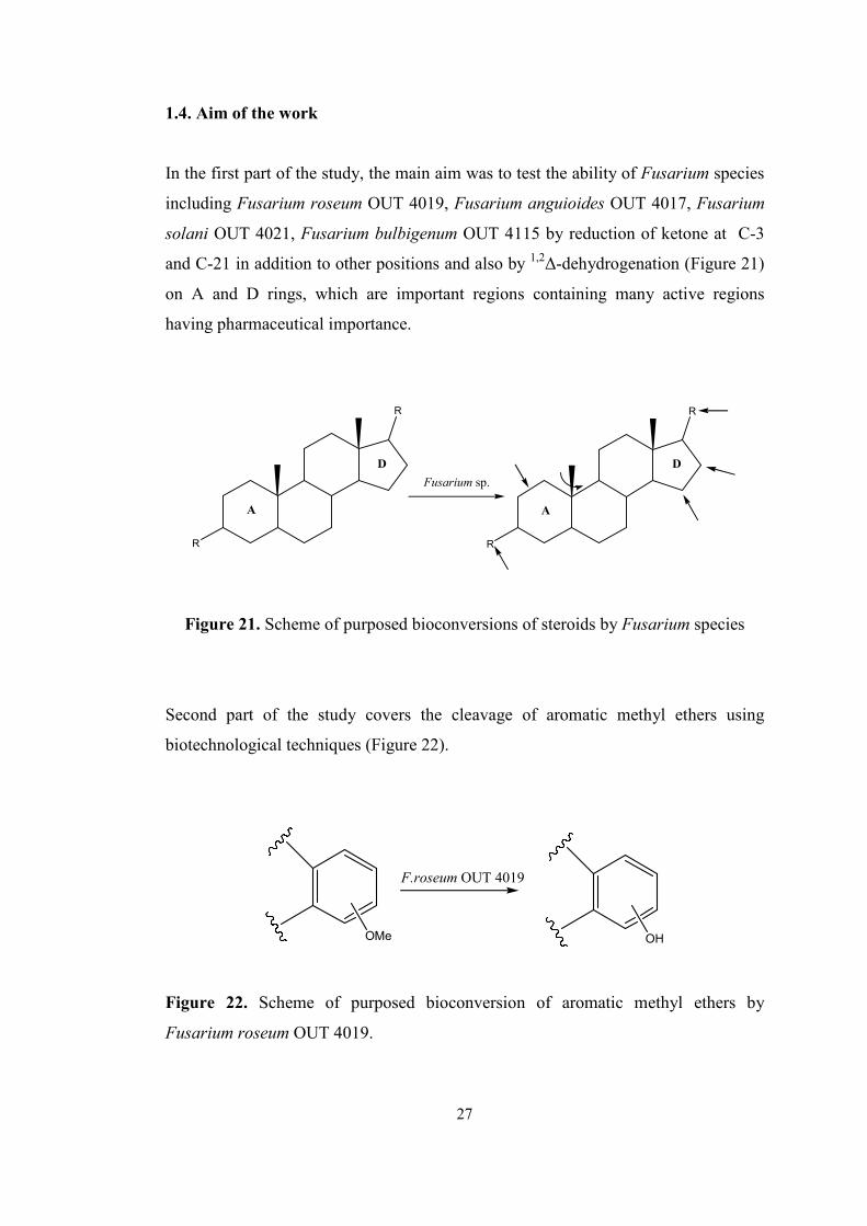
1.4. Aim of the work
In the first part of the study, the main aim was to test the ability of Fusarium species
including Fusarium roseum OUT 4019, Fusarium anguioides OUT 4017, Fusarium
solani OUT 4021, Fusarium bulbigenum OUT 4115 by reduction of ketone at C-3
and C-21 in addition to other positions and also by 1,2∆-dehydrogenation (Figure 21)
on A and D rings, which are important regions containing many active regions
having pharmaceutical importance.
R
R
A
D
Fusarium sp.
R
R
A
D
Figure 21. Scheme of purposed bioconversions of steroids by Fusarium species
Second part of the study covers the cleavage of aromatic methyl ethers using
biotechnological techniques (Figure 22).
F.roseum OUT 4019
OMe OH
Figure 22. Scheme of purposed bioconversion of aromatic methyl ethers by
Fusarium roseum OUT 4019.
27
43
CHAPTER 2
RESULTS AND DISCUSSION
2.1. STEROID BIOCONVERSIONS
2.1.1. Perspective of the work
Naturally occurring compounds are in a great interest since the early times of life of
human beings. Natural products are important compounds which are synthesized by
living organisms. They are divided into two groups according to their types: primary
and secondary metabolites.
Primary metabolites are vital for the living organisms. These metabolites start to
produce during growth phase and production continues throughout the life of the
organisms. Thus, these compounds are widespread among all organisms.
Hormones are one of the most important primary metabolites. They exist nearly in all
multi cellular organisms. They are powerful signal molecules that have roles as
regulators in the function of metabolism. In mammalians, they are involved in many
functions such as regulation of blood sugar level, sodium-potassium level,
degradation of fats and proteins, increasing muscle and bone synthesis, inhibition of
inflammatory response etc. Steroids are the most important types of hormones
providing sex hormones and adrenal cortex hormones, e.g. progesterone, and
cortisone which have extensive application in birth control pills and anti-
inflammation respectively.
On the other hand, conversion of steroids by using biotechnological methods is a
highly growing area and promising an alternative method of steroid synthesis.
28
44
Biotransformation process has many advantages over conventional chemistry. It has
environmentally safe and friendly conditions because its mild conditions and reaction
media containing only water as a solvent instead of organic substance. The most
important one is the specificity of enzymes besides their environmentally safe
conditions. By stereo- and regio-specific properties of enzymes or microorganisms
having those enzymes, desired selectivity on the product has been achieved easily. In
drug development, producing products being highly pure and also having high
enantiomeric purity and regio-specificity is important since each enantiomers of the
molecule existing in drugs show different effects on the body. Producing a drug with
a single enantiomer is possible with bioconversion methods. Therefore, in
pharmaceuticals, this specificity gives the biotechnology the leading position to
produce target specific drugs.
In bioconversion reactions, steroids are generally mostly studied substance in the
pharmaceutical. There are many precursors which are used to synthesize the desired
type of steroids. Most of them are isolated from the plant sources such as
stigmasterol and diosgenin.
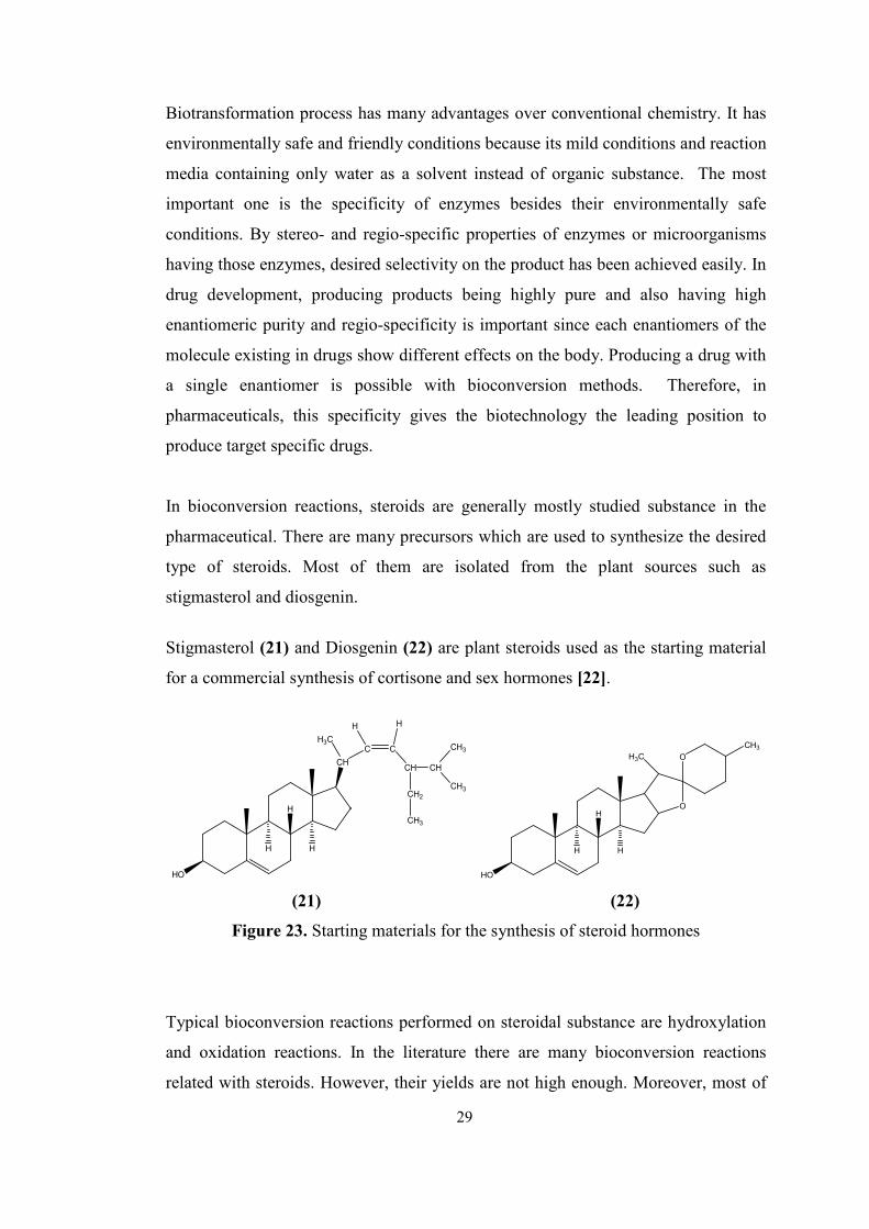
Stigmasterol (21) and Diosgenin (22) are plant steroids used as the starting material
for a commercial synthesis of cortisone and sex hormones [22].
H
H
H
HO
CH
H3CC
H
C
H
CH
CH2
CH
CH3
CH3
CH3
H
H
H
HO
O
O
CH3H3C
(21) (22)
Figure 23. Starting materials for the synthesis of steroid hormones
Typical bioconversion reactions performed on steroidal substance are hydroxylation
and oxidation reactions. In the literature there are many bioconversion reactions
related with steroids. However, their yields are not high enough. Moreover, most of
29
45
the reactions give more than one products with the same type conversion at different
positions. Therefore, the selectivities are also very low.
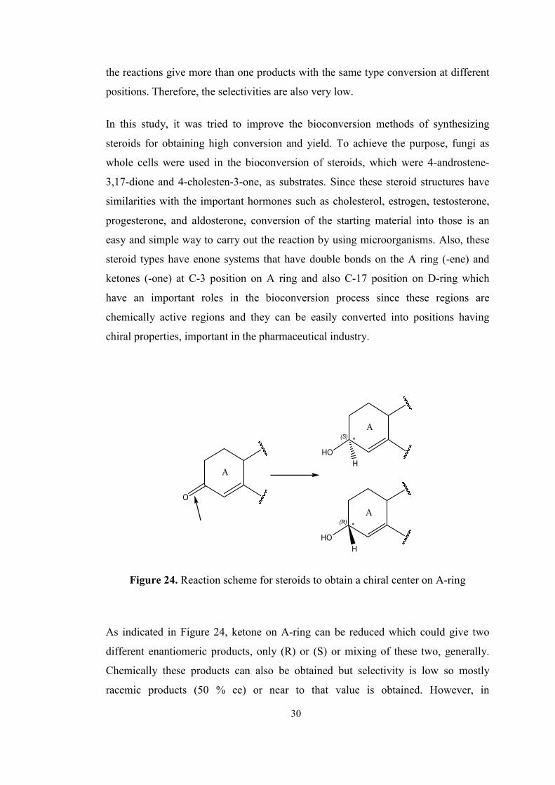
In this study, it was tried to improve the bioconversion methods of synthesizing
steroids for obtaining high conversion and yield. To achieve the purpose, fungi as
whole cells were used in the bioconversion of steroids, which were 4-androstene-
3,17-dione and 4-cholesten-3-one, as substrates. Since these steroid structures have
similarities with the important hormones such as cholesterol, estrogen, testosterone,
progesterone, and aldosterone, conversion of the starting material into those is an
easy and simple way to carry out the reaction by using microorganisms. Also, these
steroid types have enone systems that have double bonds on the A ring (-ene) and
ketones (-one) at C-3 position on A ring and also C-17 position on D-ring which
have an important roles in the bioconversion process since these regions are
chemically active regions and they can be easily converted into positions having
chiral properties, important in the pharmaceutical industry.
O
A
∗(S)
HO
A
H
(R) ∗
HO
A
H
Figure 24. Reaction scheme for steroids to obtain a chiral center on A-ring
As indicated in Figure 24, ketone on A-ring can be reduced which could give two
different enantiomeric products, only (R) or (S) or mixing of these two, generally.
Chemically these products can also be obtained but selectivity is low so mostly
racemic products (50 % ee) or near to that value is obtained. However, in
30
46
bioconversion with microorganisms, due to the selectivity of the enzyme, producing
enantiomerically pure compounds is possible.
O
(R)
∗
OH
D
D
(S)
∗
OH
D
H
H
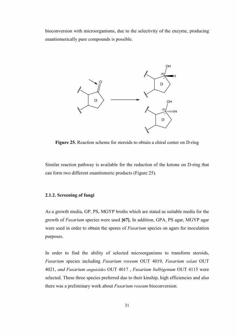
Figure 25. Reaction scheme for steroids to obtain a chiral center on D-ring
Similar reaction pathway is available for the reduction of the ketone on D-ring that
can form two different enantiomeric products (Figure 25).
2.1.2. Screening of fungi
As a growth media, GP, PS, MGYP broths which are stated as suitable media for the
growth of Fusarium species were used [67]. In addition, GPA, PS agar, MGYP agar
were used in order to obtain the spores of Fusarium species on agars for inoculation
purposes.
In order to find the ability of selected microorganisms to transform steroids,
Fusarium species including Fusarium roseum OUT 4019, Fusarium solani OUT
4021, and Fusarium anguioides OUT 4017 , Fusarium bulbigenum OUT 4115 were
selected. These three species preferred due to their kinship, high efficiencies and also
there was a preliminary work about Fusarium roseum bioconversion.
31
47
First of all, from the agars of stock cultures where the Fusarium species had been
grown, inoculation was performed into broths by sterile loop. Microorganisms,
especially fungi are widely distributed in the nature where they were isolated. Since
they have specific growth regions containing different nutritional diversity as carbon,
nitrogen, besides mineral and oxygen sources, the adaptation of those native
microorganisms is a difficult procedure. Therefore, using a synthetic media, to obtain
desired growth rates and also desired biomass values require additional studies.
Because each of different media preparations contains different carbon, nitrogen and
also mineral sources, microorganisms show different response to those parameters.
According to those, since they grow in different style and different amounts in the
media, these result in changing in conversion, yield and even in selectivity of the
reaction that affect the overall bioconversion process directly.
For all Fusarium species including Fusarium roseum OUT 4019, Fusarium solani
OUT 4021, and Fusarium anguioides OUT 4017, Fusarium bulbigenum OUT 4115
selected for this study, there was a growth observed in these broths. Obtained results
were used in the biotransformation reactions. To achieve a sufficient biomass results,
microorganisms were incubated at 25 °C for 4.5 days in shaker.
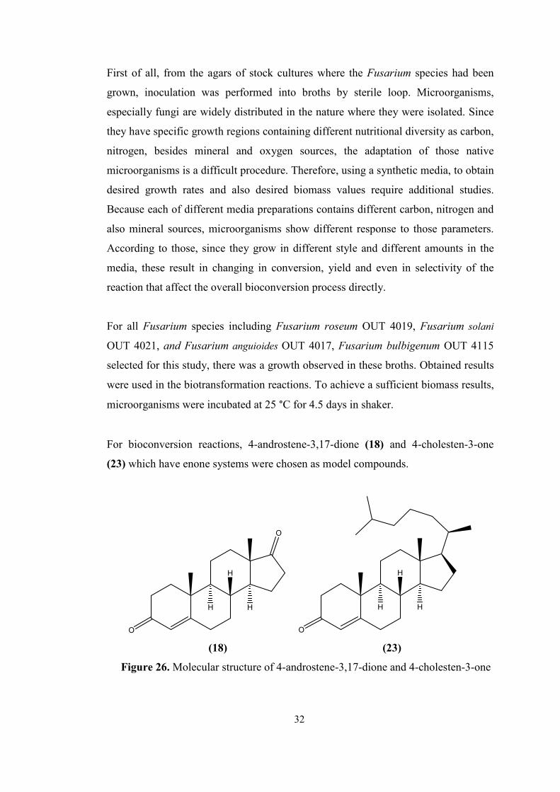
For bioconversion reactions, 4-androstene-3,17-dione (18) and 4-cholesten-3-one
(23) which have enone systems were chosen as model compounds.
O
O
H
H
H
O
H
H
H
(18) (23)
Figure 26. Molecular structure of 4-androstene-3,17-dione and 4-cholesten-3-one
32
48
After the incubation, steroids, 4-androstene-3,17-dione (18) and 4-cholesten-3-one
(23), were added into the media with obtained biomass results by dissolving them in
an organic solvent dimethylsulfoxide, DMSO, which is miscible in water easily.
During the bioconversion period, reactions of Fusarium species were monitored by
using thin layer chromatography, TLC, with a phosphomolybdic acid dye, PMA
under UV254 nm light for both substrate and products which are UV active.
In the first control experiment, 4-androstene-3,17-dione was used as substrate and it
was shown that all Fusarium species converts substrate into any products. These
steroid types which were used as starting materials for the bioconversion reactions by
Fusarium species were used readily without performing any chemical synthesis
because of their commercial availability.
Fusarium roseum OUT 4019 catalyzed the steroid bioconversion reaction in GP and
MGYP broths. Whereas Fusarium anguioides OUT 4017 and Fusarium solani OUT
4015 catalyzed the reaction in PS and MGYP broths, Fusarium bulbigenum OUT
4115 catalyzed the reaction in only MGYP broth.
O
O
H
H
HA
D
O
O
H
H
HA
D
Fusarium
?
? ?
?
?
?
?
? ?
??
?
?
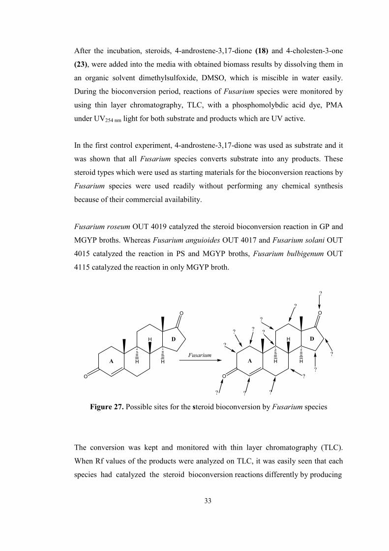
Figure 27. Possible sites for the steroid bioconversion by Fusarium species
The conversion was kept and monitored with thin layer chromatography (TLC).
When Rf values of the products were analyzed on TLC, it was easily seen that each
species had catalyzed the steroid bioconversion reactions differently by producing
33
49
different products. However, it was not possible to determine the transformation type
or product type by looking at only TLC data. Therefore, after work up, obtained
crude products were purified by column chromatography and then analyzed by 1H-
NMR and 13C-NMR spectrum and also GC-MS.
According to NMR and GC-MS spectrum data besides isolated yields, Fusarium
roseum OUT 4019 had a best conversion in GP broth. While Fusarium anguioides
OUT 4017 showed the highest performance in PS broth, Fusarium solani OUT 4021
did in MGYP broth. However, for Fusarium bulbigenum OUT 4115, although
conversion was observed with 4-androstene-3,17-dione, any identification couldn’t
be performed. Therefore, after those experiments, by optimization studies it was tried
to find the best condition to carry out this reaction in steroid conversion for obtaining
high chemical conversion and yields.
The same procedure was performed for 4-cholesten-3-one as in the case of 4-
androstene-3,17-dione. For all Fusarium species used, no conversion was observed
because of low solubility of the 4-cholesten-3-one. Therefore, 4-androstene-3,17-
dione was selected as a model substrate for the optimization of bioconversion studies
by Fusarium species.
Besides these optimization studies, blank experiments for both steroid substrates, 4-
androstene-3,17-dione and 4-cholesten-3-one were carried out in order to find the
effect of media on the steroids during bioconversion periods. Blank experiments
were monitored by TLC as in the case of bioconversion studies during two weeks.
For both substances, there was no change observed in the media selected. To sum up,
the two starting materials were identified as stable materials in those media.
2.1.3. Optimization studies for fungal bioconversion
Some parameters glucose concentration, solvent, growth media either agar or broth,
inoculum size were changed to find out the optimum conditions with the hope having
the best chemical yields in these reactions.
34
50
2.1.3.1. Effect of solvent
It is well known that fungus reactions proceed in aqueous media. Since the
compounds used in this study are steroids, major problem is solubility in water,
steroids only soluble to 0.1 - 1% in water. It is necessary to use organic solvents and
emulsions for solution of substrates and products. However, although enzymes can
be quite stable to organic solvents, membranes of cells are very sensitive and
solvents cause disruption. This can create a problem when live cells are required with
an active electron transport chain [68]. Therefore, it was necessary to find out a co-
solvent;
-which could dissolve these substrates,
-give no harm to Fusarium species
-that is miscible with water so that the reaction
will take place in a homogenous media.
For these purposes, three solvents: dimethysulfoxide (DMSO), ethanol and acetone
were selected and tested to find out the suitable solvent which has the above
mentioned properties.
These solvents were tested with substrates to see their solubilities. Three of them
have an ability to dissolve 4-androstene-3,17-dione successfully.
Then, these solvents, 5 mL, were added separately into the sterilized media, in which
Fusarium spp. were inoculated and incubated at 25 °C. Although all three have good
dissolving abilities with a decreasing order: acetone, DMSO, and ethanol, ethanol
and acetone were eliminated due to their ease to vaporize, even in the mixture with
water. Therefore, during the reaction period, since conversion period and temperature
(25 °C) was almost suitable for vaporizing totally form the media, these solvents did
not solve the solubility problem to obtain the highest yield.
The results show that of three solvent, dimethylsulfoxide was the best one which
provide the optimum conditions.
35
51
According to the studies performed by DMSO, high level of it shows toxic effect on
some fungi types. Therefore, in order to decrease the level of toxicity, as a second
co-solvent ethanol and acetone were studied by decreasing the amount of DMSO.
Each Fusarium spp. showed different effect against solvent mixture. While F.
roseum and F. anguioides showed better results against DMSO-acetone mixture, for
F. solani higher yield was obtained with DMSO-ethanol mixture. Ethanol had a
positive effect on conversion by utilizing as carbon source so it increased the yield
[57, 58].
Besides the studies of 4-androstene-3,17-dione, 4-cholesten-3-one was also studied
to solve the solubility problem in the media. In order to find the best solvent to
eliminate the solubility problem three powerful solvent were used as in the case of 4-
androstene-3,17-dione. However, although acetone and DMSO are powerful
solvents, they could not dissolve 4-cholesten-3-one totally when using suitable
amount of solvent. Although it was possible to dissolve it by using high amount of
solvent, it couldn’t be applied since those amounts would be toxic for
microorganisms. Besides, with ethanol as a solvent, solubility problem were
achieved with increasing solvent amount at the beginning. However, bioconversion
period was long so, and ethanol is volatile liquid, this problem did not completely
eliminated. There was a conversion observed with ethanol as a co-solvent. However,
due to the inabilities for the solubility of 4-cholesten-3-one conversions preventing
the complete conversion, the obtained yields for all Fusarium species were very low
to detect the structure of product by NMR and GC-MS analysis. Therefore, 4-
androstene-3,17-dione was selected as a model substrate for the optimization of
bioconversion studies of Fusarium species.
2.1.3.2. Effect of glucose concentration
It is known that glucose is the primary carbon source of living things all around the
world. In the growth media, glucose concentration affects the conversion of substrate
also the chemical yield indirectly by means of growth and biomass formation. Since
the growth and continuity of life depends on the nutrition, when there is no enough
nutrition, mainly containing carbon sources, microorganisms start to degrade the
36
52
starting material using as a carbon source as also stated by Sripalakit et al. .This
affects the total conversion and yield of the product in many different ways such as
producing side products which are difficult to eliminate from the product extract and
needs further purification steps decreasing the yield, and decrease the converted
product because of using as also carbon source. To eliminate these problems,
optimization must be required.
Glucose concentration study was performed only for Fusarium roseum OUT 4019.
Therefore, it was optimized only for it. Between the values of 2.5, 3.0 and 3.5 % w/v
of glucose concentration was studied for GP (glucose-Peptone) media and 3.0 % w/v
glucose concentration gave the best conversion and yield.
2.1.3.3. Effect of type of growth media
For microorganisms, growth media indirectly affect the conversion and yield of the
bioconversion. When there is a successful growth of microorganism in the media,
results of the reaction will be better.
F. solani OUT 4021 grown in PD agar as in the case of the others and this had a low
yield to produce spore on the surface of agar. Having fresh and better grown spores
in stock culture is important for obtaining high cell biomass in liquid media. In order
to increase the spore yield, two different agars were tested: PDA (Potato Dextrose
Agar and MGYP agar). According to results, on MGYP agar, growth of spores gave
better results. This result was also applied for the broth and the same success was
obtained for growth on MGYP broth when it was compared to the previously used
broth, PSB. Therefore, MGYP was selected as growth media as an agar and broth for
F. solani OUT 4015. The other two species F. roseum OUT 4019 in GP agar and
broth and also F. Anguioides OUT 4017 in PS agar and broth were grown and
showed successful bioconversion results when compared to old cultured samples.
37
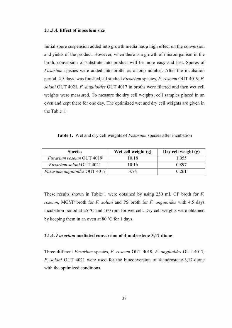
53
2.1.3.4. Effect of inoculum size
Initial spore suspension added into growth media has a high effect on the conversion
and yields of the product. However, when there is a growth of microorganism in the
broth, conversion of substrate into product will be more easy and fast. Spores of
Fusarium species were added into broths as a loop number. After the incubation
period, 4.5 days, was finished, all studied Fusarium species, F. roseum OUT 4019, F.
solani OUT 4021, F. anguioides OUT 4017 in broths were filtered and then wet cell
weights were measured. To measure the dry cell weights, cell samples placed in an
oven and kept there for one day. The optimized wet and dry cell weights are given in
the Table 1.
Table 1. Wet and dry cell weights of Fusarium species after incubation
Species Wet cell weight (g) Dry cell weight (g)
Fusarium roseum OUT 4019 10.18 1.055
Fusarium solani OUT 4021 10.16 0.897
Fusarium anguioides OUT 4017 3.74 0.261
These results shown in Table 1 were obtained by using 250 mL GP broth for F.
roseum, MGYP broth for F. solani and PS broth for F. anguioides with 4.5 days
incubation period at 25 °C and 160 rpm for wet cell. Dry cell weights were obtained
by keeping them in an oven at 80 °C for 1 days.
2.1.4. Fusarium mediated conversion of 4-androstene-3,17-dione
Three different Fusarium species, F. roseum OUT 4019, F. anguioides OUT 4017,
F. solani OUT 4021 were used for the bioconversion of 4-androstene-3,17-dione
with the optimized conditions.
38
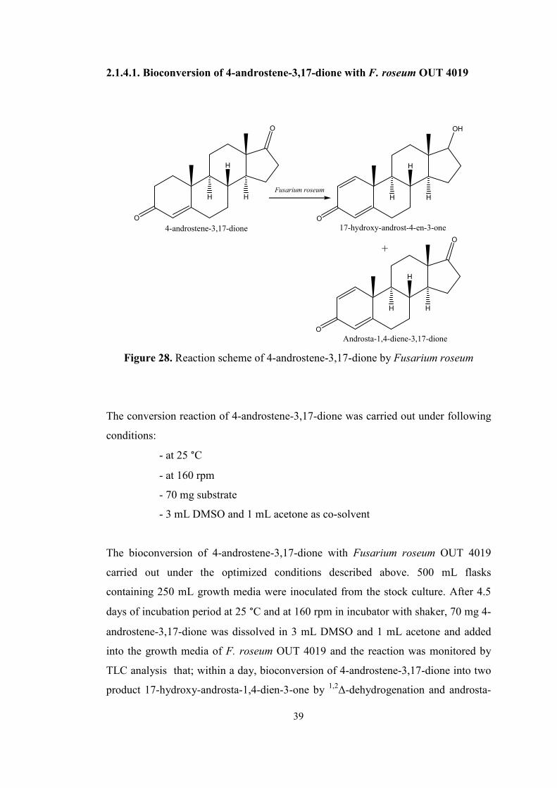
54
2.1.4.1. Bioconversion of 4-androstene-3,17-dione with F. roseum OUT 4019
O
O
H
H
H
4-androstene-3,17-dioneO
OH
H
H
H
17-hydroxy-androst-4-en-3-one
Fusarium roseum
+
O
O
H
H
H
Androsta-1,4-diene-3,17-dione
Figure 28. Reaction scheme of 4-androstene-3,17-dione by Fusarium roseum
The conversion reaction of 4-androstene-3,17-dione was carried out under following
conditions:
- at 25 °C
- at 160 rpm
- 70 mg substrate
- 3 mL DMSO and 1 mL acetone as co-solvent
The bioconversion of 4-androstene-3,17-dione with Fusarium roseum OUT 4019
carried out under the optimized conditions described above. 500 mL flasks
containing 250 mL growth media were inoculated from the stock culture. After 4.5
days of incubation period at 25 °C and at 160 rpm in incubator with shaker, 70 mg 4-
androstene-3,17-dione was dissolved in 3 mL DMSO and 1 mL acetone and added
into the growth media of F. roseum OUT 4019 and the reaction was monitored by
TLC analysis that; within a day, bioconversion of 4-androstene-3,17-dione into two
product 17-hydroxy-androsta-1,4-dien-3-one by 1,2∆-dehydrogenation and androsta-
39
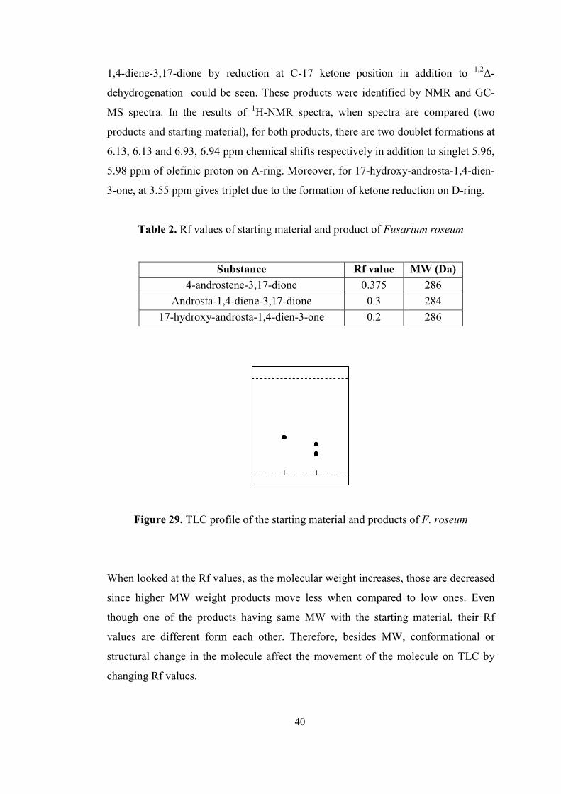
55
1,4-diene-3,17-dione by reduction at C-17 ketone position in addition to 1,2∆-
dehydrogenation could be seen. These products were identified by NMR and GC-
MS spectra. In the results of 1H-NMR spectra, when spectra are compared (two
products and starting material), for both products, there are two doublet formations at
6.13, 6.13 and 6.93, 6.94 ppm chemical shifts respectively in addition to singlet 5.96,
5.98 ppm of olefinic proton on A-ring. Moreover, for 17-hydroxy-androsta-1,4-dien-
3-one, at 3.55 ppm gives triplet due to the formation of ketone reduction on D-ring.
Table 2. Rf values of starting material and product of Fusarium roseum
Substance Rf value MW (Da)
4-androstene-3,17-dione 0.375 286
Androsta-1,4-diene-3,17-dione 0.3 284
17-hydroxy-androsta-1,4-dien-3-one 0.2 286
Figure 29. TLC profile of the starting material and products of F. roseum
When looked at the Rf values, as the molecular weight increases, those are decreased
since higher MW weight products move less when compared to low ones. Even
though one of the products having same MW with the starting material, their Rf
values are different form each other. Therefore, besides MW, conformational or
structural change in the molecule affect the movement of the molecule on TLC by
changing Rf values.
40
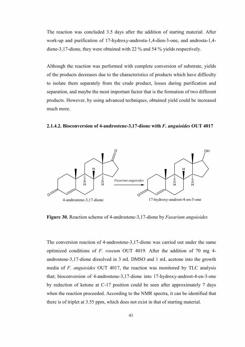
56
The reaction was concluded 3.5 days after the addition of starting material. After
work-up and purification of 17-hydroxy-androsta-1,4-dien-3-one, and androsta-1,4-
diene-3,17-dione, they were obtained with 22 % and 54 % yields respectively.
Although the reaction was performed with complete conversion of substrate, yields
of the products decreases due to the characteristics of products which have difficulty
to isolate them separately from the crude product, losses during purification and
separation, and maybe the most important factor that is the formation of two different
products. However, by using advanced techniques, obtained yield could be increased
much more.
2.1.4.2. Bioconversion of 4-androstene-3,17-dione with F. anguioides OUT 4017
O
O
H
H
H
4-androstene-3,17-dioneO
OH
H
H
H
17-hydroxy-androst-4-en-3-one
Fusarium anguioides
Figure 30. Reaction scheme of 4-androstene-3,17-dione by Fusarium anguioides
The conversion reaction of 4-androstene-3,17-dione was carried out under the same
optimized conditions of F. roseum OUT 4019. After the addition of 70 mg 4-
androstene-3,17-dione dissolved in 3 mL DMSO and 1 mL acetone into the growth
media of F. anguioides OUT 4017, the reaction was monitored by TLC analysis
that; bioconversion of 4-androstene-3,17-dione into 17-hydroxy-androst-4-en-3-one
by reduction of ketone at C-17 position could be seen after approximately 7 days
when the reaction proceeded. According to the NMR spectra, it can be identified that
there is of triplet at 3.55 ppm, which does not exist in that of starting material.
41
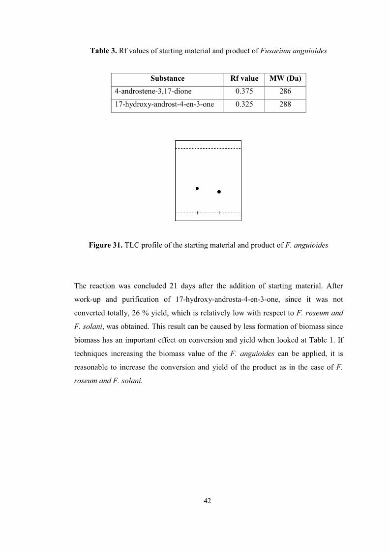
57
Table 3. Rf values of starting material and product of Fusarium anguioides
Substance Rf value MW (Da)
4-androstene-3,17-dione 0.375 286
17-hydroxy-androst-4-en-3-one 0.325 288
Figure 31. TLC profile of the starting material and product of F. anguioides
The reaction was concluded 21 days after the addition of starting material. After
work-up and purification of 17-hydroxy-androsta-4-en-3-one, since it was not
converted totally, 26 % yield, which is relatively low with respect to F. roseum and
F. solani, was obtained. This result can be caused by less formation of biomass since
biomass has an important effect on conversion and yield when looked at Table 1. If
techniques increasing the biomass value of the F. anguioides can be applied, it is
reasonable to increase the conversion and yield of the product as in the case of F.
roseum and F. solani.
42
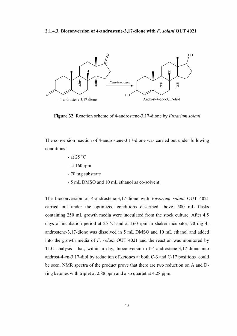
58
2.1.4.3. Bioconversion of 4-androstene-3,17-dione with F. solani OUT 4021
O
O
H
H
H
4-androstene-3,17-dioneHO
OH
H
H
H
Androst-4-ene-3,17-diol
Fusarium solani
Figure 32. Reaction scheme of 4-androstene-3,17-dione by Fusarium solani
The conversion reaction of 4-androstene-3,17-dione was carried out under following
conditions:
- at 25 °C
- at 160 rpm
- 70 mg substrate
- 5 mL DMSO and 10 mL ethanol as co-solvent
The bioconversion of 4-androstene-3,17-dione with Fusarium solani OUT 4021
carried out under the optimized conditions described above. 500 mL flasks
containing 250 mL growth media were inoculated from the stock culture. After 4.5
days of incubation period at 25 °C and at 160 rpm in shaker incubator, 70 mg 4-
androstene-3,17-dione was dissolved in 5 mL DMSO and 10 mL ethanol and added
into the growth media of F. solani OUT 4021 and the reaction was monitored by
TLC analysis that; within a day, bioconversion of 4-androstene-3,17-dione into
androst-4-en-3,17-diol by reduction of ketones at both C-3 and C-17 positions could
be seen. NMR spectra of the product prove that there are two reduction on A and D-
ring ketones with triplet at 2.88 ppm and also quartet at 4.28 ppm.
43
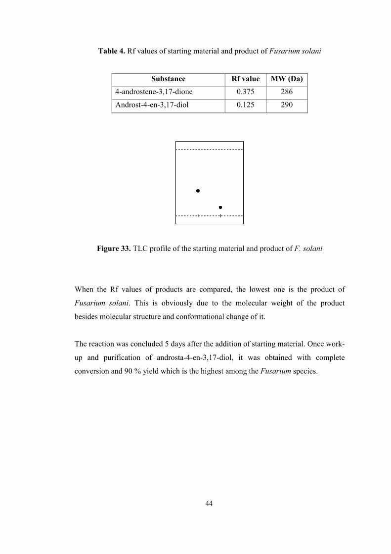
59
Table 4. Rf values of starting material and product of Fusarium solani
Substance Rf value MW (Da)
4-androstene-3,17-dione 0.375 286
Androst-4-en-3,17-diol 0.125 290
Figure 33. TLC profile of the starting material and product of F. solani
When the Rf values of products are compared, the lowest one is the product of
Fusarium solani. This is obviously due to the molecular weight of the product
besides molecular structure and conformational change of it.
The reaction was concluded 5 days after the addition of starting material. Once work-
up and purification of androsta-4-en-3,17-diol, it was obtained with complete
conversion and 90 % yield which is the highest among the Fusarium species.
44
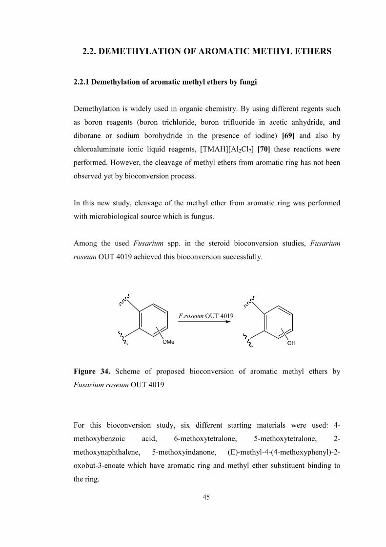
60
2.2. DEMETHYLATION OF AROMATIC METHYL ETHERS
2.2.1 Demethylation of aromatic methyl ethers by fungi
Demethylation is widely used in organic chemistry. By using different regents such
as boron reagents (boron trichloride, boron trifluoride in acetic anhydride, and
diborane or sodium borohydride in the presence of iodine) [69] and also by
chloroaluminate ionic liquid reagents, [TMAH][Al2Cl7] [70] these reactions were
performed. However, the cleavage of methyl ethers from aromatic ring has not been
observed yet by bioconversion process.
In this new study, cleavage of the methyl ether from aromatic ring was performed
with microbiological source which is fungus.
Among the used Fusarium spp. in the steroid bioconversion studies, Fusarium
roseum OUT 4019 achieved this bioconversion successfully.
F.roseum OUT 4019
OMe OH
Figure 34. Scheme of proposed bioconversion of aromatic methyl ethers by
Fusarium roseum OUT 4019
For this bioconversion study, six different starting materials were used: 4-
methoxybenzoic acid, 6-methoxytetralone, 5-methoxytetralone, 2-
methoxynaphthalene, 5-methoxyindanone, (E)-methyl-4-(4-methoxyphenyl)-2-
oxobut-3-enoate which have aromatic ring and methyl ether substituent binding to
the ring.
45
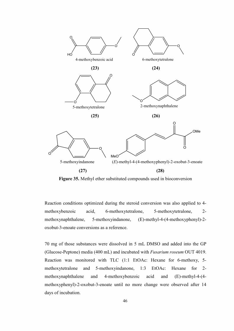
61
O
HO
O
4-methoxybenzoic acid
O
O
6-methoxytetralone
(23) (24)
O
O
5-methoxytetralone
O
2-methoxynaphthalene
(25) (26)
O
O
5-methoxyindanone
MeO
O
O
OMe
(E)-methyl-4-(4-methoxyphenyl)-2-oxobut-3-enoate
(27) (28)
Figure 35. Methyl ether substituted compounds used in bioconversion
Reaction conditions optimized during the steroid conversion was also applied to 4-
methoxybenzoic acid, 6-methoxytetralone, 5-methoxytetralone, 2-
methoxynaphthalene, 5-methoxyindanone, (E)-methyl-4-(4-methoxyphenyl)-2-
oxobut-3-enoate conversions as a reference.
70 mg of those substances were dissolved in 5 mL DMSO and added into the GP
(Glucose-Peptone) media (400 mL) and incubated with Fusarium roseum OUT 4019.
Reaction was monitored with TLC (1:1 EtOAc: Hexane for 6-methoxy, 5-
methoxytetralone and 5-methoxyindanone, 1:3 EtOAc: Hexane for 2-
methoxynaphthalene and 4-methoxybenzoic acid and (E)-methyl-4-(4-
methoxyphenyl)-2-oxobut-3-enoate until no more change were observed after 14
days of incubation.
46
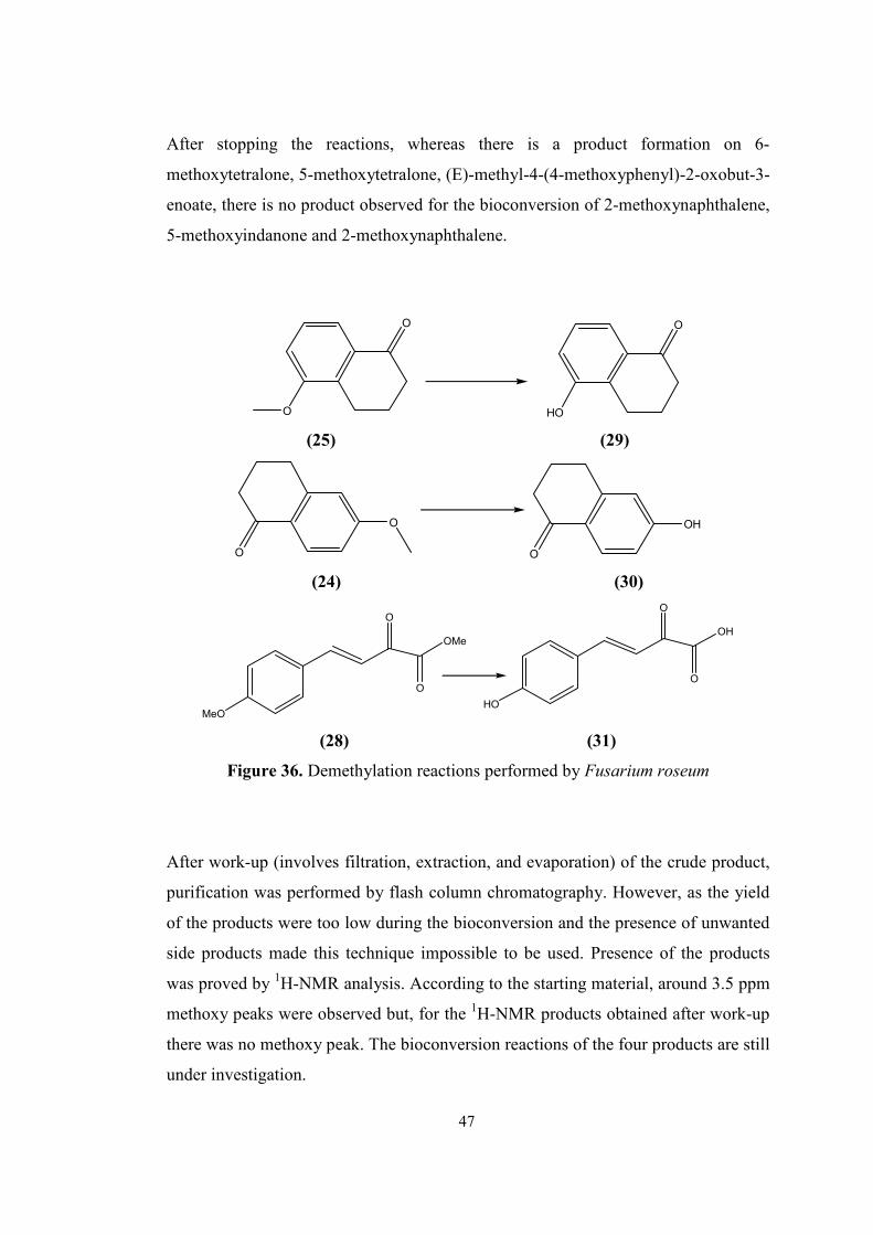
62
After stopping the reactions, whereas there is a product formation on 6-
methoxytetralone, 5-methoxytetralone, (E)-methyl-4-(4-methoxyphenyl)-2-oxobut-3-
enoate, there is no product observed for the bioconversion of 2-methoxynaphthalene,
5-methoxyindanone and 2-methoxynaphthalene.
O
O
HO
O
(25) (29)
O
O
OH
O
(24) (30)
MeO
O
O
OMe
HO
O
O
OH
(28) (31)
Figure 36. Demethylation reactions performed by Fusarium roseum
After work-up (involves filtration, extraction, and evaporation) of the crude product,
purification was performed by flash column chromatography. However, as the yield
of the products were too low during the bioconversion and the presence of unwanted
side products made this technique impossible to be used. Presence of the products
was proved by 1H-NMR analysis. According to the starting material, around 3.5 ppm
methoxy peaks were observed but, for the 1H-NMR products obtained after work-up
there was no methoxy peak. The bioconversion reactions of the four products are still
under investigation.
47
63
CHAPTER 3
MATERIAL AND METHODS
3.1. Materials
Fungal bioconversion reactions were carried out in Edmund Bühler Johama Otto
GMBH rotary shaker.
Flash column chromatographies were run on silica gel (Redisep Flash column 4g
(max. pressure 45PSI (3.1 bar)) with the solvent mixture indicated.
Thin layer chromatography was performed on commercial silica gel plates
(Macherey-Nagel, 0.2 mm Silica Gel 60 with fluorescent indicator UV254) that were
developed by immersion in 5 % phosphomolybdic acid (PMA) in 95 % ethanol.
Preparative thin layer chromatography was performed on commercial silica gel
plates (Macherey-Nagel, 2 mm silica gel, SIL G-200 UV254, with UV254 indicator)
was used with 1: 3 EtOAc - Hexane when the product was need to be purified further
for NMR analysis.
All solvents were freshly distilled before use and stored.
1H-NMR spectra were obtained on Bruker DPX400 instrument in CDCI3 and CCI4.
Chemical shifts are expressed in ppm downfield from TMS; the 1H-NMR data is
present in the order: δ value of the signal, integrated number of protons, peak
multiplicity (abbreviations are as follows: s, singlet; d, doublet; t, triplet; q, quartet;
m, multiplet; br., broad) and coupling constants in Hertz.
48
64
GC-MS spectra were determined using a ThermoQuest (TSP) TraceGC-2000 series
equipped with phenomenex Zebron ZB-5 capillary column (5 %
phenylmethylsiloxane, 30 m, 250 µm); TGC (injector) = 250 °C, TMS (ion source) =
200 °C, time programme (oven): T 0 min. = 60 °C, T 3 min. = 60 °C, T 14 min. = 280 °C
(heating rate is 20 °C min-1), T 20 min. = 280 °C, MS: ThermoQuest Finnigan multi
mass (EI, 70 eV).
4-androstene-3,17-dione and 4-cholestene-3-one were purchased form ICN
Biomedicals Inc. company.
All other reagents were analytical grade and were purchased form J. T. Baker and
Lab Scan companies and also used without further purification.
3.2. Agar preparations for fungal growth
Fusarium roseum OUT 4019, Fusarium solani OUT 4021, Fusarium anguioides
OUT 4017 species were inoculated into the agar for using in the succeeding
bioconversion reactions performed in broth.
3.2.1. Agar preparations for F. roseum OUT 4019
For 100 mL agar (glucose - peptone agar, GPA):
3 % w/v Glucose (3.3 % glucose monohydrate)
1 % w/v Peptone
1.5 % w/v Agar bacteriological powder
Dilute to 100 mL with distilled water. Sterilize in autoclave for 15 minutes at 121 °C
in a heat stable bottle. After cooling down to about 50 °C, pour liquid agar into Petri
plates to solidify it in sterile conditions.
3.2.2. Agar preparations for Fusarium solani OUT 4021
For 100 mL agar (malt extract - glucose - yeast - peptone agar, MGYP agar):
0.3 % w/v Malt extract
49
65
1 % w/v Glucose (1.1 % glucose monohydrate)
0.3 % w/v Yeast extract
0.5 % w/v Peptone
1.5 % w/v Agar bacteriological powder
Dilute to 100 mL with distilled water. Sterilize in autoclave for 15 minutes at 121 °C
in a heat stable bottle. After cooling down to about 50 °C, pour liquid agar into Petri
plates to solidify it in sterile conditions.
3.2.3. Agar preparations for Fusarium anguioides OUT 4017
For 100 mL agar (potato extract- sucrose agar, PSA):
4 % w/v Potato extract
2 % w/v Sucrose
1.5 % w/v Agar bacteriological powder
Dilute to 100 mL with distilled water. Sterilize in autoclave for 15 minutes at 121 °C
in a heat stable bottle. After cooling down to about 50 °C, pour liquid agar into Petri
plates to solidify it in sterile conditions.
3.3. Broth preparations for fungal growth
In this study, Fusarium roseum OUT 4019, Fusarium solani OUT 4021, Fusarium
anguioides OUT 4017 species were inoculated into the broths to perform the
bioconversion reactions of 4-androstane-3,17-dione.
3.3.1. Broth preparations for F. roseum OUT 4019
For 250 mL broth (glucose - peptone, GP):
3 % w/v Glucose (3.3 % glucose monohydrate)
1 % w/v Peptone
Dilute to 250 mL with distilled water. Sterilize in autoclave for 15 minutes at 121 °C.
50
66
3.3.2. Broth preparations for Fusarium solani OUT 4021
For 250 mL broth (malt extract - glucose - yeast - peptone broth, MGYP broth):
0.3 % w/v Malt extract
1.0 % w/v Glucose (1.1 % glucose monohydrate)
0.3 % w/v Yeast extract
0.5 % w/v Peptone [67]
Dilute to 250 mL with distilled water. Sterilize in autoclave for 15 minutes at 121 °C
.
3.3.3. Broth preparations for Fusarium anguioides OUT 4017
For 250 mL broth (potato extract- sucrose broth, PSB):
4 % w/v Potato extract
2 % w/v Sucrose [67]
Dilute to 250 mL with distilled water. Sterilize in autoclave for 15 minutes at 121 °C.
3.4. Fungus preparation for experiments
In this study, Fusarium roseum OUT 4019, Fusarium solani OUT 4021, Fusarium
anguioides OUT 4017 species were used for bioconversion.
These three species were obtained from “Osaka University Culture Collection”.
The preparation of Fusarium roseum OUT 4019 was maintained on the GP agar from
stock culture described above. The streaked plates were incubated at 25 °C for 6-7
days for spore production and then stored at 4 °C until use. Since Fusarium is a
filamentous fungus that do not produce colony during incubation, it is not possible to
count the colony or cell number, these species inoculates as loop numbers. After their
inoculation into the sterile flasks containing the media and they were grown in rotary
shaker at 25 °C for 4.5 days. After 4.5 days, dissolving substrate was added into these
media.
51
67
For the preparation of Fusarium solani OUT 4021, Fusarium anguioides OUT 4017,
same procedure was applied. However, growth of spores of F. solani OUT 4021 in a
dark place due to the high spore formation capacity in dark place.
3.5. Synthesis of 17-hydroxy-androsta-1,4-dien-3-one and androsta-1,4-diene-3,
17-dione from 4-androstene-3,17-dione with F. roseum OUT 4019
Method for synthesis of 17-hydroxy-androsta-1,4-dien-3-one and androsta-1,4-diene-
3,17-dione:
70 mg of starting material 4-androstene-3,17-dione was dissolved in the solvent
mixture of 3 mL DMSO and 1 mL acetone as co-solvent and was added to the media
containing F. roseum OUT 4019 (grown at 25 °C for 4.5 days). The reaction was
checked with TLC (1:1 EtOAc - Hexane).
350 mL of EtOAc was added into the media then the organic layer was separated and
washed with saturated NaCI solution and dried over anhydrous MgSO4. The crude
products was separated and purified by flash column chromatography (1:6 EtOAc -
Hexane).
17-hydroxy-androsta-1,4-dien-3-one was obtained in 22 % molar yield as light
reddish colored crystals in 3.5 days. androsta-1,4-diene-3,17-dione was obtained in
% 54 molar yield as deep reddish colored crystals in 3.5 days.
For 4-androstene-3,17-dione as a starting material:
1H-NMR (400 MHz, CDCI3-CCI4): δ (ppm) 5.65 (s, 1 H), 0.85-2.43 (m, 25 H)
13C-NMR (400 MHz, CDCI3-CCI4): δ (ppm) 218.75, 198.00, 169.06, 124.26, 76.99,
76.67, 53.89, 50.87, 47.29, 38.54, 35.75, 33.78, 32.47, 31.35, 30.79, 21.74, 20.31,
17.37, 13.64.
52
68
For 17-hydroxy-androsta-1,4-dien-3-one:
1H-NMR (400 MHz, CDCI3-CCI4): δ (ppm) 0.74-2.46 (m, 20 H), 3.55 (t, J = 8.4 Hz,
1 H), 5.96 (s, 1H), 6.13 (d, J= 10 Hz, 1 H), 6.94 (d, J = 10.4 Hz, 1 H).
For androsta-1,4-diene-3,17-dione:
1H-NMR (400 MHz, CDCI3-CCI4): δ (ppm) 0.82-2.48 (m, 21 H), 5.98 (s, 1 H), 6.14
(d, J= 10 Hz, 1 H), 6.94 (d, J= 10 Hz, 1 H).
3.6. Synthesis of 17-hydroxy-androst-4-en-3-one, from 4-androstene-3,17-dione
with F. anguioides 4017
Method for synthesis 17-hydroxy-androst-4-en-3-one was the same as that described
above. 17-hydroxy-androst-4-en-3-one was obtained in 26 % molar yield as light
yellow colored crystals in 21 days.
1H-NMR (400 MHz, CDCI3-CCI4): δ (ppm) 5.63 (s, 1H), 3.55 (t, J = 8.4, H), 0.72-
2.37 (m, 26 H).
3.7. Synthesis of androst-4-ene-3,17-diol from 4-androstene-3,17-dione with F.
solani 4021
Method for synthesis of androst-4-ene-3,17-diol:
70 mg of starting material 4-androstene-3,17-dione was dissolved in the organic
mixture of 5 mL DMSO and 10 mL EtOH as a co-solvent and used as a secondary
carbon source. Then, it was added to the media containing F. solani OUT 4021
(grown at 25 °C for 4.5 days). The reaction was checked with TLC (1:1 EtOAc -
Hexane).
350 mL of EtOAc was added into the media then the organic layer was separated and
washed with saturated NaCI solution and dried over anhydrous MgSO4. The crude
53
69
product was separated and purified by flash column chromatography (1:6 EtOAc -
Hexane).
Androst-4-ene-3,17-diol was obtained in 90 % molar yield as yellow colored crystals
in 5 days.
1H-NMR (400 MHz, CDCI3-CCI4): δ (ppm) 0.87-2.42 (m, 27 H), 2.88 (t, J = 13.8
Hz, 1H), 4.28 (q, J = 16 Hz, 1 H), 5.64 (s, 1H).
13C-NMR (400 MHz, CDCI3-CCI4): δ (ppm) 215.75, 199.07, 170.64, 123.74, 77.03,
76.71, 69.93, 57.18, 53.81, 50.25, 46.15, 38.66, 35.67, 33.73, 32.70, 31.86, 20.23,
17.46, 15.20.
54
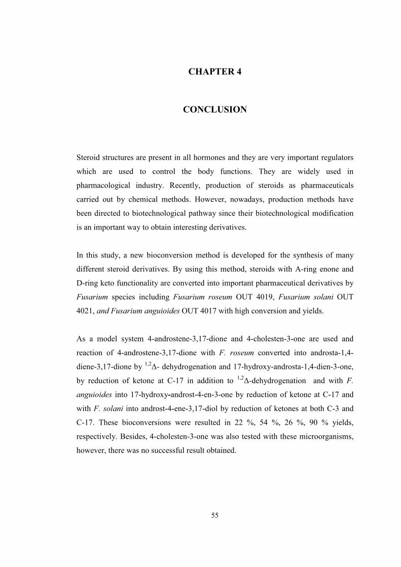
70
CHAPTER 4
CONCLUSION
Steroid structures are present in all hormones and they are very important regulators
which are used to control the body functions. They are widely used in
pharmacological industry. Recently, production of steroids as pharmaceuticals
carried out by chemical methods. However, nowadays, production methods have
been directed to biotechnological pathway since their biotechnological modification
is an important way to obtain interesting derivatives.
In this study, a new bioconversion method is developed for the synthesis of many
different steroid derivatives. By using this method, steroids with A-ring enone and
D-ring keto functionality are converted into important pharmaceutical derivatives by
Fusarium species including Fusarium roseum OUT 4019, Fusarium solani OUT
4021, and Fusarium anguioides OUT 4017 with high conversion and yields.
As a model system 4-androstene-3,17-dione and 4-cholesten-3-one are used and
reaction of 4-androstene-3,17-dione with F. roseum converted into androsta-1,4-
diene-3,17-dione by 1,2∆- dehydrogenation and 17-hydroxy-androsta-1,4-dien-3-one,
by reduction of ketone at C-17 in addition to 1,2∆-dehydrogenation and with F.
anguioides into 17-hydroxy-androst-4-en-3-one by reduction of ketone at C-17 and
with F. solani into androst-4-ene-3,17-diol by reduction of ketones at both C-3 and
C-17. These bioconversions were resulted in 22 %, 54 %, 26 %, 90 % yields,
respectively. Besides, 4-cholesten-3-one was also tested with these microorganisms,
however, there was no successful result obtained.
55
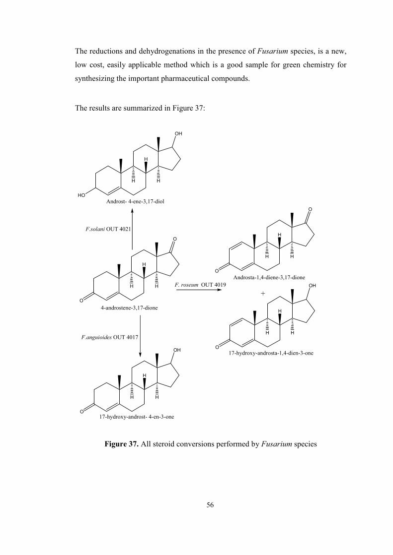
71
The reductions and dehydrogenations in the presence of Fusarium species, is a new,
low cost, easily applicable method which is a good sample for green chemistry for
synthesizing the important pharmaceutical compounds.
The results are summarized in Figure 37:
O
O
H
H
H
4-androstene-3,17-dione
O
O
H
H
H
HO
OH
H
H
H
O
OH
H
H
H
O
OH
H
H
H
+
Androst- 4-ene-3,17-diol
Androsta-1,4-diene-3,17-dione
17-hydroxy-androsta-1,4-dien-3-one
17-hydroxy-androst- 4-en-3-one
F.solani OUT 4021
F.anguioides OUT 4017
F. roseum OUT 4019
Figure 37. All steroid conversions performed by Fusarium species
56
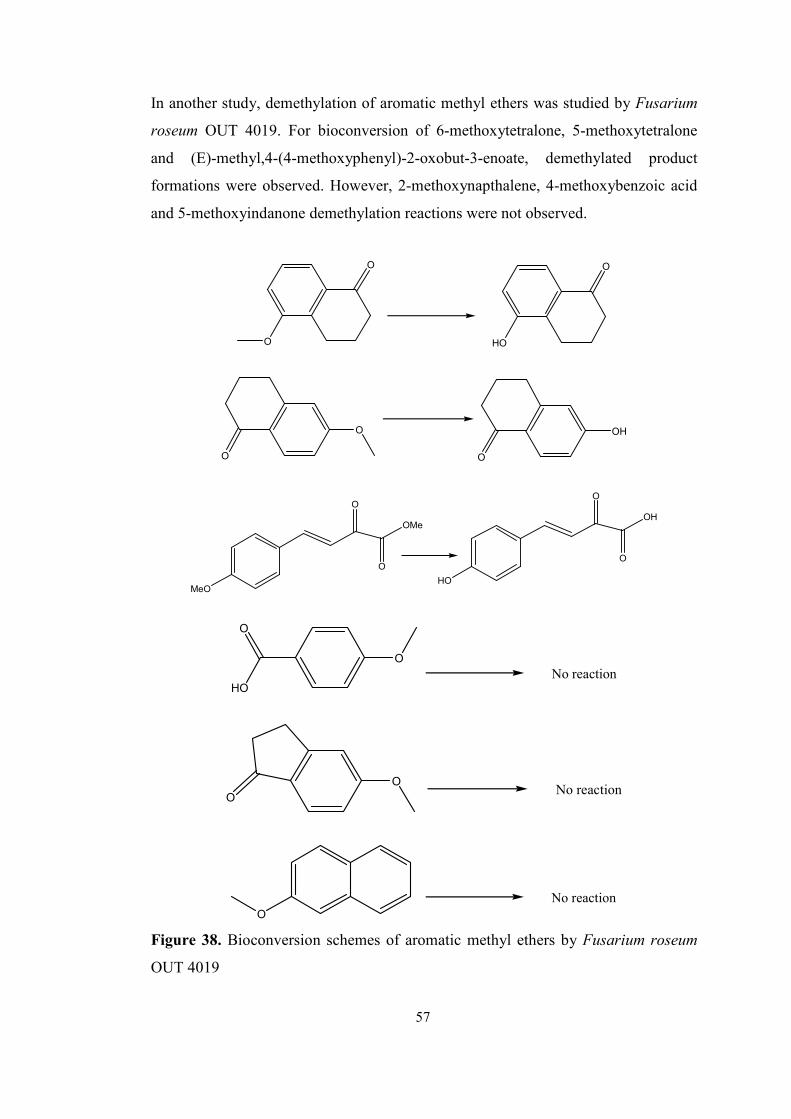
72
In another study, demethylation of aromatic methyl ethers was studied by Fusarium
roseum OUT 4019. For bioconversion of 6-methoxytetralone, 5-methoxytetralone
and (E)-methyl,4-(4-methoxyphenyl)-2-oxobut-3-enoate, demethylated product
formations were observed. However, 2-methoxynapthalene, 4-methoxybenzoic acid
and 5-methoxyindanone demethylation reactions were not observed.
O
O
HO
O
O
O
OH
O
MeO
O
O
OMe
HO
O
O
OH
O
HO
O
No reaction
No reactionO
O
No reactionO
Figure 38. Bioconversion schemes of aromatic methyl ethers by Fusarium roseum
OUT 4019
57
73
REFERENCES
[1] The World Wide Web; http://en.wikipedia.org/ last viewed 17.12.2007
[2] Kun, L. Y., Microbial Biotechnology Principles and Applications, second edition,
World Scientific, Singapore, 2006.
[3] Anke, T., Fungal Biotechnology, Chapman& Hall, London, 1997.
[4] Koskinen, A. M. P., Enzymatic Reactions in Organic Media, Blackie Academic
& Professional, Glasgow, 1996.
[5] Bains, W., Biotechnology form A to Z, third edition, Oxford University Press,
New York, 2004.
[6] Mahato, S. B., Garai, S., Steroids, 1997, 62, 332-345.
[7] Pajic, T., Vitas, M., Zigon, D., Pavko, A., Kelly, S. L., Komel, R., Yeast, 1999,
15, 639–645.
[8] Swizdor, A., Kolek, T., Steroids, 2005, 70, 817–824.
[9] Neslihanoğlu, R., Syntheses of Bioactive Compounds Using Biotechnological
Methods: Fungal Culture Mediated Syntheses of Chiral α-hydroxy Ketones and
Diols, Biotechnology Thesis, September-2001, METU.
[10] Mandal, D., Ahmadb, A., Khanb, M.I., Kumar, R., Journal of Molecular
Catalysis A: Chemical, 2002, 181, 237–241.
[11] Verdin, A., Saharaqui, A. L., Laruelle, F., Grandmougin-Ferjani, A., Durand, R.,
Mycological research, 2006, 110, 4, 479 – 484.
[12] Daiber, A., Shoun, H., Ullrich, V., Journal of Inorganic Biochemistry, 2005, 99,
185–193.
58
74
[13] Saunders, R. P., Hardman, R., Biotechnology and Bioengineering, 1985, 27,
825-831.
[14] Swizdor, A., Szpineter, A., Kolek, T., Journal of Molecular Catalysis B:
Enzymatic, 2006, 42, 10–13
[15] Cai, W., Li, J., Zhang, Z., Journal of Hazardous Materials, 2007, 148, 38–42.
[16] Sua, J., Xua, J., Lu, W., Lin, G., Journal of Molecular Catalysis B: Enzymatic,
2006, 38, 113–118.
[17] Swizdor, A., Kolek, T., Journal of Steroid Biochemistry and Molecular Biology,
1998, 67, No. 1, 63-69.
[18] The World Wide Web; http://www.chem.qmul.ac.uk/ last viewed 21.12.2007
[19] Briggs, M. H., Brotherton, J., Steroid Biochemistry and Pharmacology,
Academic Press, New York, 1970.
[20] Deacon, J., Fungal biology, fourth edition, Blackwell Publishing, 2006.
[21] Simmonds, R. J., Chemistry of Biomolecules: An Introduction, The Royal
Society of Chemistry, Cambridge, 1992.
[22] Solomons, T. W. G., Organic Chemistry, fifth edition, Wiley & Sons, Inc.,
Toronto, 1992.
[23] Donova, M. V., Applied Biochemistry and Microbiology, 2007, 43, No. 1, 1–14.
[24] Berg, J. M., Tymoczko, J. L., Stryer, L., Clarke, N. D., Biochemistry, fifth
edition, W. H. Freman and Company, New York, 2002.
[25] Voishvillo, N. E., Andryushina, V. A., Savinova, T. S., Stytsenko, T. S.,
Vasileva, N. A., Turova, T. P., Kolganova, T. V., Skryabin,K. G., Applied
Biochemistry and Microbiology, 2003, 39, No. 2, 152-157.
59
75
[26] Koskinen, A., Asymmetric Synthesis of Natural Products, John Wiley & Sons,
Chichester , 1993.
[27] Kreuzer, H., Massey, A., Biology and Biotechnology, Science, Applications and
Issues, ASM Press, Washington D.C., 2005.
[28] Vollhardt K. P. C., Schore, N. E., Organic Chemistry, Structure and Function,
fifth edition, W. H. Freeman and Company, New York, 2007.
[29] Numazawa, M., Handa, W., Matsuzaki, H., Journal of Steroid Biochemistry &
Molecular Biology, 2006, 101, 239–245.
[30] The World Wide Web; http://web.indstate.edu/ last viewed 25.12.2007
[31] Fokina, V. V., Arinbasarova, A. Y., Zubov, L. A., Lozinsky, V.L.,
Koshcheenko, A.K., Applied Biochemistry and Microbiology, 1995, 31(2), 184-189.
[32] Prave, P., Fundamentals of Biotechnology, 1987, VCH, New York.
[33] Numazawa, M., Handa W., Chem. Pharm. Bull., 2006, 54, 4, 554-556.
[34] Moses, V., Cape, R.E., Biotechnology, The Science and The Business, Harwood
Academic Publishers, New York, 1991.
[35] Fernandes, P., Cruz, A., Angelova, B., Pinheiro, H. M., Cabral, J. M. S., Enzyme
and Microbial Technology, 2003, 32, 688–705.
[36] He, X., Liu, B., Wang, G., Wang, X., Su, L., Qu, G., Yao, X., Journal of Steroid
Biochemistry and Molecular Biology, 2006, 100, 87–94.
[37] Dorfman, R.I., Ungar, F., Metabolism of Steroid Hormones, Academic Press,
New York, 1965.
[38] Manosroi, J., Manosroi, A., Saraphanchotiwittaya, A., Abe, M., Journal of
Chemical Technology and Biotechnology, 1999, 74, 364-370.
[39] Glazer, A. N., Nikaido, H., Microbial Biotechnology Fundamentals of Applied
Microbiology, W.H. Freeman and Company, 1998.
60
76
[40] Moore-Landecker, E., Fundamentals of the Fungi, fourth edition, Prentice Hall,
1996.
[41] Romano, A., Romano, D., Ragg, E., Costantino, F., Lenna, R., Gandolfi, R.,
Molinari, F., Steroids, 2006, 71, 429–434.
[42] Adham, N. Z., El-Hady, A. A., Naim, N., Process Biochemistry, 2003, 38, 897-
902.
[43] Holland, H. L., Lakshmaiah, G., Journal of Molecular Catalysis B: Enzymatic,
1999, 6, 83–88.
[44] Hanson, R. J., Hunter, A. C., Phytochemistry, 1998, 49, No.8, 2359-2362.
[45] Veizina, C., Sehgal, S. N., Singh, K., General Microbiology, 1962, Ayerst
Research Laboratories, Canada.
[46] Sedlaczek, L., Dlugonski, J., Jaworski, A., Applied Microbiology and
Biotechnology, 1984, 20, 166-169.
[47] Somal, P., Chopra, C.L., Applied Microbiol. and Biotech., 1985, 21, 5, 267-269.
[48] Cotillon, A., Morfin, R., Journal of Steroid Biochemistry and Molecular
Biology, 1999, 68, 229-237.
[49] Huszcza E., Dmochowska-Gladysz, J., Phytochemistry, 2003, 62, 155–158.
[50] Huszcza, E., Dmochowska-Gladysz, J., Journal of Basic Microbiology, 2003,
43, 2, 113–120.
[51] Bartmanska, A., Dmochowska-Gladysz, J., Enzyme and Microbial Technology,
2007, 40, 1615–1621.
[52] Songtao, B., lianxiang, D., Liming, Z., Fuping, L., Process Biochemistry, 2005,
40, 3309–3313.
61
77
[53] Smolders, A.J.J., Pinheiro, H. M., Noronha, P., Cabral, J. M. S., Biotechnology
and Bioengineering, 1991, 38, 1210-1217.
[54] Constantinides, A., Biotechnology and Bioengineering, 1980, 22, 119-136.
[55] Manosroi, J., Sripalakit, P., Manosroi, A., Journal of Chemical Technology and
Biotechnology, 1998, 73, 203-210.
[56] Boeren, S., Laane, C., Biotechnology and Bioengineering, 1987, 29, 305-309.
[57] Llanes, N., Pendas, J., Falero, A., Perez, C., Hung, B. R., Moreira, T., Journal of
Steroid Biochemistry & Molecular Biology, 2002, 80, 131–133.
[58] Sripalakit, P., Wichai, U., Saraphanchotiwitthaya, A., Journal of Molecular
Catalysis B: Enzymatic, 2006, 41, 49–54.
[59] Faber, K., Biotransformations in Organic Chemistry, A textbook, 4th edition,
Springer-Verlag, Berlin, 2000.
[60] Patil, S., Shukla, A., Shinde, K., Banerjee, T., Letters in Applied Microbiology,
2002, 35, 95–97
[61] Yazdi, M. T., Zanjanian, S. M., Faramarzi, M. A., Amini, M., Amani, A., Abdi,
K., Arch. Pharm. Chem. Life Sci., 2006, 339, 473 – 476.
[62] Faramarzi,M. A., Yazdi, M. T., Amini, M., Mohseni, F. A., Zarrini, G., Amani,
A., Shafiee, A., World Journal of Microbiology and Biotechnology, 2004, 20, 657–
660.
[63] Paul, E.A., Clark, F.E., Soil Microbiology and Biochemistry, Academic Press,
Inc., California, 1989.
[64] Pitt, J.I., Hocking, A.D., Fungi and Food Spoilage, second edition, Blackie
Academic & Professional, Cambridge, 1997.
[65] Howard, D. H., Pathogenic Fungi in Humans and Animals, 2nd edition, Marcel
Dekker, 2003.
62
78
[66] Devlin, M.T., Textbook of Biochemistry with Clinical Correlations, sixth
edition, Wiley-Liss., New York, 2006.
[67] Atlas, R.M., Handbook of microbial media, CRC press, Boca Raton, Fla., 2004.
[68] The World Wide Web; http://www-biol.paisley.ac.uk/ last viewed 25.12.2007
[69] Jung, M.E, Lyster, M. A., Organic synthesis, Coll., 1988, 6, 353; 1979, 59, 35
[70] Kemperman, G.J., Roeters, T.A., and Hilberink, P.W., European Journal of
Organic Chemistry, 2003, 1681-1686
63
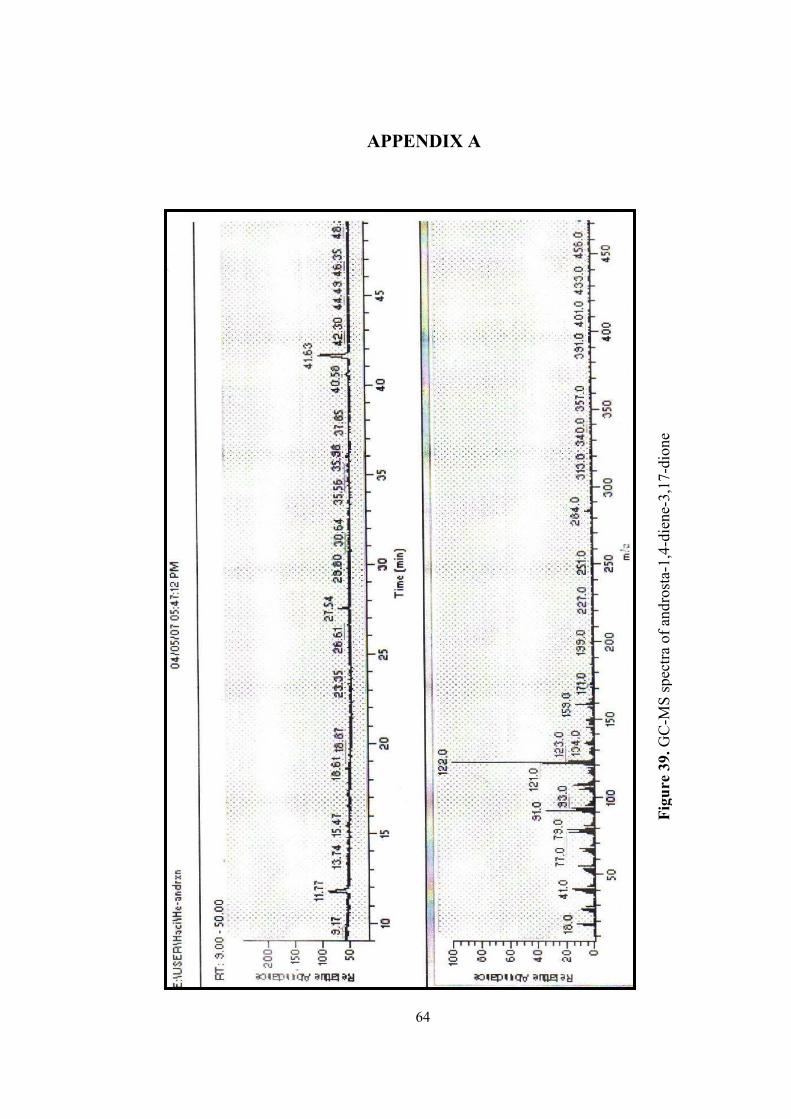
79
Figure 39.
GC
-MS
spe
ctra
of
andr
osta
-1,4
-die
ne-3
,17-
dion
e
APPENDIX A
64
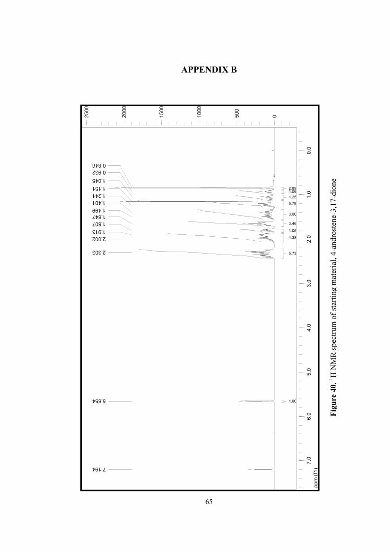
80
ppm (f1)
0.0
1.0
2.0
3.0
4.0
5.0
6.0
7.0
0500
1000
1500
2000
2500
7.194
5.654
2.303
2.002
1.913
1.807
1.647
1.499
1.401
1.241
1.151
1.045
0.932
0.846
1.00
5.73
4.38
1.05
3.46
3.00
5.79
1.25
1.092.89
Figure 40.
1 H N
MR
spe
ctru
m o
f st
arti
ng m
ater
ial, 4
-and
rost
ene-
3,17
-dio
ne
APPENDIX B
65
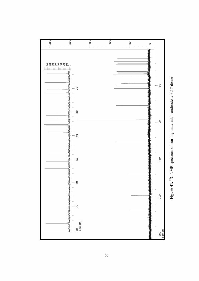
81
ppm (f1)
50
100
150
200
250
050
100
150
200
250
ppm (f1)
20
30
40
50
60
70
80
01020304050607080
Figure 41.
13C
NM
R s
pect
rum
of
star
ting
mat
eria
l, 4
-and
rost
ene-
3,17
-dio
ne
66
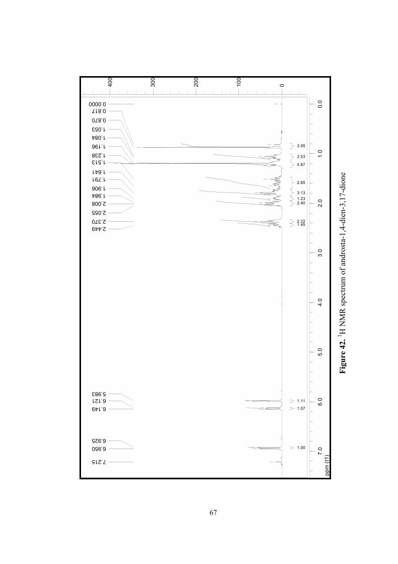
82
ppm (t1)
0.0
1.0
2.0
3.0
4.0
5.0
6.0
7.0
0100
200
300
400
7.215
6.950
6.925
6.149
6.121
5.983
2.449
2.370
2.055
2.008
1.984
1.906
1.791
1.641
1.513
1.238
1.196
1.084
1.053
0.870
0.817
0.0000
1.00
1.07
1.11
1.452.22
2.401.23
3.13
2.85
5.87
2.53
3.95
Figure 42.
1 H N
MR
spe
ctru
m o
f an
dros
ta-1
,4-d
ien-
3,17
-dio
ne
67
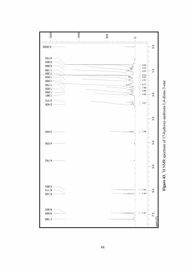
83
Figure 43.
1 H N
MR
spe
ctru
m o
f 17
-hyd
roxy
-and
rost
a-1,
4-di
ene-
3-on
e
ppm (t1)
0.0
1.0
2.0
3.0
4.0
5.0
6.0
7.0
0500
1000
1500
7.188
6.949
6.923
6.138
6.113
5.963
4.742
4.030
3.545
2.405
2.312
1.961
1.894
1.820
1.795
1.695
1.604
1.384
1.180
0.999
0.904
0.743
-0.0000
1.43
1.42
1.51
1.58
3.37
2.99
3.88
1.96
6.77
4.31
3.618.15
5.07
3.72
5.93
68
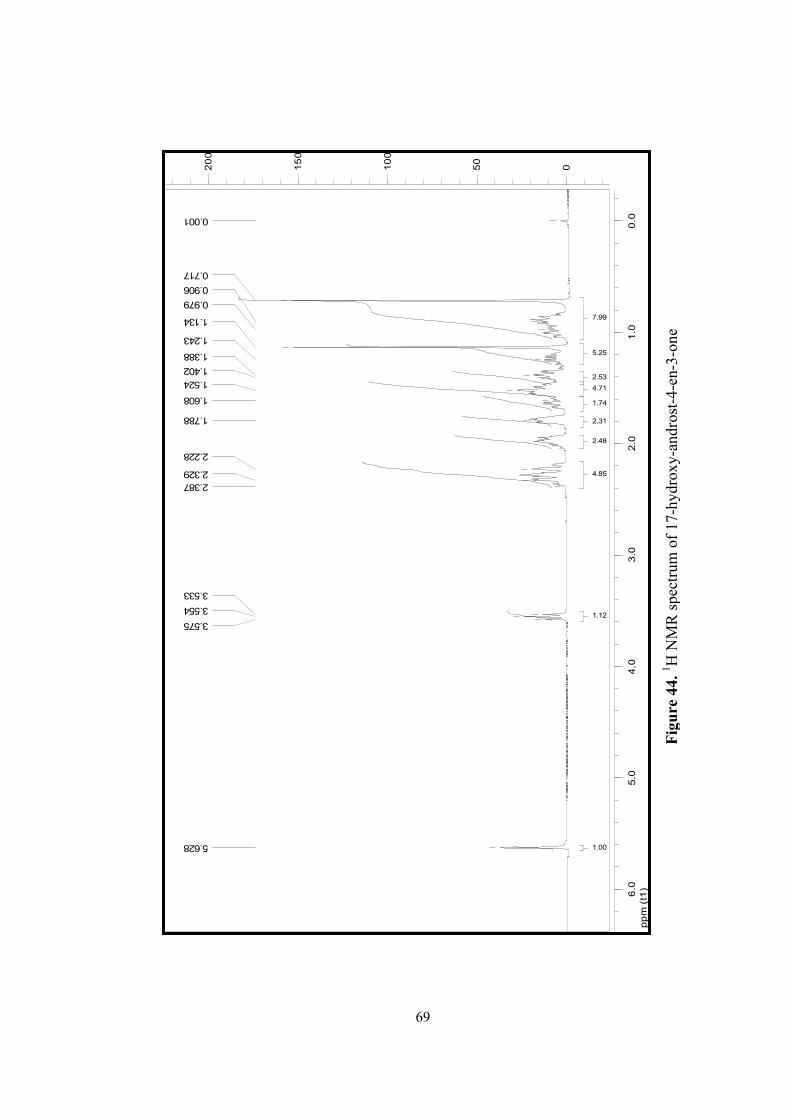
84
ppm (t1)
0.0
1.0
2.0
3.0
4.0
5.0
6.0
050
100
150
200
5.628
3.575
3.554
3.533
2.387
2.329
2.228
1.788
1.608
1.524
1.402
1.388
1.243
1.134
0.979
0.906
0.717
0.001
1.00
1.12
4.85
2.48
2.31
1.74
4.71
2.53
5.25
7.99
Figure 44.
1 H N
MR
spe
ctru
m o
f 17
-hyd
roxy
-and
rost
-4-e
n-3-
one
69
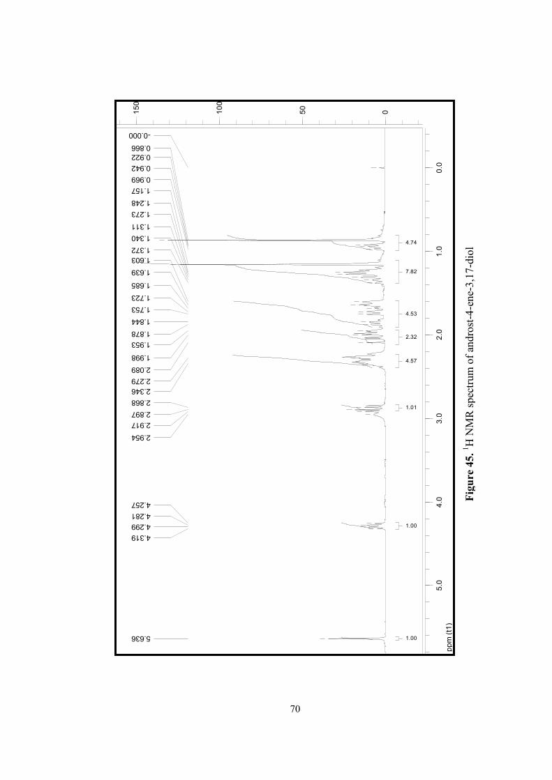
85
ppm (t1)
0.0
1.0
2.0
3.0
4.0
5.0
050
100
150
5.636
4.319
4.299
4.281
4.257
2.954
2.917
2.897
2.868
2.346
2.279
2.089
1.998
1.953
1.878
1.844
1.753
1.723
1.685
1.639
1.603
1.372
1.340
1.311
1.273
1.248
1.157
0.969
0.942
0.922
0.866
-0.0001.00
1.00
4.57
2.32
4.53
7.82
1.01
4.74
Figure 45.
1 H N
MR
spe
ctru
m o
f an
dros
t-4-
ene-
3,17
-dio
l
70
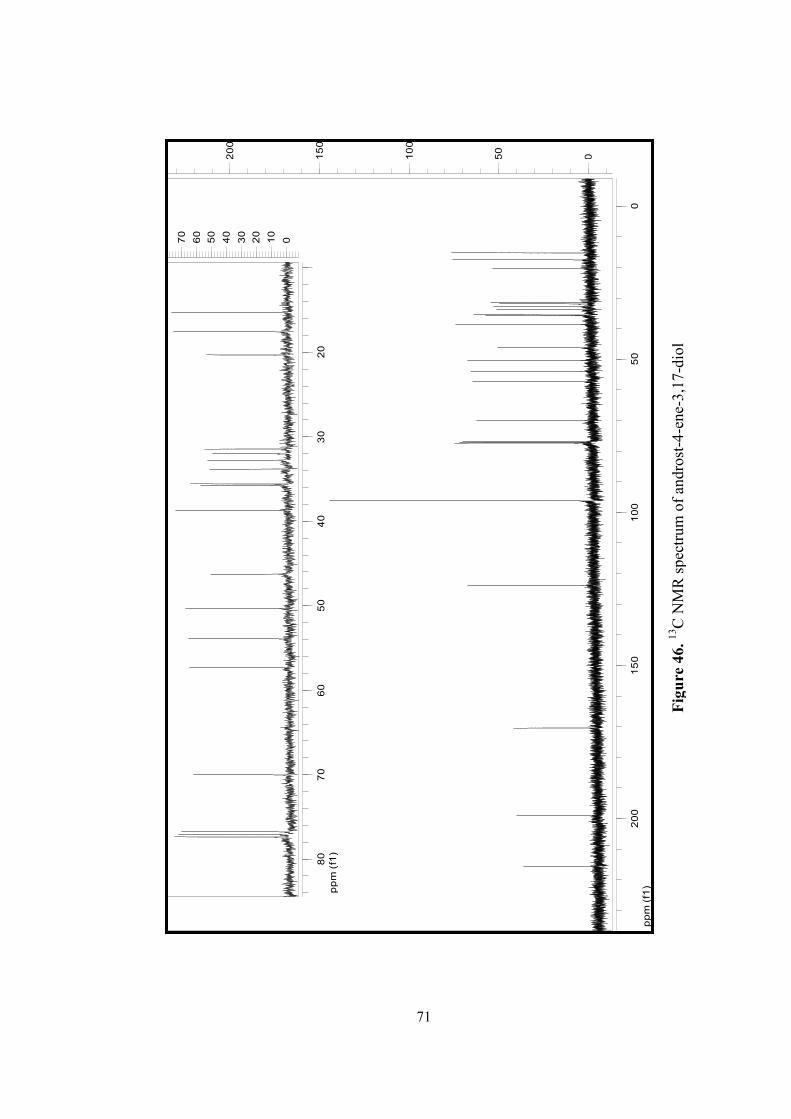
86
ppm (f1)
050
100
150
200
050
100
150
200
ppm (f1)
20
30
40
50
60
70
80
010
20
30
40
50
60
70
Figure 46.
13C
NM
R s
pect
rum
of
andr
ost-
4-en
e-3,
17-d
iol
71