Embed Size (px)
Citation preview
www.wjpls.org 80
Tauheed et al . World Journal of Pharmaceutical and Life Sciences
BIOTRANSFORMATION OF STEROLS BY ACTINOBACTERIA TO PRODUCE
PHARMACEUTICAL PRODUCT
Syeda Fareeha Tauheed1*
and Sikandar Ali1
1Institute of Industrial Biotechnology, GC University Lahore, Pakistan.
Article Received on 16/04/2017 Article Revised on 05/05/2017 Article Accepted on 26/05/2017
INTRODUCTION
Klaus Kieslich elaborate the phenomenon of
biotransformation as ―chemical reactions by
microorganisms or enzymes‖ (Kieslich, 1985). Later this
definition of biotransformation was updated, and
primarily focus on knowledge that related with the
compounds of microbial transformations of steroid in
case of differentiate the phenomenon of microbial
transformation from that of bioconversion and also biodegradation (Lilly, 1984).
Previously, for the preparation of compounds many
chemical processes are used but now microbial
transformation is more significant arsenal for the
synthesis of product, which may have been complicated
otherwise to produce by ordinary synthetic processes.
For the bioconversion of steroids this transformation has
been significantly used (Charney and Herzong, 1976). In
many past years, Biotransformation is an introductive
tool in pharmaceutical industry for the synthesis of many
drugs, hormones and antibiotics (Aharnowetz and Choe, 1981). The main benefit of the microbiological
conversion is to nullify the considerable chemical
process lie in the area of their mild reaction conditions.
The main aim of biotransformation is to emphasis on the
precise reaction with minimum of side reaction. In order
to produce some functional products, the selection of
biocatalysts is more significant approach under relatively
gentle conditions contrast to its chemical catalyst
equivalent to make biocatalysts which are more
fascinating and revolutionary (Mark and Flashman,
2016).
These transformations have significant role in the region of steroid and antibiotics. In common contingency, the
decrement of cholesterol by microorganism is we defined
and famous process. In 1913, Sohngen and et al
elaborate the process of degradation of cholesterol
including microorganism such as Nocarida (Turfitt,
1944) and Mycobacterium from soil and Aerobacter
aerogenes and Pseudomonas jaegeri. The current study
on this process of degradation of cholesterol is very
limited and has ability to improve this knowledge. A
well-known tracer experiment indicates us that divide the
ring might pave the way the cleavage of steroid chain (Stadtman, 1954). The problem of information about the
cleavage of cholesterol is still intricate, doubtful and
unsettled. In few years, many research group have been
performed experiments to break the selective cleavage of
Research Article ISSN 2454-2229 wjpls, 2017, Vol. 3, Issue 4, 80-93
World Journal of Pharmaceutical and Life Sciences WJPLS
www.wjpls.org SJIF Impact Factor: 4.223
*Corresponding Author: Syeda Fareeha Tauheed
Institute of Industrial Biotechnology, GC University Lahore, Pakistan.
ABSTRACT
This review consists two parts. In the first part of review we enhance the main aim of the development of
pharmaceutical industry by introducing micro biotechnological process on industrial scale and wipeout the many-
level chemical coalescence. Also, this article elaborates on the current development of micro sterols
biotransformation system. Actinobacteria, which has considerable importance for the production of hormonal drugs
(pharmaceutical product) also has main role as a catalyst in the steroid bioconversion and the development of biotechnology. The ability to activate the transformation process of sterol substrate in a broad way, it is feasible to
expect the effective use of these microorganism in the advancement of new technologies on the production of
pharmaceutical steroids substrate. This article is first attempt to channelize the data on the ability of Actinobacteria
to activate the distinct reaction of many biotransformation of steroid such as hydroxylation, reduction and
introduction of double bonds, oxidation of steroids, reduction of hydrocarbon (ketones) and degradation, with
focus on the importance of biotechnological process and analysis of steroid conversion over the last decade year.
The second part of this review precisely emphasis in the location of new enzymatic approaches such as cleavage of
steroid side chain.
KEYWORDS: Steroid, Microbial transformation, Side-chain degradation, Hydroxylation Dehydrogenation,
Selective cleavage.
www.wjpls.org 81
Tauheed et al . World Journal of Pharmaceutical and Life Sciences
sterol chain by microorganism and to prepare the sterol
hormones from steroid but none of them are successful.
So, it is great challenging problem for the research to
breakdown the cleavage of steroid chain and for this
purpose screening of microorganism is carried out
(Arima, 1969). This review also explains the potential to break the cholesterol by Actinobacteria and the
degradation as well.
Steroid The scope of steroids is expanded in the kingdom of
animals and plants. The fundamental architecture of
steroid constitutes of seventeen atoms of carbon designed
in the form of a perhydrocyclopentanophenanthrene.
These compounds alter significantly in architecture and
include essential compounds such as cholesterol, insect
molting hormones, corticoid hormones, sex hormones,
antibiotics, vitamin D, cardiac aglycone and bile acids (Bhatti and Khera, 2012).
Steroid biotransformation is a multimillion dollar
industry and inhabited a considerable position among the
preparation of pharmaceutical products that are mainly
used for curing and averting diseases of different groups
in endocrinology, oncology, rheumatology, gynecology,
etc. (Fernandes et al., 2003). A distinct type of steroids is
majorly utilized as anti-inflammatory, diuretic, anabolic
as well as contraceptive and anti-androgenic. Some
steroids are used as immunosuppressive while others act as presentational and anticancer agents, and several other
applications (Ahmed et al., 1992). These steroids are also
considerable to utilized in the cure of breast cancer as an
also in prostate gland cancer (Diaz-Chico et al., 2007).
Their role in the nursing of hyprcortisolism also known
as adrenal insufficiency (Hohnston, 1987) are significant
as a replacement agent. Considerably for the prevention
of coronary heart diseases (Frye and Leonard, 1987) anti-
fungal (Chung et al., 1998) agents are widely used. Their
important used in the cure of AIDS, these steroids are
used in anti-obesity agent as an active ingredient.
Currently, in modern research a glycoside which as
steroid executed anti-viral activity on herpes virus
(Arthan et al., 2002). The scope of new steroids in
business point of view is limited in now days. Whereas their scope can be enhanced to obtain desired metabolites
which is active ingredient of novel steroids. In
pharmaceutical industry, production of steroids has great
importance by biotechnology. According to commerce
point of view steroid production is second significant
source of antibiotic production (Brown, 1984).
Moreover, special microbial transformation steps have
been involved for the production of novel hormonal
steroids as drugs. The relatively broad nomenclature of
efficient steroid drugs is continually expanding. Highly
complex structure of steroids molecules renders the use
of biocatalysts for the production of pharmacologically important steroid drug intermediates of note, several
preparations administered for life-saving indications
have no non-steroid analogues. Large scale production of
hormonal steroid drugs is based on combining both of
biotechnology (i.e. microbial technology) and chemical
products (Fernandes, 2003). There is a specific rule in
which whole cells are used to develop the
biotechnological equipment. The benefit of this
biotechnology is that, it is more economical then
enzymes (considering isolation, purification and
stabilization procedures).
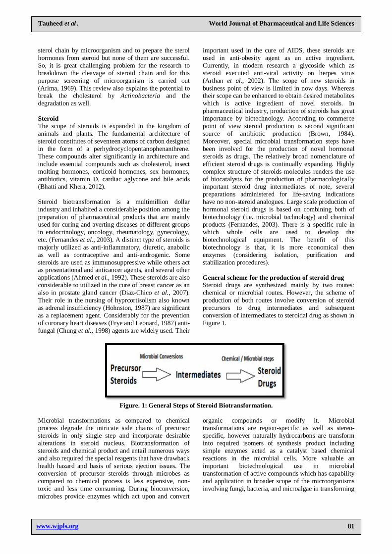
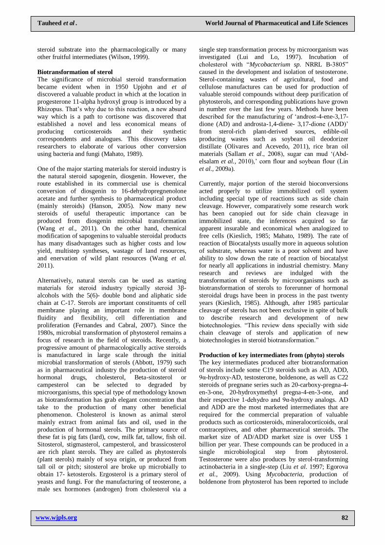
General scheme for the production of steroid drug
Steroid drugs are synthesized mainly by two routes:
chemical or microbial routes. However, the scheme of
production of both routes involve conversion of steroid
precursors to drug intermediates and subsequent
conversion of intermediates to steroidal drug as shown in
Figure 1.
Figure. 1: General Steps of Steroid Biotransformation.
Microbial transformations as compared to chemical process degrade the intricate side chains of precursor
steroids in only single step and incorporate desirable
alterations in steroid nucleus. Biotransformation of
steroids and chemical product and entail numerous ways
and also required the special reagents that have drawback
health hazard and basis of serious ejection issues. The
conversion of precursor steroids through microbes as
compared to chemical process is less expensive, non-
toxic and less time consuming. During bioconversion,
microbes provide enzymes which act upon and convert
organic compounds or modify it. Microbial transformations are region-specific as well as stereo-
specific, however naturally hydrocarbons are transform
into required isomers of synthesis product including
simple enzymes acted as a catalyst based chemical
reactions in the microbial cells. More valuable an
important biotechnological use in microbial
transformation of active compounds which has capability
and application in broader scope of the microorganisms
involving fungi, bacteria, and microalgae in transforming
www.wjpls.org 82
Tauheed et al . World Journal of Pharmaceutical and Life Sciences
steroid substrate into the pharmacologically or many
other fruitful intermediates (Wilson, 1999).
Biotransformation of sterol
The significance of microbial steroid transformation
became evident when in 1950 Upjohn and et al discovered a valuable product in which at the location in
progesterone 11-alpha hydroxyl group is introduced by a
Rhizopus. That‘s why due to this reaction, a new absurd
way which is a path to cortisone was discovered that
established a novel and less economical means of
producing corticosteroids and their synthetic
correspondents and analogues. This discovery takes
researchers to elaborate of various other conversion
using bacteria and fungi (Mahato, 1989).
One of the major starting materials for steroid industry is
the natural steroid sapogenin, diosgenin. However, the route established in its commercial use is chemical
conversion of diosgenin to 16-dehydropregnenolone
acetate and further synthesis to pharmaceutical product
(mainly steroids) (Hanson, 2005). Now many new
steroids of useful therapeutic importance can be
produced from diosgenin microbial transformation
(Wang et al., 2011). On the other hand, chemical
modification of sapogenins to valuable steroidal products
has many disadvantages such as higher costs and low
yield, multistep syntheses, wastage of land resources,
and enervation of wild plant resources (Wang et al. 2011).
Alternatively, natural sterols can be used as starting
materials for steroid industry typically steroid 3β-
alcohols with the 5(6)- double bond and aliphatic side
chain at C-17. Sterols are important constituents of cell
membrane playing an important role in membrane
fluidity and flexibility, cell differentiation and
proliferation (Fernandes and Cabral, 2007). Since the
1980s, microbial transformation of phytosterol remains a
focus of research in the field of steroids. Recently, a
progressive amount of pharmacologically active steroids is manufactured in large scale through the initial
microbial transformation of sterols (Abbott, 1979) such
as in pharmaceutical industry the production of steroid
hormonal drugs, cholesterol, Beta-sitosterol or
campesterol can be selected to degraded by
microorganisms, this special type of methodology known
as biotransformation has grab elegant concentration that
take to the production of many other beneficial
phenomenon. Cholesterol is known as animal sterol
mainly extract from animal fats and oil, used in the
production of hormonal sterols. The primary source of these fat is pig fats (lard), cow, milk fat, tallow, fish oil.
Sitosterol, stigmasterol, campesterol, and brassicosterol
are rich plant sterols. They are called as phytosterols
(plant sterols) mainly of soya origin, or produced from
tall oil or pitch; sitosterol are broke up microbially to
obtain 17- ketosterols. Ergosterol is a primary sterol of
yeasts and fungi. For the manufacturing of teosterone, a
male sex hormones (androgen) from cholesterol via a
single step transformation process by microorganism was
investigated (Lui and Lo, 1997). Incubation of
cholesterol with ―Mycobacterium sp. NRRL B-3805‖
caused in the development and isolation of testosterone.
Sterol-containing wastes of agricultural, food and
cellulose manufactures can be used for production of valuable steroid compounds without deep purification of
phytosterols, and corresponding publications have grown
in number over the last few years. Methods have been
described for the manufacturing of ‗androst-4-ene-3,17-
dione (AD) and androsta-1,4-diene- 3,17-dione (ADD)‘
from sterol-rich plant-derived sources, edible-oil
producing wastes such as soybean oil deodorizer
distillate (Olivares and Acevedo, 2011), rice bran oil
materials (Sallam et al., 2008), sugar can mud ‗(Abd-
elsalam et al., 2010),‘ corn flour and soybean flour (Lin
et al., 2009a).
Currently, major portion of the steroid bioconversions
acted properly to utilize immobilized cell system
including special type of reactions such as side chain
cleavage. However, comparatively some research work
has been canopied out for side chain cleavage in
immobilized state, the inferences acquired so far
apparent insurable and economical when analogized to
free cells (Kieslich, 1985; Mahato, 1989). The rate of
reaction of Biocatalysts usually more in aqueous solution
of substrate, whereas water is a poor solvent and have
ability to slow down the rate of reaction of biocatalyst for nearly all applications in industrial chemistry. Many
research and reviews are indulged with the
transformation of steroids by microorganisms such as
biotransformation of sterols to forerunner of hormonal
steroidal drugs have been in process in the past twenty
years (Kieslich, 1985). Although, after 1985 particular
cleavage of sterols has not been exclusive in spite of bulk
to describe research and development of new
biotechnologies. ―This review dens specially with side
chain cleavage of sterols and application of new
biotechnologies in steroid biotransformation.‖
Production of key intermediates from (phyto) sterols
The key intermediates produced after biotransformation
of sterols include some C19 steroids such as AD, ADD,
9α-hydroxy-AD, testosterone, boldenone, as well as C22
steroids of pregnane series such as 20-carboxy-pregna-4-
en-3-one, 20-hydroxymethyl pregna-4-en-3-one, and
their respective 1-dehydro and 9α-hydroxy analogs. AD
and ADD are the most marketed intermediates that are
required for the commercial preparation of valuable
products such as corticosteroids, mineralocorticoids, oral
contraceptives, and other pharmaceutical steroids. The market size of AD/ADD market size is over US$ 1
billion per year. These compounds can be produced in a
single microbiological step from phytosterol.
Testosterone were also produces by sterol-transforming
actinobacteria in a single-step (Liu et al. 1997; Egorova
et al., 2009). Using Mycobacteria, production of
boldenone from phytosterol has been reported to include
www.wjpls.org 83
Tauheed et al . World Journal of Pharmaceutical and Life Sciences
two steps via intermediate of AD and subsequent 1-
dehydrogenation of AD by Fusarium sp.
Apart from C19 steroids, valuable 23, 24-dinorcholane
derivatives were achieved from sterols biotransformation
(Andor et al., 2006). These compounds are the
significant precursors for corticosteroid synthesis. For example, 9α-hydroxy-C22 steroids can be easily
converted to C21 corticosteroids by oxidative
decarboxylation (Toro and Ambrus, 1990). Apparent
advantages of microbial transformation of valuable
precursors i.e. C19 and C22 from phytosterols are shorter
process, environmentally friendly and low-cost
procedures. However, the relatively low productivity and
insufficient selectivity of the strains often remain the
bottleneck in their industrial applications. There are
many strains of microorganisms that have been described
as biocatalysts of sterol biotransformation, e.g.,
Arthrobacter spp. (Arthrobacter oxydans 317 AL, Arthrobacter rubbelus), Brevibacterium spp.,
Pseudomonas spp., and Rhodococcus spp., but their use
necessitates the addition of inhibitors to avoid steroid
nucleus degradation (Abd-elsalam et al., 2010; Tong and
Dong, 2009).
Over the past 100 years, efforts have been made to
discover organisms that are capable to convert
phytosterols to key steroid precursor efficiently. For
example, an AD-producing Aspergillus oryzae NCIM
634 strain was selected as an efficient organism (Malaviya and Gomes, 2009); a strain isolated by soil
and identified as Fusarium moniliforme, converted
phytosterol present in corn-flour and soybean flour to
AD (Lin et al., 2009a); ADD production from
cholesterol was also shown for Chryseobacterium gleum
ATCC 35910 (Chaudhari et al., 2010). However,
Actinobacteria of the genera Mycobacterium and
Rhodococcus considered as the most efficient AD/ADD
producers (Malaviya and Gomes, 2009).
Biotranformation of Sterols by Actinobacteria
Actinobacteria are prominent among the most efficient
biocatalysts of steroid transformation. Actinobacteria are
capable of effecting diverse types of steroid
transformation, such as dehydrogenation, doubles bond
isomerization, oxidation of steroid alcohols,
hydrogenation of unsaturated bonds, reduction of steroid
ketones, deacetylation and last but not least
hydroxylation as well as complete degradation to carbon
dioxide and water or partial decrement of the side chain
(of sterols, cholinic acids, steroids of the pregnane series)
and other reactions. A remarkable number of Actinobacteria is able to degrade various sterol
(Fernandes et al., 2003). A number of strains that are
able to transform the side chains of various sterols, either
as natural or UV mutants blocked in steroid ring
cleavage or requiring inhibitors of steroid ring
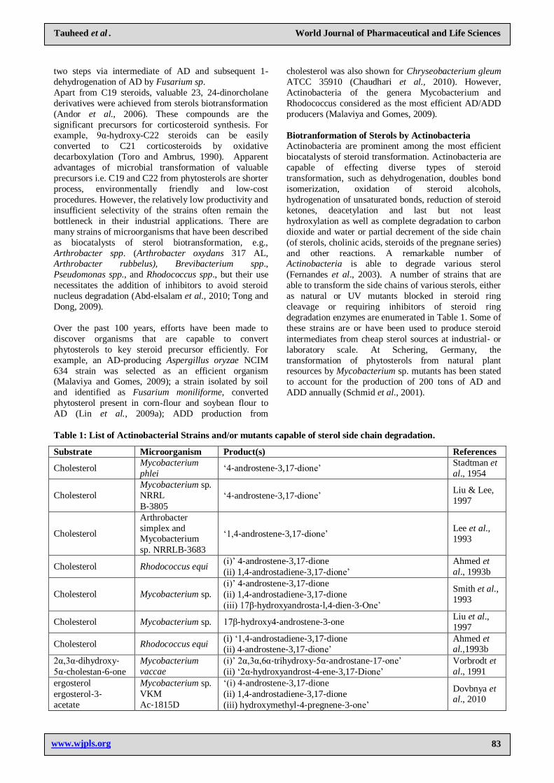
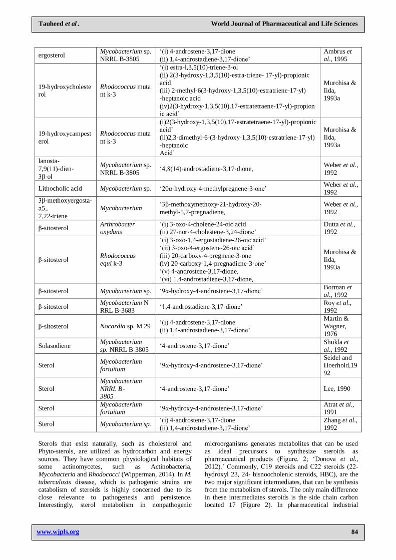
degradation enzymes are enumerated in Table 1. Some of
these strains are or have been used to produce steroid
intermediates from cheap sterol sources at industrial‐ or
laboratory scale. At Schering, Germany, the
transformation of phytosterols from natural plant resources by Mycobacterium sp. mutants has been stated
to account for the production of 200 tons of AD and
ADD annually (Schmid et al., 2001).
Table 1: List of Actinobacterial Strains and/or mutants capable of sterol side chain degradation.
Substrate Microorganism Product(s) References
Cholesterol Mycobacterium
phlei ‗4‐androstene‐3,17‐dione‘
Stadtman et
al., 1954
Cholesterol Mycobacterium sp.
NRRL B‐3805
‗4‐androstene‐3,17‐dione‘ Liu & Lee,
1997
Cholesterol
Arthrobacter
simplex and Mycobacterium
sp. NRRLB‐3683
‗1,4‐androstene‐3,17‐dione‘ Lee et al.,
1993
Cholesterol Rhodococcus equi (i)‘ 4‐androstene‐3,17‐dione
(ii) 1,4‐androstadiene‐3,17‐dione‘
Ahmed et
al., 1993b
Cholesterol Mycobacterium sp. (i)‘ 4‐androstene‐3,17‐dione (ii) 1,4‐androstadiene‐3,17‐dione (iii) 17β‐hydroxyandrosta‐l,4‐dien‐3‐One‘
Smith et al.,
1993
Cholesterol Mycobacterium sp. 17β‐hydroxy4‐androstene‐3‐one Liu et al.,
1997
Cholesterol Rhodococcus equi (i) ‗1,4‐androstadiene‐3,17‐dione (ii) 4‐androstene‐3,17‐dione‘
Ahmed et al.,1993b
2α,3α‐dihydroxy‐ 5α‐cholestan‐6‐one
Mycobacterium
vaccae (i)‘ 2α,3α,6α‐trihydroxy‐5α‐androstane‐17‐one‘ (ii) ‗2α‐hydroxyandrost‐4‐ene‐3,17‐Dione‘
Vorbrodt et
al., 1991 ergosterol ergosterol‐3‐ acetate
Mycobacterium sp.
VKM Ac‐1815D
‗(i) 4‐androstene‐3,17‐dione (ii) 1,4‐androstadiene‐3,17‐dione (iii) hydroxymethyl‐4‐pregnene‐3‐one‘
Dovbnya et al., 2010
www.wjpls.org 84
Tauheed et al . World Journal of Pharmaceutical and Life Sciences
ergosterol Mycobacterium sp.
NRRL B‐3805 ‗(i) 4‐androstene‐3,17‐dione (ii) 1,4‐androstadiene‐3,17‐dione‘
Ambrus et
al., 1995
19‐hydroxycholesterol
Rhodococcus muta
nt k‐3
‗(i) estra‐l,3,5(10)‐triene‐3‐ol (ii) 2(3‐hydroxy‐1,3,5(10)‐estra‐triene‐ 17‐yl)‐propionic
acid (iii) 2‐methyl‐6(3‐hydroxy‐1,3,5(10)‐estratriene‐17‐yl)
‐heptanoic acid (iv)2(3‐hydroxy‐1,3,5(10),17‐estratetraene‐17‐yl)‐propion
ic acid‘
Murohisa &
Iida, 1993a
19‐hydroxycampest
erol Rhodococcus muta
nt k‐3
(i)2(3‐hydroxy‐1,3,5(10),17‐estratetraene‐17‐yl)‐propionic
acid‘ (ii)2,3‐dimethyl‐6‐(3‐hydroxy‐1,3,5(10)‐estratriene‐17‐yl)
‐heptanoic Acid‘
Murohisa &
Iida, 1993a
lanosta‐ 7,9(11)‐dien‐ 3β‐ol
Mycobacterium sp.
NRRL B‐3805 ‗4,8(14)‐androstadiene‐3,17‐dione,
Weber et al.,
1992
Lithocholic acid Mycobacterium sp. ‗20α‐hydroxy‐4‐methylpregnene‐3‐one‘ Weber et al.,
1992 3β‐methoxyergosta‐a5,.
7,22‐triene Mycobacterium
‗3β‐methoxymethoxy‐21‐hydroxy‐20‐ methyl‐5,7‐pregnadiene,
Weber et al.,
1992
β‐sitosterol Arthrobacter oxydans
‗(i) 3‐oxo‐4‐cholene‐24‐oic acid (ii) 27‐nor‐4‐cholestene‐3,24‐dione‘
Dutta et al., 1992
β‐sitosterol Rhodococcus
equi k‐3
‗(i) 3‐oxo‐1,4‐ergostadiene‐26‐oic acid‘ ‗(ii) 3‐oxo‐4‐ergostene‐26‐oic acid‘ (iii) 20‐carboxy‐4‐pregnene‐3‐one (iv) 20‐carboxy‐1,4‐pregnadiene‐3‐one‘ ‗(v) 4‐androstene‐3,17‐dione, ‗(vi) 1,4‐androstadiene‐3,17‐dione,
Murohisa &
Iida, 1993a
β‐sitosterol Mycobacterium sp. ‗9α‐hydroxy‐4‐androstene‐3,17‐dione‘ Borman et
al., 1992
β‐sitosterol Mycobacterium N
RRL B‐3683 ‗1,4‐androstadiene‐3,17‐dione‘
Roy et al.,
1992
β‐sitosterol Nocardia sp. M 29 ‗(i) 4‐androstene‐3,17‐dione (ii) 1,4‐androstadiene‐3,17‐dione‘
Martin &
Wagner, 1976
Solasodiene Mycobacterium
sp. NRRL B‐3805 ‗4‐androstene‐3,17‐dione‘
Shukla et
al., 1992
Sterol Mycobacterium fortuitum
‗9α‐hydroxy‐4‐androstene‐3,17‐dione‘ Seidel and
Hoerhold,19
92
Sterol Mycobacterium
NRRL B‐ 3805
‗4‐androstene‐3,17‐dione‘ Lee, 1990
Sterol Mycobacterium fortuitum
‗9α‐hydroxy‐4‐androstene‐3,17‐dione‘ Atrat et al., 1991
Sterol Mycobacterium sp. ‗(i) 4‐androstene‐3,17‐dione (ii) 1,4‐androstadiene‐3,17‐dione‘
Zhang et al.,
1992
Sterols that exist naturally, such as cholesterol and
Phyto-sterols, are utilized as hydrocarbon and energy
sources. They have common physiological habitats of
some actinomycetes, such as Actinobacteria,
Mycobacteria and Rhodococci (Wipperman, 2014). In M.
tuberculosis disease, which is pathogenic strains are
catabolism of steroids is highly concerned due to its
close relevance to pathogenesis and persistence. Interestingly, sterol metabolism in nonpathogenic
microorganisms generates metabolites that can be used
as ideal precursors to synthesize steroids as
pharmaceutical products (Figure. 2; ‗Donova et al.,
2012).‘ Commonly, C19 steroids and C22 steroids (22-
hydroxyl 23, 24- bisnoocholenic steroids, HBC), are the
two major significant intermediates, that can be synthesis
from the metabolism of sterols. The only main difference
in these intermediates steroids is the side chain carbon located 17 (Figure 2). In pharmaceutical industrial

www.wjpls.org 85
Tauheed et al . World Journal of Pharmaceutical and Life Sciences
manufacturing of C19 steroids as well as ―androst-4-ene-
3,17-dione (AD), androst- 1,4-dien-3,17-dione (ADD)
and 9α-hydroxy-androst-4-ene-3,17-dione (9-OHAD)‖,
all of them are used to produce sex and adrenocortical
hormones which may have been largely manufactured.
The main reason of this steroid production is due to the successful development of pharmaceutical strains by
metabolic engineering or mutant breeding (Figure 2).
The optimization value of the sterol C22 is very less, is
the case of ideal industrial strains that have yet to be
organized. However, few C22 steroids, including ‗22-
hydroxy-23,24-bisnorchol-4-ene-3-one (4-HBC), 22-
hydroxy-23,24-bisnorchol-1,4-dien-3-one (1,4-HBC) and
9,22-dihydroxy-23,24-bisnorchol-4-ene-3-one (9-
OHHBC),‘ have very high significant precursors in synthesizing progestational and adrenocortical hormones
(Figure 2). (Donova et al., 2009).
Figure 2: An overview of Biotransformation of Sterols into Steroidal Pharmaceuticals.
Hydroxylation of some sterols by actinobacteria
Actinobacteria has tendency to move hydroxyl groups at
different location of the nucleus of steroid as shown in
Figure. 3. The 9α location of hydroxyl group is carried out by mentioning the matching in Nocardia,
Rhodococcus, and Mycobacterium.
Figure 3: Position within steroid molecule at which
Actinobacteria introduce Hydroxyl Group.
www.wjpls.org 86
Tauheed et al . World Journal of Pharmaceutical and Life Sciences
A ternary 9-KSH complex comprising a flavoprotein
reductase and two ferredoxin proteins was isolated from
Nocardia sp. M117 ‗(van der Gieze et al., 2007).‘ In
Arthrobacter oxydans 317, the ―9α-hydroxylase and 3-
ketosteroid-1-dehydrogenase (3-KSD)‖ are both
plasmid-encoded (Dutta et al., 1992). The 9-KSH of Rhodococcus erythropolis SQ1 includes a dimeric [2Fe–
2S] monooxygenase type IA component; the genes
encoding 9-KSH have been identified (kshA and kshB).
When any of the genes was deleted, the resulting mutants
lost the ability to grow on ―androst-4-ene-3,17-dione
(AD) or androsta-1,4-diene-3,17-dione (ADD),‖ but
retained the capacity for using ‗9α-hydroxyandrost-4-
ene-3,17-dione (9-OH-AD)‘ as a substrate. Of interest,
deletion of kshA did not affect the process of degradation
of sterol. The process of degradation continues and held
complete with AD or without ADD (intermediates),
which shows that the event of 9-KSH was preserved. Contra positively the removal of kshB inference in a
whole loss of capability to cleave the steroid side chain;
the mutants retained only the capacity for oxidizing
sitosterol to sitost-4-ene-3-one. It has been pointed that
the gene kshB is either involved in 9α-hydroxylation of
sterols as a constituent of the tentative enzyme, ‗9-KSH,
or represents a part of the C-26- hydroxylating system,
which initiates side chain degradation of sitosterol‘
(Donova et al., 2007).
Identification of novel biological activities of 7α-hydroxysteroids—which serve as antiglucocorticoid
agents or means of diagnosing and treating neoplastic,
neurological/mental, and immune disorders, as well as
Alzheimer‘s disease—justifies increasing interest in and
search for strains of Actinobacteria with high 7α-
hydroxylase activity. Proactymonyces sp. is capable of
catalyzing 7-hydroxylation of cholesterol (whether the
hydroxyl is at position α or β has not been
determined).The search for microbial biocatalysts
capable of hydroxylating sterols (both known and newly
synthesized) is a continual process driven by the high
physiological activity of hydroxy derivatives of sterols. However, recent progress in selecting new organisms
with unusual region and stereospecific activity has not
been significant (Donova et al., 2007)
Dehydrogenation of sterols by actinobacteria
Actinobacteria has a distinct feature i.e the ability to
dehydrogenate C-C bonds within steroid nucleus.
Corynebacterium, Micromonospora, Mycobacterium,
Nocardia, Nocardioides, Rhodococcus, and Streptomyces
having this feature. Strains capable of introducing ∆1(2),
∆4(5),∆7(8) , ∆8(9), ∆9(11), and ∆16(17) double bonds ‗(Atrat et al., 1991; van der Gieze et al., 2007).‘
Data from the literature indicate that Actinobacteria
contain several isoforms of 3-KSD. Selective deletion of
the gene kstD, which encodes 3-KSD 1 of R.
erythropolis SQ1, did not affect the ability of the
resulting mutant to grow on AD, ADD, and 9-OH-AD.
Biochemical data demonstrated the presence of another
3-KSD isoform, designated 3-KSD 2. This enzyme was
inactivated in R. erythropolis RG1-UV29, which was
obtained by UV-mutagenesis of the strain R. erythropolis
RG1. Deletion of both genes, i.e., those encoding 3-KSD
1 and 3-KSD 2, blocked the growth on ‗AD and 9-OH-
AD, without affecting the growth on ADD. Thus, both isoenzymes were demonstrated to be involved in sterol
degradation in R. erythropolis SQ1 in such a way that the
presence of at least one of them was sufficient for the
degradation of the sterol nucleus. The ability of some
Actinobacteria (N. restrictus, etc.) to introduce a ∆17(20)
bond in the course of sterol transformation is also
reported in this work ‗(Mahato et al., 1989; van der
Gieze et al., 2007; Donova et al, 2007).‘
Oxidation of sterols by actinobacteria
The oxidation of sterols involved in sterol degradation
have been studied at the biochemical as well as genomic level using Actinobacteria. An accepted metabolic
pathway for sterol degradation by actinobacteria was
recommended based on the documentation of their
intermediates. To date, however, not all enzymes
involved in oxidation of sterols have been fully
identified. Considerable progress in sterol bioconversion
studies was achieved due to the excellent works by van
der Geize‘s team on the disclosure of cluster of genes
involved in sterol degradation in Rhodococcus and
Mycobacterium (Petrusma et al., 2000). Those works
initiated intensive research into cholesterol catabolism by Mycobacterium tuberculosis H37Rv and other
Actinobacterial pathogens and are of importance at the
discovery of new targets for the therapy of tuberculosis
and other pulmonary diseases (Ouellet et al., 2011). The
acute role of cholesterol catabolism for these infections
has been also confirmed by other authors (Nesbitt et al.,
2010). On the other hand, these data are significant for
the future generation of strains capable of producing
important steroid precursors, and for the regulation of
process selectivity. In general, degradation of sterols by
Actinobacteria involves three main routes: (i) uptake of
sterol, (ii) removal of aliphatic ‗side chain at C17‘ (Atrat et al., 1991; Donova, 2009) and (iii) steroid nucleus
oxidation.
The mechanism of sterol uptake by actinobacteria
includes a direct contact of the cell surface with
hydrophobic sterol particles. Cells adhere to sterol
particles and gradually imbed into them. Bio-surfactants
or bio-emulsifiers produced by actinobacteria
extracellularly can increase the bioavailability of the
substrate. A flexible mesophase formation in the contact
zone, composed of extracellular components where sterols are partially solubilized, was hypothesized earlier
(Atrat et al., 1991). A confirmation was obtained that
destruction of the cell outermost leaflet of the lipid
bilayer, full or partial removal of non-covalent bound
lipids at the preserved intactness of the basal cell wall
skeleton can facilitate steroidal substrate influx and
metabolite efflux and result in the enhancement of
steroid Participation of the Mce4 steroid transporter
www.wjpls.org 87
Tauheed et al . World Journal of Pharmaceutical and Life Sciences
system in active transport of sterols into the cell was
evidenced for Rhodococcus jostii RHA1, whose
transcription was upregulated during growth of strain
RHA1 on cholesterol on contrast to pyruvate grown cells
‗(van der Geize et al., 2007).‘ The cluster consists of an
operon of 11 genes that encodes two permeases (supA
and supB) and the Mce4A‐Mce4I proteins that penetrate
the outer layer of the mycolic acids and possibly are
involved in substrate binding, together constituting a
complex ATP‐binding cassette (ABC) transporter
system. The ATPase domain, however, was neither
encoded by the mce4 locus, nor was it found elsewhere
in the strain RHA1 cholesterol catabolic gene cluster
‗(van der Geize et al., 2007; Mohn et al., 2008).‘ The
mceG gene of ‗M. tuberculosis’ encodes an ATPase
domain which has been shown to interact with proteins determined by the mce1 and mce4 loci, but is not located
proximal to either of the two loci (Joshi et al., 2006). In
strain RHA1, ro01974 and ro02744 encode MceG
orthologs, therefore, either one of them or both may be
involved in the Mce4 transport system ‗(Mohn et al.,
2008).‘ The Mce4 transport system mediates sterol
uptake specifically, since mutagenesis studies with genes
from the mce cluster revealed that they are vital for
growth of strain ―RHA1‖ on sterols, including
cholesterol, but not other steroids ‗(Mohn et al., 2008).‘
It cannot be ruled out that the Mce4 system also
transports various steroids, but most likely other mechanism(s) for uptake of steroids exist that are either
specific for steroids or that can complement steroid
uptake in strain RHA1 mce4 gene deletion mutants
(Mohn et al., 2008).It was proposed that extracellular
cholesterol oxidase may contribute in sterol
transportation into the actinobacterial cells.
Initial steps of sterol core oxidation
Biotransformation of sterols is initiated by alteration of
the ‗3β-ol-5-ene- to 3-oxo-4-ene‘ moiety. The role of
cholesterol oxidase (or 3β-hydroxysteroid oxidase) in this process has been elucidated. This enzyme is able to
oxidize the β‐hydroxyl group at C3 and further
isomerization of the Δ5 double bond to a Δ4 (Figure 3).
Cholesterol oxidase (CHO) enzymes are flavoproteins in
nature that can either contain a covalently or
non‐covalently bound FAD (Vrielink and Ghisla, 2009).
Moreover, they may function outside or inside the cell,
depending on the type of organism and enzyme used.
Cholesterol oxidases transfer the hydrogen atoms to
molecular oxygen from various steroid or sterol substrates, thus forming hydrogen peroxide. Also, some
cholesterol oxidases also perform hydroxylation reaction
of cholesterol, eventually forming
4‐cholesten‐6β‐ol‐3‐one from cholesterol. Many
Actinobacteria produce cholesterol oxidase enzymes,
including members of Rhodococcus ‘(Navas et al., 2001;
Fernández de Las Heras et al., 2011),‘ Brevibacterium
and Streptomyces (Ishizaki et al., 1989). Also
3β‐hydroxysteroid dehydrogenases (3β‐HSD) are known
to catalyze 3‐ keto‐4‐ene formation (Figure 6), in Nocardia sp. ‗(Horinouchi et al., 1991)‘ and M.
tuberculosis ‘(Yang et al., 2007).‘ This enzyme uses
NAD(P)+ as electron acceptor as a replacement for
molecular oxygen and function intracellularly. The
3β‐HSD enzyme Rv1106c of M. tuberculosis was shown
to be active on cholesterol, pregnenolone and dehydroepiandrosterone, while the highest activities
were found for the latter two compounds (Yang et al.,
2007) which are C21 and C19 steroids, respectively, and
are expected pathway intermediates of cholesterol
degradation. Therefore, it is likely that in M. tuberculosis
cholesterol side chain degradation occurs prior to ring
oxidation. Recent mutational and biochemical studies
have shown that in ‗R. jostii RHA1‘ C26 hydroxylation
is the obligate first step in cholesterol degradation, prior
to the action of CHO or 3β‐HSD, while in R.
rhodochrous DSM43269 there was no clear preference for either of these two reactions (Rosłoniec et al., 2009).
Cholesterol oxidase ChoD‘ is not crucial for sterol
catabolism in the fast-growing AD producing
‗Mycobacterium sp. VKM Ac-1815D‘ strain, and the
knock-out of choD gene does not abrogate sterol ring-A
oxidation (Ivashina et al., 2012). Similar conclusions
were made earlier for Mycobacterium smegmatis mc2
155 (Uhía et al., 2011a). In Rhodococcus erythropolis
CECT3014, cholesterol oxidase gene ChoG was shown
to be a major inducible extracellular cholesterol oxidase,
but its disruption did not alter cell growth on cholesterol
(Fernández et al., 2011). However, in Streptomyces virginiae IB L-14, inactivation of cholesterol oxidase
ChoL led to abrogate the oxidation of diosgenin to
diosgenone and other 3-oxosteroids.Two cholesterol
oxidases genes, ChoM1 and ChoM2, were identified in
Mycobacterium neoaurum NwIB and described to be
essential for consumption of phytosterol as a carbon
source (Wei et al., 2010). In addition to its proposed
function in sterol transport and A-ring oxidation,
cholesterol oxidase can play a title role in the
pathogenicity of ‗M. tuberculosis H37Rv‘ (Brzostek et
al., 2009) and ‗Rhodococcus equi (Navas et al. 2001),‘ and along with other sterol-modifying enzymes can
regulate the exceptional ability of pathogenic
mycobacteria to stay alive in macrophages ‗(van der
Gieze et al., 2007).‘
It was reported that utilization of cholesterol in
mycobacteria is controlled by two TetR-type
transcriptional regulator genes: kstR and. KstR controls
the expression of 83 cholesterolic catabolism genes.
These results were generally confirmed by recent results
of Uhía with co-authors (2012) on the comparative
transcriptome studies of M. smegmatis mc2 155 cells growing on cholesterol and glycerol as the only ‗carbon
sources.‘ The microarray analyses publicized that total
89 genes were upregulated three times during the growth
of strain on cholesterol with 39 catabolic genes
organized in three specific clusters. The function of
‗KstR and KstR2‘ as auto-regulated repressors of
cholesterol degradation was supported.
www.wjpls.org 88
Tauheed et al . World Journal of Pharmaceutical and Life Sciences
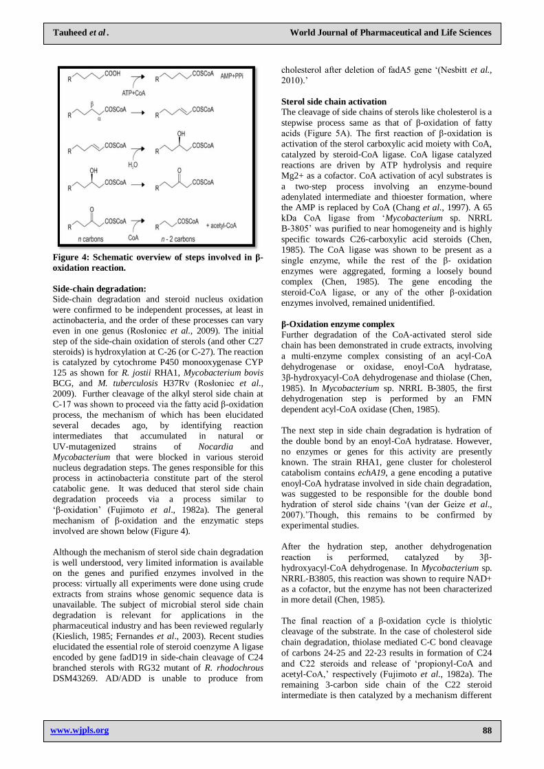
Figure 4: Schematic overview of steps involved in β-
oxidation reaction.
Side-chain degradation:
Side-chain degradation and steroid nucleus oxidation
were confirmed to be independent processes, at least in
actinobacteria, and the order of these processes can vary
even in one genus (Rosłoniec et al., 2009). The initial
step of the side-chain oxidation of sterols (and other C27
steroids) is hydroxylation at C-26 (or C-27). The reaction is catalyzed by cytochrome P450 monooxygenase CYP
125 as shown for R. jostii RHA1, Mycobacterium bovis
BCG, and M. tuberculosis H37Rv (Rosłoniec et al.,
2009). Further cleavage of the alkyl sterol side chain at
C-17 was shown to proceed via the fatty acid β-oxidation
process, the mechanism of which has been elucidated
several decades ago, by identifying reaction
intermediates that accumulated in natural or
UV‐mutagenized strains of Nocardia and
Mycobacterium that were blocked in various steroid
nucleus degradation steps. The genes responsible for this process in actinobacteria constitute part of the sterol
catabolic gene. It was deduced that sterol side chain
degradation proceeds via a process similar to
‗β‐oxidation‘ (Fujimoto et al., 1982a). The general
mechanism of β‐oxidation and the enzymatic steps
involved are shown below (Figure 4).
Although the mechanism of sterol side chain degradation
is well understood, very limited information is available on the genes and purified enzymes involved in the
process: virtually all experiments were done using crude
extracts from strains whose genomic sequence data is
unavailable. The subject of microbial sterol side chain
degradation is relevant for applications in the
pharmaceutical industry and has been reviewed regularly
(Kieslich, 1985; Fernandes et al., 2003). Recent studies
elucidated the essential role of steroid coenzyme A ligase
encoded by gene fadD19 in side-chain cleavage of C24
branched sterols with RG32 mutant of R. rhodochrous
DSM43269. AD/ADD is unable to produce from
cholesterol after deletion of fadA5 gene ‗(Nesbitt et al.,
2010).‘
Sterol side chain activation
The cleavage of side chains of sterols like cholesterol is a
stepwise process same as that of β‐oxidation of fatty
acids (Figure 5A). The first reaction of β‐oxidation is
activation of the sterol carboxylic acid moiety with CoA,
catalyzed by steroid‐CoA ligase. CoA ligase catalyzed
reactions are driven by ATP hydrolysis and require
Mg2+ as a cofactor. CoA activation of acyl substrates is
a two‐step process involving an enzyme‐bound
adenylated intermediate and thioester formation, where
the AMP is replaced by CoA (Chang et al., 1997). A 65
kDa CoA ligase from ‗Mycobacterium sp. NRRL
B‐3805‘ was purified to near homogeneity and is highly
specific towards C26‐carboxylic acid steroids (Chen,
1985). The CoA ligase was shown to be present as a
single enzyme, while the rest of the β‐ oxidation
enzymes were aggregated, forming a loosely bound
complex (Chen, 1985). The gene encoding the
steroid‐CoA ligase, or any of the other β‐oxidation
enzymes involved, remained unidentified.
β‐Oxidation enzyme complex
Further degradation of the CoA‐activated sterol side
chain has been demonstrated in crude extracts, involving
a multi‐enzyme complex consisting of an acyl‐CoA
dehydrogenase or oxidase, enoyl‐CoA hydratase,
3β‐hydroxyacyl‐CoA dehydrogenase and thiolase (Chen,
1985). In Mycobacterium sp. NRRL B‐3805, the first dehydrogenation step is performed by an FMN
dependent acyl‐CoA oxidase (Chen, 1985).
The next step in side chain degradation is hydration of
the double bond by an enoyl‐CoA hydratase. However,
no enzymes or genes for this activity are presently
known. The strain RHA1, gene cluster for cholesterol
catabolism contains echA19, a gene encoding a putative
enoyl‐CoA hydratase involved in side chain degradation, was suggested to be responsible for the double bond
hydration of sterol side chains ‗(van der Geize et al.,
2007).‘Though, this remains to be confirmed by
experimental studies.
After the hydration step, another dehydrogenation
reaction is performed, catalyzed by 3β‐ hydroxyacyl‐CoA dehydrogenase. In Mycobacterium sp.
NRRL‐B3805, this reaction was shown to require NAD+ as a cofactor, but the enzyme has not been characterized
in more detail (Chen, 1985).
The final reaction of a β‐oxidation cycle is thiolytic
cleavage of the substrate. In the case of cholesterol side
chain degradation, thiolase mediated C‐C bond cleavage
of carbons 24‐25 and 22‐23 results in formation of C24
and C22 steroids and release of ‗propionyl‐CoA and
acetyl‐CoA,‘ respectively (Fujimoto et al., 1982a). The
remaining 3-carbon side chain of the C22 steroid
intermediate is then catalyzed by a mechanism different
www.wjpls.org 89
Tauheed et al . World Journal of Pharmaceutical and Life Sciences
from β‐oxidation, most likely involving a reverse
aldol‐lyase reaction (Fujimoto et al., 1982a).
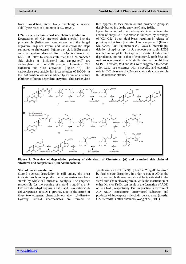
C24‐Branched chain sterol side chain degradation
Degradation of ‗C24‐branched chain sterols,‘ like the
phytosterols β‐sitosterol, campesterol and the fungal
ergosterol, requires several additional enzymatic steps
compared to cholesterol. Fujimoto et al. (1982b) used a
cell‐free system derived from ―Mycobacterium sp.
NRRL B‐3805‖ to demonstrate that the C24‐branched
side chains of ―β‐sitosterol and campesterol‖ are
carboxylated at the C28 position, following C26 oxidation and CoA activation (Figure 5B). The
carboxylase responsible for incorporation of HCO3‐ at
the C28 position was not inhibited by avidin, an effective
inhibitor of biotin dependent enzymes. This carboxylase
thus appears to lack biotin or this prosthetic group is
deeply buried inside the enzyme (Chen, 1985).
Upon formation of the carboxylate intermediate, the
action of enoyl‐CoA hydratase is followed by breakage
of ‗C24‐C25‘ by an aldol lyase, resulting in release of
propionyl‐CoA from β‐sitosterol and campesterol (Figure
5B, ‘Chen, 1985; Fujimoto et al., 1982a‘). Interestingly,
deletion of ltp3 or ltp4 in R. rhodochrous strain RG32
resulted in complete blockage of β‐sitosterol side chain
degradation, but not of that of cholesterol. Both ltp3 and
ltp4 encode proteins with similarities to the thiolase
SCPx. Therefore, ltp3 and ltp4 were suggested to encode
aldol lyase type enzymes with a specific and essential
role in C‐C cleavage of C24‐branched side chain sterols in Rhodococcus strains.
Figure 5: Overview of degradation pathway of side chain of Cholesterol (A) and branched side chain of
sitosterol and campsterol (B) in Actinobacteria.
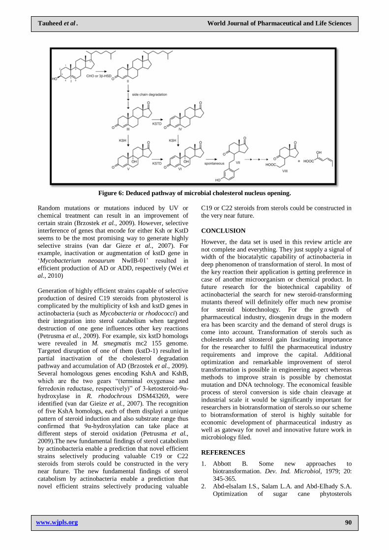
Steroid nucleus oxidation
Steroid nucleus degradation is still among the most
intricate problems in production of androstenones from
sterols by whole-cell microbial catalysis. The enzymes
responsible for the opening of steroid ‗ring-B‘ are ‗3-
ketosteroid-9α-hydroxylase (Ksh) and 3-ketosteroid-1-
dehydrogenase‘ (KstD; Figure 6). Due to the action of
these two enzymes, chemically unstable ‗1,4-dien-9α-
hydroxy‘ steroid intermediates are formed to
spontaneously break the 9(10) bond in ―ring-B‖ followed
by further core disruption. In order to obtain AD as the
only product, both enzymes should be inactivated in the
sterol side-chain cleaving strain, while the inactivation of
either Kshs or KstDs can result in the formation of ADD
or 9-OH-AD, respectively. But, in practice, a mixture of
AD, ADD, testosterone, unconverted substrate, and
products of incomplete side-chain degradation (mostly,
C22 steroids) is often obtained (Wang et al., 2011).
www.wjpls.org 90
Tauheed et al . World Journal of Pharmaceutical and Life Sciences
Figure 6: Deduced pathway of microbial cholesterol nucleus opening.
Random mutations or mutations induced by UV or
chemical treatment can result in an improvement of
certain strain (Brzostek et al., 2009). However, selective
interference of genes that encode for either Ksh or KstD
seems to be the most promising way to generate highly
selective strains (van dar Gieze et al., 2007). For example, inactivation or augmentation of kstD gene in
‗Mycobacterium neoaurum NwIB-01‘ resulted in
efficient production of AD or ADD, respectively (Wei et
al., 2010)
Generation of highly efficient strains capable of selective
production of desired C19 steroids from phytosterol is
complicated by the multiplicity of ksh and kstD genes in
actinobacteria (such as Mycobacteria or rhodococci) and
their integration into sterol catabolism when targeted
destruction of one gene influences other key reactions
(Petrusma et al., 2009). For example, six kstD homologs were revealed in M. smegmatis mc2 155 genome.
Targeted disruption of one of them (kstD-1) resulted in
partial inactivation of the cholesterol degradation
pathway and accumulation of AD (Brzostek et al., 2009).
Several homologous genes encoding KshA and KshB,
which are the two gears ―(terminal oxygenase and
ferredoxin reductase, respectively)‖ of 3-ketosteroid-9α-
hydroxylase in R. rhodochrous DSM43269, were
identified (van dar Gieize et al., 2007). The recognition
of five KshA homologs, each of them displayi a unique
pattern of steroid induction and also substrate range thus confirmed that 9α-hydroxylation can take place at
different steps of steroid oxidation (Petrusma et al.,
2009).The new fundamental findings of sterol catabolism
by actinobacteria enable a prediction that novel efficient
strains selectively producing valuable C19 or C22
steroids from sterols could be constructed in the very
near future. The new fundamental findings of sterol
catabolism by actinobacteria enable a prediction that
novel efficient strains selectively producing valuable
C19 or C22 steroids from sterols could be constructed in
the very near future.
CONCLUSION
However, the data set is used in this review article are
not complete and everything. They just supply a signal of width of the biocatalytic capability of actinobacteria in
deep phenomenon of transformation of sterol. In most of
the key reaction their application is getting preference in
case of another microorganism or chemical product. In
future research for the biotechnical capability of
actinobacterial the search for new steroid-transforming
mutants thereof will definitely offer much new promise
for steroid biotechnology. For the growth of
pharmaceutical industry, diosgenin drugs in the modern
era has been scarcity and the demand of sterol drugs is
come into account. Transformation of sterols such as cholesterols and sitosterol gain fascinating importance
for the researcher to fulfil the pharmaceutical industry
requirements and improve the capital. Additional
optimization and remarkable improvement of sterol
transformation is possible in engineering aspect whereas
methods to improve strain is possible by chemostat
mutation and DNA technology. The economical feasible
process of sterol conversion is side chain cleavage at
industrial scale it would be significantly important for
researchers in biotransformation of sterols.so our scheme
to biotransformation of sterol is highly suitable for
economic development of pharmaceutical industry as well as gateway for novel and innovative future work in
microbiology filed.
REFERENCES
1. Abbott B. Some new approaches to
biotransformation. Dev. Ind. Microbiol, 1979; 20:
345-365.
2. Abd-elsalam I.S., Salam L.A. and Abd-Elhady S.A.
Optimization of sugar cane phytosterols
www.wjpls.org 91
Tauheed et al . World Journal of Pharmaceutical and Life Sciences
bioconversion using Arthrobacter rubellus. J Appl
Sci Res, 2010; 6(9):1334–1339.
3. Aharnowetz V. and Choe G. The microbiological
production of pharmaceuticals. Scientific American,
1981; 240: 140-153.
4. Ahmad S., Garg S.K. and Johri B.N. Biotransformation of sterols: selective cleavage of
the side chain. Biotechnol Adv, 1992; 10: 1–67.
5. Ahmed S., Roy P.K. and Basu S.K. Cholesterol
side‐chain cleavage by immobilized cells of
Rhodococcus equi DSM 89‐133. Indian J. Exp. Biol,
1993; 31: 319‐322.
6. Ambrus G., Jekkel A., Ilkoy E., Horvath G. and
Bocksei Z.. Novel 26‐oxygenated products in
microbial degradation of ergosterol. Steroids, 1995;
60: 626‐629.
7. Andor A., Jekkel A., Hopwood D.A., Jeanplong F.,
Ilkoy E., Konya A. and Ambrus G. Appl Envir
Microbiol, 2006; 72(10): 6554–6559.
8. Arima K. Microbial Transformation of Sterols. Agr.
Biol. Chern, 1969; 33:1636-1643.
9. Arthan D., Svasti J., Kittakoop J.,
Pittayakhachonwut D., Tanticharoen M. and
Thebtaranonth Y. Antiviral isoflavonoid sulfate and
steroidal glycosides from the fruits of Solanum torvum. Phytochemistry, 2002; 59:459–63.
10. Atrat P., Hösel P., Richter W., Meyer H.W. and
Hörhold C. Interactions of Mycobacterium fortuitum
with solid sterol substrate particles. J Basic
Microbiol, 1991; 31: 413–422.
11. Bhatti H.N. and Khera. R.A. Biological
transformations of steroidal compounds: a review.
Elsevier Inc, 2012; 77: 1267-1290.
12. Borman E.A., Redikulifsev Y.V., Koshcheenko
K.A., Turuta A.M. and Kamernitskii A.V.
Transformation of sitosterol to
9alpha‐hydroxy‐androstene‐dione by Mycobacterium sp. 207 cells in the presence of an
adsorption resin. Prikl. Biokhim. Mikrobiol, 1992;
28: 551‐556.
13. Brown W.E. The impact of biotechnology to health
care industry. Ann Rep Fermen Proc, 1984; 7: 135.
14. Brzostek A., Pawelczyk J., Rumijowska-Galewicz
D.B. and Dziadek J. Mycobacterium tuberculosis is
able to accumulate and utilize cholesterol. J
Bacteriol, 2009; 191: 6584–6591.
15. Chang K.H., Xiang H. and Dunaway‐Mariano D.
Acyl‐adenylate motif of the acyladenylate/
thioester‐forming enzyme superfamily: a
site‐directed mutagenesis study with the
Pseudomonas sp. strain CBS3 4‐chlorobenzoate:
coenzyme A ligase. Biochemistry, 1997; 36:
15650‐15659.
16. Charney W. and Herzong L.H. Microbial transformations of steroids. New York: Academic
Press, 1976, pp. 5–73.
17. Chaudhari P.N., Chaudhari B.L. and Chincholkar
S.B. Cholesterol biotransformation to androsta-1,4-
diene-3,17-dione by growing cells of
Chryseobacterium gleum. Biotechnol Lett, 2010; 32:
695–699.
18. Chen C.S. The mechanism of degradation of side
chains of phytosterols by microorganisms. Ph.D.
thesis. University of Wisconsin‐Madison, Ann Arbor, M.I, 1985.
19. Chung S.K., Ryoo C.H.., Yang H.W., Shim J.Y.,
Kang M.G., Lee K.W. and Kang H.I. Synthesis and
bioactivities of steroid derivatives as antifungal
agents. Tetrahedron, 1998; 52: 15899–914.
20. Diaz-Chico B.N., Rodrıguez F.G., Gonzalez A.,
Ramırez R., Bilbao C., Leon A.C., Jaime A.A.,
Chirino R.and Navarro D. Androgens and androgen
receptors in breast cancer. J Steroid Biochem Mol
Biol, 2007; 105:1–15.
21. Donova M.V. Transformation steroid compounds by
actinobacteria. Appl Biochem Microbiol, 2007; 43:1–14.
22. Donova M.V. and Egorova O.V . Microbial steroid
transformations: current state and prospects. Appl.
Microbiol. Biotechnol, 2012; 94:1423–1447.
23. Dovbnya D.V., Egorova O.V. and Donova M.V.
Microbial side‐chain degradation of ergosterol and
its 3‐substituted derivatives: a new route for
obtaining of deltanoids. Steroids, 2010; 75: 653‐658.
24. Dutta R.K., Roy M. and Singh H.D.S. Metabolic
blocks in the degradation of beta‐sitosterol by a
plasmid‐cured strain of Arthrobacter oxydans. J.
Basic Microbiol, 1992; 32: 167‐176.
25. Egorova O.V., Nikolayeva V.M, Sukhodolskaya G.,
Donova M.V. Transformation of C19-steroids and
testosterone production by sterol-transforming
strains of Mycobacterium spp. J Mol Cat B: Enzym,
2009; 5: 198–203.
26. Fujimoto Y., Chen C.S, Szeleczky Z., Di Tullio D. and Sih C.J. Microbial degradation of the
phytosterol side chain. I. Enzymatic conversion of
3‐oxo‐24‐ethylcholest‐4‐en‐26‐oic acid into
3‐oxochol‐4‐en‐24‐oic acid and
androst‐4‐ene‐3,17‐dione. J. Am. Chem. Soc, 1982;
104: 4718‐4720.
27. Fernandes P., Cruz A., Angelova B., Pinheiro H.M.,
and Cabral J.M.S. Microbial conversion of steroid
compounds: recent developments. Enzyme Microb Tec, 2003; 32: 688–705.
28. Fernandes P. and Cabral J.M.S. Phytosterols:
applications and recovery methods. Biores Technol,
2007; 98: 2335–2350.
29. Fernández de las Heras L., Mascaraque L.,
Fernández E.G., Navarro- Llorens J.M, Perera J. and
Drzyzga O. ChoG is the main inducible extracellular
cholesterol oxidase of Rhodococcus sp. strain
CECT3014. Microb Research, 2011; 166(5):
403–418.
30. Frye L.L. and Leonard D.A. Lanosterol analog: dual action inhibitors of cholesterol biosynthesis. In:
Parish EJ, Nes WD, editors. Biochemistry and
function of sterols. Boca Raton, FL: CRC Press,
1987; 23–53.
www.wjpls.org 92
Tauheed et al . World Journal of Pharmaceutical and Life Sciences
31. Hanson J.R. Steroids: reactions and partial synthesis.
Nat Prod Rep, 2005; 22:104–110.
32. Hohnston J.O. Aromatase inhibitors. In: Parish EJ,
Nes WD, editors. Biochemistry and function of
sterols. CRC Press, 1987; 23–53.
33. Horinouchi S., Ishizuka H. and Beppu T. Cloning, nucleotide sequence, and transcriptional analysis of
the NAD(P)‐dependent cholesterol dehydrogenase
gene from a Nocardia sp. and its hyperexpression in
Streptomyces spp. Appl. Environ. Microbiol, 1991;
57: 1386‐1393.
34. Ishizaki T.,Hirayama N., Shinkawa H., Nimi O. and
Murooka Y. Nucleotide sequence of the gene for
cholesterol oxidase from a Streptomyces sp. J.
Bacteriol, 1989; 171: 596‐601. 35. Ivashina T.V., Nikolayeva V.M, Dovbnya D.V. and
Donova M.V. Cholesterol oxidase ChoD is not a
critical enzyme accounting for oxidation of sterols to
3-keto-4-ene steroids in fast-growing
Mycobacterium sp. VKM Ac-1815D. J Steroid
Biochem Mol Biol, 2012; 129: 47–53.
36. Joshi S.M., Pandey A.K., Capite N., Rubin E.J. and
Sassetti C.M. Characterization of mycobacterial
virulence genes through genetic interaction
mapping. Proc. Natl. Acad. Sci. USA, 2006; 103:
11760‐11765. 37. Kieslich K. Microbial side chain degradation of
sterols. J. Basic Microbiol, 1985; 25: 461-475.
38. Lee C.Y., Chen C.D. and Liu W.H. Production of
androsta‐l,4‐diene‐3,17‐dione from cholesterol using
two‐step microbial transformation. Appl. Microbiol.
Biotechnol, 1993; 38: 447‐452
39. Lilly M.D. Advances in biotransformation
processes. Trans Inst Chem Eng, 1984; 72: 27-34.
40. Lin Y., Song X, Fu J, Lin J. and Qu Y.. Microbial transformation of phytosterol in corn flour and
soybean flour to 4-androstene-3,17-dione by
Fusarium moniliforme Sheld. Biores Technol, 2009;
10: 1864–1867.
41. Liu W.H. and Lo C.K. Production of testosterone
from cholesterol using a single step microbial
transformation of Mycobacterium sp.. J Ind
Microbiol Biotechnol, 1997; 19: 269–72.
42. Mahato S.B. Steroid transformation by
microorganism III. Phytochemistry, 1989; 28: 7-40.
43. Malaviya A. and Gomes J. Rapid screening and
isolation of a fungus for sitosterol to androstenedione biotransformation. Appl Biochem
Biotechnol, 2009; 158: 374–386.
44. Martin C.K.A. and Wagner F. Microbial
transformation of beta‐sitosterol by Nocardia sp.
M29. Eur. J. Appl. Microbiol, 1976; 2: 243‐255.
45. Mark D. W. and Flashman E. Catalytic strategies of
the non-heme iron dependent oxygenases and their
roles in plant biology. Elsevier. Inc, 2016; 31: 126-
135.
46. Mohn W.W., van der Geize R., Stewart G.R., Okamoto S., Liu J., Dijkhuizen L.and Eltis L.D. The
actinobacterial mce4 locus encodes a steroid
transporter. J. Biol. Chem, 2008; 283: 35368‐35374.
47. Murohisa T. and Iida M. Studies on microbial
transformation (XXVI). Microbial degradation of
19‐hydroxysterol side‐chains. J. Ferment. Bioeng,
1993; 75: 13‐17.
48. Nesbitt N.M., Yang X., Fontan P., Kolesnikova I., Smith I., Sampson N.S. and Dubnau E. A thiolase of
Mycobacterium tuberculosis is required for
virulence and production of androstenedione and
androstanedione from cholesterol. Infect Immun,
2010; 78: 275–282.
49. Navas J., Gonzalez-Zorn B., Ladron N., Garrido P.
and Vazquez-Boland J.A. Identification and
mutagenesis by allelic exchange of choE, encoding a
cholesterol oxidase from the intracellular pathogen
Rhodococcus equi. J. Bacteriol, 2001; 183(16):
4796–4805. 50. Roy P.K., Khan A.W., Kumar J., Chopra S.D.K and
Basu S.K. Steroid transformation in a
laboratory‐scale glass airlift fermentor. World J.
Microbiol. Biotechnol, 1992; 8: 399‐401.
51. Olivares A. and Acevedo F. Effect of inoculation
strategies, substrate to biomass ratio and nitrogen
sources on the bioconversion of wood sterols by
Mycobacterium sp. World J Microbiol Biotechnol,
2011; 27(11): 2513–2520.
52. Ouellet H., Johnston J.B. and de Montellano P.R.O. Cholesterol catabolism as a therapeutic target in
Mycobacterium tuberculosis. Trends Microbiol,
2011; 19: 530–553.
53. Petrusma M., Dijkhuizen L. and van der Geize R.
Rhodococcus rhodochrous DSM 43269 3-
ketosteroid-9α-hydroxylase, a two-component iron-
sulfur-containing monooxygenase with subtle
steroid substrate specificity. Appl Environ
Microbiol, 2009; 75: 5300–5307.
54. Rosłoniec K.Z., Wilbrink M. H., Capyk J.K., Mohn
W.W., Ostendorf M.,Van der Geize R., Dijkhuizen
L. and Eltis L.D. Cytochrome P450 125 (CYP125)
catalyses C26‐hydroxylation to initiate sterol
side‐chain degradation in Rhodococcus jostii RHA1.
Mol. Microbiol, 2009; 74: 1031‐43.
55. Sallam L.A.R., Osman M.E.S, Hamdy A.A. and
Zaghlol G.M. Microbial transformation of
phytosterols mixture from rice bran oil
unsaponifiable matter by selected bacteria. World J
Microbiol Biotechnol, 2008; 24:1643–1656.
56. Schaefer C.M. et al. FadA5 a thiolase from Mycobacterium tuberculosis: a steroid-binding
pocket reveals the potential for drug development
against tuberculosis. Structure, 2015; 23: 21–33.
57. Schmid A., Dordick J.S., Hauer B., Kiener A.,
Wubbolts M. and Witholt B. Industrial biocatalysis
today and tomorrow. Nature, 2001; 409: 258‐268.
58. Seidel L. and Hoerhold C. Selection and
characterization of new microorganisms for the
manufacture of 9‐OH‐AD from sterols. J. Basic
Microbiol, 1992; 32: 49‐55. 59. Shukla A, Patil S. and Bharti S. Microbial
conversion of solasodine to 4‐androstene‐3,17‐
www.wjpls.org 93
Tauheed et al . World Journal of Pharmaceutical and Life Sciences
dione (AD), a key intermediate for androgen
synthesis. Lett. Appl. Microbiol, 1992; 15: 86‐88.
60. Smith M., Zahnley J., Pfeifer D. and Goff. Growth
and cholesterol oxidation by Mycobacterium species
in Tween 80 medium. Appl. Environ. Microbiol,
1993; 59: 1425‐1429.
61. Stadtman T.C. Steroidal Alcohol. J. Biol. chem,
1954; 206: 511.
62. Tong W.Y. and Dong X. Microbial
biotransformation: recent developments on steroid
drugs. Recent Patents on Biotechnol, 1940; 3: 141–
153.
63. Toro A. and Ambrus G. Oxidative Decarboxylation
of 17(20)-Dehydro-23,24-dinorcholanoic acids.
Tetrahed Let, 1990; 31: 3475–3476.
64. Turfitt G.E. The Microbial Degradation of Steroids. Biochem J, 1944; 38: 492-496.
65. Van der Geize, Yam R.K, Heuser T, Wilbrink M.H,
Hara H., Anderton M.C. and Eltis L.D. A gene
cluster encoding cholesterol catabolism in a soil
actinomycete provides insight into Mycobacterium
tuberculosis survival in macrophages. Proc Nat
Acad Sci USA, 2007; 104: 1947–1952.
66. Uhía I., Galán B., Morales V. and García J.L. Initial
step in the catabolism of cholesterol by
Mycobacterium smegmatis mc2 155. Environ
Microbiol, 2011; 13(4): 943.
67. Vorbrodt H.M., Adam G., Porzel A., Hoerhold C., Daenhardt S. and Boehme K.H. Microbial
degradation of
2alpha,3alpha‐dihydroxy‐5alpha‐cholestan‐6‐one by
Mycobacterium vaccae. Steroids, 1991; 56: 586‐588.
68. Vrielink A. and Ghisla S. Cholesterol oxidase:
biochemistry and structural features. FEBS J, 2009;
276: 6826‐6843.
69. Wang F.Q., Yao K. and Wei D.Z, From soybean
phytosterols to steroid hormones. In: El-Shemy H(ed) Soybean and health. InTech—Open Access
Publisher, Rijeka, 2011; 232–252.
70. Weber A, Kennecke M. and Neef G.
20‐Methyl‐5,7‐pregnadiene‐3beta,2l‐diol derivatives
and their manufacture with Mycobacterium. World
Patent 0903, 1992; 465 (Schering A‐G).
71. Wei W., Wang F.Q, Fan S.Y. and Wei D. Z.
Inactivation and augmentation of the primary 3-
ketosteroid-delta(1)-dehydrogenase in
Mycobacterium neoaurum NwIB-01: biotransformation of soybean phytosterols to 4-
androstene-3,17-dione or 1,4-androstadiene-3,17-
dione. Appl Environ Microbiol, 2010; 76:4578–
4582.
72. Wilson M.R., Gallimore W.A and Reese P.D.
Steroid transformations with Fusari-um oxysporum
var. cubense and Colleto-trichum musae. Steroids.,
2014; 64: 834-843.
73. Wipperman M.F., Sampson N.S. and Thomas S.T.
Pathogen roid rage: cholesterol utilization by
Mycobacterium tuberculosis. Crit. Rev. Biochem. Mol. Biol, 2014; 49: 269–293.
74. Yang X. Dubnau E., Smith I. and Sampson N.H.
Rv1106c from Mycobacterium tuberculosis is a
3β‐hydroxysteroid dehydrogenase. Biochemistry,
2007; 46: 9058‐9067.
75. Zhang L.Q., Bian E.P. and Wang Y. Side‐chain cleavage of sterols by Mycobacterium sp. MI2. Yao
Xue Xue Bao, 1992; 27: 903‐907.