Embed Size (px)
Citation preview

Linköping University Medical Dissertations No 1348
Bisphosphonates and implants in the jaw bone
Jahan Abtahi DDS, MD
Department of Clinical and Experimental Medicine, Linköping University, Sweden.
Department of Oral & Maxillofacial Surgery, University Hospital, Linköping, Sweden.
Linköping 2013.


©Jahan Abtahi, 2013 Cover/picture/Illustration/Design: Jahan Abtahi. Published article has been reprinted with the permission of the copyright holder. Printed in Sweden by LiU-Tryck, Linköping, Sweden, 2013 ISBN 978-91-7519-724-1 ISSN 0345-0082

Supervisor Per Aspenberg, Department of Clinical and Experimental Medicine, Division of Orthopedics, Linköping University. Co-supervisor Agneta Marcusson Department of Oral & Maxillofacial Surgery, University Hospital, Linköping. Faculty opponent Prof. Lars Rasmusson, Department of Oral and Maxillofacial Surgery, Institute of Odontology, the Sahlgrenska Academy at University of Gothenburg. Committee board Prof. Christer Tagesson, Division of Occupational and Environmental Medicine, Department of Clinical and Experimental Medicine, Faculty of Health Sciences, Linköping University. Prof. Thomas Albrektsson, Institute of Clinical Sciences, Department of Biomaterials, Gothenburg University. Johan Thorfinn, Department of Plastic Surgery, Hand Surgery and Burns, University Hospital, Linkoping.

Table of Contents
POPULÄRVETENSKAPLIG SAMMANFATTNING ......................... 11
ABSTRACT ............................................................................................ 13
LIST OF PAPERS .................................................................................. 15
ABBREVIATIONS ................................................................................. 17
INTRODUCTION .................................................................................. 19
BONE METABOLISM ............................................................................. 21
Bone tissue structure ....................................................................... 21
Bone cells ....................................................................................... 22
Osteoclast-osteoblast interplay ........................................................ 26
INTEGRATION OF TITANIUM IMPLANTS IN BONE TISSUE ....................... 29
The initial events ............................................................................. 29
Bone-implant interface .................................................................... 30
The role of micromovement ............................................................ 31
The role of surface topography ........................................................ 33
Surface Roughness ..................................................................... 33
Surface Chemistry ...................................................................... 34
Surface Orientation .................................................................... 35
IMPLANT STABILITY MEASUREMENTS .................................................. 36
Evaluation before and during implantation ...................................... 37
Post-implantation evaluation ........................................................... 39
Experimental studies ....................................................................... 39
Clinical studies ............................................................................... 40
Resonance-frequency analysis ......................................................... 42

Table of Contents
The technique ............................................................................ 42
Clinical and experimental studies ............................................... 43
BISPHOSPHONATES.............................................................................. 46
Structure and bioactivity of bisphosphonates ................................... 46
Local and systemic delivery, experimental studies ........................... 50
Local and systemic delivery, clinical studies ................................... 51
OSTEONECROSIS OF THE JAW (ONJ).................................................... 53
Definition ....................................................................................... 53
Pathogenesis ................................................................................... 54
Treatment ....................................................................................... 55
Prevention....................................................................................... 57
THOUGHTS BEHIND THE START OF THE PROJECT .................. 59
HYPOTHESES ....................................................................................... 61
MATERIAL AND METHODS .............................................................. 63
STUDY DESIGNS (I AND II), CLINICAL STUDIES ..................................... 63
Study I ............................................................................................ 63
Study II ........................................................................................... 63
RESONANCE-FREQUENCY MEASUREMENT (STUDIES I AND II) ............. 64
STUDY DESIGN (III-V), EXPERIMENTAL STUDIES ................................. 65
Study III ......................................................................................... 65
Study IV ......................................................................................... 66
Study V .......................................................................................... 67
COATING TECHNIQUE .......................................................................... 68
RESULTS................................................................................................ 71

Table of Contents
SHORT SUMMARY OF RESULTS OF CLINICAL STUDIES (I AND II) ........... 71
Marginal bone height (I and II)........................................................ 71
Resonance-frequency analysis (I and II) .......................................... 72
Histology (I). .................................................................................. 74
SHORT SUMMARY OF RESULTS OF EXPERIMENTAL STUDIES (III) ......... 75
SHORT SUMMARY OF RESULTS OF EXPERIMENTAL STUDIES (IV) ......... 76
SHORT SUMMARY OF RESULTS OF EXPERIMENTAL STUDIES (V) ........... 77
DISCUSSION .......................................................................................... 79
IMPLANTS AND LOCAL DELIVERY OF BISPHOSPHONATE ....................... 79
RESONANCE-FREQUENCY ANALYSIS..................................................... 83
RAT MODEL OF ONJ ............................................................................ 84
IMPLANTS AND COATING TECHNIQUE .................................................. 86
LACTATE DEHYDROGENASE ANALYSIS ................................................. 88
PATHOPHYSIOLOGY OF ONJ ................................................................ 89
CONCLUSIONS ..................................................................................... 93
WHAT NEXT? ....................................................................................... 95
ACKNOWLEDGEMENTS .................................................................... 97
REFERENCES ....................................................................................... 99


Populärvetenskaplig sammanfattning
11
Populärvetenskaplig sammanfattning
Insättning av metall-implantat för ersättning av förlorade kroppsdelar är ett
vanligt förekommande behandlingsmetod inom odontologi och ortopedi.
Lyckandefrekvensen för dessa behandlingar är direkt kopplade till
implantatens stabilitet, som i sin tur beror på kringliggande benvävnad. När
man sätter in en skruv av titan i käkbenet utlöses ett frakturläkningssvar som
skapar nytt ben runt skruven. Frakturläkningssvaret innehåller både
uppbyggnad och nedbrytning av ben. Genom att selektivt minska
bennedbrytningen med ett läkemedel (bisfosfonat) kan man med bibehållen
uppbyggnadskomponent få mer och starkare ben. Bisfosfonater används
kliniskt bland annat för att hämma bennedbrytning hos patienter med
benskörhet eller skelettmetastaser. Under de senaste åren har
bisfosfonatbehandling för att förbättra implantatfixation testats i både
djurförsök och kliniska studier, men inte i käkar. Detta kan bero på att det
finns ett samband mellan användningen av bisfosfonat (speciellt intravenöst)
och förekomst av ett tillstånd som kallas för ”osteonekros i käken”.
Patofysiologin och behandlingen av detta tillstånd är kontroversiell.
Syftet med denna avhandling är att öka förståelsen om hur bisfosfonater
förstärker benvävnaden runt ett implantat. Kan bisfosfonatbeläggning på
metallytan förbättra fixeringen av implantat i käken? Kan man reproducera
eller förhindra uppkomsten av osteonekros i käken i en djurmodell?
Totalt opererades 96 implantat i överkäken på 21 patienter, som alla fick ett
implantat med bisfosfonat.

Populärvetenskaplig sammanfattning
12
Resonansfrekvensmätning visade att de bisfosfonat beklädda
tandimplantaten hade bättre stabilitet jämfört med kontrollimplantaten efter
6 månaders läkning.
Röntgenundersökning visade mindre benförlust kring bisfosfonatbeklädda
implantat. Vi utvecklade tre djurmodeller för att studera osteonekros i käken.
I ett experiment studerades effekten av lokal och systemisk
bisfosfonatbehandling på käkbenet. Skruvar beklädda med ett potent
bisfosfonat (zoledronat) orsakade bättre implantatinläkning, även under
betingelser där systemisk bisfosfonat framkallar osteonekros i käken. Vi har
också visat att osteonekros i käken inte uppkommer förrän benet exponerats,
t ex genom tandborttagning. Slutligen kunde vi förebygga uppkomsten av
detta tillstånd genom omedelbar täckning med slemhinna efter
tandborttagning.
Slutsatsen är att lokalbehandling med bisfosfonat ger bättre fixering av
implantat i käkarna. Detta kan leda till nya möjligheter för ortopedisk och
dental implantatkirurgi. Patofysiologin av osteonekros i käken är relaterad
till exponering av benvävnad och till läkemedel som förhindrar nedbrytning
av benvävnad.

Abstract
13
Abstract
Insertion of metal implants in bone is one of the commonest of all surgical
procedures. The success of these operations is dependent on the fixation of
the implants, which, in turn, depends on the strength of the bone that holds
them. If the quality of the bone holding the implant could be improved
locally, surgical procedures would become simpler and rehabilitation would
become faster. Bisphosphonates are anti-resorptive drugs that act specifically
on osteoclasts, thereby maintaining bone density and strength. Once released
from the surface of a coated implant, bisphosphonates reduce osteoclast
activity, thereby changing the balance of bone turnover in favor of bone
formation, leading to a net gain in local bone density. During the last
decades, the effects of bisphosphonate treatment on the stability of implants
have been tested in several clinical and animal studies, but not in human
jaws. This may be because it has been suggested that there is a link between
the use of bisphosphonates (especially those given intravenously) and a
condition called osteonecrosis of the jaw (ONJ). The pathophysiology and
treatment of ONJ is controversial. The difficulty in treating ONJ has
highlighted the importance of prevention.
The overall aim of the present thesis was to evaluate the effect of local and
systemic use of bisphosphonates on bone tissue. Could a thin,
bisphosphonate-eluting fibrinogen coating improve the fixation of metal
implants in the human jaw? Would it be possible to reproduce ONJ and
prevent the development of this condition in an animal model?
In two clinical studies, a total number of 96 implants were inserted in 21
patients. In a randomized trial with a paired design, one implant in each pair

Abstract
14
was coated with a thin fibrinogen layer containing two bisphosphonates
(pamidronate and ibandronate). The bisphosphonate-coated implants showed
better stability as measured by resonance-frequency analysis. Radiographic
intraoral films also showed less bone loss. Three animal models were
developed. In a study comparing local and systemic effects of
bisphosphonates, zoledronate-coated screws inserted in rats showed better
fixation in spite of a drug treatment that is known to induce ONJ-like lesions
when given systemically. In another rat model, ONJ-like lesions were
reproducibly induced at sites of tooth extraction whereas there were no signs
of bone cell death in uninjured sites. Finally, rat experiments showed that the
development of ONJ-like lesions after tooth extraction could be prevented
by early mucoperiosteal coverage.
In conclusion, a thin, bisphosphonate-eluting fibrinogen coating can improve
the fixation of dental implants in human bone. This principle may lead to
new possibilities in orthopaedic surgery and dentistry. The pathophysiology
of ONJ is strongly linked to bone exposure in combination with drugs that
reduce resorption.

List of papers
15
List of papers
This thesis is based on the following papers, which will be referred to in the text by their Roman numerals. I. Bisphosphonate coating might improve fixation of dental implants in the maxilla: A pilot study. Abtahi J, Tengvall P, Aspenberg P Int J Oral & Maxillofac Surg 2010 39(7): 673-7. II. Bisphosphonate-coating improves the fixation of metal implants in human bone. A randomized trial of dental implants. Abtahi J, Tengvall P, Aspenberg P Bone 2012 50(5): 1148-51. III. Bisphosphonate-induced osteonecrosis of the jaw in a rat model arises first after the bone has become exposed. No primary necrosis in unexposed bone. Abtahi J, Agholme F, Sandberg O, Aspenberg P J Oral Pathol Med 2012 41(6): 494-9. IV. Effect of local versus systemic bisphosphonate on dental implant fixation in a model of ONJ. Abtahi J, Agholme F, Sandberg O, Aspenberg P Journal of Dental Research 2012 [Epub ahead of print]. V. Prevention of osteonecrosis of the jaw by mucoperiosteal coverage in a rat model. Jahan Abtahi, Fredrik Agholme, Per Aspenberg Int J Oral & Maxillofac Surg 2013, accepted, manuscript number: IJOMS-D-12-00927R1.


Abbreviations
17
Abbreviations
ATP Adenosine triphosphate
BIC Bone-to-implant contact
BMP Bone morphogenetic protein
BRONJ Bisphosphonate-related osteonecrosis of the jaw
Cbfa 1 Core binding factor 1
CSF Colony-stimulating factor
ISQ Implant stability quotient
FPPS Farnesyl diphosphate synthase
GTP Guanosine triphosphate
HAC Hydroxyapatite coating
OH Hydroxyl group
ONJ Osteonecrosis of the jaw
OPG Osteoprotegerin
PTH Parathyroid hormone
PTV Periotest value
RANK-L Receptor activator of NF-kappa B ligand
RFA Resonance-frequency analysis
TGF-β Transforming growth factor-β
TNF- α Tumor necrosis factor-α


Introduction
19
Introduction
During the last decades there have been problems with the condition called
“bisphosphonate-related osteonecrosis of the jaw” (BRONJ). This condition
is defined as an area of exposed bone in the maxillofacial region that does
not heal within 8 weeks of identification by a healthcare provider, in a
patient who currently receives or has been exposed to a bisphosphonate and
has not had radiation therapy to the craniofacial region (from here on, I use
the shorter acronym ONJ). The pathophysiology of ONJ is poorly
understood and as maxillofacial surgeon, I have wondered why these lesions
localize specifically in the jaws. It is remarkable that orthopedic surgeons
and osteoporosis researchers consider bisphosphonates to be beneficial and
useful in many areas, while dental practitioners reject these drugs. By
working with an orthopedic research team, I had the opportunity to address
the problem of the paradoxical effects of bisphosphonates. Previous animal
studies by this team have shown that bisphosphonate coatings can improve
the fixation of implants in bone. Clinically, this idea has been tested in
orthopedics but not in dentistry. From the literature it can be deduced that
high implant survival rates would be expected in jaws. However, dental
implant surgery can be risky in bone of low density. We therefore decided to
use bisphosphonates in the hope of improving the fixation of implants in the
maxilla. Considering that ONJ was apparently non-existent ten years ago,
the field has progressed through knowledge gained from case reports,
population-based studies and emerging animal models. Still, there are
preconceptions that need to be tested and important clues that need to be
investigated in order to translate pathophysiology into improved patient care.
In the clinic, I have also met cancer patients who have suffered from
exposed bone in the jaw associated with intravenous bisphosphonate

Introduction
20
therapy. Is it possible to treat or prevent the development of ONJ? I hope
that my research will improve our knowledge oft this condition.

Introduction
21
Bone metabolism
Bone tissue structure
The skeleton consists of specialized cells, mineralized and unmineralized
connective tissue matrix, and spaces that include bone marrow cavities,
vascular canals, canaliculi, and lacunae. At the nano-scale level, bone tissue
is a composite material composed of an organic phase, consisting mainly of
the protein-based material collagen, and a mineral phase, consisting
primarily of hydroxyapatite (1, 2). Hydroxyapatite is present as plate-like
crystals, 20-80 nm long and 2-5 nm thick, which are themselves composed
of calcium and phosphate. The crystals are found in and around collagen
fibers and give bone its compressive strength. The organic matrix determines
the structure and mechanical properties of the bone. Of the organic matrix,
approximately 90% is type-1 collagen. The remainder consists of non-
collagenous matrix proteins, minor collagen types, proteoglycans, and lipids.
At the microscopic level, there are two types of bone tissue, woven bone and
lamellar bone. Woven bone is considered to be immature, with collagen
arranged randomly. At birth, it makes up all the bone in the body, and in
later years it is found at sites of fracture healing or in response to extreme
mechanical loading (3). Lamellar bone is the name given to bone that
eventually replaces woven bone. By the age of 4 years, most of the skeleton
is lamellar bone. Anatomically, both woven bone and lamellar bone can be
organized into compartments as either cortical or trabecular bone (cancellous
bone). An important difference between cortical bone and trabecular bone is
in the way the bone matrix and cellular elements are arranged. Between 80%
and 90% of cortical bone volume is mineral, but only 15-25% of trabecular

Introduction
22
bone volume is mineral (4). The trabecular arrangement allows bone
marrow, blood vessels, and connective tissues to be in contact with bone.
The main function of cortical bone is to give structure and protection. In
cortical bone, lamellae are arranged concentrically around a central vascular
channel (Haversian canal). This arrangement of cortical bone around a vessel
is called an osteon. Osteons are usually aligned with the long axis of bone
and are connected to each another by Volkmann’s canal, which runs at right
angles to the osteon. The outer surface of cortical bone, facing the soft tissue
is covered by periosteum, while the inner surface that faces the bone marrow
is covered by endosteum. Cells lining the endosteum are metabolically active
and very involved in bone formation and resorption.
Bone cells
There are mainly four types of bone cells. These are the osteoprogenitor
cells, osteoblasts, osteocytes and osteoclasts. The osteoblasts are
mononucleate bone-forming cells that are derived from the local
osteoprogenitor cell line (mesenchymal cells) located in the deeper layer of
periosteum and the bone marrow (5-7). These progenitors are capable of
differentiating into other mesenchymal cell lineages such as
chondrocytes, fibroblasts, myoblasts, and bone marrow stromal cells
including adipocytes (8-12). Mature osteoblasts have an average lifespan
of 1 month, after which they either undergo apoptosis, to be replaced by
newly differentiated osteoblasts, or alternatively about one-third of them
may be incorporated into deposited bone matrix as osteocytes (15).

Introduction
23
Several growth factors and hormones regulate osteoblast differentiation.
Growth factors are soluble proteins that act as signaling agents for cells and
influence critical functions, such as cell division, matrix synthesis, and tissue
differentiation, by receptor-ligand binding. Of these, bone morphogenetic
proteins (BMPs) are the most potent inducers and stimulators of osteoblast
differentiation (13, 14). These proteins not only stimulate osteoprogenitors to
differentiate into mature osteoblasts but also induce non-osteogenic cells to
differentiate into cells of the osteoblast lineage. Proliferation and
differentiation of osteoblasts is also regulated by many transcription factors.
Expression of the transcription factor core binding factor 1 (Cbfa 1) is an
absolute requirement for osteoblast differentiation and bone formation (15).
Similarly, the transcription factor ppar-c 2 can specify adipocyte
differentiation (16), and sox-9 expression is required for chondrocyte
differentiation (17).
Osteoblasts express receptors for various hormones including parathyroid
hormone (PTH), glucocorticoids, 1α, 25-dihydroxyvitamin D3, and estrogen,
which are involved in the regulation of osteoblast differentiation (18-20).
Parathyroid hormone acts directly on the skeleton to promote calcium
release from bone and on the kidney to enhance calcium reabsorption by
binding to its receptor. The anabolic effect of PTH on bone tissue occurs by
upregulation of expression of transcription factor c-fos, which is a key
regulator of osteoblast and osteoclast differentiation (18). Inactivation of c-
fos causes the bone-remodeling disease osteopetrosis, which is characterized
by impaired osteoclastic bone resorption, resulting in a net increase in
skeletal mass (21). In contrast, when c-fos is overexpressed in tissues, bone
tumors develop that are typically chondroblastic osteosarcomas, containing
large amounts of neoplastic bone with foci of cartilage (22, 23).

Introduction
24
Glucocorticoids also play an important role in the normal regulation of bone
remodeling (24). The precise role of glucocorticoids in bone formation is
still poorly understood. In vivo studies have shown that continued exposure
of bone to pharmacological doses of glucocorticoids excess results in
osteoporosis (25, 26). This event is due to the effect of glucocorticoids on
bone cells. Glucocorticoids increase the expression of receptor activator of
NF-kappa B ligand (RANK-L) and reduce the expression of its decoy
receptor, osteoprotegerin (OPG), in stromal and osteoblastic cells (27). They
also enhance the expression of colony-stimulating factor (CSF)-1, which in
the presence of RANK-L induces osteoclastogenesis (28). Furthermore,
several in vitro studies have shown that glucocorticoids reduce the number
of cells of the osteoblastic lineage and shift the differentiation of stromal
cells towards the adipocytic lineage (29) Glucocorticoids also increase peri-
lacunar osteocytic bone resorption to an extent that negatively influences
bone material properties (30).
Osteoclasts are large, multinucleate cells derived from hematopoietic stem
cells, and they are equipped with phagocytic-like mechanisms similar to
those of circulating macrophages (31). Osteoclast literally means “bone
eater”. Osteoclast differentiation has various characteristic features, such as
multinucleation induced by the cell fusion of mononuclear osteoclasts to
cover a larger area, synthesis of the vacuolar proton pump and acid to
dissolve the bone mineral, the formation of ruffled borders to secrete protons
and acid, and the formation of a sealing zone to prevent proton and acid
leakage (31). Their proliferation and differentiation (osteoclastogenesis)
depend on the presence of two different osteoblast expressed cytokines,
macrophage colony-stimulating factor (M-CSF) and RANK-L (32, 33). The
protein osteoprotegerin (OPG), which is secreted by osteoblasts, acts as a

Introduction
25
decoy receptor that competes with RANK for RANK-L and thereby inhibits
osteoclast differentiation (34, 35).
Osteoclasts have developed efficient and unique machinery to dissolve
mineral and degrade bone matrix. To maximize bone resorption, osteoclasts
expand their surface area by fusion to many mononucleated macrophages
(36). After migration of the osteoclast to a resorption site, a specific
membrane domain, the sealing zone, forms adjacent to the bone surface (37,
38). Inside the sealed region, extensive infoldings of the cell membrane
form, the so-called “ruffled border” which increases the membrane surface
(38). A cytoplasmic proton pump (H+-ATPase) produces protons that
generate a pH of 4-5 in the extracellular space adjacent to the bone surface
(39). This event results degradation of the mineral component of bone,
which is composed of hydroxyapatite (31). The organic matrix of the bone
(collagen) is removed through enzymatic activity, by cathepsin K. This
enzyme reaches the bone surface by exocytosis through the basolateral
membrane of the osteoclast (40).
Osteocytes are found within individual lacunae in the mineralized bone
matrix. The lifespan of osteocytes is higher than that of osteoblasts, which is
an estimated 3 months in human bone (41) and 10–20 days in newly formed
murine bone (42). The lifespan of osteocytes is probably largely determined
by bone turnover and they may have a half-life of decades if the particular
bone they reside in has a slow turnover rate (4).
The morphology of embedded osteocytes is dependent on the bone type.
Indeed, osteocytes found in trabecular bone are more rounded than
osteocytes from cortical bone (43). Each osteocyte communicates with its

Introduction
26
neighbors and with the cells lining the surface of bone through long, slender
cytoplasmatic processes (canalicular processes) that connect by means of
gap junctions (44-46). Osteocytes are capable of detecting mechanical
stimuli, which are mediated by loading-induced dynamic fluid flow in the
canaliculi (47, 48). Osteocytes differentiation is under the influence of
several bone markers such as alkaline phosphatase, bone sialoprotein,
osteocalcin, and collagen type I (49). Once the osteoid mineralizes, osteocyte
ultrastructure undergoes further changes including a reduction in
endoplasmic reticulum and Golgi apparatus corresponding to a decrease in
protein synthesis and secretion (50). At this stage, many of the previously
expressed bone markers are downregulated in the osteocyte (50).
Osteocytes are cells that not only play a physiological role during their
lifetime, but also achieve functions through their apoptosis. Osteocytes have
been hypothesized to play a role in this targeted remodeling process (51, 52).
Damage to the bone matrix, such as micro-cracks induces apoptotic death of
osteocytes, which initiate signals for bone resorption by expression of
osteoclast-stimulatory factors, such as RANKL and M-CSF (53, 54).
Osteoclast-osteoblast interplay
Throughout life, bone is constantly renewed through a two-stage process
called remodeling (55, 56). This condition is a dynamic process that relies on
the correct balance between bone resorption by osteoclasts and bone
deposition by osteoblasts. In healthy adults, under normal circumstances,
bone resorption is always followed by an equal degree of bone formation, a
tightly balanced process referred to as coupling (57). The regulation of bone
resorption involves a complicated set of hormonal and/or cytokine

Introduction
27
interactions that initially stimulate osteoblasts, which then elaborate factors
that signal osteoclasts to degrade bone (58, 59). Osteoblast differentiation is
promoted by lipid-modified glycoproteins of wingless (Wnt), bone
morphogenic proteins (BMPs), and several transcription factors (15, 60).
Furthermore, Wnt signaling has been shown to reduce osteoblast and
osteocyte apoptosis in vivo and to increase bone formation by stimulating
differentiation and replication of osteoblasts (61).
Cells of the Osteoblast lineage produce the osteoclastogenic cytokines
RANKL and M-CSF, which recognize their respective receptors RANK and
c-fms on macrophages, prompting them to take on the osteoclast phenotype
(32, 33). RANKL activity is negatively regulated in the circulation by
osteoprotegerin (OPG), which competes with RANK as a soluble decoy
receptor (34). Administration of RANKL to mice causes osteoporosis (35),
whereas disruption of the RANKL gene in mice leads to severe
osteopetrosis, impaired tooth eruption, and the absence of osteoclasts (60).
Several hormones including calcitonin, parathyroid hormone, vitamin D,
estrogen, interleukins, glucocorticoids and tumor necrosis factor-α (TNF- α),
transforming growth factor-β (TGF-β), among others, regulate osteoclast and
osteoblast function (18-20, 24, 62, 63) (Figure 1).

Introduction
28
Figure 1. Differentiation and activation of osteoclasts. M-CSF and RANKL are essential for osteoclastogenesis. OPG can bind to RANKL and thereby inhibit osteoclast differentiation.
The coupling of bone resorption and formation suggests that autocrine and
paracrine factors are produced and released within the local bone
environment (64, 65). Transforming growth factor β (TGF-β) is a
multifunctional cytokine with potent effects on bone metabolism (66, 67).
Both osteoblasts and osteoclasts synthesize and secrete latent TGF-β (65).
Resorption of bone by osteoclasts releases latent TGF-β from the organic
matrix, where it potently stimulates osteoblastogenesis and at the same time
inhibits RANKL expression by osteoblasts. (65).

Introduction
29
Integration of titanium implants in bone tissue
The initial events
Insertion of metal implants in bone is one of the most common of all surgical
procedures. Brånemark et al. first defined “osseointegration” in 1969 as a
direct structural and functional contact (at the light-microscopic level)
between living bone and implant.
The initial host response after implantation is characterized by an
inflammatory reaction elicited mainly by the surgical trauma. Inflammatory
cells, initially polymorphonuclear granulocytes and later monocytes,
emigrate from post-capillary venules into the tissue surrounding the implant
(69). At this stage, damage to the pre-existing bone in the implant- bone
cavity is often a consequence of heating, located within 100 μm (70).
Immediately after the surgical damage, the walls of bone are covered with
blood, which initiates a clotting reaction. This is the first tissue to come into
contact with the implant surface after insertion of the implant in the bone
cavity (71). It has been shown that the implant-blood interface is composed
of a fibrin film containing platelets and red blood cells, and they appear to
respond differently to different implant surface topographies (71).
A strong correlation has been found in several studies between the
adsorption of fibrinogen and surface adhesion of platelets (71-74). The
enhanced aggregation may be due to the increased surface area of the micro-
roughened surface, suggesting that this topography induces more
agglomeration of red blood cells/platelets than machined surfaces (71).

Introduction
30
A few days after implantation, osteoblasts produce collagen matrix directly
on the early- formed lamina limitans layer on the implant surface (69-75).
During the first week, mesenchymal cells and multinuclear giant cells are
present in the area around the implant (75). Areas of newly formed bone
(woven bone) can be seen at the endosteal surfaces towards the implant 1-2
weeks after implantation. Woven bone contains osteocytes, and the
trabeculae are lined with osteoblasts. At this time, in areas that are
responsible for primary mechanical stability, the bone tissue shows signs of
ongoing bone remodeling, resorption, and apposition. After 4 weeks, the
dense woven bone often combined with lamellar bone approaches the
implant surface, to fill the threads. The remodeling process for rabbits and
dogs starts after 4 weeks and is complete after 90 days (69, 75).
Bone-implant interface
The interface zone between bone and implant has been the subject of a vast
number of recent publications (76-86). Many investigators have shown an
interface zone consisting of connective tissue (76, 77). Initially, it was
suggested that the goal for the surgeon should be a periodontal membrane
around the implant (76). Several authors believe that direct contact between
implant and bone is possible only if the implant is ceramic, and not if it is
metal (78, 79). Furthermore, light microscopy of the interface zone has
revealed intimate contact between newly formed bone and the oxidized
surface of titanium implant (80). Early studies on bone-implant contact at the
electron microscopic level showed a close relationship between implants and
collagenous filaments from bone (81). Albrektsson and co-workers (82)
compared interfacial arrangements around stainless steel implants with those
seen around commercially pure titanium. The titanium implants, in contrast

Introduction
31
to stainless steel ones, became directly anchored in bone without any cellular
layer at the interface. Moreover, the authors found a thin (20-40 nm) layer of
amorphous material consisting proteoglycans adjacent to the implant surface
(83). Similar results have been found by others (84-86). Further
ultrastructural studies of machined implants by Sennerby et al. (85, 86)
showed a 100-nm- wide electron-dense line (lamina limitans) at the border
between the mineralized bone and the non-calcified amorphous layer. This
finding indicated that the stability of the implant is mainly mechanical.
The role of micromovement
In the literature, implant movement relative to the surrounding bone has
been suggested to be a crucial parameter in the prognosis of implant
osseointegration (87, 88). Early movement of the implant during the initial
healing phase will lead to a preponderance of interfacial connective tissue
(89-92). There appears to be a consensus that excessive micromotion impairs
osseintegration (93, 94). However, many of these studies are not comparable
due to the influence of other factors such as implant geometry, surface
characteristics, and implant site. The threshold of micromotion, as
experimentally evaluated in animals, is between 50 and 100 µm (95). In a
cadaver study by Burke et al. (96) micromotion of knee and hip prostheses
was measured using sensitive displacement transducers. Such movement has
been shown to be in the range of 100-600 µm (97).
Søblle et al. (97) studied the influence of micromotion between bone and
titanium implant with and without a hydroxyapatite coating (HA) in a dog
model. They showed that micromotion of 150 µm inhibits bone ingrowth
and results in development of a fibrous membrane. Moreover, 4 weeks after
Introduction
32
implantation, a fibro-cartilaginous membrane was seen around unstable HA-
coated implants, whereas around titanium implants, only fibrous connective
tissue was found. Furthermore, continuous loading of initially unstable
titanium implants resulted in the development of a permanent fibrous
membrane, whereas HA-coating had the capacity to replace the motion-
induced fibrous membrane with bone. These findings show that micromotion
has a role in tissue differentiation (98).
In an experimental study by Akagawa et al. (99) dental implants were
inserted in dog mandible by one-stage or two-stage procedures. The animals
were fed with hard pellet food for 3 months. The submerged implants
showed direct bone apposition and sparse fibrous, dense connective tissue.
The unsubmerged implants also showed direct bone apposition. However,
the apical part of the implant was frequently in contact with dense
connective tissue. From these experimental studies, it appears that
micromovements like those induced by early loading of dental implants
should be avoided if the intention is osseointegration.
Trisi et al. (100) evaluated in vitro the correlation between the micromotion
of cylindrical- screw implants and the insertion torque in fresh bovine bone
of different densities ex vivo. They found that increasing the peak insertion
torque reduced the micromotion between the implant and the bone.
However, micromotion in soft bone is always high and immediate loading of
implants in low-density entails a higher risk of loosening.

Introduction
33
The role of surface topography
Surface Roughness
Titanium and its alloys are the materials most often used in implant
manufacture because of their excellent biocompatibility, favorable
mechanical properties, and well-documented beneficial results. When
exposed to air, titanium immediately develops a stable oxide layer which
forms the basis of its exceptional biocompatibility. In the last decade, most
dental implant manufacturers have focused on implant surfaces to improve
bone-to-implant contact (101). It is well known that modification of the
implant surface (including topography and chemistry) alters the cellular and
bone tissue responses (102, 103). Earlier studies (104, 81) have suggested
that implant surface topography is the only parameter that significantly
affects bone-to-implant contact (BIC). These findings were later confirmed
by studies with animals, which indicated that a certain degree of surface
roughness favored BIC, as assessed by the removal torque test (105-108).
Albrektsson and Wennerberg (109) defined smooth surfaces to have an Sa
value of < 0.5 μm; minimally rough surfaces were identified with an Sa of
0.5-1 μm, moderately rough surfaces with an Sa of 1-2 μm, and rough
surfaces with an Sa of > 2 μm.
Another advantage of a roughened titanium surface is a shorter healing
period and the option of using shorter implants, still with a good long-term
prognosis because of the better bone anchorage (110). Therefore, many
surface modifications of titanium implants have been developed to achieve
better osseointegration (electron-polished, anodization, plasma-spraying grit-
blasting or acid-etching).

Introduction
34
One way to produce an increased surface roughness is to blast the surface.
Wennerberg et al. (111) showed higher bone-to-implant contact and higher
removal torque values for implants blasted with TiO (25 µm) rather than
Al2O3 (75 µm) and machined implants. However, no differences were seen
when implants were blasted with different materials (A12O3 and TiO), but
the same degree of roughness (25 µm) was used (112). In recent years, high-
strength ceramics have become attractive as new materials for dental
implants, due to their biocompatibility and higher fracture resilience (113).
At the ultrastructural level, there was no difference between the tissue
response of zircornium implants and that of titanium implants (114, 115).
The TiOblast ™ surface is grit-blasted with titanium dioxide particles to
achieve a moderately rough surface. Several studies (116-118) have shown
promising clinical outcome after 5 years of loading of this surface. In vivo
and experimental studies (119-121) have shown a high cell biocompatibility
and better anchorage in bone when using titanium oxide-blasted implants
rather than implants with a machine-prepared surface.
Surface Chemistry
Beyond surface topography, surface chemistry may provide important and
possibly synergistic cues for bone formation at titanium implants. Recent
studies advocated that surface chemistry is changed by many surface
topographic modifications (122, 123).
Ellingsen et al. (124) placed 80 implants with and without a fluoride-
modified surface in the tibia of 20 rabbits. Removal torque measurements
were performed and biopsies were obtained after 1 and 3 months of healing.
While no differences in removal torque were detected between the two types
of implants after 1 month, the fluoride-modified (test) implants in the 3-

Introduction
35
month healing group had significantly higher torque values than the control
implants. Furthermore, examinations done in ground sections representing
both 1 and 3 months of healing revealed that at fluoride-modified implants,
there were larger proportions of BIC than at the control implants. Berglundh
et al. (125) found that the amount of new bone that formed in the voids
within the first 2 weeks of healing was larger at fluoride-modified (test)
implants than at TiOblast (control) implants. Similar findings have been
reported by Cooper et al. (126). Calcium phosphates, such as hydroxyapatite
(HA), have been successfully used as bone replacement materials in the case
of periodontal lesions, or as a pulp-capping agent (127, 128). Synthetic
hydroxyapatite that is very similar to the inorganic component of bone has
been found to be osteoinductive. It is capable of supporting ingrowth of
osteoprogenitor cells into the graft or implant. Hydroxyapatite coatings on
titanium alloy are used in dental and orthopedic procedures (129).
Surface Orientation
The orientation of the surface topography has been the subject of a number
of studies (130-132). Ivanoff and co-workers (131) investigated
microimplants in a human test model. They found that when blasted and
turned implants with similar roughness were compared, the blasted implants
had significantly more bone-to-implant contact than turned implants. Despite
the fact that both implants had the same grade of roughness, the turned
implants had a clear orientation of the topography with an orientation
perpendicular to the long axis of the implant. This finding indicated that
surface orientation may be as important as the degree of surface roughness.
Moreover, Wennerberg et al (132) found no differences between the
Introduction
36
horizontally grooved implants and the vertically grooved implants when
measuring bone-to-implant contact.
Burgos et al. (133) showed that titanium implants with a turned or an
oxidized surface are integrated in bone in different ways. As observed at the
light-microscopic level, bone formation occurs directly on the moderately
oxidized surface, while turned titanium surfaces are integrated by growth of
bone from the adjacent bone marrow and bone tissue. The mechanisms
behind the different integration patterns are not fully known, but surface
topography most certainly plays an important role. Moreover, a positive
effect of titanium surface roughness on osteoblast proliferation and gene
expression associated with collagen biosynthesis has been discussed by
several authors (134-136). The surface profile in the nanometer range plays
an important role in the adsorption of proteins, adhesion of osteoblast cells,
and thus, the rate of osseointegration (137). However, the optimal size of
nonometer particles applied on implant surfaces is still unknown.
Implant stability measurements
It is clear that stability both at placement and during function is an important
criterion for the success of dental implants. Primary implant stability is a
mechanical phenomenon influenced by factors related to the implant (design
and dimensions of the fixture), the patient (quality and quantity of bone), and
the operator (surgical technique). Primary implant stability is highest just
after implant placement, because of mechanical compression of the fixture
on bone walls, and it decreases with time. Secondary stability is the
progressive increase in stability related to biological events at the bone-
implant interface such as new bone formation and remodeling (138). The

Introduction
37
clinical definition of implant osseointegration considers the level of stable
marginal bone and absence of mobility in the bone (139). The diagnosis is
therefore based on radiographic and mechanical stability criteria.
Evaluation before and during implantation
Imaging studies
Different radiographic techniques (intraoral radiographs, panoramic
radiographs, lateral cephalograms, and tomographic images) are commonly
used for evaluation of jaw bone quality and volume when planning for
implant treatment (140). The use of computerized tomography (CT) for the
evaluation of the bone density of patients requiring implant therapy was
introduced by Schwarz et al. (141) and this method has been used in several
studies (142-146). The authors suggested that there are strong correlations
between bone density as estimated by CT, the insertion torque, and
resonance-frequency values at implant placement. These findings support the
idea that the preoperative CT examination may be a helpful technique for
predicting primary stability. Although CT examinations have advantages
over conventional radiography techniques, CT exposes the patient to higher
doses of radiation. The recently developed limited cone-beam CT involving
a two-dimensional X-ray sensor has been reported to be useful in presurgical
evaluation. Limited cone-beam CT provides high-resolution images and does
not need the use of high doses of radiation (147, 148).
Drilling/cutting resistance
Currently the most popular method of bone quality assessment is that
developed by Lekholm and Zarb, who introduced a scale of 1to 4, based on
both the radiographic assessment and the sensation of resistance experienced
by the surgeon when preparing the implant site (149). The grading refers to

Introduction
38
individual experience and provides only a rough mean value for the entire
jaw. This classification has therefore been questioned recently due to its poor
objectivity and reproducibility (150, 151). A similar index of four different
density classes, based on the tactile feeling during drilling and implant
insertion was introduced by Mish and Friberg (152, 153). Norton et al (143)
concluded that there is a need for an objective quantitative classification of
bone quality, which can be applied preoperatively and is not operator-
dependent. They proposed a new classification based on the Hounsfield units
of the bone on the CT scan, and related it to the existing classification of
Lekholm & Zarb (149). A Hounsfield unit represents a normalized index of
x-ray attenuation based on a scale where air corresponds to -1000 units
water at standard pressure and temperature to 0 (154). Klinge et al. (155)
proposed an individualized healing period after implant placement based on
an objective score of bone quality. Altogether, 15 bone biopsies from 12
patients were harvested at mandibular sites before insertion of an implant.
However, this idea has not been developed for practical reasons.
Furthermore, a more objective method has been described by Johansson and
Strid (156), who measured the cutting resistance during implant insertion as
a function of the electric current drawn by the handpiece. In a series of
studies by Friberg et al. (157-159), a positive correlation was found between
values of cutting resistance, bone density, and resonance-frequency
measurements. However, it was not possible to use this instrument to
identify implants that are at risk of failure already at implant placement
(159). The majority of failures were seen in bone of medium-to-high density,
while implants inserted in bone of poor density gave a better outcome,
perhaps due to an adapted surgical protocol and an extended healing period.

Introduction
39
Post-implantation evaluation
Invasive and non-invasive clinical tools are available for objective
assessment of implant stability. Invasive biomechanical tests including
removal torque measurements, pullout tests, and histological and
histomorphometric evaluation can give valuable information about the
fixation of the implant. However, these destructive methods can be used
mainly in experimental studies.
Experimental studies
Biomechanical tests
The removal torque test is a simple method that has often been used to
evaluate the tissue response to titanium and other materials in experimental
studies (160-166). Johansson and co-workers (160) reported a gradually
increasing implant removal torque and bone-to-implant contact within a 12-
month period. The stability and resistance to shear forces of implants also
appear to be dependent on the mechanical properties of the bone at the bone-
implant interface. Eulenberger et al. (161) used small cortical screws of
stainless steel and titanium to evaluate the stability of implants by
measurement of removal torque in rabbit tibia. They found higher removal
torque value for titanium implants after 12 weeks. In the rabbit study by
Sennerby et al. (162), higher removal torque values were recorded in
commercially pure titanium implants than in vitallium implants.
The importance of cortical bone fixation of implants has been discussed in
several studies (26, 27). Ivanoff et al. (163) studied the influence of different
implant diameters on removal torque after 12 weeks of healing in the rabbit

Introduction
40
tibia. The authors also showed higher removal torque values in rabbit tibia
when implants engaged two cortical layers rather than one (164).
Rasmusson et al. (165) studied the effect of barrier membranes on bone
resorption and implant stability in onlay bone grafts. Disc-shaped bone grafts
were harvested from the calvarium and placed with titanium implants in the
tibia of rabbits. On one side (test), the bone graft/implant was covered with a
membrane, while the contralateral side (with no membrane) served as a
control. The results showed that postsurgical bone graft resorption was
inhibited as long as the membrane was in place. However, after removal of
the membrane at 8 weeks, the resorption rate was higher on the test side. No
differences were found between the test and control sites after 24 weeks, as
measured by removal torque. Furthermore, Rasmussen et al. (166) used a
rabbit model to study the healing and stability of titanium implants in free
bone grafts, placed simultaneously or after 8 weeks of healing and followed
for 24 weeks. Removal torque tests after 24 weeks did not reveal any
differences between the two procedures.
Clinical studies
Non-destructive conventional methods, such as clinical evaluation (through
manipulation with forceps or judgment of percussion sound) are highly
subjective and lack reliability. Other objective methods such as Periotest®
(Bensheim, Germany) or the Dental Fine Tester® (Kyocera, Kyoto, Japan)
have been used for monitoring of the stability of implants over the healing
period. Their lack of resolution, however, and their poor sensitivity and
susceptibility to operator variables has been criticized (167).

Introduction
41
The Periotest system is an electronic instrument that was originally designed
to quantify signs of stress resorption by the periodontal ligament surrounding
the tooth, as a measure of mobility (168). The Periotest instrument comprises
a hand piece containing a metal slug that is accelerated towards a tooth by an
electromagnet. The duration of contact of the slug with the tooth is measured
by an accelerometer. The software in the instrument is designed to relate
contact time as a function of tooth mobility. The result is displayed digitally
and audibly as Periotest values (PTVs) on a scale from -8 (low mobility) to
50 (high mobility). Inter-operator and inter-instrument variability has been
studied extensively (169-171). Several studies (172, 173) have shown the
sensitivity of this method for the variation in the occlusal-gingival position.
Furthermore, it has also been found that Periotest values are dependent on
the angulations of the hand piece. A change in position of 1 mm in striking
height may produce a difference in Periotest values of between 1 and 2 (169,
174).
Osseointegrated implants placed in the mandible have shown systematically
lower PTVs than those placed in the maxilla (175). Moreover, Salonen et al.
(176) recorded Periotest values of four different implant systems after an
average of 22.5 months after installation. 14 of 204 implants lost stability.
The lost implants showed significantly higher Periotest values than stable
implants, except for ITI implants in the maxilla. In a clinical study by Drago
et al. (177), implant stability was evaluated by Periotest at abutment
connection, 6 and 12 months after occlusal loading. Periotest values
recorded at abutment connection and after 12 months of functional loading
failed to predict loss of implant stability. The positive predictive value for
these two occasions was 64%. Thus, the prognostic value of Periotest to
detect loss of implant stability has been questioned.

Introduction
42
In recent years, the Ostell™ device for resonance-frequency analysis (RFA)
has been advocated to provide an objective measurement of primary implant
stability and to monitor implant stability over the healing period.
Resonance-frequency analysis
The technique
Resonance-frequency analysis (RFA) is a non-invasive, objective method for
evaluation of implant stability and has been validated through several in
vitro and in vivo studies (178-180). Meredith et al. (181) measured the
frequency response of the transducer attached to an implant fixture in an
aluminium block using abutments of various lengths (0-5 mm). A strong
correlation was found between the RFA value and the exposed height of the
implant above the block, while the overall implant length was of no
significance. Resonance frequency was determined by the stiffness of the
bone–implant complex as demonstrated by performing repeated
measurements of implants placed in self-curing resin. A significant increase
in resonance frequency was found to be correlated with increase in stiffness.
The RFA technique used in this study (Osstell; Integration Diagnostics,
Sävedalen, Sweden) is based on magnetic pulses (3,500 to 8,500 kHz)
instead of electrical excitement. A transducer called a “SmartPeg” is
attached to an implant or abutment and a measurement is made by holding a
probe near to the peg. The peg is excited and the resonance frequency is
expressed electromagnetically in implant stability quotient (ISQ) units, on a
scale from 1 (lowest) to 100 (highest). An increase in ISQ of 1 unit appears
to correspond to 50Hz in resonance.

Introduction
43
Clinical and experimental studies
Early studies by Meredith et al. (182) showed in increase in implant stability
from placement to abutment connection in 54 of 56 implants inserted in the
maxilla, as measured by RF. Moreover, the authors found a significant
correlation between effective implant length (abutment length and bone loss)
and RF.
Friberg et al. (183) correlated cutting resistance (bone density) with primary
stability for 61 maxillary implants placed in different densities of bone.
Repeated measurements indicated that all implants reached similar ISQ
values at abutment connection and after 1 year of loading irrespective of
initial stability at the time of installation. Similar findings have also been
reported by other authors (184-186). These results indicate that the stiffness
of the implant-bone contact is low in soft bone and high in dense bone.
Furthermore, the remodeling process of soft trabecular bone seems to result
in increased stiffness of the peri-implant bone.
Moreover, the influence of implant diameter and location on RFA values has
been studied by Östman et al. (187). Measurements of a total of 905
Brånemark dental implants in 267 consecutive patients showed higher ISQ
values for wide-platform implants in comparison to regular/narrow-platform
implants. Moreover, a lower stability was seen with increased implant
length, which may be explained by the fact that long implants may have a
reduced diameter in the coronal direction.
The resonance-frequency technique has shown higher implant stability in
mandibular bone than in maxillary bone (185, 188). A correlation has been
found between bone quality (Lekholm & Zarb) and ISQ values by some

Introduction
44
authors (187, 188) but not by others (189). In a clinical study by Miyamoto
et al. (190), a total of 225 implant stability measurements were made at the
time of implant placement using a resonance frequency analyzer. Before
surgery, cortical thickness was determined by using CT scans. The authors
showed that dental implant stability at the time of surgery was weakly
influenced by implant length, but strongly related to cortical bone thickness.
To monitor the outcome of implant installation and determine the prognostic
value of RFA in predicting loss of implant stability, Huwiler et al. (191)
assessed ISQ values at the time of implant installation and 1, 2, 3, 4, 5, 6, 8,
and 12 weeks thereafter. ISQ values of 57–70 represented stable implants.
One implant lost stability at 3 weeks. At this time, its ISQ value had dropped
from 68 to 45. However, the latter value was determined after the clinical
diagnosis of instability. In a longitudinal study by Glauser et al. (192), ISQ
values of 72 stable implants were compared with those of nine implants that
had lost stability during 1 year according to an immediate/early-loading
protocol. The implants that failed during the course of the study showed
significantly lower stability already after 1 month. The risk of loss of
stability was 18%, if ISQ values were between 49 and 58. In contrast to
these findings Bischof et al. (188) performed RFA measurements on
immediate and delayed loaded implants during the 3 months of healing. The
resonance-frequency analysis method did not reveal any differences between
these groups.
It is well known that when performing longitudinal measurements of implant
stability, the position of the transducer must be highly reproducible because
the measured values are dependent on the orientation of the transducer. The
role of direction-dependence of the OsstellTM transducer was evaluated in a

Introduction
45
parametrical finite element study (193). The data indicated that, when
measuring perpendicularly to the long axis of the alveolar crest, the
deviation must not exceed 30° from the ideal perpendicular position. In this
case, the first resonance frequency is recorded. When measuring in the
position parallel to the long axis of the alveolar crest, however, the deviation
must not exceed 10°. In order to monitor the stability of an implant over time
correctly, it seems important that the same transducer orientation is kept
during the different measurements. In a prospective study (194), resonance-
frequency measurements at different orientation were performed in a total of
55 implants in the maxillae of nine patients. The results showed that, when
measuring the RFA perpendicular (buccopalatal) to the bony crest, the ISQ
values may be up to approximately 10 units lower compared to paralel
(mesiodistal) orientations. Similar results were found by Pattijn et al. (195).
Moreover, Park et al. (196) found no differences when measuring RF from
different directions.
The resonance frequency technique has also been used to measure implant
stability in grafted bone (197-198). Recently, Rasmusson et al. (199)
evaluated implant stability in particulate bone, onlay block bone,
interpositional bone, and non-grafted maxillary bone using RFA. A total of
260 TiO2-blasted implants were placed 5-6 months after bone grafting, and
the abutments another 6 months later. The results showed that implants
placed in non-grafted and grafted maxillary bone using a two-stage protocol
had similar stability. Similar findings have been presented by others (200).
Degidi et al. (201) showed higher RFA values for implants in a site
previously treated with a sinus augmentation procedure than for implants in
non-grafted maxillary bone.

Introduction
46
Bisphosphonates
Structure and bioactivity of bisphosphonates
Bisphosphonates are anti-resorptive drugs that act specifically on osteoclasts,
thereby maintaining bone density and strength (202). Bisphosphonates are
used in many clinical settings, including prevention and treatment of primary
and secondary osteoporosis, Paget’s disease of bone, hypercalcemia,
multiple myeloma and osteolysis associated with bone metastases of
malignant tumors (203, 204). They may directly inhibit the bone-resorbing
activity of osteoclasts by mechanisms that can lead to osteoclast apoptosis
(205). Moreover, a study by Sahni et al. (206) suggested that part of the
inhibitory action of bisphosphonates on the osteoclasts is mediated through
an action on the osteoblasts. However, it is not yet known whether this plays
any important role in vivo.
Bisphosphonates also directly promote the proliferation and differentiation
of human osteoblast-like cells in vitro (207). It has been reported that these
drugs cause a number of effects on other cells, including inhibition of cell
proliferation (208) and causing a decrease in cell adhesion, in fibroblasts
(209) and in macrophages (210, 211).
Bisphosphonates are synthetic pyrophosphate analogs with a P-C-P bond
instead of the P-O-P bond of inorganic pyrophosphates, which are used as
anti-tarter agents in toothpastes and as a bone-specific radionuclide in
technetium 99m methylene diphosphonate (Tc 99m MDP) bone scans.
Unlike pyrophosphates, bisphosphonates are resistant to breakdown by
enzymatic hydrolysis, which explains their accumulation in the bone matrix

Introduction
47
and their extremely long half-life (212). The P-C-P structure (Figure 2)
allows a great number of possible variations, especially by changing the two
lateral chains (R1 and R2) in the carbon atom. The two phosphate groups are
essential for binding to bone mineral such as hydroxyapatite and together
with the R1 side chain they act as a “bone hook”. A hydroxyl (OH) group or
an amino group at the R1 position increases the affinity for calcium and thus
for bone mineral (213, 214).
Figure 2. The chemical structure of pyrophosphate and bisphosphonate. R1 and R2 signify the side chains of bisphosphonate.
The structure and three-dimensional conformation of the R2 side chain
determine the anti-resorptive potency and the enhanced binding to
hydroxyapatite (213, 215). It has been shown that bisphosphonates
containing a basic primary nitrogen atom in an alkyl chain are 10-100 times
more potent at inhibiting bone resorption than earlier-generation
bisphosphonates, such as clodronate, which lack this feature. Compounds
that contain tertiary nitrogen, such as ibandronate and olpadronate, are even
more potent at inhibiting bone resorption. Residronate and zoledronate are
among the most potent of bisphosphonates, containing a nitrogen atom

Introduction
48
within a heterocyclic ring; they are up to 10,000 times more potent than
etidronate (216).
The non-nitrogen-containing bisphosphonates (etidronate, clodronate, and
tiludronate) inhibit bone resorption by generating the cytotoxic, methylene-
containing analogs of ATP that interfere with mitochondrial function and
induce apoptosis of osteoclasts (217-219). In contrast, the nitrogen-
containing bisphosphonates (alendronate, zoledronate, pamidronate,
risedronate, and ibandronate) bind to and inhibit farnesyl pyrophosphate
synthase (FPPS), a key enzyme of the mevalonate pathway, thereby
preventing the prenylation and activation of small GTPases that are essential
for the bone-resorbtion activity and survival of osteoclasts (220-222).
There is no evidence that orally or intravenously administered
bisphosphonates are metabolized in animals or humans (223, 224). The
gastrointestinal uptake of oral bisphosphonates is low, with a bioavailability
of 0.7 % for alendronate (225) and 0.3% for pamidronate (226). The poor
absorption of bisphosphonates can probably be attributed to their very poor
lipophilicity, which prevents transcellular transport across the epithelial
barriers. Consequently bisphosphonates must be absorbed by the paracellular
route, which means passage through the pores of the tight junctions between
the epithelial cells. Oral absorption of alendronate in rats is fourfold to
fivefold higher in the fasted state than in the fed state (227). The same effect
of food on the absorption of alendronate has also been observed in healthy
volunteers (225).
Intravenous administration of a single dose of alendronate leads to rapid
accumulation of this drug in bone tissue: 30% in 5 min and 60% in 1 hour.

Introduction
49
At 5minutes after dosing, 63% of the dose is present in non-calcified tissues.
This drops to about 1% 6-24 hours post-dose (227). The distribution of
alendronate in bone is determined by blood flow and favors deposition at
sites of the skeleton that is undergoing active resorption (227). Following
administration of a single dose of 14C-alendronate in rats, over 70% of the
bone resorption surface was densely labeled in comparison with 2% of the
bone formation surfaces (228). This preferential localization of alendronate
(in areas of high bone turnover) could be due to exposure of hydroxyapatite
at sites that are undergoing bone resorption and the accessibility of these
bone surfaces to substances in the circulation
Following administration of 14C-alendronate in rats a larger proportion of the
dose is taken up by trabecular bone than by cortical bone, and in the latter at
the metaphysis rather than the diaphysis (229, 230). Similar results were
observed in dogs when etidronate was given intravenously (231).
Furthermore, Lin et al. (230) showed that the uptake of alendronate is
proportional to the intravenous dose up to 5 mg/kg IV, but at 10 mg/kg or
higher, the concentration of drug in bone increases less than linearly
In recent years, it has been hypothesized that another target of
bisphosphonates may be osteoblasts, which subsequently influence
osteoclasts. The mitogenic effect of bisphosphonates on osteoblasts has
been reported by several authors (232, 233). Furthermore, it has been shown
that these drugs inhibited the expression of RANKL in a rat osteoblast cell
line (234) and increase the expression of OPG in human osteoblastic cells
(235), suggesting that the anti-resorptive effect of bisphosphonates is
mediated by the influence of osteoblasts on RANKL signaling (234, 235).
Moreover, it has been shown that bisphosphonates promote osteoblast

Introduction
50
differentiation in cultures of osteoblast-like cell lines in a dose-dependent
manner and inhibit the osteoblast apoptosis (236). However, the clinical
relevance of this is unclear.
Local and systemic delivery, experimental studies
Extraction of teeth necessitated by factors such as developmental problems,
trauma, severe periodontal disease, or unsolvable endodontic problems often
causes reduction of the residual alveolar ridge height and width in the jaws.
These reductions usually cause difficulties in prosthetic restoration, poor
aesthetics and insufficient function. Many investigators have shown that
tooth extraction stimulates osteoclastic activity with varying amounts of
alveolar crest loss (237, 238). Systemic alendronate was found to be
significantly effective in reducing bone loss associated with experimental
periodontitis in monkeys and beagle dogs (239, 240). Yaffe et al. (241)
found that local delivery of alendronate reduced alveolar bone resorption
activated by mucoperiosteal surgery. In orthodontics, topical administration
of amino bisphosphonate caused significant reduction of tooth movement in
rats, when orthodontic force was applied (242, 243).
Several methods have been reported to increase the bone density around
experimental porous implants, but to varying degrees (244-254). In animal
models, several investigators have shown that surface-immobilized
bisphosphonates improve the mechanical fixation of metal screws in terms
of an increased pullout force and bone-to-implant contact (255-259).
Yoshinari et al. (256) used plasma-sprayed HA-coated titanium dental
implants that were immersed in pamidronate and implanted in mandibular
bone of beagles. This study showed a 10% increase in bone contact area.
Tengvall et al. (260) showed an increase (by 28%) of the pullout force of

Introduction
51
steel screws inserted in rat femurs by using fibrinogen/pamidronate
/ibandronate coating. Hydroxyapatite-coated implants releasing zoledronate
induced an increase in pullout force by up to 42% compared to implants
without zoledronate (257). However, at higher zoledronate concentrations (>
2.1 µg/implant) the pullout force decreased by 35%. The authors’
hypothesized that this might be correlated to the lower bone mineral density
close to the implant, due to a negative effect on osteoblast function.
Several authors who performed animal studies have also described the
efficacy of bisphosphonates on mechanical fixation of implants in
osteoporotic bone (258, 259, 261). However, there are many differences
between the bone metabolism of small animals that of humans such as
mineral density and healing capacity (262, 263). Even after different
strategies such as ovariectomy and steroid application, the bone density is
higher than in healthy human bone equivalents (264). Thus, it is questionable
to extrapolate the in vivo results from small animals to specific clinical
situations with osteoporotic patients.
Local and systemic delivery, clinical studies
Bisphosphonate have been given orally or systemically in order to improve
fixation of orthopedic implants (265-267). A single infusion of 4 mg
zoledronate showed promise in improving initial fixation of a cementless
implant (265). In a randomized, double-blind trial of a hybrid-type total hip
arthroplasty in patients with osteoarthritis, Wilkinson et al. (267) found that
a single dose of 90 mg of pamidronate significantly reduced femoral bone
loss. Hilding and Aspenberg (268) showed that local application of a
bisphosphonate during total joint surgery reduces migration of metal
prostheses as measured by radiostereometry. The authors applied 1 mg

Introduction
52
ibandronate (1 mL) to the tibial bone surface 1 min before cementation. The
role of bisphosphonate as an adjunct to conventional periodontal therapy in
management of periodontal disease has recently been in focus. In a
prospective investigation on possible effects of alendronate on alveolar bone,
335 patients with moderate or severe periodontal disease were randomized to
either placebo or 70 mg alendronate once a week (269). After 2 years of
treatment with bisphosphonate, there was no detectable effect on alveolar
bone loss, except in those patients with low mandibular bone mineral density
at baseline. In contrast, local treatment appears to be efficacious. In a recent
series of three randomized controlled trials, local treatment of parodontitis
with a gel containing a very high concentration of alendronate was
successful in regenerating a large part of the lost bone, whereas placebo had
little effect (270-272).

Introduction
53
Osteonecrosis of the jaw (ONJ)
Definition
There is strong evidence for a link between the use of systemic
bisphosphonates (especially those given intravenously) and osteonecrosis of
the jaw in cancer patients (273-275). This condition of the jaw is defined as
non-healing, exposed bone for more than 8 weeks in patients receiving a
bisphosphonate and without any history of local radiation therapy (276,
277). Clinically, the disease presents as exposed alveolar bone (Figure 3)
that occurs spontaneously or becomes evident following a surgical procedure
such as tooth removal, periodontal surgery, apicoectomy or dental implant
placement (273-275). These lesions most often become frequently
symptomatic when surrounding tissues are inflamed or when there is clinical
evidence of infection. Signs and symptoms that may occur before the
development of clinically detectable osteonecrosis include pain, tooth
mobility, mucosal swelling, erythema, and ulceration. The incidence of ONJ
is estimated to be 1-12% in cancer patients receiving high-dose intravenous
bisphosphonates (273-275). The frequency of ONJ in bone malignancy
cases, mainly treated intravenous bisphosphonates, was found to be 1 in 100
(278). If tooth extractions were carried out, the calculated frequency of ONJ
was 1 in 10 (278). In a retrospective study, Wang et al (279) found that the
incidence of ONJ associated with intravenous bisphosphonates was at least
3.8 per 100 patients with multiple myeloma, 2.5 per 100 patients with breast
cancer, and 2.9 per 100 patients with prostate cancer. In osteoporosis
patients, bisphosphonate-associated osteonecrosis of the jaw is rare and the
incidence may not be greater than the natural background incidence of the
condition. Epidemiological studies have indicated an estimated incidence of

Introduction
54
less than 1 case per 100 000 person-years of exposure to oral amino
bisphosphonates (273-275).
Figure 3. Exposed alveolar bone in the mandible of a patient with intravenous bisphosphonate therapy.
Pathogenesis
The etiopathogenesis of ONJ remains uncertain. When the condition known
as bisphosphonate-related osteonecrosis of the jaw (BRONJ) was first
described, its similarities with radiation-induced osteonecrosis led to the
assumption that the condition started with sterile necrosis of the jaw bone,
which acquired a clinical appearance of classical chronic osteomyelitis after
exposure to the oral cavity (273, 274). Hence the name osteonecrosis, a term
otherwise reserved for sterile bone death, usually because of impaired blood
supply. At that time, it was speculated that bisphosphonates could cause
osteonecrosis through effects on blood vessels in bone, possibly by
inhibition of vascular endothelial growth factor (280-282). It was soon

Introduction
55
suggested that BRONJ does not begin as a form of classical osteonecrosis,
but is in fact osteomyelitits from the start (283, 284). Bacterial
contamination with Actinomyces appears to play an important role in
maintaining osteomyelitic wounds (285-286). Because jaw bone containing
bisphosphonates will be resorbed slowly, it is conceivable that bacterially
contaminated bone cannot be removed fast enough to prevent the
development of chronic osteomyelitis. This view is supported by the
observation that similar lesions appear after treatment with an anti-RANKL
antibody which reduces osteoclast recruitment (287). Thus, it appears that
reduced resorptive activity is a key factor behind the reduced ability of these
lesions to heal.
Corticosteroids and chemotherapeutic drugs have been suggested as factors
that can predispose to ONJ or increase the risk of developing ONJ (288).
The duration of bisphosphonate therapy also appears to be related to the
likelihood of developing necrosis with longer treatment regimens associated
with a greater risk (289). The mean time to ONJ after zoledronate treatment
was calculated to be 1.8 years and the minimum was 10 months; after
pamidronate, the mean time was 2.8 years and the minimum was 1.5 years;
and after oral BP therapy, the mean time was 4.6 years and the minimum
was 3 years (290). Similar findings have been reported by others (291, 292).
Treatment
The optimal treatment strategy for ONJ is still to be established. No effective
treatment has been developed yet and interrupting drug therapy does not
seem to be sufficient. In general patients with suspected ONJ should be
evaluated and managed by a team including a dental specialist, an oral and
maxillofacial surgeon, and an oncologist. The presentation and

Introduction
56
symptomatology of ONJ can vary in patients despite similar disease
processes, bisphosphonate dosage regimens and treatment duration. A
clinical staging system (Figure 4) has been developed in order to more
accurately categorize patients with ONJ (277, 293).
Staging
Stage 1 Exposed, necrotic bone that is asymptomatic and has no evidence of infection
Stage 2 Exposed, necrotic bone associated with pain and infection
Stage 3 Exposed, necrotic bone in patients with pain, infection, and pathological fracture, extraoral fistula, or osteolysis extending to the inferior border
Figure 4. Clinical staging of BRONJ according to the American Association of Oral and Maxillofacial Surgeons (AAOMS).
In early stages, surgical debridement and coverage has been successful
(294). Segmental osteotomies are recommended only for severe cases (295-
297). However, this controversial treatment has a high morbidity and affects
the quality of life of patients (298). The difficulty in treating ONJ has
highlighted the importance of prevention. Before starting on bisphosphonate
therapy, patients should be screened for dental comorbidities and invasive
dental procedures should be performed.
Introduction
57
Prevention
There are currently no evidence-based guidelines on the management of
bisphosphonate- induced ONJ; therefore, emphasis is placed on preventive
measures.
Before the start of bisphosphonate therapy, the patient should be referred for
a thorough dental evaluation to identify and treat any potential sources of
infection. The dentist should emphasize oral hygiene instructions and routine
dental prophylaxis to ensure optimal dental health. If dental health is
suboptimal and tooth extraction is required, bisphosphonate therapy should
be delayed by 4–6 weeks to allow appropriate bone healing (293).
If a patient is already on bisphosphonate therapy with no evidence of
osteonecrosis, it is paramount to emphasize good oral hygiene and
preventive dental care. However, oral surgical procedures should be avoided,
as bone healing may be compromised. If dental extraction is necessary,
immediate mucoperiosteal coverage of the alveoli might be performed to
minimize bacterial contamination and the risk of ONJ.
If ONJ has already developed, dental management will depend on the
severity of the lesion. Treatment objectives for these patients will be directed
at eliminating pain, and controlling soft and hard tissue infections. At early
stages, however, surgical debridement and coverage has been successful
(especially for patients on oral bisphosphonates). Segmental osteotomies are
recommended only for severe cases. However, this controversial treatment
has a high morbidity and affects the quality of life in patients. Antibiotics can
be useful in preventing the development of ONJ, at least in an animal model,
but they have not been tested in clinical trials.

Introduction
58
Although there is no reason to stop bisphosphonate treatment in patients who
are about to receive routine dental care, there is a debate about whether
treatment should be withheld temporarily (drug holiday) when more invasive
dental care, such as a surgical procedure, is needed. Given the long half-life
of bisphosphonates in bone (measured in years) whether or not temporary
cessation of treatment with these agents would reduce associated risks is not
known. These questions require further study.

Thoughts behind the start of the project
59
Thoughts behind the start of the project
Bisphosphonates have been tested extensively for treating osteoporosis and
they are in clinical use. These drugs can also be used to reduce peri-implant
resorption allowing orthopedic implants to achieve a stronger primary
fixation. In a series of experimental studies, Aspenberg and co-workers
found better fixation of metal implants with local delivery of
bisphosphonates. Once released from the surface of a coated implant,
bisphosphonates reduce osteoclast activity, thereby changing the balance of
bone turnover in favor of bone formation, leading to a net gain in local bone
density. Clinically, this idea has been tested in orthopedics but not in
dentistry. For dental implants, improved fixation would enable surgeons to
push the limits regarding the quality of bone (in a surgical sense) in which
implants can be inserted. One could possibly also widen the indications for
immediate loading. The possibility to use resonance-frequency analysis to
estimate postoperative stability opened the door for studies of implant
fixation in a way that would be impossible in orthopedics, and we decided to
explore this in a pilot study followed by a randomized trial. While these
studies were proceeding, there were many reports about the development of
ONJ in patients who are on bisphosphonate therapy. However, our team
believes that local treatment of bone tissue with bisphosphonate is beneficial,
while systemic treatment may be associated with complications, such as
ONJ. We therefore developed a rat model from the literature to study the
pathogenesis of ONJ.


Hypotheses
61
Hypotheses
A bisphosphonate coating improves the fixation of dental implants
An ONJ-like lesion can be reproducibly produced in a rat model
Bone exposure is required for development of an ONJ-like lesion in rats
An immune deficiency (modelled by corticosteroid treatment) is required for
development of an ONJ-like lesion in rats
A bisphosphonate coating improves implant fixation also under conditions
where there is a high risk of developing ONJ-like lesions in rats
Mucoperiostal coverage prevents the development of ONJ-like lesions after
molar extraction in rats

Material and methods
63
Material and methods
Study designs (I and II), clinical studies
Study I
Is it feasible to use bisphosphonate-coated implants in the human jaw?
In total, 35 implants (Brånemark MK III Ti Unite, 3.75 mm in diameter)
were inserted in 5 patients with completely edentulous jaws by a two stage
protocol. The coated implants were 10 mm long. The other implants varied
between 11.5 and 13 mm. Implant stability was measured by using
resonance-frequency analysis at the implant placement and 6 months later, at
abutment connection. Radiographic intraoral films were obtained in a
standardized manner using a long-cone technique preoperatively, after 8
weeks, and after 6 months at abutment connection. Finally, coated implants
were removed en bloc for histological examination (Figure 5).
Study II
Does bisphosphonate coating improve the fixation of dental implants?
A randomized clinical trial with internal controls was performed in 16
patients. In total, 61 implants (Brånemark MK III Ti Unite, 3.75 mm in
diameter) were inserted in 16 patients with both completely and partially
edentulous jaws by a two-stage protocol. The coated and control implants
were both 11.5 mm long and visually indistinguishable. The other implants
varied between 11.5 and 13 mm. The resonance frequency analysis and
dental radiography were performed as in study I. However, in study II,
radiological examination was also performed one year after functional
loading (Figure 5).

Material and methods
64
Figure 5. Summary of clinical studies I and II.
Resonance-frequency measurement (studies I and II)
The present RFA technique (Osstell Mentor, Integration Diagnostics,
Sävedalen, Sweden) in these studies is based on magnetic pulses (3,500 to
8,500 kHz) instead of electrical excitement. The resonance frequency
instrument converts kHz units to implant stability quotient (ISQ) value
running from 1 (lowest) to 100 (highest).
Following implant insertion, a SmartPeg was connected to the implant and
the registration was then performed by holding a probe near to the peg.
SmartPeg is a small aluminium rod with a magnet attached to its top. To
excite the SmartPeg, magnetic pulses with four different frequencies are sent
by the coil in the measurement probe. As a consequence the SmartPeg starts
to vibrate mostly in two directions perpendicular to each other. These two
directions correspond to the lowest and the highest frequencies possible.

Material and methods
65
Vibration of the SmartPeg generates an alternating magnetic field that is
detected by a receiving coil in the probe. To suppress electromagnetic
“noise” in the environment, each pulse frequency is sent out 4 times and an
average signal is created. So for each measurement, a total of 16 pulses are
sent. The average signal of each set of four pulses is converted into a
frequency spectrum by using Fast Fourier Transform. These four spectra are
analyzed by the instrument in order to find the two highest peaks, which are
in turn used to calculate the two ISQ values. If the difference between the
two peaks is less than 3 ISQs or if only one peak is detected, only one ISQ
value is presented. Measurements were repeated three times for each implant
with the probe oriented perpendicular to the long axis of the implant. The
mean of these measurements was recorded.
Study design (III-V), experimental studies
Study III
Can an ONJ-like lesion be reproducibly produced in a rat model?
After a number of preliminary experiments, forty rats were randomly
allocated to four groups of 10. All animals underwent unilateral molar
extraction and received different drug treatments (Figure 6). The animals
were euthanized 2 weeks after tooth extraction, using carbon dioxide. The
presence of osteonecrosis was determined by clinical and histological
observations of groups I–III. To test the hypothesis that ONJ can develop
without preceding sterile bone death, osteocyte viability in the contralateral
mandibular was examined using lactate dehydrogenase histochemistry
(LDH) in group IV.

Material and methods
66
Figure 6. Summary of experimental study III (AL: Alendronate; DX: Dexamethasone; LDH: Lactate dehydrogenase; CO: Clinical observation; His: Histology).
Study IV
Can a bisphosphonate coating improve implant fixation also under
conditions with a high risk of ONJ-like lesions in rats?
In this study we used 4 groups of 10 rats. All rats underwent extraction of
the left maxillary first molar, a titanium screw was implanted into the socket
of the mesiopalatal root of the molar and the animals received different drug
treatments (Figure 7). We measured removal torque as the primary variable.
As a secondary endpoint, we noted the presence of any ONJ-like lesions.
Finally, we estimated the amount of remaining bone at the entire extraction
site, as well as in the immediate vicinity of the implant, by micro-CT.

Material and methods
67
Figure 7. Summary of experimental study IV (AL: Alendronate; DX: Dexamethasone).
Study V
Can early mucoperiostal coverage prevent ONJ-like lesions?
In this study we used 3 groups of 10 rats, which underwent extraction of the
left maxillary first molar and received different treatments (Figure 8). The
animals were anesthetized and inspected after 1, 2 and 3 weeks. To reduce
suffering in the animal, those with ONJ-like lesions were euthanized using
carbon dioxide after 2 weeks. The others were kept alive until 3 weeks, to
ensure that no wounds developed. The presence of ONJ-like lesions was
evaluated by clinical observation, and this was confirmed by histology.

Material and methods
68
Figure 8. Summary of experimental study V. Animals with ONJ-like lesions were euthanized using carbon dioxide after 2 weeks. The others were kept alive until 3 weeks, to make sure that no wounds developed (AL: Alendronate; DX: Dexamethasone; CO: Clinical observation).
Coating technique
The coating procedure in studies I and II was performed as described by
Tengvall et al (260). Briefly, a cross-linked layer of fibrinogen was
covalently bound to the metal, and then small amounts of pamidronate and
ibandronate were bound and adsorbed to the fibrinogen matrix. The
thickness of the fibrinogen and bisphosphonate layers was estimated to be 23
nm as measured by ellipsometry (299). The amount of bisphosphonate,
approximately 60% pamidronate and 40% ibandronate, on similarly treated
surfaces has been measured to be less than 1μg per cm2. The coating used in
clinical studies is a “first-generation ” bisphosphonate coating (260). While
these studies were proceeding, improved bisphosphonate coatings were
developed using zoledronate. The screws in study IV were coated with
fibrinogen matrix into which zoledronate was embedded. By using of

Material and methods
69
isotope-labeled bisphosphonate in a parallel series, the amount of
bisphosphonate per screw was found to be approximately 400 ng.
In study IV, the stability of the bisphosphonate layer was also been tested by
screwing and unscrewing an implant in extraction sockets of rats. Implant-
insertion trauma did not have any influence on this layer, as measured by
ellipsometry and scintillator.


Results
71
Results
Short summary of results of clinical studies (I and II) Is it feasible to use bisphosphonate-coated implants in the human jaw?
Does bisphosphonate-coating improve the fixation of dental implants?
There was no loss to follow-up. No complications were seen from insertion
to abutment connection and 1 year after functional loading in any of the all
together 21 patients.
Marginal bone height (I and II)
In study I, 105 intraoral radiographs were taken and no significant
differences were found for this variable.
In study II, 244 intraoral films were taken. At 6 months, the marginal bone
loss was less with bisphosphonate coating than in the controls (p =0.012)
(Figure 9). The difference was already apparent at 2 months (p =0.017). The
independent observer also found a significant treatment effect at 6 months (p
=0.003). The measurements for the bisphosphonate implants at 6 months
were in complete agreement between the two observers for 10 of the
patients, and the difference did not exceed 0.25 mm for the remaining 6
patients. Study II extends only to abutment connection at 6 months.
However, we have now followed these patients with a new radiography at 18
months. The difference between coated and control implants remained
significant (p = 0. 04).

Results
72
Figure 9. Dental radiograph (patient 16) showing a bisphosphonate coated implant (right) and a control (left). Arrows show reference points for measurement of marginal bone level.
Resonance-frequency analysis (I and II)
In study I, Comparing the seven implants in each patient, the
bisphosphonate-coated implants always had the largest increase in ISQ
value, although in one patient there was a tie (Figure 10). Although
bisphosphonate-coated implants were inserted in bone of poor quality (at
posterior site), they had the largest increase in ISQ values.

Results
73
-20
-15
-10
-5
0
5
10
15
20
1 2 3 4 5 6 BCI
pat 1
pat 2
pat 3
pat 4
pat 5
Change in ISQ valu esChange in ISQ value
Figure 10. Change in ISQ values between insertion and abutment connection. All implants in each patient are connected by a line. Implant values are in the same order as inserted, starting opposite to the bisphosphonate implant, which is encircled.
In study II, the bisphosphonate-coated implants showed a larger increase in
ISQ value from baseline to 6 months than did the controls (a difference in
increase of 6.9 units between experimental and control implants; 95% CI:
4.1–9.8; p =0.0001) (Figure 11). All the coated implants except two showed
a higher increase in ISQ value at 6 months than their paired controls. The
absolute ISQ value at 6 months was higher for the coated implant in all cases
but one.
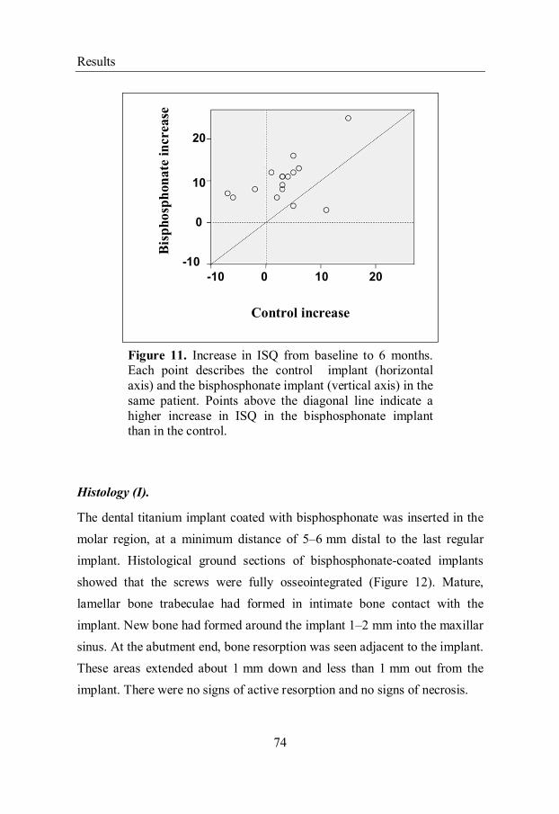
Results
74
Control increase
Bis
ph
osp
ho
nate
inc
rea
se
0
10
20
0 10 20-10
-10
Control increase
Bis
ph
osp
hon
ate
incr
ease
Figure 11. Increase in ISQ from baseline to 6 months. Each point describes the control implant (horizontal axis) and the bisphosphonate implant (vertical axis) in the same patient. Points above the diagonal line indicate a higher increase in ISQ in the bisphosphonate implant than in the control.
Histology (I).
The dental titanium implant coated with bisphosphonate was inserted in the
molar region, at a minimum distance of 5–6 mm distal to the last regular
implant. Histological ground sections of bisphosphonate-coated implants
showed that the screws were fully osseointegrated (Figure 12). Mature,
lamellar bone trabeculae had formed in intimate bone contact with the
implant. New bone had formed around the implant 1–2 mm into the maxillar
sinus. At the abutment end, bone resorption was seen adjacent to the implant.
These areas extended about 1 mm down and less than 1 mm out from the
implant. There were no signs of active resorption and no signs of necrosis.

Results
75
Figure 12. Histological cross-sectional image of a bisphosphonate-coated implant after 6 months.
Short summary of results of experimental studies (III)
Can an ONJ-like lesion be reproducibly produced in a rat model?
All 10 animals in the systemic group developed large ONJ-like lesions
(Figure 13). The alendronate and control groups showed an intact overlying
mucosa in all rats (Figure 13). This was confirmed by histology. All animals
with ONJ-like lesions showed discontinuity of the overlying epithelium,
bony sequestra and inflammatory cells. Lactate dehydrogenase assay showed
living osteocytes in the maxillae without tooth extraction. On the side of
tooth extraction, most osteocytes also appeared normal, except in regions
close to the necrotic wound. Because of technical problems, mainly with
cutting of the specimens, only six of 10 samples could be examined. The
negative controls showed no living (i.e. stained) osteocytes.

Results
76
Figure 13. Normal healing after extraction of first molar in the Control and Alendronate groups (A and B). Bone exposure in rat treated with dexamethasone and alendronate (C).
Short summary of results of experimental studies (IV)
Can a bisphosphonate coating improve implant fixation also under
conditions where there is a high risk of ONJ-like lesions in rats?
All 10 animals with systemic alendronate treatment developed large ONJ-
like changes, while all of them with local treatment were completely healed
(Figure 14). Implant removal torque was higher for the bisphosphonate-
coated implants than in the other groups (p < 0.03 for each comparison).

Results
77
Micro-CT of the maxilla showed more bone loss in the systemic alendronate
group than in groups receiving local treatment (p = 0.001). The bone density
in the immediate vicinity of the implant was higher for the Local group than
for the Controls, and lower for the Systemic group compared with controls
(p = 0.001 for both comparisons).
Figure 14. Normal healing after extraction of first molar and insertion of oral implant into the Control group (A), the Local group (B), and the Dexamethasone group (C). Bone exposure in rat treated with dexamethasone and alendronate (Systemic) (D).
Short summary of results of experimental studies (V)
Can early mucoperiostal coverage prevent ONJ-like lesions?
All animals in the non-coverage group developed large ONJ-like changes
(Figure 15). The coverage and control groups showed an intact overlying

Results
78
mucosa in all rats (Figure 15). Findings were confirmed with histology. Due
to technical problems, mainly with cutting the specimens, only 9 of the
control and coverage samples could be examined. These two samples were
then excluded from histological examination. The blinded evaluation showed
epithelial discontinuity in all non-coverage cases and in none of the others.
Large sequestrae or areas with empty osteocyte lacunae were seen in all non-
coverage cases. In the other groups, minor sequestrae were seen
occasionally. Inflammatory cells were also more common in the non-
coverage group.
Figure 15. Extraction site at harvest. Control (A), Coverage (B), and Non-coverage (C) rats (right panel: overview; left panel: magnified view of gingival healing area).

Discussion
79
Discussion
Implants and local delivery of bisphosphonate
Throughout history, humans have sought ways to replace lost teeth.
However, it is only within the past 100 years that members of the dental and
medical professions have made substantial progress in the permanent
replacement of missing teeth by intraosseous anchorage of artificial metal
fixtures. Fifty years later, the term osseointegration was coined by
Brånemark. Direct contact between living bone tissue and titanium implants
can lead to biological adhesion. Osseointegration is observed in several
areas, not only with dental implants, but also with maxillofacial implants,
replacement of damaged joints, and placement of artificial limbs. The
success of these operations is dependent of the fixation of the implants,
which, in turn, depends on the strength of the bone that holds them. If bone
quality is poor, surgical procedures can be modified to provide sufficient
mechanical fixation by adding more screws or larger devices, or by
protecting the implant from mechanical loading for a considerable time after
surgery, for osseointegration. Thus, if the quality of the bone holding an
implant could be improved locally, surgical procedures would become
simpler and rehabilitation would become faster.
To improve bone-to-implant contact, most manufacturers of dental implants
have focused on implant surfaces (101). Common methods of treating
titanium dental implant surfaces are blasting, acid-etching, and chemical
modification (124, 300).
Bisphosphonates are anti-resorptive drugs that act specifically on osteoclasts,
thereby maintaining bone density and strength (202). Once released from the
Discussion
80
surface of a coated implant, bisphosphonates reduce osteoclast activity,
thereby changing the balance of bone turnover in favor of bone formation,
leading to a net gain in local bone density.
During the last decades, the stability of implants with local bisphosphonate
treatment has been tested in clinical studies (268) and animal studies (255-
259), but not in human jaws. One problem is to find a method to measure the
effect of bisphosphonate treatment on implant fixation in terms of stability.
To our knowledge, this is available only for dental implants, by
measurement of mechanical resonance frequency, which provides unique
opportunities for implant research. One reason for this is that dental implants
are accessible for examination without surgical exposure, which is in the
case with orthopedic implants.
Resonance-frequency analysis is a reliable and non-invasive method to
measure the quality of fixation in humans. The method, its validation, and
clinical use have been comprehensively reviewed (179, 182-184). The
changes in implant stability expressed by differences in ISQ value over time
reflect the biologic events associated with the bone-implant interface. In two
clinical studies, a total of 96 implants were inserted in 21 patients. In a
randomized trial with a paired design, one implant of each pair was coated
with a thin fibrinogen layer containing two bisphosphonates (pamidronate
and Ibandronate). The bisphosphonate-coated implants showed a larger
increase in ISQ value from baseline to 6 months than did the controls. It is
unclear at what time the bisphosphonate exerted its effect during the healing
process. It is a weakness of these studies that early stages of the healing
period of the implants were not studied by resonance-frequency analysis.
The reason is that the studies were conducted as two-stage procedures.

Discussion
81
However, the positive effect on radiographic structure already at 2 months
might suggest that early changes in stability could be seen in the initial phase
of the healing process.
Periapical radiographs have been used routinely to evaluate longitudinal
bone loss around implants. The obvious advantages are that they are non-
invasive, non-destructive techniques that can be used at multiple sites and
several times in clinical trials. The disadvantage is that radiographs provide a
two-dimensional representation of a three-dimensional volume. Despite this,
differences around implants over time can be detected in radiographs and are
included in the success criteria in most clinical trials.
The level of marginal bone around the implant is widely considered to be
one of the most important reference criteria for monitoring of peri-implant
health and for evaluation of the long-term success of dental implants.
Several factors may contribute to marginal bone loss. It may be the result of
bacterial contamination, promoting the occurrence of peri-implantitis and
progressive bone resorption (301). Incorrect fixture positioning may also
cause marginal bone loss. The most coronal portion of peri-implant bone
may tend to resorb if the fixture is placed with a residual buccal wall that is
too thin. Following implant placement, a small amount of marginal bone
resorption occurs and these small changes are considered to be part of a
physiological process.
Study II extends only to abutment connection at 6 months. We have now
followed these patients with a new radiography at 18 months. The difference
between coated and control implants remained significant (p = 0. 04).
Discussion
82
The largest available follow-up material after routine implant insertion (n =
431) showed a mean marginal resorption from insertion to 18 months of 0.7
mm with an SD of 1.0 (302). Judging by the SD, the distribution is quite
skewed. Our figures for uncoated implants are similar to these, with a mean
of 0.63 and SD of 0.58 (although we prefer to report median and range).
Based on their findings in submerged implants, Albrektsson et al. (139, 303)
proposed criteria for implant success, including the absence of implant
mobility and absence of pain. Thus, 1 mm of bone loss was considered
acceptable during the first year of function, and 0.2 mm annually thereafter.
In our studies, the marginal bone loss was less with bisphosphonate- coated
implants than with the controls at 2, 6 and 18 months. The difference
between coated and control implants did not change significantly between 6
and 18 months (Figure 16). A clinical difference of 1 mm can be regarded as
negligible. However, in highly demanding cases such as implants in the
anterior maxilla, preservation of the marginal bone level is critical for an
“esthetic” result.

Discussion
83
Figure 16. Difference in marginal bone loss between control implants and bisphosphonate-coated implants at baseline and 2, 6, and 18 months postoperatively. Note that study II reports only 2 and 6 months.
Resonance-frequency analysis
Resonance-frequency analysis (RFA) is a non-invasive, objective method for
evaluation of implant stability and has been validated through several in
vitro and in vivo studies (178-180). This technique does not correlate with
the insertion torque, as the torque is mostly due to surface friction (304,
305). However the method correlates well with cortical bone thickness (306)
and the cutting resistance at the time of implant placement (183). The use of
resonance- frequency analysis may provide the possibility of identifying
implants with higher risk of failure and the ability to individualize implant
treatment. For example, early overload of an implant may cause the
resonance frequency to decrease, which serves as a warning, so that

Discussion
84
loading can be interrupted and the implant allowed to regain fixation
(307). Furthermore, in a longitudinal study by Glauser et al. (192), ISQ
values of 72 stable implants was compared with those of nine implants that
had lost stability over 1 year according to an immediate/early-loading
protocol. The implants that failed during the course of the study showed
significantly lower stability already after 1 month. Similar findings have
been published by Atieh et al. (308).
Rat model of ONJ
The Pathophysiology of ONJ is not well understood. An animal model of
ONJ has therefore been developed to mimic bisphosphonate-related
osteonecrosis in cancer patients. Following tooth extractions, a combination
of zoledronate and dexamethasone caused ONJ-like lesions in the majority
of rats (309). The importance of surgical trauma in the development of ONJ
has been emphasized by these authors (309). It is important to note that the
animals underwent a rigorous dental procedure (extraction of all three
molars in any given jaw bone) that generally exceeds the sort of dental
trauma usually experienced by humans who develop BRONJ. In our animal
studies, only one molar was extracted, but we added partial excision of
gingiva adjacent to the extraction site, to increase the area of bone exposure.
This might explain our higher success rate, with lesions in all cases.
As with any animal study involving pharmaceutical agents, it is important to
consider the doses administered with respect to those used clinically.
Clinically, cancer patients are given zoledronate as an intravenous infusion
every 4 weeks at a dose of 4 mg for an individual weighing 60 kg (~66
μg/kg) (310). In an attempt to mimic dosing for multiple myeloma, animals

Discussion
85
in our studies were dosed daily with alendronate using a subcutaneous
injection of 200 μg/kg for 14 days.
Previous experience in our group has shown that strong inhibition of
osteoclastic activity in rats can be achieved with this dose (311). When
calculated as dose per body weight per day, the rat dose is 100 times higher
than the human dose. However, the serum concentration after each injection
may be quite similar, as the metabolic rate of rats is estimated to be 3 times
higher than that of humans (312).
The relevance of our model for human ONJ is not certain. One important
difference between rats and humans may be size. Vascular outgrowth,
epithelial proliferation and other processes of healing are likely to occur at
roughly the same speed (in mm per day) in animals of different sizes. Thus,
our model may be relevant mainly for millimeter-sized lesions rather than
for complete extraction defects in humans. On the other hand, similar rat
models are regarded as relevant, at least for the principal aspects of ONJ
(309, 313).
A weakness in our studies was that the bisphosphonate treatment in our
model started at tooth extraction, whereas in clinical ONJ the treatment has
lasted for a long time before this. However, if ONJ starts with a
contaminated lesion rather than primary sterile osteonecrosis, the
pathophysiological processes will take place under the influence of
bisphosphonates in both cases. Moreover, this timing was chosen because it
further demonstrates that no bone pathology pre-dating tooth extraction is
necessary for the development of BRONJ.

Discussion
86
Implants and coating technique
The coating technique for dental implants in this study was presented by
Tengvall et al. (260). Briefly, a crosslinked layer of fibrinogen was
covalently bound to the metal, and then small amounts of pamidronate and
ibandronate were bound and adsorbed to the fibrinogen matrix. In an animal
model, the thickness of this bisphosphonate layer (a few nm) was measured
by ellipsometry (255). Ellipsometry is an optical method that is often used to
measure the thickness of thin films adsorbed to flat surfaces. However,
because the surface area of the screw is much greater than a corresponding
flat surface, it may be difficult to translate the amount of drug on a flat
surface to that of the screw.
The recommended human dose of alendronate for treatment of osteoporosis
is 10 mg/day. The gastrointestinal uptake of oral bisphosphonates is about
0.7% for alendronate (225) and 0.3% for pamidronate (226). For individuals
weighing 75 kg, this means that of the prescribed human daily dose of
bisphosphonates, approximately 0.5-1 µg/kg/day reaches the bloodstream.
For comparison, the total amount of bisphosphonate on coated human dental
implants is in the order of 1 µg ibandronate (less than 1µg /cm2). Clinically,
osteoporosis patients are given ibandronate as an oral dose of 50mg per day
for individuals weighing 60 kg (~ 833 μg/kg) (314). Thus, the amount of
bisphosphonate on the surface of the dental implant corresponds to the total
body dose of one day of osteoporosis treatment.
After insertion of bisphosphonate-coated implants, the bisphosphonate is
released from the surface and rapidly accumulates in the surrounding bone
tissues. In an animal model using a fibrinogen immobilization matrix and

Discussion
87
14C-alendronate, 60% of the immobilized bisphosphonate was released after
8 h, but the release continued slowly for up to 8 days (255).
Once released from the surface of a coated implant, bisphosphonates reduce
osteoclast activity, thereby changing the balance of bone turnover in favor of
bone formation, leading to a net gain in local bone density (314). Fast
formation of a shell of new woven bone surrounding the implant is seen,
which becomes slowly remodelled into lamellar bone (315). Furthermore, it
has been shown that the amount of bone increases adjacent to the implant,
with a maximum density 250 μm from the implant surface (316).
One question regarding local bisphosphonate therapy of any type is the
nature of the systemic side effects that the drug may have on skeletal
remodeling. Following elevation of the mucoperiosteal flap Yaffe et al.
(317) applied 22 μg of 14C-labeled alendronate directly to alveolar bone with
a soaked gelatin sponge in a rat model. The local absorbtion of alendronate
and its disposition in the contralateral side of the mandible as well as in the
tibia bone were analyzed. The mean total amount of drug measured in the
entire left tibia after 60 min was 3.2% of the dose applied (0.7 μg).
McKenzie et al. (318) inserted hydroxyapatite-coated implants with 100 μg 14C-labeled zoledronate in dogs’ femoral bone. Bone samples adjacent to and
distant from the implant were harvested and the concentration of
radiolabeled bisphosphonate in each sample was quantified using liquid
scintillation spectrophotometry. The mean zoledronate concentration in the
cortical bone adjacent to the implant (peri-implant bone) was 733 ng/g bone
at 6 weeks and 377 ng/g bone at 52 weeks. At 6 weeks small amounts of
zoledronate (≤ 7.2 ng/g) were detected throughout the skeleton, indicating
some escape into the circulation after local elution. This indicated that most
of the bisphosphonate remained at the implant site. By using the isotope-

Discussion
88
labeled bisphosphonate in a parallel series in our animal model (study IV),
the bisphosphonate amount per screw was found to be approximately 400
ng. This is a very low dose compared with the dose currently used in clinical
practice.
These studies support our notion that local delivery of bisphosphonates and
their affinity for bone may become an important treatment modality to
prevent resorption of bone during dental and orthopedic procedures. It seems
that the risk of systemic side effects of local delivery of bisphosphonate
would be small.
Lactate dehydrogenase analysis
Bisphosphonates have been associated with osteonecrosis of the jaw, and it
has been suggested that this condition starts with sterile bone death. We
hypothesized that BRONJ can develop without being preceded by sterile
bone death. The viability of osteocytes was therefore the subject of this
examination. Lactate dehydrogenase (LDH) is a cytoplasmic enzyme found
in most living cells. The lactate dehydrogenase assay is a popular method to
detect cell viability in bone sections. The major advantage of the LDH assay
is the stability of the LDH enzyme for up to 36 h after cell death, eliminating
any false negative viability results due to processing of the tissue. With this
method, viable osteocytes react to form non-reversible tetrazolium-formazan
granules, while non-viable osteocytes are distinguished with methyl green
stain.
The use of staining of LDH activity as a measurement of osteocyte viability
goes back to 1982 when Wong et al. (319) introduced this histological

Discussion
89
method for use on bone sections derived from human femoral heads. They
demonstrated that viable osteocytes stained positive for LDH activity, while
it was already known that necrotic tissue does not show any staining of LDH
activity staining. Sambrook et al. (320) showed that the number of viable
osteocytes was significantly reduced in femoral head bone of glucocorticoid-
induced osteoporosis patients compared with controls. They use frozen bone
samples for this verification. We used a lactate dehydrogenase assay
according to a modified protocol by Phillips et al. (321). By increasing the
concentrations of coenzyme and tetrazole, these authors obtained excellent
reactivity. Fresh, unfixed specimens were used to preserve LDH activity.
Since the enzyme persists only for 36–48 h at 37 °C after sacrifice (319), the
specimens should be processed within 24 h to minimize artifact (321).
However, freezing results in disruption of the structural integrity of the cell
(322).
Reported specimen thicknesses for sawn fresh bone vary from 200 μm (320,
323) to 300 μm (320, 324). Phillips et al. (319) used 400-μm-thick fresh
bone and demonstrated that section thickness may not affect osteocyte
reactivity. However, it may affect the length of time required for
decalcification. In the present study, we used 800-μm-thick fresh bone.
However, sawing a thinner section would probably cause damage due to the
low density of the maxillary bone.
Pathophysiology of ONJ
The pathophysiology of ONJ is not well defined, and the condition is
probably multifactorial in nature. While it this is most likely associated with
bisphosphonate use, a causal effect has yet to be demonstrated. There are
Discussion
90
several features that make the oral cavity a unique environment. The alveolar
bone in both the mandible and the maxilla is covered by a thin layer of
periosteum and epithelium, which are subjected to a wide variety of stresses
such as mastication forces, dental procedures, and periodontal disease. This
combination of constant stress predisposes the thin mucosa to trauma,
leading to exposure of bone.
When bisphosphonate-related osteonecrosis of the jaw (BRONJ) was first
described, its similarities with radiation-induced osteonecrosis led to the
assumption that the condition started with sterile necrosis of the jaw bone. At
that time, it was speculated that bisphosphonates could cause osteonecrosis
through effects on blood vessels in bone, possibly by inhibition of vascular
endothelial growth factor (VEGF) (280-282). It has been demonstrated that
patients with advanced solid cancers and associated bone metastases have
significantly reduced levels of serum VEGF when treated with zoledronate
or pamidronate (280, 325, 326). However, a recent clinical trial compared
the incidence of ONJ in breast cancer patients receiving bevacizumab
(avastin, anti-VEGF-1 monoclonal antibody) or placebo (327). The analysis
failed to show any increased incidence of ONJ in the risk patients. The anti-
angiogenic role of bisphosphonate is still unclear. The notable incidence of
ONJ in the maxilla appears to refute the idea of the anti-angiogenic effect of
bisphosphonate being the sole cause of ONJ.
It was later suggested that BRONJ does not begin as a form of classical
osteonecrosis, but is in fact osteomyelitits already from the beginning (283,
284). Microbial contamination with Actinomyces appears to play an
important role in maintaining the osteomyelitic wounds (285, 286). Hansen
et al (285) examined histopathological specimens from patients with

Discussion
91
actinomycosis of the jaw. Out of a total number of 45 patients with
actinomycosis, 43 (93%) suffered from ONJ (58%) or infected
osteoradionecrosis (35%). Furthermore, the oral cavity and teeth are
colonized by a complex microbial flora, many of which are pathogenic
organisms. The intimate relationship between teeth and jaws allows a portal
of entry for microbes and other inflammatory products to the underlying
bone, a situation that is not found in any other part of the body. Hence, oral
surgical procedures exposing bone to the oral cavity increase the risk of
ONJ. In a rat model, we showed that ONJ-like lesions can be prevented
when extraction sockets are covered with a mucoperiosteal flap (328). This
shows the importance of bacterial role in the development of this condition.
Because jaw bone containing bisphosphonates will resorbed slowly, it is
conceivable that bacterially contaminated bone cannot be removed fast
enough to prevent the development of chronic osteomyelitis. This view is
supported by the observation that similar lesions appear after treatment with
an anti-RANKL antibody that reduces osteoclast recruitment (287).
Thus, it appears that resorptive activity is a key factor in bone healing.
Based on these considerations, this condition might be called
bisphosphonate-induced osteomyelitis of the jaw.

Conclusions
93
Conclusions
Studies I and II
A thin, bisphosphonate-eluting fibrinogen coating can improve the fixation
of metal implants in human bone. This might lead to new possibilities for
orthopedic surgery in osteoporotic bone and for dental implants.
Study III
ONJ-like lesions were reproducibly induced in a rat model at sites of tooth
extraction, whereas there were no signs of osteocyte death in uninjured sites.
Osteonecrosis of the jaw appears to arise first after the bone has been
exposed.
Study IV
In a rat model, local bisphosphonate treatment with zoledronate improved
implant fixation in a setting where systemic treatment caused ONJ.
Improved fixation of bisphosphonate-coated dental implants in humans
might be achieved with a smaller risk of such complications in comparison
with systemic treatment.
Study V
Mucoperiosteal coverage of newly exposed bone prevented ONJ-like lesions
in rats. Immediate coverage of extraction sites might be recommended for
patients at risk of ONJ.
This treatment has now been implemented at our department.


What next?
95
What next?
We started this project by showing that local treatment of implants with
bisphosphonate may have a future place in orthopedic surgery and dental
surgery, since bisphosphonate coatings improved the fixation of dental
implants in the human jaw. However, the clinical benefits of this technique
are still not understood. One could speculate that if bisphosphonates have an
effect in the early phase of healing, then rehabilitation after implantation
would become faster. This hypothesis still has to be tested.
Hypothetical positive long term effects would include reduced rates of
mechanical loosening and peri-implantitis. In order to study such effects, it
will be required a very large number of patients that is not currently within
reach. Another important issue is the risk that patients might be exposed to
with the use of locally delivered bisphosphonates. A potential risk would be
the peri-implantitis, which may jeopardize the entire perception of local
bisphosphonate treatment. However, to exclude that such risks exist (with
sufficient confidence intervals), again a very large number of patients would
be required. At present, we can only say that if any such local adverse effects
would appear, the problem would be easily solved by removing the
bisphosphonate-containing bone in the immediate vicinity of the implant.
The posterior maxilla has been described as the most difficult and
problematic part of the mouth for the implant practitioner, and it requires the
most ingenuity for achievement of successful results. Anatomical
considerations include reduced bone quantity, especially in patients who
have experienced alveolar resorption in the wake of tooth loss. In these
cases, it may be necessary to perform a bone augmentation procedure before
placement of the implant. However, one drawback in using autogenous bone

What next?
96
is the unpredictable resorption of bone, which might be reduced by the use
of bisphosphonates, perhaps locally. More short- and long-term data are
needed for us to fully evaluate the benefits of bisphosphonate use in oral
surgery.

Acknowledgements
97
Acknowledgements
I am particularly grateful to Prof. Per Aspenberg, my main supervisor and
co-worker, for his friendship, encouragement, and guidance through the
years.
I also thank my co-supervisor Agneta Marcusson for support and for
introducing me to the field of science.
My co-authors:
Prof. Pentti Tengvall, thank you for inspiring discussions on coating
techniques and for support.
Fredrik Agholme and Olof Sandberg, thank you both for valuable
discussions, for support, and for helping me with laboratory work.
Other collaborators of great importance
This study could not have been performed without generous financial
support from Folktandvården Östergötland and Sinnescentrum.
My friends and colleagues in Linköping, Bergen and Örebro
I express my sincere appreciation and thanks to my colleagues:
Sten Sahlholm, Björn Olaisson, Johan Nilsson, Maths Bellinetto Ericson,
Reine Sjöman, Anders Ottosson, Christer Lindgren, Gustav Henefalk, Vera
Alstad, Lennart Andersson, Elionor Ärleskog, Nils Ravald, Börje Svensson,
Arezo Tardast, John Helge Heimdal and Prof. Jan Olofsson.

Acknowledgements
98
I also thank Prof. Lena Norberg Spaak for valuable advice and support
during this never- ending journey (The Doors).
I express my sincere appreciation and thanks to my friends Kristina and
Svante Nagy, Birgitta and Per-Otto Waern, Masoud Amozegar and Sara
Yildiz and Hamest Martirosian for all your kind words and for all the laughs.
I also thank Trine Vikinge and AddBio AB for coating of titanium screws in
study 4 and for valuable discussions.
My family
I thank my family Lina and Nick for their love, great understanding, and
incredible patience. My mother and my brother, Dr. Farmehr Abtahi, for
very fruitful discussions and support throughout my life. Finally, in memory
of my father who unfortunately passed away much too early in my life, but
who has given me the power to produce this thesis.

References
99
References
1. Raspanti M, Congiu T, Guizzardi S. Tapping-mode atomic force
microscopy in fluid of hydrated extracellular matrix. Matrix Biol 20: 601-04
2001.
2. Baselt DR, Revel JP, Baldschwieler JD. Subfibrillar structure of type I
collagen observed by atomic force microscopy. Biophys J 65: 2644-55 1993.
3. Forwood MR, Burr DB. Physical activity and bone mass: exercises in
futility? Bone Miner 21: 89-112 1993.
4. Khan K, Mckay H, Kannus P et al. Physical activity and bone health.
Human kinetics 2001.
5. Owen, M. Marrow stromal stem cells. J Cell Sci Suppl 10: 63-76 1988.
6. Caplan, AI. Mesenchymal stem cells. J Orthop Res 9: 641-50 1991.
7. Agata H, Asahina I, Yamazaki Y, Uchida M, Shinohara Y, Honda MJ,
Kagami H, Ueda M. "Effective bone engineering with periosteum-derived
cells.". Journal of dental research 86: 79-83 2007.
8. Pittenger MF, Mackay AM, Beck SC, Jaiswal RK, Douglas R, Mosca JD,
Moorman MA, Simonetti DW, Craig S, Marshak DR. Multilineage potential
of adult human mesenchymal stem cells. Science 284: 143-47 1999.
9. Friedenstein AJ, Chailakhjan RK, Lalykina KS. The development of
fibroblast colonies in monolayer cultures of guinea pig bone marrow and
spleen cells. Cell Tissue Kinet 3: 393-402 1970.

References
100
10. Friedenstein AJ. Precursor cells of mechanocytes. Int Rev Cytol 47: 327-
55 1976.
11. Friedenstein AJ, Deriglasova UF, Kulagina NN, Panasuk AF, Rudokowa
SF, Luria EA, Rudakowa IA. Precursors for fibroblasts in different
populations of hematopoietic cells as detected by the in vitro colony assay
method. Exp Hematol 2: 83-92 1974.
12. Friedenstein AJ. Osteogenic stem cells in the bone marrow. Bone Miner
Res 7: 243-72 1990.
13. Wang EA, Rosen V, Cordes P, Hewick RM, Kriz MJ, Luxenberg DP,
Sibley BS, Wozney JM. Purification and characterization of other distinct
bone-inducing factors. Proc Natl Acad Sci U S A 85: 9484-8 1988.
14. Wozney JM, Rosen V, Celeste AJ, Mitsock LM, Whitters MJ, Kriz RW,
Hewick RM, Wang EA. Novel regulators of bone formation: molecular
clones and activities. Science 16: 1528-34 1988.
15. Ducy P, Zhang R, Geoffroy V, Ridall AL, Karsenty G. Osf2/Cbfa1, a
transcriptional activator of osteoblast differentiation. Cell 89: 747-54 1997.
16. Rosen ED, Sarraf P, Troy AE, Bradwin G, Moore K, Milstone DS,
Spiegelman BM, Mortensen RM. PPAR gamma is required for the
differentiation of adipose tissue.
in vivo and in vitro. Mol Cell 4: 611-17 1999.
17. de Crombrugghe B, Lefebvre V, Behringer RR, Bi W, Murakami S,
Huang W. Transcriptional mechanisms of chondrocyte differentiation.
Matrix Biol: 19: 389-94 2000.
References
101
18. Dempster DW, Cosman F, Parisien M, Shen V, Lindsay R. Anabolic
actions of parathyroid hormone on bone. Endocr Rev 14: 690-709 1993.
19. Turner RT, Riggs BL, Spelsberg TC. Skeletal effects of estrogen. Endocr
Rev 15: 275-300 1994.
20. Lian JB, Stein GS, Stein JL, van Wijnen AJ. Regulated expression of the
bone specific osteocalcin gene by vitamins and hormones. Vitam Horm 55:
443-509 1999.
21. Wang ZQ, Ovitt C, Grigoriadis AE, Mohle-Steinlein U, Ruther U,
Wagner EF. Bone and haematopoietic defects in mice lacking c-fos. Nature
360: 741-45 1992.
22. Grigoriadis AE, Wang ZQ, Wagner EF. Fos and bone cell development:
lessons from a nuclear oncogene. Trends Genet 11: 436-41 1995.
23. Watanabe H, Saitoh K, Kameda T, Murakami M, Niikura Y, Okazaki S,
Morishita Y, Mori S, Yokouchi Y, Kuroiwa A, Iba H. Chondrocytes as a
specific target of ectopic Fos expression in early development. Proc Natl
Acad Sci U S A 15: 3994-9 1997.
24. Delany AM, Dong Y, Canalis E. Mechanisms of glucocorticoid action in
bone cells. J Cell Biochem 56: 295-302 1994.
25. Vanderoost J, Søe K, Merrild DM, Delaissé JM, van Lenthe GH.
Glucocorticoid-Induced Changes in the Geometry of Osteoclast Resorption
Cavities Affect Trabecular Bone Stiffness. Calcif Tissue Int Nov 28. 2012.
26. Canalis E, Bilezikian JP, Angeli A, Giustina A. Perspectives on
glucocorticoid-induced osteoporosis. Bone 34: 593-8 2004.
References
102
27. Hofbauer LC, Gori F, Riggs BL, Lacey DL, Dunstan CR, SpelsbergTC,
et al. Stimulation of osteoprotegerin ligand and inhibition of osteoprotegerin
production by glucocorticoids in human osteoblastic lineage cells: potential
paracrine mechanisms of glucocorticoid-induced osteoporosis.
Endocrinology 140: 4382-9 1999.
28. Rubin J, Biskobing DM, Jadhav L, Fan D, Nanes MS, Perkins S, et al.
Dexamethasone promotes expression of membrane-bound macrophage
colony-stimulating factor in murine osteoblast-like cells. Endocrinology 139:
1006-12 1998.
29. Pereira RMR, Delany AM, Canalis E. Cortisol inhibits the differentiation
and apoptosis of osteoblasts in culture. Bone 28: 484-90 2001.
30. Xia X, Kar R, Gluhak-Heinrich J, Yao W, Lane NE, Bonewald LF,
Biswas SK, Lo WK, Jiang JX. Glucocorticoid-induced autophagy in
osteocytes. J Bone Miner Res 11: 2479-88 2010.
31. Väänänen HK, Laitala-Leinonen T. Osteoclast lineage and function.
Arch Biochem Biophys 15: 132-8 2008.
32. Felix R, Halasy-Nagy J, Wetterwald A, Cecchini MG, Fleisch H,
Hofstetter W. Synthesis of membrane- and matrix-bound colony-stimulating
factor-1 by cultured osteoblasts. J Cell Physiol 166: 311-22 1996.
33. Boyle WJ, Simonet WS, Lacey DL. Osteoclast differentiation and
activation. Nature 15: 337-42 2003.
34. Simonet WS, Lacey DL, Dunstan CR, Kelley M, Chang MS, Lüthy R,
Nguyen HQ, Wooden S, Bennett L, Boone T, Shimamoto G, DeRose M,

References
103
Elliott R, Colombero A, Tan HL, Trail G, Sullivan J, Davy E, Bucay N,
Renshaw-Gegg L, Hughes TM, Hill D, Pattison W, Campbell P. Sander S,
Van G, Tarpley J, Derby P, Lee R, Boyle WJ. Osteoprotegerin: a novel
secreted protein involved in the regulation of bone density.Cell 18: 309-19
1997.
35. Lacey DL, Timms E, Tan HL, Kelley MJ, Dunstan CR, Burgess T,
Elliott R, Colombero A, Elliott G, Scully S, Hsu H, Sullivan J, Hawkins N,
Davy E, Capparelli C, Eli A, Qian YX, Kaufman S, Sarosi I, Shalhoub V,
Senaldi G, Guo J, Delaney J, Boyle WJ. Osteoprotegerin ligand is a cytokine
that regulates osteoclast differentiation and activation. Cell 17: 165-76 1998.
36. Vignery A. Osteoclasts and giant cells: macrophage-macrophage fusion
mechanism. Int J Exp Pathol 81: 291-304 2000.
37. Tolar J, Teitelbaum SL, Orchard PJ. Osteopetrosis. N Engl J Med 30:
2839-49 2004.
38. Väänänen HK, Zhao H, Mulari M, Halleen JM. The cell biology of
osteoclast function. J Cell Sci 113: 377-81 2000.
39. Silver IA, Murrills RJ, Etherington DJ. Microelectrode studies on the
acid microenvironment beneath adherent macrophages and osteoclasts. Exp
Cell Res 175: 266-76 1988.
40. Blair HC. How the osteoclast degrades bone. Bioessays 20: 837-46 1998.
41. Manolagas SC. Birth and death of bone cells: basic regulatory
mechanisms and implications for the pathogenesis and treatment of
osteoporosis. Endocr Rev 21: 115-37 2000.
References
104
42. McCulloch CA, Heersche JN. Lifetime of the osteoblast in mouse
periodontium. Anat Rec 222: 128-35 1988.
43. Currey JD. The many adaptations of bone. J Biomech 36: 1487-95 2003.
44. Doty SB. Morphological evidence of gap junctions between bone cells.
33: 509-12 1981.
45. Palumbo C, Palazzini S, Marotti G. Morphological study of intercellular
junctions during osteocyte differentiation. Bone 11: 401-6 1990.
46. Yellowley CE, Li Z, Zhou Z, Jacobs CR, Donahue HJ. Functional gap
junctions between osteocytic and osteoblastic cells. J Bone Miner Res 15:
209-17 2000.
47. Burger EH, Klein-Nulend J. Mechanotransduction in bone-role of the
lacuno-canalicular network. FASEB J 13: 101-12 1999.
48. Alford AI, Jacobs CR, Donahue HJ. Oscillating fluid flow regulates gap
junction. Communication in osteocytic MLO-Y4 cells by an ERK1/2 MAP
kinase-dependen mechanism small star, filled. Bone 33: 64-70 2003.
49. Mullender MG, van der Meer DD, Huiskes R, Lips P. Osteocyte density
changes in aging and osteoporosis. Bone 18: 109-13 1996.
50. Cameron DA, Paschall HA, Robinson RA Changes in the fine structure
of bone cells after the administration of parathyroid extract. J Cell Biol 33:
1-14 1967.

References
105
51. B.S. Noble, H. Stevens, N. Loveridge, J. Reeve Identification of
apoptotic changes in osteocytes in normal and pathological human bone.
Bone 20: 273-82 1997.
52. O. Verborgt, G.J. Gibson, M.B. Schaffler. Loss of osteocyte integrity in
association with microdamage and bone remodeling after fatigue in vivo. J
Bone Miner Res 15: 60-67 2000.
53. Qiu S-J, Hoshaw SJ, Gibson GJ, Lundin-Cannon KD, Schaffler MB.
Osteocyte apoptosis in reaction to matrix damage in compact bone Trans.
Orthop Res Soc 22: 89 1997.
54. Heino TJ, Kurata K, Higaki H, Väänänen HK. Evidence for the role of
osteocytes in the initiation of targeted remodeling. Technol Health Care 17:
49-56 2009.
55. Zaidi M. Skeletal remodelling in health and disease. Nature Med 13:
791-801 2007.
56. Cohen MM. The new bone biology: pathologic, molecular and clinical
correlates. Am J Med Genetics 140: 2646-2706 2006.
57. Martin TJ, Sims NA. Osteoclast-derived activity in the coupling of bone
formation to resorption. Trends Mol Med 11: 76-81 2005.
58. Martin TJ, Udagawa N. Hormonal regulation of osteclast function. TEM
9: 6-12 1998.
59. Suda T, Takahashi N, Udagawa N, Jimi E, Gillespie MT, Martin TJ.
Modulation of osteoclast differentiation and function by the new members of

References
106
the tumor necrosis factor receptor and ligand families. Endocrinol. Rev 20:
345-357 1999..
60. Bennett CN, Longo KA, Wright WS, et al. Regulation of
osteoblastogenesis and bone mass by Wnt 10b. Proc Natl Acad Sci USA
102: 3324-29 2005.
61. Krishnan V, Bryant HU, Macdougald OA. Regulation of bone mass by
Wnt signaling. J Clin Invest 11: 1202-9 2006.
62. Kong YY, Yoshida H, Sarosi I, Tan H L, Timms E, Capparelli C,
Morony S, Oliveira-dos-Santos AJ, Van G, Itie A, Khoo W, Wakeham A,
Dunstan CR, Lacey DL, Mak TW, Boyle WJ, Penninger JM. OPGL is a key
regulator of osteoclastogenesis, lymphocyte development and lymph-node
organogenesis. Nature 397: 315-23 1999.
63. Hofbauer LC, Lacey DL, Dunstan CR, Spelsberg TC, Riggs BL, Khosla
S. Interleukin-1beta and tumor necrosis factor-alpha, but not interleukin-6,
stimulate osteoprotegerin ligand gene expression in human osteoblastic cells.
Bone 25: 255-259 1999.
64. Mundy G R. The effects of TGF-beta on bone. Ciba Foundation 157: 137
1991.
65. Oursler, MJ. Osteoclast synthesis and secretion and activation of latent
transforming growth factor beta. J. Bone Miner. Res 9: 443–452 1994.
66. Quinn, JM, Itoh K, Udagawa N, Hausler K, Yasuda H, Shima N, Mizuno
A, Higashio K, Takahashi N, Suda T, Martin TJ, Gillespie MT.

References
107
Transforming growth factor beta affects osteoclast differentiation via direct
and indirect actions. J Bone Miner Res 16: 1787-1794 2001.
67. Bonewald LF. Transforming growth factor beta. In: Bilezikian JP, Raisz
LG, Rodan GA (eds.) Principles of Bone Biology. Academic Press, San
Diego, CA, USA 647–659 1996.
68. Bonewald LF. Regulation and regulatory activities of transforming
growth factor beta. Crit Rev Eukaryot Gene Expr 9: 33-44 1999.
69. Sennerby L, Thomsen P, Ericson L.E. Early tissue response to titanium
implants inserted in rabbit cortical bone. Light microscopic observations.
Journal of materials science, materials in medicine 4: 240-250 1993.
70. Futami T, Fujii N, Ohnishi H, Taguchi N, Kusakari H, Ohshima H,
Maeda T. Tissue response to titanium implants in the rat maxilla:
ultrastructural and histochemical observations of the bonetitanium interface.
Journal of Periodontology 71: 287-98 2000.
71. Park JY, Davies JE. Red blood cell and platelet interactions with
titanium implant surfaces. Clinical Oral Implants Research 11: 530-39 2000.
72. Nygren H, Eriksson C, Lausmaa J. Adhesion and activation of platelets
and polymorphonuclear granulocyte cells at TiO2 surfaces. J Lab Clin Med
129: 35-46 1997.
73. Weiss HJ, Hawiger J, Ruggeri ZM, Turitto VT, Thiagarajan P, Hoffmann
T. Fibrinogen-independent platelet adhesion and thrombus formation on
subendothelium mediated by glycoprotein IIb-IIIa complex at high shear
rate. J Clin Invest 83: 288-97 1989.

References
108
74. Chinn JA, Horbett TA, Ratner BD. Baboon fibrinogen adsorption and
platelet adhesion to polymeric materials. Thromb Haemost 6: 608-17 1991.
75. Berglundh T, Abrahamsson I, Lang NP, Lindhe J. De novo alveolar bone
formation adjacent to endosseous implants. Clin Oral Implants Res 14: 251-
62 2003.
76. Cook, H. P. Immidiate recintruction of the mandible by metallic implant
following resection for neoplasm. Ann R Coll Surg Engl 42: 233-39 1967.
77. Linder L, Lundskog, J. Incorporation of stainless steel, titanium and
vitallium in bone. Injury 6: 277-85 1975.
78. Jacobs CH, Berry JT, Pope HM, Hoaglund FT. A study of the bone
machining process drilling. J Biomech 9: 343-49 1976.
79. Muster D, Champy M. Le probleme d interface os-biomateriaux.
Actualites Odonto-Stomat 121: 109-24 1978.
80. Schüpbach P, Glauser R, Rocci A, Martignoni M, Sennerby L, Lundgren
A, Gottlow J. The human bone-oxidized titanium implant interface: A light
microscopic, scanning electron microscopic, back-scatter scanning electron
microscopic, and energy-dispersive x-ray study of clinically retrieved dental
implants. Clin Implant Dent Relat Res 1: 36-43 2005.
81. Albrektsson T, Brånemark PI, Hansson HA, Lindström J.
Osseointegrated titanium implants. Requirements for ensuring a long-lasting,
direct bone-to-implant anchorage in man. Acta Orthop Scand 52: 155-70
1981.

References
109
82. Albrektsson T, Hansson HA. An ultrastructural characterization of the
interface between bone and sputtered titanium or stainless steel surfaces.
Biomaterials 7: 201-5 1986.
83. Albrektsson T, Hansson HA, Ivarsson B. Interface analysis of titanium
and zirconium bone implants. Biomaterials 6: 97-101 1985.
84. Linder L, Albrektsson T, Brånemark PI, Hansson HA, Ivarsson B,
Jönsson U, Lundström I. Electron microscopic analysis of the bone-titanium
interface. Acta Othop Scand 54: 45-52 1983.
85. Sennerby L, Ericson LE, Thomsen P, Lekholm U, Astrand P. Structure
of the bone-titanium interface in retrieved clinical oral implants. Clin Oral
Implants Res 2: 103-11 1991.
86. Sennerby L, Thomsen P, Ericson L.E. Ultrastructure of the bone-titanium
interface in rabbits. J Mater Sci Mater Med 3: 262-71 1992.
87. Pilliar RM, Lee JM, Maniatopoulos C. Observations on the effect of
movement on bone ingrowth into porous-surfaced implants. Clin Orthop
208: 108-13 1986.
88. Szmukler-Moncler S, Salama H, Reingewirtz Y, Dubruille JH. Timing of
loading and effect of micromotion on bone-dental implant interface: review
of the experimental literature. J Biomed Mater Res 43: 192-203 1998.
89. Ducheyne P, De Meester P, Aernoudt E. Influence of a functional
dynamic loading on bone ingrowth into surface pores of orthopedic implants.
J Biomed Mater Res 6: 811-38 1977.

References
110
90. Cameron H, Macnab I, Pilliar R. Porous surfaced Vitallium staples. S
Afr J Surg 10: 63-70 1972.
91. Aspenberg P, Goodman S, Toksvig-Larsen S, Ryd L, Albrektsson T.
Intermittent micromotion inhibits bone ingrowth. Titanium implants in
rabbits. Acta Orthop Scand 63: 141-5 1992.
92. Brunski JB, Moccia AF, Pollock SR, Korostoff E, Trachtenberg DI. The
influence of functional use of endosseous dental implants on the tissue
implant interface: I. Histological aspects. J Dent Res 58: 1953-69 1979.
93. Cameron HU, Pilliar RM, MacNab I. The effect of movement on the
bonding of porous metal to bone. J Biomed Mater Res 7: 301-11 1973.
94. Søballe K, Hansen ES, Brockstedt–Rasmussen H, Bunger C. Tissue
ingrowth into titanium and hydroxyapatitecoated implants during stable and
unstable mechanical conditions. J Orthop Res 10: 285-99 1992.
95. Brunski JB. In vivo bone response to biomechanical loading at the
bone/dental-implant interface. Adv Dent Res 13: 99-119 1999.
96. Burke DW, O'Connor DO, Zalenski EB, Jasty M, Harris WH.
Micromotion of cemented and uncemented femoral components. J Bone
Joint Surg Br73: 33-7 1991.
97. Søballe K, Brockstedt-Rasmussen H, Hansen ES, Bünger C.
Hydroxyapatite coating modifies implant membrane formation. Controlled
micromotion studied in dogs. Acta Orthop Scand 63: 128-40 1992.

References
111
98. Søballe K, Hansen ES, Brockstedt-Rasmussen H, Bünger C.
Hydroxyapatite coating converts fibrous tissue to bone around loaded
implants. J Bone Joint Surg Br 75: 270-8 1993.
99. Akagawa Y, Hashomoto M, Kondo N, Satomi K, Takata T, Tsuru H.
Initial bone-implant interface of submergible and supramergible endosseous
single crystal-sapphire implants. J Prosthet Dent 55: 96-100 1986.
100. Trisi P, De Benedittis S, Perfetti G, Berardi D. Primary stability,
insertion torque and bone density of cylindric implant ad modum
Branemark: is there a relationship? An in vitro study. Clin Oral Implants Res
22: 567-70 2011.
101. T. Albrektsson, A. Wennerberg. Oral implant surfaces: Part 2 – review
focusing on clinical knowledge of different surface. Int J Prosthodont 5: 544-
64 2004.
102. Brunette DM, Chehroudi B. The effects of the surface topography of
micromachined titanium substrata on cell behavior in vitro and in vivo. J
Biomech Eng 121: 49-57 1999.
103. Kieswetter K, Schwartz Z, Dean DD, Boyan BD. The role of implant
surface characteristics in the healing of bone. Crit Rev Oral Biol Med 7:
329-45 1996.
104. Thomas K, Cook SD. Relationship between surface characteristics and
the degree of bone-implant integration. J Biomed Mater Res 26: 831–33
1992.

References
112
105. Gotfredsen K, Hjorting-Hansen E, Jensen JS, Holmen A.
Histomorphometric and removal torque analysis for TiO2-blasted titanium
implants. Clin Oral Implants Res 3: 77-84 1992.
106. Gotfredsen K, Wennerberg A, Johansson C, Skovgaard LT, Hjorting-
Hansen E. Anchorage of TiO2-blasted, HA-coated, and machined implants:
An experimental study in rabbits. J Biomed Mater Res 29: 1223-31 1995.
107. Buser D, Schenk RK, Steinemann S, Fiorellini JP, Fox CH, Stich H.
Influence of surface characteristics on bone integration of titanium implants.
A histomorphometric study in miniature pigs. J Biomed Mater Res 25: 889-
902 1991.
108. Buser D, Nydegger T, Hirt HP, Cochran DL, Nolte LP. Removal
torques values of titanium implants in the maxilla of miniature pigs. Int J
Oral Maxillofac Implants 13: 611-19 1998.
109. Albrektsson T, Wennerberg A. Oral implant surfaces: Part 1--review
focusing on topographic and chemical properties of different surfaces and in
vivo responses to them. Int J Prosthodont 17: 536-43 2004.
110. Schenk RK, Buser D. Osseointegration: a reality. Periodontol 2000 17:
22–35 1998.
111. Wennerberg A, Albrektsson T, Andersson B, Kroll JJ. A
histomorphometrical and removal torque study of screw-shaped titanium
implants with three different surface topographies. Clin Oral Implants Res 6:
24-30 1995.

References
113
112. Wennerberg A, Albrektsson T, Johanson CB, Andersson B.
Experimental study of turned and grit blasted screw-shaped implants with
special emphasis on effects of blasting material and surface topography.
Biomaterials 17: 15-22 1996.
113. Piconi C, Maccauo G. Zirconia as a ceramic biomaterial. Biomaterials
20: 1-25 1999.
114. Depprich R, Zipprich H, Ommerborn M, Mahn E, Lammers L,
Handschel J, Naujoks C, Wiesmann HP, Kübler NR, Meyer U.
Osseointegration of zirconia implants: an SEM observation of the bone-
implant interface. Head Face Med 6:4:25 2008.
115. Sennerby L, Dasmah A, Larsson B, Iverhed M. Bone tissue responses
to surface-modified zirconia implants: A histomorphometric and removal
torque study in the rabbit. Clin Implant Dent Relat Res 1: 13-20 2005.
116. Palmer RM, Palmer PJ, Smith BJ. A 5-year prospective study of Astra
single tooth implants. Clin Oral Implants Res 11: 179-82 2000.
117. Gotfredsen K. A 5-year prospective study of single-tooth replacements
supported by the Astra Tech implant: a pilot study. Clin Implant Dent Relat
Res 6: 1-8 2004.
118. Arvidson K, Bystedt H, Frykholm A, von Konow L, Lothigius E. Five-
year prospective follow-up report of the Astra Tech Dental Implant System
in the treatment of edentulous mandibles. Clin Oral Implants Res 9: 225-34
1998.

References
114
119. Cooper LF, Masuda T, Whitson SW, Yliheikkila P, Felton DA.
Formation of mineralizing osteoblast cultures on machined, titanium oxide
grit-blasted and plasma-sprayed titanium surfaces. Int J Oral Maxillofac
Implants 14: 37-47 1999.
120. Mustafa K, Silva Lopez B, Hultenby K, Wennerberg A, Arvidson K.
Attachment and proliferation of human oral fibroblasts to titanium surfaces
blasted with TiO2 particles. A scanning electron microscopic and
histomorphometric analysis. Clin Oral Implants Res 9: 195-207 1998.
121. Gotfredsen K, Nimb L, Hjörting-Hansen E, Jensen JS, Holmén A.
Histomorphometric and removal torque analysis for TiO2-blasted titanium
implants. An experimental study on dogs. Clin Oral Implants Res 3: 77-84
1992.
122. Morra M, Cassinelli C, Bruzzone G, Carpi A, Di Santi G, Giardino R,
et al. Surface chemistry effects of topographic modification of titanium
dental implant surfaces: 1. Surface analysis. Int J Oral Maxillofac Implants
18: 40-5 2003.
123. Cassinelli C, Morra M, Bruzzone G, Carpi A, Di Santi G, Giardino R,
Fini M. Surface chemistry effects of topographic modification of titanium
dental implant surfaces: 2. In vitro experiments. Int J Oral Maxillofac
Implants 18: 46-52 2003.
124. Ellingsen JE, Johansson CB, Wennerberg A. Holmén A. Improved
retention and bone-tolmplant contact with fluoride-modified titanium
implants. Int J Oral Maxillofacial Implants, 5: 659–66 2004.

References
115
125. Berglundh T, Abrahamsson I, Albouy JP, Lindhe J. Bone healing at
implants with a fluoride-modified surface: an experimental study in dogs.
Clin Oral Implants Res 18: 147-52 2007.
126. Cooper LF, Zhou Y, Takebe J, Guo J, Abron A, Holme´n A, Ellingsen,
JE. Fluoride modification effects on osteoblast behavior and bone formation
at TiO2 grit-blasted c.p. titanium endosseous implants. Biomaterials 27: 926-
36 2006.
127. Ogilvie A, Frank RM, Benqué EP, Gineste M, Heughebaert M,
Hemmerle J. The biocompatibility of hydroxyapatite implanted in the human
periodontium. J Periodontal Res 22: 270-83 1987.
128. Jaber L, Mascrès C, Donohue WB. Reaction of the dental pulp to
hydroxyapatite. Oral Surg Oral Med Oral Pathol 73: 92-8 1992.
129. Sandrini E, Morris C, Chiesa R, Cigada A, Santin M. In vitro
assessment of the osteointegrative potential of a novel multiphase anodic
spark deposition coating for orthopaedic and dental implants. J Biomed
Mater Res B Appl Biomater 73: 392-9 2005.
130. Shibli JA, Grassi S, de Figueiredo LC, Feres M, Marcantonio E Jr, Iezzi
G, Piattelli A. Influence of implant surface topography on early
osseointegration: a histological study in human jaws. J Biomed Mater Res B
Appl Biomater 80: 377-85 2007.
131. Ivanoff CJ, Hallgren C, Widmark G, Sennerby L, Wennerberg A.
Histologic evaluation of the bone integration of TiO2-blasted and turned
titanium microimplants in humans. Clin Oral Implants Res 12: 128-34 2001.

References
116
132. Hallgren C, Sawase T, Ortengren U, Wennerberg A.
Histomorphometric and mechanical evaluation of the bone-tissue response to
implants prepared with different orientation of surface topography. Clin
Implant Dent Relat Res 3: 194-203 2001.
133. Burgos PM, Rasmusson L, Meirelles L, Sennerby L. Early bone tissue
responses to turned and oxidized implants in the rabbit tibia. Clin Implant
Dent Relat Res 10: 181-90 2008.
134. Davies JE. Understanding peri-implant endosseous healing. J Dent
Educ 67: 932-49. 2003.
135. Marinucci L, Balloni S, Becchetti E, Belcastro S, Guerra M, Calvitti M,
Lilli C, Calvi EM Locci P. Effect of titanium surface roughness on human
osteoblast proliferation and gene expression in vitroInt J Oral Maxillofac
Implants 21: 719-25 2006.
136. Mendonça DB, Miguez PA, Mendonça G, Yamauchi M, Aragão FJ,
Cooper LF. Titanium surface topography affects collagen biosynthesis of
adherent cells. Bone 49: 463-72 2011.
137. Wennerberg A, Albrektsson T. Effects of titanium surface topography
on bone integration: a systematic review. Clin Oral Implants Res 4: 172-84
2009.
138. Raghavendra S, Wood MC, Taylor TD. Early wound healing around
endosseous implants: a review of the literature. Int J Oral Maxillofac
Implants 20: 425-31 2005.

References
117
139. Albrektsson T, Zarb G, Worthington P, Eriksson AR. The long-term
efficacy of currently used dental implants: a review and proposed criteria of
success. Int J Oral Maxillofac Implants. Summer 1: 11-25 1986.
140. Grøndahl K, Ekestubbe A, Grondahl HG. Radiography in oral
endosseous prosthetics. Goteborg, Sweden: Nobel Biocare AB 1996.
141. Schwarz MS, Rothman SLG, Rhodes ML, Chafetz N. Computed
tomography: Part I. Preoperative assessment of the mandible for endosseous
implant surgery. Int J Oral Maxillofac Implants 2: 137-41 1987.
142. Beer A, Gahleitner A, Holm A, Tschabitscher M, Homolka P.
Correlation of insertion torques with bone mineral density from dental
quantitative CT in the mandible. Clin Oral Implants Res 14: 616-20 2003.
143. Norton MR, Gamble C. Bone classification: an objective scale of bone
density using the computerized tomography scan. Clin Oral implants Res 12:
79-84 2001.
144. Turkyilmaz I, Tozum TF, Tumer C, et al. Assessment of Correlation
Between Computerized Tomography Values of the Bone, and Maximum
Torque and Resonance Frequency Values at Dental Implant Placement. J
Oral Rehabil 33: 881-88 2006.
145. Ikumi N, Tsutsumi S. Assessment of correlation between computerized
tomography values of the bone and cutting torque values at implant
placement: a clinical study. Int J Oral Maxillofac Implants 20: 253-260
2005.

References
118
146. Lee S, Gantes B, Riggs M, et al. Bone density assessments of dental
implant sites: 3. Bone quality evaluation during osteotomy and implant
placement. Int J Oral Maxillofac Implants 22: 208-12 2007.
147. Kobayashi K, Shimoda S, Nakagawa Y, Yamamoto A. Accuracy in
measurement of distance using limited cone-beam computed tomography.
Int J Oral Maxillofac Implants 19: 228-31 2004.
148. Hua Y, Nackaerts O, Duyck J, Maes F, Jacobs R. Bone quality
assessment based on cone beam computed tomography imaging. Clin Oral
Implants Res 20: 767-71 2009.
149. Lekholm U, Zarb GA, Albrektsson T. Patient selection and preparation.
Tissue integrated prostheses. Chicago: Quintessence Publishing Co Inc 199–
209 1985.
150. Shapurian T, Damoulis PD, Reiser GM, Griffin TJ, Rand WM.
Quantitative evaluation of bone density using the Hounsfield index. Int J
Oral Maxillofac Implants 21: 290-7 2006.
151. Todisco M, Trisi P. Bone mineral density and bone histomorphometry
are statistically related. Int J Oral Maxillofac Implants 20: 898-904 2005.
152. Misch CE. Density of bone: effect on surgical approach, and healing.
In: Misch CE, ed. Contemporary implant dentistry. St Louis: Mosby-Year
Book 371-84 1999.
153. Friberg B, Sennerby L, Roos J, Lekhom U. Identification of bone
quality in conjunction with insertion of titanium implants. A pilot study in
jaw autopsy specimens. Clinical Oral Implants Research 6: 213-19 1995.
References
119
154. Brooks RA. A quantitative theory of the Hounsfield unit and its
application to dual energy scanning. J Comput Assist Tomogr 1: 487-93
1977.
155. Klinge B, Johansson C, Albrektsson T, Hallström H, Engdahl T. A new
method to obtain bone biopsies at implant sites peri-operatively: technique
and bone structure. Clin Oral Implants Res 6: 91-5 1995.
156. Johansson p, Strid K-G. Assessment of bone quality from cutting
resistance during implant surgery. Int J Oral maxillofac Implants 9: 279-88
1994.
157. Friberg B, Sennerby L, Roos J, Johansson P, Strid CG, Lekholm U.
Evaluation of bone density using cutting resistance measurements and
microradiography: an in vitro study in pig ribs. Clin Oral Implants Res 6:
164-71 1995.
158. Friberg B, Sennerby L, Meredith N, Lekholm U. A comparison
between cutting torque and resonance frequency measurements of maxillary
implants. A 20-month clinical study. Int J Oral Maxillofac Surg 28: 297-303
1999.
159. Friberg B, Sennerby L, Gröndahl K, Bergström C, Bäck T, Lekholm U.
On cutting torque measurements during implant placement: a 3-year clinical
prospective study. Clin Implant Dent Relat Res 1: 75-83 1999.
160. Johansson C, Albrektsson T. Integration of screw implants in the rabbit:
a 1-year follow-up of removal torque of titanium implants. Int J Oral
Maxillofac Implants 2: 69-75 1987.
References
120
161. Eulenberger J, Steinemann SG. [Removal torques of small screws of
steel and titanium with different surfaces]. Unfallchirurg 93: 96-9 1990.
162. Johansson CB, Sennerby L, Albrektsson T. A removal torque and
histomorphometric study of bone tissue reactions to commercially pure
titanium and Vitallium implants. Int J Oral Maxillofac Implants 6: 437-41
1991.
163. Ivanoff CJ, Sennerby L, Johansson C, Rangert B, Lekholm U. Influence
of implant diameters on the integration of screw implants. An experimental
study in rabbits. Int J Oral Maxillofac Surg 26: 141-8 1997.
164. Ivanoff CJ, Sennerby L, Lekholm U. Influence of mono- and bicortical
anchorage on the integration of titanium implants. A study in the rabbit tibia.
Int J Oral Maxillofac Surg 25: 229-35 1996.
165. Rasmusson L, Meredith N, Kahnberg KE, Sennerby L. Effects of
barrier membranes on bone resorption and implant stability in onlay bone
grafts. An experimental study. Clin Oral Implants Res 10: 267-77 1999.
166. Rasmusson L, Meredith N, Cho IH, Sennerby L. The influence of
simultaneous versus delayed placement on the stability of titanium implants
in onlay bone grafts. A histologic and biomechanic study in the rabbit. Int J
Oral Maxillofac Surg 28: 224-31 1999.
167. Meredith N. Assessment of implant stability as a prognostic
determinant. Int J Prosthodont 11: 491-501 1998.

References
121
168. Schulte W, d'Hoedt B, Lukas D, Muhlbradt L, Scholz F, Bretschi J,
Frey D, Gudat H, Konig M, Markl M.[Periotest--a new measurement process
for periodontal function]. Zahnarztl Mitt 73:1229-30, 1233-6, 1239-40 1983.
169. Teerlinck, J., Quirynen, M., Darius, P. & van Steenberghe, D. Periotest:
an objective clinical diagnosis of bone apposition toward implants. Int J Oral
Maxillofac Implants 6: 55-61 1991.
170. Manz MC, Morris HF, Ochi S. An evaluation of the Periotest system.
Part I: examiner reliability and repeatability of readings. Dental Implant
Clinical Group (Planning Committee). Implant Dentistry 1: 142-46 1992.
171. Manz MC, Morris HF, Ochi S. An evaluation of the Periotest system.
Part II: reliability and repeatability of instruments. Dental Implant Clinical
Research Group (Planning Committee). Implant Dentistry 1: 221–26 1992.
172. Chai JY, Yamada J, Pang IC. In vitro consistency of the Periotest
instrument. Journal of Prosthodontics 2: 9–12 1993.
173. Haas R, Saba M, Mensdorff-Pouilly N, Mailath G. Examination of the
damping behaviour of IMZ implants. Int J Oral Maxillofac Implants 10: 410-
14 1995.
174. Olivé J, Aparicio C. Periotest method as a measure of osseointegrated
oral implant stability. Int J Oral Maxillofac Implants 5: 390-400 1990.
175. McGlun~phy EA, Larsen PE, Peterson LJ, Carr AB. A comparison of
implant mobility with respect to intraoral location [Abstract]. J Dent Res
73:300 1994.

References
122
176. Salonen MA, Raustia AM, Kainulainen V, Oikarinen KS. Factors
related to Periotest values in endosseal implants: a 9-year follow-up. J Clin
Periodontol 24: 272-7 1997.
177. Drago CJ. A prospective study to assess osseointegration of dental
endosseous implants with the Periotest instrument. Int J Oral Maxillofac
Implants 15:389-95 2000.
178. Meredith N, Cawley P, Alleyne D. Quantitative determination of the
stability of the implant-tissue interface using resonance frequency analysis.
Clin Oral Implants Res 7: 261-267 1996.
179. Balleri P, Cozzolino A, Ghelli L, Momicchioli G, Varriale A. Stability
measurements of osseointegrated implants using Osstell in partially
edentulous jaws after 1 year of loading: a pilot study. Clin Implant Dent
Relat Res 4: 128-32 2002.
180. Meredith N, Shagaldi F, Alleyne D, Sennerby L, Cawley P. The
application of resonance frequency measurements to study the stability of
titanium implants during healing in the rabbit tibia. Clin Oral Implants Res
8: 234-43 1997.
181. Meredith N, Book K, Friberg B, Jemt T, Sennerby L. Resonance
frequency measurements of implant stability in vivo. Clin Oral Implants Res
8: 226-33 1997.
182. Meredith N, Book K, Friberg B, Jemt T, Sennerby L. Resonance
frequency measurements of implant stability in vivo. A cross-sectional and
longitudinal study of resonance frequency measurements on implants in the

References
123
edentulous and partially dentate maxilla. Clin Oral Implants Res 8: 226-33
1997.
183. Friberg B, Sennerby L, Meredith N, Lekholm U. A comparison
between cutting torque and resonance frequency measurements of maxillary
implants. A 20-month clinical study. Int J Oral Maxillofac Surg 28: 297-303
1999.
184. Sennerby L, Andersson P, Verrocchi D, Viinamäki R. One-year
outcomes of Neoss bimodal implants. A prospective clinical, radiographic,
and RFA study. Clin Implant Dent Relat Res 14: 313-20 2012.
185. Barewal RM, Oates TW, Meredith N, Cochran DL. Resonance
frequency measurement of implant stability in vivo on implants with a
sandblasted and acid-etched surface. Int J Oral Maxillofac Implants 18: 641-
51 2003.
186. Nedir R, Bischof M, Szmukler-Moncler S, Bernard JP, Samson J.
Predicting osseointegration by means of implant primary stability. Clin Oral
Implants Res 15: 520-8 2004.
187. Ostman PO, Hellman M, Wendelhag I, Sennerby L. Resonance
frequency analysis measurements of implants at placement surgery. Int J
Prosthodont 19: 77-83 2006.
188. Bischof M, Nedir R, Szmukler-Moncler S, Bernard JP, Samson J.
Implant stability measurement of delayed and immediately loaded implants
during healing. Clin Oral Implants Res 15: 529-39 2004.

References
124
189. Zix J, Kessler-Liechti G, Mericske-Stern R. Stability measurements of
1-stage implants in the maxilla by means of resonance frequency analysis: a
pilot study. Int J Oral Maxillofac Implants 20: 747-52 2005.
190. Miyamoto I, Tsuboi Y, Wada E, Suwa H, Iizuka T. Influence of cortical
bone thickness and implant length on implant stability at the time of surgery-
-clinical, prospective, biomechanical, and imaging study. Bone 37: 776-80
2005.
191. Huwiler MA, Pjetursson BE, Bosshardt DD, Salvi GE, Lang NP.
Resonance frequency analysis in relation to jaw bone characteristics and
during early healing of implant installation. Clin Oral Implants Res 18: 275-
80 2007.
192. Glauser R, Sennerby L, Meredith N, Ree A, Lundgren A, Gottlow J,
Hämmerle CH. Resonance frequency analysis of implants subjected to
immediate or early functional occlusal loading. Successful vs. failing
implants. Clinical Oral Implants Research 15: 428-34 2004.
193. Capek L, Simunek A, Slezak R, Dzan L. Influence of the orientation of
the Osstell transducer during measurement of dental implant stability using
resonance frequency analysis: a numerical approach. Med Eng Phys 31: 764-
9 2009.
194. Veltri M, Balleri P, Ferrari M. Influence of transducer orientation on
Osstell stability measurements of osseointegrated implants. Clin Implant
Dent Relat Res 9: 60-4 2007.
195. Pattijn SVN, Jaecques E, De Smet L, Muraru C, Van Lierde C, Van der
Perre G, Naert I, Vander Sloten J. Resonance frequency analysis of implants

References
125
in the guinea pig model: influence of boundary conditions and orientation of
the transducer. Med Eng Phys 29: 182–90 2007.
196. Park JC, Kim HD, Kim SM, Kim MJ, Lee JH. A comparison of implant
stability quotients measured using magnetic resonance frequency analysis
from two directions: a prospective clinical study during the initial healing
period. Clin Oral Implants Res 216: 591-7 2010.
197. Rasmusson L, Meredith N, Cho IH, Sennerby L. The influence of
simultaneous versus delayed placement on the stability of titanium implants
in onlay bone grafts. A histologic and biomechanic study in the rabbit. Int J
Oral Maxillofac Surg 28: 224-31 1999.
198. Rasmusson L, Meredith N, Sennerby L. Measurements of stability
changes of titanium implants with exposed threads subjected to barrier
membrane induced bone augmentation. An experimental study in the rabbit
tibia. Clin Oral Implants Res 8: 316-22 1997.
199. Rasmusson L, Thor A, Sennerby L. Stability evaluation of implants
integrated in grafted and nongrafted maxillary bone: a clinical study from
implant placement to abutment connection. Clin Implant Dent Relat Res 14:
61-6 2012.
200. Sjöström M, Lundgren S, Nilson H, Sennerby L. Monitoring of implant
stability in grafted bone using resonance frequency analysis. A clinical study
from implant placement to 6 months of loading. Int J Oral Maxillofac Surg
34: 45-51 2005.
201. Degidi M, Daprile G, Piattelli A, Carinci F. Evaluation of factors
influencing resonance frequency analysis values, at insertion surgery, of

References
126
implants placed in sinus-augmented and nongrafted sites. Clin Implant Dent
Relat Res 9: 144-9 2007.
202. S.-J.H.S. Qui, G. Gibson, K. Lundin-Cannon, M. Schaffler. Osteocyte
apoptosis After Acute Matrix Injury in Compact Bone. The Orthopaedic
Research Society, San Francisco 1997.
203. Coleman RE. Future directions in the treatment and prevention of bone
metastases. Am J Clin Oncol 25: 2-8 2002.
204. Holen I, Coleman RE. Bisphosphonates as treatment of bone
metastases. Curr Pharm Des 16: 262-7 2010.
205. Hughes DE, Wright KR, Uy HL, Sasaki A, Yoneda T, Roodman GD,
Mundy GR, Boyce BF. Bisphosphonates promote apoptosis in murine
osteoclasts in vitro and in vivo. J Bone Miner Res 10: 1478-87 1995.
206. Sahni M, Guenther HL, Fleisch H, Collin P, Martin TJ.
Bisphosphonates act on rat bone resorption through the mediation of
osteoblasts. J Clin Invest 91: 2004-11 1993.
207. Im GI, Qureshi SA, Kenney J, Rubash HE, Shanbhag AS. Osteoblast
proliferation and maturation by bisphosphonates. Biomaterials 25: 4105-15
2004.
208. Rogers MJ. New insights into the molecular mechanisms of actions of
bisphosphonates. Curr Pharm 9: 2643-58 2003.
209. Fast DK, Felix R, Dowse C, Neuman WF, Fleisch H. The effects of
diphosphonates on the growth and glycolysis of connective-tissue cells in
culture. Biochem J 15: 97-107 1978.

References
127
210. Cecchini MG, Felix R, Fleisch H, Cooper PH. Effect of
bisphosphonates on proliferation and viability of mouse bone marrow-
derived macrophages. J Bone Miner Res 2: 135-42 1987.
211. Reitsma PH, Teitelbaum SL, Bijvoet OL, Kahn AJ. Differential action
of the bisphosphonates (3-amino-1-hydroxypropylidene)-1,1-bisphosphonate
(APD) and disodium dichloromethylidene bisphosphonate (Cl2MDP) on rat
macrophage-mediated bone resorption in vitro. J Clin Invest 70: 927-33
1982.
212. Cremers SC, Pillai G, Papapoulos SE. Pharmacokinetics/
pharmacodynamics of bisphosphonates: use for optimisation of intermittent
therapy for osteoporosis. Clin Pharmacokinet 44: 551-70 2005.
213. van Beek ER, Löwik CW, Ebetino FH, Papapoulos SE. Binding and
antiresorptive properties of heterocycle-containing bisphosphonate analogs:
structure-activity relationships. Bone 23: 437-42 1998.
214. Russell RG, Xia Z, Dunford JE, Oppermann U, Kwaasi A, Hulley PA,
Kavanagh KL, Triffitt JT, Lundy MW, Phipps RJ, Barnett BL, Coxon FP,
Rogers MJ, Watts NB, Ebetino FH. Bisphosphonates: an update on
mechanisms of action and how these relate to clinical efficacy. Ann N Y
Acad Sci 11: 209-57 2007.
215. Nancollas GH, Tang R, Phipps RJ, Henneman Z, Gulde S, Wu W,
Mangood A, Russell RG, Ebetino FH. Novel insights into actions of
bisphosphonates on bone: differences in interactions with hydroxyapatite.
Bone 38: 617-27 2006.

References
128
216. Russell RG, Rogers MJ. Bisphosphonates: from the laboratory to the
clinic and back again. Bone 25: 97-106 1999.
217. Lehenkari PP, Kellinsallmi M, Napankangas JP, et al. Further insight
into mechanism of action of clodronate: inhibition of mitochondrial
ADP/ATP translocase by a nonhydrolyzable adenine-containing metabolite.
Mol Pharmacol 61: 1255-62 2002.
218. Frith JC, Mönkkönen J, Blackburn GM, Russell RG, Rogers MJ.
Clodronate and liposome-encapsulated clodronate are metabolized to a toxic
ATP analog, adenosine 5'-(beta, gamma-dichloromethylene) triphosphate, by
mammalian cells in vitro. J Bone Miner Res 12: 1358-67 1997.
219. Grey A, Reid IR. Differences between the bisphosphonates for the
prevention and treatment of osteoporosis. Ther Clin Risk Manag 2: 77–86
2006.
220. Rodan GA, Reszka AA. Bisphosphonate mechanism of action. Curr
Mol Med 2: 571-7 2002.
221. Reszka AA, Rodan GA. Bisphosphonate mechanism of action. Curr
Rheumatol Rep 5: 65-74 2003.
222. Schindeler A, Little DG. Bisphosphonate action: revelations and
deceptions from in vitro studies. J Pharm Sci 96: 1872-8 2007.
223. Michael WR, King WR, Wakim JM. Metabolism of disodium ethane-1-
hydroxy-1,1-diphosphonate (disodium etidronate) in the rat, rabbit, dog and
monkey. Toxicol Appl Pharmacol 21: 503-15 1972.

References
129
224. Wingen F, Schmähl D. Pharmacokinetics of the osteotropic
diphosphonate 3-amino-1-hydroxypropane-1,1-diphosphonic acid in
mammals. Arzneimittelforschung 9: 1037-42 1987.
225. Gertz BJ, Holland SD, Kline WF, Matuszewski BK, Porras AG.
Clinical pharmacology of alendronate sodium. Osteoporos Int 3: 13-6 1993.
226. Daley-Yates PT, Dodwell DJ, Pongchaidecha M, Coleman RE, Howell
A. The clearance and bioavailability of pamidronate in patients with breast
cancer and bone metastases. Calcif Tissue Int 49: 433-5 1991.
227. Lin JH, Chen IW, deLuna FA. On the absorption of alendronate in rats.
J Pharm Sci 83: 1741-6 1994.
228. Sato M, Grasser W, Endo N, Akins R, Simmons H, Thompson DD,
Golub E, Rodan GA. Bisphosphonate action. Alendronate localization in rat
bone and effects on osteoclast ultrastructure. J Clin Invest 88: 2095-105
1991.
229. Lin JH, Duggan DE, Chen IW, Ellsworth RL. Physiological disposition
of alendronate, a potent anti-osteolytic bisphosphonate, in laboratory
animals. Drug Metab Dispos 19: 926-32 1991.
230. Lin JH, Chen IW, Duggan DE. Effects of dose, sex and age on the
disposition of alendronate, a potent antiosteolytic bisphosphonate, in rats.
Drug Metab Dispos 20: 473-8 1992.
231. Kasting GB, Francis MD. Retention of etidronate in human, dog, and
rat. J Bone Miner Res 7: 513-22 1992.

References
130
232. Im GI, Qureshi SA, Kenney J, Rubash HE, Shanbhag AS. Osteoblast
proliferation and maturation by bisphosphonates. Biomaterials 25: 4105-15
2004.
233. N Giuliani, M Pedrazzoni, G Negri, G Passeri, M Impicciatore, G
Girasole. Bisphosphonates stimulate formation of osteoblast precursors and
mineralized nodules in murine and human bone marrow cultures in vitro and
promote early osteoblastogenesis in young and aged mice in vivo. Bone 22:
455–61 1998.
234. Mackie PS, Fisher JL, Zhou H, Choong PF. Bisphosphonates regulate
cell growth and gene expression in the UMR 106–01clonal rat osteosarcoma
cell line. Br J Cancer 84: 951-958 2001.
235. Viereck V, Emons G, Lauck V, Frosch KH, Blaschke S, Grundker C,
Hofbauer LC. Bisphosphonates pamidronate and zoledronic acid stimulate
osteoprotegerin production by primary human osteoblasts. Biochem Biophys
Res Commun 291: 680-86 2002.
236. Corrado A, Neve A, Maruotti N, Gaudio A, Marucci A, Cantatore FP.
Dose-dependent metabolic effect of zoledronate on primary human
osteoblastic cell cultures. Clin Exp Rheumatol 28: 873-9 2010.
237. Jahangari L, Devlin H, Ting K, Nishimura I. Current perspectives in
residual ridge remodeling and its clinical implications: a review. J Prosthet
Dent 80: 224-37 1998.
238. Shimizu M, Sasaki T, Ishihara A, Furuya R, Kawawa T. Bone wound
healing after maxillary molar extraction in ovariectomized aged rats. J
Electron Microsc 47: 517-26 1998.

References
131
239. Reddy MS, Weatherford TW 3rd, Smith CA, West BD, Jeffcoat MK,
Jacks TM. Alendronate treatment of naturally-occurring periodontitis in
beagle dogs. J Periodontol 66: 211-7 1995.
240. Weinreb M, Quartuccio H, Seedor JG, Aufdemorte TB, Brunsvold M,
Chaves E, Kornman KS, Rodan GA. Histomorphometrical analysis of the
effects of the bisphosphonate alendronate on bone loss caused by
experimental periodontitis in monkeys. J Periodontal Res 29: 35-40 1994.
241. Yaffe A, Iztkovich M, Earon Y, Alt I, Lilov R, Binderman I. Local
delivery of an amino bisphosphonate prevents the resorptive phase of
alveolar bone following mucoperiosteal flap surgery in rats. J Periodontol
68: 884-9 1997.
242. Adachi H, Igarashi K, Mitani H, Shinoda H. Effects of topical
administration of a bisphosphonate (risedronate) on orthodontic tooth
movements in rats. J Dent Res 73: 1478-86 1994.
243. Liu L, Igarashi K, Haruyama N, Saeki S, Shinoda H, Mitani H. Effects
of local administration of clodronate on orthodontic tooth movement and
root resorption in rats. Eur J Orthod 26: 469-73 2004.
244. Berry JL, Geiger JM, Moran JM, Skraba JS, Greenwald AS. Use of
tricalcium phosphate or electrical stimulation to enhance the bone-porous
implant interface. J Biomed Mater Res 20: 65-77 1986.
245. Cole BJ, Bostrom MP, Pritchard TL, Sumner DR, Tomin E, Lane JM,
Weiland AJ. Use of bone morphogenetic protein 2 on ectopic porous coated
implants in the rat. Clin Orthop Relat Res 345: 219-28 1997.

References
132
246. Cook SD, Thomas KA, Kay JF, Jarcho M. Hydroxyapatite-coated
porous titanium for use as an orthopedic biologic attachment system. Clin
Orthop Relat Res 230: 303-12 1988.
247. Cook SD, Salkeld SL, Patron LP, Barrack RL. The effect of
demineralized bone matrix gel on bone ingrowth and fixation of porous
implants. J Arthroplasty 17: 402-8 2002.
248. Kienapfel H, Sumner DR, Turner TM, Urban RM, Galante JO. Efficacy
of autograft and freeze-dried allograft to enhance fixation of porous coated
implants in the presence of interface gaps. J Orthop Res 10: 423-33 1992.
249. McDonald DJ, Fitzgerald RH Jr, Chao EY. The enhancement of
fixation of a porous-coated femoral component by autograft and allograft in
the dog. J Bone Joint Surg Am 70: 728-37 1988.
250. Rivero DP, Fox J, Skipor AK, Urban RM, Galante JO. Calcium
phosphate-coated porous titanium implants for enhanced skeletal fixation. J
Biomed Mater Res 22: 191-201 1988.
251. Sumner DR, Turner TM, Purchio AF, Gombotz WR, Urban RM,
Galante JO. Enhancement of bone ingrowth by transforming growth factor-
beta. J Bone Joint Surg Am 77: 1135-47 1995.
252. Sumner DR, Turner TM, Urban RM, Turek T, Seeherman H, Wozney
JM. Locally delivered rhBMP-2 enhances bone ingrowth and gap healing in
a canine model. J Orthop Res 22: 58-65 2004.

References
133
253. Tanzer M, Harvey E, Kay A, Morton P, Bobyn JD. Effect of
noninvasive low intensity ultrasound on bone growth into porous-coated
implants. J Orthop Res 14: 901-6 1996.
254. Susin C, Qahash M, Polimeni G, Lu PH, Prasad HS, Rohrer MD, Hall
J, Wikesjö UM. Alveolar ridge augmentation using implants coated with
recombinant human bone morphogenetic protein-7 (rhBMP-7/rhOP-1):
histological observations. J Clin Periodontol 37: 574-81 2010.
255. Wermelin K, Aspenberg P, Linderbäck P, Tengvall P. Bisphosphonate
coating on titanium screws increases mechanical fixation in rat tibia after
two weeks. J Biomed Mater Res A 86: 220-7 2008.
256. M. Yoshinari, Y. Oda, T. Inoue, K. Matsuzaka, M. Shimono. Bone
response to calcium phosphate-coated and bisphosphonate-immobilized
titanium implants. Biomaterials 14: 2879-85 2002.
257. Peter B, Gauthier O, Laïb S, Bujoli B, Guicheux J, Janvier P, van
Lenthe GH, Müller R, Zambelli PY, Bouler JM, Pioletti DP. Local delivery
of bisphosphonate from coated orthopedic implants increases implants
mechanical stability in osteoporotic rats. J Biomed Mater Res A 76: 133-43
2006.
258. Roshan-Ghias A, Arnoldi J, Procter P, Pioletti DP. In vivo assessment
of local effects after application of bone screws delivering bisphosphonates
into a compromised cancellous bone site. Clin Biomech 26: 1039-43 2011.
259. Stadelmann VA, Gauthier O, Terrier A, Bouler JM, Pioletti DP.
Implants delivering bisphosphonate locally increase periprosthetic bone

References
134
density in an osteoporotic sheep model. A pilot study. Eur Cell Mater 31: 10-
6 2008.
260. Tengvall P, Skoglund B, Askendal A, Aspenberg P. Surface
immobilized bisphosphonate improves stainless-steel screw fixation in rats.
Biomaterials 25: 2133-8 2004.
261. Narai S, Nagahata S. Effects of alendronate on the removal torque of
implants in rats with induced osteoporosis. Int J Oral Maxillofac Implants
18: 218-23 2003.
262. Egermann M, Goldhahn J, Schneider E. Animal models for fracture
treatment in osteoporosis. Osteoporos Int 16: 129-38 2005.
263. Holy C, Fialkov J, Shoichet M, Davies J. In vivo models for bone
tissue-engineering constructs. Toronto, em squared inc. 496-504.
264. Buma P, Schreurs W, Verdonschot N. Skeletal tissue engineering -
from in vitro studies to large animal models. Biomaterials 25: 1487-95 2004.
265. Friedl G, Radl R, Stihsen C, Rehak P, Aigner R, Windhager R. The
effect of a single infusion of zoledronic acid on early implant migration in
total hip arthroplasty. A randomized, double-blind, controlled trial. J Bone
Joint Surg AM 91: 274-81 2009.
266. Carulli C, Civinini R, Matassi F, Villano M, Innocenti M. The use of
anti-osteoporosis drugs in total knee arthroplasty. Aging Clin Exp Res 23:
38-9 2011.

References
135
267. Wilkinson JM, Eagleton AC, Stockley I, Peel NF, Hamer AJ, Eastell R.
Effect of pamidronate on bone turnover and implant migration after total hip
arthroplasty: a randomized trial. J Orthop Res 23:1-8 2005.
268. Hilding M, Ryd L, Toksvig-Larsen S, Aspenberg P. Clodronate
prevents prosthetic migration: a randomized radiostereometric study of 50
total knee patients. Acta Orthop Scand 71: 553-7 2000.
269. Jeffcoat MK, Cizza G, Shih WJ, Genco R, Lombardi A. Efficacy of
bisphosphonates for the control of alveolar bone loss in periodontitis. J Int
Acad Periodonto 9: 70-6 2007.
270. Pradeep AR, Kumari M, Rao NS, Naik SB. 1% alendronate gel as local
drug delivery in the treatment of Degree II furcation defects: a randomized
controlled clinical trial. J Periodontol May 3 2012.
271. Sharma A, Pradeep AR. Clinical efficacy of 1% alendronate gel as a
local drug delivery system in the treatment of chronic periodontitis: a
randomized, controlled clinical trial. J Periodontol 83: 11-18 2012.
272. Sharma A, Pradeep AR. Clinical efficacy of 1% alendronate gel in
adjunct to mechanotherapy in the treatment of aggressive periodontitis: a
randomized controlled clinical trial. J Periodontol 83: 19-26 2012b.
273. Marx RE, Sawatari Y, Fortin M, Broumand V. Bisphosphonate-induced
exposed bone (osteonecrosis/osteopetrosis) of the jaws: risk factors,
recognition, prevention, and treatment. J Oral Maxillofac Surg 63: 1567-75
2005.

References
136
274. Ruggiero SL, Mehrotra B, Rosenberg TJ, Engroff SL.Osteonecrosis of
the jaws associated with the use of bisphosphonates: a review of 63 cases. J
Oral Maxillofac Surg 62: 527-34 2004.
275. Barasch A, Cunha-Cruz J, Curro FA, Hujoel P, Sung AH, Vena D,
Voinea-Griffin AE; CONDOR Collaborative Group, Beadnell S, Craig RG,
DeRouen T, Desaranayake A, Gilbert A, Gilbert GH, Goldberg K, Hauley R,
Hashimoto M, Holmes J, Latzke B, Leroux B, Lindblad A, Richman J,
Safford M, Ship J, Thompson VP, Williams OD, Yin W. Risk factors for
osteonecrosis of the jaws: a case-control study from the CONDOR dental
PBRN. J Dent Res 90: 439-44 2011.
276. Khosla S, Burr D, Cauley J, Dempster DW, Ebeling PR, Felsenberg D,
Gagel RF, Gilsanz V, Guise T, Koka S, McCauley LK, McGowan J, McKee
MD, Mohla S, Pendrys DG, Raisz LG, Ruggiero SL, Shafer DM, Shum L,
Silverman SL, Van Poznak CH, Watts N, Woo SB, Shane E;
Bisphosphonate-associated osteonecrosis of the jaw: report of a task force of
the American Society for Bone and Mineral Research. American Society for
Bone and Mineral Research 22: 1479-91 2007.
277. American Association of Oral and Maxillofacial Surgeons position
paper on bisphosphonate-related osteonecrosis of the jaws. Advisory Task
Force on Bisphosphonate-Related Ostenonecrosis of the Jaws, American
Association of Oral and Maxillofacial Surgeons. Source AAOMS,
Rosemont IL 60018, USA 65: 369-76 2007.
278. Mavrokokki T, Cheng A, Stein B, Goss A. Nature and frequency of
bisphosphonate-associated osteonecrosis of the jaws in Australia. J Oral
Maxillofac Surg 65: 415-23 2007.

References
137
279. Wang EP, Kaban LB, Strewler GJ, Raje N, Troulis MJ. Incidence of
osteonecrosis of the jaw in patients with multiple myeloma and breast or
prostate cancer on intravenous bisphosphonate therapy. J Oral Maxillofac
Surg 65: 1328-31 2007.
280. Santini D, Vincenzi B, Avvisati G, Dicuonzo G, Battistoni F, Gavasci
M, Salerno A, Denaro V, Tonini G. Pamidronate induces modifications of
circulating angiogenetic factors in cancer patients. Clin Cancer Res 8: 1080-
4 2002.
281. Marx RE. Pamidronate (Aredia) and zoledronate (Zometa) induced
avascular necrosis of the jaws: a growing epidemic. J Oral Maxillofac Surg
61: 1115-7 2003.
282. Bigi MM, Escudero ND, Ubios AM, Mandalunis PM. Evaluation of
vascular endothelial growth factor (VEGF) in interradicular bone marrow in
olpadronate treated animals. Acta Odontol Latinoam 23: 265-9 2010.
283. Aspenberg P. Osteonecrosis of the jaw: what do bisphosphonates do?
Expert Opin Drug Saf 5: 743-5 2006.
284. Dodson TB, Raje NS, Caruso PA, Rosenberg AE. Case records of the
Massachusetts General Hospital. Case 9-2008. A 65-year-old woman with a
nonhealing ulcer of the jaw. N Engl J Med 358: 1283-91 2008.
285. Hansen T, Kunkel M, Springer E, et al. Actinomycosis of the jaws—
histopathological study of 45 patients shows significant involvement in
bisphosphonate-associated osteonecrosis andinfected osteoradionecrosis.
Virchows Arch 451: 1009-17 2007.

References
138
286. Sedghizadeh PP, Kumar SK, Gorur A, et al. Identification of microbial
biofilms in osteonecrosis of the jaws secondary to bisphosphonate therapy. J
Oral Maxillofac Surg 66: 767-75 2008.
287. Taylor KH, Middlefell LS, Mizen KD. Osteonecrosis of the jaws
induced by anti-RANK ligand therapy. Br J Oral Maxillofac Surg 48: 221-3
2010.
288. Rutkowski JL. Combined use of glucocorticoids and bisphosphonates
may increase severity of bisphosphonate-related osteonecrosis of the jaw. J
Oral Implantol 37:505 2011.
289. Woo SB, Hellstein JW, Kalmar JR. Systematic review: bisphosphonates
and osteonecrosis of the jaws. Ann Intern Med 144: 753-61 2006.
290. Palaska PK, Cartsos V, Zavras AI. Bisphosphonates and time to
osteonecrosis development. Oncologist 14: 1154-66 2009.
291. Durie BG. Use of bisphosphonates in multiple myeloma: IMWG
response to Mayo Clinic consensus statement. Mayo Clin Proc 82: 516-7
2007.
292. Bamias A, Kastritis E, Bamia C, Moulopoulos LA, Melakopoulos I,
Bozas G, Koutsoukou V, Gika D, Anagnostopoulos A, Papadimitriou C,
Terpos E, Dimopoulos MA. Osteonecrosis of the jaw in cancer after
treatment with bisphosphonates: incidence and risk factors. J Clin Oncol 23:
8580-7 2005.
293. Ruggiero SL, Fantasia J, Carlson E. Bisphosphonate-related
osteonecrosis of the jaw: background and guidelines for diagnosis, staging

References
139
and management. Oral Surg Oral Med Oral Pathol Oral Radiol Endod 102:
433-41 2006.
294. Lemound J, Eckardt A, Kokemüller H, von See C, Voss PJ, Tavassol F,
Rücker M, Rana M, Gellrich NC. Bisphosphonate-associated osteonecrosis
of the mandible: reliable soft tissue reconstruction using a local myofascial
flap. Clin Oral investig 16: 1143-52 2011.
295. Carlson ER, Basile JD. The role of surgical resection in the
management of bisphosphonate-related osteonecrosis of the jaws. J Oral
Maxillofac Surg 67: 85-95 2009.
296. Mucke T, Koschinski J, Deppe H, Wagenpfeil S, Pautke C, Mitchell
DA, Wolff KD, Holzle F. Outcome of treatment and parameters influencing
recurrence in patients with bisphosphonate-related osteonecrosis of the jaws.
J Cancer Res Clin Oncol 137: 907-13 2010.
297. Seth R, Futran ND, Alam DS, Knott PD. Outcomes of vascularized
bone graft reconstruction of the mandible in bisphosphonate-related
osteonecrosis of the jaws. Laryngoscope 120: 2165-71 2010.
298. Filleul O, Crompot E, Saussez S. Bisphosphonate-induced
osteonecrosis of the jaw: a review of 2,400 patient cases. J Cancer Res Clin
Oncol 136: 1117-24 2010.
299. Aronson H. Local delivery of bisphosphonates from FibMat. Matrix
[Thesis]. Linköping University, Linköping, Sweden 2008.
300. Sykaras N, Iacopino AM, Marker VA, Triplett RG, Woody RD.
Implant materials, designs, and surface topographies: their effect on

References
140
osseointegration. A literature review. Int J Oral Maxillofacial Implants 5:
675-90 2000.
301. Lindhe J, Meyle J. Peri-implant diseases: consensus Report of the Sixth
European Workshop on Periodontology. Journal of Clinical Periodontology
35: 282-85 2008.
302. Adell R, Lekholm U, Rockler B, Brånemark PI. A 15-year study of
osseointegrated implants in the treatment of the edentulous jaw. Int J Oral
Surg 10: 387-416 1981.
303. Albrektsson T, Zarb GA. Current interpretations of the osseointegrated
response: clinical significance. Int J Prosthodont 6: 95-105 1993.
304. O’Sullivan D, Sennerby L, Meredith N. Measurements comparing the
initial stability of five designs of dental implants: a human cadaver study.
Clinical Implant Dentistry and Related Research 2: 85-92 2000.
305. Nkenke E, Hahn M, Weinzierl K, Radespiel-Troger M, Neukam FW,
Engelke K. Implant stability and histomorphometry: a correlation study in
human cadavers using stepped cylinder implants. Clinical Oral Implants
Research 14: 601-09 2003.
306. Merheb J, Van Assche N, Coucke W, Jacobs R, Naert, I. & Quirynen,
M. Relationship between cortical bone thickness or computerized
tomography-derived bone density values and implant stability. Clinical Oral
Implants Research 21: 612-17 2010.

References
141
307. Sennerby L, Meredith N. Implant stability measurements using
resonance frequency analysis: biological and biomechanical aspects and
clinical implications. Periodontol 2000 4: 51-66 2008.
308. Atieh MA, Alsabeeha NH, Payne AG, de Silva RK, Schwass DS,
Duncan WJ. The prognostic accuracy of resonance frequency analysis in
predicting failure risk of immediately restored implants. Clin Oral Implants
Res Oct 31. doi: 10.1111/clr.12057 2012.
309. Sonis ST, Watkins BA, Lyng GD, Lerman MA, Anderson KC. Bony
changes in the jaws of rats treated with zoledronic acid and dexamethasone
before dental extractions mimic bisphosphonate-related osteonecrosis in
cancer patients. Oral Oncol 45: 164-72 2009.
310. Clemons MJ, Dranitsaris G, Ooi WS, Yogendran G, Sukovic T, Wong
BY, Verma S, Pritchard KI, Trudeau M, Cole DE. Phase II trial evaluating
the palliative benefit of second-line zoledronic acid in breast cancer patients
with either a skeletal-related event or progressive bone metastases despite
first-line bisphosphonate therapy. J Clin Oncol 24: 4895–900 2006.
311. Astrand J, Skripitz R, Skoglund B, Aspenberg P: A rat model for testing
pharmacologic treatments of pressure-related bone loss. Clin Orthop Relat
Res 409: 296-305 2003.
312. Fuchs RK, Allen MR, Condon KW, Reinwald S, Miller LM,
McClenathan D, Keck B, Phipps RJ, Burr DB. Calculating clinically
relevant drug doses to use in animal studies. Osteoporos Int 19: 1815-17
2008.

References
142
313. López-Jornet P, Camacho-Alonso F, Molina-Miñano F, Gómez-García
F, Vicente-Ortega V. An experimental study of bisphosphonate-induced
jaws osteonecrosis in Sprague-Dawley rats. J Oral Pathol Med 39: 697-702
2010.
314. Devitt B, McLachlan SA. Use of ibandronate in the prevention of
skeletal events in metastatic breast cancer. Ther Clin Risk Manag 4: 453-58
2008.
315. Bobyn JD, McKenzie K, Karabasz D, Krygier JJ, Tanzer M. Locally
delivered bisphosphonate for enhancement of bone formation and implant
fixation. J Bone Joint Surg Am 6: 23-31 2009.
316. Wermelin K, Suska F, Tengvall P, Thomsen P, Aspenberg P. Stainless
steel screws coated with bisphosphonates gave stronger fixation and more
surrounding bone. Histomorphometry in rats. Bone 42: 365-71 2008.
317. Yaffe A, Binderman I, Breuer E, Pinto T, Golomb G. Disposition of
alendronate following local delivery in a rat jaw. J Periodontol 70: 893-5
1999.
318. McKenzie K, Dennis Bobyn J, Roberts J, Karabasz D, Tanzer M.
Bisphosphonate remains highly localized after elution from porous implants.
Clin Orthop Relat Res 469: 14-22 2011.
319. Wong SY, Dunstan CR, Evans RA, Hills E. The determination of bone
viability: a histochemical method for identification of lactate dehydrogenase
activity in osteocytes in fresh calcified and decalcified sections of human
bone.Pathology 14: 39-42 1982.

References
143
320. Sambrook PN, Hughes DR, Nelson AE, Robinson BG, Mason RS.
Osteocyte viability with glucocorticoid treatment: relation to
histomorphometry. Ann Rheum Dis 62: 1215-17 2003.
321. Phillips CA, Hughes DR, Huja SS. Modifications of the lactate
dehydrogenase assay, a histochemical determinant of osteocyte viability--a
qualitative study. Acta Histochem 11: 166-71 2009.
322. Dunstan CR, Somers NM, Evans RA. Osteocyte death and hip fracture.
Calcif Tissue Int 53: 113-19 1993.
323. Stoddart MJ, Furlong PI, Simpson A, Davies CM, Richards RG. A
comparison of non-radioactive methods for assessing viability in ex vivo
cultured cancellous bone: technical note. Eur Cell Mater 12: 16-25 2006.
324. Dunstan CR, Evans RA, Hills E, Wong SY, Higgs RJ. Bone death in
hip fracture in the elderly. Calcif Tissue Int 47: 270-75 1990.
325. Santini, D. et al. Zoledronic acid induces significant and long-lasting
modifications of circulating angiogenic factors in cancer patients. Clin
Cancer Res 9: 2893-97 2003.
326. Santini, D. et al. Repeated intermittent low-dose therapy with
zoledronic acid induces an early, sustained, and long-lasting decrease of
peripheral vascular endothelial growth factor levels in cancer patients.Clin
Cancer Res 13: 4482-86 2007.
327. Guarneri V, Miles D, Robert N, Diéras V, Glaspy J, Smith I, Thomssen
C, Biganzoli L, Taran T, Conte P. Bevacizumab and osteonecrosis of the
jaw: incidence and association with bisphosphonate therapy in three large

References
144
prospective trials in advanced breast cancer. Breast Cancer Res Treat 122:
181-8 2010.
328. Abtahi J, Agholme F, Aspenberg P. Prevention of bisphosphonate
induced osteomyelitis of the jaw by mucoperiosteal coverage in rats.
Accepted at: Int J Oral & Maxillofac Surg, Manuscript number: IJOMS-D-
12-00927R1.