Embed Size (px)
Citation preview
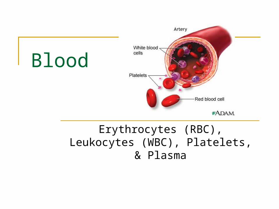
Blood
Erythrocytes (RBC), Leukocytes (WBC), Platelets, & Plasma

Blood Function
Transport nutrients, wastes, body heat, etc thru bv
Components 55% Plasma – fluid matrix 45% Erythrocytes (rbc) – O2 transport >1% Leukocytes (wbc) – protect body >1% Platelets – cell fragments used
for clotting Fun Facts
4-6 liters of blood per person 7% of body weight Temp 38oC pH between 7.35 – 7.45 Salty-metallic taste 5x thicker than water


Blood Plasma
90% water Distributes body heat through out body Contains dissolved nutrients, salts, resp.
gases, hormones, plasma proteins, wastes, and products of cell metabolism
Plasma Proteins Albumin – maintains osmotic pressure of blood Clotting Proteins – prevents blood loss from injured
bv Antibodies – protect body from pathogens

Erythrocytes (rbc)
Biconcave disc shape (large s.a. to volume) Anucleate - lacks a nucleus Lack cellular organelles including mitochondria (so do
not use O2 themselves) Hemoglobin (Hb) – Fe carries 4 molecules O2
1rbc carries 1 billion molecules O2
Avg male 13-18g Hb/100ml blood Avg female 12-16g Hb/100ml blood
Anemia – low rbc or low Hb Polycythemia – high rbc (high alt or bone marrow
cancer) Last 120 days – broken down by liver and spleen

Leukocytes (wbc) Live days, months, or even years About 4,000-11,000 wbc/mm3 blood Can move in and out of bv Respond to chems sent out from damaged cells When in action – body doubles #wbc w/in hrs Leukocytosis
>11,000 wbc indicates bacterial or viral infection Leukopenia
Decrease in wbc Typically due to corticosteroids or anticancer drugs
Leukemia Cancer of bone marrow Too many immature wbc to defend off pathogens

Types of Leukocytes Granulocytes – granule containing wbc w/lobed nuclei
Neutrophils – phagocytes at site of infection Eosinophils – increases w/allergies and infections by parasitic
worms Basophils – histamine (chem incr. wbc to site) containing granules
Agranulocytes – lack cytolasmic granules Lymphocytes – in lymphatic tissue (T cells and B cells) Monocytes – largest wbc
Become macrophages when enter tissues Fight off chronic infections

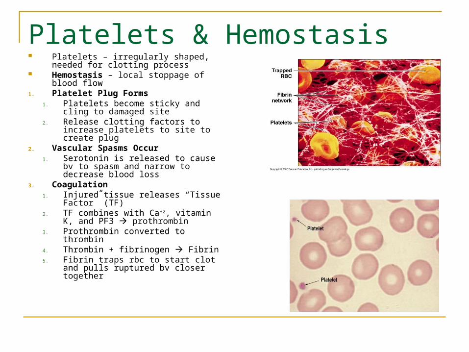
Platelets & Hemostasis Platelets – irregularly shaped, needed
for clotting process Hemostasis – local stoppage of blood
flow1. Platelet Plug Forms
1. Platelets become sticky and cling to damaged site
2. Release clotting factors to increase platelets to site to create plug
2. Vascular Spasms Occur1. Serotonin is released to cause bv to
spasm and narrow to decrease blood loss
3. Coagulation1. Injured tissue releases “Tissue
Factor” (TF)2. TF combines with Ca+2, vitamin K,
and PF3 prothrombin3. Prothrombin converted to thrombin4. Thrombin + fibrinogen Fibrin5. Fibrin traps rbc to start clot and pulls
ruptured bv closer together

Hematopoiesis – Blood Cell Formation Hemocytoblast
stem cell in rbm takes 3-5 days to form rbc
Rbc divides and synthesizes Hb Incr. Hb ejects nucleus and most
organelles Enters bloodstream
Rbc falls apart in 100-120 days Eliminated by phagocytes, spleen, and
liver Erythropoietin
hormone controls rate of rbc production Produced by kidneys and targets bone
marrow Thrombopoietin – increases production of
platelets Interleukins and colony stimulating factors
increase production of wbc from bone marrow
Released in response to inflam chems, bacteria, or toxins

Blood Types & Transfusions
Phlebotomist – hs degree + training + exam
ABO Blood Groups Based on 2 antigens A&B Antibodies formed in infancy Determined by anti-A, and anti-
B serums Rh Blood Groups
Once Rh- contacts Rh+ produces antibodies
Pregnant Rh- mom carrying Rh+ baby
RhoGAM prevents moms antibodies from attacking fetus
Hemolysis – rupture of rbc

Fetal Hb vs. Adult Hb By day 28 of fetal
development – embryonic blood cells circulate in blood vessels
Fetal Hb (HbF) differs from adult HbA
HbF has a greater ability to pick up O2 b/c fetal blood less O2 rich than mothers
If fetal rbc destroyed so quick that liver cannot rid of bile fast enough causes infant jaundice