Embed Size (px)
Citation preview

Chapter 19: Blood Vessels 12/02/2012
Blood Vessel Structure and Function
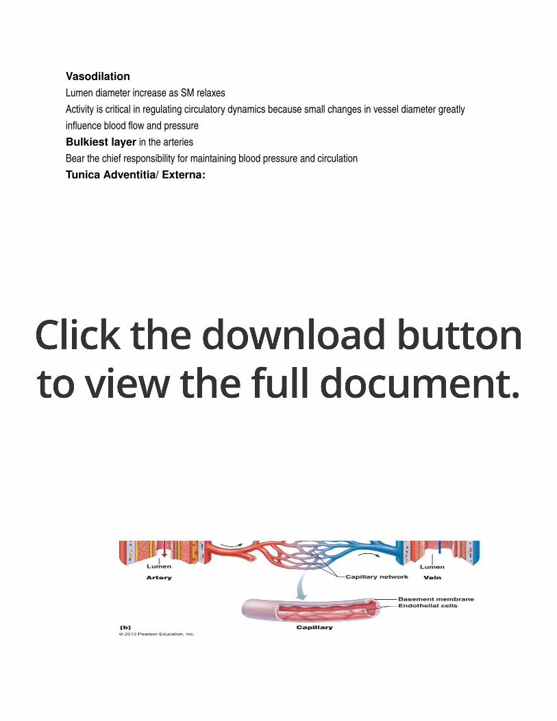
3 major types:arteriescapillariesveins as heart contracts it forces blood into the large arteries leaving the ventricles
then blood moves into arteries that get smaller and smaller, finally reaching smallest branches the arterioles, which feed into the capillary beds of body organs and tissues
blood drains from capillaries into venules, which are the smallest veins.
then on to the larger veins that merge to form the large veins that empty into the heart arteries arterioles capillaries venules veinsBV stretch for about 100,000 km through the internal body Arteries carry blood away from the heartBranch and fork as they form smaller and smaller divisions Veins carry blood towards the heartJoin/ merge together into larger vessels approaching the heartIn systemic circuit Arteries carry oxygenated blood Veins carry deoxygenated blood

In pulmonary circuit Arteries carry deoxygenated blood to the lungsThe veins carry oxygenated blood from the lungs to the heartUmbilical vessels of a fetus also differ in the roles of veins and arteries Only capillaries have intimate contact with tissue cells and directly serve cellular needs Exchanges between blood and tissue occur primarily through the gossamerthin capillary walls
Structure of Blood Vessel Walls 3 distinct layers/ tunics (covering)that surround a central bloodcontaining space> lumen Tunica intima Innermost tunic Tunic is in intimate contact with the blood in the lumen Contain simple squamous epithelium that lines the lumen of all vessels Endothelium is continuous with the endocardial lining of the heart and its flat cells fit closely together> forming a slick surface that minimizes friction as blood moves through the lumen In vessels larger then 1 mm a sub endothelial layer> consisting of basement membrane and loose CT > supports the endothelium Arteries have an internal elastic membrane > VEINS DON’TTunica media Mostly circularly arranged smooth muscleSheets of elastin Contains the most elastinActivity of the smooth muscle is regulated by sympathetic vasomotor nerve fibers of the autonomic nervous system & chemicals Depending on body’s need at any given moment regulation causes:Vasoconstriction Lumen diameter decreases as smooth muscle contracts
Vasodilation Lumen diameter increase as SM relaxes Activity is critical in regulating circulatory dynamics because small changes in vessel diameter greatly influence blood flow and pressure Bulkiest layer in the arteries Bear the chief responsibility for maintaining blood pressure and circulation Tunica Adventitia/ Externa:Outer most layerComposed largely of loosely woven collagen fibers that protect & reinforce the vessel & anchor it to surrounding structures Is infiltrated with nerve fibers, lymphatic vessels and in larger veins> a network of elastic fibers In larger arteries> it contains a system of tiny BV, the vasa vasorum vessels of vesselsnourish the more external tissues of the blood vessel wallthe innermost (luminal) portion of vessels obtain their nutrients directly from blood in lumen. The 3 vessel types vary in length, diameter, wall thickness and tissue make up
Arterial System 3 groups:elasticmusculararterioles
Elastic Arteries / Conducting Arteries thickwalled arteries near the heart aorta and its major brancheslongest in diameter2.5 cm 1 cmthe most elastic > highest portion of elastin their large lumens make them lowresistance pathways that conduct blood from the heart to medium sized arteries arteries are sometimes called conducting arteries pressure reservoirsexpanding and recoiling as the heart ejects blood blood flows fairly continuously rather than starting and stopping with pulsating rhythm of heart beatatherosclerosis BV become hard Blood flows more intermittently Smooth out pressure fluctuationsRecoil helps maintain pressure & flow of blood
Muscular Arteries/ Distributing Arteries Deliver blood to specific organs .3mm 1cm diameterHave the thickest tunic media of all vessels Contains more smooth muscle and less elastic tissueMore active in vasoconstriction and less capable of stretching There is an elastic membrane on each face of tunica media
Arterioles

Smallest of arteries have a lumen diameter of 10um .3mm larger arteries have all 3 tunicsbut tunica media is chiefly smooth muscle with a few scattered elastic fiberssmaller arteries> lead into capillary bedsa little bit more then a single layer of smooth muscle cells spiraling around endothelial lining minutetominute blood flow into capillary beds is determined by arteriolar diametervaries in response to changing:neural hormonallocal chemical influences when arterioles constrict > the tissues served are largely bypassed when arterioles dilate> blood flow into the local capillaries increases
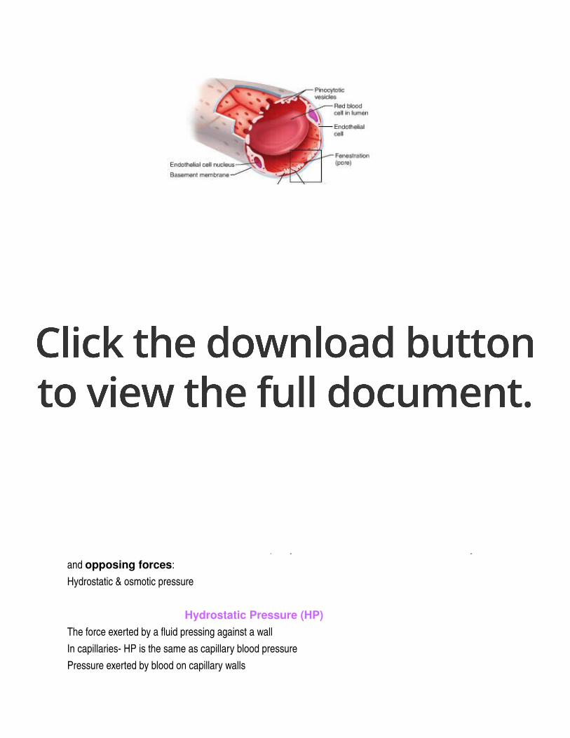
CapillariesMicroscopic capillaries are the smallest BV Exceedingly thin walls consist of just a thin tunica intima Length = 1mmLumen diameter = 810 um Just large enough for RBCs to slip through single file Function= exchange PericytesAt strategic locations along the outer surface of some capillaries Smooth muscle like cells that stabilize the capillary wall & help control capillary permeability

Most tissues have a rich capillary supply Exceptions:Tendon & ligaments= poorly vascularizedCartilage & epithelial lack capillariesReceive nutrients from BV in nearby CTAvascular cornea & lens of eye receive nutrients from aqueous humorcapillaries= back alleys & driveways that provide direct access to nearly every cell in the bodyideal suited for their role of exchange between blood and interstitial fluid. gasesnutrientshormones
Types of Capillaries 3 types:continuousfenestratedsinusoidal
Continuous Capillaries abundant in skin and muscles continuousendothelial cells are joined by tight junctions

providing an uninterrupted liningthese junctions are usually incomplete & leave gap of unjoined membranecalled intercellular cleftslarge enough to allow limited passage of fluids and small solutes
Fenestrated Capillaries similar to continuous capillaries except:endothelial cells are riddled with oval pores/ fenestrations delicate membrane covers the fenestrations much more permeable to fluids and small solutes than continuous capillaries found wherever active capillary absorption or filtrate formation occurs in small intestine receive nutrients from digested food endocrine organs allow homes rapid entry into blood fenestrated capillaries with perpetually open pores occur in kidneys where rapid filtration of blood plasma is essential areas of active absorption or filtration
Sinusoidal Capillaries highly modified, leaky capillaries found inliverbone marrowspleen
adrenal medullahave large irregular shaped lumens are usually fenestrated endothelial lining has fewer tight junctions & larger intercellular clefts than ordinary capillaries structural adaptations allow large molecules, proteins and even blood cells to pass between the blood and surrounding tissues most permeable
Capillary Beds do not function independently they form capillary bedsinterweaving networkmicrocirculation flow of blood from an arteriole to a venule through a capillary bedconsists of 2 types of BV vascular shuntmetarteriole + throughfare channels short vessel that directly connects the arteriole and venules at opposite ends of the bed true capillaries actual exchange vessels 10100 per bed

usually branch off the metarteriole end (proximal end of the shunt) and return to the thoroughfare channel (distal end)occasionally they spring from the terminal arteriole and empty directly into the venule the terminal arteriole feeding the bed leads into a metarteriolea vessel that is structurally intermediate between an arteriole and a capillary
metarteriole is continuous with the thoroughfare channelintermediate between a capillary and a venule
thoroughfare channel joins the postcapillary venule that drains in the beds. Precapillary sphinctersCuff of smooth muscle Surrounds root of each true capillary at metarterioleActs as a valve to regulate blood flow into the capillaryBlood flowing through a terminal arteriole may go either through the true capillaries or the shunt When relaxed/ open Blood flows through the true capillaries and takes part in exchanges with tissue cellsWhen contracted/ closedBlood flows through shunts and bypasses the tissue cells Local chemical condition and arteriole vasomotor nerve fibers regulate the amount of blood entering a capillary bedA bed may be completely flooded with blood or bypassed depending on the condition Vigorous exerciseBlood is rerouted from your digestive organs to the capillary beds of your skeletal muscle where it is more immediately neededHelps explain why vigorous exercise right after a meal can cause indigestion/ abdominal cramps
Venous System Carry blood from the capillary beds toward the heartalong route, the diameter of successive venous vessels increases

walls gradually thicken as progress from venules to larger and larger veins
Venules capillaries unit to form venules range from 8100 um diametersmallest venules> the post capillary venules > consist entirely of endothelium around which pericytes congregateextremely porousfluid and WBCs move easily from the bloodstream through their walls just endothelium & a few fibroblasts larger venules have one or two layers of SM cells ( a scanty tunica media) and a thin tunica externa/ adventiitia
Veins venules join to form veins usually have 3 distinct tunicsbut walls are always thinner & lumen are larger then corresponding arteries little smooth muscle or elastin in the tunica media poorly developed and tends to be thin even in largest veins tunica externa is the heaviest wall layerconsisting of thick bundles of collagen fibers & elastic networks can accommodate a fairly large supply at any time because of their large lumens and thin walls.Are capacitance vessels and blood reservoirs Can hold up to 65% of the body’s blood supply at any time. Normally only partly filled.Walls can be much thinner then those of arterial walls without danger of bursting because the blood pressure in veins is low.

Lowpressure condition demands several structural adaptations to ensure veins return blood o the hear at the same rate it was pumped into the circulation One adaptation: Largediameter lumensOffer relatively little resistance to flow
Venous ValvesAnother adaptation is valves that prevent blood from flowing backwardVenous valvesFormed from folds of the tunica intimaResemble the SL valves in both structure and function Most abundant in veins of limbs where gravity opposes the upward flow of blood Absent in veins of the thoracic and abdominal body cavities Effectiveness is demonstrated by ability for the vein to remain flat/ collapsed despite the pull of gravity
Homeostatic ImbalanceVaricose veins Veins that are tortuous and dilated because of incompetent/ leaky valves Adults usually suffer in lower limbs Factors :HereditaryAgeConditions that hinder venous return Prolonged standing in one positionPregnancy

Obesity Elevated venous pressure
Venous SinusesSuch as:Coronary sinuses of heart Dural venous sinuses of the brain Receive cerebrospinal fluid and blood draining from the brain Reinforced by the though dura matter that covers brains surfaceHighly specialized, flattened veins with extremely thin walls Composed of only endothelium Supported by tissues that surround them Rather then tunics
Vascular AnastomosesBV form special interconnections called vascular anastomosesMost organs receive blood from more than one arterial branch and the arteries supplying the same territory often merge forming> arterial anastomosesProvide alternate pathways called collateral channels for blood to reach a given body regionIf one branch is cut or blocked by a clot this channel can provide sufficient blood to areaOccur around joints where active movement may hinder blood flow through one channel Also common in abdominal organ, heart and brain Arteries that supply the retina, kidneys and spleen either do no anastomose or have a poorly developed collateral circulation If blood flow is interrupted> cells supplied by such vessel dieArteriovenous anastomoses

Ex: The metarteriolethoroughfare channel shunts of capillary beds that connect arterioles and venules Veins interconnect much more freely then arteriesVenous anastomoses are common Are abundant so on occluded vein rarely blocks blood flow or leads to tissue death
Part 2Physiology of Circulation
Introduction to Blood Flow, Blood Pressure and Resistance
Blood Flow (BF)The volume of blood flowing through a vessel, an organ or the entire circulation in a given period ml/min considering entire vascular systemblood flow is equivalent to CO under resting conditions its relatively constant at any given moment blood flow through individual body organs may vary widely according to their immediate needs can be regulated independently for various tissues & organs
Blood Pressure (BP)force per unit area exerted on a vessel wall by the contained blood expressed by mm Hgrefers to systemic blood pressure in the largest arteries near the heart average aortic bp= 90mm Hg

the differences in bp within the vascular system pressure gradientprovides driving force that keeps blood moving from high pressure to low pressure through the body 90> 90 = no flow!90>30= flow!
Resistance opposition to flow/ inversely related is a measure of the amount of friction blood encounters as it passes through the vesselsmeasure of total frictional forces that impede flow
generally use the term peripheral resistance most friction is encountered in the peripheral/ systemic circulation well away from the heart major determinant of BF because a change in blood vessels radius increases resistance to the 4th power.
3 important sources of resistance:blood viscosity vessel diametervessel length
F = P1 - P2
R
Resistance varies inversely with
(radius)4!!

Blood Viscosityviscosity internal resistance to flow that exists in all fluids related to the thickness/ stickiness of fluidthe great the viscositythe less easily molecules slide past one another & the more difficult it is to get and keep the fluid moving BV is fairly constantConditions such as polycythemia (excessive RBCs) can increase blood viscosity ResistanceIf RBC is lowAnemia’sBlood is less viscous Peripheral resistance declines Due to formed elements, plasma proteins
Total Blood Vessel Length Relationship between total BV length + resistance is straight forward:Longer vessel= greater resistance
BV DiameterChanges frequently & significantly alters peripheral resistanceBlood viscosity and vessel length are normally unchanging How?Principles of fluid flow

Fluids close to the wall of a tube/ channel is slowed by friction as its passes along the wallFluids in the center flows more freely and fasterLaminar flow / streamliningIn a tube of any given size the relative speed and position of fluid in the different regions of the tubes cross section remain constant The smaller the tube= the greater the friction Relatively more fluid contacts the walls Resistance varies inversely with the 4th power of the vessel radius If the radius of a vessel doubles, the resistance drops to 1/16 of its original value Large arteries close to the heart which do not change dramatically in diameter contribute little to peripheral resistance Smalldiameter arterioles are major determinants of peripheral resistanceBecause they can enlarge or constrict in response to neural and chemical controlsWhen blood encounters an abrupt change in vessel diameter or rough or protruding areas of the tube wall (fatty plaque)> the smooth laminar blood is replaced by turbulent flow Irregular fluid motion where blood from the difference lamina mixes Turbulence dramatically increases resistance
Relationship Between Flow, Pressure, and ResistanceBlood flow (F) is directly proportional to the difference in BP (ΔP) between two points in the circulation that is blood pressure or hydrostatic pressure gradient When ΔP Blood flow speeds upΔP Blood flow declinesBF is inversely proportional to the peripheral resistance (R) in the systemic circuit If R BFF= ΔP/ R

R is far more important than ΔP in influencing local BF because R can easily be changed by altering blood vessel diameter Arterioles dilate> resistance= BF to that area Even though systemic pressure is unchanged or may actually be failing
Systemic Blood Pressure Any fluid driven by a pump through a circuit of closed channels operates under pressure The nearer the fluid is to the pump > the greater the pressure exerted on the fluid. BF in BV is the same Blood flows through the BVs along a pressure gradient Always moving from higher > lower pressure areas The pumping action of the heart generates blood flowPressure results when flow is opposed by resistance Systemic blood pressure= highest in aorta Declines through pathway to finally reach 0 mmHg in the right atrium The steepest drop in BP occurs in the arterioles Offers the greatest resistance to BF As long as pressure gradients exist, no matter how small, blood continues to flow until it completes the circuit back to the heart
Arterial Pressure 2 factors:
1. How much the elastic arteries close to the heart can stretch compliance/ dispensability
2. The volume of blood forced into them at any time If amounts of blood entering and leaving the elastic arteries in a given period were equal, arterial pressure would be constant

Instead BP is pulsatile in the elastic arteries near the heartIt increases and decreases with rise and fall
Systolic Pressure As L ventricle contracts/ systole & expels blood into the aorta > it imparts kinetic pressure energy to the blood Stretches the elastic aorta as aortic pressure reaches its peak Averages 120 mm Hg in healthy adults Blood moves forward into arterial bed bc pressure in aorta is higher than the pressure in the more distal vessels
Diastolic Pressure During diastole> the aortic valve closes> preventing blood from flowing back into the heartWalls recoil Maintaining sufficient pressure to keep the blood from flowing forward into the smaller vessels During this time aortic pressure drops to its lowest levels Elastic arteries = pressure reservoirs that operate as auxiliary pumps to keep blood circulating throughout the period of diastole/ relaxation The volume and energy of blood stored in the elastic arteries during systole are given back during diastole
Pulse pressureDifference between the systolic and diastolic pressures systolic bp diastolic bpfelt as a pulse during systole, as ventricular contraction forces blood into the elastic arteries and expands them indicates a vigor contraction of ventricle provides info on elasticity of aorta & major arteries
stroke volume & faster blood ejection from heart (result of increased contractility) raises pulse pressure temporarily

atherosclerosis chronically increases pulse pressure because they elastic arteries become less stretchy
Mean Arteriole Pressure (MAP)pressure that propels the blood to the tissues diastole lasts longer then systole so MAP is not simply the value halfway between systolic and diastolic pressuresits roughly equal to the diastolic pressure pulse 1/3 of the pulse pressure. MAP= diastolic pressure + pulse pressure/3 Systolic bp = 120 mmHg Diastolic bp= 80 mmHg MAP= 80 mmHg + (12080= 40mm Hg)/ 3= 93 mm Hg MAP & Pulse pressure both decline with increasing distance from the heart MAP loses ground between the blood and vessels wallsPulse pressure is gradually phased out in the less elastic muscular arteries Elastic rebound of vessels ceases to occur At end of arterial tree Blood flow is steady and the pulse pressure has dissipated
Capillary Blood Pressure by time blood reaches capillaries blood pressure has dropped to approximately 35 mm Hg and by the end of the capillary beds is only around 17 mm Hg low capillary pressures are desirable because:
1. Capillaries are fragile and high pressures would rupture them 2. Capillaries are extremely permeable and thus even the low capillaries
pressure can force solutecontaining fluids (filtrate) out of the blood stream into the interstitial space

Fluids are important for continuously refreshing the interstitial fluid Lost of exchange at low bp
Venous Blood Pressure Steady and changes very little during the cardiac cycle The pressure gradient in veins, from venules to the termini of the vena cavae, is only about 15 mmHg Difference in pressure between an artery and a vein becomes very clear when the vessels are cutThe blood flows evenly from the wound, but a lacerated artery spurts bloodThe very low pressure in the venous system reflects the cumulative effects of peripheral resistance Dissipates most of the energy of blood pressure as heat during each circuit Despite the structural modifications of veins > large lumens and valves > venous pressure is normally too low to promote adequate venous return. 3 functional adaptations are critically important to venous return:the muscular pump consists of skeletal muscle activity as the muscles surrounding the deep veins contract and relax, they “milk” blood toward the heart once blood passes each successive valves > it can’t flow back standing professions such as hairdressers and dentists often have swollen ankles> blood pools in their feet and legs skeletal muscle inactivity reduces venous return the respiratory pumpmoves blood upward toward the heart as pressure changes in the ventral body cavity during breathing as we inhale > abdominal pressure increases, squeezing local veins and forcing blood towards the heartat the same time, the pressure in the chest decreases allowing thoracic veins to expand and speeding blood entry into the right atrium Sympathetic venoconstriction Reduces the volume of blood in the veins

The capacitance vessels As layer of smooth muscle around the veins constricts under sympathetic control, venous volume is reduced and blood is pushed toward the heartAll 3 functional adaptations increase venous return > which increases stroke volume (by Frank sterling law), therefore increases cardiac output.
Maintaining Blood Pressure Homeostatic mechanisms that regulate cardiovascular dynamics are those that maintain BP Cardio outputBlood flow of entire circulation Peripheral resistanceBlood volume Homeostatic mechanisms in place to maintain adequate flow to tissues under a variety of circumstances Keeps us from keeling over when we get up too quickly etc. CO and peripheral resistance relate to blood pressure:F= ΔP/RCO= ΔP/RΔP= CO X RBP varies directly with CO and RAlso with blood volume because CO depends on blood volume Heart cant pump out what doesn’t enter the chambersIn theory, a change in any of these variables would cause a corresponding change in blood pressure What really happens:Changes in one variable that threaten blood pressure homeostasis are quickly compensated for by changes in the other variables CO is equal to stroke volume ml/beat times HR beats/min Normal CO is 5 5.5 L/min
Main factors determining cardiac output:Venous return SVNeural and hormonal controlsHRMedulla is in charge of heart rate most of the time Parasympathetic vagus nerves maintains the resting heart rate During resting conditions venous return (end diastolic volume) largely controls SVDuring stress the cardio acceleratory center takes overactivating sympathetic NS increasing:HRBy acting on SA nodeSV By enhancing cardiac muscle contractility, which decreases end systolic volumeEnhanced CO increases MAP
Factors that regulate Blood Pressure:Shortterm Regulation By the nervous system and blood borne hormones alters blood pressure by changing peripheral resistance and CO Longterm resistance

Alters blood volume via the kidneys
ShortTerm Regulation: NEURAL CONTROLS Alter both CO & peripheral resistance Neural controls of peripheral resistance are directed at 2 main goals:Maintaining adequate MAP By altering blood vessel diameter on a momentto moment basis Very small changes in blood vessels diameter cause substantial changes in peripheral resistance Systemic blood pressureUnder conditions of low volume All vessels except those supplying the heart and brain are constricted to allow as much blood as possible to flow to those two vital organs Altering Blood distribution To respond to specific demands of various organs During exercise blood is shunted temporarily from the digestive organs to the skeletal muscles Most neural controls operate via reflex arcs involving baroreceptors & afferent fibers Reflexes are integrated in the cardiovascular center of the medulla Their output travels via autonomic fibers to the heart and vascular smooth muscles Inputs from chemoreceptors & higher brain centers also influence the neural control mechanism
Role of the Cardiovascular Center Clusters of neurons in the medulla oblongata act together to integrate BP control by altering CO & blood vessel diameter Cardiovascular centerConsists of:Cardiac centersCardioacceleratory and cardioinhibitory centersVasomotor center Controls diameter of BVs

Transmits impulses at a fairly steady rate along sympathetic efferent called vasomotor fibersExit from T1 through L2 Innervate the smooth muscle of BV, mainly arterioles Arterioles are almost always in a state of moderate constriction called vasomotor tone Constant input, especially to arterioles (NE)Degree of vasomotor varies from organ to organ Arterioles of the skin & digestive viscera receive vasomotor impulses more frequently & tend to be more strongly constricted than skeletal muscles Any increase in sympathetic activity produces generalized vasoconstriction & raises BP Decreased sympathetic activity allows the vascular muscle to relax somewhat + lowers BP to basal levels
Modified by inputs from:1. Baroreceptors
Pressuresensitive mechanoreceptors that respond to changes in arterial pressure and stretch) 2. Chemoreceptors
Receptors that respond to changes in blood levels of CO2, O2, H+3. Higher brain centers
Baroreceptors ReflexesWhen arterial BP rises it activates BaroreceptorsStretch receptors located in the carotid sinusesdilations in the internal carotid arteries which provide the major blood supply to the brain aortic arch walls of nearly every large artery of the neck and thorax

when stretchedbaroreceptors send a rapid stream of impulses to the cardiovascular centerinhibiting the vasomotor and Cardioacceleratory centers & stimulating cardioinhibitory centerresult is a decrease in BPincreasing MAP stretches receptorsresponse via vasomotor center is:3 mechanisms bring this about:Arteriolar Vasodilation Decreased output from the vasomotor center allows arterioles to dilateAs peripheral resistance falls> so does MAP Venodilation Decreased output from the vasomotor center also allows veins to dilateShifts blood to the venous reservoirs Decreases venous return & CO Decreased CO Impulses to the cardiac centers inhibit sympathetic activity and stimulate parasympathetic activity Reducing HR and contractile forceAs CO falls> so does MAP
In opposite situation:A decline in MAP initiates reflex vasoconstriction and increases COBringing BP back up Peripheral resistance and cardiac output are regulated in tandem to minimize changes in blood pressure Goal
Rapidly responding baroreceptors protect the circulation against shortterm/ acute changes in BPEx: when you stand up after reclining blood pressure falls Baroreceptors take part in carotid sinus reflexProtect the blood supply to your brain Whereas those activated in the aortic reflexHelp maintain adequate blood pressure in your systemic circuit as a whole They are relatively ineffective in protecting us against sustained pressure changes Development of chronic hypertension Baroreceptors adapt to monitor pressure changes at a higher set point
Chemoreceptor Reflexes when CO2 levels rise or pH levels falls or O2 content of the blood drops sharply chemoreceptors in the aortic arch and large arteries of the neck transmit impulses to the Cardioacceleratory center which then increases CO & to the vasomotor centerwhich causes reflex vasoconstriction rise in blood pressure that follows speeds the return of blood to the heart and lungs
most prominent chemoreceptors are the carotid and aortic bodies located close by the baroreceptors in the carotid sinuses and aortic arch chemoreceptors play a larger role in regulating respiratory rate than BP
Influence of Higher Brain Centersreflexes that regulate BP are integrated in the medulla oblongata of the brain stem fightorflight response mediated by hypothalamusalso mediates redistribution of blood flow and other cardiovascular responses that occur during exercises and changes in body temp, stress
ShortTerm Regulation: HORMONAL CONTROLS help regulate BP in shortterm regulation by changes in peripheral resistancein longterm regulation by changes in blood volume Paracrines (local chemicals) Primarily serve to match blood flow to the metabolic need of a particular tissue Massive release of paracrines can affect BP Shortterm effects of hormones:Adrenal Medulla Hormones During periods of stressAdrenal gland release epinephrine and NE to the blood FightorflightEnhance sympathetic NS response by CO and promoting generalized vasoconstriction Nicotine mimics effects of catecholamines Amino acid hormones??Angiotensin IIWhen BP or Blood volume are low Kidneys release renin Acts as an enzyme, ultimately generating angiotensin IIStimulates intense vasoconstriction> promoting rapid rise in systemic BP Also stimulates release of aldosterone & ADH

Act in longterm regulation of BP by enhancing blood volume Atrial Natriuretic Peptide (ANP) Atria of the heart produces ANPLeads to reduction in blood volume and BP Antagonizes aldosterone and prods the kidneys to excrete more Na & water from the body Reducing blood volume Also causes generalized vasoconstriction Antidiuretic Hormone (ADH)Produced by hypothalamus / posterior pituitaryStimulates kidneys to conserve water Not usually important in shortterm BP regulation > therefore longterm effectWhen BP falls to dangerously low levels (during hemorrhage) much more ADH is released and helps restore arterial pressure by causing intense vasoconstriction Endotheliumderived factors:Endothelin is a potent vasoconstrictor Plateletderived growth factor vasoconstrictor Nitric oxide (NO)Induces brief vasodilationInflammatory chemicals:Histamine, prostacyclinRelease during inflammatory responsePotent vasodilators Alcohol
Inhibits ADH releaseDepresses vasomotor centerPromotes vasodilation Especially skin
Long Term Regulation: RENAL MECHANISMS Unlike shortterm controls of BP that alter peripheral resistance and CO Longterm controls alter Blood Volume Renal mechanisms mediate longterm regulation as well as responding to shortterm changes in blood Baroreceptors quickly adapt to prolonged chronic episodes of high or low pressure this is where the kidneys step in to restore and maintain BP homeostasis by regulating blood volume blood volume varies with age, body size and sex but renal mechanisms keep it close to 5Lanything that changes blood volume will change BP blood volume= major determinant of COexcessive salt intake> promotes water retention an increase in blood volume is followed by a increase in BP anything that increases blood volume raises MAP because of the greater fluid load in the vascular treealso stimulate the kidneys to eliminate waterthis reduces blood volume & BPa decrease in blood volume = a fall/ decrease in BP blood loss & dehydration that occurs during vigorous exercise are causes of reduced Blood volume a sudden drop in pressure signals internal bleeding and blood volume to low to support normal circulation triggers renal mechanisms that increase blood volume and BPblood volume can be stabilized or maintained within normal limits only when blood volume is stable

kidneys act both directly & indirectly to regulate arterial pressure and provide the major longterm mechanisms of BP control
Direct Renal Mechanismalters blood volume independently of hormones when either blood volume or BP is increase increases the rate at which the fluids filter from the blood stream to the kidney tubules = insufficient time to reclaim water, not enough time to absorb
more of it leaves the body as urine BP and blood volume decrease Therefore water is conserved and returned to the blood stream and blood pressure rises. As BV go so does arterial blood pressure
Arterial Pressure
Filtration by Kidneys

Urine Formation
Blood Volume
Mean Arterial Pressure
Indirect Renal MechanismRegulate BP indirectly via the reninangiotensinaldosterone mechanism When arterial BP Certain cells in kidneys release then enzyme renin into the bloodRenin enzymatically cleaves angiotensin a plasma protein made by the liverconverting it to angiotensin Iangiotensin converting enzyme (ACE) converts angiotensin I angiotensin IIACE activity is associated with the capillary endothelium in various body tissues > particularly the lungsAngiotensin II acts in 4 ways to stabilize arterial BP & extracellular fluid volume:Stimulate adrenal cortex to secrete aldosteroneEnhances renal reabsorption of NaAs Na moves into blood stream water followsconserves blood volume.
Also it directly stimulates Na reabsorption by kidneys Prods the posterior pituitary to release ADH promotes more water reabsorption by the kidneys Triggers the sensation of thirst by activating the hypothalamic thirst center Encourages water consumption, ultimately resorting blood volume and so BP Is a potent vasoconstrictor Increasing BP by increasing peripheral resistance. MAP
Clinical Monitoring of Circulatory Efficiency Measuring pulse and blood pressure Vital signs PulseBPBody temp
Resp rateMeasured by:
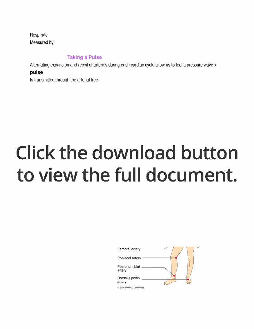
Taking a Pulse Alternating expansion and recoil of arteries during each cardiac cycle allow us to feel a pressure wave > pulseIs transmitted through the arterial treeCan feel it in any artery that lies close to the surfacePush surface artery against firm tissueUsually radial pulse is used to measure pulse Many more spots to take pulsePressure pointsPulse pointsCompressed to stop blood flow into distal tissues during hemorrhageActivity, postural changes and emotions effect pulse rates Normal healthy man:sitting down pulse = 70Lying down = 66Suddenly stands= 80Vigorous exercise or emotional upsets= 140180Sympathetic NS effects on heart
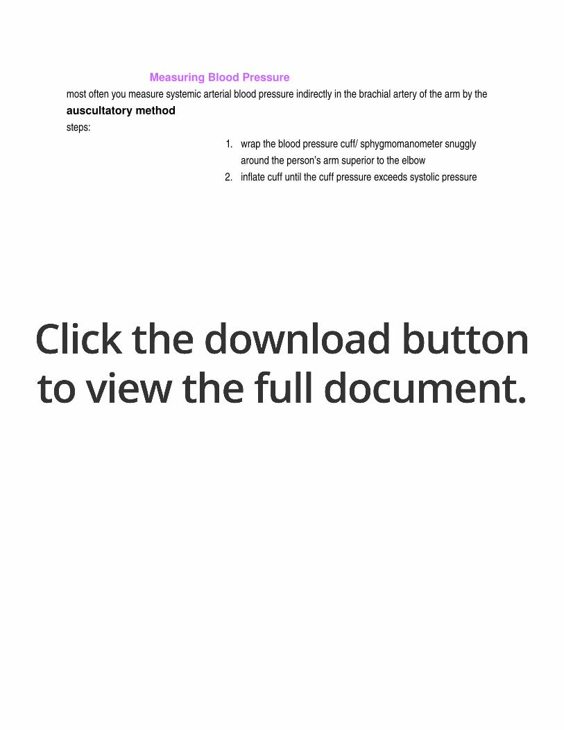
Measuring Blood Pressure most often you measure systemic arterial blood pressure indirectly in the brachial artery of the arm by the auscultatory method steps:
1. wrap the blood pressure cuff/ sphygmomanometer snuggly around the person’s arm superior to the elbow
2. inflate cuff until the cuff pressure exceeds systolic pressure blood flow into the arm stops and a brachial pulse cannot be felt or hear artery fully collapsed> no flow
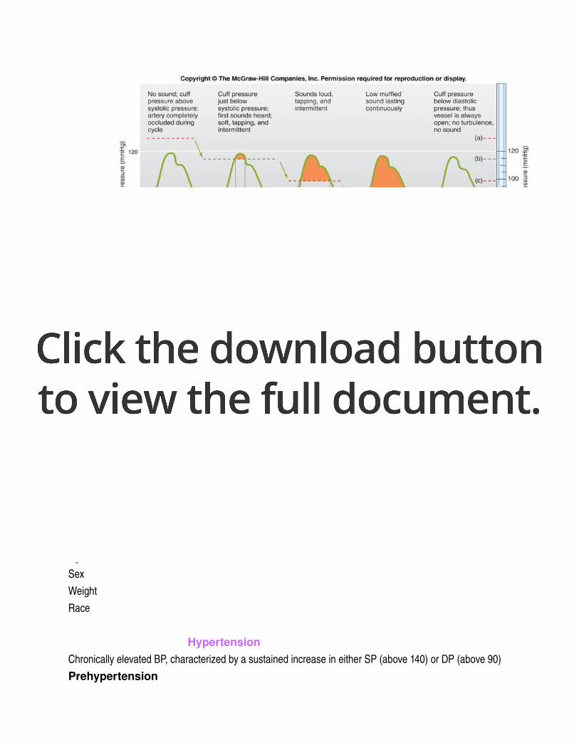
3. reduce cuff pressure gradually and listen (auscultate) with a stethoscope for sounds of brachial artery
systolic pressurethe 1st soft tapping sounds are heard> first point at which a small amount of blood is spurting through the constricted artery
as pressure cuff is reduced further, sounds called the sounds of Korotkoff become louder and more distinct
when artery is no longer constricted and blood flows freely the sounds can no longer be hearddiastolic pressurepressure at which the sounds disappear
Homeostatic Imbalances in Blood Pressurenormal adult at rest:SP= 1101140DP= 75 80Elevations in BP occur as normal adaptations during changes in PosturePhysical exertionEmotional upsetFeverAgeSexWeightRace
HypertensionChronically elevated BP, characterized by a sustained increase in either SP (above 140) or DP (above 90)Prehypertension
If BP valves are elevated but not yet in hypertensive rangeHigh risk individuals Common and dangerous disease Primary HypertensionHereditaryDietObesityAgeDiabetesStressSmokingCan be curedSecondary HypertensionObstructed renal arteries Kidney diseaseEndocrine disorders
HypotensionLow blood pressureBelow 90/60 mm HgNo cause for concern
Look in text book for more information pg. 711
Blood Flow Through Body Tissues: Tissue PerfusionBlood flow through body tissue> tissue perfusion is involve in:
1. Delivering O2 and nutrients to tissue cells & removing wastes from them 2. Exchanging gases in the lungs3. Absorbing nutrients from the digestive tract 4. Forming urine in the kidneys
The rate of blood flow to each tissue and organ is almost exactly the right amount for proper function At rest :brain receives 13% of total BFheart 4%kidneys 20%abdominal organs 24%
skeletal muscles 20%during exercise nearly all of the increased CO flushes into the skeletal muscles and bf to the kidneys and digestive organ declines
Velocity of BFinversely related to cross sectional areaspeed/velocity of BF changes as blood travels through the systemic circulation fastest at the aorta & other larger arteries like a river total cross sectional area is leastslowest in capillaries large total cross sections like a lake picks up speed again in veins river again as arteriole system branchesfi total cross sectional area of vascular bed increases= velocity of blood flow proportionatelyeven though individual branches have smaller lumen > their combined gross sectional areas, therefore the volume of blood they can hold, are much greater then that of aortas aorta 2.5cm squaredcombined crosssection 4500 cm squared this difference results in fast blood flow in aortaslow blood flow in capillaries beneficial because it allows adequate time for exchanges between blood and tissue cells as capillaries combine to form 1st venules and then veins, total cross sectional area declines and velocity increases cross sectional area of vena cava is 8cm squared velocity of bf varies from 10 to 30 cm/s in those vesselsdepending on activity of skeletal muscle pump
Autoregulation: Local Regulation of Blood Flow AutoregulationAutomatic adjustment of bf at any instant Local conditions regulate this process independently of any control by nerves or hormones MAP is the same everywhere in the body & homeostatic mechanisms adjust CO as needed to maintain that constant pressure
Changes in bf through individual organs are controlled intrinsically by modifying the diameter of local arterioles feeding the capillaries Local conditions in the arterioles feeding the capillaries beds of an organ have little effect on pressure in the muscular artery feeding that organ or in the large elastic arteries As long as the circulatory feedback mechanisms maintain a relatively constant MAPLocal demand regulates the amount of blood delivered to various areasSummary:Organs regulate their own bf by varying the resistance of their arterioles Classed as MetabolicChemicalMyogenicPhysical
Metabolic Controls (Local)When BF is too low to meet a tissue’s metabolic needs> oxygen levels and metabolic products accumulateMetabolic products act as paracrinesChanges serve as Autoregulation stimuli that lead to autonomic increases in tissue bfMetabolic factors that regulate bf:Low O2
in H+ from CO2 & lactic acidK+ AdenosineProstaglandins Inflammatory chemicals Many act directly to relax vascular SMSome may act by causing vascular endothelial cells to release nitric oxideNitric Oxide (NO)Powerful vasodilator

Acts via a cyclic GMP secondmessenger system Quickly destroyed Potent vasodilator effects are briefMajor player in controlling local vasodilationOften overriding sympathetic vasoconstriction when tissues need more blood flow Endothelium also releases potent vasoconstrictors , including endothelins, are among the most potent vasoconstrictors Net result of metabolically controlled Autoregulation is immediate vasodilation of the arterioles serving the capillary beds of the “needy” tissues and dilation of their pre capillary sphincters Bf to the area rises temporarily Allowing blood to surge through the true capillaries and become available to the tissue cells Inflammatory chemicals (histamine, kinins and prostaglandins) released in injury, infection or allergice reactions also cause local vasodilation Helps defense mechanisms clear microorganisms and toxins from the area Promotes healing
Myogenic Controls Fluctuations in systemic blood pressure would cause problems for individual organs if it were not for the myogenic responses of vascular SM Myo= muscleGen= origin Inadequate blood perfusion through an organ is quickly followed by decline in organs metabolic rate and possible organ death if prolongedExcessively high arterial pressure and tissue perfusion can be dangerous > combination can rupture more fragile BVs Vascular SM prevents these problems by responding directly to passive stretch (caused by increased intravascular pressure) with increased tone Tone resists the stretch > causes vasoconstriction Reduced stretch vasodilation increased blood flow into tissue Keep tissue perfusion fairly constant despite most variations in systemic pressure
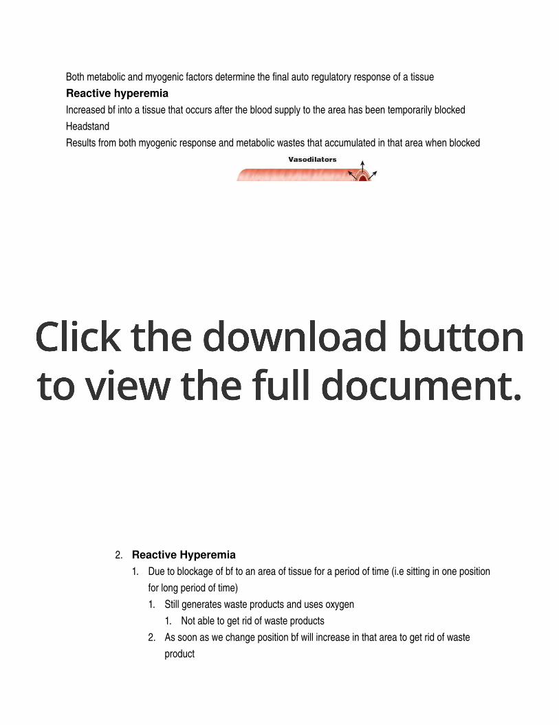
Both metabolic and myogenic factors determine the final auto regulatory response of a tissue Reactive hyperemiaIncreased bf into a tissue that occurs after the blood supply to the area has been temporarily blockedHeadstand Results from both myogenic response and metabolic wastes that accumulated in that area when blocked
1. Active Hyperemia1. Increased accumulation of waste products due to increased O2
1. Result of increased activity of tissues2. Stimulation of vasodilation
2. Reactive Hyperemia1. Due to blockage of bf to an area of tissue for a period of time (i.e sitting in one position
for long period of time)1. Still generates waste products and uses oxygen
1. Not able to get rid of waste products2. As soon as we change position bf will increase in that area to get rid of waste
product
Blood Flow in Special AreasEach organ has special requirements and functions that are revealed in its pattern of Autoregulation Autoregulation in the brain, heart and kidneys is extraordinarily efficient Maintains adequate perfusion even when MAP fluctuates
Skeletal Muscles Bf varies with fiber type and muscle activity Capillary density and bf are greater in red/slow oxidative fibers than in white/ fast glycolytic fibers Resting skeletal muscles receive about 1L of blood/ minuteOnly about 25% of their capillaries are open Myogenic and general neural mechanisms predominate Active muscles> bf increases (hyperemia) in direct proportion to their greater metabolic activity Called active hyperemia Occurs mostly in response to the decreased oxygen concentration and accumulated metabolic factors that result from the “revvedup” metabolism of working muscles Systemic adjustments mediated by the vasomotor center must also occur to ensure that blood delivery to the muscle is both faster and more abundant During exerciseSympathetic NS system activity increases, releases:NE > causes vasoconstriction of the vessels of blood reservoirs Digestive viscera and skin Therefore more blood goes to the muscles working SNS and local metabolic controls have opposing effects on arteriolar diameterDuring:Local controls override sympathetic vasoconstriction Blood flow to skeletal muscles can increase tenfoldVirtually all capillaries in the active muscles open to accommodate the increased flow Factor determining how long muscles can contract vigorously is the ability of the CVS to deliver adequate O2 & nutrients & remove waste products
The Brain Blood flow is maintained and constant Brain is unable to store essential nutrients Metabolic Controls:

Cerebral bf is regulated by one of the body’s most precise autoregulatory system & is tailored to local neural needEx: Make a fist with your right hand > the neurons in the left cerebral cortex controlling that movement receive more blood than the adjoining neurons Very sensitive to declining pH & increased CO2 levels cause vasodilation Resulting in acidic conditions in brain tissue Low levels of O2 are a much less potent stimulus for AutoregulationVery high levels of CO2 abolish autoregulatory mechanisms & depress brain activity Myogenic mechanismProtects it from possibly damaging changes in bp When MAP Cerebral vessels dilate to ensure adequate brain perfusion When MAP Cerebral vessels constrict to protect the small more fragile vessels father along the pathway from excessive pressure During special cases:Brain ischemia > rising intracranial pressure (as with brain tumors)Brain regulates its own bf by triggering a rise in systemic bp However, when systemic bp changes are extreme:Brain becomes vulnerable Fainting occurs when MAP falls below 60mm Hg
The Skin Bf through skin
a. Supplies nutrients to cellsAutoregulation
b. Helps regulate body temp

neural interventionprimary function of the cutaneous circulation
c. Provides blood reservoir Neural intervention Body tempBf can change from 50 ml/min to 2500ml/min Capability reflects neural adjustments of bf through arterioles and arteriovenous anastomoses (richly supplied with sympathetic nerve endings)Controlled by reflexes initiated by temperature receptors or signals from higher CNS centers Exposure to heat/ body temp = hypothalamic “thermostat” signals for vasomotor stimulation of skin vessels Result: warm blood flushes into the capillary beds and heat radiates from the skin surfaceWhen cold/ body temp = superficial skin vessels strongly constrict Blood almost entirely bypasses the capillaries associated with the arteriovenous anastomoses > diverting warm blood inward to organs
The Lungs UnusualRelatively short pathway Pulmonary arteries and arterioles are structurally like veins and venules Thin walls & large lumens Resistance to bf is low > less pressure is needed to propel blood through those vessels Therefore pressure is much lower then systemic 24/10 vs. 120/80in pulmonary circulation:autoregulatory mechanism is opposite:low pulmonary O2 levels cause local vasoconstriction
high levels of O2 promote vasodilation persistent with gas exchange role in this circulationwhen air sacs are flooded with O2rich air the pulmonary capillaries become flushed with blood and are ready to receive the O2 load if air sacs are collapsed or blocked with mucus> the O2 content in those areas is low> blood largely bypasses those nonfunctional areas
The Heart pumping action of ventricles influences movement of blood through smaller vessels of the coronary circulation contraction= compression of coronary vesselsbf through myocardium stopsrelaxation= high aortic pressure forces blood through the coronary circulation myoglobin in cardiac cells store sufficient O2 to satisfy the cell’s O2 needs during systole abnormal/ rapid heartbeat reduces the ability of myocardium to receive adequate O2 and nutrients during diastole resting conditions:bf= 250 ml/min controlled by myogenic mechanismscardiac cells use as much as 65% of the O2 carried to them in blood bf remains fairly constant despite wide variations in coronary perfusion pressure strenuous exercise:coronary vessels dilate in response to local accumulation of vasodilators (adenosine) bf may 34 Xany event that decreases the O2 content of the blood releases vasodilators that adjust the O2 supply to the O2 demand enhanced bf is important bc it’s the only way to provide more O2 to a vigorously working heart
Blood Flow Through Capillaries and Capillary Dynamics Capillaries

major point of communication between interstitial fluid & blood bf through capillary networks is slow and intermittentdue to vasomotionthe on/off opening and closing of precapillary sphincters in response to local autoregulatory controls
Capillary Exchange of Respiratory Gases and Nutrients Diffusion is how O2, CO2, most nutrients and metabolic wastes pass between blood and interstitial fluid Net movement always occurs along a concentrated gradient Moving from an area of its higher concentration to lower concentrationO2 and nutrients pass from the blood> high concentrations through interstitial fluid to tissue CO2 and metabolic wastes leave the cells> high concentration diffuse into the capillary blood4 different routes across capillaries for different types of molecules Diffuse through membrane/ lipid bilayer of endothelial cell plasma membranes.
Lipid soluble respiratory gasesMovement through intercellular clefts small watersoluble solutes> amino acids & sugars Fenestrations/ pore water soluble substancesTransported in vesicles or caveolae large molecules > proteins Capillaries differ in their leakiness
Fluid Movements: Bulk Flow Nutrient gas exchanges = across the capillary walls by diffusion Bulk fluid flows are also going on Fluid is forced out of the capillaries through the clefts at the arterial end of the bedMost of it returns to the bloodstream at venous end Important in determining the relative fluid volumes in the bloodstream and the interstitial spaceAprox. 20L of fluid filter out of the capillaries each day before being returned to the blood The direction and amount of flow across capillary walls reflects the balance between two dynamic and opposing forces:Hydrostatic & osmotic pressure
Hydrostatic Pressure (HP)The force exerted by a fluid pressing against a wall In capillaries HP is the same as capillary blood pressure Pressure exerted by blood on capillary walls
Capillary Hydrostatic Pressure (HPC)Forces fluids through capillary walls = filtration Leaving behind cells and most proteins BP drops as bfs along a capillary bedHPC is higher at arterial end (35mm Hg) then at venous end (17 mm Hg)BP which forces fluid out of the capillaries is opposed by the interstitial fluid hydrostatic pressure (HPif) acting outside the capillaries and pushing fluid in Usually very little fluid in the interstitial spaceLymphatic vessels constantly withdraw it May vary from slightly – to +, traditionally it is assumed to be 0
Colloid Osmotic Pressures (OP)The force opposing HPCreated by large nondiffusible molecules Plasma proteins Unable to cross the capillary wall These molecules draw water toward themselves Encouraging osmosis bc the water concentration in their vicinity is lower than it is on opposite side of capillary walls *”HP pushes & OP pulls/sucks”capillary colloid osmotic pressure (OPc)/ oncotic pressure developed by the abundant amount of plasma proteins, primarily albumin moleculesOPc is 26mm Hg Interstitial colloid osmotic pressure (OPif)Is lower.1 5 mm Hg unlike HP> OP doesn’t vary significantly from one end of the capillary to the other
HydrostaticOsmotic Pressure Interactions net filtration pressure ( NFP)considers all the forces acting at the capillary bedwhile net filtration is occurring at the arteriole end a – value for NFP at the venous end of the capillary indicates that fluid is moving into the capillaries (called reabsorption) result: net fluid flow is out of the circulation at the arterial end of capillary beds and into the circulation at the venous end

more fluid enters the tissue spaces than return to the blood result: a net loss of fluid from the circulation of about 1.5 ml/minlymphatic vessels pick up this fluid and any leaky proteins and return it to the vascular system > account for the relatively low levels of both fluid and proteins in the interstitial space


Circulatory Shock Any condition that blood vessels are inadequately filled and blood cannot circulate normally Cells die> organ dies
Hypovolemic Shock Most common form Low blood volume Results from largescale blood or fluid loss Acute hemorrhage Sever vomiting or diarrhea Extensive burns If blood volume drops rapidly = HR increase in attempt to correct problem Weak “thread pulse” is first sign Intense vasoconstriction also occurs Shifts blood from the various blood reservoirs into the major circulatory channels & enhances venous return BP is stable at 1st then eventually drops if blood loss continues Replace fluid volume as quickly as possible
Vascular Shock Blood volume is normal

Circulation is poor as a result of extreme vasodilation Huge drop in peripheral resistance follows because of rapidly falling BPLoss of vasomotor toneDue to anaphylactic shock Massive release of histamine triggers bodywide vasodilation Neurogenic shock & septic shock
Cardiogenic Shock / pump failureOccurs when heart is so inefficient that it cannot sustain adequate circulation Myocardial damage after numerous heart attacks

12/02/2012

12/02/2012