Embed Size (px)
Citation preview
BioMed Central
BMC Ear, Nose and Throat Disorders
ss
Open AcceCase reportA thyroid tumor extending to the parapharyngeal spaceFikret Cetik†1, Demet Yazici*†1 and Aysun Uguz2Address: 1Depatment of Otolaryngology, Cukurova University, Adana, Turkey and 2Department of Pathology, Cukurova University, Adana, Turkey
Email: Fikret Cetik - [email protected]; Demet Yazici* - [email protected]; Aysun Uguz - [email protected]
* Corresponding author †Equal contributors
AbstractBackground: The metastasis of papillary thyroid carcinoma to the parapharyngeal space is rareand discussed in the English literature before. Encountering a parapharyngeal mass with cysticappearance on imaging, one should rule out thyroid malignancy as differential diagnosis.
Case presentation: The case presented here is a 22-year-old woman who was referred to ourclinic with complaints of painless neck mass, dysphagia and hoarseness for two years. Afterradiologic and pathological examination, the mass thought to be relevant with the thyroid gland.Peroperatively, the tumor was found to originate from the superior pole of the right thyroid gland,with a narrow stalk, and extended following the neurovascular bundle to the lower part of theparapharyngeal space. The bulk was removed via transservical approach with total thyroidectomy.
Conclusion: The occurrence of the follicular variant of papillary thyroid carcinoma in theparapharyngeal space is extremely rare. The management of this rare case was discussed with thereview of literature.
BackgroundThe parapharyngeal space, an inverted pyramid-shapedregion, extends from the skull base to the greater cornu ofthe hyoid bone [1]. Tumors of this space are rare, account-ing for 0, 5 % of head and neck neoplasms [2]. Only 20%of these neoplasms are malignant and 50% of these neo-plasms arise from the deep lobe of the parotid gland orminor salivary glands [3].
Although thyroid neoplasms are the most common endo-crine tumors in head and neck, thyroid cancer is a rela-tively uncommon neoplasm [4]. Presentation of thethyroid carcinoma as a neck mass extending into the PPSis very rare [5]. A 22-year-old female who has papillarycarcinoma of thyroid extending to the parapharygealspace is presented in this case report.
Case presentationA 22-year-old female patient was referred our clinic forevaluation of a painless neck mass, hoarseness and dys-phagia. She first noticed the mass in the upper right necktwo years earlier without any symptoms. Her medical andfamily histories were unremarkable.
Physical examination revealed a submucosal mass in theright lateral oropharyngeal wall with medial displacementof the right tonsil. A non-tender, firm, mobile mass meas-uring 4 × 4 cm. was detected deep to the right sternocleid-omastoid muscle below the angle of the mandible.Superior extent of the mass could not be palpated in theneck. The telescopic examination of the larynx demon-strated a right sided, smooth mass narrowing the rimaglottis. There were not any cranial nerve deficits otherthan the paralysis of the right vocal cord. The computed
Published: 01 March 2006
BMC Ear, Nose and Throat Disorders2006, 6:3 doi:10.1186/1472-6815-6-3
Received: 16 August 2005Accepted: 01 March 2006
This article is available from: http://www.biomedcentral.com/1472-6815/6/3
© 2006Cetik et al; licensee BioMed Central Ltd.This is an Open Access article distributed under the terms of the Creative Commons Attribution License (http://creativecommons.org/licenses/by/2.0), which permits unrestricted use, distribution, and reproduction in any medium, provided the original work is properly cited.
Page 1 of 7(page number not for citation purposes)
BMC Ear, Nose and Throat Disorders 2006, 6:3 http://www.biomedcentral.com/1472-6815/6/3
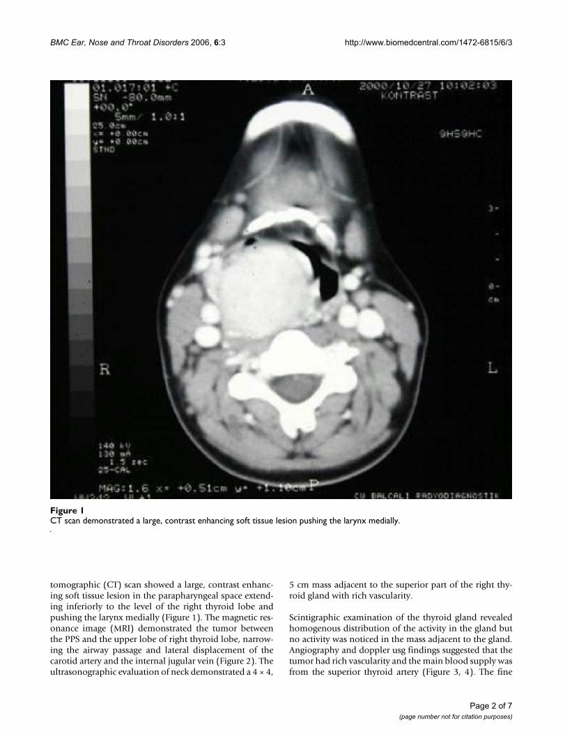
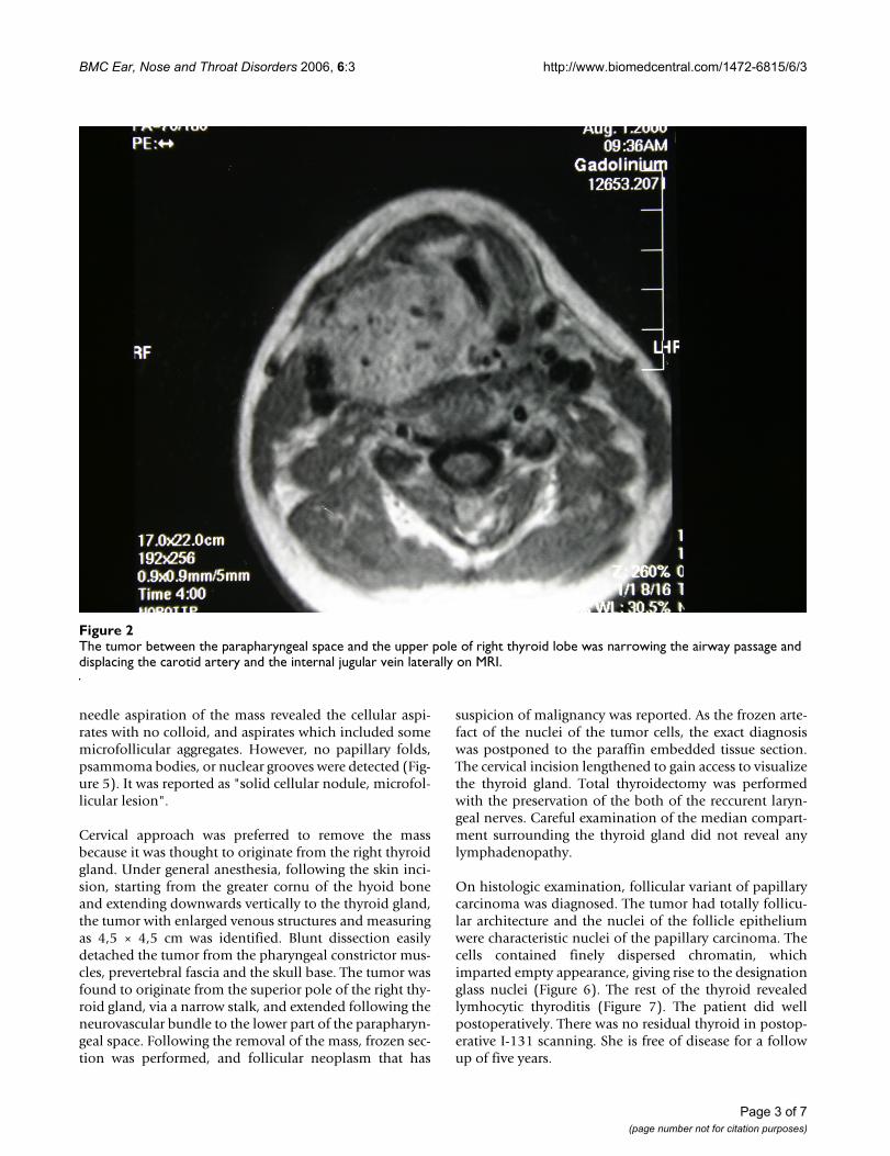
tomographic (CT) scan showed a large, contrast enhanc-ing soft tissue lesion in the parapharyngeal space extend-ing inferiorly to the level of the right thyroid lobe andpushing the larynx medially (Figure 1). The magnetic res-onance image (MRI) demonstrated the tumor betweenthe PPS and the upper lobe of right thyroid lobe, narrow-ing the airway passage and lateral displacement of thecarotid artery and the internal jugular vein (Figure 2). Theultrasonographic evaluation of neck demonstrated a 4 × 4,
5 cm mass adjacent to the superior part of the right thy-roid gland with rich vascularity.
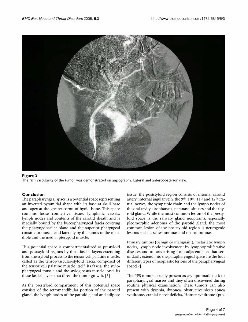
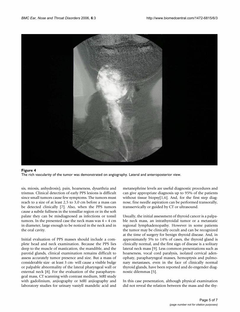
Scintigraphic examination of the thyroid gland revealedhomogenous distribution of the activity in the gland butno activity was noticed in the mass adjacent to the gland.Angiography and doppler usg findings suggested that thetumor had rich vascularity and the main blood supply wasfrom the superior thyroid artery (Figure 3, 4). The fine
CT scan demonstrated a large, contrast enhancing soft tissue lesion pushing the larynx mediallyFigure 1CT scan demonstrated a large, contrast enhancing soft tissue lesion pushing the larynx medially.
Page 2 of 7(page number not for citation purposes)
BMC Ear, Nose and Throat Disorders 2006, 6:3 http://www.biomedcentral.com/1472-6815/6/3
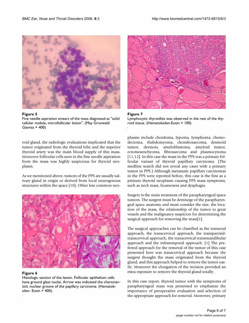
needle aspiration of the mass revealed the cellular aspi-rates with no colloid, and aspirates which included somemicrofollicular aggregates. However, no papillary folds,psammoma bodies, or nuclear grooves were detected (Fig-ure 5). It was reported as "solid cellular nodule, microfol-licular lesion".
Cervical approach was preferred to remove the massbecause it was thought to originate from the right thyroidgland. Under general anesthesia, following the skin inci-sion, starting from the greater cornu of the hyoid boneand extending downwards vertically to the thyroid gland,the tumor with enlarged venous structures and measuringas 4,5 × 4,5 cm was identified. Blunt dissection easilydetached the tumor from the pharyngeal constrictor mus-cles, prevertebral fascia and the skull base. The tumor wasfound to originate from the superior pole of the right thy-roid gland, via a narrow stalk, and extended following theneurovascular bundle to the lower part of the parapharyn-geal space. Following the removal of the mass, frozen sec-tion was performed, and follicular neoplasm that has
suspicion of malignancy was reported. As the frozen arte-fact of the nuclei of the tumor cells, the exact diagnosiswas postponed to the paraffin embedded tissue section.The cervical incision lengthened to gain access to visualizethe thyroid gland. Total thyroidectomy was performedwith the preservation of the both of the reccurent laryn-geal nerves. Careful examination of the median compart-ment surrounding the thyroid gland did not reveal anylymphadenopathy.
On histologic examination, follicular variant of papillarycarcinoma was diagnosed. The tumor had totally follicu-lar architecture and the nuclei of the follicle epitheliumwere characteristic nuclei of the papillary carcinoma. Thecells contained finely dispersed chromatin, whichimparted empty appearance, giving rise to the designationglass nuclei (Figure 6). The rest of the thyroid revealedlymhocytic thyroditis (Figure 7). The patient did wellpostoperatively. There was no residual thyroid in postop-erative I-131 scanning. She is free of disease for a followup of five years.
The tumor between the parapharyngeal space and the upper pole of right thyroid lobe was narrowing the airway passage and displacing the carotid artery and the internal jugular vein laterally on MRIFigure 2The tumor between the parapharyngeal space and the upper pole of right thyroid lobe was narrowing the airway passage and displacing the carotid artery and the internal jugular vein laterally on MRI.
Page 3 of 7(page number not for citation purposes)
BMC Ear, Nose and Throat Disorders 2006, 6:3 http://www.biomedcentral.com/1472-6815/6/3
ConclusionThe parapharyngeal space is a potential space representingan inverted pyramidal shape with its base at skull baseand apex at the greater cornu of hyoid bone. This spacecontains loose connective tissue, lymphatic vessels,lymph nodes and contents of the carotid sheath and ismedially bound by the buccopharyngeal fascia coveringthe pharyngobasilar plane and the superior pharyngealconstrictor muscle and laterally by the ramus of the man-dible and the medial pterygoid muscle.
This potential space is compartmentalized as prestyloidand poststyloid regions by thick fascial layers extendingfrom the styloid process to the tensor veli palatine muscle,called as the tensor-vascular-styloid fascia, composed ofthe tensor veli palatine muscle itself, its fascia, the stylo-pharyngeal muscle and the styloglossus muscle. And, itsthese fascial layers that direct the tumor growth. [3]
As the prestyloid compartment of this potential spaceconsists of the retromandibular portion of the parotidgland, the lymph nodes of the parotid gland and adipose
tissue, the poststyloid region consists of internal carotidartery, internal jugular vein, the 9th, 10th, 11th and 12th cra-nial nerves, the sympathic chain and the lymph nodes ofthe oral cavity, oropharynx, paranasal sinuses and the thy-roid gland. While the most common lesion of the presty-loid space is the salivary gland neoplasms, especiallypleomorphic adenoma of the parotid gland, the mostcommon lesion of the poststyloid region is neurogeniclesions such as schwannomas and neurofibromas.
Primary tumors (benign or malignant), metastatic lymphnodes, lymph node involvement by lymphoproliferativediseases and tumors arising from adjacent sites that sec-ondarily extend into the parapharyngeal space are the fourdifferent types of neoplastic lesions of the parapharyngealspace[2].
The PPS tumors usually present as asymptomatic neck orparapharyngeal masses and they often discovered duringroutine physical examination. These tumors can alsopresent with dysphia, dyspnea, obstructive sleep apneasyndrome, cranial nerve deficits, Horner syndrome (pto-
The rich vascularity of the tumor was demonstrated on angiographyFigure 3The rich vascularity of the tumor was demonstrated on angiography. Lateral and anteroposterior view.
Page 4 of 7(page number not for citation purposes)
BMC Ear, Nose and Throat Disorders 2006, 6:3 http://www.biomedcentral.com/1472-6815/6/3
sis, miosis, anhydrosis), pain, hoarseness, dysarthria andtrismus. Clinical detection of early PPS lesions is difficultsince small tumors cause few symptoms. The tumors mustreach to a size of at least 2,5 to 3,0 cm before a mass canbe detected clinically [7]. Also, when the PPS tumorscause a subtle fullness in the tonsillar region or in the softpalate they can be misdiagnosed as infections or tonsiltumors. In the presented case the neck mass was 4 × 4 cmin diameter, large enough to be noticed in the neck and inthe oral cavity.
Initial evaluation of PPS masses should include a com-plete head and neck examination. Because the PPS liesdeep to the muscle of mastication, the mandible, and theparotid glands, clinical examination remains difficult toassess accurately tumor presence and size. But a mass ofconsiderable size -at least 3 cm- will cause a visible bulgeor palpable abnormality of the lateral pharyngeal wall orexternal neck [8]. For the evaluation of the parapharyn-geal mass, CT scanning with contrast medium, MRI studywith gadolinium, anjiography or MRI anjiography andlaboratory studies for urinary vaniyll mandelic acid and
metanephrine levels are useful diagnostic procedures andcan give appropriate diagnosis up to 95% of the patientswithout tissue biopsy[1,6]. And, for the first step diag-nose, fine needle aspiration can be performed transorally,transservically or guided by CT or ultrasound.
Usually, the initial assessment of thyroid cancer is a palpa-ble neck mass, an intrathyroidal tumor or a metastaticregional lymphadenopathy. However in some patientsthe tumor may be clinically occult and can be recognizedat the time of surgery for benign thyroid disease. And, inapproximately 5% to 14% of cases, the thyroid gland isclinically normal, and the first sign of disease is a solitarylateral neck mass [9]. Less common presentations such ashoarseness, vocal cord paralysis, isolated cervical aden-ophaty, parapharyngeal masses, hemoptysis and pulmo-nary metastases, even in the face of clinically normalthyroid glands, have been reported and do engender diag-nostic dilemmas [5].
In this case presentation, although physical examinationdid not reveal the relation between the mass and the thy-
The rich vascularity of the tumor was demonstrated on angiographyFigure 4The rich vascularity of the tumor was demonstrated on angiography. Lateral and anteroposterior view.
Page 5 of 7(page number not for citation purposes)
BMC Ear, Nose and Throat Disorders 2006, 6:3 http://www.biomedcentral.com/1472-6815/6/3
roid gland, the radiologic evaluations implicated that thetumor originated from the thyroid lobe and the superiorthyroid artery was the main blood supply of this mass.Moreover follicular cells seen in the fine needle aspirationfrom the mass was highly suspicious for thyroid neo-plasm.
As we mentioned above, tumors of the PPS are usually sal-ivary gland in origin or derived from local neurogenousstructures within the space [10]. Other less common neo-
plasms include chordoma, lypoma, lymphoma, chemo-dectoma, rhabdomyoma, chondrosarcoma, desmoidtumor, dermois, ameloblastoma, amyloid tumor,ectomesenchyoma, fibrosarcoma and plasmocytoma[11,12]. In this case the mass in the PPS was a primary fol-licular variant of thyroid papillary carcinoma. (Themedline search did not reveal any cases with a primarytumor in PPS.) Although metastatic papillary carcinomasin the PPS were reported before, this case is the first as aprimary thyroid neoplasm causing PPS mass symptoms,such as neck mass, hoarseness and dysphagia.
Surgery is the main treatment of the parapharyngeal spacetumors. The surgent must be demiurge of the parapharyn-geal space anatomy and must consider the size, the loca-tion of the mass, the relationship of the tumor to greatvessels and the malignancy suspicion for determining thesurgical approach for removing the mass[1].
The surgical approaches can be classified as the transoralapproach, the transcervical approach, the transparotid-transcervical approach, the transcervical-transmandibularapproach and the infratemporal approach. [1] The pre-ferred approach for the removal of the tumor of this casepresented here was transcervical approach because thesurgent thought the mass originated from the thyroidgland, and this approach helped to remove the tumor eas-ily. Moreover the elongation of the incision provided anextra exposure to remove the thyroid gland totally.
In this case report, thyroid tumor with the symptoms ofparapharyngeal mass was presented to emphasize theimportance of preoperative evaluation and selection ofthe appropriate approach for removal. Moreover, primary
Lymphocytic thyroiditis was observed in the rest of the thy-roid tissueFigure 7Lymphocytic thyroiditis was observed in the rest of the thy-roid tissue. (Hematoksilen-Eosin × 100)
Fine needle aspiration smears of the mass diagnosed as "solid cellular nodule, microfollicular lesion"Figure 5Fine needle aspiration smears of the mass diagnosed as "solid cellular nodule, microfollicular lesion". (May Grunwald Giemsa × 400)
Histologic section of the lesionFigure 6Histologic section of the lesion. Follicular epithelium cells have ground glass nuclei. Arrow was indicated the character-istic nuclear groove of the papillary carcinoma. (Hematok-silen- Eosin × 400).
Page 6 of 7(page number not for citation purposes)
BMC Ear, Nose and Throat Disorders 2006, 6:3 http://www.biomedcentral.com/1472-6815/6/3
Publish with BioMed Central and every scientist can read your work free of charge
"BioMed Central will be the most significant development for disseminating the results of biomedical research in our lifetime."
Sir Paul Nurse, Cancer Research UK
Your research papers will be:
available free of charge to the entire biomedical community
peer reviewed and published immediately upon acceptance
cited in PubMed and archived on PubMed Central
yours — you keep the copyright
Submit your manuscript here:http://www.biomedcentral.com/info/publishing_adv.asp
BioMedcentral
thyroid tumor should always be remembered in the differ-ential diagnosis of parapharyngeal masses.
Competing interestsThe author(s) declare that they have no competing inter-ests.
Authors' contributionsThe authors have equally contributed to this case report.
References1. Attia A, El-Shafiey M, El-Shazly S, Shouman T, Zaky I: Management
of parapharyngeal space tumors at the National CancerInstitute, Egypt. J Egypt Natl Canc Inst 2004, 16(1):34-42.
2. Stell PM, Mansfield AO, Stoney PJ: Surgical approaches to tumorsof the parapharyngeal space. Am J Otolaryngol 1985, 6(2):92-7.
3. Lombardi D, Nicolai P, Antonelli AR, Maroldi R, Farina D, Shaha AR:Parapharyngeal lymph node metastasis: an unusual presen-tation of papillary thyroid carcinoma. Head Neck 2004,26(2):190-6.
4. Zohar Y, Strauss M: Occult distant metastases of well-differen-tiated thyroid carcinoma. Head Neck 1994, 16(5):438-42.
5. Gagel RF, Goepfert H, Callender DL: Changing concepts in thepathogenesis and management of thyroid carcinoma. CACancer J Clin 1996, 46(5):261-83.
6. Miller FR, Wanamaker JR, Lavertu P, Wood BG: Magnetic reso-nance imaging and the management of parapharyngealspace tumors. Head Neck 1996, 18(1):67-77.
7. Som PM, Biller HF, Lawson W, Sacher M, Lanzieri CF: Parapharyn-geal space masses: an updated protocol based upon 104cases. Radiology 1984, 153(1):149-56.
8. Carrau RL, Myers EN, Johnson JT: Management of tumors arisingin the parapharyngeal space. Laryngoscope 1990, 100(6):583-9.
9. Attie JN, Setzin M, Klein I: Thyroid carcinoma presenting as anenlarged cervical lymph node. Am J Surg 1993, 166(4):428-30.
10. Work WP, Hybels RL: A study of tumors of the parapharyngealspace. Laryngoscope 1974, 84(10):1748-55.
11. Fluur E: Parapharyngeal tumors. Arch Otolaryngol 1964,80:557-65.
12. Shapiro RS, Stool SE, Snow JB Jr, Chamorro H: Parapharyngealrhabdomyoma. Arch Otolaryngol 1975, 101(5):323-6.
Pre-publication historyThe pre-publication history for this paper can be accessedhere:
http://www.biomedcentral.com/1472-6815/6/3/prepub
Page 7 of 7(page number not for citation purposes)