Embed Size (px)
Citation preview
BioMed CentralBMC Microbiology
ss
Open AcceResearch articlePresence of Helicobacter pylori in a Mexican Pre-Columbian MummyGonzalo Castillo-Rojas1, Marco A Cerbón2 and Yolanda López-Vidal*1Address: 1Programa de Inmunología Molecular Microbiana, Departamento de Microbiología y Parasitología. Facultad de Medicina, Universidad Nacional Autónoma de México, Mexico City, Mexico and 2Departamento de Biología. Facultad de Química, Universidad Nacional Autónoma de México, Mexico City, Mexico
Email: Gonzalo Castillo-Rojas - [email protected]; Marco A Cerbón - [email protected]; Yolanda López-Vidal* - [email protected]
* Corresponding author
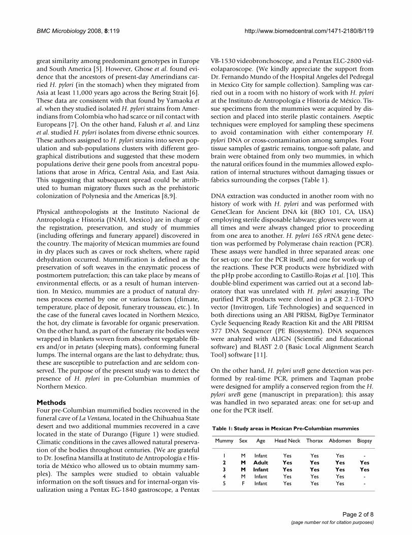
AbstractBackground: Recent studies showed that Helicobacter pylori existed in the New World prior tothe arrival of Columbus. The purpose of the present study was to detect the presence ofHelicobacter pylori in pre-Columbian mummies from Northern Mexico.
Methods: Six samples were studied (four samples of gastric remains, tongue-soft palate, and brainremained as negative controls) from two of the six naturally mummified corpses studied (adult maleand infant male). Samples were taken from tissues suitable for DNA amplification by Polymerasechain reaction (PCR). DNA was extracted and H. pylori detection was carried out by PCR andhybridized with the pHp probe from 16S rRNA gene. The purified PCR products were cloned andsequenced in both directions. DNA sequences were analyzed with ALIGN and BLAST software. Asecond amplification was performed using ureB gene by real-time PCR.
Results: From four samples of gastric remnant, only two were H. pylori-positive for amplificationof a 109 bp DNA fragment; the remaining two were negative, as were the tongue-soft palate andthe brain biopsies as well. These PCR products were hybridized with a pHp probe. Nucleotidesequence analysis showed homology with H. pylori in 98 of 99% when compared with the gene banknucleotide sequence. Only one sample of gastric remnant H. pylori-positive with 16S rRNA gene wasalso positive for ureB gene from H. pylori.
Conclusion: This data supported infection with H. pylori in Mexican pre-Columbian mummiesdating from approximately 1,350 AC.
BackgroundHelicobacter pylori (H. pylori) microaerophilic Gram-nega-tive bacteria, which colonize the human stomach, areassociated with an increased risk of developing gastriccancer and peptic ulcer disease [1]. In a study on the sur-vival of antigenic material in mummified human remainsfrom the Andean area of South America, Allison et al.
found that fecal specimens harbored antigens from H.pylori nearly 3,000 years old [2]. Several studies based ongenotypic analysis of H. pylori strains isolated from Latin-American patients exhibited European genotypes [3,4].Kersulyte et al. have suggested that H. pylori may have beenbrought to the New World by European conquerors andcolonist's ca 500 years ago, because the authors found a
Published: 15 July 2008
BMC Microbiology 2008, 8:119 doi:10.1186/1471-2180-8-119
Received: 6 December 2007Accepted: 15 July 2008
This article is available from: http://www.biomedcentral.com/1471-2180/8/119
© 2008 Castillo-Rojas et al; licensee BioMed Central Ltd. This is an Open Access article distributed under the terms of the Creative Commons Attribution License (http://creativecommons.org/licenses/by/2.0), which permits unrestricted use, distribution, and reproduction in any medium, provided the original work is properly cited.
Page 1 of 8(page number not for citation purposes)
BMC Microbiology 2008, 8:119 http://www.biomedcentral.com/1471-2180/8/119
great similarity among predominant genotypes in Europeand South America [5]. However, Ghose et al. found evi-dence that the ancestors of present-day Amerindians car-ried H. pylori (in the stomach) when they migrated fromAsia at least 11,000 years ago across the Bering Strait [6].These data are consistent with that found by Yamaoka etal. when they studied isolated H. pylori strains from Amer-indians from Colombia who had scarce or nil contact withEuropeans [7]. On the other hand, Falush et al. and Linzet al. studied H. pylori isolates from diverse ethnic sources.These authors assigned to H. pylori strains into seven pop-ulation and sub-populations clusters with different geo-graphical distributions and suggested that these modernpopulations derive their gene pools from ancestral popu-lations that arose in Africa, Central Asia, and East Asia.This suggesting that subsequent spread could be attrib-uted to human migratory fluxes such as the prehistoriccolonization of Polynesia and the Americas [8,9].
Physical anthropologists at the Instituto Nacional deAntropología e Historia (INAH, Mexico) are in charge ofthe registration, preservation, and study of mummies(including offerings and funerary apparel) discovered inthe country. The majority of Mexican mummies are foundin dry places such as caves or rock shelters, where rapiddehydration occurred. Mummification is defined as thepreservation of soft weaves in the enzymatic process ofpostmortem putrefaction; this can take place by means ofenvironmental effects, or as a result of human interven-tion. In Mexico, mummies are a product of natural dry-ness process exerted by one or various factors (climate,temperature, place of deposit, funerary trousseau, etc.). Inthe case of the funeral caves located in Northern Mexico,the hot, dry climate is favorable for organic preservation.On the other hand, as part of the funerary rite bodies werewrapped in blankets woven from absorbent vegetable fib-ers and/or in petates (sleeping mats), conforming funerallumps. The internal organs are the last to dehydrate; thus,these are susceptible to putrefaction and are seldom con-served. The purpose of the present study was to detect thepresence of H. pylori in pre-Columbian mummies ofNorthern Mexico.
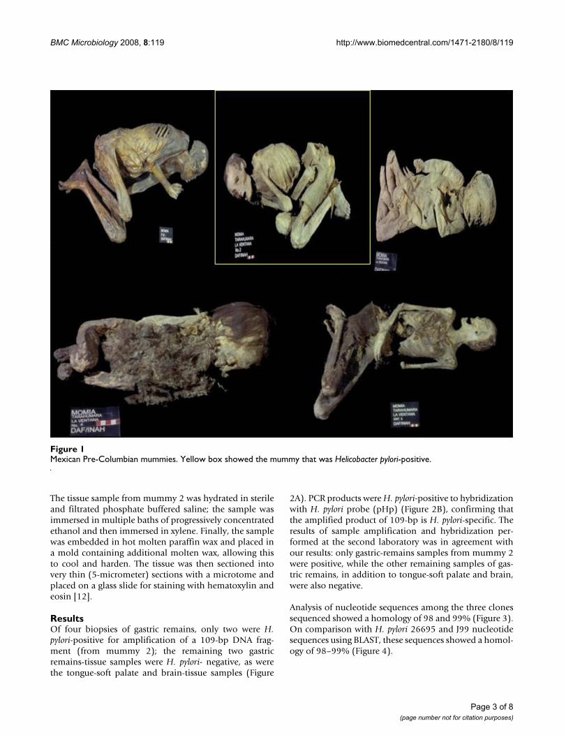
MethodsFour pre-Columbian mummified bodies recovered in thefuneral cave of La Ventana, located in the Chihuahua Statedesert and two additional mummies recovered in a cavelocated in the state of Durango (Figure 1) were studied.Climatic conditions in the caves allowed natural preserva-tion of the bodies throughout centuries. (We are gratefulto Dr. Josefina Mansilla at Instituto de Antropología e His-toria de México who allowed us to obtain mummy sam-ples). The samples were studied to obtain valuableinformation on the soft tissues and for internal-organ vis-ualization using a Pentax EG-1840 gastroscope, a Pentax
VB-1530 videobronchoscope, and a Pentax ELC-2800 vid-eolaparoscope. (We kindly appreciate the support fromDr. Fernando Mundo of the Hospital Angeles del Pedregalin Mexico City for sample collection). Sampling was car-ried out in a room with no history of work with H. pyloriat the Instituto de Antropología e Historia de México. Tis-sue specimens from the mummies were acquired by dis-section and placed into sterile plastic containers. Aseptictechniques were employed for sampling these specimensto avoid contamination with either contemporary H.pylori DNA or cross-contamination among samples. Fourtissue samples of gastric remains, tongue-soft palate, andbrain were obtained from only two mummies, in whichthe natural orifices found in the mummies allowed explo-ration of internal structures without damaging tissues orfabrics surrounding the corpses (Table 1).
DNA extraction was conducted in another room with nohistory of work with H. pylori and was performed withGeneClean for Ancient DNA kit (BIO 101, CA, USA)employing sterile disposable labware; gloves were worn atall times and were always changed prior to proceedingfrom one area to another. H. pylori 16S rRNA gene detec-tion was performed by Polymerase chain reaction (PCR).These assays were handled in three separated areas: onefor set-up; one for the PCR itself, and one for work-up ofthe reactions. These PCR products were hybridized withthe pHp probe according to Castillo-Rojas et al. [10]. Thisdouble-blind experiment was carried out at a second lab-oratory that was unrelated with H. pylori assaying. Thepurified PCR products were cloned in a pCR 2.1-TOPOvector (Invitrogen, Life Technologies) and sequenced inboth directions using an ABI PRISM, BigDye TerminatorCycle Sequencing Ready Reaction Kit and the ABI PRISM377 DNA Sequencer (PE Biosystems). DNA sequenceswere analyzed with ALIGN (Scientific and Educationalsoftware) and BLAST 2.0 (Basic Local Alignment SearchTool) software [11].
On the other hand, H. pylori ureB gene detection was per-formed by real-time PCR, primers and Taqman probewere designed for amplify a conserved region from the H.pylori ureB gene (manuscript in preparation); this assaywas handled in two separated areas: one for set-up andone for the PCR itself.
Table 1: Study areas in Mexican Pre-Columbian mummies
Mummy Sex Age Head Neck Thorax Abdomen Biopsy
1 M Infant Yes Yes Yes -2 M Adult Yes Yes Yes Yes3 M Infant Yes Yes Yes Yes4 M Infant Yes Yes Yes -5 F Infant Yes Yes Yes -
Page 2 of 8(page number not for citation purposes)
BMC Microbiology 2008, 8:119 http://www.biomedcentral.com/1471-2180/8/119
The tissue sample from mummy 2 was hydrated in sterileand filtrated phosphate buffered saline; the sample wasimmersed in multiple baths of progressively concentratedethanol and then immersed in xylene. Finally, the samplewas embedded in hot molten paraffin wax and placed ina mold containing additional molten wax, allowing thisto cool and harden. The tissue was then sectioned intovery thin (5-micrometer) sections with a microtome andplaced on a glass slide for staining with hematoxylin andeosin [12].
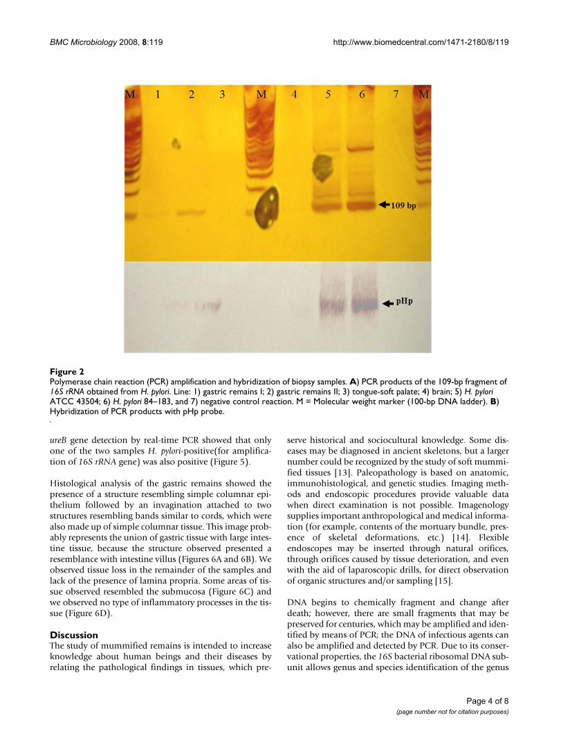
ResultsOf four biopsies of gastric remains, only two were H.pylori-positive for amplification of a 109-bp DNA frag-ment (from mummy 2); the remaining two gastricremains-tissue samples were H. pylori- negative, as werethe tongue-soft palate and brain-tissue samples (Figure
2A). PCR products were H. pylori-positive to hybridizationwith H. pylori probe (pHp) (Figure 2B), confirming thatthe amplified product of 109-bp is H. pylori-specific. Theresults of sample amplification and hybridization per-formed at the second laboratory was in agreement withour results: only gastric-remains samples from mummy 2were positive, while the other remaining samples of gas-tric remains, in addition to tongue-soft palate and brain,were also negative.
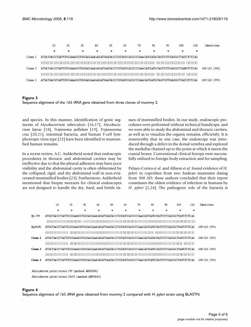
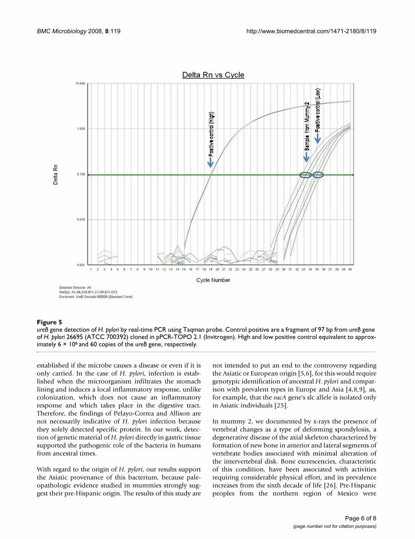
Analysis of nucleotide sequences among the three clonessequenced showed a homology of 98 and 99% (Figure 3).On comparison with H. pylori 26695 and J99 nucleotidesequences using BLAST, these sequences showed a homol-ogy of 98–99% (Figure 4).
Mexican Pre-Columbian mummiesFigure 1Mexican Pre-Columbian mummies. Yellow box showed the mummy that was Helicobacter pylori-positive.
Page 3 of 8(page number not for citation purposes)
BMC Microbiology 2008, 8:119 http://www.biomedcentral.com/1471-2180/8/119
ureB gene detection by real-time PCR showed that onlyone of the two samples H. pylori-positive(for amplifica-tion of 16S rRNA gene) was also positive (Figure 5).
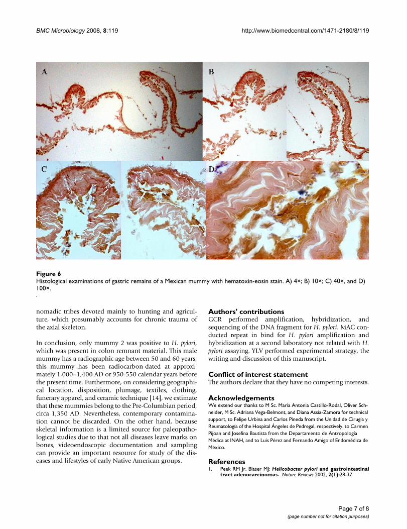
Histological analysis of the gastric remains showed thepresence of a structure resembling simple columnar epi-thelium followed by an invagination attached to twostructures resembling bands similar to cords, which werealso made up of simple columnar tissue. This image prob-ably represents the union of gastric tissue with large intes-tine tissue, because the structure observed presented aresemblance with intestine villus (Figures 6A and 6B). Weobserved tissue loss in the remainder of the samples andlack of the presence of lamina propria. Some areas of tis-sue observed resembled the submucosa (Figure 6C) andwe observed no type of inflammatory processes in the tis-sue (Figure 6D).
DiscussionThe study of mummified remains is intended to increaseknowledge about human beings and their diseases byrelating the pathological findings in tissues, which pre-
serve historical and sociocultural knowledge. Some dis-eases may be diagnosed in ancient skeletons, but a largernumber could be recognized by the study of soft mummi-fied tissues [13]. Paleopathology is based on anatomic,immunohistological, and genetic studies. Imaging meth-ods and endoscopic procedures provide valuable datawhen direct examination is not possible. Imagenologysupplies important anthropological and medical informa-tion (for example, contents of the mortuary bundle, pres-ence of skeletal deformations, etc.) [14]. Flexibleendoscopes may be inserted through natural orifices,through orifices caused by tissue deterioration, and evenwith the aid of laparoscopic drills, for direct observationof organic structures and/or sampling [15].
DNA begins to chemically fragment and change afterdeath; however, there are small fragments that may bepreserved for centuries, which may be amplified and iden-tified by means of PCR; the DNA of infectious agents canalso be amplified and detected by PCR. Due to its conser-vational properties, the 16S bacterial ribosomal DNA sub-unit allows genus and species identification of the genus
Polymerase chain reaction (PCR) amplification and hybridization of biopsy samplesFigure 2Polymerase chain reaction (PCR) amplification and hybridization of biopsy samples. A) PCR products of the 109-bp fragment of 16S rRNA obtained from H. pylori. Line: 1) gastric remains I; 2) gastric remains II; 3) tongue-soft palate; 4) brain; 5) H. pylori ATCC 43504; 6) H. pylori 84–183, and 7) negative control reaction. M = Molecular weight marker (100-bp DNA ladder). B) Hybridization of PCR products with pHp probe.
Page 4 of 8(page number not for citation purposes)
BMC Microbiology 2008, 8:119 http://www.biomedcentral.com/1471-2180/8/119
and species. In this manner, identification of genic seg-ments of Mycobacterium tuberculosis [16,17], Mycobacte-rium leprae [18], Treponema pallidum [19], Trypanosomacruz [20,21], intestinal bacteria, and human T-cell lym-photropic virus type [22] have been identified in mummi-fied human remains.
In a recent review, A.C. Aufderheid noted that endoscopicprocedures in thoracic and abdominal cavities may beineffective due to that the pleural adhesion may have poorvisibility and the abdominal cavity is often obliterated bythe collapsed, rigid, and dry abdominal wall in non-evis-cerated mummified bodies [23]. Furthermore, Aufderheidmentioned that biopsy tweezers for clinical endoscopesare not designed to handle the dry, hard, and brittle tis-
sues of mummified bodies. In our study, endoscopic pro-cedures were performed without technical handicaps, andwe were able to study the abdominal and thoracic cavities,as well as to visualize the organic remains, efficiently. It isnoteworthy that in one case, the endoscope was intro-duced through a defect in the dorsal vertebra and exploredthe medullar channel up to the point at which it meets thecranial bones. Conventional clinical forceps were success-fully utilized to foreign-body extraction and for sampling.
Pelayo-Correa et al. and Allison et al. found evidence of H.pylori in coprolites from two Andean mummies datingfrom 300 AD; these authors concluded that their reportconstitutes the oldest evidence of infection in humans byH. pylori [2,24]. The pathogenic role of the bacteria is
Sequence alignment of the 16S rRNA gene obtained from three clones of mummy 2Figure 3Sequence alignment of the 16S rRNA gene obtained from three clones of mummy 2.
Sequence alignment of 16S rRNA gene obtained from mummy 2 compared with H. pylori strain using BLASTNFigure 4Sequence alignment of 16S rRNA gene obtained from mummy 2 compared with H. pylori strain using BLASTN.
Page 5 of 8(page number not for citation purposes)
BMC Microbiology 2008, 8:119 http://www.biomedcentral.com/1471-2180/8/119
established if the microbe causes a disease or even if it isonly carried. In the case of H. pylori, infection is estab-lished when the microorganism infiltrates the stomachlining and induces a local inflammatory response, unlikecolonization, which does not cause an inflammatoryresponse and which takes place in the digestive tract.Therefore, the findings of Pelayo-Correa and Allison arenot necessarily indicative of H. pylori infection becausethey solely detected specific protein. In our work, detec-tion of genetic material of H. pylori directly in gastric tissuesupported the pathogenic role of the bacteria in humansfrom ancestral times.
With regard to the origin of H. pylori, our results supportthe Asiatic provenance of this bacterium, because pale-opathologic evidence studied in mummies strongly sug-gest their pre-Hispanic origin. The results of this study are
not intended to put an end to the controversy regardingthe Asiatic or European origin [5,6], for this would requiregenotypic identification of ancestral H. pylori and compar-ison with prevalent types in Europe and Asia [4,8,9], as,for example, that the vacA gene's slc allele is isolated onlyin Asiatic individuals [25].
In mummy 2, we documented by x-rays the presence ofvertebral changes as a type of deforming spondylosis, adegenerative disease of the axial skeleton characterized byformation of new bone in anterior and lateral segments ofvertebrate bodies associated with minimal alteration ofthe intervertebral disk. Bone excrescencies, characteristicof this condition, have been associated with activitiesrequiring considerable physical effort, and its prevalenceincreases from the sixth decade of life [26]. Pre-Hispanicpeoples from the northern region of Mexico were
ureB gene detection of H. pylori by real-time PCR using Taqman probeFigure 5ureB gene detection of H. pylori by real-time PCR using Taqman probe. Control positive are a fragment of 97 bp from ureB gene of H. pylori 26695 (ATCC 700392) cloned in pPCR-TOPO 2.1 (Invitrogen). High and low positive control equivalent to approx-imately 6 × 10⁶ and 60 copies of the ureB gene, respectively.
Page 6 of 8(page number not for citation purposes)
BMC Microbiology 2008, 8:119 http://www.biomedcentral.com/1471-2180/8/119
nomadic tribes devoted mainly to hunting and agricul-ture, which presumably accounts for chronic trauma ofthe axial skeleton.
In conclusion, only mummy 2 was positive to H. pylori,which was present in colon remnant material. This malemummy has a radiographic age between 50 and 60 years;this mummy has been radiocarbon-dated at approxi-mately 1,000–1,400 AD or 950-550 calendar years beforethe present time. Furthermore, on considering geographi-cal location, disposition, plumage, textiles, clothing,funerary apparel, and ceramic technique [14], we estimatethat these mummies belong to the Pre-Columbian period,circa 1,350 AD. Nevertheless, contemporary contamina-tion cannot be discarded. On the other hand, becauseskeletal information is a limited source for paleopatho-logical studies due to that not all diseases leave marks onbones, videoendoscopic documentation and samplingcan provide an important resource for study of the dis-eases and lifestyles of early Native American groups.
Authors' contributionsGCR performed amplification, hybridization, andsequencing of the DNA fragment for H. pylori. MAC con-ducted repeat in bind for H. pylori amplification andhybridization at a second laboratory not related with H.pylori assaying. YLV performed experimental strategy, thewriting and discussion of this manuscript.
Conflict of interest statementThe authors declare that they have no competing interests.
AcknowledgementsWe extend our thanks to M Sc. María Antonia Castillo-Rodal, Oliver Sch-neider, M Sc. Adriana Vega-Belmont, and Diana Assia-Zamora for technical support, to Felipe Urbina and Carlos Pineda from the Unidad de Cirugía y Reumatología of the Hospital Ángeles de Pedregal, respectively, to Carmen Pijoan and Josefina Bautista from the Departamento de Antropología Médica at INAH, and to Luís Pérez and Fernando Amigo of Endomédica de México.
References1. Peek RM Jr, Blaser MJ: Helicobacter pylori and gastrointestinal
tract adenocarcinomas. Nature Reviews 2002, 2(1):28-37.
Histological examinations of gastric remains of a Mexican mummy with hematoxin-eosin stainFigure 6Histological examinations of gastric remains of a Mexican mummy with hematoxin-eosin stain. A) 4×; B) 10×; C) 40×, and D) 100×.
Page 7 of 8(page number not for citation purposes)
BMC Microbiology 2008, 8:119 http://www.biomedcentral.com/1471-2180/8/119
Publish with BioMed Central and every scientist can read your work free of charge
"BioMed Central will be the most significant development for disseminating the results of biomedical research in our lifetime."
Sir Paul Nurse, Cancer Research UK
Your research papers will be:
available free of charge to the entire biomedical community
peer reviewed and published immediately upon acceptance
cited in PubMed and archived on PubMed Central
yours — you keep the copyright
Submit your manuscript here:http://www.biomedcentral.com/info/publishing_adv.asp
BioMedcentral
2. Allison MJ, Bergman T, Gerszten E: Further studies on fecal par-asites in antiquity. American Journal of Clinical Pathology 1999,112(5):605-609.
3. Achtman M, Azuma T, Berg DE, Ito Y, Morelli G, Pan ZJ, Suerbaum S,Thompson SA, Ende A van der, van Doorn LJ: Recombination andclonal groupings within Helicobacter pylori from differentgeographical regions. Molecular Microbiology 1999, 32(3):459-470.
4. Van Doorn LJ, Figueiredo C, Megraud F, Pena S, Midolo P, QueirozDM, Carneiro F, Vanderborght B, Pegado MD, Sanna R, et al.: Geo-graphic distribution of vacA allelic types of Helicobacterpylori. Gastroenterology 1999, 116(4):823-830.
5. Kersulyte D, Mukhopadhyay AK, Velapatino B, Su W, Pan Z, GarcíaC, Hernández V, Valdez Y, Mistry RS, Gilman RH, et al.: Differencesin genotypes of Helicobacter pylori from different humanpopulations. Journal of Bacteriology 2000, 182(11):3210-3218.
6. Ghose C, Pérez-Pérez GI, Domínguez-Bello MG, Pride DT, Bravi CM,Blaser MJ: East Asian genotypes of Helicobacter pylori strainsin Amerindians provide evidence for its ancient human car-riage. Proceedings of the National Academy of Sciences of the UnitedStates of America 2002, 99(23):15107-15111.
7. Yamaoka Y, Orito E, Mizokami M, Gutiérrez O, Saitou N, Kodama T,Osato MS, Kim JG, Ramírez FC, Mahachai V, et al.: Helicobacterpylori in North and South America before Columbus. FEBSLetters 2002, 517(1–3):180-184.
8. Falush D, Wirth T, Linz B, Pritchard JK, Stephens M, Kidd M, BlaserMJ, Graham DY, Vacher S, Pérez-Pérez GI, et al.: Traces of humanmigrations in Helicobacter pylori populations. Science 2003,299(5612):1582-1585.
9. Linz B, Balloux F, Moodley Y, Manica A, Liu H, Roumagnac P, FalushD, Stamer C, Prugnolle F, Merwe SW van der, et al.: An African ori-gin for the intimate association between humans and Helico-bacter pylori. Nature 2007, 445(7130):915-918.
10. Castillo-Rojas G, Ballesteros MA, Ponce de León S, Morales-EspinosaR, Cravioto A, López-Vidal Y: Bleeding peptic ulcers and pres-ence of Helicobacter pylori by various tests: a case-controlstudy. European Journal of Gastroenterology & Hepatology 2002,14(10):1113-1118.
11. Altschul SF, Gish W, Miller W, Myers EW, Lipman DJ: Basic localalignment search tool. Journal of Molecular Biology 1990,215(3):403-410.
12. Mekota AM, Vermehren M: Determination of optimal rehydra-tion, fixation and staining methods for histological andimmunohistochemical analysis of mummified soft tissues.Biotechnic and Histochemistry 2005, 80(1):7-13.
13. Cockburn A, Cockburn E: [Paleopathology in Peru]. Paleopathol-ogy Newsletter 1977:3-6,9.
14. Pineda C, Mansilla J, Pijoan C, Fernández S, Martínez-Lavin M: Radio-graphs of an ancient mortuary bundle support theory for theNew World origin of syphilis. AJR Am J Roentgenol 1998,171(2):321-324.
15. Tapp E, Stanworth P, Wildsmith K: The endoscope in mummyresearch. In Evidence Embalmed Edited by: David R, Tapp E. Man-chester, England: Manchester University Press; 1984:65-77.
16. Donoghue HD, Spigelman M, Greenblatt CL, Lev-Maor G, Bar-GalGK, Matheson C, Vernon K, Nerlich AG, Zink AR: Tuberculosis:from prehistory to Robert Koch, as revealed by ancientDNA. The Lancet Infectious Diseases 2004, 4(9):584-592.
17. Salo WL, Aufderheide AC, Buikstra J, Holcomb TA: Identificationof Mycobacterium tuberculosis DNA in a pre-ColumbianPeruvian mummy. Proceedings of the National Academy of Sciencesof the United States of America 1994, 91(6):2091-2094.
18. Rafi A, Spigelman M, Stanford J, Lemma E, Donoghue H, Zias J: Myco-bacterium leprae DNA from ancient bone detected by PCR.Lancet 1994, 343(8909):1360-1361.
19. Kolman CJ, Centurion-Lara A, Lukehart SA, Owsley DW, Tuross N:Identification of Treponema pallidum subspecies pallidum in a200-year-old skeletal specimen. The Journal of Infectious Diseases1999, 180(6):2060-2063.
20. Aufderheide AC, Salo W, Madden M, Streitz J, Buikstra J, Guhl F, Arri-aza B, Renier C, Wittmers LE Jr, Fornaciari G, et al.: A 9,000-yearrecord of Chagas' disease. Proceedings of the National Academy ofSciences of the United States of America 2004, 101(7):2034-2039.
21. Guhl F, Jaramillo C, Vallejo GA, Yockteng R, Cárdenas-Arroyo F, For-naciari G, Arriaza B, Aufderheide AC: Isolation of Trypanosomacruzi DNA in 4,000-year-old mummified human tissue from
northern Chile. American Journal of Physical Anthropology 1999,108(4):401-407.
22. Li HC, Fujiyoshi T, Lou H, Yashiki S, Sonoda S, Cartier L, Núñez L,Muñoz I, Horai S, Tajima K: The presence of ancient human T-cell lymphotropic virus type I provirus DNA in an Andeanmummy. Nature Medicine 1999, 5(12):1428-1432.
23. Aufderheide AC: Progress in soft tissue paleopathology. Journalof the American Medical Association 2000, 284(20):2571-2573.
24. Correa P, Willis D, Allison M, Gerszten E: Helicobacter pylori inpre-Columbian mummies [AGA Abstract #G3919]. Gastroen-terology 1998, 114(4):A956.
25. Castillo-Rojas G, Mazari-Hiriart M, López-Vidal Y: [Helicobacterpylori: focus on CagA and VacA major virulence factors].Salud Publica de Mexico 2004, 46(6):538-548.
26. Resnick D: Degenerative diseases of the vertebral column.Radiology 1985, 156(1):3-14.
Page 8 of 8(page number not for citation purposes)