Embed Size (px)
Citation preview

BioMed CentralBMC Neuroscience
ss
Open AcceResearch articleBrain-derived neurotrophic factor modulation of Kv1.3 channel is disregulated by adaptor proteins Grb10 and nShcBeverly S Colley†, Melissa A Cavallin†, KC Biju, David R Marks and Debra A Fadool*Address: Department of Biological Science, Programs in Neuroscience and Molecular Biophysics, The Florida State University, Tallahassee, Florida, USA
Email: Beverly S Colley - [email protected]; Melissa A Cavallin - [email protected]; KC Biju - [email protected]; David R Marks - [email protected]; Debra A Fadool* - [email protected]
* Corresponding author †Equal contributors
AbstractBackground: Neurotrophins are important regulators of growth and regeneration, and acutely, they canmodulate the activity of voltage-gated ion channels. Previously we have shown that acute brain-derivedneurotrophic factor (BDNF) activation of neurotrophin receptor tyrosine kinase B (TrkB) suppresses theShaker voltage-gated potassium channel (Kv1.3) via phosphorylation of multiple tyrosine residues in the Nand C terminal aspects of the channel protein. It is not known how adaptor proteins, which lack catalyticactivity, but interact with members of the neurotrophic signaling pathway, might scaffold with ion channelsor modulate channel activity.
Results: We report the co-localization of two adaptor proteins, neuronal Src homology and collagen(nShc) and growth factor receptor-binding protein 10 (Grb10), with Kv1.3 channel as demonstratedthrough immunocytochemical approaches in the olfactory bulb (OB) neural lamina. To further explore thespecificity and functional ramification of adaptor/channel co-localization, we performedimmunoprecipitation and Western analysis of channel, kinase, and adaptor transfected human embryonickidney 293 cells (HEK 293). nShc formed a direct protein-protein interaction with Kv1.3 that wasindependent of BDNF-induced phosphorylation of Kv1.3, whereas Grb10 did not complex with Kv1.3 inHEK 293 cells. Both adaptors, however, co-immunoprecipitated with Kv1.3 in native OB. Grb10 wasinterestingly able to decrease the total expression of Kv1.3, particularly at the membrane surface, andsubsequently eliminated the BDNF-induced phosphorylation of Kv1.3. To examine the possibility that theSrc homology 2 (SH2) domains of Grb10 were directly binding to basally phosphorylated tyrosines inKv1.3, we utilized point mutations to substitute multiple tyrosine residues with phenylalanine. Removal ofthe tyrosines 111–113 and 449 prevented Grb10 from decreasing Kv1.3 expression. In the absence ofeither adaptor protein, channel co-expression reciprocally down-regulated expression and tyrosinephosphorylation of TrkB kinase and related insulin receptor kinase. Finally, through patch-clampelectrophysiology, we found that the BDNF-induced current suppression of the channel was prevented byboth nShc and Grb10.
Conclusion: We report that adaptor protein alteration of kinase-induced Kv1.3 channel modulation isrelated to the degree of direct protein-protein association and that the channel itself can reciprocallymodulate receptor-linked tyrosine kinase expression and activity.
Published: 23 January 2009
BMC Neuroscience 2009, 10:8 doi:10.1186/1471-2202-10-8
Received: 4 August 2008Accepted: 23 January 2009
This article is available from: http://www.biomedcentral.com/1471-2202/10/8
© 2009 Colley et al; licensee BioMed Central Ltd. This is an Open Access article distributed under the terms of the Creative Commons Attribution License (http://creativecommons.org/licenses/by/2.0), which permits unrestricted use, distribution, and reproduction in any medium, provided the original work is properly cited.
Page 1 of 19(page number not for citation purposes)

BMC Neuroscience 2009, 10:8 http://www.biomedcentral.com/1471-2202/10/8
BackgroundVoltage-dependent potassium (Kv) channels are regula-tors of neuronal excitability. The channels are responsiblefor maintaining the resting potential of cells, they deter-mine the width and maximum amplitude of the actionpotential, and they govern the interpulse interval or tim-ing patterns of firing in order to relay sensory informationto the brain or coordinate motor output [1,2]. As recentlyreviewed by Kaczmarek [3], Kv channels may lead a "dou-ble life" by possessing non-conducting functions, whichallow them to participate in coupled biochemical reac-tions or communicate directly with cytoplasmic andnuclear signaling pathways. Kv1.3, a member of theShaker subfamily of Kv channels, is particularly wellpoised to participate in multiple cell signaling pathwaysgiven a number of molecular motifs that serve as protein-protein interaction domains in the N and C terminalaspects of the channel protein (Fig. 1). Multiple and dif-ferent combinations of tyrosine residues become phos-phorylated upon activation of cellular and receptortyrosine kinase signaling cascades to elicit changes in ionchannel current magnitude, inactivation kinetics, orcumulative/use-dependent inactivation [4-9]. Further lev-els of complexity exist, however, because the channel isnot just a substrate for tyrosine phosphorylation. Thephosphorylated tyrosine residue(s) now become recogni-tion motifs for a variety of src homology 2 (SH2) domaincontaining proteins [10] that are down-stream signalingmolecules, or adaptor proteins, without catalytic activity[11], and which functionally subserve to "modulate themodulation" [12,13]. Two proline-rich domains havebeen shown to serve as phosphorylation-independentbinding domains for src homology 3 (SH3) containingproteins and ubiquitin ligases to change Kv1.3 channelclustering and surface expression [14]. Gene-targeteddeletion of Kv1.3 demonstrates that the channel's associ-ated scaffold of kinases and adaptor proteins becomesunbalanced in the absence of the channel gene, wherebythere is a significant increase in the expression of thesemodulatory cell signaling proteins, including neuro-trophin receptor tyrosine kinase B (TrkB), the cellularkinase Src, the adaptor proteins 14-3-3, neuronal Srchomology and collagen (nShc), postsynaptic density pro-tein-95 (PSD-95), and the growth factor receptor-boundprotein 10 (Grb10) [15]. Because Kv1.3 protein has anabundance of molecular modules for protein-proteininteractions (Fig. 1), it is not surprising to discover a diver-sity of non-conducting functions following targeted dele-tion of the core of the scaffold (the channel protein)[15,16].
This study focuses upon two adaptor proteins enriched inthe olfactory bulb, namely Grb10 and nShc. The adaptorprotein Grb10 is a member of a superfamily of adaptorproteins that also includes Grb7, Grb14, and Mig-10 [17].
The characterization of conserved molecular architectureof this family has shown that members all contain a C-ter-minal SH2 domain, a central segment containing a pleck-strin homology domain (PH domain), and a principlebinding region for association with insulin-like growthfactor receptor type 1 that also acts to stabilize insulinreceptor kinase (IR) binding to the SH2 domain (Fig. 1).The N terminal domain of Grb10 is less well conservedbut contains multiple PXXP domains that serve as puta-tive binding sites for SH3 domain containing proteins[18] (Fig. 1). While Grb10 was originally discovered to bean interacting protein with the epidermal growth factorreceptor (EGF-R) [19,20] and later found to be a negative
Cartoon schematic of important regulatory structures in Kv1.3 ion channel, neurotrophin receptor tyrosine kinase B (TrkB), and the adaptor proteins, growth factor receptor binding protein (Grb10) and neuronal and Src homology and collagen (nShc)Figure 1Cartoon schematic of important regulatory struc-tures in Kv1.3 ion channel, Neurotrophin receptor tyrosine kinase B (TrkB), and the adaptor proteins, growth factor receptor binding protein (Grb10) and neuronal and Src homology and collagen (nShc). Brain-derived neurotrophic factor (BDNF) binds to TrkB, which induces receptor dimerization, autophosphorylation of multiple Y residues in the β chains, and subsequent phospho-rylation of the Kv1.3 downstream substrate. Site-directed mutagenesis has been used to map the molecular targets for phosphorylation of the channel via activation of insulin recep-tor kinase (+), Src kinase (*), or TrkB (#). ● = Tyrosine (Y) to phenylalanine (F) point mutations made to eliminate vari-ous phosphorylation recognition motifs. Proline-rich PXXP domains in channel and adaptors as noted that are known to bind to SH3- domain containing proteins. Note SH2 domains of Grb10 and nShc that recognize Y-phosphorylated residues in protein partners. Shc binding site on TrkB (Y490) is also noted (TrkBShc-). SH3 = Src homology 3 domain, SH2 = Src homology 2 domain, PTB = protein tyrosine binding, PH = pleckstrin homology domain, BPS = binding phosphorylated substrate, PLC = phospholipase C, β = beta subunit.
Page 2 of 19(page number not for citation purposes)

BMC Neuroscience 2009, 10:8 http://www.biomedcentral.com/1471-2202/10/8
regulator of the IR via ubiquitination [21-24], it may alsointeract with other RTKs. Grb10 has been linked withhuman growth retardation and type 2 diabetes whenupregulated or mutated [25,26], which alters Grb10 pro-tein-protein interactions. The adaptor protein nShcbelongs to a family that includes three major genes, ShcA(Shc), ShcB (Sck), and ShcC (nShc, or neuronal Shc). AllShc isoforms possess two distinct domains that bindphosphotyrosine containing sequences, namely, thephosphotyrosine binding domain (PTB) and the SH2domain, and a central domain (called CH1) that containsmultiple tyrosine phosphorylation sites [27,28] (Fig. 1).nShc is specifically enriched in adult brain and has beenfound to direct survival and phenotypic maturation ofneurons via association with neurotrophin receptors [29-32]. More recently, nShc has been also implicated in themodulation of hippocampal synaptic plasticity via modu-lation of the NMDA receptor, independent of neuro-trophin receptor signaling [33].
Previous site-directed mutagenesis studies in heterolo-gous expression systems, have demonstrated that BDNF-activation of TrkB kinase phosphorylates Kv1.3 at threetyrosine (Y) residues or clusters in the N- and C-termini ofthe channel, Y111–113, Y137, Y479, to cause suppression ofcurrent magnitude without changes in channel inactiva-tion or deactivation kinetics [9]. Our current strategy wasto co-express channel and kinase with one of two adaptorproteins that varied in their reported interactions: (1) anadaptor protein that was not known to directly associatewith the neurotrophin receptor or the channel (Grb10),and (2) an adaptor protein that was known to bind to Y490
(NPQpY motif) of the neurotrophin receptor (nShc).Unexpectedly, we discovered that nShc adaptor proteinsreportedly characterized to interact with RTKs also usedKv1.3 channel for a binding partner directly. Grb10 didnot interact with either the channel or kinase, but couldindirectly modulate the channel by changing its surfaceexpression. In native OB, however, both adaptor proteinsco-IP with Kv1.3 and TrkB kinase. Our data suggest thatthere is a complex regulation between the channel, thekinase, and adaptor proteins such that the expression ofKv1.3 down regulates that of TrkB kinase, while theexpression of Grb10 likely acts to induce ubiquitinationof the channel thus decreasing channel surface expression.
ResultsCo-localization of adaptor proteins and Kv1.3 channel in the olfactory bulbAlthough the goal of our study was to define the molecu-lar targets for interaction of Kv1.3 plus TrkB kinase in thepresence of adaptor proteins using a well-defined heterol-ogous expression system (HEK 293 cells), it was impor-tant to ascertain the physiological significance of suchinteractions by first exploring the potential co-localiza-
tion of the adaptor proteins with the channel in nativebrain. Kv1.3 has a limited distribution in the central nerv-ous system, where it has been studied primarily in regionsof highest expression, namely in the olfactory bulb (OB),the olfactory cortex, and the dentate gyrus of the hippoc-ampus [34]. We have previously reported the expressionof several adaptor proteins (nShc, Grb10, 14-3-3, andPSD-95) in cytoplasmic lysates of the OB by Western blotanalysis [12,15] as well as the co-localization and co-IP ofthe channel and TrkB kinase in the OB and olfactory cor-tex [35]. Thus, we explored co-localization of Kv1.3 withthat of nShc (Fig. 2) and Grb10 (Fig. 3) in the OB of post-natal day 20–30 mice where Kv1.3 channel properties arewell characterized [9,36] and it is known that modulationof Kv1.3 alters olfactory threshold [15]. Kv1.3 and nShcdemonstrated greatest overlap of expression in the periph-eral structures surrounding the glomeruli (Fig. 2A–C) andweaker co-localization patterns in the external plexiform
Co-localization of nShc and Kv1.3 in the olfactory bulb of postnatal miceFigure 2Co-localization of nShc and Kv1.3 in the olfactory bulb of postnatal mice. Ten micron coronal cryosections were sequentially, double immunolabeled with α AU13 (Kv1.3 antiserum, 1:500) and αShc (1:500), and visualized by incubation in species-specific fluorophore-conjugated sec-ondary antibodies where the green channel represents Kv1.3 channel, the red channel represents Shc adaptor, and the merged image (co-localization of proteins) is yellow. Note that the channel and adaptor overlap in the peripheral struc-tures surrounding the glomeruli and in the external plexi-form layer (EPL), the mitral cell layer (MCL), the internal plexiform layer (IPL), and the granule cell layer (GCL). ONL = outer nerve layer.
Page 3 of 19(page number not for citation purposes)

BMC Neuroscience 2009, 10:8 http://www.biomedcentral.com/1471-2202/10/8
layer (EPL), the mitral cell layer (MCL), the internal plexi-form layer (IPL), and the granule cell layer (GCL)(Fig.2D–I). Kv1.3 and Grb10 demonstrated greatest overlap ofexpression in the IPL and GCL (Fig. 3A–C) and weaker co-localization patterns in some fibers surrounding theglomeruli (Fig. 3D–I).
Adaptors can directly co-IP with Kv1.3 channel independent of TrkB kinaseSince both adaptors demonstrated a degree of colocaliza-tion with Kv1.3 in native brain tissue, we sought more rig-orous conformation for a potential protein-proteininteraction using HEK293 cells in which site specificmutation of the channel could be performed to map themolecular targets for channel-adaptor scaffolding. Wehave previously demonstrated that BDNF-induced Kv1.3current suppression is mediated through tyrosine phos-phorylation of three sites in the N and C termini of thechannel, Y111–113, Y137 and Y449 [9]. Since both nShc andGrb10 have SH2 domains that could target any or a com-bination of these tyrosine residues upon BDNF-inducedTrkB phosphorylation, we wanted to test whether eitheradaptor protein could associate with the channel underBDNF-stimulated conditions. As demonstrated in Fig. 4A(lanes 5–6), immunoprecipitation of myc-tagged Kv1.3(IP anti-c-myc), followed by Western analysis and blot-
ting for Shc (blot anti-Shc) labels predominantly a single52 kDa band in HEK 293 cells that were only co-trans-fected with mycKv1.3 + Shc (no TrkB). Therefore, thechannel and the Shc adaptor protein form a protein-pro-tein interaction complex independent of BDNF-inducedphosphorylation of the channel or at least in the absenceof TrkB kinase. Inclusion of TrkB kinase or Y490F TrkB(TrkBShc-) cDNA into the transfection scheme does notprevent or increase the channel/adaptor interaction (Fig.4A, lanes 7–10), further exemplifying the lack of require-ment of TrkB in the channel/adaptor complex. As shownin Fig. 4B, we were not able to co-immunoprecipitate TrkBand Grb10 (lanes 9–10) or Kv1.3 and Grb10 (lanes 1–6),indicating that unlike nShc adaptor protein [29,37],Grb10 does not (tightly) associate with the kinase. Also,note the expression of native levels of adaptor proteins,apparently endogenous to HEK 293 cells in lanes untrans-fected with Shc or Grb10 cDNA (Fig. 4A, lanes 1–4, Fig.4B, lane 7).
Adaptor/kinase/channel scaffold can be precipitated from olfactory bulb and hippocampusGiven our ability to co-immunoprecipitate nShc andKv1.3 channel, we next explored whether the proteinsinteracted in native tissue by using two regions known toexpress Kv1.3 [34]. As demonstrated in Fig. 5, immuno-precipitation of Kv1.3, followed by Western analysis andblotting for anti-TrkB, anti-Shc, or anti-Grb10, labels pro-tein bands of predicted molecular mass for the kinase, andadaptors respectively. As we have previously reported forthe olfactory bulb [36], TrkB labeling demonstrated agreater intensity for the full-length receptor protein (140–145 kDa) over that of its truncated form (90–95 kDa) thatlacks a kinase domain (Fig. 5A). The predicted molecularmass for Shc is multiple depending upon the variant (46/52/66 kDa) and in previous cytosolic fractions we haveobserved all three variants in olfactory bulb preparations[15]. It is interesting that in the HEK 293 cells, the molec-ular migration was closest to the 52 kDa variant, whereasin the olfactory bulb, the variant associated with Kv1.3(66 kDa) was different than those associated with TrkB(46/66 kDa) (Fig. 5B). Moving to the hippocampus, thevariants (52/66 kDa) forming a protein-protein interac-tion with Kv1.3 were also different than that in the olfac-tory bulb (Fig. 5C). It is also noteworthy, that while Grb10could not be consistently co-immunoprecipitated witheither the kinase nor the channel via heterologous expres-sion (Fig. 4B), both were complexed with the adaptor pro-tein in the olfactory bulb and the hippocampus (Fig. 5). Itis possible in the HEK 293 cells that the interaction is atthe limit of our detection. It may also suggest either a amissing scaffolding component in the HEK 293 cells thatis essential for protein-protein interactions with theGrb10 adaptor or a different regulation of Grb10 in neu-rons.
Co-localization of Grb10 and Kv1.3 in the olfactory bulb of postnatal miceFigure 3Co-localization of Grb10 and Kv1.3 in the olfactory bulb of postnatal mice. As in Fig. 2 with the substitution of αGrb10 (1:500). Note that the channel and adaptor overlap mainly in the IPL, GCL, and slightly in some fibers surround-ing the glomeruli.
Page 4 of 19(page number not for citation purposes)

BMC Neuroscience 2009, 10:8 http://www.biomedcentral.com/1471-2202/10/8
Page 5 of 19(page number not for citation purposes)
nShc adaptor forms a protein-protein interaction with Kv1.3 channelFigure 4nShc adaptor forms a protein-protein interaction with Kv1.3 channel. (A) HEK 293 cells were transiently transfected with cDNA encoding myc-tagged Kv1.3 (mK) plus TrkB (T) or nShc (S) or triply transfected with channel, kinase, adaptor com-bination using either wild-type (T) or mutant TrkB kinase (TShc-) lacking the Shc binding site. Forty-eight hour (h) post-trans-fection, cells were stimulated with 1 ng/ml BDNF (+) or vehicle control (-) for 10 minutes at 37°C. Five to 10 μg of whole-cell lysates from each respective transfection condition were separated by SDS-PAGE, electro-transferred to nitrocellulose, and probed with αShc (anti-Shc). Upper panel shows representative expression bands for three such experiments in which endog-enous (lanes 1–4) versus transfected levels (lanes 5–10) of Shc protein are compared. In the lower panel, lysates were immuno-precipitated (IP) with an antiserum against the myc epitope to pull down Kv1.3 channel protein (anti-myc), separated by SDS-PAGE and nitrocellulose was probed with αShc (anti-Shc). Note direct co-IP of Shc and Kv1.3 (lanes 5–6) is independent of BDNF stimulation. Addition (lanes 7–8) and loss of TrkB Shc binding site (lanes 9–10) does not affect the co-IP. Lower panel expression bands are representative of three such experiments. The predicted molecular mass (Mr) for Shc is 46/52/66 kDa. (B) Similar experimental paradigm, protocol, notation, and sample size as in (A) but for Grb10 adaptor. Grb10 = G. Top paired panels show three repetitions in which myc-tagged Kv1.3 is transfected alone (mK) or including Grb10 (mKG). Lysates were probed with αGrb10 (anti-Grb10) to compare endogenous versus transfected levels of Grb10. IP of the channel (anti-myc) fol-lowed by probing with αGrb10 (anti-Grb10) fails to indicate any channel/adaptor complex. Bottom paired panels demonstrate that αGrb10 can effectively be immunoprecipitated (IP: Grb10, blot Grb10, lanes 7–8) but that it also does not co-IP with TrkB (lanes 9–10). Mr for Grb10 is 60 kDa.

BMC Neuroscience 2009, 10:8 http://www.biomedcentral.com/1471-2202/10/8
Grb10 decreases surface expression density of the channelWe have previously reported that Kv1.3 channel modula-tion can be perturbed by adaptor proteins via disruptionof src- or insulin-induced Kv1.3 phosphorylation [12,13].In the cellular tyrosine kinase signaling pathway (srckinase), adaptor proteins that do not disrupt tyrosinephosphorylation have no effect on channel function;however, those that decrease tyrosine phosphorylationcan relieve kinase-induced current suppression. In thereceptor tyrosine kinase signaling pathway (IR), adaptorproteins that disrupt tyrosine phosphorylation relieveboth current suppression and simultaneously act toincrease the rate of channel inactivation. For the neuro-trophin pathway (a different receptor tyrosine kinase), wequestioned how the degree of channel phosphorylation asmodified by an adaptor protein might be linked to func-tion. Thus, we immunoprecipitated Kv1.3 channel plusTrkB kinase under various transfection conditions thatincluded either Shc or Grb10 adaptors (Fig. 6A). Interest-ingly, inclusion of nShc in the transfection scheme (Kv1.3+ TrkB + nShc; KTS) increased the Y phosphorylation ofthe channel over that observed under the co-transfectioncondition alone (Kv1.3 + TrkB; KT) (Fig. 6A, bottom gel,lanes 7–8 versus lanes 3–4). Moreover, elimination of theY490 TrkB site via substitution with TrkBShc- in the chan-nel/kinase/adaptor transfection scheme (Fig. 6B; mKTShc-
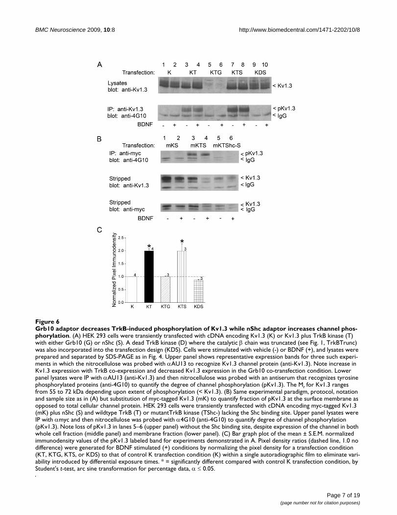
S, top gel, lanes 5–6) completely prevented tyrosine phos-phorylation of the channel. Basal phosphorylation ofKv1.3 (- BDNF conditions) was higher than expected orreported in our previous studies [9,35], but may be attrib-uted to the solution used as a vehicle carrier (patch solu-tion) that may have slightly depolarized the cells (Fig. 6A,lanes 3 versus 4). None the less, all basal phosphorylationcould be completely eliminated in the absence of TrkB(Fig. 6A, bottom gel, lanes 1–2) or via substitution with adead TrkB kinase construct in which the catalytic domainwas truncated (Fig. 6A, bottom gel, lanes 9–10) [37]. Mostinterestingly, inclusion of Grb10 in the transfectionscheme (Kv1.3 + TrkB + Grb10) not only completely elim-inated the Y phosphorylation of the channel over thatobserved in the co-transfection condition (Kv1.3 +TrkB)(Fig. 6A, bottom gel, lanes 5–6 versus lanes 3–4),blotting the lysates with anti-Kv1.3 to control for expres-sion of the channel, revealed that Grb10 was greatlyreducing the expression of Kv1.3 (Fig. 6A, top gel, lanes 5–6). The degree of BDNF-induced Kv1.3 phosphorylationin each transfection condition was quantified by densit-ometry, normalized to that of the Kv1.3 channel transfec-tion alone, and statistically compared in the bar plot ofFig. 6C (Student's t-test, arc sine transformation for per-centage data, α ≤ 0.05).
Expression levels of Grb10, Kv1.3, and TrkB are cross-regulatedTo further explore the robustness of our finding of Grb10downregulation of Kv1.3, we compared the surface frac-tion of Kv1.3 channels (mycKv1.3) and the total Kv1.3pool (Kv1.3) in the presence and absence of Grb10. Asshown in Fig. 7A, B both the surface fraction of Kv1.3channels as well as the total pool of Kv1.3 is dramaticallydown regulated by Grb10 adaptor protein (quantitativedensitometry, significantly-different Student's t-test, α ≤0.05). Grb10 also had the capacity to down regulate Kv1.3protein expression in the presence of TrkB kinase (Fig.7B). In conjunction with the ability to downregulate totalchannel protein, immunocytochemical experiments dem-onstrated a significant reduction in fluorescent signal atthe membrane under Grb10 co-transfection conditions(Fig. 7C). Albeit lack of Grb10/Kv1.3 co-immunoprecipi-tation in vitro, if Grb10 was downregulating the expres-sion of Kv1.3 channel via the SH2 recognition domainson Kv1.3, then substitution of Y to F mutations in the N-and C-terminal aspects of the channel should completelyeliminate any reduction in expression. Single point muta-genesis at the selected Y residues of this study does notalter expression of the derived channel mutants as deter-mined by either Western analysis or electrophysiology [7].Western analysis of HEK 293 cells in which Grb10 was co-transfected with wild-type or various Y to F channelmutants revealed that residues YYY111–113 and Y449 repre-sent important residues for regulation between channel
Kv1.3 channel forms protein-protein interactions with both adaptor proteins in the olfactory bulb and hippocampusFigure 5Kv1.3 channel forms protein-protein interactions with both adaptor proteins in the olfactory bulb and hippocampus. (A-B) Olfactory bulb or (C) hippocampus lysates were immunoprecipitated with either anti-Kv1.3 (IP: anti-Kv1.3) or anti-TrkB (IP: anti-TrkB), separated by SDS-PAGE, and Western blots were probed (blot) with channel, kinase, or adaptor antisera as noted. Mr as specified.
Page 6 of 19(page number not for citation purposes)
BMC Neuroscience 2009, 10:8 http://www.biomedcentral.com/1471-2202/10/8
Page 7 of 19(page number not for citation purposes)
Grb10 adaptor decreases TrkB-induced phosphorylation of Kv1.3 while nShc adaptor increases channel phosphorylationFigure 6Grb10 adaptor decreases TrkB-induced phosphorylation of Kv1.3 while nShc adaptor increases channel phos-phorylation. (A) HEK 293 cells were transiently transfected with cDNA encoding Kv1.3 (K) or Kv1.3 plus TrkB kinase (T) with either Grb10 (G) or nShc (S). A dead TrkB kinase (D) where the catalytic β chain was truncated (see Fig. 1, TrkBTrunc) was also incorporated into the transfection design (KDS). Cells were stimulated with vehicle (-) or BDNF (+), and lysates were prepared and separated by SDS-PAGE as in Fig. 4. Upper panel shows representative expression bands for three such experi-ments in which the nitrocellulose was probed with αAU13 to recognize Kv1.3 channel protein (anti-Kv1.3). Note increase in Kv1.3 expression with TrkB co-expression and decreased Kv1.3 expression in the Grb10 co-transfection condition. Lower panel lysates were IP with αAU13 (anti-Kv1.3) and then nitrocellulose was probed with an antiserum that recognizes tyrosine phosphorylated proteins (anti-4G10) to quantify the degree of channel phosphorylation (pKv1.3). The Mr for Kv1.3 ranges from 55 to 72 kDa depending upon extent of phosphorylation (< Kv1.3). (B) Same experimental paradigm, protocol, notation and sample size as in (A) but substitution of myc-tagged Kv1.3 (mK) to quantify fraction of pKv1.3 at the surface membrane as opposed to total cellular channel protein. HEK 293 cells were transiently transfected with cDNA encoding myc-tagged Kv1.3 (mK) plus nShc (S) and wildtype TrkB (T) or mutantTrkB kinase (TShc-) lacking the Shc binding site. Upper panel lysates were IP with αmyc and then nitrocellulose was probed with α4G10 (anti-4G10) to quantify degree of channel phosphorylation (pKv1.3). Note loss of pKv1.3 in lanes 5–6 (upper panel) without the Shc binding site, despite expression of the channel in both whole cell fraction (middle panel) and membrane fraction (lower panel). (C) Bar graph plot of the mean ± S.E.M. normalized immunodensity values of the pKv1.3 labeled band for experiments demonstrated in A. Pixel density ratios (dashed line, 1.0 no difference) were generated for BDNF stimulated (+) conditions by normalizing the pixel density for a transfection condition (KT, KTG, KTS, or KDS) to that of control K transfection condition (K) within a single autoradiographic film to eliminate vari-ability introduced by differential exposure times. * = significantly different compared with control K transfection condition, by Student's t-test, arc sine transformation for percentage data, α ≤ 0.05.

BMC Neuroscience 2009, 10:8 http://www.biomedcentral.com/1471-2202/10/8
Page 8 of 19(page number not for citation purposes)
Grb10 protein-protein interactions with Kv1.3 causes decreased channel expression that require multiple Y residuesFigure 7Grb10 protein-protein interactions with Kv1.3 causes decreased channel expression that require multiple Y residues. (A) HEK 293 cells were transiently transfected with cDNA encoding Kv1.3 (K) or Kv1.3 plus Grb10 (G) using either myc-tagged Kv1.3 (Upper panel) or wildtype Kv1.3 (Lower panel). Lysates were prepared and separated by SDS-PAGE as in Fig. 4. The Mr for Kv1.3 ranges from 55 to 72 kDa depending upon extent of phosphorylation (< Kv1.3). (B) Bar graph plot of the mean ± S.E.M. normalized immunodensity values of the Kv1.3 labeled band for experiments demonstrated in A and also transfection schemes including TrkB kinase (KT, KTG). Pixel density ratios (dashed line) were generated by normalizing to control K transfection condition within a single autoradiographic film to eliminate variability introduced by differential expo-sure times. * = significantly different compared with control K or KT transfection condition, respectively, by Student's t-test, arc sine transformation for percentage data, α ≤ 0.05. (C) Representative HEK 293 cells (of 30) transiently transfected with myc-tagged Kv1.3 (Myc) alone or plus Grb10 (Myc + Grb10). Channel protein was detected by incubation with anti-myc under non-permeabilizing conditions and then visualized with species-appropriate fluorescent-conjugated secondary antiserum. (D) (TOP) As in (A) using wildtype (K) or various Y to F point channel mutations or a W to F point channel mutation (W386F Kv1.3); transfection scheme was minus (-) or plus (+) Grb10 adaptor. The Mr for Kv1.3 ranges from 55 to 72 kDa depending upon extent of phosphorylation (< Kv1.3). (BOTTOM) As in (A) using Kv1.3 or two other Shaker subfamily members; trans-fection scheme was minus (-) or plus (+) Grb10 adaptor. NOTE: The Western analyses were performed in pairs for each chan-nel member and therefore are not aligned to a single migration standard; Mr for Kv1.3 (55 to 72 kDa), Kv1.4 (74 kDa), Kv1.5 (64 kDa), and actin (42 kDa) varied. (E) Bar graph plot of the mean ± S.E.M. normalized immunodensity values for experiments demonstrated in (D, TOP). Pixel density ratios (dashed line) were generated as in Part (B) above, by normalizing to control transfection condition without Grb10 and comparing within a single autoradiographic film to eliminate variability introduced by differential exposure times. * = significantly different compared to minus Grb10 treatment by Student's t-test, Arc sine trans-formation for percentage data, α ≤ 0.05. (F) Bar graph plot of the mean ± S.E.M. normalized immunodensity values for experi-ments demonstrated in (D, BOTTOM). Analyses and statistical metric as in E.
BMC Neuroscience 2009, 10:8 http://www.biomedcentral.com/1471-2202/10/8
and adaptor protein (Fig. 7D and 7E). Removal of theseresidues prevented Kv1.3 downregulation by Grb10 co-transfection. Mutation of residues Y137 and Y479 retainedthe trend for down-regulation of Kv1.3, thus these resi-dues are likely not involved in the adaptor's regulation ofchannel expression. Next, we tested whether channel con-ductance was necessary for Grb10 down-regulation of
Kv1.3 by substituting the pore-conducting mutant W386FKv1.3 [38,39]. As shown in Fig. 7D and 7E, mutation ofthe pore had no effect in the adaptor's ability to down-reg-ulate Kv1.3 channel expression. Finally, we tested whetherthe Grb10 down-regulation of Kv1.3 was specific to thisparticular Shaker subfamily member or could be extendedto other potassium channels. Substitution in the transfec-tion scheme of either Kv1.4 or Kv1.5, two family membersthat are also expressed in olfactory bulb neurons [15], elic-ited no change in channel expression when co-transfectedwith Grb10 (Fig. 7D and 7F). Furthermore, expression ofgeneral structural proteins (actin) also demonstrated nochange in expression in conditions of Grb10 transfection.
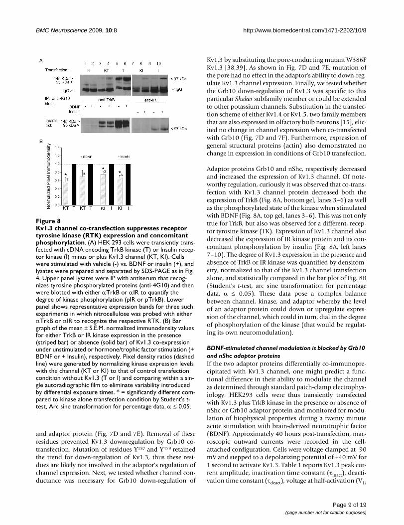
Adaptor proteins Grb10 and nShc, respectively decreasedand increased the expression of Kv1.3 channel. Of note-worthy regulation, curiously it was observed that co-trans-fection with Kv1.3 channel protein decreased both theexpression of TrkB (Fig. 8A, bottom gel, lanes 3–6) as wellas the phosphorylated state of the kinase when stimulatedwith BDNF (Fig. 8A, top gel, lanes 3–6). This was not onlytrue for TrkB, but also was observed for a different, recep-tor tyrosine kinase (TK). Expression of Kv1.3 channel alsodecreased the expression of IR kinase protein and its con-comitant phosphorylation by insulin (Fig. 8A, left lanes7–10). The degree of Kv1.3 expression in the presence andabsence of TrkB or IR kinase was quantified by densitom-etry, normalized to that of the Kv1.3 channel transfectionalone, and statistically compared in the bar plot of Fig. 8B(Student's t-test, arc sine transformation for percentagedata, α ≤ 0.05). These data pose a complex balancebetween channel, kinase, and adaptor whereby the levelof an adaptor protein could down or upregulate expres-sion of the channel, which could in turn, dial in the degreeof phosphorylation of the kinase (that would be regulat-ing its own neuromodulation).
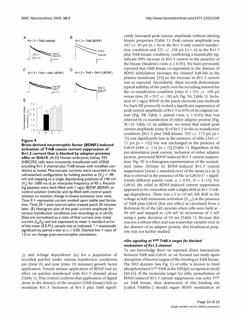
BDNF-stimulated channel modulation is blocked by Grb10 and nShc adaptor proteinsIf the two adaptor proteins differentially co-immunopre-cipitated with Kv1.3 channel, one might predict a func-tional difference in their ability to modulate the channelas determined through standard patch-clamp electrophys-iology. HEK293 cells were thus transiently transfectedwith Kv1.3 plus TrkB kinase in the presence or absence ofnShc or Grb10 adaptor protein and monitored for modu-lation of biophysical properties during a twenty minuteacute stimulation with brain-derived neurotrophic factor(BDNF). Approximately 40 hours post-transfection, mac-roscopic outward currents were recorded in the cell-attached configuration. Cells were voltage-clamped at -90mV and stepped to a depolarizing potential of +40 mV for1 second to activate Kv1.3. Table 1 reports Kv1.3 peak cur-rent amplitude, inactivation time constant (τinact), deacti-vation time constant (τdeact), voltage at half-activation (V1/
Kv1.3 channel co-transfection suppresses receptor tyrosine kinase (RTK) expression and concomitant phosphorylationFigure 8Kv1.3 channel co-transfection suppresses receptor tyrosine kinase (RTK) expression and concomitant phosphorylation. (A) HEK 293 cells were transiently trans-fected with cDNA encoding TrkB kinase (T) or Insulin recep-tor kinase (I) minus or plus Kv1.3 channel (KT, KI). Cells were stimulated with vehicle (-) vs. BDNF or insulin (+), and lysates were prepared and separated by SDS-PAGE as in Fig. 4. Upper panel lysates were IP with antiserum that recog-nizes tyrosine phosphorylated proteins (anti-4G10) and then were blotted with either αTrkB or αIR to quantify the degree of kinase phosphorylation (pIR or pTrkB). Lower panel shows representative expression bands for three such experiments in which nitrocellulose was probed with either αTrkB or αIR to recognize the respective RTK. (B) Bar graph of the mean ± S.E.M. normalized immunodensity values for either TrkB or IR kinase expression in the presence (striped bar) or absence (solid bar) of Kv1.3 co-expression under unstimulated or hormone/trophic factor stimulation (+ BDNF or + Insulin), respectively. Pixel density ratios (dashed line) were generated by normalizing kinase expression levels with the channel (KT or KI) to that of control transfection condition without Kv1.3 (T or I) and comparing within a sin-gle autoradiographic film to eliminate variability introduced by differential exposure times. * = significantly different com-pared to kinase alone transfection condition by Student's t-test, Arc sine transformation for percentage data, α ≤ 0.05.
Page 9 of 19(page number not for citation purposes)
BMC Neuroscience 2009, 10:8 http://www.biomedcentral.com/1471-2202/10/8
2), and voltage dependence (κ) for a population ofrecorded patches under various transfection conditionspre (time 0) and post (time 20 minutes) growth factorapplication. Twenty minute application of BDNF had noeffect on patches transfected with Kv1.3 channel alone(Table 1). This control confirms that application of ligandalone in the absence of the receptor (TrkB kinase) fails tomodulate Kv1.3. Inclusion of Kv1.3 plus TrkB signifi-
cantly increased peak current amplitude without alteringkinetic properties (Table 1). Peak current amplitude was287 +/- 49 pA (n = 4) in the Kv1.3-only control transfec-tion condition and 551 +/- 198 pA (n = 4) in the Kv1.3plus TrkB kinase condition, confirming a statistically sig-nificant 90% increase in Kv1.3 current in the presence ofthe kinase (Student's t-test, α ≤ 0.05). We have previouslyreported that TrkB kinase co-expression in the absence ofBDNF stimulation increases the channel half-life at theplasma membrane [35] so the increase in Kv1.3 currentwas as expected. Secondarily, these records demonstratetypical stability of the patch over the recording interval forthe co-transfection condition (time 0 = 551 +/- 198 pAversus time 20 = 557 +/- 183 pA; Fig. 9A, Table 1). Inclu-sion of 1 ng/μl BDNF in the patch electrode (see methodsfor back-fill protocol) evoked a significant suppression ofpeak current amplitude of Kv1.3 to 85% of its original cur-rent (Fig. 9B; Table 1, paired t-test, α ≤ 0.05) that wasrelieved by co-transfection of either adaptor protein (Fig.9C–D; Table 1). In addition, we noted that initial peakcurrent amplitude (time 0) of Kv1.3 in the co-transfectioncondition (Kv1.3 plus TrkB kinase; 555 +/- 173 pA (n =7)) was significantly less in the presence of nShc (343 +/-71 pA (n = 13)) but was unchanged in the presence ofGrb10 (488 +/- 114 (n = 7)) (Table 1). Regardless of thepre-stimulation peak current, inclusion of either adaptorprotein, prevented BDNF-induced Kv1.3 current suppres-sion. Fig. 9E is a histogram representation of the normal-ized (time 20/time 0) BDNF-induced Kv1.3 currentsuppression (mean ± standard error of the mean (s.e.m.))that is relieved in the presence of Shc or Grb10 (* = signif-icantly-different paired t-test, α ≤ 0.05, N = 4–13). ForGrb10, the relief in BDNF-induced current suppressionappeared to be coincident with a slight shift in Kv1.3 volt-age dependence. There was a 5 to 10 mV left shift in thevoltage at half-maximum activation (V1/2) in the presenceof TrkB plus Grb10 (but not nShc) as calculated from aBoltzman fit of the tail currents when cells were held at -90 mV and stepped to +20 mV in increments of 5 mVusing a pulse duration of 50 ms (Table 1). Because thiswas not a robust effect and was somewhat variable even inthe absence of an adaptor protein, this biophysical prop-erty was not further studied.
nShc signaling at Y490 TrkB is target for blocked modulation of Kv1.3 channelTo our knowledge there no reported direct interactionsbetween TrkB and Grb10, so we focused our study upondisruption of known targets of Shc binding to TrkB kinase.The SH2 domain (see Fig. 1) of nShc is known to bindphosphorylated Y490 TrkB at the NPQpY recognition motif[40,41]. If the molecular target for nShc perturbation ofBDNF-induced Kv1.3 current suppression was soley Y490
on TrkB kinase, then destruction of this binding site(called TrkBShc-) should regain BDNF modulation of
Brain-derived neurotrophic factor (BDNF)-induced activa-tion of TrkB causes current suppression of Kv1.3 current that is blocked by adaptor proteins nShc or Grb10Figure 9Brain-derived neurotrophic factor (BDNF)-induced activation of TrkB causes current suppression of Kv1.3 current that is blocked by adaptor proteins nShc or Grb10. (A-D) Human embryonic kidney 293 (HEK293) cells were transiently transfected with cDNA encoding Kv1.3 channel plus TrkB kinase with modified con-ditions as noted. Macroscopic currents were recorded in the cell-attached configuration by holding patches at (Vh) = -90 mV and stepping to a single depolarizing potential of +40 mV (Vc) for 1000 ms at an interpulse frequency of 45 s. Record-ing pipettes were back-filled with 1 ng/μl BDNF (BDNF) or control solution (vehicle) and tip-filled with control patch solution to monitor change in kinase activation over time. Time 0 = represents current evoked upon stable seal forma-tion, Time 20 = post-neurotrophin treated patch 20 minutes later. (E) Histogram plot of the peak current amplitude for various transfection conditions and recordings as in (A-D). Data are normalized as a ratio of final current over initial current (I20/I0) and are expressed as mean ± standard error of the mean (S.E.M.); sample size as indicated. * = statistically significant by paired t-test at α ≤ 0.05. Dashed line = ratio of 1.0 or no change post-neurotrophin stimulation.
Page 10 of 19(page number not for citation purposes)

BMC Neuroscience 2009, 10:8 http://www.biomedcentral.com/1471-2202/10/8
Kv1.3 (prevent relief of current suppression by adaptorprotein). What we found by substituting the TrkB kinasemutant construct, however, was an intermediate effect.Co-transfection of Kv1.3 + TrkBShc- plus nShc cDNAexhibited BDNF-induced current suppression of Kv1.3(533 +/- 109 pA (time 0) versus 473 +/- 115 pA (time 20)that did not reach statistical significance (not significantlydifferent paired t-test, α ≤ 0.05; n = 10, 89% suppression)compared with that observed with Kv1.3 + TrkB co-expres-sion (356 +/- 71 pA (time 0) versus 298 +/- 64 pA (time20); significantly different paired t-test, α ≤ 0.05; n= 5,84% suppression) (Table 2, Fig. 10B, C). If, however, wesubstituted Grb10 in the transfection scheme instead ofnShc (Kv1.3 + TrkBshc- + Grb10), we observed retentionof BDNF-induced current suppression that was notrelieved by the adaptor protein (533 +/- 122 pA (time 0)versus 379 +/- 84 pA (time 20); significantly differentpaired t-test, α ≤ 0.05; n = 4, 71% suppression) (Table 2,Fig. 10D). Although Grb10 has never been reported todirectly interact with TrkB kinase signaling cascades, andwe also do not find that this adaptor co-immunoprecipi-tates with the kinase in vitro (Fig. 4B), residue Y490 of TrkBkinase is an important target for nShc, and its mutationappears to somehow uncouple Grb10 modulation of thechannel. With the removal of Y490 of TrkB kinase, Grb10can no longer relieve BDNF-induced current suppressionof the channel (Fig. 10D and 10G). Interestingly,
although neither adaptor protein induces a change inpeak current amplitude of Kv1.3 alone (Fig. 10E and 10F),Grb10 also evoked a faster rate of channel inactivation(762 +/- 151 msec (minus Grb10) versus 553 +/- 46 msec(plus Grb10) as demonstrated in Fig. 10E and statisticallycompared in Table 1.
DiscussionKv1.3 is a substrate for multiple tyrosine kinase signalingcascades including IR, TrkB, and src kinase [5,7-9,35,36].Each exerts the phosphorylation of multiple tyrosine resi-dues, and different combinations of residues, to producea unified response, of Kv1.3 current suppression. Inclu-sion of adaptor proteins causes perturbation of the chan-nel current suppression [12,13], and in the case ofneurotrophin signaling, we find that disruption of thechannel modulation is not directly correlated to the extentof channel phosphorylation. The scenario is more com-plex. This may be attributed to the fact that the interactionbetween kinase and channel is no longer a binary relation-ship, with the added adaptor having the capacity to bindto both channel and kinase to elicit different responses.Not only can an adaptor protein alter how the channelwill respond to neurotrophin-induced current suppres-sion, the adaptors alter expression levels of the channeland the channel itself can downregulate TrkB expressionand its phosphorylated state. Such a dynamic equilibrium
Table 1: The effect of adaptor proteins on TrkB kinase-induced Kv1.3 current properties.
Transfection Condition Modulator Peak current(pA)
τInact(msec)
τDeact(msec)
V1/2(mV)
κ
Kv1.3 BDNFPre 287 ± 49 (4) 794 ± 252 31.5 ± 5.4 -38.2 ± 5.8 4.2 ± 0.7Post 303 ± 68 961 ± 263 38.3 ± 13.2 -41.5 ± 6.8 4.9 ± 1.1
Kv1.3 + Grb10 BDNFPre 506 ± 271 (4) 774 ± 110 31.3 ± 4.5 -39.4 ± 6.1 5.4 ± 0.4Post 498 ± 246 *610 ± 127 34.2 ± 4.7 -41.5 ± 6.8 4.4 ± 2.6
Kv1.3 + nShc BDNFPre 543 ± 176 (7) 770 ± 45 32.6 ± 4.3 -45.0 ± 2.5 4.3 ± 0.6Post 586 ± 107 817 ± 54 31.4 ± 4.0 -41.4 ± 3.3 3.9 ± 0.9
Kv1.3 + TrkB VehiclePre 551 ± 198 (4) 788 ± 186 37.2 ± 3.3 -40.4 ± 5.0 5.1 ± 0.5Post 557 ± 183 742 ± 130 32.3 ± 3.3 -38.6 ± 5.8 5.4 ± 0.7
Kv1.3 + TrkB BDNFPre 555 ± 173 (7) 900 ± 243 36.8 ± 2.9 -36.2 ± 4.6 5.8 ± 1.1Post *442 ± 173 762 ± 151 38.8 ± 6.7 -40.3 ± 6.7 5.3 ± 1.4
Kv1.3 + TrkB + Grb10 BDNFPre 488 ± 114 (7) 862 ± 110 29.3 ± 3.5 -42.6 ± 3.7 6.7 ± 0.6Post 545 ± 133 *553 ± 46 35.4 ± 5.5 *-54.1 ± 2.8 6.3 ± 1.4
Kv1.3 + TrkB + nShc BDNFPre 343 ± 71 (13) 618 ± 67 37.4 ± 3.2 -40.9 ± 1.9 4.4 ± 0.7Post 381 ± 92 549 ± 68 36.4 ± 2.0 -43.5 ± 2.9 4.4 ± 1.1
HEK 293 cells were transfected with cDNAs encoding Kv1.3 ± TrkB ki nase ± adaptor protein. Several voltage-stimulating paradigms were completed (see "Methods") to collect the tabled biophysical properties by cell-attached patch-clamp recording. Mean values (± s.e.m.) for peak current amplitude (pA), inactivation time constant (τInact), deactivation time constant (τDeact), voltage at half activation (V1/2), and slope (κ) of the voltage dependence were compared across various transfection conditions at the start of the patch recording (Pre) versus twenty minutes following 1 ng/μg BDNF (Post) using a paired t-test, α ≤ 0.05. * = Significantly different.
Page 11 of 19(page number not for citation purposes)

BMC Neuroscience 2009, 10:8 http://www.biomedcentral.com/1471-2202/10/8
between Kv1.3/TrkB/adaptors may allow a full spectrumof modulation of channel properties dependent upon thecompliment of proteins co-expressed in a neuron at agiven physiological state.
Biochemically, the degree of channel phosphorylation byBDNF-induced TrkB activation is no longer directlyrelated to the degree of functional current suppressionwhen adaptors are added to the mix. This is very different
than what is reported for the cellular tyrosine kinase, Src.Here, phosphorylation was directly linked to the degree ofcurrent suppression and one could predict the modula-tion of the selected adaptor protein based upon how itinfluenced tyrosine phosphorylation of the channel [12].This is in stark contrast to RTKs. The addition of nShc toKv1.3 plus TrkB transfected conditions induced anincrease in both channel expression as well as BDNF-induced channel phosphorylation whereas the additionof Grb10 resulted in the opposite. Despite the fact that theadaptors evoke an opposite response biochemically, thefunctional ramification is identical, the prevention ofBDNF-induced current suppression. In fact, nShc/Grbhave structurally similar SH2 domains whereas PSD-95lacks such phosphorylation-dependent interactivedomains [13], and yet each has the capacity to function-ally perturb the BDNF-induced current suppression. Thismight suggest that direct interaction of the adaptor andthe channel may play a predominant role in preventingkinase modulation and that changed phosphorylation is asecondary effect. In the case of the two SH2 domain con-taining adaptor proteins, nShc and Grb10, binding of Y490
TrkB by nShc is an additional key regulatory site impact-ing modulation of the channel. Certainly there are endog-enous levels of both nShc and Grb10 expressed in HEK293 cells, thus our Kv1.3 + TrkB conditions are not com-pleted devoid of adaptor protein, however, when nShc isco-transfected with these conditions, binding of Y490 TrkBinduces a block of BDNF-induced channel suppression.
Kv1.3 channel expression is changed in the presence ofTrkB kinase without BDNF stimulation and now we canalso see that Grb10 adaptor significantly modifies surfaceexpression of Kv1.3. With TrkB kinase co-expression, thereis a correlate increase in channel surface expression,increase in channel protein expression, and increase inchannel macroscopic current [35]. Interestingly, withGrb10 co-expression (also not BDNF dependent) there isoppositely a decrease in channel surface expression, adecrease in protein expression of the channel, but nochange in channel macroscopic current, rather anincreased rate of channel inactivation. The adaptor/chan-nel do not form a complex in vitro that we can detect, nev-ertheless, co-expression apparently changes surfacedistribution and channel density but not current magni-tude or conduction. This would imply that there must beMANY Kv1.3 channels that are not contributing to cur-rent. Such a pool of silent or "sleeping channels" has beenreported for specific Kv subfamily members (Kv6 and Kv9family members) [42,43] and uncovered for other potas-sium channels during FRAC (functional recovery afterchemobleaching) [44]. Our data [35] and that reported bySun et al. [44] support that molecular/protein density of achannel may not always exhibit linearity with functionaldensity.
Mutation of Y490 TrkB Shc binding site differentially affects block of TrkB-induced current suppression of Kv1.3 current by adaptor proteins nShc and Grb10Figure 10Mutation of Y490 TrkB Shc binding site differentially affects block of TrkB-induced current suppression of Kv1.3 current by adaptor proteins nShc and Grb10. (A-F) Same experimental paradigm, notation, and analysis protocol as Fig. 9 with the substitution of mutant TrkB kinase (TrkBShc-) in which the recognition site (Y490) for nShc bind-ing was altered by point mutation to F. Note that TrkB-induced current suppression observed in (B) is still blocked by co-transfection of nShc in (C) but not with Grb10 (D). Note that Grb10 co-transfection (E) but not nShc co-trans-fection (F) with Kv1.3 alone induced an increased rate of inactivation. (G) Histogram plot of the peak current ampli-tude for various transfection conditions and recordings as in (A-F). Data are normalized as a ratio of final current over ini-tial current (I20/I0) and are expressed as mean ± S.E.M.; sam-ple size as indicated. * = statistically significant by paired t-test at α ≤ 0.05. Dashed line = ratio of 1.0 or no change post-neurotrophin stimulation.
Page 12 of 19(page number not for citation purposes)

BMC Neuroscience 2009, 10:8 http://www.biomedcentral.com/1471-2202/10/8
Based upon the crystal structure of Grb10 SH2 domaindetermined at 1.65 Å resolution, it is suggested that bind-ing of Grb10 is dimeric, using turn containing phosphoty-rosine sequences [45]. It is unknown (but doubtful) if theKv1.3 channel contains turn containing phosphotyrosinemotifs unless the tetrameric structure of the subunits per-mitted such an alignment, which would be contrary topositioning of the α transmembrane helices 1 and 6 forKv1.2, a Shaker family member for which crystal structurehas been reported [46]. On the basis of structural analysisand previous biochemical studies for the adaptor, Grb10and Grb14 have a partially impaired ability to bind phos-photyrosine-containing ligands due to a non-glycyl resi-due at the end of the BD loop and lack of a P + 3 bindingpocket [47]. These combined data are consistent with ourfindings that we cannot detect Grb10 and Kv1.3 co-immu-noprecipition in vitro, albeit the fact that channel/adaptorfunctional interactions appear to modulate channelexpression and can be found as a complex in unstimu-lated native tissues. The fact that mutation of several Y res-idues on the N and C terminal aspects of the channelclearly perturbs the negative modulatory affect of Grb10on channel expression, indicates a specific modulatoryrole that does not require tight association. Moreover, themodulation does not require channel activity, or at leastfunctional ion conductance. While all Y to F channelmutations blocked Grb10 down regulation of Kv1.3expression, YYY111-113FFF Kv1.3 and Y449F Kv1.3appear to be the most robust at blocking down regulation.Both of these sites (along with Y137) were previouslyreported as targets for BDNF-induced Kv1.3 current sup-pression and phosphorylation [9] but only Y449 has a largehydrophobic M downstream of the phosphotyrosine,reported as a preferred Grb motif [48].
It has previously been reported that the E3 ubiquitin pro-tein ligase called Nedd4-2 regulates voltage-gated ionchannels, including Nav, Kir, and Kv family members [49]via targeting phosphotyrosine motifs located in the C-ter-minal aspect of the channel. It has also been demon-strated that the SH2 domains of Grb10 form a constitutivecomplex with Nedd4-1 so as to mediate ligand-dependentubiquitination of IGF-IR [17]. Since co-expression ofNedd4-2 with Kv1.3 in Xenopus oocytes induces a strongsuppression of Kv1.3 current [14] we conjecture thatGrb10 might bind to endogenous Nedd4-2 in our systemto cause ubiquitination of Kv1.3 and/or TrkB kinase. Thismight explain a potential mechanism for decreased Kv1.3surface expression in the presence of Grb10 and correlatedecreased channel tyrosine phosphorylation.
We have shown that TrkB and Grb10 act as modulators ofchannel expression, while others have demonstrated thatthe Grb10 adaptor acts as a negative modulator of IRkinase activity (reviewed by Holt and Siddle [47]). Ramoset al. [50] elucidated the mechanism by which the SH2and binding phosphorylated substrate (BPS) domains ofGrb10 might induce proteasomal degradation of the IRand subsequent inhibition of its downstream signalingpathways. The question arises as to whether the adaptor orkinases themselves are regulated and if so, how? Our datademonstrate that there is a reciprocal regulation of RTKphosphorylation activity and expression in the presenceof Kv1.3. Holmes et al. [4] similarly reported that treat-ments that induce membrane hyperpolarization (K chan-nel activity) markedly attenuate tyrosine phosphorylationcatalyzed by cellular tyrosine kinases. Unlike their report,centralizing on reciprocal downregulation of Kv1.3 andcellular TKs, our data demonstrate that both TrkB and IRkinase protein levels are reduced when co-expressed with
Table 2: The effect of adaptor protein signaling at Y490 TrkB for Kv1.3 current properties.
Transfection Condition Peak Current(pA)
τInact(msec)
τDeact(msec)
Kv1.3 + TrkBPre 356 ± 71 (5) 697 ± 130 32.2 ± 2.1Post *298 ± 64 780 ± 59 24.2 ± 2.9
Kv1.3 + TrkBShc-Pre 372 ± 144 (3) 693 ± 45 31.8 ± 3.3Post 386 ± 140 843 ± 114 26.5 ± 3.7
Kv1.3 + TrkBShc- + nShcPre 533 ± 109 (10) 1070 ± 163 31.2 ± 4.3Post 473 ± 115 1040 ± 196 35.3 ± 3.8
Kv1.3 + TrkBShc- + Grb10Pre 533 ± 122 (4) 894 ± 173 29.0 ± 1.6Post *379 ± 84 660 ± 229 32.6 ± 7.8
HEK 293 cels were transfected with cDNAs encoding Kv1.3 +TrkB kinase or TrkBShc- (Y490 mutation) ± adaptor protein. Several voltage-stimulating paradigms were completed (see "Methods") to collect the tabled biophysical properties by cell-attached patch-clamp recording. BDNF stimulation, notations, and statistical analysis as in Table 1.
Page 13 of 19(page number not for citation purposes)

BMC Neuroscience 2009, 10:8 http://www.biomedcentral.com/1471-2202/10/8
Kv1.3, which then in turn, decreases the total amount ofkinase phosphorylation. At the same time, we alsoobserve that both nShc and Grb10 adaptor protein expres-sion is reduced when co-expressed with Kv1.3 (data notshown), and that the expression of both adaptors isincreased in Kv1.3-null mice [15], offering another tier ofregulation of the channel/kinase/adaptor interactioninvolving receptor-linked as opposed to cellular tyrosinekinases. Another suggested method in which Grb10action might be terminated is via phosphorylation; in itsinactive state it is thought to form tetramers [51,52].Although we did not test whether Grb10 was phosphor-ylated in channel/kinase/adaptor transfected conditionsunder BDNF-stimulated conditions (Fig. 6), this certainlydid not functionally affect downregulation of channelexpression.
Grb10 appears to be promiscuous in interacting with mul-tiple receptor and non-receptor TKs and other signalingproteins in vitro [47], therefore, gene-targeted deletion ofthe adaptor could afford physiological relevance to manyof these interactions. Grb10-null mice have dispropor-tionate overgrowth of the embryo and placenta, imbal-anced glucose homeostasis, and are approximately thirtypercent larger at birth [24,25,53]. This is an interestingphenotype given the fact that Kv1.3-null mice, oppositely,are resistant to diet-induced obesity, are smaller and phys-iologically thinner than age-matched wild-type animals,and have increased mobility and metabolism [15,54,55] –while having an increased expression of Grb10 proteinthat reinforces its function as a growth inhibitor [47].
In heterologous expression systems and in vivo, Shc adap-tor protein can bind directly to the channel (where it doesnot alter function or expression). Shc can also bind toTrkB at Y490 to increase phosphorylation of Kv1.3 andrelieve BDNF-induced current suppression. To whatextent is this mimicked in native olfactory bulb neurons(OBNs)? We know that exogenous application of BDNFto native OBNs [9,36], where Shc and TrkB are bothpresent [12,15], causes an increase in Kv1.3 tyrosine phos-phorylation and current suppression that is significantlygreater than what we observe in heterologous expressionsystems [35] (typically 60% (mice) to 40% (rats) of mitralcell outward current is suppressed by acute BDNF stimu-lation). We also know that RTKs explicitly target Kv1.3,because in mice with Kv1.3 gene-targeted deletion, nei-ther insulin nor BDNF has any affect on mitral cell cur-rent. Thus, there is a divergence in gain strength betweenHEK 293 cells and OB neurons for neurotrophic factor sig-naling that might lie at a missing adaptor protein notmimicked in the HEK 293 cell system, a lesser abundanceof inhibitory adaptor proteins, or perhaps another regula-tory molecule as yet not accounted for.
ConclusionIn native OB, adaptor proteins may change with develop-mental profile, regeneration, or disease [26,28,32,56,57]– hence expression patterns of co-localization may benon-static, which would result in state-dependent channelproperties. For example, it is well documented that neuro-trophins are altered with injury [58-60] and that insulin ismodified with metabolic state and meals [61], while bothare modified with growth or development [62-64]. There-fore, the conductance through a Kv1.3 channel, whichunderlies the shape and frequency of the action potentialoutput of the OB, would inherently be modified bydynamic change in physiological state. This in turn mightpredict that odor perception is very state dependent as dic-tated by its microenvironment of protein partners andadaptors that would influence a major voltage-gated ionchannel in this system.
MethodsSolutions and reagentsHuman embryonic kidney cell (HEK 293) patch solutioncontained (in mM): 30 KCl, 120 NaCl,10 4-(2-hydrosye-thyl)-1-peperazineethanesulfonic acid (HEPES), and 2CaCl2 (pH 7.4). HEK 293 cell recording bath solutioncontained (in mM): 150 KCl, 10 HEPES, 1 EGTA, and 0.5MgCl2 (pH 7.4). Cell lysis buffer contained (in mM): 25tris (hydroxymethyl) aminomethane (pH 7.5), 150 NaCl,150 NaF, 0.5 EDTA, and 1.0% Triton X-100 (pH 8.0). Pro-tease and phosphatase inhibitor (PPI) solution was addedto the lysis buffer just prior to use for a final concentrationas follows: 1 μg/ml pepstatin A, 1 μg/ml leupeptin, 2 μg/ml aprotinin, 10 μg/ml phenylmethylsulfonyl fluoride,and 10 mM Na3VO4. Nonidet-NP40 protease and phos-phataseinhibitor (NP40 PPI) solution was prepared in thesame PPI solution as above but contained (in mM): 20Tris base (pH 7.5), 150 NaCl, 1% nonidet-NP40 and 10%glycerol. Homogenization buffer contained (in mM): 320sucrose, 10 Tris, 50 KCl, 1 ETDA (pH 7.8). Wash buffercontained (in mM): 25 Tris base (pH 7.5), 150 NaCl, 150NaF, 0.5 EDTA and 0.1% Triton X-100. Tris strippingbuffer (TSB) contained (in mM): 10 Tris, 10 β-mercap-toethanol, with 1% SDS (pH 8.8). Sodium citrate strip-ping buffer (SCSB) contained (in mM): 100 Na3C6H5O72H20, 10 β-mercaptoethanol, with 1% SDS; pH 3.0. Phos-phate-buffered saline (PBS) contained (in mM): 136.9NaCl, 2.7 KCl, 10.1 Na2HPO4, and 1.8 KH2PO4 (pH 7.4).Human recombinant brain-derived neurotrophic factor(rhBDNF) was purchased from Promega (Madison, WI).All salts and other reagents were purchased from SigmaChemical Co. (St. Louis, MO) or Fisher Scientific (Atlanta,GA).
cDNA constructs and antiseraAll channel, kinase, and adaptor protein coding regionswere downstream from a cytomegalovirus (CMV) pro-
Page 14 of 19(page number not for citation purposes)

BMC Neuroscience 2009, 10:8 http://www.biomedcentral.com/1471-2202/10/8
moter or a lac promoter. TrkB cDNA was a generous giftfrom Dr. P. Barker, McGill University, in a CMX vector asused previously [9]. A dead TrkB kinase (TrkBTrunc) anda targeted mutation at the Shc binding site of TrkB (Y490TrkB or TrkBShc-) were created by Dr. David Kaplan, Uni-versity of Toronto, as described previously [37] and weregenerous gifts to our study. Kv1.3 channel was subclonedinto the multiple cloning region of pcDNA3 (Invitrogen,Carlsbad, CA) at the unique restriction HindIII site of themultiple cloning region. The ten amino acid c-mycepitope (EQKLISEEDL) was inserted into the extracellularS1/S2 loop between residues 226–227 of Kv1.3 channelvia two consecutive polymerase chain reactions (PCRs)using the Expand Long Template PCR System (Roche,Indianapolis, IN). Both the untagged channel andepitope-tagged version of the channel were used in thisstudy as required for surface expression and localizationexperiments. We have previously demonstrated that thetag does not alter channel biophysical properties or pro-tein interactions [35]. Site-directed mutagenesis to gener-ate Y to phenylalanine (F) at positions 111–113, 137,449, and 479 was done as described previously [5,7]. ThecDNA for a non-conducting Kv1.3 mutant with a pointmutation exchanging a tryptophan (W) to F at position386 was a generous gift from Dr. Todd Holmes (Univer-sity of California Irvine, Irvine, CA) [38]. Neuronal Shc(nShc) cDNA was a generous gift from Dr. T. Nakamura(Sumitomo Electric Industries, Yokohama, Japan) andwas expressed in the vector pCMV1 [29]. Grb10 cDNAwas a gift from Dr. R. Roth (Stanford University) and wasexpressed in pBlueScript SK (-) vector (Stratagene, La Jolla,CA). Kv1.4 and Kv1.5 cDNA were both expressed inpcDNA3 and were a generous gift from Todd Holmes(University of California Irvine, Irvine, CA) [65].
AU13, a rabbit polyclonal antiserum, was generatedagainst a 46 amino acid sequence(478MVIEEGGMNHSAFPQTPFKTGNSTATCTTNNNPNDCVNIKKIFTDV523) representing the unique codingregion of Kv1.3 on the C- terminus. Genemed Synthesis(San Antonio, TX) purified the peptide and Cocalico Bio-logicals (Reamstown, PA) the antisera as previously char-acterized [36]. This antibody was used forimmunoprecipitation (1:1000) and Western blot detec-tion (1:800) of Kv1.3. Kv1.4 and Kv1.5 polyclonal antis-era were used for Western blot detection (1:1000) aspreviously characterized and were generous gifts fromDrs. J.O. Dolly and T.C. Holmes, respectively [15]. Tyro-sine phosphorylated proteins were visualized by the anti-phosphotyrosine antibody 4G10 (Millipore/Upstate Bio-chemical; St. Louis, MO) and used at 1:1000 for Westernblots. Tyrosine phosphorylated proteins were immuno-precipitated with 4G10 antibody (3 μg per 600–1200 μgof whole lysate protein). Monoclonal antiserum directedagainst amino acids 156–322 of human TrkB was pur-
chased from BD Transduction Laboratories (San Jose, CA)and used at 1:1000 for Western blots. Grb10 (K-20) rabbitpolyclonal antibody was purchased from Santa Cruz Bio-technology, Inc. (Santa Cruz, CA) and used at 1:800 forWestern blots and at 1:500 for immunocytochemistry.Shc rabbit polyclonal antibody (cat # 610082) from BDTransduction Laboratories (San Jose, CA) was used forimmunohistochemistry (1:500) and Western blots(1:750). Monoclonal actin antibody (A 2066) was pur-chased from Sigma and used at 1:1000 for Western blotsas a secondary confirmation for equivalent protein load-ing. Anti-c-myc mouse monoclonal (clone E910; antige-netic peptide EQKLISEEDL against from the human mycprotein) was purchased from Roche and used at 1:800 forWestern blots. Donkey anti-rabbit FITC-conjugate (1:100)and goat anti-rabbit Texas Red-conjugate (1:200) werefrom Southern Biotech (Birmingham, AL).
ImmunocytochemistryProcedures for surface labeling of mycKv1.3 in the pres-ence and absence of Grb10 as expressed in HEK293 cellswas as previously described [35]. Briefly, cells werelabeled under non-permeabilizing conditions to imagethe surface distribution of the channel. Visualization andmicroscopic analysis was performed on a Zeiss Axioplan 2Microscope attached to a Zeiss LSM510 confocal system(Carl Zeiss, Thornwood, NY) in conjunction with Meta-morph software (Molecular Devices/Universal Imaging,Dowingtown, PA). Samples were excited at 488 nm (forGFP) and 595 nM (for Texas Red) through a 63× oil-immersion objective (NA = 1.40) and fluorescencebetween 500–545 and 565–615 nm was detected from0.8 μm optical sections. Images were taken from the cellsat their maximum cross section. After removing back-ground pixels by thresholding, integrated pixel intensitywas calculated both from the total cross section of the celland from an area that encompassed the cross section ofthe cell excluding the outermost region.
Double-color immunofluorescence procedures for co-localization of Kv1.3 channel and either Grb10 or nShcadaptor protein in the olfactory bulb were as described inBiju et al. [16]. Briefly, C57 BL/6 mice (postnatal day 20)were anaesthetized with pentabarbitol, trancardially per-fused, and post-fixed with 4% paraformaldehyde. Follow-ing sucrose cryoprotection, olfactory bulbs were coronallycut at 10 μm thickness on a Leica CM1850 microtome-cry-ostat (Leica, Meyer Instruments, Houston, TX). Sectionswere mounted on 2% gelatin coated slides and stored at -20°C until immunoprocessing. Sequential labeling wasperformed [16,35] using AU13 as the first primary anti-body (visualized with FITC-conjugated, species-specificsecondary antibody), followed by anti-Shc or anti-Grb10as the second primary antibody, which was visualized bya Texas Red-conjugated, species-specific secondary anti-
Page 15 of 19(page number not for citation purposes)

BMC Neuroscience 2009, 10:8 http://www.biomedcentral.com/1471-2202/10/8
body. Processed slides were viewed at 40× magnificationusing an Axiovert S-100 Microscope (Carl Zeiss) equippedwith epifluorescence. Images were captured using an Axi-ocam digital camera and Axiovision associated software(v3.0.6) at a maximum pixel resolution of 1300 × 1300.
Maintenance and transfection of HEK 293 cellsHEK 293 cells were maintained in Minimum EssentialMedium (MEM), 2% penicillin/streptomycin, and 10%FBS (Invitrogen/Gibco/BRL). Before transfection, cellswere grown to 100% confluency (7 days), dissociatedwith trypsin-EDTA (Sigma) and mechanical trituration,diluted in MEM to a concentration of 600 cells/ml, andreplated on Corning dishes (Catalog # 25000, Fisher Sci-entific). The Corning dishes have a growing surface ofapproximately 8 cm2 (electrophysiology) or 21 cm2 (bio-chemistry) to favorably allow cells to divide logarithmi-cally at the point of transfection. cDNA was introducedinto HEK 293 cells with a lipofectamine reagent (Invitro-gen/Gibco/BRL) 3–5 days after passage as previouslydescribed [7,8]. Briefly, cells were transfected for 4–5hours with 0.5–0.75 μg of each cDNA construct per 35mm dish for electrophysiology or 2–3.5 : μg of each cDNAconstruct per 60 mm dish for biochemistry. Plasmid DNAwith no coding insert (control vector) served as the con-trol to equalize total μg of cDNA added to each dish. Cellswere either harvested for biochemical analysis or used forelectrophysiological recordings approximately 30–40hours after transfection. Efficiency of co-transfection(greater than 95%) with our transfection method hasbeen tested using double, sequential labeling and confo-cal microscopic visualization, as reported previously [8].
Immunoprecipitation and Electrophoretic SeparationTransfected cells were rinsed in PBS and then stimulatedwith 10 ng/μl BDNF or vehicle control solution for tenminutes at room temperature. The selection of vehiclecontrol (patch pipette solution), temperature, and incu-bation time was designed to parallel conditions for bio-chemistry with those used for electrophysiologicalrecordings. Cells were then harvested by lysis in ice-coldPPI solution (see Solutions and Reagents). The lysateswere clarified by centrifugation at 14,000 × g for 10 min-utes at 4°C and incubated for 1 hour (h) with 0.2–0.3 mg/ml protein A-sepharose (GE Healthcare Bio-SciencesCorp/Amersham-Pharmacia, Piscataway, NJ), followed byre-centrifugation to remove the protein A-sepharose. Totest the phosphorylation state of Kv1.3 in the presence ofBDNF-activated TrkB kinase plus or minus an adaptorprotein, tyrosine-phosphorylated proteins were immuno-precipitated from the clarified lysate by overnight incuba-tion at 4°C with 3–4 μg/ml 4G10 antibody (Millipore/Upstate Biochemical). Protein-protein interactions werealso determined by a similar co-immunoprecipitationstrategy by substituting 4G10 antibody for antisera
directed against Kv1.3, c-myc, Shc, Grb10, or TrkB kinase.In experiments where tyrosine-phosphorylated proteinswere immunoprecipitated from native olfactory bulb orhippocampus, tissues were harvested from postnatal day20 (P20) C57BL/6 mice that had been euthanized via CO2inhalation followed by decapitation according to AVMA-and NIH-approved methods. Brain regions were quicklyremoved from the cranium and homogenized 50 strokesby Kontes tissue grinder (size 20) in homogenizationbuffer on ice [8]. The lysate clarification, immunoprecipi-tation, and SDS-PAGE analysis were as described for HEK293 cells above. In experiments in which surface labeledKv1.3 was probed using c-myc antiserum, wholemycKv1.3 transfected HEK 293 cells were first incubatedwith c-myc antiserum (0.4 μg/μl) for 30 minutes at 37°Cprior to BDNF stimulation as above. Cells with surface-labeled mycKv1.3 were then rinsed in PBS, lysed in ice-cold PPI solution. Both immunoprecipitated proteins andsurface-labeled mycKv1.3 protein were harvested by a 2-hincubation with protein A-sepharose and centrifugationas above. The immunoprecipitates (IPs) were washed 3times with ice-cold wash buffer (0.1% Triton). Lysates andwashed IPs were diluted in sodium dodecyl sulfate (SDS)gel loading buffer containing 1 mM Na3VO4. Protein con-centration was determined by Bradford protein assay(Biorad Laboratories, Hercules, PA). Standard curves weregenerated using the small percentage of Triton (final con-centration of detergent = 0.001%) that did not signifi-cantly interfer with protein concentration determinationin this assay. Proteins were separated on 10% acrylamideSDS gels and electrophoretically transferred to nitrocellu-lose for Western blot analysis. Nitrocellulose membraneswere blocked with 5% nonfat milk and incubated over-night at 4°C in primary antibody against Kv1.3 or othersought protein partner. Membranes were then exposed tospecies-specific peroxidase-conjugated secondary (GEHealthcare Bio-Sciences Corp/Amersham-Pharmacia orSigma) for 90 minutes at room temperature. Enhancedchemiluminescence (ECL; GE Healthcare BioSciencesCorp/Amersham-Pharmacia) exposure of Fuji RX film(Fisher Scientific) was used to visualize labeled protein.
In experiments comparing Kv1.3 expression in the pres-ence and absence of Grb10 adaptor protein, labeled Kv1.3bands from cell lysates were quantified by densitometryusing a Hewlett-Packard Photosmart Scanner in conjunc-tion with Quantiscan software (Biosoft, Cambridge, UK).Pixel densities for each band were normalized to Kv1.3within the same cell passage, transfection, and autoradio-graph. Mean pixel densities were then calculated acrosssets of normalized data contained in single autoradio-graphs. This type of quantification was designed to reduceinherent variability in cell culture, transient transfectionefficiencies, and ECL exposure times. Statistical signifi-cance was set at the 95% confidence level for immun-
Page 16 of 19(page number not for citation purposes)

BMC Neuroscience 2009, 10:8 http://www.biomedcentral.com/1471-2202/10/8
odensitometry data that were analyzed by one-wayanalysis of variance (ANOVA) with Student NewmanKeuls (snk) follow-up test. In experiments comparingKv1.3 mutant expression in the presence of Grb10 adap-tor protein, labeled Kv1.3 bands were quantified as aboveand normalized to the channel mutant alone conditionwithin the same cell passage, transfection, and autoradio-graph. Mean pixel densities were calculated as above, andstatistical significance was set at the 95% confidence levelfor immunodensitometry data that were analyzed by Stu-dent's t-test with an arcsine transformation for percentagedata.
ElectrophysiologyPatch electrodes were fabricated from Jencons glass (Jen-cons Limited, Bridgeville, PA), fire-polished to approxi-mately 1 μm, and coated near the tip with beeswax toreduce the electrode capacitance. Pipette resistances werebetween 9 and 14 MΩ. Hoffman modulation contrastoptics was used to visualize cells at 40× magnification(Axiovert 135, Carl Zeiss). Macroscopic currents in cell-attached membrane patches were recorded using an Axo-patch-200B amplifier (MDS Analytical Technologies/Axon Instruments, Sunnyvale, CA), filtered at 2 kHz, dig-itized at 2–5 kHz, and stored for later analysis. All voltagesignals were generated and data were acquired with theuse of an Axon Digidata 1200 board with pClamp soft-ware (Axon Instruments). Data were analyzed using soft-ware from Microcal Origin (Northampton, MA) andQuattro Pro (Borland International, Scotts Valley, CA).
Outward macroscopic currents were recorded in the cell-attached rather than whole-cell configuration; Kv1.3channel expression is so robust in the HEK 293 expressionsystem that it is not routinely possible to record whole-cellcurrents without saturating the amplifier [4-6]. Patcheswere held routinely at a holding potential (Vh) of -90 mVand stepped in 20 mV depolarizing potentials (Vc) using apulse duration of 1000 milliseconds. Pulses were gener-ally delivered at intervals of 60 seconds or longer to pre-vent cumulative inactivation of the Kv1.3 channel [66].Kv1.3 peak current amplitude, channel inactivation (τin-
act) and deactivation (τdeact) kinetics, voltage at one-halfmaximal activation (V1/2), and slope of voltage depend-ence (κ) were measured in the presence of TrkB kinase andthe presence or absence of an adaptor protein (N-Shc orGrb10). Each biophysical property was measured priorand following BDNF stimulation using a blocked (withincell patch) design as described previously [9,36]. Briefly,BDNF stimulation was accomplished by tip-filling (~0.01mm) the patch-pipette with control solution to acquirethe basal measurement (time 0) and then back filling(~35 mm) with BDNF (1 ng/μl) to acquire the post-simu-lation measurement (20 minutes). Each biophysical prop-erty was analyzed in the form of non-normalized data by
blocked factorial, two-way ANOVA with a snk follow-uptest at the 95% confidence level to determine any statisti-cal difference in Kv1.3 channel function in the presence ofkinase or adaptor proteins, with or without neurotrophinstimulation. For graphical representations of peak currentamplitudes among different transfection conditions,measurements were normalized to the transfection condi-tion of Kv1.3 alone within a single recording session andrespective transfection set. Kinetic properties of Kv1.3have been reported to be independent of current magni-tude [66], thus peak current amplitude but not kineticdata were normalized to adjust for differences in channelexpression between transfections.
Fitting parameters for inactivation and deactivation kinet-ics were as previously described [8]. Briefly, the inactiva-tion of the macroscopic current, during a 1000 ms voltagestep from -90 to +40 mV, was fit to the sum of two expo-nentials by minimizing the sums of squares using a bi-exponential function-(y = y0 + A1e-(x-x0)/τ1 + A2e-(x-x0)/τ2).The two inactivation time constants (τ1 and τ2) were com-bined by multiplying each by its weight (A) and sum-ming. The summed inactivation rate was calculated withthe equation Tau Inactivation (τinact) = [A1 * τ1) + (A2 *τ2)]/(A1 + A2). The deactivation of the macroscopic cur-rent (τDeact) was fit similarly but to a single exponential (y= y0+ Ae-(x-x0)/τ). Tail current amplitudes were plotted in acurrent-voltage relationship and fit to a Boltzmann sig-moidal curve (Y = [(A1 + A2)/(1 + e(x-x0)/dx)] + A2) to calcu-late the slope of voltage dependence (κ) and voltage athalf-activation (V1/2) for Kv1.3.
AbbreviationsANOVA: analysis of variance; anti-4G10: phosphotyro-sine antiserum; AU13: antiserum directed against Kv1.3ion channel; BDNF: brain-derived neurotrophic factor;BPS: binding phosphorylated substrate; BSA: bovineserum albumin; cDNA: copy DNA; CH1: collagen homol-ogous region 1; CMV: cytomegalovirus promoter; C termi-nus: carboxyl terminus of a protein; D: dead TrkB kinase;ECL: enhanced chemiluminescence; EGF-R: epidermalgrowth factor receptor; EPL: external plexiform layer of theolfactory bulb; F: phenylalanine; G: Grb10; GFP: greenfluorescence protein; GCL: glomerular cell layer of theolfactory bulb; GL: granule cell layer of the olfactory bulb;G-protein: GTP-binding protein; Grb10: growth factorreceptor-binding protein 10; h: hour; HB: homogeniza-tion buffer; HEK 293: human embryonic kidney 293 cells;HRP: horseradish perioxidase; IP: immunoprecipitation;IPL: internal plexiform layer of the olfactory bulb; IR:insulin receptor kinase; κ: voltage dependence; K: potas-sium channel; kDa: kilodalton; Kv: Voltage-dependentpotassium channel; Kv1.3: Kv subfamily member 1.3;Kv1.3-/-: Kv1.3 gene-targeted deletion; MCL: mitral celllayer of the olfactory bulb; MEM: minimum essential
Page 17 of 19(page number not for citation purposes)

BMC Neuroscience 2009, 10:8 http://www.biomedcentral.com/1471-2202/10/8
media; Mig-10: C. elegans gene for cell migration; mK:myc-tagged Kv1.3; Myc: myc epitope tag; NA: numericalaperture; NGF: nerve growth factor; NPQpY motif: aminoacid sequence of TrkB where nShc binds; nShc: neuronalSrc homology and collagen; N: terminus amino terminusof a protein; OB: olfactory bulb; PBS: phosphate bufferedsaline; PBST: phosphate buffered saline + tween; PCR:polymerase chain reaction; PH: pleckstrin homologydomain; PLC: phospholipase C; PPI: protease and phos-phatase inhibitor; PSD-95: post-synaptic density 95 kDaprotein; PTB: phosphotyrosine binding domain; PXXP:proline-rich amino acid sequence bound by SH3domains; rt: room temperature; RTK: receptor-linked tyro-sine kinase; S: nShc; Sck: ShcB; SCSB: sodium citrate strip-ping buffer; SDS-PAGE: sodium dodecyl sulfatepolyacrylamide gel electrophoresis; SH2: Src homology 2domain; SH3: Src homology 3 domain; Shc: Src homol-ogy and collagen; snk: Student Newman Keuls post-hocanalysis; Src: protein encoded by the gene of Rous sar-coma virus; τDeact: deactivation time constant; τinact: inac-tivation time constant; T: TrkB; TK: tyrosine kinase; TrkB:neurotrophin receptor tyrosine kinase B; TrkBShc: Y490FTrkB kinase (lacking Shc binding site); V1/2: voltage athalf-maximum activation; Vc: command voltage; Vh: hold-ing voltage; W: tryptophan; W386F: W386F Kv1.3 (non-conducting channel mutant); Y: tyrosine; YYY: YYY111-113FFF Kv1.3; 137: Y137F Kv1.3; 14-3-3: a signaling pro-tein that binds phosphoserine/threonine residues; 449:Y449F Kv1.3; 479: Y479F Kv1.3; 2X, 3X, etc., fold.
Authors' contributionsBC carried out the immunocytochemistry experiments intissue sections, participated in the electrophysiologyexperiments, and conducted a share of the SDS-PAGE/Western analysis. MC conducted the SDS-PAGE/Westernanalysis and helped to draft the manuscript. KCB pre-pared the histological sections and performed the confo-cal imaging. DM conducted the SDS-PAGE/Westernanalysis on native tissues. DF conceived of the study andcoordinated its design, constructed the channel mutants,conducted a share of the SDS-PAGE/Western analysis, andwrote the manuscript. All authors read and approved thefinal manuscript.
AcknowledgementsWe would like to thank Ms. Kimberly Riddle for imaging assistance through the Florida State University Microscopy Core Facility in the Department of Biological Science. We would like to thank Mr. Robert Daly, Mr. Jeffery Godbey, and Mr. Michael Henderson for routine technical assistance and mouse colony maintenance. We would like to thank Ms. Kristal Tucker for assistance with additional control experiments. This work was supported by National Institutes of Health (NIH) Grants DC03387 and T32 DC00044 from the NIDCD.
References1. Yellen G: The voltage-gated potassium channels and their rel-
atives. Nature (London) 2002, 419:35-42.
2. Bean BP: The action potential in mammalian central neurons.Nat Rev Neurosci 2007, 8:451-465.
3. Kaczmarek LK: Non-conducting functions of voltage-gated ionchannels. Nat Rev Neurosci 2006, 7:761-771.
4. Holmes TC, Fadool DA, Levitan IB: Tyrosine phosphorylation ofthe Kv1.3 potassium channel. J Neurosci 1996, 16:1581-1590.
5. Fadool DA, Holmes TC, Berman K, Dagan D, Levitan IB: Multipleeffects of tyrosine phosphorylation on a voltage-dependentpotassium channel. J Neurophysiol 1997, 78:1563-1573.
6. Bowlby MR, Fadool DA, Holmes TC, Levitan IB: Modulation of theKv1.3 potassium channel by receptor tyrosine kinases. J GenPhysiol 1997, 110:601-610.
7. Fadool DA, Levitan IB: Modulation of olfactory bulb neuronpotassium current by tyrosine phosphorylation. J Neurosci1998, 18:6126-6137.
8. Fadool DA, Tucker K, Phillips JJ, Simmen JA: Brain insulin receptorcauses activity-dependent current suppression in the olfac-tory bulb through multiple phosphorylation of Kv1.3. J Neu-rophysiol 2000, 83:2332-2348.
9. Colley B, Tucker K, Fadool DA: Comparison of modulation ofKv1.3 channel by two receptor tyrosine kinases in olfactorybulb neurons of rodents. Receptors Channels 2004, 10:25-36.
10. Songyang Z, Shoelson SE, Chaudhuri M, Gish G, Pawson T, HaserWG, King F, Roberst T, Ratnofsky S, Lechieider RJ, Neel BG, BirgeRB, Fajardo JE, Chou MM, Hanafusa J, Schaffhausen B, Cantley LC:SH2 domains recognize specific phosphopeptide sequences.Cell 1993, 72:767-778.
11. Pawson T, Scott JD: Signaling through scaffold, anchoring, andadaptor proteins. Science 1997, 278:2075-2080.
12. Cook KK, Fadool DA: Two adaptor proteins differentially mod-ulate the phosphorylation and biophysics of Kv1.3 ion chan-nel by SRC kinase. J Biol Chem 2002, 277:13268-13280.
13. Marks DR, Fadool DA: Post-synaptic density 95 (PSD-95)affects insulin-induced Kv1.3 channel modulation of theolfactory bulb. J Neurochem 2007, 103:1608-1627.
14. Henke G, Maier G, Wallisch S, Boehmer C, Lang F: Regulation ofthe voltage gated K+ channel Kv1.3 by the ubiquitin ligaseNedd4-2 and the serum and glucocorticoid inducible kinaseSGK1. J Cell Physiol 2004, 199:194-199.
15. Fadool DA, Tucker K, Perkins R, Fasciani G, Thompson RN, ParsonsAD, Overton JM, Koni PA, Flavell RA, Kaczmarek LK: Kv1.3 channelgene-targeted deletion produces "Super-Smeller Mice" withaltered glomeruli, interacting scaffolding proteins, and bio-physics. Neuron 2004, 41:389-404.
16. Biju KC, Marks DR, Mast TG, Fadool DA: Deletion of voltage-gated channel affects glomerular refinement and odorantreceptor expression in the mouse olfactory system. J CompNeurol 2008, 506:161-179.
17. Morrione A: Grb10 adapter protein as regulator of insulin-likegrowth factor receptor signaling. J Cell Physiol 2003,197:307-311.
18. Han DC, Shen TL, Guan JL: The Grb7 family proteins: structure,interactions with other signaling molecules and potentialcellular functions. Oncogene 2001, 20:6315-6321.
19. Frantz JD, Giorgetti-Peraldi S, Ottinger EA, Shoelson SE: HumanGrb-IRbeta/Grb10: Splice variants of an insulin and growthfactor receptor-binding protein with PH and SH2 domains. JBiol Chem 1997, 272:2659-2667.
20. He W, Rose DW, Olefsky JM, Gustafson TA: Grb10 interacts dif-ferentially with the insulin receptor, insulin-like growth fac-tor I receptor, and epidermal growth factor receptor via theGrb10 Src homology 2 (SH2) domain and a second noveldomain located between the pleckstrin homology and SH2domains. J Biol Chem 1998, 273:6860-6867.
21. Liu F, Roth RA: Grb-IR: A SH2-domain-containing protein thatbinds to the insulin receptor and inhibits its function. Proc NatlAcad Sci USA 1995, 92:10287-10291.
22. Kasus-Jacobi A, Perdereau D, Auzan C, Clauser E, Van Obberghen E,Mauvais-Jarvis F, Girard J, Burnol A: Identification of the ratadapter Grb14 as an inhibitor of insulin actions. J Biol Chem1998, 273:26026-26035.
23. Vecchione A, Marchese A, Henry P, Rotin D, Morrione A: TheGrb10/Nedd4 complex regulates ligand-induced ubiquitina-tion and stability of the insulin-like growth factor I receptor.Mol Cell Biol 2003, 23:3363-3372.
Page 18 of 19(page number not for citation purposes)

BMC Neuroscience 2009, 10:8 http://www.biomedcentral.com/1471-2202/10/8
24. Wang L, Balas B, Christ-Roberts CY, Kim RY, Ramos FJ, Kikani CK,Li C, Deng C, Reyna S, Musi N, Dong LQ, DeFronzo RA, Liu F:Peripheral disruption of the Grb10 gene enhances insulin sig-naling and sensitivity in vivo. Mol Cell Biol 2007, 27:6497-6505.
25. Charalambous M, Smith FM, Bennett WR, Crew TE, Mackenzie F,Ward A: Disruption of the imprinted Grb10 gene leads to dis-proportionate overgrowth by an Igf2-independent mecha-nism. Proc Natl Acad Sci USA 2003, 100:8292-8297.
26. Di Paola R, Ciociola E, Boonyasrisawat W, Nolan D, Duffy J, MiscioG, Cisternino C, Fini G, Tassi V, Doria A, Trischitta V: Associationof hGrb10 genetic variations with type 2 diabetes in cauca-sian ubjects. Diabetes Care 2006, 29:1181-1182.
27. Pelicci G, Dente L, De Giuseppe A, Verducci-Galletti B, Giuli S, MeleS, Vetriani C, Giorgio M, Pandolfi PP, Cesareni G, Pelicci PG: A fam-ily of Shc related proteins with conserved PTB, CH1 andSH2 regions. Oncogene 1996, 13:633-641.
28. Cattaneo E, Pelicci PG: Emerging roles for SH2/PTB-containingShc adaptor proteins in the developing mammalian brain.Trends Neurosci 1998, 21:476-481.
29. Nakamura T, Sanokawa R, Sasaki Y, Ayusawa D, Oishi M, Mori N: N-Shc: a neural specific adapter molecule that mediates signal-ing from neurotrophin/Trk to Ras/MAPK pathway. Oncogene1996, 13:1111-1121.
30. Nakamura T, Muraoka S, Sanokawa R, Mori N: N-Shc and Sck, twoneuronally expressed shc adapter homologs. J Biol Chem 1998,273:6960-6967.
31. Sakai R, Henderson JT, O'Bryan JP, Elia AJ, Saxton TM, Pawson T:The mammalian ShcB and ShcC phosphotyrosine dockingproteins function in the maturation of sensory and sympa-thetic neurons. Neuron 2000, 28:819-833.
32. Conti L, Sipione S, Magrassi L, Bonfanti L, Rigamonti D, Pettirossi V,Peschanski M, Haddad B, Pelicci PG, Milanesi G, Pelicci G, CattaneoE: Shc signaling in differentiating neural progenitor cells. NatNeurosci 2001, 4:579-586.
33. Miyamoto Y, Chen L, Sato M, Sokabe M, Nabeshima T, Pawson T,Sakai R, Mori N: Hippocampal synaptic modulation by thephosphotyrosine adapter protein ShcC/N-Shc via interac-tion with the NMDA receptor. J Neurosci 2005, 25:1826-1835.
34. Kues WA, Wunder F: Heterogeneous expression patterns ofmammalian potassium channel genes in developing andadult rat brain. Eur J Neurosci 1992, 4:1296-1308.
35. Colley BS, Biju KC, Visegrady A, Campbell S, Fadool DA: Neuro-trophin B receptor kinase increases Kv subfamily member1.3 (Kv1.3) ion channel half-life and surface expression. Neu-roscience 2007, 144:531-546.
36. Tucker K, Fadool DA: Neurotrophin modulation of voltage-gated potassium channels in rat through TrkB receptors istime and sensory-experience dependent. J Physiol 2002,542.2:413-429.
37. Atwal JK, Massie B, Miller FD, Kaplan DR: The TrkB-Shc Site Sig-nals Neuronal Survival and Local Axon Growth via MEK andPI3-Kinase. Neuron 2000, 27:265-277.
38. Holmes TC, Berman K, Schwartz JE, Dagan D, Levitan IB: Expres-sion of voltage-gated potassium channels decreases cellularprotein tyrosine phosphorylation. J Neurosci 1997,17:8964-8974.
39. Holmes TC, Fadool DA, Levitain IB: Tyrosine phosphorylation ofthe Kv1.3 potasssium channel. J Neurosci 1996, 16:1581-1590.
40. Geer P Van der, Wiley S, Lai VK, Olivier JP, Gish GD, Stephens R,Kaplan D, Shoelson S, Pawson T: A conserved amino-terminalShc domain binds to phosphotyrosine motifs in activatedreceptors and phosphopeptides. Curr Biol 1995, 5:404-412.
41. Kavanaugh VM, Turck CW, Williams LT: PTB domain binding tosignaling proteins through a sequence motif containing phos-photyrosine. Science 1995, 268:1177-1179.
42. Stocker M, Hellwig M, Kerschensteiner D: Subunit assembly anddomain analysis of electrically silent K+ channel alpha-subu-nits of the rat Kv9 subfamily. J Neurochem 1999, 72:1725-1734.
43. Ottschytsch N, Raes AL, Timmermans J-P, Snyders DJ: Domainanalysis of Kv6.3, an electrically silent channel. J Physiol 2005,568.3:737-747.
44. Sun H, Shikano S, Siong Q, Li M: Function recovery after chem-obleaching (FRAC) evidence for activity silent membranereceptors on cell surface. Proc Natl Acad Sci USA 2004,101:16964-16969.
45. Stein EG, Ghirlando R, Hubbard SR: Structural basis for dimeri-zation of the Grb10 Src homology 2 domain. Implications forligand specificity. J Biol Chem 2003, 278:13257-13264.
46. Long SB, Campbell EB, MacKinnon R: Crystal structure of a mam-malian voltage-dependent Shaker family K+ channel. Science2005, 309:897-903.
47. Holt LJ, Siddle K: Grb10 and Grb14: Enigmatic regulators ofinsulin action – and more? Biochem J 2005, 388:393-406.
48. Laviola L, Giorgino F, Chow JC, Baquero JA, Hansen H, Ooi J, Zhu J,Riedel H, Smith RJ: The adaptor protein Grb10 associates pref-erentially with the insulin receptor as compared with theIGF-1 receptor in mouse fibroblasts. J Clin Invest 1997,99(5):830-837.
49. Abriel H, Staub O: Ubiquitylation of ion channels. Physiology2005, 20:398-407.
50. Ramos FJ, Langlai PR, Hu D, Dong LQ, Liu F: Grb10 mediates insu-lin-stimuluated degradation of the insulin receptor: a mech-anism of negative regulation. Am J Physiol Endocrinol Metab 2006,290:E1262-E1266.
51. Mano H, Ohya K, Miyazato A, Yamashita Y, Ogawa W, Inazawa J,Ikeda U, Shimada K, Hatake K, Kasunga M, Ozawa K, Kajigaya S:Grb10/GrbIR as an in vivo substrate of Tec tyrosine kinase.Genes Cells 1998, 3:431-441.
52. Langlais P, Dong LQ, Hu D, Liu F: Identification of Grb10 as adirect substrate for members of the src tyrosine kinase fam-ily. Oncogene 2000, 19:2895-2903.
53. Smith FM, Garfield AS, Ward A: Regulation of growth andmetabolism by imprinted genes. Cytogenet Genome Res 2006,113(1–4):279-91.
54. Xu J, Koni PA, Wang P, Li G, Kaczmarek LK, Li Y, Flavell RA, DesirGV: The voltage-gated potassium channel Kv1.3 regulatesenergy homeostasis and body weight. Hum Mol Genetics 2003,12(5):551-559.
55. Tucker K, JM Overton, Fadool DA: Kv1.3 gene-targeted deletionalters longevity and reduces adiposity by increasing locomo-tion and metabolism in melanocortin-4 receptor-null mice.Int J Obes (Lond) 2008, 32(8):1222-1232.
56. Riedel H: Grb10 exceeding the boundaries of a common sign-aling adapter. Front Biosci 2004, 9:603-618.
57. Zuccato C, Conti L, Reitano E, Tartari M, Cattaneo E: The functionof the neuronal proteins Shc and huntingtin in stem cells andneurons: pharmacologic exploitation for human brain dis-eases. Ann NY Acad Sci 2005, 1049:39-50.
58. Carter LA, Roskams AJ: Neurotrophins and their receptors inthe primary olfactory neurons. Microsc Res Tech 2002,58:189-196.
59. Ruitenberg MJ, Levison DB, Lee SV, Verhaagen J, Harvey AR, PlantGW: NT-3 expression from engineered olfactory enshealth-ing glia promotes spinal sparing and regeneration. Brain 2005,128:839-853.
60. Simpson PJ, Moon C, Kleman AM, Connolly E, Ronnett GV: Progres-sive and inhibitory cell cycle proteins act simultaneously toregulate neurotrophin-mediated proliferation and matura-tion of neuronal precursors. Cell Cycle 2007, 6:1077-1089.
61. Solomon TP, Chambers ES, Jeukendrup AE, Toogood AA, BlanninAK: The effect of feeding frequency on insulin and ghrelinresponses in human subjects. Br J Nutr 2008, 100:810-819.
62. Kaplan D, Miller FD: Neurotrophin signal transduction in thenervous system. Curr Opin Neurobiol 2000, 10:381-391.
63. Kahn SE, Hull RL, Utzschneider KM: Mechanisms linking obesityto insulin resistance and type 2 diabetes. Nature (London) 2006,444:840-846.
64. Singh KK, Park KJ, Hong EJ, Kramer BM, Greenberg ME, Kaplan D,Miller FD: Developmental axon pruning mediated by BDNF-p75NTR-dependent axon degeneration. Nat Neurosci 2008,11(6):649-58.
65. Nitabach , Llamas DA, Thompson IJ, Collins KA, Holmes TC: Phos-phorylation-dependent and phosphorylation-independentmodes of modulation of shaker family voltage-gated potas-sium channels by SRC family protein tyrosine kinases. J Neu-rosci 2002, 22:7913-7922.
66. Marom S, Goldstein SAN, Kupper J, Levitan IB: Mechanism andmodulation of inactivation of the Kv3 potassium channel.Receptors & Channels 1993, 1:81-88.
Page 19 of 19(page number not for citation purposes)