Embed Size (px)
Citation preview
BME NEUROSCIENCE
PRINCIPLES OF NEURAL SCIENCE
1ST SEMESTER
GRADUATE COURSE
HYOUNG F. KIM
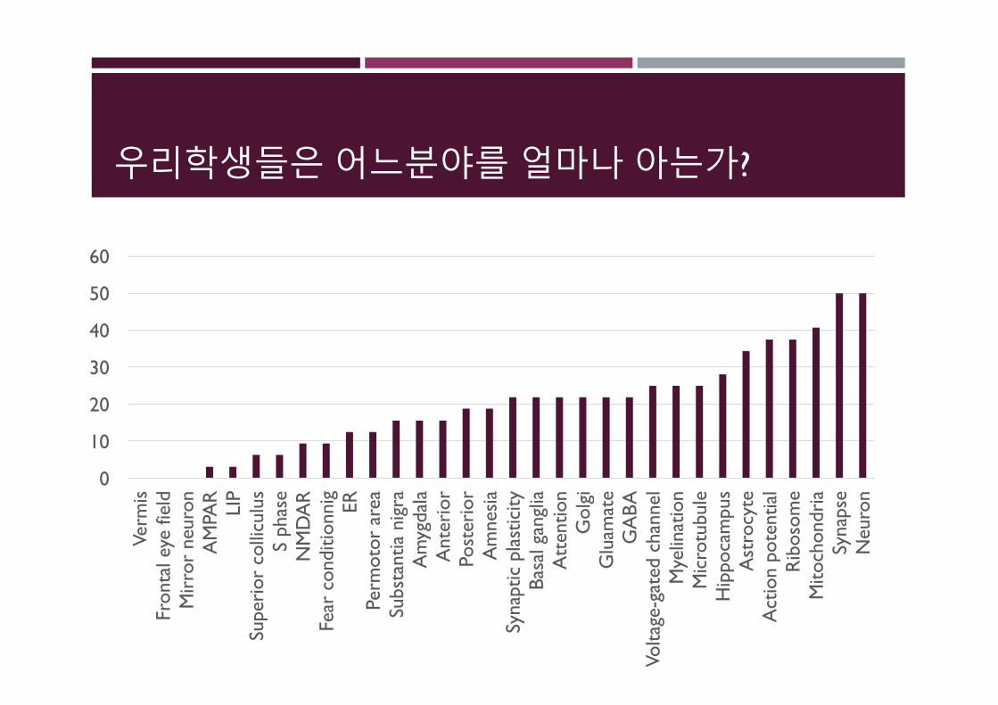
우리학생들은어느분야를얼마나아는가?
0
10
20
30
40
50
60
Verm
isFr
onta
l eye
fiel
dM
irro
r ne
uron
AM
PAR
LIP
Supe
rior
col
licul
usS
phas
eN
MD
AR
Fear
con
ditio
nnig ER
Perm
otor
are
aSu
bsta
ntia
nig
raA
myg
dala
Ant
erio
rPo
ster
ior
Am
nesi
aSy
napt
ic p
last
icity
Basa
l gan
glia
Att
entio
nG
olgi
Glu
amat
eG
ABA
Volta
ge-g
ated
cha
nnel
Mye
linat
ion
Mic
rotu
bule
Hip
poca
mpu
sA
stro
cyte
Act
ion
pote
ntia
lR
ibos
ome
Mito
chon
dria
Syna
pse
Neu
ron
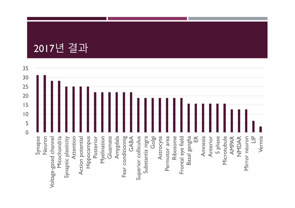
2017년결과
0
5
10
15
20
25
30
35
Syna
pse
Neu
ron
Volta
ge-g
ated
cha
nnel
Mito
chon
dria
Syna
ptic
pla
stic
ityA
tten
tion
Act
ion
pote
ntia
lH
ippo
cam
pus
Post
erio
rM
yelin
atio
nG
luam
ate
Am
ygda
laFe
ar c
ondi
tionn
igG
ABA
Supe
rior
col
licul
usSu
bsta
ntia
nig
raG
olgi
Ast
rocy
tePe
rmot
or a
rea
Rib
osom
eFr
onta
l eye
fiel
dBa
sal g
angl
ia ERA
mne
sia
Ant
erio
rS
phas
eM
icro
tubu
leA
MPA
RN
MD
AR
Mir
ror
neur
on LIP
Verm
is
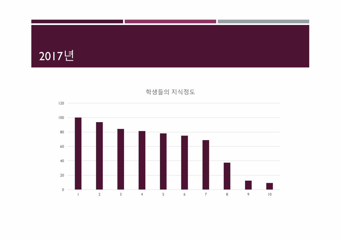
2017년
0
20
40
60
80
100
120
1 2 3 4 5 6 7 8 9 10
학생들의지식정도
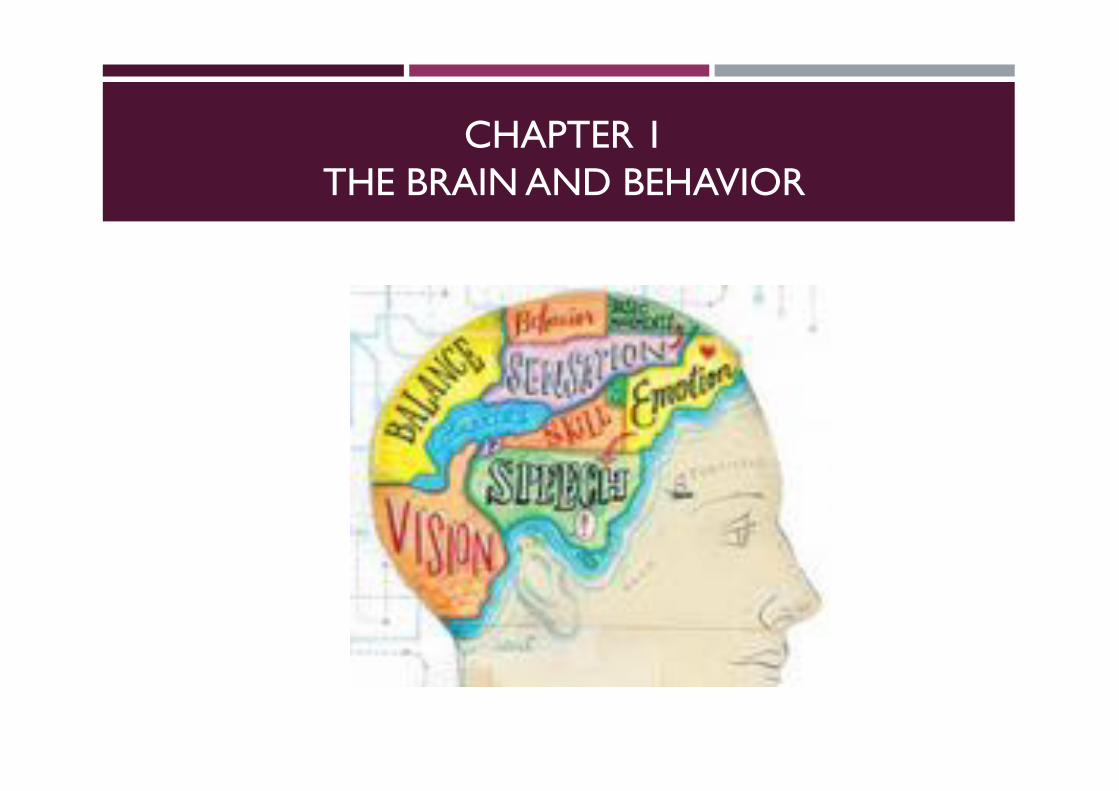
CHAPTER 1THE BRAIN AND BEHAVIOR
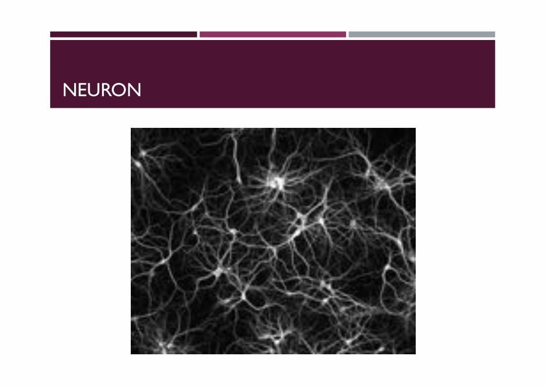
NEURON
HISTORY ABOUT NEUROSCIENCE
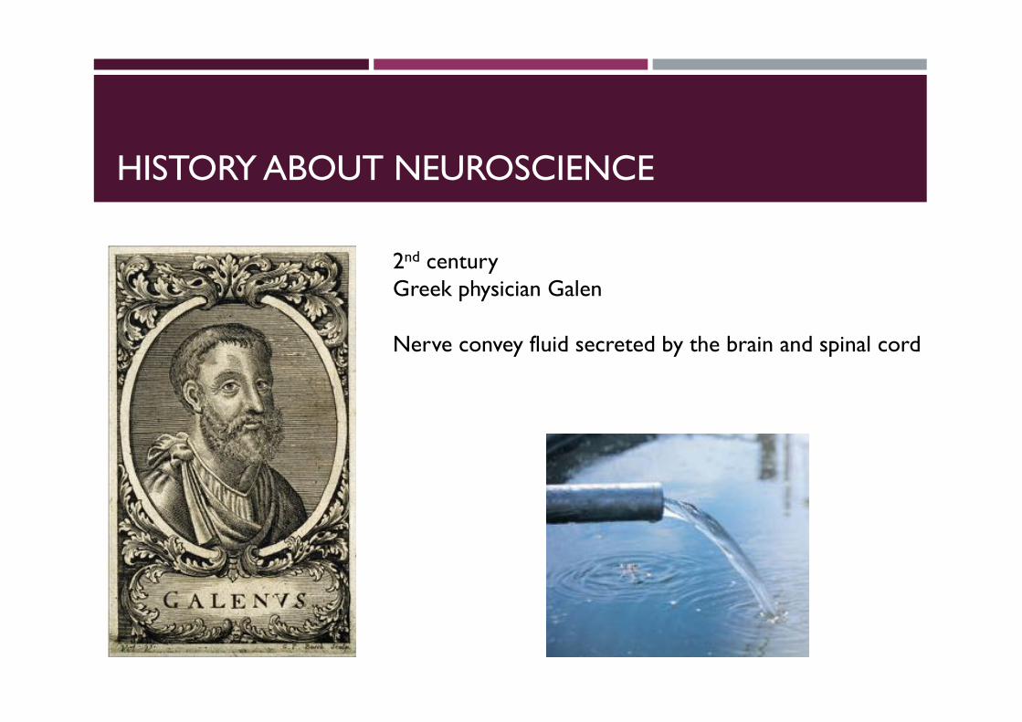
2nd centuryGreek physician Galen
Nerve convey fluid secreted by the brain and spinal cord
HISTORY ABOUT NEUROSCIENCE
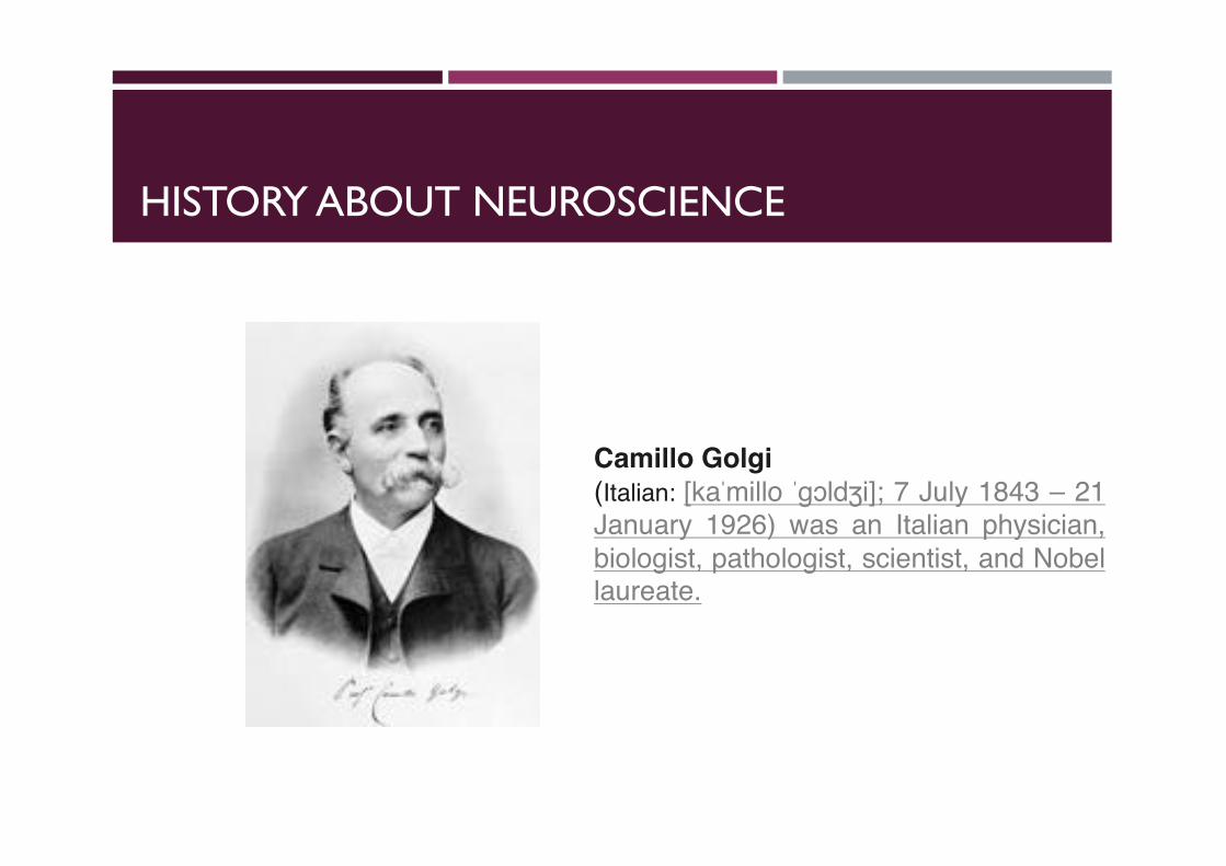
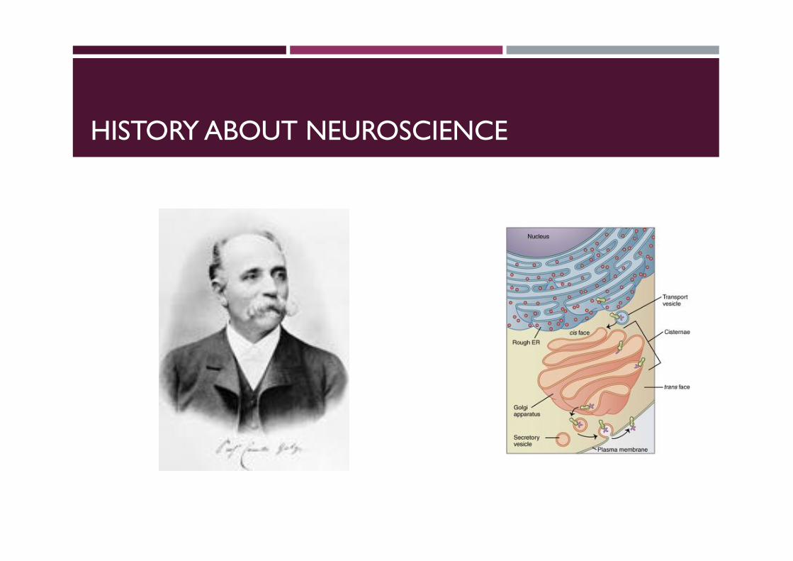
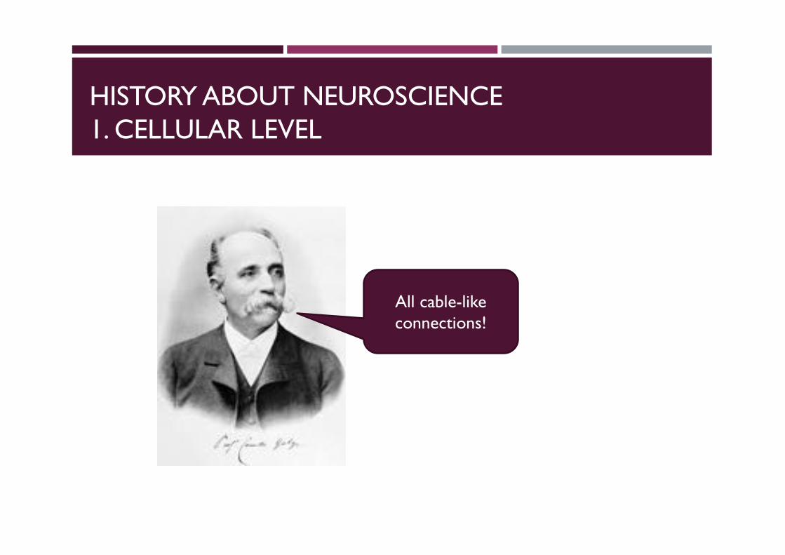
Camillo Golgi(Italian: [kaˈmillo ˈɡɔldʒi]; 7 July 1843 – 21January 1926) was an Italian physician,biologist, pathologist, scientist, and Nobellaureate.
HISTORY ABOUT NEUROSCIENCE
HISTORY ABOUT NEUROSCIENCE
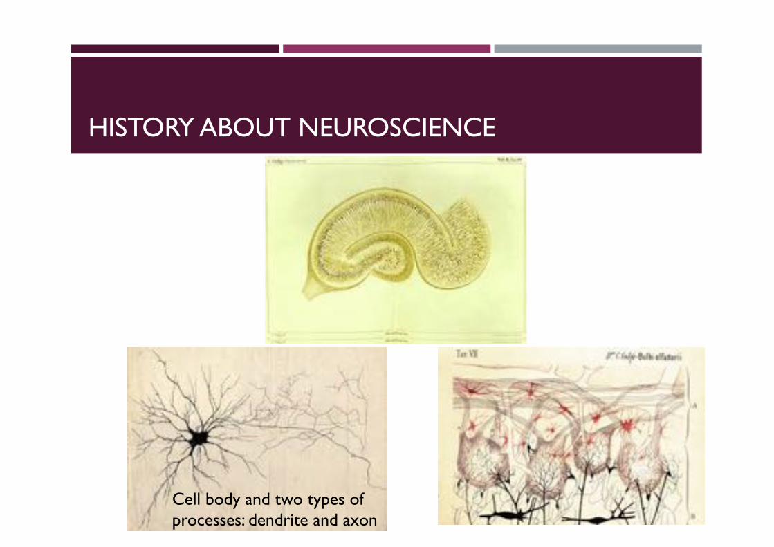
Cell body and two types of processes: dendrite and axon
HISTORY ABOUT NEUROSCIENCE1. CELLULAR LEVEL
All cable-like connections!
HISTORY ABOUT NEUROSCIENCE1. CELLULAR LEVEL
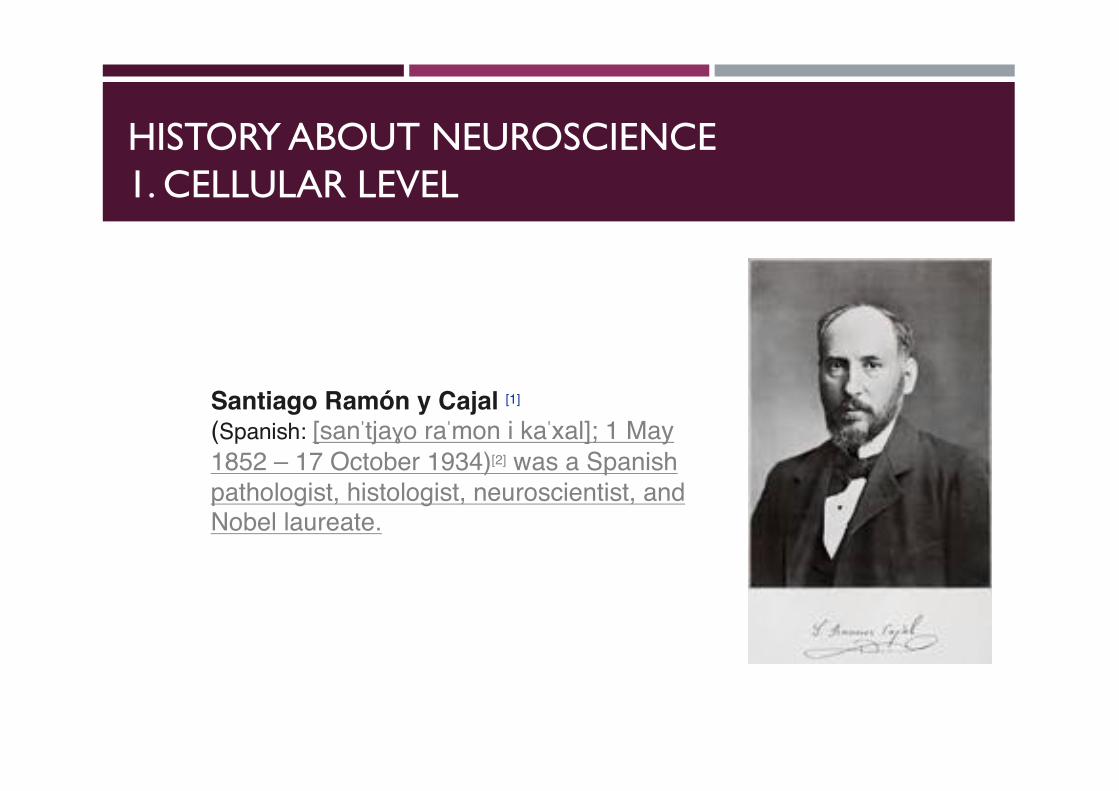
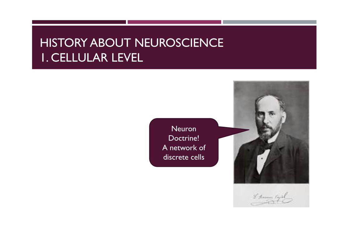
Santiago Ramón y Cajal [1]
(Spanish: [sanˈtjaɣo raˈmon i kaˈxal]; 1 May 1852 – 17 October 1934)[2] was a Spanishpathologist, histologist, neuroscientist, and Nobel laureate.
HISTORY ABOUT NEUROSCIENCE1. CELLULAR LEVEL
HISTORY ABOUT NEUROSCIENCE1. CELLULAR LEVEL
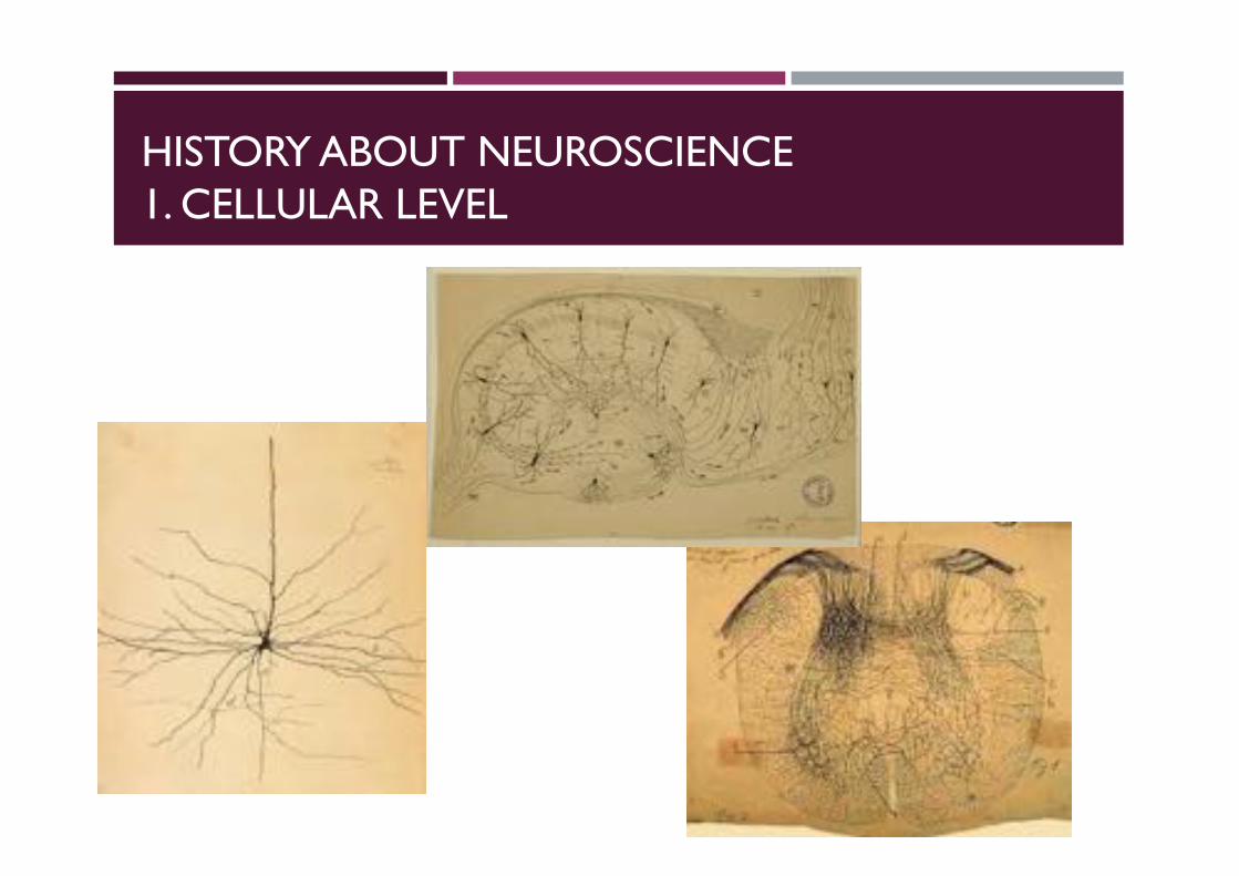
NeuronDoctrine!
A network of discrete cells
HISTORY ABOUT NEUROSCIENCE1. CELLULAR LEVEL
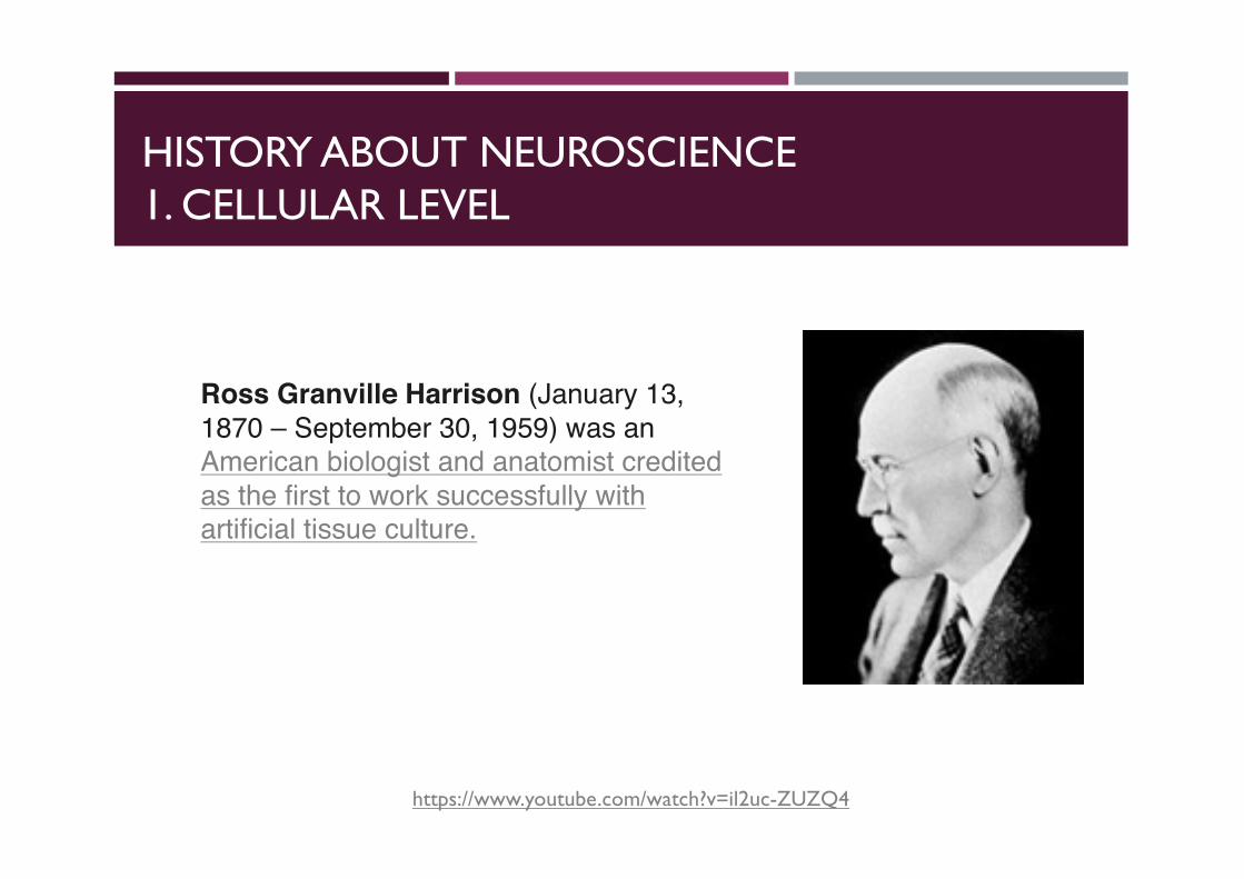
Ross Granville Harrison (January 13, 1870 – September 30, 1959) was an American biologist and anatomist credited as the first to work successfully with artificial tissue culture.
https://www.youtube.com/watch?v=il2uc-ZUZQ4
HISTORY ABOUT NEUROSCIENCE1. CELLULAR LEVEL
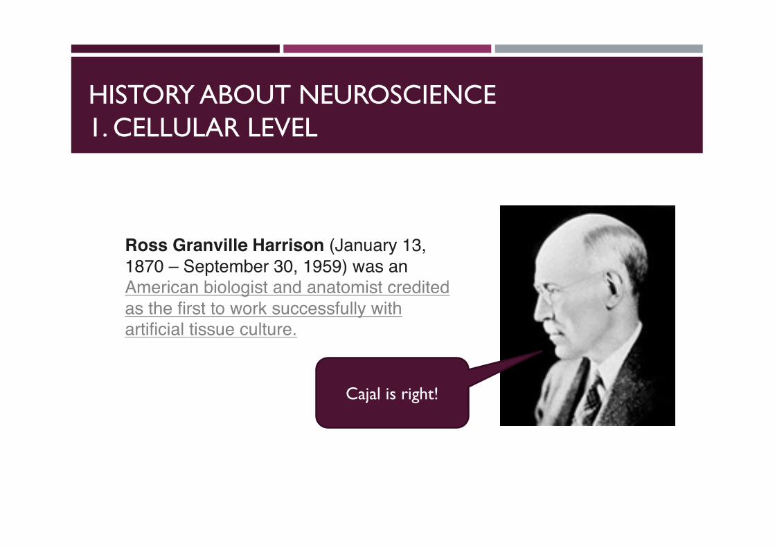
Ross Granville Harrison (January 13, 1870 – September 30, 1959) was an American biologist and anatomist credited as the first to work successfully with artificial tissue culture.
Cajal is right!

PHYSIOLOGY LEVEL
HISTORY ABOUT NEUROSCIENCE2. PHYSIOLOGY
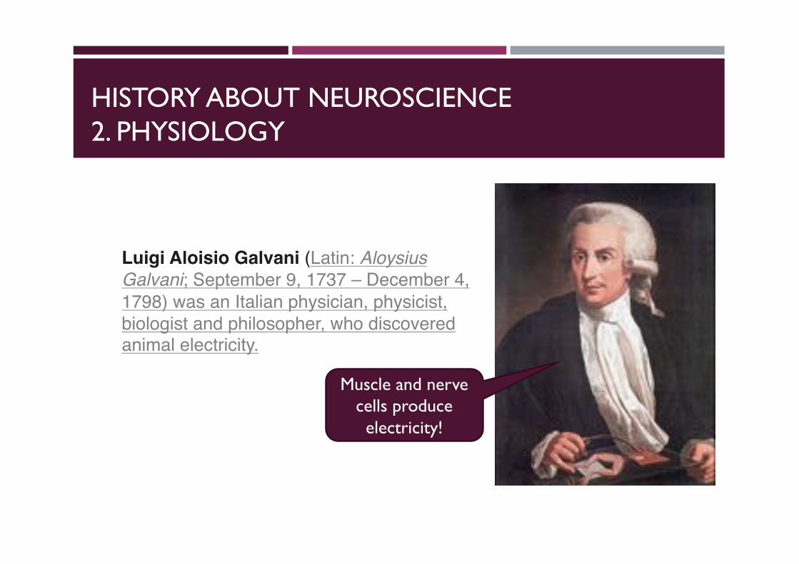
Muscle and nerve cells produce electricity!
Luigi Aloisio Galvani (Latin: Aloysius Galvani; September 9, 1737 – December 4, 1798) was an Italian physician, physicist, biologist and philosopher, who discovered animal electricity.
HISTORY ABOUT NEUROSCIENCE2. PHYSIOLOGY
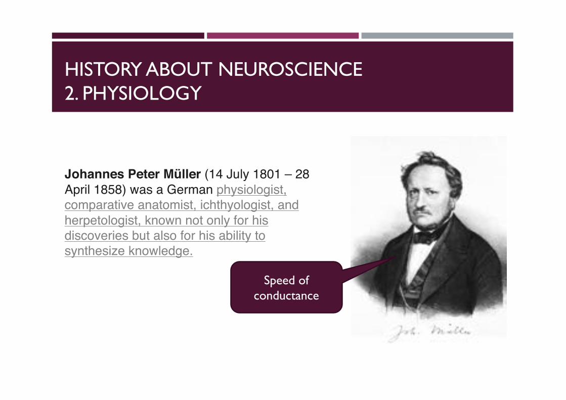
Speed of conductance
Johannes Peter Müller (14 July 1801 – 28 April 1858) was a German physiologist, comparative anatomist, ichthyologist, and herpetologist, known not only for his discoveries but also for his ability to synthesize knowledge.
HISTORY ABOUT NEUROSCIENCE2. PHYSIOLOGY
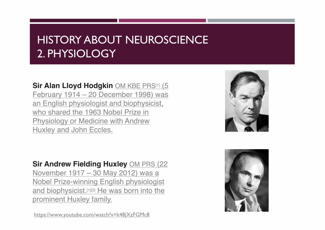
Sir Alan Lloyd Hodgkin OM KBE PRS[1] (5 February 1914 – 20 December 1998) was an English physiologist and biophysicist, who shared the 1963 Nobel Prize in Physiology or Medicine with Andrew Huxley and John Eccles.
Sir Andrew Fielding Huxley OM PRS (22 November 1917 – 30 May 2012) was a Nobel Prize-winning English physiologistand biophysicist.[1][2] He was born into the prominent Huxley family.
https://www.youtube.com/watch?v=k48jXzFGMc8
HISTORY ABOUT NEUROSCIENCE2. PHYSIOLOGY
500
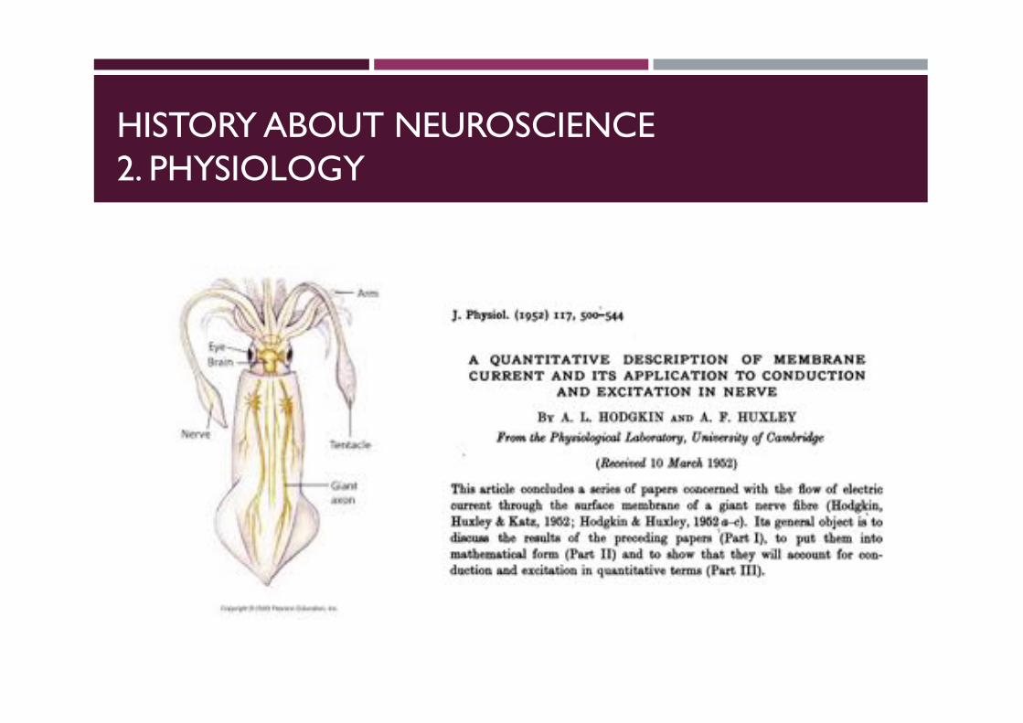
J. Physiol. (I952) I I7, 500-544
A QUANTITATIVE DESCRIPTION OF MEMBRANECURRENT AND ITS APPLICATION TO CONDUCTION
AND EXCITATION IN NERVE
BY A. L. HODGKIN AND A. F. HUXLEYFrom the Physiological Laboratory, University of Cambridge
(Received 10 March 1952)
This article concludes a series of papers concerned with the flow of electriccurrent through the surface membrane of a giant nerve fibre (Hodgkin,Huxley & Katz, 1952; Hodgkin & Huxley, 1952 a-c). Its general object is todiscu the results of the preceding papers (Part I), to put them intomathematical form (Part II) and to show that they will account for con-duction and excitation in quantitative terms (Part III).
PART I. DISCUSSION OF EXPERIMENTAL RESULTSThe results described in the preceding papers suggest that the electricalbehaviour of the membrane may be represented by the network shown inFig. 1. Current can be carried through the membrane either by charging themembrane capacity or by movement of ion-s through the resistances in parallelwith the capacity. The ionic current is divided into components carried bysodium and potassium ions (INa and IK), and a small 'leakage current' (I,)made up by chloride and other ions. Each component of the ionic current isdetermined by a driving force which may conveniently be measured as anelectrical potential difference and a permeability coefficient which has thedimensions of a conductance. Thus the sodium current (INa) is equal to thesodium conductance (9Na) multiplied by the difference between the membranepotential (E) and the equilibrium potential for the sodium ion (ENa). Similarequations apply to 'K and I, and are collected on p. 505.Our experiments suggest that gNa and 9E are functions of time and
membrane potential, but that ENa, EK, El, CM and g, may be taken asconstant. The influence of membrane potential on permeability can be sum-marized by stating: first, that depolarization causes a transient increase insodium conductance and a slower but maintained increase in potassium con-ductance; secondly, that these changes are graded and that they can bereversed by repolarizing the membrane. In order to decide whether theseeffects are sufficient to account for complicated phenomena such as the actionpotential and refractory period, it is necessary to obtain expressions relating
HISTORY ABOUT NEUROSCIENCE2. PHYSIOLOGY
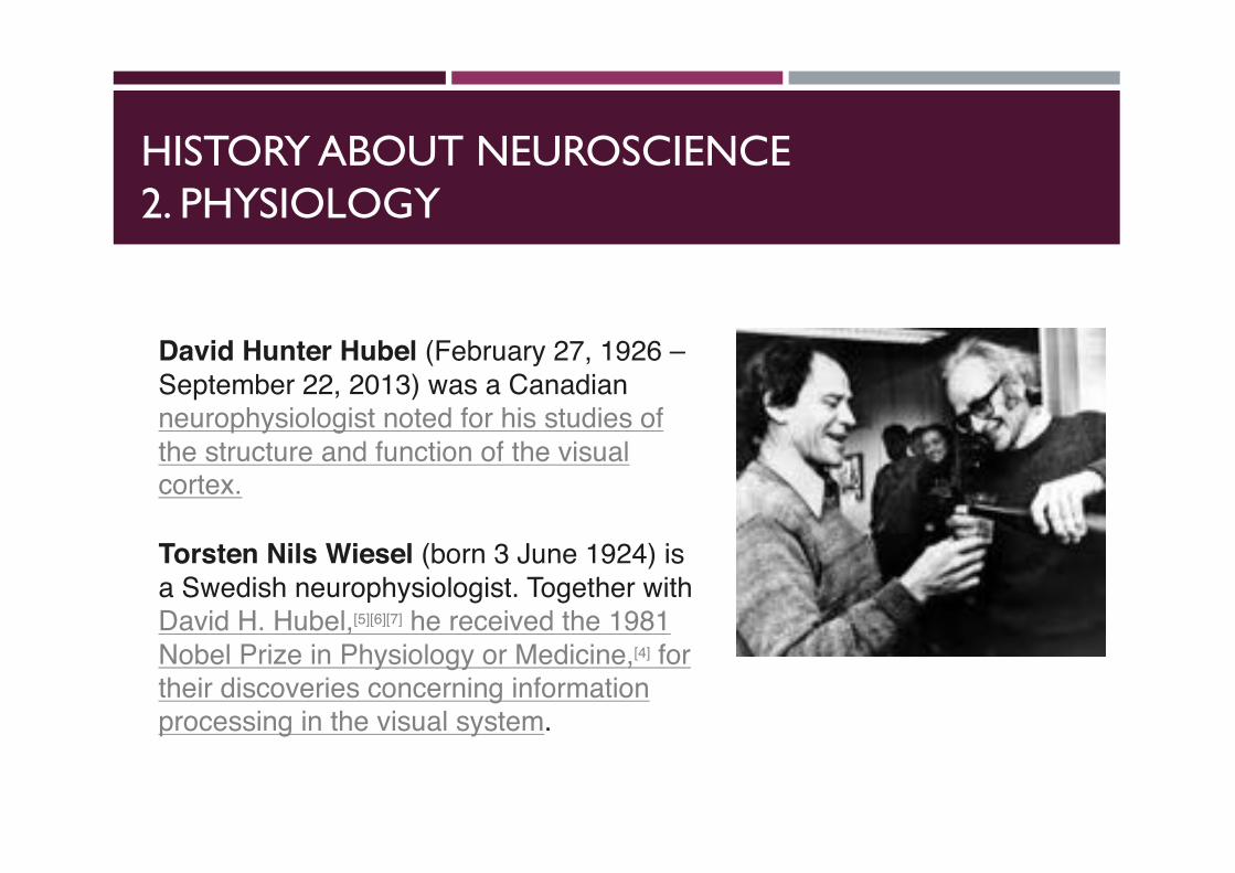
David Hunter Hubel (February 27, 1926 –September 22, 2013) was a Canadian neurophysiologist noted for his studies of the structure and function of the visual cortex.
Torsten Nils Wiesel (born 3 June 1924) is a Swedish neurophysiologist. Together with David H. Hubel,[5][6][7] he received the 1981 Nobel Prize in Physiology or Medicine,[4] for their discoveries concerning information processing in the visual system.
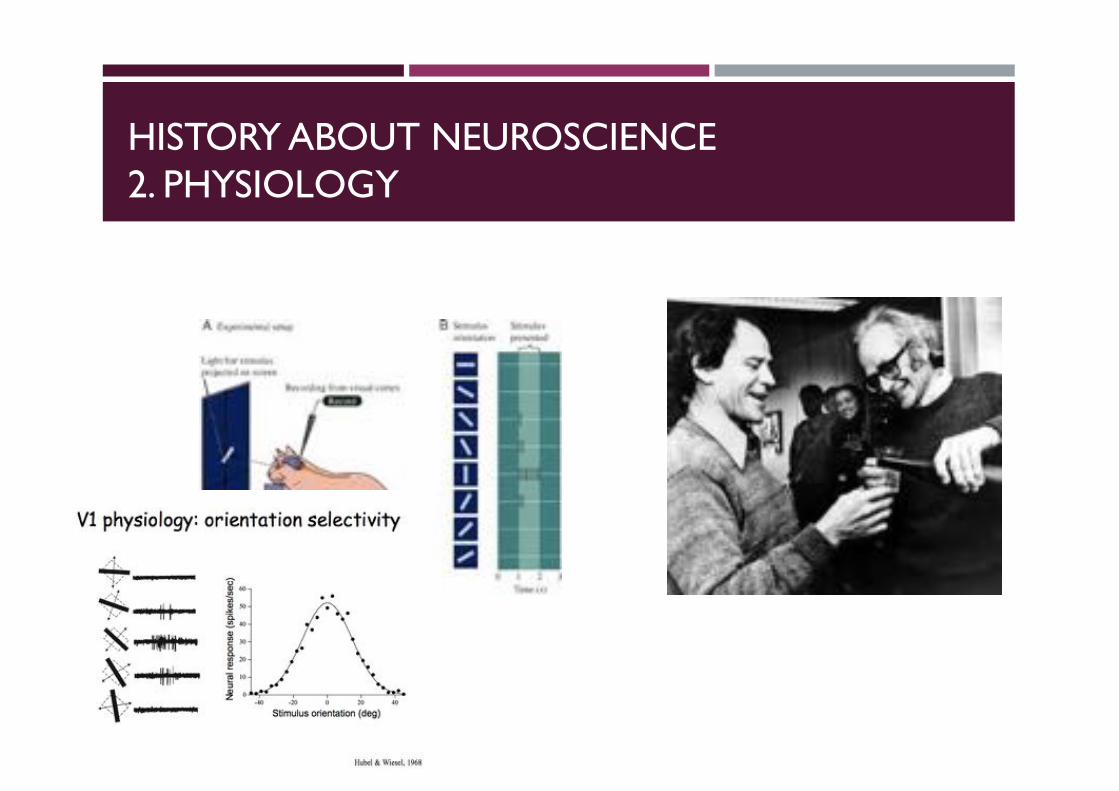
HISTORY ABOUT NEUROSCIENCE2. PHYSIOLOGY
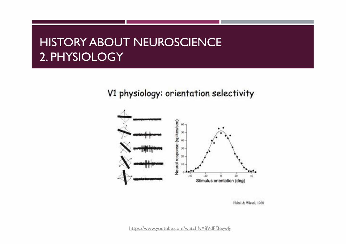
HISTORY ABOUT NEUROSCIENCE2. PHYSIOLOGY
https://www.youtube.com/watch?v=8VdFf3egwfg
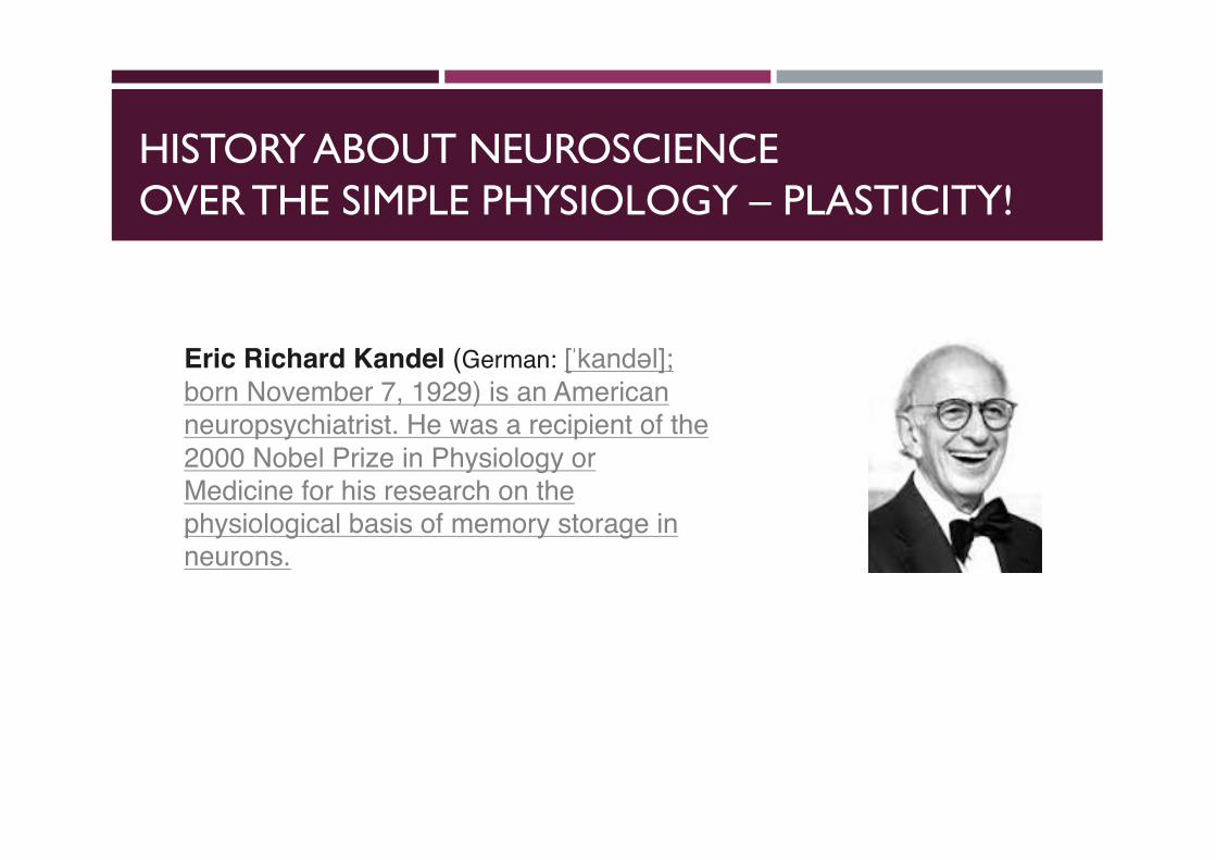
HISTORY ABOUT NEUROSCIENCEOVER THE SIMPLE PHYSIOLOGY – PLASTICITY!
Eric Richard Kandel (German: [ˈkandəl]; born November 7, 1929) is an Americanneuropsychiatrist. He was a recipient of the 2000 Nobel Prize in Physiology or Medicine for his research on the physiological basis of memory storage in neurons.
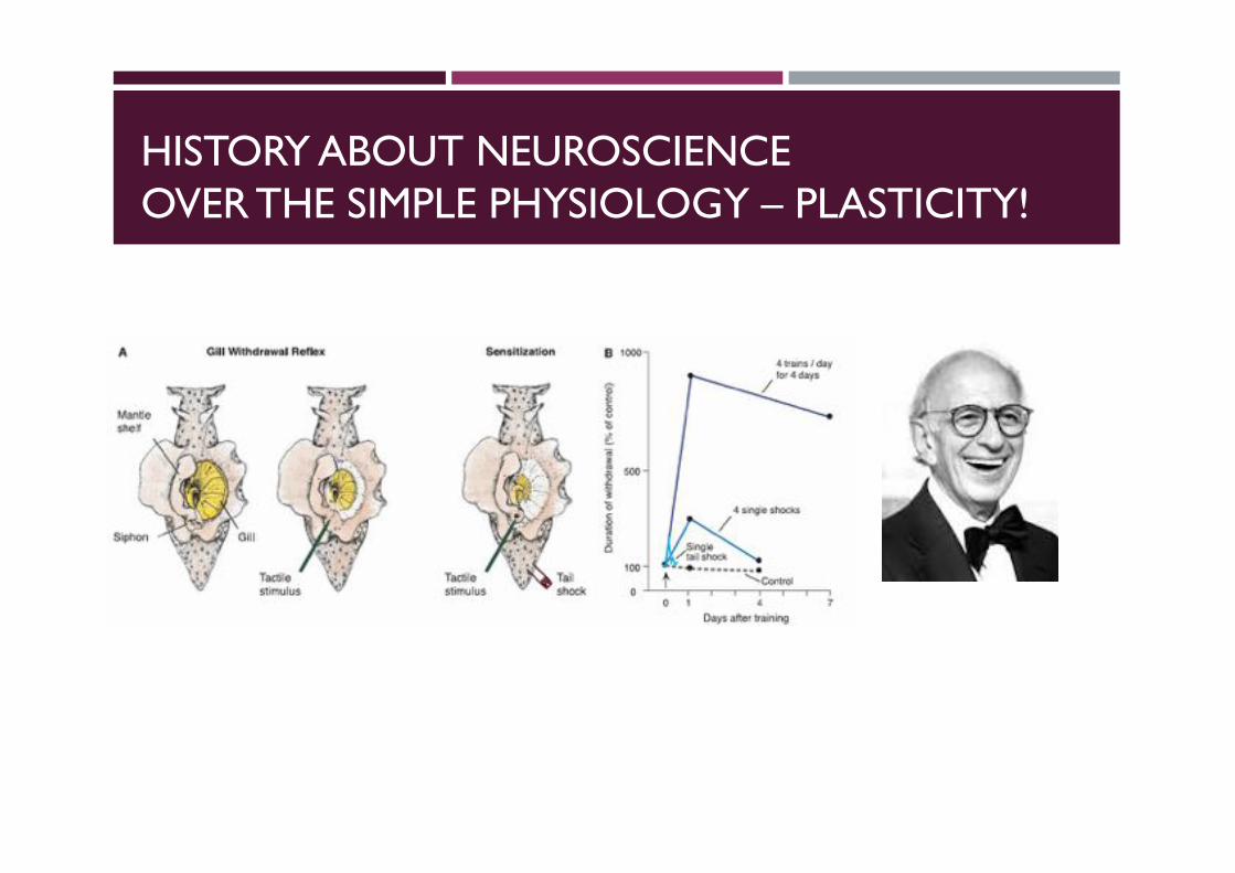
HISTORY ABOUT NEUROSCIENCEOVER THE SIMPLE PHYSIOLOGY – PLASTICITY!

PSYCHOLOGY
HISTORY ABOUT NEUROSCIENCE3. PSYCHOLOGY
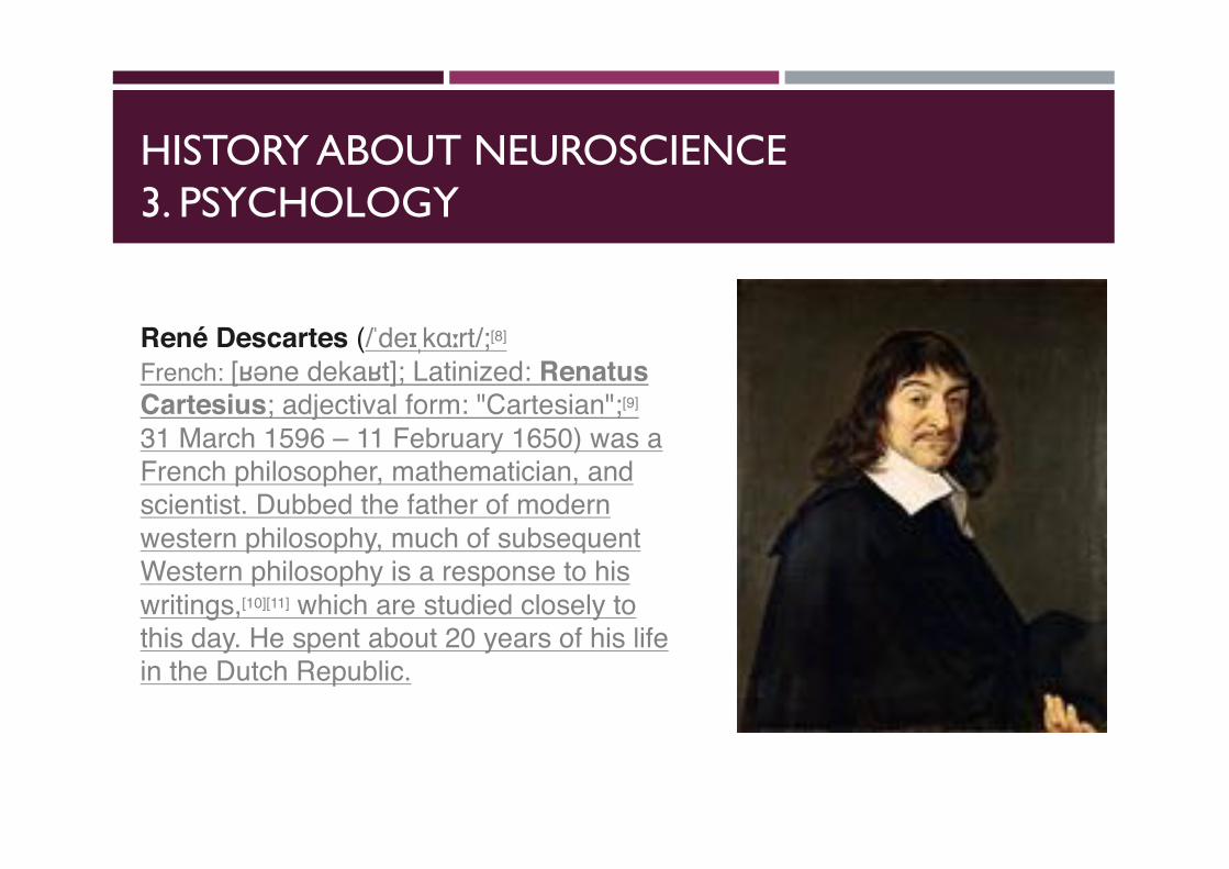
René Descartes (/ˈdeɪˌkɑːrt/;[8]
French: [ʁəne dekaʁt]; Latinized: Renatus Cartesius; adjectival form: "Cartesian";[9]
31 March 1596 – 11 February 1650) was a French philosopher, mathematician, and scientist. Dubbed the father of modern western philosophy, much of subsequent Western philosophy is a response to his writings,[10][11] which are studied closely to this day. He spent about 20 years of his life in the Dutch Republic.
HISTORY ABOUT NEUROSCIENCE3. PSYCHOLOGY
Body MindSoulSpirit
HISTORY ABOUT NEUROSCIENCE3. PSYCHOLOGY
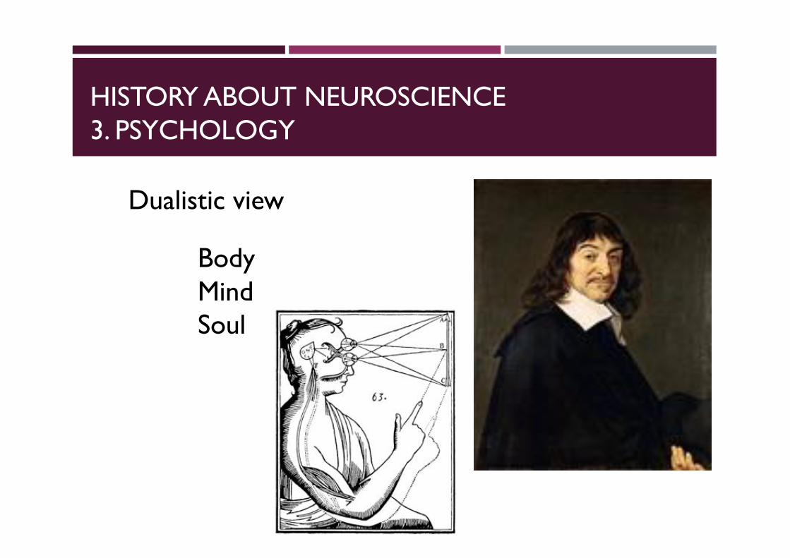
BodyMindSoul
Dualistic view
HISTORY ABOUT NEUROSCIENCE3. PSYCHOLOGY
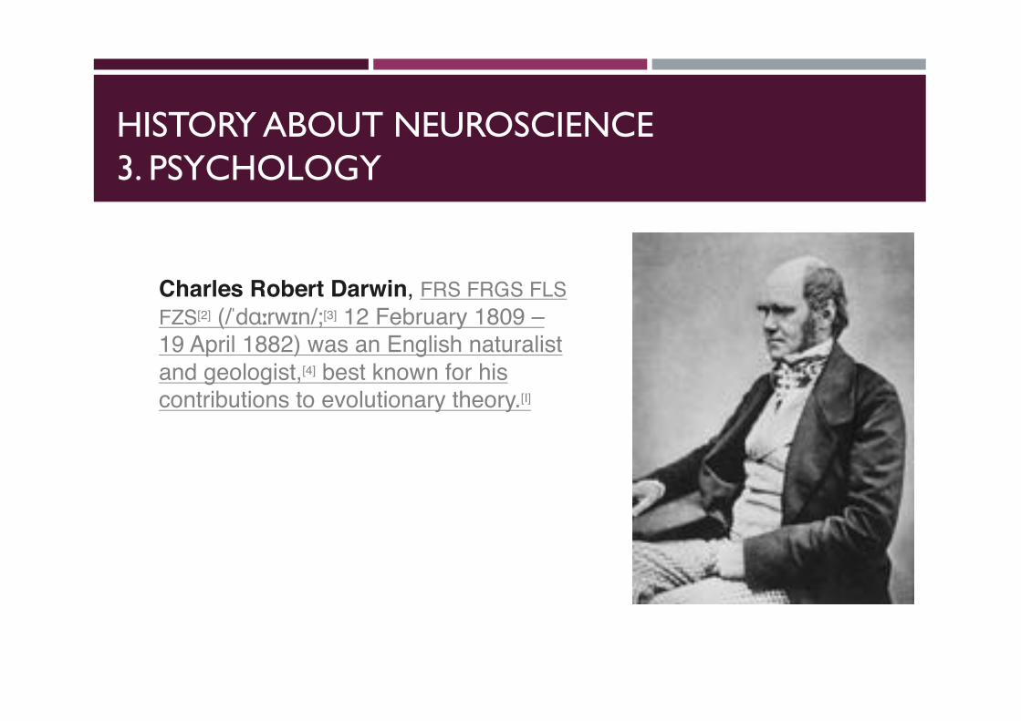
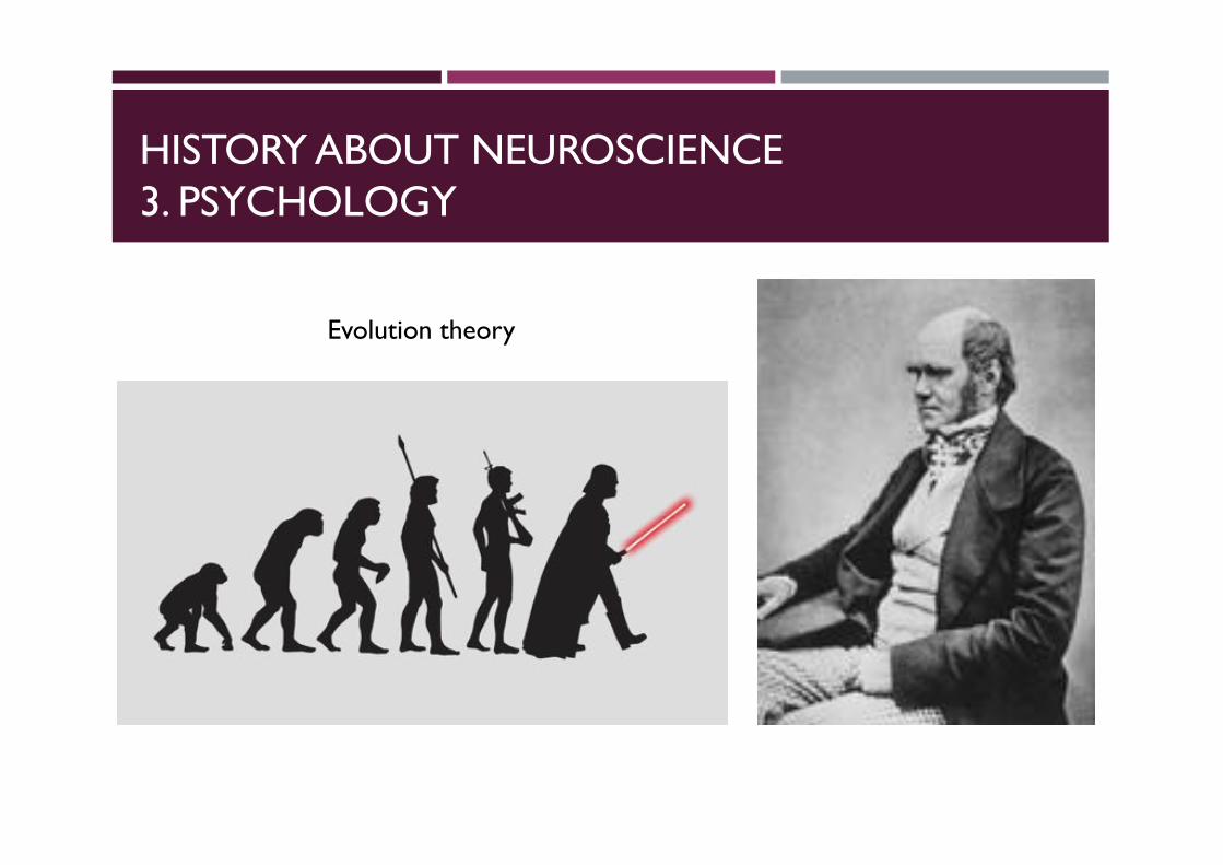
Charles Robert Darwin, FRS FRGS FLSFZS[2] (/ˈdɑːrwɪn/;[3] 12 February 1809 –19 April 1882) was an English naturalistand geologist,[4] best known for his contributions to evolutionary theory.[I]
HISTORY ABOUT NEUROSCIENCE3. PSYCHOLOGY
Evolution theory
HISTORY ABOUT NEUROSCIENCE3. PSYCHOLOGY
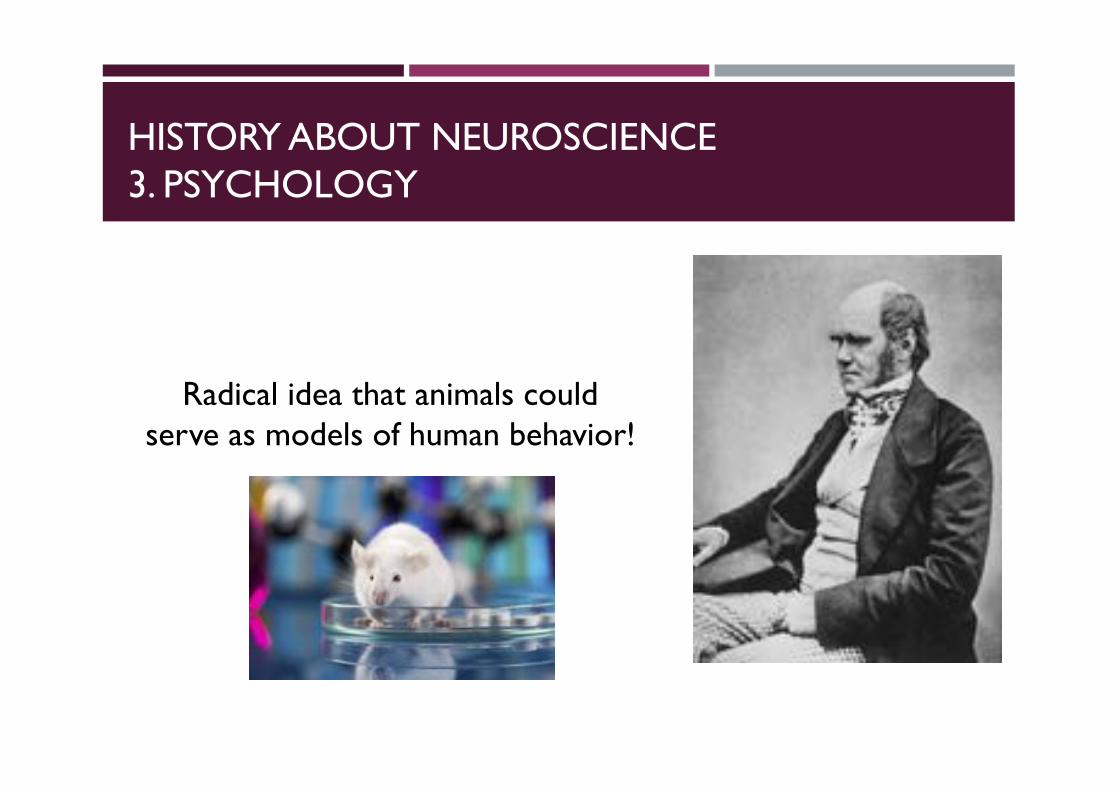
Radical idea that animals could serve as models of human behavior!
HISTORY ABOUT NEUROSCIENCE3. PSYCHOLOGY
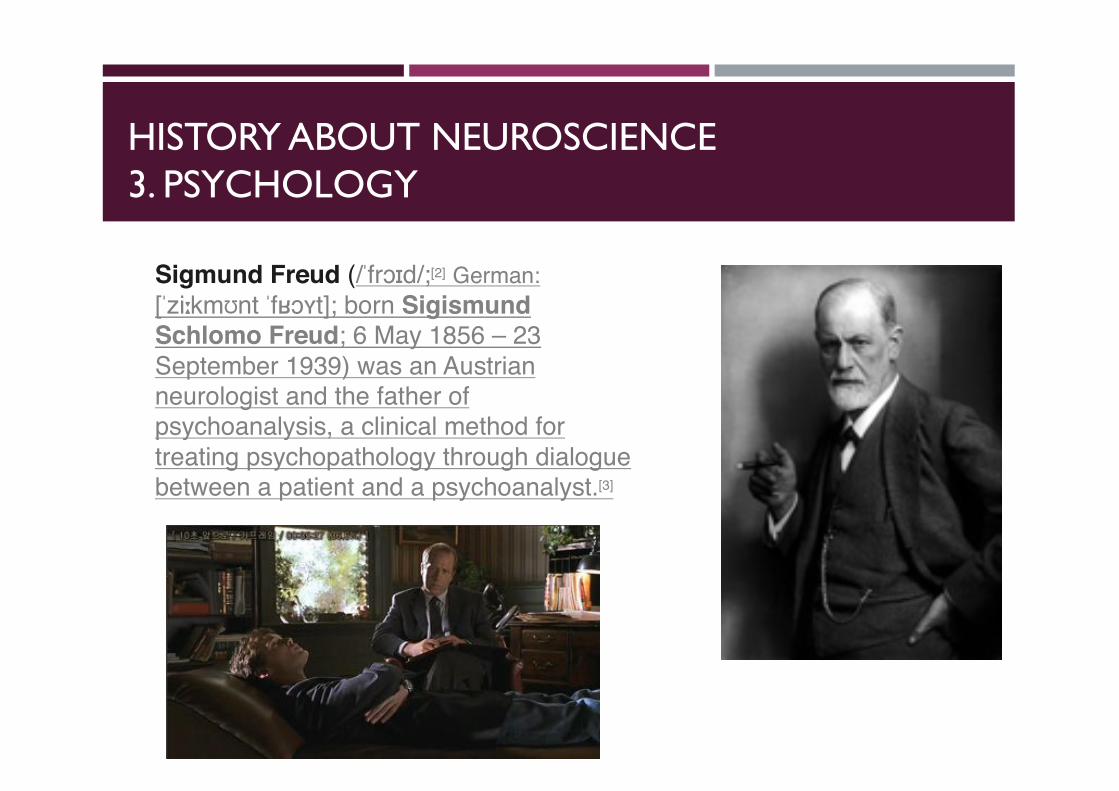
Sigmund Freud (/ˈfrɔɪd/;[2] German:[ˈziːkmʊnt ˈfʁɔʏt]; born Sigismund Schlomo Freud; 6 May 1856 – 23 September 1939) was an Austrianneurologist and the father of psychoanalysis, a clinical method for treating psychopathology through dialogue between a patient and a psychoanalyst.[3]
HISTORY ABOUT NEUROSCIENCE3. PSYCHOLOGY
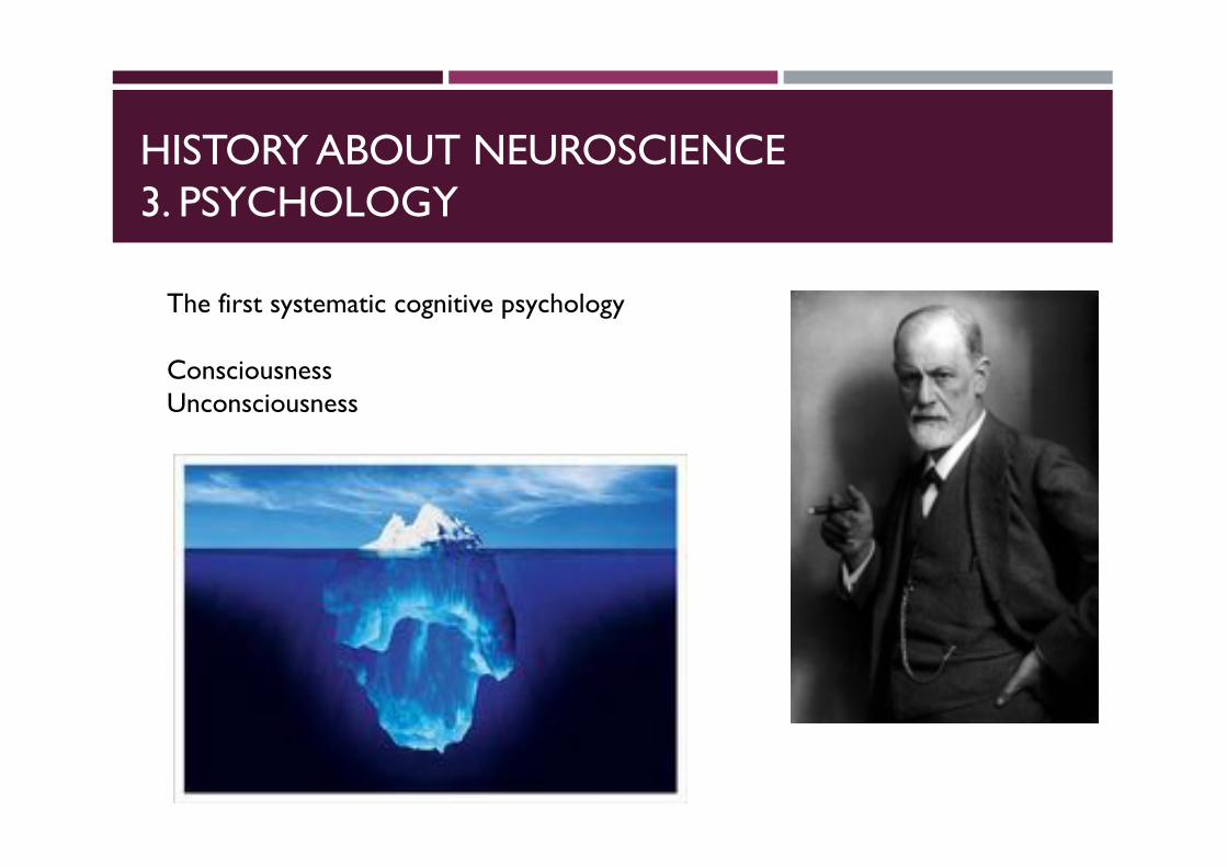
The first systematic cognitive psychology
ConsciousnessUnconsciousness

PSYCHOLOGY MET BIOLOGY
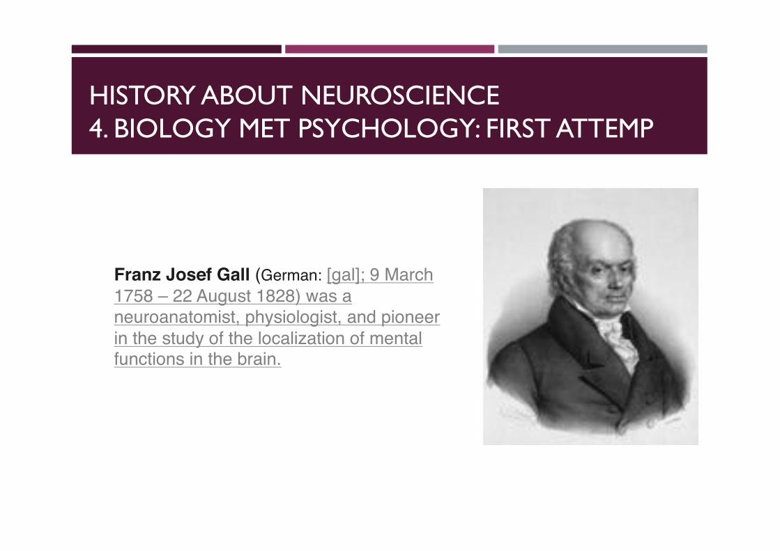
HISTORY ABOUT NEUROSCIENCE4. BIOLOGY MET PSYCHOLOGY: FIRST ATTEMP
Franz Josef Gall (German: [gal]; 9 March 1758 – 22 August 1828) was a neuroanatomist, physiologist, and pioneer in the study of the localization of mental functions in the brain.
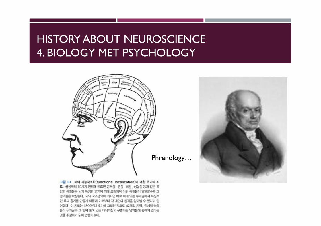
HISTORY ABOUT NEUROSCIENCE4. BIOLOGY MET PSYCHOLOGY
Phrenology…

HISTORY ABOUT NEUROSCIENCE4. BIOLOGY MET PSYCHOLOGY
Phrenology…
Why??
HISTORY ABOUT NEUROSCIENCE4. BIOLOGY MET PSYCHOLOGY
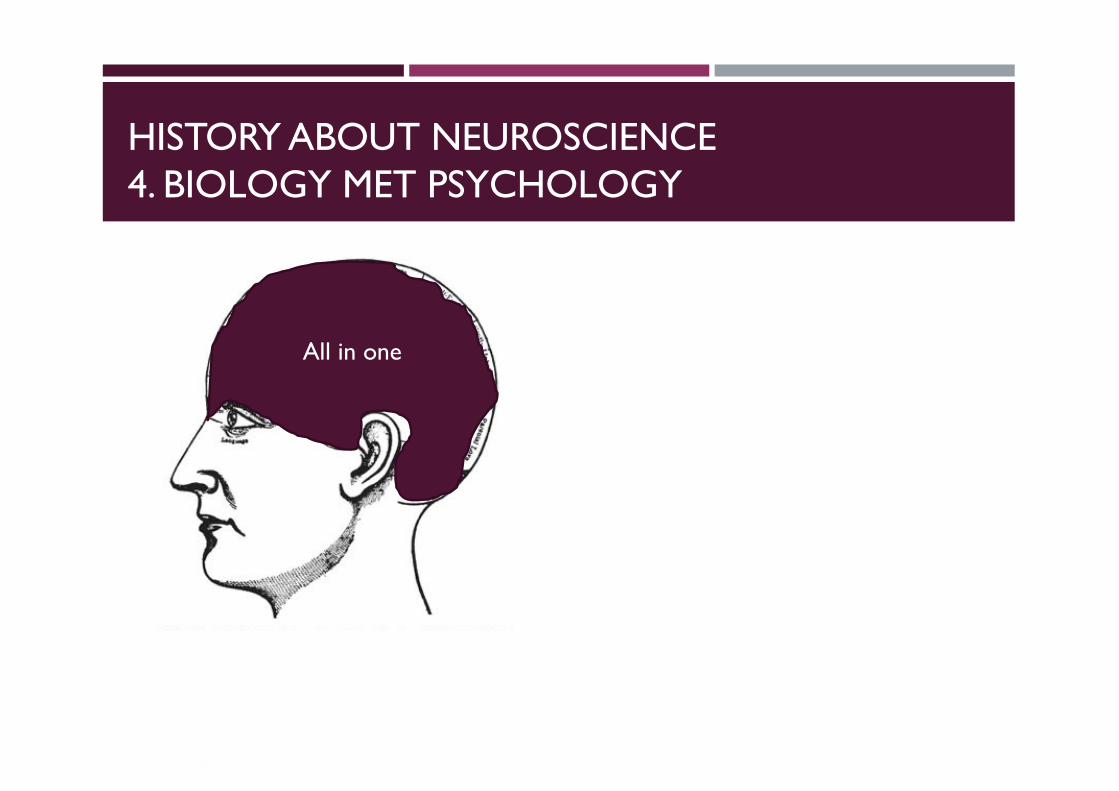
All in one
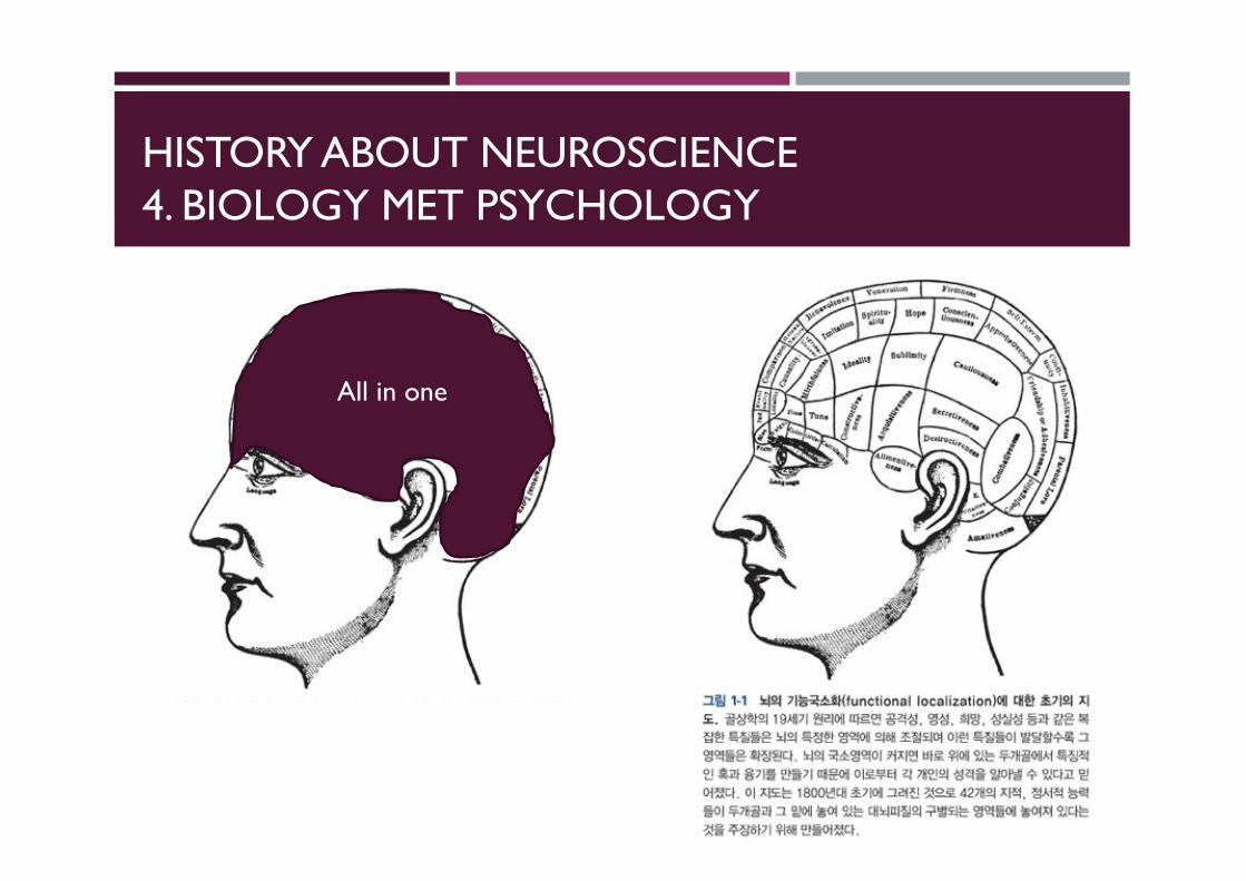
HISTORY ABOUT NEUROSCIENCE4. BIOLOGY MET PSYCHOLOGY
All in one
HISTORY ABOUT NEUROSCIENCE4. BIOLOGY MET PSYCHOLOGY
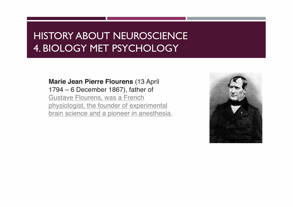
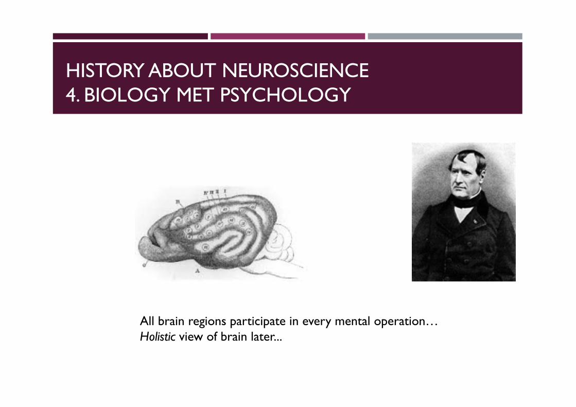
Marie Jean Pierre Flourens (13 April 1794 – 6 December 1867), father of Gustave Flourens, was a French physiologist, the founder of experimental brain science and a pioneer in anesthesia.
HISTORY ABOUT NEUROSCIENCE4. BIOLOGY MET PSYCHOLOGY
All brain regions participate in every mental operation…Holistic view of brain later...
HISTORY ABOUT NEUROSCIENCE4. BIOLOGY MET PSYCHOLOGY
HISTORY ABOUT NEUROSCIENCE4. BIOLOGY MET PSYCHOLOGY
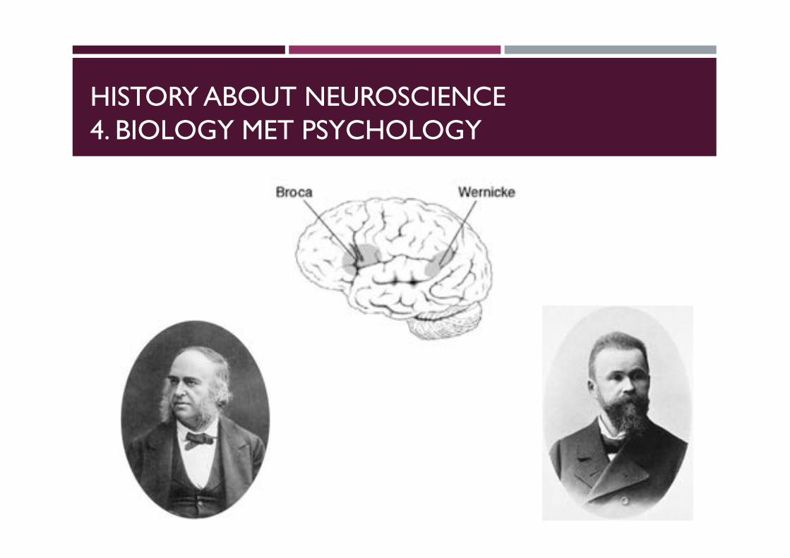
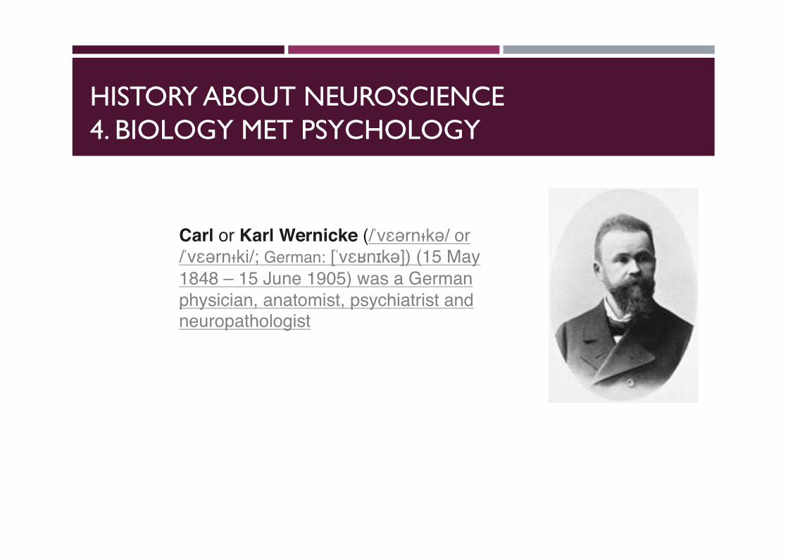
Carl or Karl Wernicke (/ˈvɛərnᵻkə/ or /ˈvɛərnᵻki/; German: [ˈvɛʁnɪkə]) (15 May 1848 – 15 June 1905) was a German physician, anatomist, psychiatrist and neuropathologist
HISTORY ABOUT NEUROSCIENCE4. BIOLOGY MET PSYCHOLOGY
Pierre Paul Broca (/broʊˈkɑː/ or /ˈbroʊkə/; 28 June 1824 – 9 July 1880) was a French physician, surgeon, anatomist, and anthropologist. He was born in Sainte-Foy-la-Grande, Gironde.
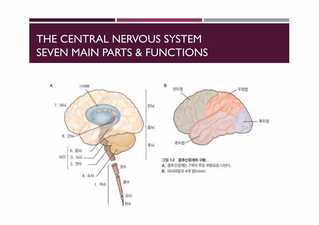
THE CENTRAL NERVOUS SYSTEMSEVEN MAIN PARTS & FUNCTIONS
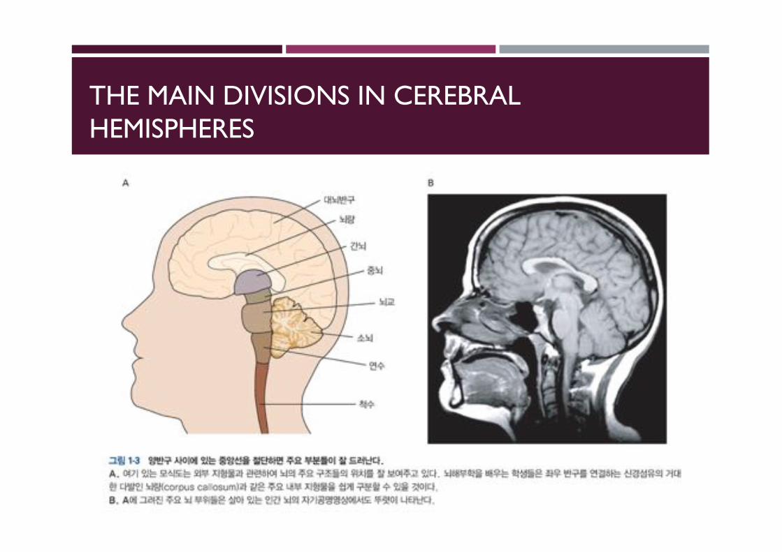
THE MAIN DIVISIONS IN CEREBRAL HEMISPHERES
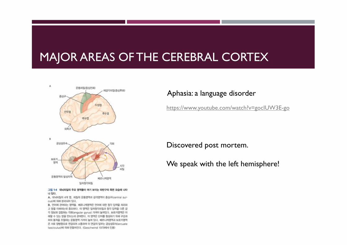
MAJOR AREAS OF THE CEREBRAL CORTEX
Aphasia: a language disorder
https://www.youtube.com/watch?v=gocIUW3E-go
Discovered post mortem.
We speak with the left hemisphere!
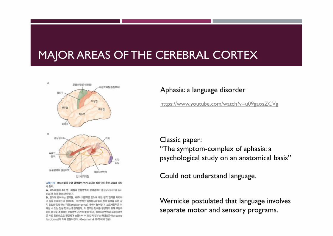
MAJOR AREAS OF THE CEREBRAL CORTEX
Aphasia: a language disorder
Classic paper:“The symptom-complex of aphasia: a psychological study on an anatomical basis”
Could not understand language.
Wernicke postulated that language involves separate motor and sensory programs.
https://www.youtube.com/watch?v=u09gaosZCVg
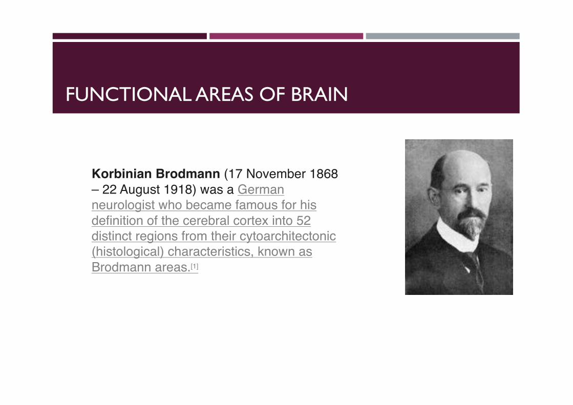
FUNCTIONAL AREAS OF BRAIN
Korbinian Brodmann (17 November 1868 – 22 August 1918) was a Germanneurologist who became famous for his definition of the cerebral cortex into 52 distinct regions from their cytoarchitectonic(histological) characteristics, known as Brodmann areas.[1]
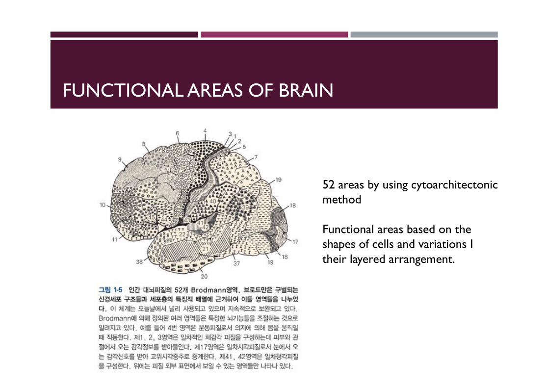
FUNCTIONAL AREAS OF BRAIN
52 areas by using cytoarchitectonicmethod
Functional areas based on the shapes of cells and variations I their layered arrangement.
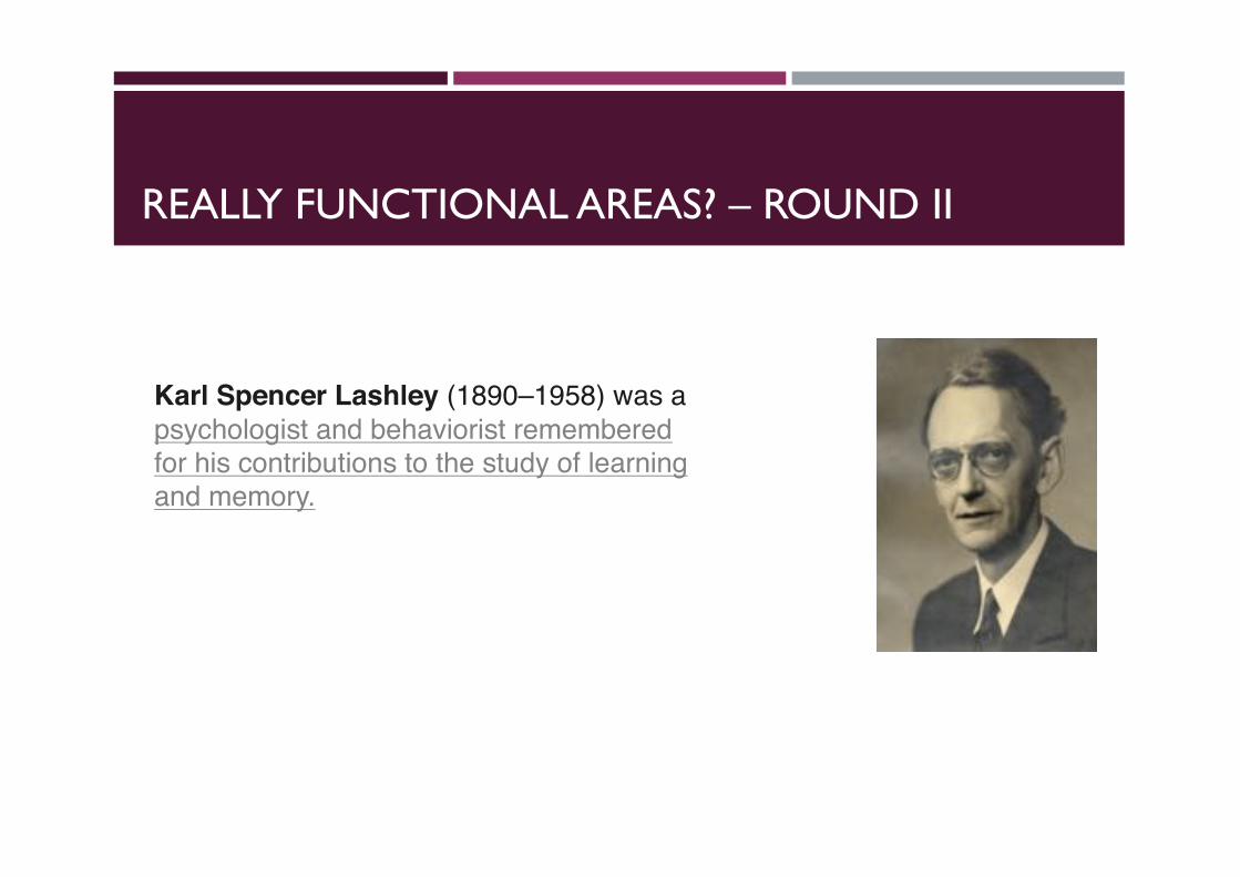
REALLY FUNCTIONAL AREAS? – ROUND II
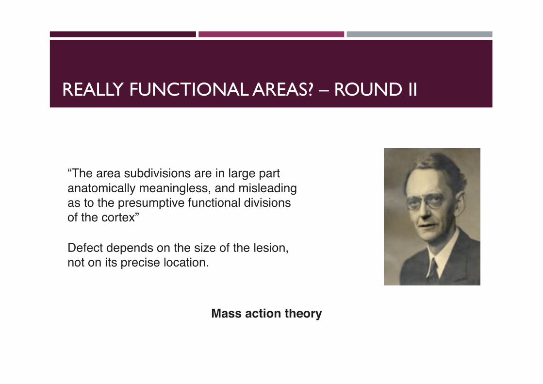
Karl Spencer Lashley (1890–1958) was a psychologist and behaviorist remembered for his contributions to the study of learning and memory.
REALLY FUNCTIONAL AREAS? – ROUND II
“The area subdivisions are in large part anatomically meaningless, and misleading as to the presumptive functional divisions of the cortex”
Defect depends on the size of the lesion, not on its precise location.
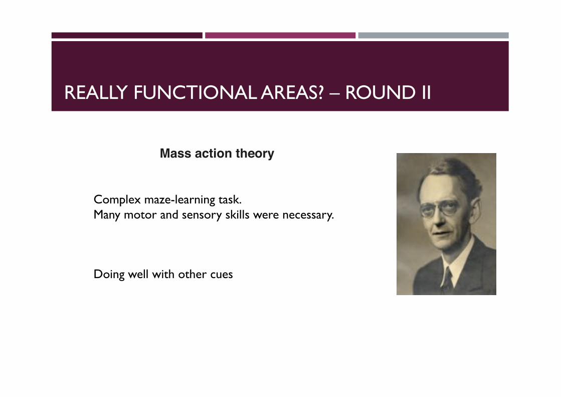
Mass action theory
REALLY FUNCTIONAL AREAS? – ROUND II
Mass action theory
Complex maze-learning task.Many motor and sensory skills were necessary.
Doing well with other cues
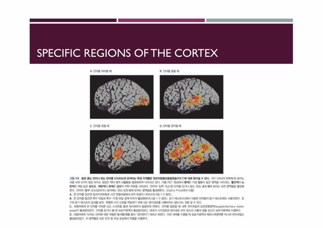
SPECIFIC REGIONS OF THE CORTEX
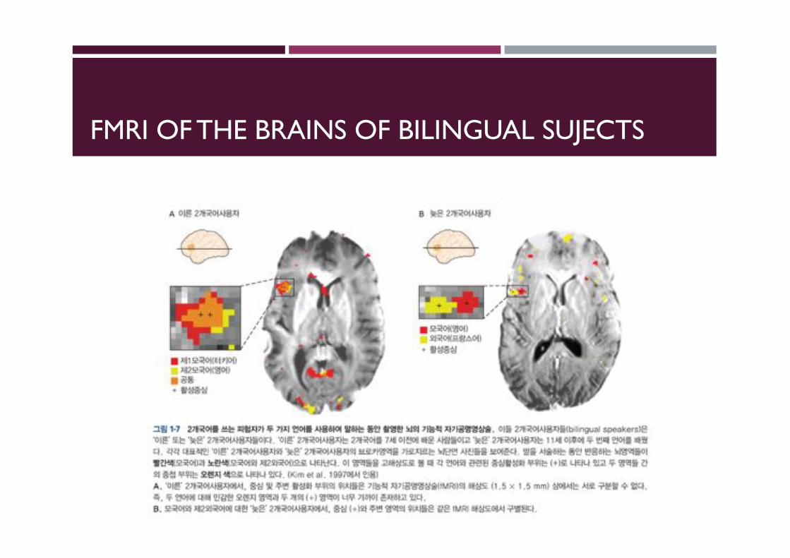
FMRI OF THE BRAINS OF BILINGUAL SUJECTS
NEURO TREEYOU CAN SEE ALL HISTORY OF NEUROSCIENTIST
http://neurotree.org/
CONCLUSION
Now you should make the history of neuroscience!
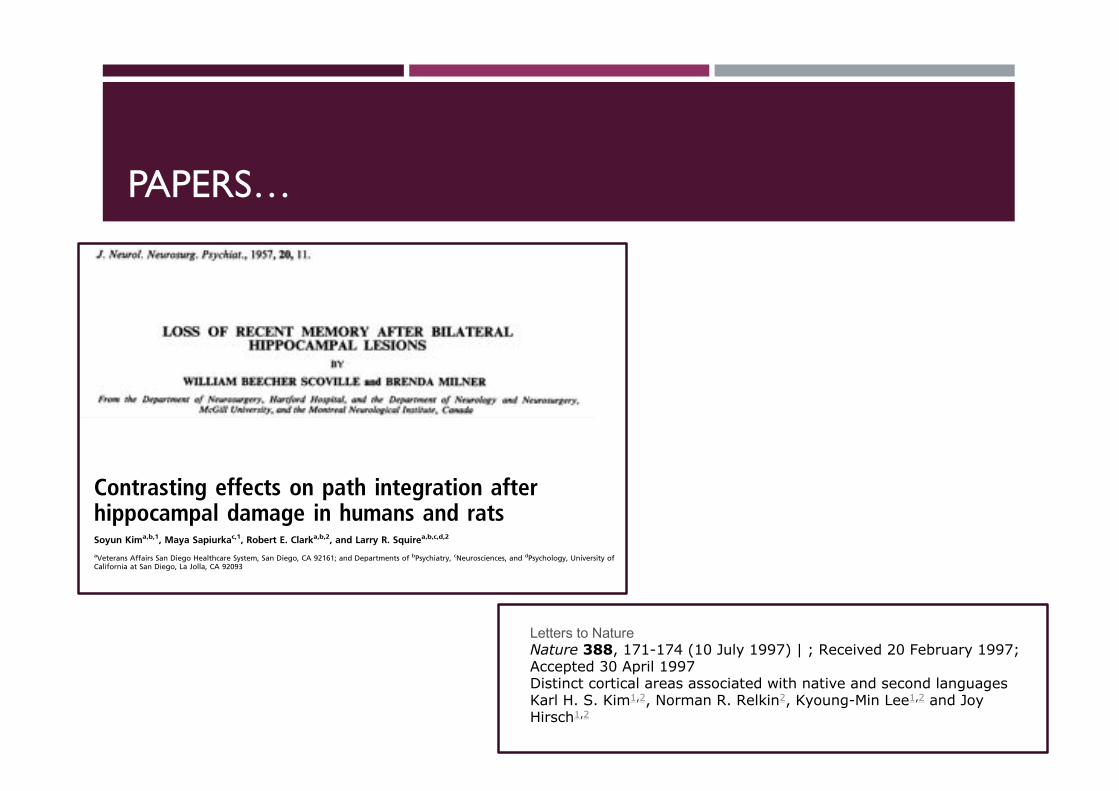
PAPERS…
J. Neurol. Neurosurg. Psychiat., 1957, 20, 11.
LOSS OF RECENT MEMORY AFTER BILATERALHIPPOCAMPAL LESIONS
BY
WILLIAM BEECHER SCOVIILLE and BRENDA MILNERFrom the Department of Neurosurgery, Hartford Hospital, and the Department of Neurology and Neurosurgery,
McGill University, and the Montreal Neurological Institute, Canada
In 1954 Scoville described a grave loss of recentmemory which he had observed as a sequel tobilateral medial temporal-lobe resection in onepsychotic patient and one patient with intractableseizures. In both cases the operations had beenradical ones, undertaken only when more conserva-tive forms of treatment had failed. The removalsextended posteriorly along the mesial surface of thetemporal lobes for a distance of approximately 8 cm.from the temporal tips and probably destroyed theanterior two-thirds of the hippocampus and hippo-campal gyrus bilaterally, as well as the uncus andamygdala. The unexpected and persistent memorydeficit which resulted seemed to us to merit furtherinvestigation. We have therefore carried out formalmemory and intelligence testing of these two patientsand also of eight other patients who had undergonesimilar, but less radical, bilateral medial temporal-lobe resections.* The present paper gives the resultsof these studies which point to the importance ofthe hippocampal complex for normal memory func-tion. Whenever the hippocampus and hippocampalgyrus were damaged bilaterally in these operationssome memory deficit was found, but not otherwise.We have chosen to report these findings in full,partly for their theoretical significance, and partly asa warning to others of the risk to memory involvedin bilateral surgical lesions of the hippocampalregion.
OperationsDuring the past seven years in an effort to preserve
the overall personality in psychosurgery some 300fractional lobotomies have been performed, largelyon seriously ill schizophrenic patients who had failedto respond to other forms of treatment. The aim inthese fractional procedures was to secure as far aspossible any beneficial effects a complete frontallobotomy might have, while at the same time avoid-ing its undesirable side-effects. And it was in fact
found that undercutting limited to the orbital sur-faces of both frontal lobes has an appreciabletherapeutic effect in psychosis and yet does not causeany new personality deficit to appear (Scoville,Wilk, and Pepe, 1951). In view of the known closerelationship between the posterior orbital and mesialtemporal cortices (MacLean, 1952; Pribram andKruger, 1954), it was hoped that still greaterpsychiatric benefit might be obtained by extendingthe orbital undercutting so as to destroy parts of themesial temporal cortex bilaterally. Accordingly, in30 severely deteriorated cases, such partial temporal-lobe resections were carried out, either with or with-out orbital undercutting. The surgical procedurehas been described elsewhere (Scoville, Dunsmore,Liberson, Henry, and Pepe, 1953) and is illustratedanatomically in Figs. 1 to 4. All the removals havebeen bilateral, extending for varying distances alongthe mesial surface of the temporal lobes. Five werelimited to the uncus and underlying amygdaloidnucleus; all others encroached also upon the anteriorhippocampus, the excisions being carried back 5 cm.or more after bisecting the tips of the temporal lobes,with the temporal horn constituting the lateral edgeof resection. In one case only in this psychoticgroup all tissue mesial to the temporal horns for adistance of at least 8 cm. posterior to the temporaltips was destroyed, a removal which presumablyincluded the anterior two-thirds of the hippocampalcomplex bilaterally.An equally radical bilateral medial temporal-lobe
resection was carried out in one young man (H. M.)with a long history of major and minor seizuresuncontrollable by maximum medication of variousforms, and showing diffuse electro-encephalographicabnormality. This frankly experimental operationwas considered justifiable because the patient wastotally incapacitated by his seizures and these hadproven refractory to a medical approach. It wassuggested because of the known epileptogenic quali-ties of the uncus and hippocampal complex andbecause of the relative absence of post-operative
* These further psychological examinations by one of the authors,B. M., were made possible through the interest of Dr. Wilder Penfield.
11
group.bmj.com on September 11, 2016 - Published by http://jnnp.bmj.com/Downloaded from
Contrasting effects on path integration afterhippocampal damage in humans and ratsSoyun Kima,b,1, Maya Sapiurkac,1, Robert E. Clarka,b,2, and Larry R. Squirea,b,c,d,2
aVeterans Affairs San Diego Healthcare System, San Diego, CA 92161; and Departments of bPsychiatry, cNeurosciences, and dPsychology, University ofCalifornia at San Diego, La Jolla, CA 92093
Contributed by Larry R. Squire, January 15, 2013 (sent for review December 6, 2012)
The hippocampus and other medial temporal lobe structures havebeen linked to both memory and spatial cognition, but it has beenunclear how these ideas are connected. We carried out parallelstudies of path integration in patients with medial temporal lobelesions and rats with hippocampal lesions. Subjects entered a cir-cular arena without vision, searched for a target, and then attemp-ted to return to the start location. Patients performed accurately,and as well as controls, so long as the outward path was relativelydirect and the target was found within 20 s. In sharp contrast, ratswith hippocampal lesions were impaired, even when the outwardpath was shorter than 1 m, involved no turns, and the target wasfound within 3 s. We suggest that patients succeeded becauseperformance could be supported by working memory and thatpatients and rats differ after hippocampal lesions in their abilityto construct a coherent working memory of spatial environments.
amnesia | navigation
Two ideas have been central to recent discussions about thefunction of the hippocampus and other medial temporal lobe
(MTL) structures. One idea emphasizes the role of these struc-tures in memory (1–3) and the other emphasizes their role inspatial cognition, including spatial navigation and path in-tegration (4–6). Path integration refers to the ability to use self-motion cues as one moves through space to keep track of a ref-erence location (7, 8). These two ideas are compatible with eachother to a large extent, because path integration requires mem-ory, but there is potential mismatch as well, and it has beenunclear how the two ideas relate to each other.Discussion of the MTL and memory typically draws a funda-
mental distinction between working memory and long-termmemory. Working memory (the limited amount of informationthat can be held in mind by active maintenance) is thought to beindependent of the MTL and spared after MTL damage (9–12),whereas long-term memory is impaired (13). One might there-fore expect that path integration should be intact after MTLdamage whenever performance can be managed within workingmemory. In one study (14), memory-impaired patients with bi-lateral damage to the hippocampus or adjacent MTL structureswere able to path integrate as well as controls in conditions whenworking memory likely supported performance (i.e., for pathsinvolving only one or two turns and trial durations shorter than35 s). In this study, however, the procedure was quite differentfrom the standard methods traditionally used to test path in-tegration in experimental animals.Discussions about path integration in rodents emphasize the
possible role of hippocampal place cells and entorhinal grid cellsin computing information about spatial location (5, 6). If MTLstructures are needed to carry out the computations needed forpath integration, then MTL damage should impair path in-tegration even in the case of short paths and short trial dura-tions. That is, in the case of path integration, the distinctionbetween working memory and long-term memory might be ir-relevant. Most studies of path integration after hippocampal orentorhinal damage in rats have found impairment (15-18; butsee ref. 19). However, it is notable that none of these studies
reported how long it took to complete the trials. Accordingly, itremains possible that the animals in these studies might haveperformed well whenever trials were accomplished quickly, be-cause in those instances performance might have been sup-ported by working memory.To address these issues, we carried out parallel experiments of
path integration in humans and rodents. In both experiments,subjects searched for a target in a circular arena in the absence ofvision and then tried to return to the start location. We assessedthe accuracy of path integration as a function of three differentmeasures: the distance traveled on the outward path, the timeneeded to find the target, and the number of turns taken on theoutward path.
ResultsExperiment 1: Path Integration in Humans. Overall performanceacross all trials was similar for the two groups [controls = 51.6 ±4.2° error; patients = 57.8 ± 5.4° error; t(14) = 0.9, P > 0.1].Both scores were better than chance (90°) (all P < 0.05). Toassess variability in individual performance, the SD of eachparticipant’s return scores was also calculated, and the in-dividual SDs were then averaged for each group. These scores(68.6 ± 5.3° for controls and 77.2 ± 2.7° for patients) indicatedthat the two groups exhibited a similar dispersion in their returnpaths [t(14) = 1.0, P > 0.1].For both groups, the accuracy of the return path was better
and well above chance levels when the distance traveled on theoutward path was short (Fig. 1A), when the tile was foundquickly (Fig. 1B), and when only a small number of turns weretaken on the outward path (Fig. 1C). The two groups performedsimilarly according to each of the three measures and at everybin size (all P > 0.15 with two exceptions; at three turns, Fig. 1C,P = 0.07; at 20 s, Fig. 1B, P = 0.08). For both groups, the ac-curacy of the return path gradually declined to chance levelsbecause participants had more difficulty finding the tile. Even forcontrols, performance approached (or reached) chance levelswhen the outward path was > 8 m, when > 30 s was needed tofind the tile, and when more than one turn was taken on theoutward path.In the rotation condition, participants were unable to return
accurately to the start location (Fig. 1A). Because the perceiveddirection heading was shifted systematically by rotation, accuracywas even worse than chance levels (all P < 0.05). There was nodifference between groups [t(14) = 0.9, P > 0.1]. This resultconfirmed that participants were relying on self-motion cues toaccomplish the task and did not have available other external cues.
Author contributions: R.E.C. and L.R.S. designed research; S.K. and M.S. performed re-search; S.K., M.S., and R.E.C. analyzed data; and S.K., R.E.C., and L.R.S. wrote the paper.
The authors declare no conflict of interest.1S.K. and M.S. contributed equally to this work.2To whom correspondence may be addressed. E-mail: [email protected] or [email protected].
This article contains supporting information online at www.pnas.org/lookup/suppl/doi:10.1073/pnas.1300869110/-/DCSupplemental.
4732–4737 | PNAS | March 19, 2013 | vol. 110 | no. 12 www.pnas.org/cgi/doi/10.1073/pnas.1300869110
Letters to NatureNature 388, 171-174 (10 July 1997) | ; Received 20 February 1997; Accepted 30 April 1997Distinct cortical areas associated with native and second languagesKarl H. S. Kim1,2, Norman R. Relkin2, Kyoung-Min Lee1,2 and Joy Hirsch1,2