Embed Size (px)
Citation preview
1
Division of Genetics, Department of Biosciences Faculty of Biological and Environmental Sciences
Institute of Biotechnology Doctoral Program in Integrative Life Science
University of Helsinki
BMP/Dpp signaling and epithelial morphogenesis in Drosophila development
Jinghua Gui
ACADEMIC DISSERTATION
To be presented for public examination, with the permission of the Faculty of Biological and Environmental Sciences, University of Helsinki, in the auditorium 1041, Biocenter 2, Viikinkaari 5, Helsinki, on January 26th, 2018, at 12 noon.
Helsinki 2018
2
Supervisor Dr. Osamu Shimmi Institute of Biotechnology University of Helsinki Pre-examiners Professor Mattias Mannervik Department of Molecular Biosciences The Wenner-Gren Institute Stockholm University and Dr. Giorgos Pyrowolakis BIOSS Centre for Biological Signaling Studies University of Freiburg Thesis committee Dr. Marja Mikkola Institute of Biotechnology University of Helsinki and Associate professor Ville Hietakangas Department of Biosciences Division of Genetics Institute of Biotechnology University of Helsinki Opponent Professor Tatsushi Igaki Graduate School of Biostudies Kyoto University Custodian Professor Juha Partanen Department of Biosciences Division of Genetics University of Helsinki
Dissertationes Scholae Doctoralis Ad Sanitatem Investigandam Universitatis Helsinkiensis
ISSN 2342-3161 (Print) ISSN 2342-317X (Online) ISBN 978-951-51-3953-5 (paperback) ISBN 978-951-51-3954-2 (PDF) Cover image: Gastrulation of Drosophila embryo Unigrafia Oy, Helsinki 2018

3
To my beloved wife and parents
4
Table of Contents List of original publications: ................................................................................................................ 6
Abstract .................................................................................................................................................. 7
Abbreviations ......................................................................................................................................... 8
1. Review of articles ............................................................................................................................. 10
1.1 The basics of epithelium ............................................................................................................ 10
1.1.1 Intercellular junctions ........................................................................................................ 11
1.1.2 Apicobasal (AB) polarity ................................................................................................... 11
1.1.3 Planar cell polarity (PCP) .................................................................................................. 12
1.1.4 Basement membrane (BM) and Extracellular matrix (ECM) ....................................... 13
1.2 Developmental programs in epithelial morphogenesis .......................................................... 14
1.2.1 Cell proliferation ................................................................................................................ 14
1.2.2 Cell differentiation.............................................................................................................. 15
1.2.3 Cell death ............................................................................................................................. 15
1.2.4 Epithelial-mesenchymal and mesenchymal-epithelial transition (EMT and MET) ..... 16
1.3 From local cell morphogenesis to global patterning............................................................... 17
1.3.1 Cell shape changes .............................................................................................................. 17
1.3.2 Cell intercalation ................................................................................................................ 18
1.3.3 Collective cell behavior ...................................................................................................... 19
1.4. BMP/Dpp in Drosophila ........................................................................................................... 19
1.4.1 A general view of the BMP/Dpp pathway in Drosophila ................................................ 19
1.4.2 Divergence of BMP ligands in Drosophila ........................................................................ 20
1.5 Dpp as a morphogen .................................................................................................................. 20
1.5.1 Dpp morphognen gradient in wing disc ........................................................................... 20
1.5.2 Formation of the Dpp gradient in wing imaginal disc .................................................... 20
1.5.3 Dpp in embryonic dorsal-ventral (D-V) patterning ........................................................ 21
1.5.4 Extracellular environment in Dpp diffusion .................................................................... 22
1.6 Dpp in wing disc development .................................................................................................. 23
1.6.1 Dpp in patterning ............................................................................................................... 23
1.6.2 Dpp in growth ..................................................................................................................... 23
1.7 Metamorphosis from wing imaginal disc to pupal wing ........................................................ 25
1.7.1 First apposition of the pupal wing .................................................................................... 25
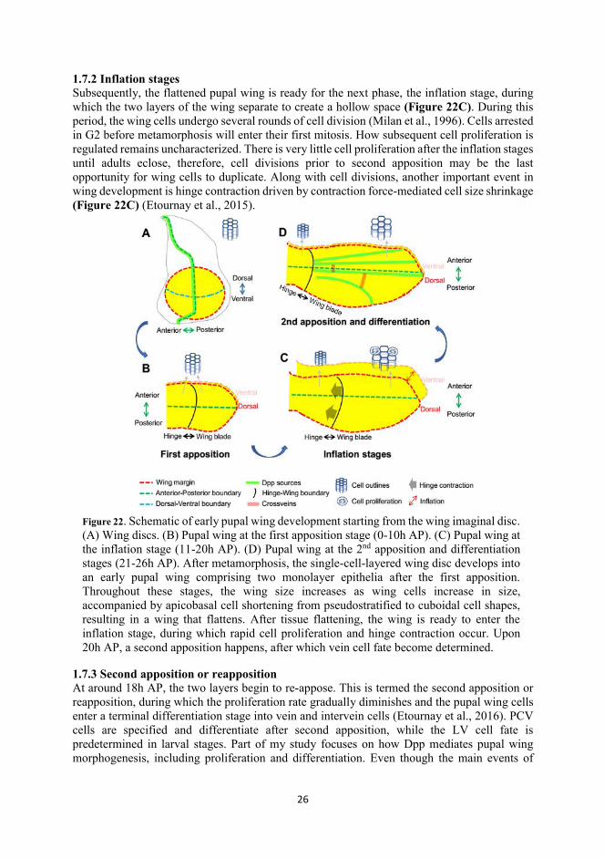
1.7.2 Inflation stages .................................................................................................................... 26
1.7.3 Second apposition or reapposition .................................................................................... 26
1.7.4 Vein cell differentiation ...................................................................................................... 27
1.8 Posterior crossvein (PCV) morphogenesis ................................................................................. 27
2. Aims of the study ............................................................................................................................. 29
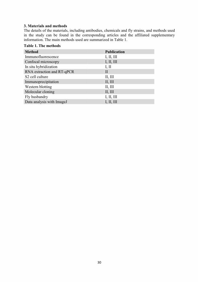
3. Materials and methods .................................................................................................................... 30
5
4. Summary of results and discussion .................................................................................................. 31
4.1 Dpp and pupal wing growth ..................................................................................................... 31
4.1.1 Dpp is indispensable for proper growth of the pupal wing ............................................ 31
4.1.2 Dpp promotes cell proliferation during inflation stages ................................................. 31
4.1.3 How does Dpp regulate cell proliferation in pupal wing?............................................... 31
4.1.4 What other factors are involved in proliferation regulation in addition to Dpp signaling? ...................................................................................................................................... 32
4.2 Dpp diffusibility in pupal stages ............................................................................................... 32
4.2.1 Dpp is diffusible before 18h AP ......................................................................................... 32
4.2.2 Lateral diffusion of Dpp is restricted and the signaling activity is refined to the future longitudinal veins after 18hAP ................................................................................................... 33
4.2.3 Dpp can diffuse vertically after 18hAP............................................................................. 34
4.3 Interplanar communication of Dpp signaling and pupal wing morphogenesis ................... 35
4.3.1 Interplanar communication is critical for Dpp signal refinement and proper wing patterning ..................................................................................................................................... 35
4.3.2 Refinement of the Dpp signal is initiated upon reapposition .......................................... 35
4.3.3 How is Dpp signal refinement achieved? .......................................................................... 36
4.3.4 Is the TKV expression pattern critical during Dpp signal refinement? ........................ 36
4.3.5 How is vertical diffusion achieved? ................................................................................... 37
4.4 Basolateral determinants in PCV cell differentiation ............................................................ 38
4.4.1 Basolateral determinants are indispensable for optimizing BMP/Dpp signal .............. 38
4.4.2 BMP/Dpp positively regulates the transcription of scrib and dlg1 ................................. 38
4.4.3 Endosome-based BMP/Dpp signal is indispensable for PCV formation ....................... 39
4.4.4 Scrib promotes endosome-based BMP/Dpp signaling activity ....................................... 39
4.4.5 Do Scrib, Dlg1 and Lgl regulate the BMP signaling and PCV morphogenesis in the same manner? .............................................................................................................................. 40
4.4.6 Physical interaction of Scrib, Tkv and Rab5 ................................................................... 40
4.4.7 Are autoregulatory mechanisms of Scrib present? ......................................................... 40
4.4.8 How do early endosomes boost Dpp signaling activity? ................................................. 41
4.5 The signaling divergence between Gbb and Scw .................................................................... 41
4.5.1 Scw harbors a unique N-glycosylation modification site ................................................ 41
4.5.2 N-glycosylation modifications of Scw in embryonic D-V patterning ............................. 41
4.5.3 N-glycosylation modifications of Scw in PCV formation ................................................ 41
4.5.4 What are mechanisms conferring the signaling divergences between Scw and Gbb? . 41
5. Final remarks and future perspectives .......................................................................................... 42
Acknowledgements .............................................................................................................................. 44
References............................................................................................................................................. 45

6
List of original publications: This thesis is based on the following manuscript and publications. The figures and content are referred to according to the Roman numerals. The articles are re-printed with the kind permission from the publishers. I Gui, J., Toddie-Moore, D., Huang, Y., Kracklauer, M., Kikushima, K., Nix, S., Ishimoto, Y., and Shimmi, O. Dynamic 3D structure directs BMP morphogen signaling during Drosophila wing morphogenesis. Unpublished manuscript. II Gui, J., Huang, Y., & Shimmi, O. (2016). Scribbled optimizes BMP signaling through its receptor internalization to the Rab5 endosome and promote robust epithelial morphogenesis. PLoS genetics, 12(11), e1006424. III Tauscher, P. M., Gui, J., & Shimmi, O. (2016). Adaptive protein divergence of BMP ligands takes place under developmental and evolutionary constraints. Development, 143(20), 3742-3750. The author’s contributions: I JG performed the experiments for Figures 1 b,c,e,f, Figure 2, Figure 3, Figures 4b-e, Figures s1 c-g, Figure s2e, Figure s3, Figure s4 II JG performed the experiments for all figures. JG and OS wrote the manuscript. III JG performed the experiments for Figure 4D and Figure S4.
7
Abstract In this thesis, I mainly investigate how BMP/Dpp signaling is involved in development of the early pupal wing of Drosophila, and the mechanisms coupling Dpp signaling with morphogenesis. There are many merits for early development of pupal wing to be an excellent models, e.g. drastic epithelial remodeling and intense proliferation, differentiation. While little is known about how these are regulated and how Dpp signaling is involved and coupled. Following the time course of pupal wing development, my study first unveils that laterally diffused Dpp promotes proliferation of pupal wing cells. Nonetheless, Dpp diffusion is tightly control since 18 hours AP (h AP), with sustained vertical diffusion (termed interplanar communication) and restricted lateral diffusion, leading to Dpp signal refinement in future vein cells. Lateral refinement of Dpp signaling is in accompany with reattachment (2nd apposition or reapposition) of dorsal and ventral wing layers. The experimental and computational data suggest that either impaired interplanar Dpp signaling or postponed wing reapposition compromises the Dpp signaling refinement and subsequent cell differentiation, proposing that epithelial architecture dynamics contributes to morphogenesis through BMP/Dpp signaling. Although lateral diffusion is restricted, Dpp can be transported into the future posterior crossvein (PCV) region and promotes PCV cell differentiation. The mechanisms underlying Dpp transport were well studied, but intrinsic mechanisms optimizing Dpp signaling during PCV cell differentiation are elusive. I characterize Scrib, a basolateral determinant, as an indispensable factor optimizing Dpp signaling by promoting the internalization of Tkv and Rab5-positive endosomal Dpp signaling during PCV morphogenesis. In the end, I set out to understand the molecular basis underlying the differential activities of Scw and Gbb, type5/6/7/8 BMPs in different developmental settings in Drosophila. My data suggest that the N-glycosylation modifications of Scw are indispensable for peak signaling during embryonic D-V patterning, while these modifications weaken signaling robustness during PCV formation. I conclude that the structural modifications by N-glycosylation and gain and loss of N-glycosylation of type-5/6/7/8 BMP ligands are adaptive to developmental and evolutionary constraints, respectively.
8
Abbreviations ada adaptor complex-2α AL apicolateral AJ adhererns junction aPKC atypical protein kinase C AP-2 Adaptor complex-2 A-P anterior-posterior BL basolateral BM basement membrane BMP Bone Morphogenetic Protein brk brinker CaTKV constitutively active Thick Veins Crb Crumb CT Carboxy-terminal domain ctrl control Cv Crossveinless Cv-2 Crossveinless-2 Cv-C Crossveinless-C DAD daughter against Dpp DAPI 4',6-diamidino-2-phenylindole DLG1 Disc Large 1 Dpp Decapentaplegic Ds Dachsous Dsh Dishehevelled D-V dorsal-ventral ECM extracellular matrix EMT epithelial to mesenchymal transition FGF fibroblast growth factor Fj Four-jointed FL full-length Fmi Flamingo Frz Frizzled Ft Fat GAG glycosaminoglycan Gbb Glass bottom boat GDP Guanosine-5'-biphosphate GFP Green fluorescent protein GTP Guanosine-5'-triphosphate h AP hours after pupariation HSPG Heparan Sulfate proteoglycan HS Heparan Sulfate IP immunoprecipitation KD knock down LGL Lethal(2) giant larvae protein
9
LRR leucine-rich repeats LVs longitudinal veins MAD Mothers against Dpp MARCM Mosaic analysis with a repressible cell marker MTs microtubules omb optomotor-blind Par3 partitioning-defective 3 Par6 partitioning-defective 6 PCV posterior cross-vein PDZ PSD-95, Dlg, ZO-1 homology PSD95 post synaptic density protein TJ tight junction pMAD phosphorylated-MAD Put Punt RAB5 Ras-related protein 5 RNA ribonucleic acid sal Spalt Sax Saxophone Scw Screw Scrib Scribble/Scribbled shRNA short hairpined RNA SJ septate junction SMAD Mothers against DPP homolog 3D three dimension Tkv Thick veins TGFβ Transforming growth factor beta Wg Wingless Wnt Wingless-related integration site wt wild type YFP yellow fluorescent protein ZO-1 zonula occludens-1
10
1. Review of articles In this thesis, I mainly studied the principles whereby epithelial morphogenesis and BMP/Dpp signaling are coupled, using the early pupal wing of Drosophila as a model system. In the first part of this literature review, I summarize the characteristics of epithelium, mainly, the properties of polarization, and how these vectoral characteristics may contribute to morphogenesis. Next, I discuss how developmental programs and local cell behavior contribute to epithelial patterning, morphogenesis and homeostasis. Thereafter, I introduce how BMP/Dpp, together with other factors, regulates development of the Drosophila wing imaginal disc and blastoderm embryo; additionally, I underscore the ‘facilitated transport’ mechanism which couples Dpp with dorsal-ventral (D-V) patterning during embryogenesis and posterior crossvein (PCV) formation in the pupal wing. Finally, I introduce developmental processes of the early pupal wing (before 26h AP) from wing imaginal disc, summarizing key aspects of the model system and introducing the questions pertinent to my studies. 1.1 The basics of epithelium
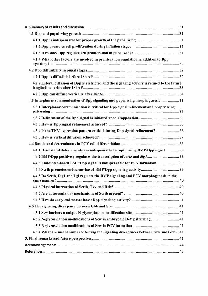
Epithelia is one of four tissue types, composing the epidemis and lining many organs. Epithelial cells are highly polarized, manifested by apicobasal and planar cell polarity, subcellular microdomains, and directional secretion and absorption. (Cao et al., 2012; Nusrat et al., 2000; Rodriguez-Boulan and Macara, 2014; Stoops and Caplan, 2014). The integration and dynamics of these vectoral cues is critical during epithelial development, morphogenesis and homeostasis (Rodriguez-Boulan and Macara, 2014), and compromising any of these properties may cause defective organogenesis and diseases, including cancer.
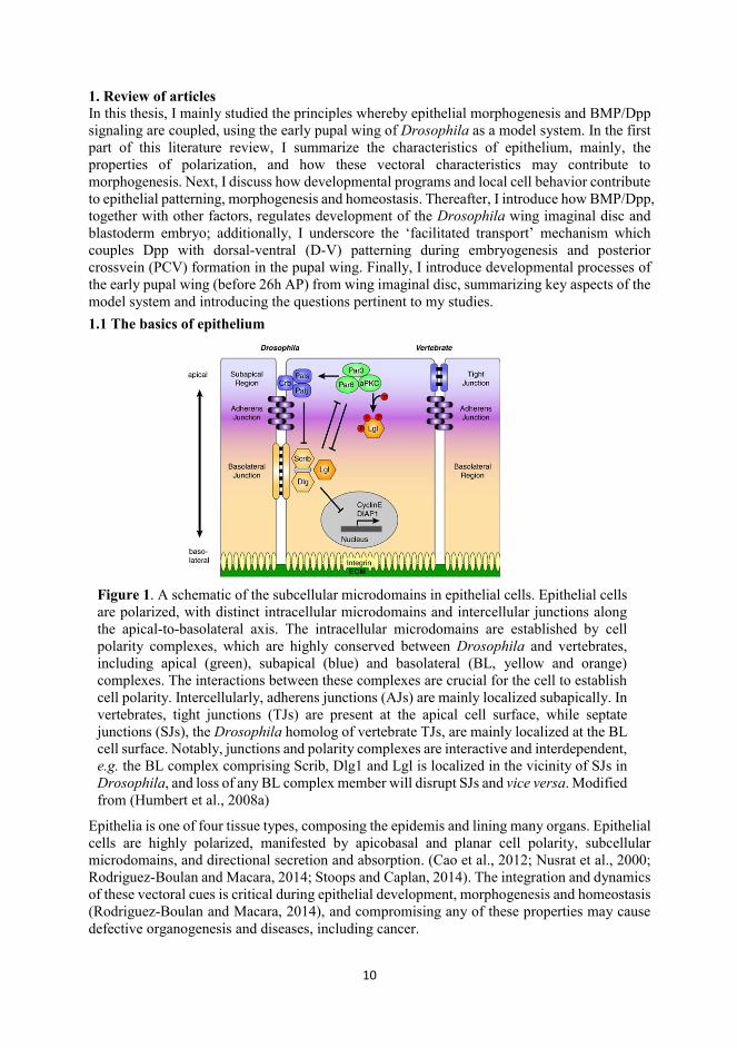
Figure 1. A schematic of the subcellular microdomains in epithelial cells. Epithelial cells are polarized, with distinct intracellular microdomains and intercellular junctions along the apical-to-basolateral axis. The intracellular microdomains are established by cell polarity complexes, which are highly conserved between Drosophila and vertebrates, including apical (green), subapical (blue) and basolateral (BL, yellow and orange) complexes. The interactions between these complexes are crucial for the cell to establish cell polarity. Intercellularly, adherens junctions (AJs) are mainly localized subapically. In vertebrates, tight junctions (TJs) are present at the apical cell surface, while septate junctions (SJs), the Drosophila homolog of vertebrate TJs, are mainly localized at the BL cell surface. Notably, junctions and polarity complexes are interactive and interdependent, e.g. the BL complex comprising Scrib, Dlg1 and Lgl is localized in the vicinity of SJs in Drosophila, and loss of any BL complex member will disrupt SJs and vice versa. Modified from (Humbert et al., 2008a)
11
1.1.1 Intercellular junctions Intercellular junctions play many important roles at various levels in epithelia. Intercellularly, they stitch individual cells together to form a collective sheet. Intracellularly, they are the prerequisites to establish and maintain cell polarity and microdomains. Moreover, junctions form a paracellular barrier to regulate permeability and defense from pathogens. As the main regulator of intercellular adhesion and communication, epithelial adherens junctions (AJs) comprise calcium-dependent E-Cadherin and Catenin family proteins, including β-Catenin (β-Cat) and α-Catenin (α-Cat). α-Cat, in turn, anchors the actin filaments to the AJs, enabling the AJs’ active involvement in mechanosensation and transduction (Drees et al., 2005; Jamora and Fuchs, 2002; Lecuit and Yap, 2015). In addition, AJs are reported to regulate a variety of signaling pathways, e.g. the Wnt signaling pathway, by titrating nuclear β-Cat (Nelson and Nusse, 2004). Distinct from AJs, tight junctions (TJs) act mainly to provide a paracellular barrier and to control paracellular transport (Chalcroft and Bullivant, 1970; Jamora and Fuchs, 2002). Note that septate junctions (SJs) in insects are homologous/analogous to TJs in vertebrates. Notably, AJs and TJs (or SJs) are localized at distinct but abutting subcellular microdomains at the cell surface (Figure 1), thus supporting cell polarization processes (Boggiano and Fehon, 2012; Humbert et al., 2008a; Ohno, 2001). 1.1.2 Apicobasal (AB) polarity Orthogonal to the planar cell axis, epithelial cells establish AB polarity. There are several main complexes involved in establishment and maintenance of AB polarity (Figure 1): the apical complex, consisting of Crumbs, PATJ and PALs, is localized at the apical cortex; apicolateral (AL) complex, which is subapical to the apical complex, is composed of aPKC, PAR3 and PAR6. In addition, Scrib, Dlg1 and Lgl constitute the basolateral (BL) complex (Roignot et al., 2013). The reciprocal but antagonistic interactions between these complexes form the biochemical basis of AB cell polarity. These complexes are highly conserved in both vertebrates and invertebrates.
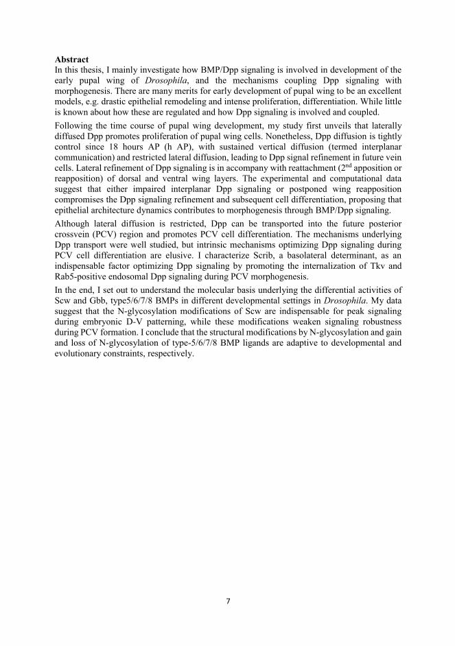
Among the most studied cell polarity molecules, Scrib, Dlg1 and Lgl were first identified in Drosophila imaginal discs as tumor suppressor genes (Azim et al., 1995; Bilder and Perrimon,
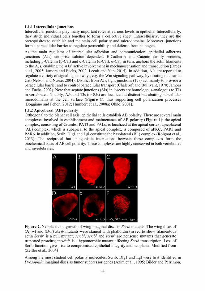
Figure 2. Neoplastic outgrowth of wing imaginal discs in Scrib mutants. The wing discs of (A) wt and (B-F) Scrib mutants were stained with phalloidin (in red to show filamentous actin Scrib2 is a null mutant; scrib3, scrib4 and scrib5 are nonsense mutants that generate truncated proteins; scribj7B3 is a hypomorphic mutant affecting Scrib transcription. Loss of Scrib function gives rise to compromised epithelial integrity and neoplasia. Modified from (Zeitler et al., 2004)
12
2000; Dow et al., 2003). The deletion of any of these genes leads to loss of cell polarity, disorganization of tissue structure and formation of neoplastic tumors in Drosophila imaginal discs (Figure 2) (Zeitler et al., 2004). The existing evidence demonstrates that scrib, dlg1 and lgl interact genetically in a multitude of processes in addition to growth control (Elsum et al., 2012; Humbert et al., 2003; Su et al., 2012). Recent studies suggest that BL complex participates in the endosomal pathway, regulating directional transport or secretion of cargo proteins (de Vreede et al., 2014; Gui et al., 2016; Walch, 2013), shedding light on their roles in signaling in addition to cell polarization. 1.1.3 Planar cell polarity (PCP)
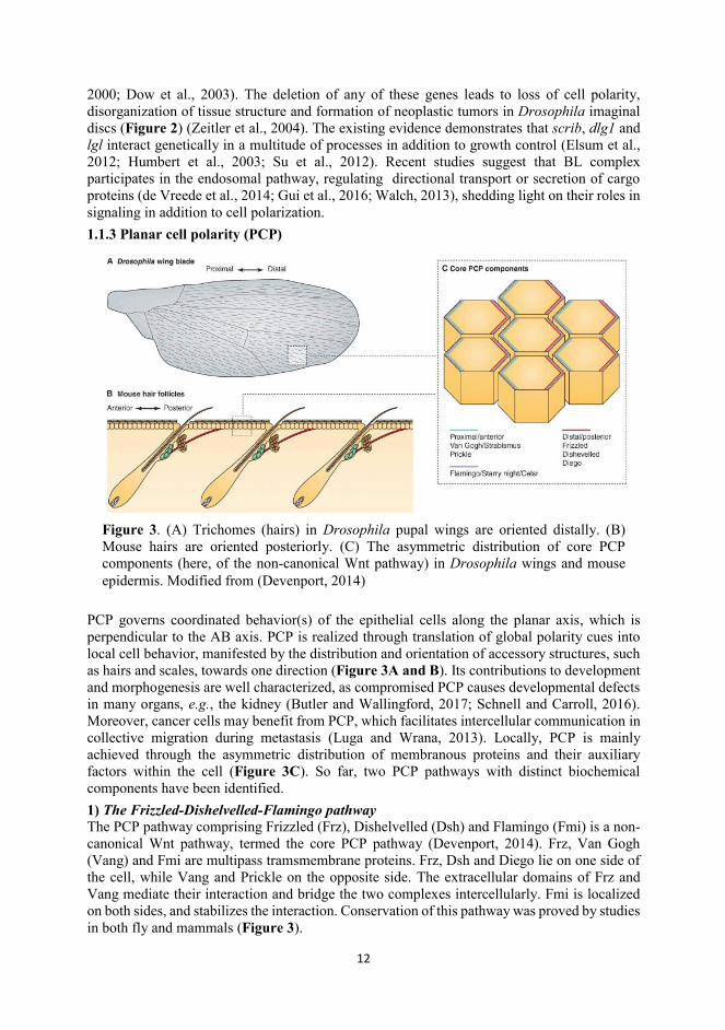
PCP governs coordinated behavior(s) of the epithelial cells along the planar axis, which is perpendicular to the AB axis. PCP is realized through translation of global polarity cues into local cell behavior, manifested by the distribution and orientation of accessory structures, such as hairs and scales, towards one direction (Figure 3A and B). Its contributions to development and morphogenesis are well characterized, as compromised PCP causes developmental defects in many organs, e.g., the kidney (Butler and Wallingford, 2017; Schnell and Carroll, 2016). Moreover, cancer cells may benefit from PCP, which facilitates intercellular communication in collective migration during metastasis (Luga and Wrana, 2013). Locally, PCP is mainly achieved through the asymmetric distribution of membranous proteins and their auxiliary factors within the cell (Figure 3C). So far, two PCP pathways with distinct biochemical components have been identified. 1) The Frizzled-Dishelvelled-Flamingo pathway The PCP pathway comprising Frizzled (Frz), Dishelvelled (Dsh) and Flamingo (Fmi) is a non-canonical Wnt pathway, termed the core PCP pathway (Devenport, 2014). Frz, Van Gogh (Vang) and Fmi are multipass tramsmembrane proteins. Frz, Dsh and Diego lie on one side of the cell, while Vang and Prickle on the opposite side. The extracellular domains of Frz and Vang mediate their interaction and bridge the two complexes intercellularly. Fmi is localized on both sides, and stabilizes the interaction. Conservation of this pathway was proved by studies in both fly and mammals (Figure 3).
Figure 3. (A) Trichomes (hairs) in Drosophila pupal wings are oriented distally. (B) Mouse hairs are oriented posteriorly. (C) The asymmetric distribution of core PCP components (here, of the non-canonical Wnt pathway) in Drosophila wings and mouse epidermis. Modified from (Devenport, 2014)
13
2) The Fat-Dachsous pathway Both the Frz-Dsh-Fmi pathway and the Fat-Dachsous (Ds) pathway are indispensable for proper PCP formation during the hair orientation in Drosophila wing, making it an excellent model to illustrate the genetic interactions between these two pathways (Figure 4) (Devenport, 2014). Fat and Ds are atypical Cadherins, interacting with the other, while Four-joints (Fj), another important component, is a Golgi-resident transmembrane kinase, which promotes Fat activity. In vertebrates, the conservation of this pathway remains questionable. While both molecular systems can establish PCP through asymmetric distribution of components, the causes of such asymmetry are distinct. In core PCP, the expression of components is relatively uniform, and biased Frz activity is the key to establish the asymmetry.
Thus, local expression of Wnt proteins, which are ligands for Frz, act upstream of the asymmetry (Wu et al., 2013). However, the asymmetry of Fat-Ds is achieved through the graded expression of Ds and Fj (Matakatsu and Blair, 2004). What might bethe mechanisms whereby these components are positioned? It has been reported that an acentrosomal MT array is polarized at the apices of cells, with MT(+) ends extending towards one side, to mediate the transport of PCP components to their destination so that symmetry can be broken (Matis et al., 2014; Shimada et al., 2006; Vladar et al., 2012). Thus, these acentrosomal MTs per se pre-establish PCP. Interestingly, previous studies suggest that polarized MTs are sufficient for PCP in some context (McCue et al., 2006). 1.1.4 Basement membrane (BM) and Extracellular matrix (ECM) Epithelial cells adhere to the BM and are anchored in a collection of secreted molecules termed the ECM, which is deposited in the BM. The ECM constitutes a large part of the microenvironment of epithelial cells. The composition of the ECM varies in different epithelial tissues. Physiologically, the ECM provides not only a physical support for the cells, but also biochemical and mechanical signals necessary for cell adhesion, differentiation, proliferation and migration. The main receptors mediating cell-ECM communication are integrin family members (Brown, 2000). In addition to signaling through integrins, some ECM molecules can function as coreceptors in signaling pathways. For instance, Collagen IV was reported to be the coreceptor of BMP2 (Wang et al., 2008). The Dystroglycan-Dystrophin complex, also bridging the ECM and the intracellular cytoskeleton (Chen et al., 2003), is involved in morphogenesis of mammalian skin and Drosophila wing (Christoforou et al., 2008; Sirour et al., 2011).
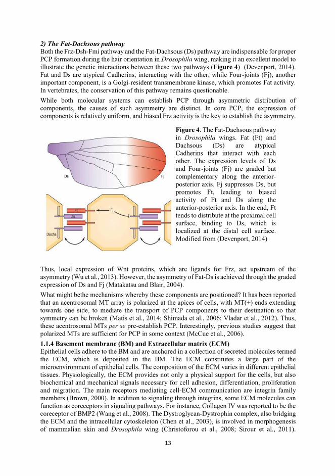
Figure 4. The Fat-Dachsous pathway in Drosophila wings. Fat (Ft) and Dachsous (Ds) are atypical Cadherins that interact with each other. The expression levels of Ds and Four-joints (Fj) are graded but complementary along the anterior-posterior axis. Fj suppresses Ds, but promotes Ft, leading to biased activity of Ft and Ds along the anterior-posterior axis. In the end, Ft tends to distribute at the proximal cell surface, binding to Ds, which is localized at the distal cell surface. Modified from (Devenport, 2014)
14
Moreover, some proteoglycans, such as Dally in fly, are involved in the distribution of growth factors, including fibroblast growth factors (FGFs) and BMPs (Matsuo and Kimura-Yoshida, 2014). Together, the ECM is actively involved in patterning and morphogenesis. 1.2 Developmental programs in epithelial morphogenesis 1.2.1 Cell proliferation Cell proliferation is a means to build up a tissue mass through duplication. The progression and rate of proliferation are tightly monitored and controlled. The division of epithelial cells is in
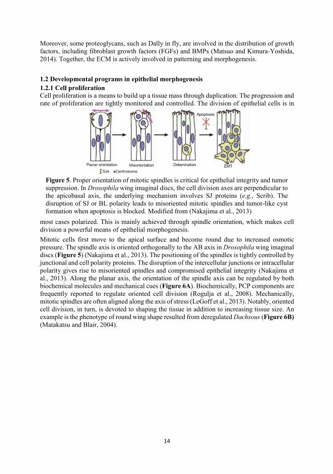
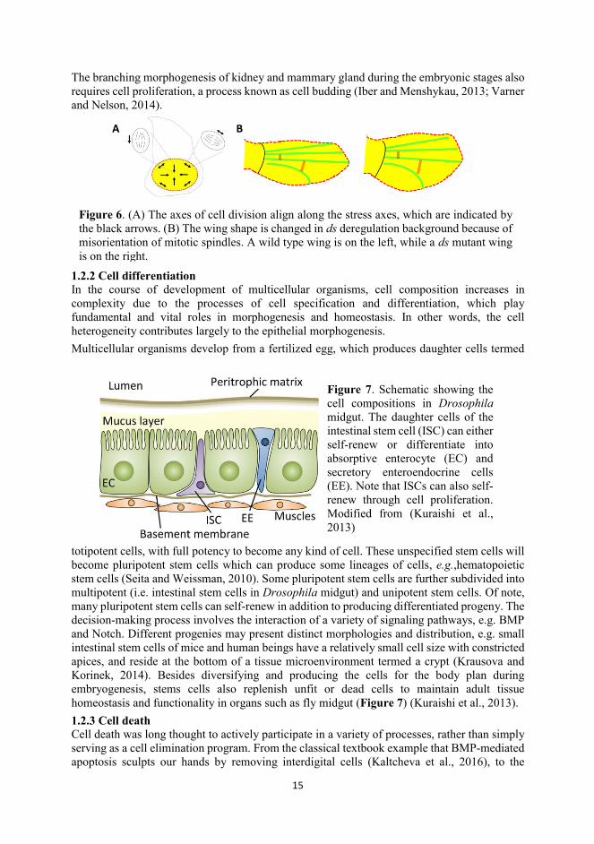
most cases polarized. This is mainly achieved through spindle orientation, which makes cell division a powerful means of epithelial morphogenesis. Mitotic cells first move to the apical surface and become round due to increased osmotic pressure. The spindle axis is oriented orthogonally to the AB axis in Drosophila wing imaginal discs (Figure 5) (Nakajima et al., 2013). The positioning of the spindles is tightly controlled by junctional and cell polarity proteins. The disruption of the intercellular junctions or intracellular polarity gives rise to misoriented spindles and compromised epithelial integrity (Nakajima et al., 2013). Along the planar axis, the orientation of the spindle axis can be regulated by both biochemical molecules and mechanical cues (Figure 6A). Biochemically, PCP components are frequently reported to regulate oriented cell division (Rogulja et al., 2008). Mechanically, mitotic spindles are often aligned along the axis of stress (LeGoff et al., 2013). Notably, oriented cell division, in turn, is devoted to shaping the tissue in addition to increasing tissue size. An example is the phenotype of round wing shape resulted from deregulated Dachsous (Figure 6B) (Matakatsu and Blair, 2004).
Figure 5. Proper orientation of mitotic spindles is critical for epithelial integrity and tumor suppression. In Drosophila wing imaginal discs, the cell division axes are perpendicular to the apicobasal axis, the underlying mechanism involves SJ proteins (e.g., Scrib). The disruption of SJ or BL polarity leads to misoriented mitotic spindles and tumor-like cyst formation when apoptosis is blocked. Modified from (Nakajima et al., 2013)
15
The branching morphogenesis of kidney and mammary gland during the embryonic stages also requires cell proliferation, a process known as cell budding (Iber and Menshykau, 2013; Varner and Nelson, 2014).
1.2.2 Cell differentiation In the course of development of multicellular organisms, cell composition increases in complexity due to the processes of cell specification and differentiation, which play fundamental and vital roles in morphogenesis and homeostasis. In other words, the cell heterogeneity contributes largely to the epithelial morphogenesis. Multicellular organisms develop from a fertilized egg, which produces daughter cells termed
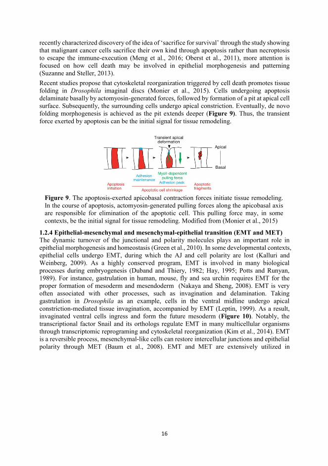
totipotent cells, with full potency to become any kind of cell. These unspecified stem cells will become pluripotent stem cells which can produce some lineages of cells, e.g.,hematopoietic stem cells (Seita and Weissman, 2010). Some pluripotent stem cells are further subdivided into multipotent (i.e. intestinal stem cells in Drosophila midgut) and unipotent stem cells. Of note, many pluripotent stem cells can self-renew in addition to producing differentiated progeny. The decision-making process involves the interaction of a variety of signaling pathways, e.g. BMP and Notch. Different progenies may present distinct morphologies and distribution, e.g. small intestinal stem cells of mice and human beings have a relatively small cell size with constricted apices, and reside at the bottom of a tissue microenvironment termed a crypt (Krausova and Korinek, 2014). Besides diversifying and producing the cells for the body plan during embryogenesis, stems cells also replenish unfit or dead cells to maintain adult tissue homeostasis and functionality in organs such as fly midgut (Figure 7) (Kuraishi et al., 2013). 1.2.3 Cell death Cell death was long thought to actively participate in a variety of processes, rather than simply serving as a cell elimination program. From the classical textbook example that BMP-mediated apoptosis sculpts our hands by removing interdigital cells (Kaltcheva et al., 2016), to the
Figure 7. Schematic showing the cell compositions in Drosophila midgut. The daughter cells of the intestinal stem cell (ISC) can either self-renew or differentiate into absorptive enterocyte (EC) and secretory enteroendocrine cells (EE). Note that ISCs can also self-renew through cell proliferation. Modified from (Kuraishi et al., 2013)
Figure 6. (A) The axes of cell division align along the stress axes, which are indicated by the black arrows. (B) The wing shape is changed in ds deregulation background because of misorientation of mitotic spindles. A wild type wing is on the left, while a ds mutant wing is on the right.
A B
16
recently characterized discovery of the idea of ‘sacrifice for survival’ through the study showing that malignant cancer cells sacrifice their own kind through apoptosis rather than necroptosis to escape the immune-execution (Meng et al., 2016; Oberst et al., 2011), more attention is focused on how cell death may be involved in epithelial morphogenesis and patterning (Suzanne and Steller, 2013). Recent studies propose that cytoskeletal reorganization triggered by cell death promotes tissue folding in Drosophila imaginal discs (Monier et al., 2015). Cells undergoing apoptosis delaminate basally by actomyosin-generated forces, followed by formation of a pit at apical cell surface. Subsequently, the surrounding cells undergo apical constriction. Eventually, de novo folding morphogenesis is achieved as the pit extends deeper (Figure 9). Thus, the transient force exerted by apoptosis can be the initial signal for tissue remodeling.
1.2.4 Epithelial-mesenchymal and mesenchymal-epithelial transition (EMT and MET) The dynamic turnover of the junctional and polarity molecules plays an important role in epithelial morphogenesis and homeostasis (Green et al., 2010). In some developmental contexts, epithelial cells undergo EMT, during which the AJ and cell polarity are lost (Kalluri and Weinberg, 2009). As a highly conserved program, EMT is involved in many biological processes during embryogenesis (Duband and Thiery, 1982; Hay, 1995; Potts and Runyan, 1989). For instance, gastrulation in human, mouse, fly and sea urchin requires EMT for the proper formation of mesoderm and mesendoderm (Nakaya and Sheng, 2008). EMT is very often associated with other processes, such as invagination and delamination. Taking gastrulation in Drosophila as an example, cells in the ventral midline undergo apical constriction-mediated tissue invagination, accompanied by EMT (Leptin, 1999). As a result, invaginated ventral cells ingress and form the future mesoderm (Figure 10). Notably, the transcriptional factor Snail and its orthologs regulate EMT in many multicellular organisms through transcriptomic reprograming and cytoskeletal reorganization (Kim et al., 2014). EMT is a reversible process, mesenchymal-like cells can restore intercellular junctions and epithelial polarity through MET (Baum et al., 2008). EMT and MET are extensively utilized in
Figure 9. The apoptosis-exerted apicobasal contraction forces initiate tissue remodeling. In the course of apoptosis, actomyosin-generated pulling forces along the apicobasal axis are responsible for elimination of the apoptotic cell. This pulling force may, in some contexts, be the initial signal for tissue remodeling. Modified from (Monier et al., 2015)
17
organogenesis and secondary tumor formation (Baum et al., 2008; Kreidberg et al., 1993; Nakajima et al., 2000). 1.3 From local cell morphogenesis to global patterning Global tissue patterning is achieved by local behavior of individual cells collectively. The epithelium progresses from the originally simple flat sheet into a tissue with more complex 3D architecture after a series of morphogenetic elaborations. Epithelial cells contribute to such
morphogenetic changes mainly through shape changes, intercalation and migration (Pilot and Lecuit, 2005). 1.3.1 Cell shape changes
Figure 10. Schematic showing mesoderm formation in Drosophila embryo. All illustrations represent cross-sections in the middle along anterior-posterior axis In the top panel, the ventral midline cells constrict apically, and this apical constriction drives ventral furrow formation and invagination. In the middle panel, cells have invaginated; in the bottom panel, invaginated ventral cells undergo EMT and become mesodermal cells. Modified from (Leptin, 1999)
Figure 11. Illustrations showing different types of cell shape (the bottom panel) and arrangement of layers (the upper panel). In epithelia, there are mainly three types of cell shape: squamous, cuboidal and columnar. Epithelial cells can be arranged into single-layered or multi-layered epithelium. Single-layered epithelium can be organized into either a simple or the pseudostratified model. Modified from http://vle.du.ac.in/mod/book/view.php?id=11580&chapterid=22238
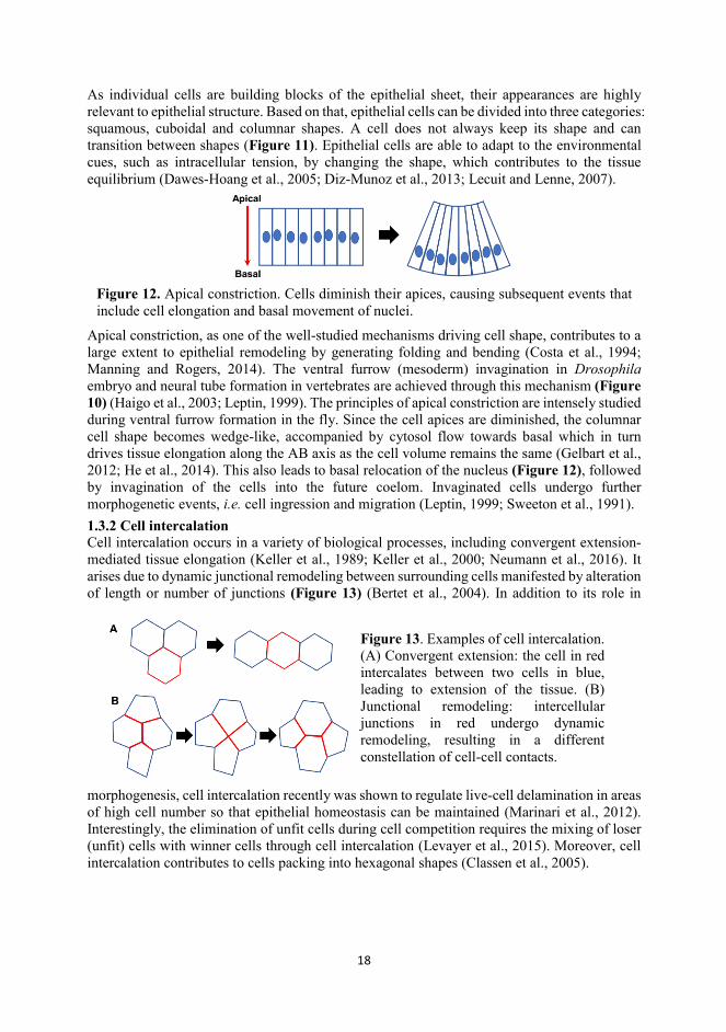
18
As individual cells are building blocks of the epithelial sheet, their appearances are highly relevant to epithelial structure. Based on that, epithelial cells can be divided into three categories: squamous, cuboidal and columnar shapes. A cell does not always keep its shape and can transition between shapes (Figure 11). Epithelial cells are able to adapt to the environmental cues, such as intracellular tension, by changing the shape, which contributes to the tissue equilibrium (Dawes-Hoang et al., 2005; Diz-Munoz et al., 2013; Lecuit and Lenne, 2007).
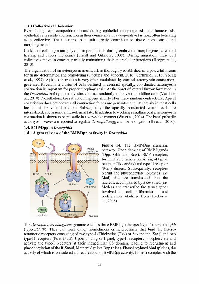
Apical constriction, as one of the well-studied mechanisms driving cell shape, contributes to a large extent to epithelial remodeling by generating folding and bending (Costa et al., 1994; Manning and Rogers, 2014). The ventral furrow (mesoderm) invagination in Drosophila embryo and neural tube formation in vertebrates are achieved through this mechanism (Figure 10) (Haigo et al., 2003; Leptin, 1999). The principles of apical constriction are intensely studied during ventral furrow formation in the fly. Since the cell apices are diminished, the columnar cell shape becomes wedge-like, accompanied by cytosol flow towards basal which in turn drives tissue elongation along the AB axis as the cell volume remains the same (Gelbart et al., 2012; He et al., 2014). This also leads to basal relocation of the nucleus (Figure 12), followed by invagination of the cells into the future coelom. Invaginated cells undergo further morphogenetic events, i.e. cell ingression and migration (Leptin, 1999; Sweeton et al., 1991). 1.3.2 Cell intercalation Cell intercalation occurs in a variety of biological processes, including convergent extension-mediated tissue elongation (Keller et al., 1989; Keller et al., 2000; Neumann et al., 2016). It arises due to dynamic junctional remodeling between surrounding cells manifested by alteration of length or number of junctions (Figure 13) (Bertet et al., 2004). In addition to its role in
morphogenesis, cell intercalation recently was shown to regulate live-cell delamination in areas of high cell number so that epithelial homeostasis can be maintained (Marinari et al., 2012). Interestingly, the elimination of unfit cells during cell competition requires the mixing of loser (unfit) cells with winner cells through cell intercalation (Levayer et al., 2015). Moreover, cell intercalation contributes to cells packing into hexagonal shapes (Classen et al., 2005).
Figure 12. Apical constriction. Cells diminish their apices, causing subsequent events that include cell elongation and basal movement of nuclei.
Figure 13. Examples of cell intercalation. (A) Convergent extension: the cell in red intercalates between two cells in blue, leading to extension of the tissue. (B) Junctional remodeling: intercellular junctions in red undergo dynamic remodeling, resulting in a different constellation of cell-cell contacts.
19
1.3.3 Collective cell behavior Even though cell competition occurs during epithelial morphogenesis and homeostasis, epithelial cells reside and function in their community in a cooperative fashion, often behaving as a collective. Their actions as a unit largely contribute to tissue homeostasis and morphogenesis. Collective cell migration plays an important role during embryonic morphogenesis, wound healing and cancer metastasis (Friedl and Gilmour, 2009). During migration, these cell collectives move in concert, partially maintaining their intercellular junctions (Haeger et al., 2015). The organization of an actomyosin meshwork is thoroughly established as a powerful means for tissue deformation and remodeling (Ducuing and Vincent, 2016; Gorfinkiel, 2016; Young et al., 1993). Apical constriction is very often modulated by cortical actomyosin contraction-generated forces. In a cluster of cells destined to contract apically, coordinated actomyosin contraction is important for proper morphogenesis. At the onset of ventral furrow formation in the Drosophila embryo, actomyosins contract randomly in the ventral midline cells (Martin et al., 2010). Nonetheless, the retraction happens shortly after these random contractions. Apical constriction does not occur until contraction forces are generated simultaneously in most cells located at the ventral midline. Subsequently, the apically constricted ventral cells are internalized, and assume a mesodermal fate. In addition to working simultaneously, actomyosin contraction is shown to be pulsatile in a wave-like manner (Wu et al., 2014). The basal pulsatile actomyosin waves are reported to regulate Drosophila egg chamber elongation (He et al., 2010). 1.4. BMP/Dpp in Drosophila 1.4.1 A general view of the BMP/Dpp pathway in Drosophila
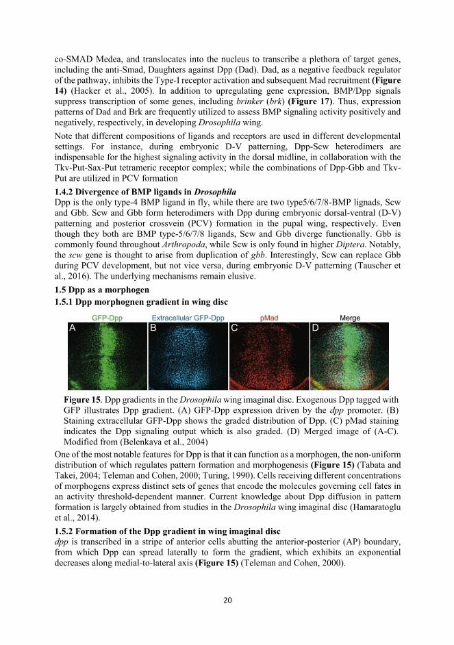
The Drosophila melanogaster genome encodes three BMP ligands: dpp (type-4), scw, and gbb (type-5/6/7/8). They can form either homodimers or heterodimers that bind the hetero-tetrameric receptors consisting of two type-I (Thickveins (Tkv) or Saxophone (Sax)) and two type-II receptors (Punt (Put)). Upon binding of ligand, type-II receptors phosphorylate and activate the type-I receptors at their intracellular GS domain, leading to recruitment and phosphorylation of the R-Smad, Mothers Against Dpp (Mad). Phosphorylated Mad (pMad), the activity of which is considered a direct readout of BMP/Dpp activity, forms a complex with the
Figure 14. The BMP/Dpp signaling pathway. Upon docking of BMP ligands (Dpp, Gbb and Scw), BMP receptors form heterotetramers consisting of type-I receptor (Tkv or Sax) and type-II receptor (Punt) dimers. Subsequently, receptors recruit and phosphorylate R-Smads (i.e. Mad) that are translocated into the nucleus, accompanied by a co-Smad (i.e. Medea) and transcribe the target genes involved in cell differentiation and proliferation. Modified from (Hacker et al., 2005)
20
co-SMAD Medea, and translocates into the nucleus to transcribe a plethora of target genes, including the anti-Smad, Daughters against Dpp (Dad). Dad, as a negative feedback regulator of the pathway, inhibits the Type-I receptor activation and subsequent Mad recruitment (Figure 14) (Hacker et al., 2005). In addition to upregulating gene expression, BMP/Dpp signals suppress transcription of some genes, including brinker (brk) (Figure 17). Thus, expression patterns of Dad and Brk are frequently utilized to assess BMP signaling activity positively and negatively, respectively, in developing Drosophila wing. Note that different compositions of ligands and receptors are used in different developmental settings. For instance, during embryonic D-V patterning, Dpp-Scw heterodimers are indispensable for the highest signaling activity in the dorsal midline, in collaboration with the Tkv-Put-Sax-Put tetrameric receptor complex; while the combinations of Dpp-Gbb and Tkv-Put are utilized in PCV formation 1.4.2 Divergence of BMP ligands in Drosophila Dpp is the only type-4 BMP ligand in fly, while there are two type5/6/7/8-BMP lignads, Scw and Gbb. Scw and Gbb form heterodimers with Dpp during embryonic dorsal-ventral (D-V) patterning and posterior crossvein (PCV) formation in the pupal wing, respectively. Even though they both are BMP type-5/6/7/8 ligands, Scw and Gbb diverge functionally. Gbb is commonly found throughout Arthropoda, while Scw is only found in higher Diptera. Notably, the scw gene is thought to arise from duplication of gbb. Interestingly, Scw can replace Gbb during PCV development, but not vice versa, during embryonic D-V patterning (Tauscher et al., 2016). The underlying mechanisms remain elusive. 1.5 Dpp as a morphogen 1.5.1 Dpp morphognen gradient in wing disc
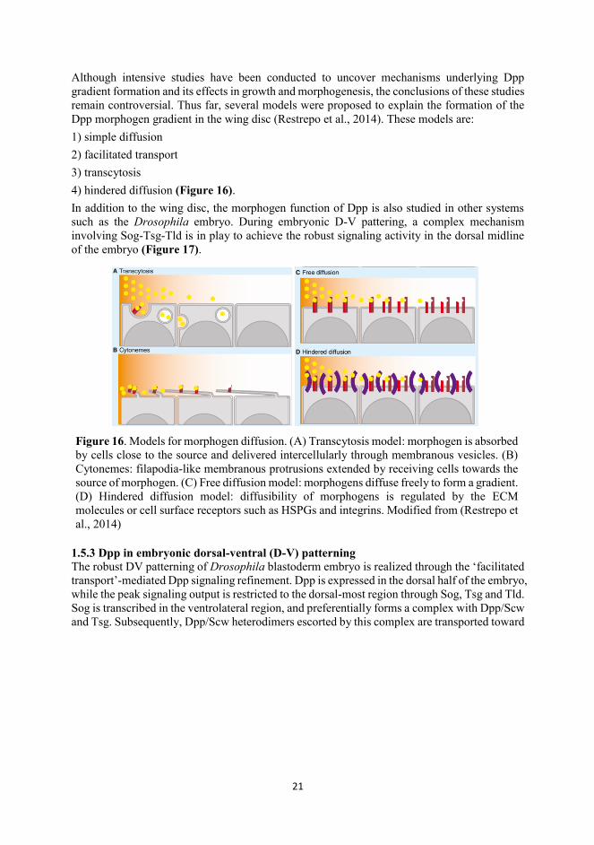
One of the most notable features for Dpp is that it can function as a morphogen, the non-uniform distribution of which regulates pattern formation and morphogenesis (Figure 15) (Tabata and Takei, 2004; Teleman and Cohen, 2000; Turing, 1990). Cells receiving different concentrations of morphogens express distinct sets of genes that encode the molecules governing cell fates in an activity threshold-dependent manner. Current knowledge about Dpp diffusion in pattern formation is largely obtained from studies in the Drosophila wing imaginal disc (Hamaratoglu et al., 2014). 1.5.2 Formation of the Dpp gradient in wing imaginal disc dpp is transcribed in a stripe of anterior cells abutting the anterior-posterior (AP) boundary, from which Dpp can spread laterally to form the gradient, which exhibits an exponential decreases along medial-to-lateral axis (Figure 15) (Teleman and Cohen, 2000).
Figure 15. Dpp gradients in the Drosophila wing imaginal disc. Exogenous Dpp tagged with GFP illustrates Dpp gradient. (A) GFP-Dpp expression driven by the dpp promoter. (B) Staining extracellular GFP-Dpp shows the graded distribution of Dpp. (C) pMad staining indicates the Dpp signaling output which is also graded. (D) Merged image of (A-C). Modified from (Belenkaya et al., 2004)
21
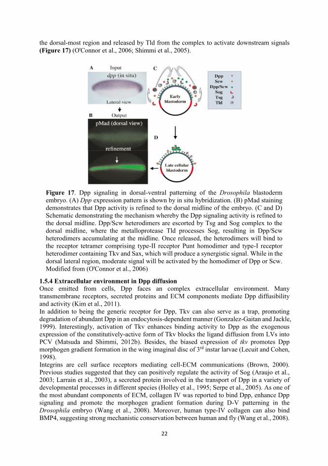
Although intensive studies have been conducted to uncover mechanisms underlying Dpp gradient formation and its effects in growth and morphogenesis, the conclusions of these studies remain controversial. Thus far, several models were proposed to explain the formation of the Dpp morphogen gradient in the wing disc (Restrepo et al., 2014). These models are: 1) simple diffusion 2) facilitated transport 3) transcytosis 4) hindered diffusion (Figure 16). In addition to the wing disc, the morphogen function of Dpp is also studied in other systems such as the Drosophila embryo. During embryonic D-V pattering, a complex mechanism involving Sog-Tsg-Tld is in play to achieve the robust signaling activity in the dorsal midline of the embryo (Figure 17).
1.5.3 Dpp in embryonic dorsal-ventral (D-V) patterning The robust DV patterning of Drosophila blastoderm embryo is realized through the ‘facilitated transport’-mediated Dpp signaling refinement. Dpp is expressed in the dorsal half of the embryo, while the peak signaling output is restricted to the dorsal-most region through Sog, Tsg and Tld. Sog is transcribed in the ventrolateral region, and preferentially forms a complex with Dpp/Scw and Tsg. Subsequently, Dpp/Scw heterodimers escorted by this complex are transported toward
Figure 16. Models for morphogen diffusion. (A) Transcytosis model: morphogen is absorbed by cells close to the source and delivered intercellularly through membranous vesicles. (B) Cytonemes: filapodia-like membranous protrusions extended by receiving cells towards the source of morphogen. (C) Free diffusion model: morphogens diffuse freely to form a gradient. (D) Hindered diffusion model: diffusibility of morphogens is regulated by the ECM molecules or cell surface receptors such as HSPGs and integrins. Modified from (Restrepo et al., 2014)
22
the dorsal-most region and released by Tld from the complex to activate downstream signals (Figure 17) (O'Connor et al., 2006; Shimmi et al., 2005).
1.5.4 Extracellular environment in Dpp diffusion Once emitted from cells, Dpp faces an complex extracellular environment. Many transmembrane receptors, secreted proteins and ECM components mediate Dpp diffusibility and activity (Kim et al., 2011). In addition to being the generic receptor for Dpp, Tkv can also serve as a trap, promoting degradation of abundant Dpp in an endocytosis-dependent manner (Gonzalez-Gaitan and Jackle, 1999). Interestingly, activation of Tkv enhances binding activity to Dpp as the exogenous expression of the constitutively-active form of Tkv blocks the ligand diffusion from LVs into PCV (Matsuda and Shimmi, 2012b). Besides, the biased expression of tkv promotes Dpp morphogen gradient formation in the wing imaginal disc of 3rd instar larvae (Lecuit and Cohen, 1998). Integrins are cell surface receptors mediating cell-ECM communications (Brown, 2000). Previous studies suggested that they can positively regulate the activity of Sog (Araujo et al., 2003; Larrain et al., 2003), a secreted protein involved in the transport of Dpp in a variety of developmental processes in different species (Holley et al., 1995; Serpe et al., 2005). As one of the most abundant components of ECM, collagen IV was reported to bind Dpp, enhance Dpp signaling and promote the morphogen gradient formation during D-V patterning in the Drosophila embryo (Wang et al., 2008). Moreover, human type-IV collagen can also bind BMP4, suggesting strong mechanistic conservation between human and fly (Wang et al., 2008).
Figure 17. Dpp signaling in dorsal-ventral patterning of the Drosophila blastoderm embryo. (A) Dpp expression pattern is shown by in situ hybridization. (B) pMad staining demonstrates that Dpp activity is refined to the dorsal midline of the embryo. (C and D) Schematic demonstrating the mechanism whereby the Dpp signaling activity is refined to the dorsal midline. Dpp/Scw heterodimers are escorted by Tsg and Sog complex to the dorsal midline, where the metalloprotease Tld processes Sog, resulting in Dpp/Scw heterodimers accumulating at the midline. Once released, the heterodimers will bind to the receptor tetramer comprising type-II receptor Punt homodimer and type-I receptor heterodimer containing Tkv and Sax, which will produce a synergistic signal. While in the dorsal lateral region, moderate signal will be activated by the homodimer of Dpp or Scw. Modified from (O'Connor et al., 2006)
23
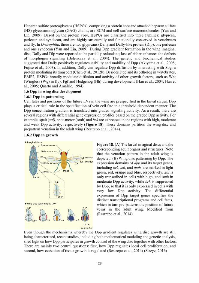
Heparan sulfate proteoglycans (HSPGs), comprising a protein core and attached heparan sulfate (HS) glycosaminoglycan (GAG) chains, are ECM and cell surface macromolecules (Yan and Lin, 2009). Based on the protein core, HSPGs are classified into three families: glypican, perlecan and syndecan, and are highly structurally and functionally conserved in vertebrates and fly. In Drosophila, there are two glypicans (Dally and Dally-like protein (Dlp), one perlecan and one syndecan (Yan and Lin, 2009). During Dpp gradient formation in the wing imaginal disc, Dally and Dlp were reported to be partially redundant; loss of either enhances the defects of morphogen signaling (Belenkaya et al., 2004). The genetic and biochemical studies suggested that Dally positively regulates stability and mobility of Dpp (Akiyama et al., 2008; Fujise et al., 2003). In addition, Dally can regulate Dpp diffusion by interacting with Sog, a protein mediating its transport (Chen et al., 2012b). Besides Dpp and its ortholog in vertebrates, BMP2, HSPGs broadly modulate diffusion and activity of other growth factors, such as Wnt (Wingless (Wg) in fly), Fgf and Hedgehog (Hh) during development (Han et al., 2004; Han et al., 2005; Quarto and Amalric, 1994). 1.6 Dpp in wing disc development 1.6.1 Dpp in patterning Cell fates and positions of the future LVs in the wing are prespecified in the larval stages. Dpp plays a critical role in the specification of vein cell fate in a threshold-dependent manner. The Dpp concentration gradient is translated into graded signaling activity. As a result, there are several regions with differential gene expression profiles based on the graded Dpp activity. For example, spalt (sal), spot-motor (omb) and brk are expressed in the regions with high, moderate and weak Dpp activity, respectively (Figure 18). These domains partition the wing disc and prepattern venation in the adult wing (Restrepo et al., 2014). 1.6.2 Dpp in growth
Even though the mechanisms whereby the Dpp gradient regulates wing disc growth are still being characterized, recent studies, including both mathematical modeling and genetic analysis, shed light on how Dpp participates in growth control of the wing disc together with other factors. There are mainly two central questions: first, how Dpp regulates local cell proliferation, and second, how cessation of tissue growth is regulated (Restrepo et al., 2014) (Strzyz, 2016)
Figure 18. (A) The larval imaginal discs and the corresponding adult organs and structures. Note that the venation pattern in the adult wing is depicted. (B) Wing disc patterning by Dpp. The expression domains of dpp and its target genes, including brk, sal, and omb, are marked in light green, red, orange and blue, respectively. Sal is only transcribed in cells with high, and omb in moderate Dpp activity, while brk is suppressed by Dpp, so that it is only expressed in cells with very low Dpp activity. The differential expression of Dpp target genes specifies the distinct transcriptional programs and cell fates, which in turn pre-patterns the position of future veins in the adult wing. Modified from (Restrepo et al., 2014)
24
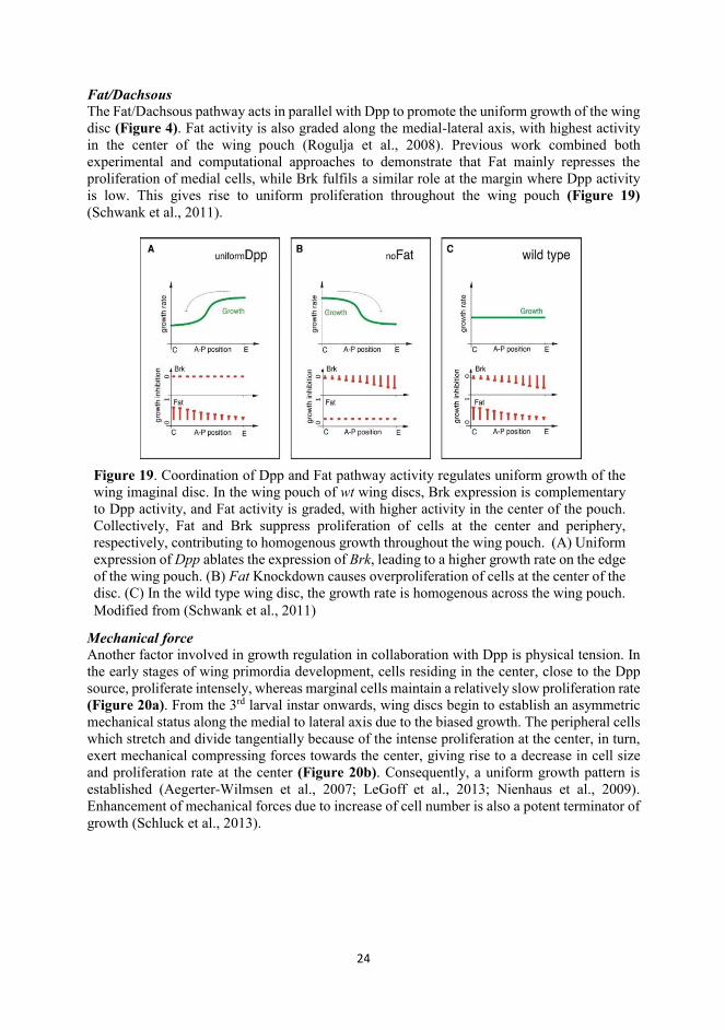
Fat/Dachsous The Fat/Dachsous pathway acts in parallel with Dpp to promote the uniform growth of the wing disc (Figure 4). Fat activity is also graded along the medial-lateral axis, with highest activity in the center of the wing pouch (Rogulja et al., 2008). Previous work combined both experimental and computational approaches to demonstrate that Fat mainly represses the proliferation of medial cells, while Brk fulfils a similar role at the margin where Dpp activity is low. This gives rise to uniform proliferation throughout the wing pouch (Figure 19) (Schwank et al., 2011).
Mechanical force Another factor involved in growth regulation in collaboration with Dpp is physical tension. In the early stages of wing primordia development, cells residing in the center, close to the Dpp source, proliferate intensely, whereas marginal cells maintain a relatively slow proliferation rate (Figure 20a). From the 3rd larval instar onwards, wing discs begin to establish an asymmetric mechanical status along the medial to lateral axis due to the biased growth. The peripheral cells which stretch and divide tangentially because of the intense proliferation at the center, in turn, exert mechanical compressing forces towards the center, giving rise to a decrease in cell size and proliferation rate at the center (Figure 20b). Consequently, a uniform growth pattern is established (Aegerter-Wilmsen et al., 2007; LeGoff et al., 2013; Nienhaus et al., 2009). Enhancement of mechanical forces due to increase of cell number is also a potent terminator of growth (Schluck et al., 2013).
Figure 19. Coordination of Dpp and Fat pathway activity regulates uniform growth of the wing imaginal disc. In the wing pouch of wt wing discs, Brk expression is complementary to Dpp activity, and Fat activity is graded, with higher activity in the center of the pouch. Collectively, Fat and Brk suppress proliferation of cells at the center and periphery, respectively, contributing to homogenous growth throughout the wing pouch. (A) Uniform expression of Dpp ablates the expression of Brk, leading to a higher growth rate on the edge of the wing pouch. (B) Fat Knockdown causes overproliferation of cells at the center of the disc. (C) In the wild type wing disc, the growth rate is homogenous across the wing pouch. Modified from (Schwank et al., 2011)
25
1.7 Metamorphosis from wing imaginal disc to pupal wing After metamorphosis, intense remodeling at cellular and structural levels occur in becoming the pupal wing, which eventually assumes a morphology very distinct from its larval anlage. How Dpp signal is involved and coupled during this extensive tissue architecture reorganization is
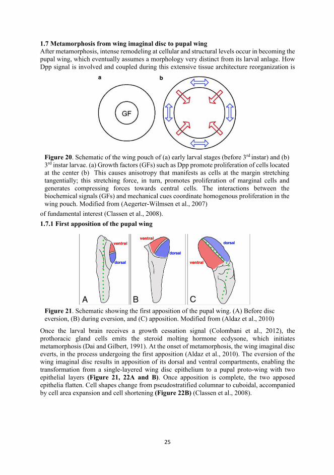
of fundamental interest (Classen et al., 2008). 1.7.1 First apposition of the pupal wing
Once the larval brain receives a growth cessation signal (Colombani et al., 2012), the prothoracic gland cells emits the steroid molting hormone ecdysone, which initiates metamorphosis (Dai and Gilbert, 1991). At the onset of metamorphosis, the wing imaginal disc everts, in the process undergoing the first apposition (Aldaz et al., 2010). The eversion of the wing imaginal disc results in apposition of its dorsal and ventral compartments, enabling the transformation from a single-layered wing disc epithelium to a pupal proto-wing with two epithelial layers (Figure 21, 22A and B). Once apposition is complete, the two apposed epithelia flatten. Cell shapes change from pseudostratified columnar to cuboidal, accompanied by cell area expansion and cell shortening (Figure 22B) (Classen et al., 2008).
Figure 20. Schematic of the wing pouch of (a) early larval stages (before 3rd instar) and (b) 3rd instar larvae. (a) Growth factors (GFs) such as Dpp promote proliferation of cells located at the center (b) This causes anisotropy that manifests as cells at the margin stretching tangentially; this stretching force, in turn, promotes proliferation of marginal cells and generates compressing forces towards central cells. The interactions between the biochemical signals (GFs) and mechanical cues coordinate homogenous proliferation in the wing pouch. Modified from (Aegerter-Wilmsen et al., 2007)
Figure 21. Schematic showing the first apposition of the pupal wing. (A) Before disc eversion, (B) during eversion, and (C) apposition. Modified from (Aldaz et al., 2010)
26
1.7.2 Inflation stages Subsequently, the flattened pupal wing is ready for the next phase, the inflation stage, during which the two layers of the wing separate to create a hollow space (Figure 22C). During this period, the wing cells undergo several rounds of cell division (Milan et al., 1996). Cells arrested in G2 before metamorphosis will enter their first mitosis. How subsequent cell proliferation is regulated remains uncharacterized. There is very little cell proliferation after the inflation stages until adults eclose, therefore, cell divisions prior to second apposition may be the last opportunity for wing cells to duplicate. Along with cell divisions, another important event in wing development is hinge contraction driven by contraction force-mediated cell size shrinkage (Figure 22C) (Etournay et al., 2015).
1.7.3 Second apposition or reapposition At around 18h AP, the two layers begin to re-appose. This is termed the second apposition or reapposition, during which the proliferation rate gradually diminishes and the pupal wing cells enter a terminal differentiation stage into vein and intervein cells (Etournay et al., 2016). PCV cells are specified and differentiate after second apposition, while the LV cell fate is predetermined in larval stages. Part of my study focuses on how Dpp mediates pupal wing morphogenesis, including proliferation and differentiation. Even though the main events of
Figure 22. Schematic of early pupal wing development starting from the wing imaginal disc. (A) Wing discs. (B) Pupal wing at the first apposition stage (0-10h AP). (C) Pupal wing at the inflation stage (11-20h AP). (D) Pupal wing at the 2nd apposition and differentiation stages (21-26h AP). After metamorphosis, the single-cell-layered wing disc develops into an early pupal wing comprising two monolayer epithelia after the first apposition. Throughout these stages, the wing size increases as wing cells increase in size, accompanied by apicobasal cell shortening from pseudostratified to cuboidal cell shapes, resulting in a wing that flattens. After tissue flattening, the wing is ready to enter the inflation stage, during which rapid cell proliferation and hinge contraction occur. Upon 20h AP, a second apposition happens, after which vein cell fate become determined.
27
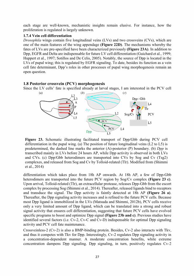
each stage are well-known, mechanistic insights remain elusive. For instance, how the proliferation is regulated is largely unknown. 1.7.4 Vein cell differentiation Drosophila wings contain five longitudinal veins (LVs) and two crossveins (CVs), which are one of the main features of the wing appendage (Figure 22D). The mechanisms whereby the fates of LVs are pre-specified have been characterized previously (Figure 23A). In addition to Dpp, EGFR and Delta are indispensable for future LV cell differentiation (Guichard et al., 1999; Huppert et al., 1997; Sotillos and De Celis, 2005). Notably, the source of Dpp is located in the LVs of pupal wing; this is regulated by EGFR signaling. To date, besides its function as a vein cell fate determinant, Dpp’s roles in other processes of pupal wing morphogenesis remain an open question. 1.8 Posterior crossvein (PCV) morphogenesis Since the LV cells’ fate is specified already at larval stages, I am interested in the PCV cell
differentiation which takes place from 18h AP onwards. At 18h AP, a few of Dpp-Gbb heterodimers are transported into the future PCV region by Sog/Cv complex (Figure 23 c). Upon arrival, Tolloid-related (Tlr), an extracellular protease, releases Dpp-Gbb from the escort complex by processing Sog (Shimmi et al., 2014). Thereafter, released ligands bind to receptors and transduce the signal. The Dpp activity is faintly detected at 18h AP (Figure 26 a). Thereafter, the Dpp signaling activity increases and is refined to the future PCV cells. Because most Dpp ligand is immobilized in the LVs (Matsuda and Shimmi, 2012b), PCV cells receive only a very limited amount of Dpp ligand, which can be translated into a strong and robust signal activity that ensures cell differentiation, suggesting that future PCV cells have evolved specific programs to boost and optimize Dpp signal (Figure 23b and c). Previous studies have identified several factors (i.e. Cv-2, Cv-C and Cv-D) indispensable for optimal Dpp signaling activity and PCV cell fate maintenance. Crossveinless-2 (Cv-2) is also a BMP-binding protein. Besides, Cv-2 also interacts with Tkv, and thus it competes with Tkv for Dpp. Interestingly, Cv-2 regulates Dpp signaling activity in a concentration-dependent manner. A moderate concentration benefits, while extreme concentration dampens Dpp signaling. Dpp signaling, in turn, positively regulates Cv-2
Figure 23. Schematic illustrating facilitated transport of Dpp/Gbb during PCV cell differentiation in the pupal wing. (a) The position of future longitudinal veins (L2 to L5) is predetermined, the dashed line marks the anterior (A)-posterior (P) boundary. (b) Dpp is transcribed mainly in LVs before 24 hours AP, while Dpp activity is observed in both LVs and CVs. (c) Dpp/Gbb heterodimers are transported into CVs by Sog and Cv (Tsg2) complexes, and released from Sog and Cv by Tolloid-related (Tlr). Modified from (Shimmi et al., 2014)
28
expression (Serpe et al., 2008). This positive-feedback mechanism ensures the fine-tuning of Dpp signal during PCV morphogenesis. Aforementioned Sog, a BMP-binding protein, suppresses Dpp signaling by sequestering Dpp from its receptors (e.g. in intervein regions), whilst promoting Dpp signaling through transporting it (e.g. in PCV region) (Matsuda and Shimmi, 2012b). Thus, additional mechanisms are needed to steer Dpp transport by Sog. Crossveinless-C (Cv-C) is part of such mechanisms. Cv-V is a RhoGTPase activating protein (RhoGAP), which inactivates small GTPases (i.e. Rho1) (Denholm et al., 2005). During PCV morphogenesis, Cv-C negatively regulates the distribution of integrin on the cell surface of future vein cell, so that Sog complex can move smoothly from LV to PCV (Matsuda et al., 2013). Even though mechanisms whereby Dpp is transported into PCV are well studied, how limited Dpp ligand transduces robust signal activity remains an open question. This makes the PCV an ideal model to understand mechanisms how cell intrinsic factors couple BMP/Dpp signaling with epithelial cell differentiation.
29
2. Aims of the study Mechanisms underlying epithelial morphogenesis are a key topic for developmental biologists. These mechanisms are involved in self-organization of a few cells into an organ with dedicated functions, and remodeling of simple epithelial sheets into complex three-dimensional tissues with distinct morphologies. Many evolutionarily conserved genetic “toolkits” are used for determining body plan in developing multicellular organisms across taxa. The BMP signaling pathway is one of those participating in a multitude of biological processes, for example, cell differentiation and proliferation. The study was conducted
1. to explore the roles of BMP/Dpp signaling during epithelial morphogenesis, and 2. to decipher the interactions between epithelial characteristics and BMP/Dpp signaling
and the relative roles during the morphogenesis
30
3. Materials and methods The details of the materials, including antibodies, chemicals and fly strains, and methods used in the study can be found in the corresponding articles and the affiliated supplementary information. The main methods used are summarized in Table 1. Table 1. The methods Method Publication Immunofluorescence I, II, III Confocal microscopy I, II, III In situ hybridization I, II RNA extraction and RT-qPCR II S2 cell culture II, III Immunoprecipitation II, III Western blotting II, III Molecular cloning II, III Fly husbandry I, II, III Data analysis with ImageJ I, II, III

31
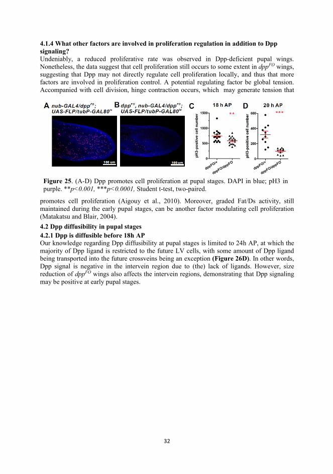
44. Summary of results and discussion 4.1 Dpp and pupal wing growth 4.1.1 Dpp is indispensable for proper growth of the pupal wing Previously, BMP/Dpp was only thought to be the determinant of provein cell fate in the pupal
wing. The evidence for this came from flies of genotype dppshv, a dpp allele in which the mid-distal part of the vein is lost in adult wings without other manifest phenotypes (Matsuda and Shimmi, 2012a). Recently, with the development of novel genetic tools, we can silence dpp through flip-out of the first exon (dppFO) at the genomic level in a spatiotemporal manner (Akiyama and Gibson, 2015). To investigate the detailed roles BMP/Dpp play in pupal wing development, we eliminated Dpp protein at the prepupal stages using this technique by collecting the white pupae which were shifted to 29° 24 hours before pupariation as described in (Akiyama and Gibson, 2015). Ablating dpp expression does not prevent the wing imaginal disc from growing to the default size (I, Figure 1B, E and H). However, we observed that, in addition to loss of venation, the size of adult wings is significantly decreased compared with the control, suggesting that Dpp promotes growth of the pupal wing (Figure 24). 4.1.2 Dpp promotes cell proliferation during inflation stages These results hint that Dpp may have some more comprehensive roles rather than simply as a vein cell fate determinant in developing pupal wings. To examine this hypothesis, I first assayed whether proliferation is affected in dppFO wings by detecting phosphorylated histone H3 (pH3), a mitotic marker (Goto et al., 1999). My results suggest that the number of pH3(+) foci in dppFO wings are significantly reduced, indicating proliferative rate is dimished due to silencing of Dpp signaling is responsible for the small wing phenotype (Figure 25). 4.1.3 How does Dpp regulate cell proliferation in pupal wing? In spite of the evidence that Dpp signaling is indispensable for pupal wing cell proliferation, the mechanism remains to be elucidated. One possibility is that Dpp acts through Brk, which is a growth suppressor in the wing imaginal disc (Schwank et al., 2011). Our data also demonstrate a negative correlation between Brk expression and proliferation rate (I, Figure 2A-F). Moreover, overexpression of Brk is sufficient to reduce the pupal wing size (I, Figure s2F and G).
Figure 24. The phenotype of dppFO wings. The comparison between (A and B) control and (C and D) dppFO pupal wings (A and C) and adult wings (B and D); pMad is in purple ( b, c, e, f.) Note that the pMad signal were undetectable in wing epithelial cells, but still detectable in neuron cells, and veins in the adult wing (D) are missing. (E and F) Quantitation of wing size. Larvae were maintained at 18°C until the mid-3rd instar larval stage and shifted to 29°C for 24 hours before white pupa formation.
32
4.1.4 What other factors are involved in proliferation regulation in addition to Dpp signaling? Undeniably, a reduced proliferative rate was observed in Dpp-deficient pupal wings. Nonetheless, the data suggest that cell proliferation still occurs to some extent in dppFO wings, suggesting that Dpp may not directly regulate cell proliferation locally, and thus that more factors are involved in proliferation control. A potential regulating factor be global tension. Accompanied with cell division, hinge contraction occurs, which may generate tension that
promotes cell proliferation (Aigouy et al., 2010). Moreover, graded Fat/Ds activity, still maintained during the early pupal stages, can be another factor modulating cell proliferation (Matakatsu and Blair, 2004). 4.2 Dpp diffusibility in pupal stages 4.2.1 Dpp is diffusible before 18h AP Our knowledge regarding Dpp diffusibility at pupal stages is limited to 24h AP, at which the majority of Dpp ligand is restricted to the future LV cells, with some amount of Dpp ligand being transported into the future crossveins being an exception (Figure 26D). In other words, Dpp signal is negative in the intervein region due to (the) lack of ligands. However, size reduction of dppFO wings also affects the intervein regions, demonstrating that Dpp signaling may be positive at early pupal stages.
Figure 25. (A-D) Dpp promotes cell proliferation at pupal stages. DAPI in blue; pH3 in purple. **p<0.001, ***p<0.0001, Student t-test, two-paired.
33
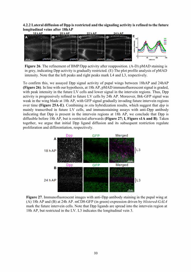
4.2.2 Lateral diffusion of Dpp is restricted and the signaling activity is refined to the future longitudinal veins after 18hAP
To confirm this, we assayed Dpp signal activity of pupal wings between 18hAP and 24hAP (Figure 26). In line with our hypothesis, at 18h AP, pMAD immunofluorescent signal is graded, with peak intensity in the future LV cells and lower signal in the intervein regions. Thus, Dpp activity is progressively refined to future LV cells by 24h AP. Moreover, Brk-GFP signal was weak in the wing blade at 18h AP, with GFP signal gradually invading future intervein regions over time (Figure 25A-E). Combining in situ hybridization results, which suggest that dpp is mainly transcribed in future LV cells, and immunostaining assays with anti-Dpp antibody indicating that Dpp is present in the intervein regions at 18h AP, we conclude that Dpp is diffusible before 18h AP, but is restricted afterwards (Figure 27; I, Figure s1A and B). Taken together, we argue that initial Dpp ligand diffusion and its subsequent restriction regulate proliferation and differentiation, respectively.
Figure 27. Immunofluorescent images with anti-Dpp antibody staining in the pupal wing at (A) 18h AP and (B) at 24h AP. mCD8-GFP (in green) expression driven by blistered-GAL4 mark the future intervein cells. Note that Dpp ligands are spread into the intervein region at 18h AP, but restricted in the LV. L3 indicates the longitudinal vein 3.
Figure 26. The refinement of BMP/Dpp activity after reapposition. (A-D) pMAD staining is in grey, indicating Dpp activity is gradually restricted. (E) The plot profile analysis of pMAD intensity. Note that the left peaks and right peaks mark L4 and L3, respectively.
34
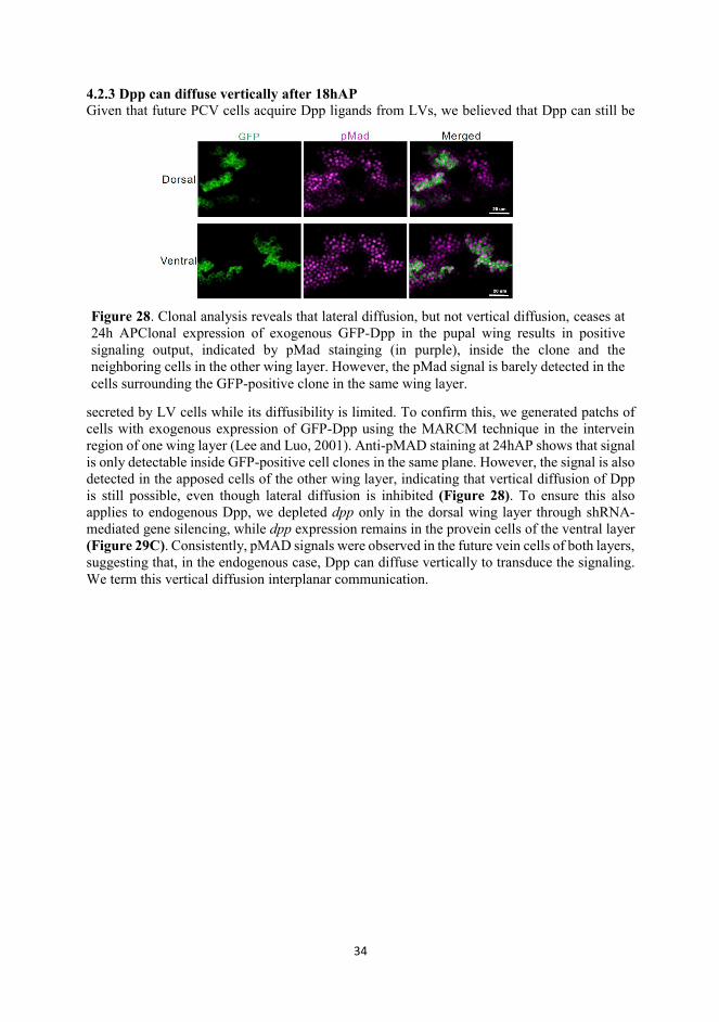
4.2.3 Dpp can diffuse vertically after 18hAP Given that future PCV cells acquire Dpp ligands from LVs, we believed that Dpp can still be
secreted by LV cells while its diffusibility is limited. To confirm this, we generated patchs of cells with exogenous expression of GFP-Dpp using the MARCM technique in the intervein region of one wing layer (Lee and Luo, 2001). Anti-pMAD staining at 24hAP shows that signal is only detectable inside GFP-positive cell clones in the same plane. However, the signal is also detected in the apposed cells of the other wing layer, indicating that vertical diffusion of Dpp is still possible, even though lateral diffusion is inhibited (Figure 28). To ensure this also applies to endogenous Dpp, we depleted dpp only in the dorsal wing layer through shRNA-mediated gene silencing, while dpp expression remains in the provein cells of the ventral layer (Figure 29C). Consistently, pMAD signals were observed in the future vein cells of both layers, suggesting that, in the endogenous case, Dpp can diffuse vertically to transduce the signaling. We term this vertical diffusion interplanar communication.
Figure 28. Clonal analysis reveals that lateral diffusion, but not vertical diffusion, ceases at 24h APClonal expression of exogenous GFP-Dpp in the pupal wing results in positive signaling output, indicated by pMad stainging (in purple), inside the clone and the neighboring cells in the other wing layer. However, the pMad signal is barely detected in the cells surrounding the GFP-positive clone in the same wing layer.
35
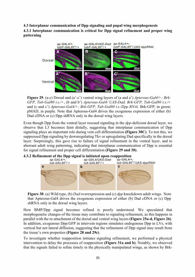
4.3 Interplanar communication of Dpp signaling and pupal wing morphogenesis 4.3.1 Interplanar communication is critical for Dpp signal refinement and proper wing patterning
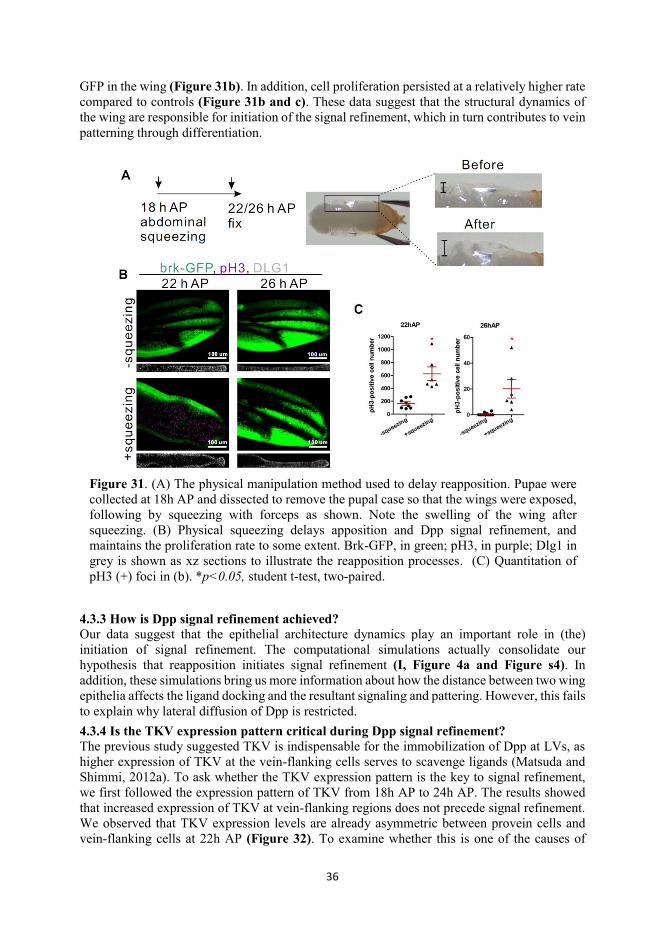
Even though Dpp from the ventral layer rescued signaling in the dpp-deficient dorsal layer, we observe that L3 becomes faint distally, suggesting that interplanar communication of Dpp signaling plays an important role during vein cell differentiation (Figure 30C). To test this, we suppressed Dpp signaling by downregulating Tkv or upregulating Dad specifically in the dorsal layer. Surprisingly, this gave rise to failure of signal refinement in the ventral layer, and to aberrant adult wing patterning, indicating that interplanar communication of Dpp is essential for signal refinement and proper cell differentiation (Figure 29 and 30). 4.3.2 Refinement of the Dpp signal is initiated upon reapposition
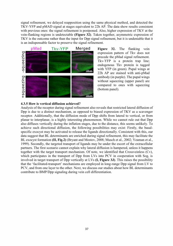
How BMP/Dpp signal becomes refined is poorly understood. We speculated that morphogenetic changes of the tissue may contribute to signaling refinement, as this happens in parallel with the re-attachment of the dorsal and ventral wing layers (Figure 25a-d, Figure 26). In addition, exogenous Dpp:GFP in intervein regions simulates endogenous Dpp in LVs, with vertical but not lateral diffusion, suggesting that the refinement of Dpp signal may result from the tissue’s own properties (Figure 28 and 29c). To investigate whether reapposition initiates signaling refinement, we performed a physical intervention to delay the processes of reapposition (Figure 31a and b). Notably, we observed that the signals failed to refine timely in the physically manipulated wings, as shown by Brk-
Figure 30. (a) Wild type, (b) Dad overexpression and (c) dpp knockdown adult wings. Note that Apterous-Gal4 drives the exogenous expression of either (b) Dad cDNA or (c) Dpp shRNA only in the dorsal wing layers.
Figure 29. (a-c) Dorsal and (a’-c’) ventral wing layers of (a and a’) Apterous-Gal4/+; Brk-GFP, Tub-Gal80 t.s./+, (b and b’) Apterous-Gal4/ UAS-Dad; Brk-GFP, Tub-Gal80 t.s./+ and (c and c’) Apterous-Gal4/+; Brk-GFP, Tub-Gal80 t.s./Dpp RNAi. Brk-GFP, in green; pMAD, in purple. Note that Apterous-Gal4 drives the exogenous expression of either (b) Dad cDNA or (c) Dpp shRNA only in the dorsal wing layers.
36
GFP in the wing (Figure 31b). In addition, cell proliferation persisted at a relatively higher rate compared to controls (Figure 31b and c). These data suggest that the structural dynamics of the wing are responsible for initiation of the signal refinement, which in turn contributes to vein patterning through differentiation.
4.3.3 How is Dpp signal refinement achieved? Our data suggest that the epithelial architecture dynamics play an important role in (the) initiation of signal refinement. The computational simulations actually consolidate our hypothesis that reapposition initiates signal refinement (I, Figure 4a and Figure s4). In addition, these simulations bring us more information about how the distance between two wing epithelia affects the ligand docking and the resultant signaling and pattering. However, this fails to explain why lateral diffusion of Dpp is restricted. 4.3.4 Is the TKV expression pattern critical during Dpp signal refinement? The previous study suggested TKV is indispensable for the immobilization of Dpp at LVs, as higher expression of TKV at the vein-flanking cells serves to scavenge ligands (Matsuda and Shimmi, 2012a). To ask whether the TKV expression pattern is the key to signal refinement, we first followed the expression pattern of TKV from 18h AP to 24h AP. The results showed that increased expression of TKV at vein-flanking regions does not precede signal refinement. We observed that TKV expression levels are already asymmetric between provein cells and vein-flanking cells at 22h AP (Figure 32). To examine whether this is one of the causes of
Figure 31. (A) The physical manipulation method used to delay reapposition. Pupae were collected at 18h AP and dissected to remove the pupal case so that the wings were exposed, following by squeezing with forceps as shown. Note the swelling of the wing after squeezing. (B) Physical squeezing delays apposition and Dpp signal refinement, and maintains the proliferation rate to some extent. Brk-GFP, in green; pH3, in purple; Dlg1 in grey is shown as xz sections to illustrate the reapposition processes. (C) Quantitation of pH3 (+) foci in (b). *p<0.05, student t-test, two-paired.
37
signal refinement, we delayed reapposition using the same physical method, and detected the TKV-YFP and pMAD signal at stages equivalent to 22h AP. The data show results consistent with previous ones: the signal refinement is postponed. Also, higher expression of TKV at the vein-flanking regions is undetectable (Figure 32). Taken together, asymmetric expression of TKV is the outcome rather than the input for Dpp signal refinement, but it is undeniable that it is an indispensable factor to preserve the signal refinement.
4.3.5 How is vertical diffusion achieved? Analysis of the receptor during signal refinement also reveals that restricted lateral diffusion of Dpp is due to a distinct mechanism, as opposed to biased expression of TKV as a scavenger receptor. Additionally, that the diffusion mode of Dpp shifts from lateral to vertical, or from planar to interplanar, is a highly interesting phenomenon. While we cannot rule out that Dpp also diffuses vertically during the inflation stages, due to the distance, this seems unlikely. To achieve such directional diffusion, the following possibilities may exist: Firstly, the basal-specific exocyst may be activated to release the ligands directionally. Consistent with this, our data suggest that BL determinants are enriched during signal refinement, this may facilitate the BL exocyst formation (II, Fig.2) (Bryant and Mostov, 2008; Musch et al., 2002; Yeaman et al., 1999). Secondly, the targeted transport of ligands may be under the escort of the extracellular partners. The first scenario cannot explain why lateral diffusion is hampered, unless it happens together with the target transport mechanism. Of note, we identified that Crossveinless (Cv), which participates in the transport of Dpp from LVs into PCV in cooperation with Sog, is involved in target transport of Dpp vertically at LVs (I, Figure 3J). This raises the possibility that the ‘facilitated-transport’ mechanisms are employed in long-range Dpp signal from LV to PCV, and from one layer to the other. Next, we discuss our studies about how BL determinants contribute to BMP/Dpp signaling during vein cell differentiation.
Figure 32. The flanking vein expression pattern of Tkv does not precede the pMad signal refinement. Tkv-YFP is a protein trap line; endogenous Tkv protein is tagged with YFP (in green). Pupal wings at 22h AP are stained with anti-pMad antibody (in purple). The pupal wings without squeezing (upper panel) are compared to ones with squeezing (bottom panel).
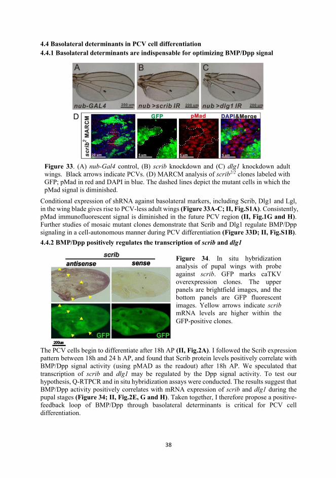
38
4.4 Basolateral determinants in PCV cell differentiation 4.4.1 Basolateral determinants are indispensable for optimizing BMP/Dpp signal
Conditional expression of shRNA against basolateral markers, including Scrib, Dlg1 and Lgl, in the wing blade gives rise to PCV-less adult wings (Figure 33A-C; II, Fig.S1A). Consistently, pMad immunofluorescent signal is diminished in the future PCV region (II, Fig.1G and H). Further studies of mosaic mutant clones demonstrate that Scrib and Dlg1 regulate BMP/Dpp signaling in a cell-autonomous manner during PCV differentiation (Figure 33D; II, Fig.S1B). 4.4.2 BMP/Dpp positively regulates the transcription of scrib and dlg1
The PCV cells begin to differentiate after 18h AP (II, Fig.2A). I followed the Scrib expression pattern between 18h and 24 h AP, and found that Scrib protein levels positively correlate with BMP/Dpp signal activity (using pMAD as the readout) after 18h AP. We speculated that transcription of scrib and dlg1 may be regulated by the Dpp signal activity. To test our hypothesis, Q-RTPCR and in situ hybridization assays were conducted. The results suggest that BMP/Dpp activity positively correlates with mRNA expression of scrib and dlg1 during the pupal stages (Figure 34; II, Fig.2E, G and H). Taken together, I therefore propose a positive-feedback loop of BMP/Dpp through basolateral determinants is critical for PCV cell differentiation.
Figure 34. In situ hybridization analysis of pupal wings with probe against scrib. GFP marks caTKV overexpression clones. The upper panels are brightfield images, and the bottom panels are GFP fluorescent images. Yellow arrows indicate scrib mRNA levels are higher within the GFP-positive clones.
Figure 33. (A) nub-Gal4 control, (B) scrib knockdown and (C) dlg1 knockdown adult wings. Black arrows indicate PCVs. (D) MARCM analysis of scrib2/2 clones labeled with GFP; pMad in red and DAPI in blue. The dashed lines depict the mutant cells in which the pMad signal is diminished.
D
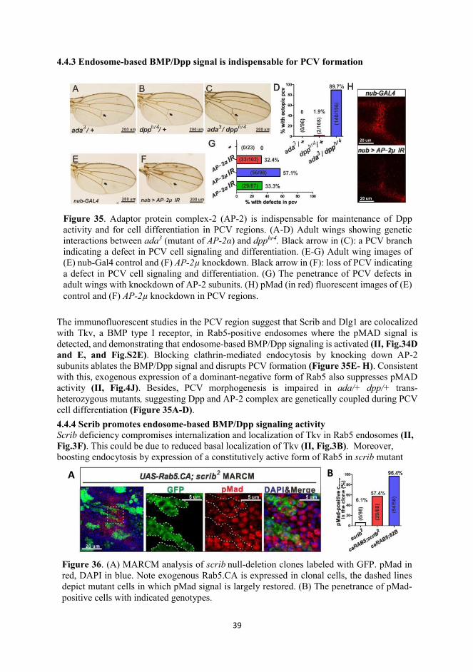
39
4.4.3 Endosome-based BMP/Dpp signal is indispensable for PCV formation
The immunofluorescent studies in the PCV region suggest that Scrib and Dlg1 are colocalized with Tkv, a BMP type I receptor, in Rab5-positive endosomes where the pMAD signal is detected, and demonstrating that endosome-based BMP/Dpp signaling is activated (II, Fig.34D and E, and Fig.S2E). Blocking clathrin-mediated endocytosis by knocking down AP-2 subunits ablates the BMP/Dpp signal and disrupts PCV formation (Figure 35E- H). Consistent with this, exogenous expression of a dominant-negative form of Rab5 also suppresses pMAD activity (II, Fig.4J). Besides, PCV morphogenesis is impaired in ada/+ dpp/+ trans-heterozygous mutants, suggesting Dpp and AP-2 complex are genetically coupled during PCV cell differentiation (Figure 35A-D). 4.4.4 Scrib promotes endosome-based BMP/Dpp signaling activity Scrib deficiency compromises internalization and localization of Tkv in Rab5 endosomes (II, Fig.3F). This could be due to reduced basal localization of Tkv (II, Fig.3B). Moreover, boosting endocytosis by expression of a constitutively active form of Rab5 in scrib mutant
Figure 35. Adaptor protein complex-2 (AP-2) is indispensable for maintenance of Dpp activity and for cell differentiation in PCV regions. (A-D) Adult wings showing genetic interactions between ada3 (mutant of AP-2α) and dpphr4. Black arrow in (C): a PCV branch indicating a defect in PCV cell signaling and differentiation. (E-G) Adult wing images of (E) nub-Gal4 control and (F) AP-2μ knockdown. Black arrow in (F): loss of PCV indicating a defect in PCV cell signaling and differentiation. (G) The penetrance of PCV defects in adult wings with knockdown of AP-2 subunits. (H) pMad (in red) fluorescent images of (E) control and (F) AP-2μ knockdown in PCV regions.
A B
Figure 36. (A) MARCM analysis of scrib null-deletion clones labeled with GFP. pMad in red, DAPI in blue. Note exogenous Rab5.CA is expressed in clonal cells, the dashed lines depict mutant cells in which pMad signal is largely restored. (B) The penetrance of pMad-positive cells with indicated genotypes.
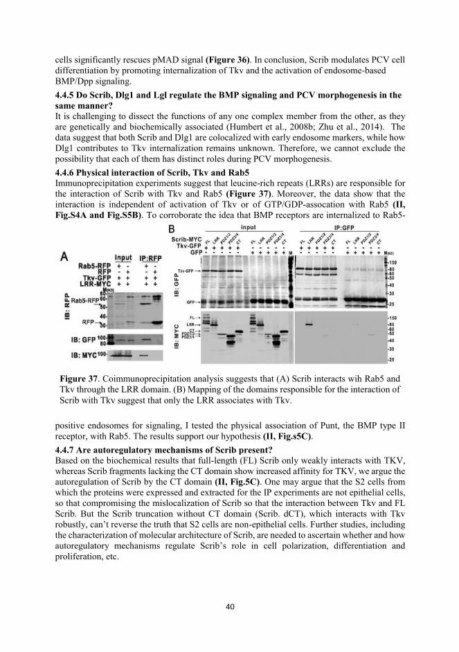
40
cells significantly rescues pMAD signal (Figure 36). In conclusion, Scrib modulates PCV cell differentiation by promoting internalization of Tkv and the activation of endosome-based BMP/Dpp signaling. 4.4.5 Do Scrib, Dlg1 and Lgl regulate the BMP signaling and PCV morphogenesis in the same manner? It is challenging to dissect the functions of any one complex member from the other, as they are genetically and biochemically associated (Humbert et al., 2008b; Zhu et al., 2014). The data suggest that both Scrib and Dlg1 are colocalized with early endosome markers, while how Dlg1 contributes to Tkv internalization remains unknown. Therefore, we cannot exclude the possibility that each of them has distinct roles during PCV morphogenesis. 4.4.6 Physical interaction of Scrib, Tkv and Rab5 Immunoprecipitation experiments suggest that leucine-rich repeats (LRRs) are responsible for the interaction of Scrib with Tkv and Rab5 (Figure 37). Moreover, the data show that the interaction is independent of activation of Tkv or of GTP/GDP-assocation with Rab5 (II, Fig.S4A and Fig.S5B). To corroborate the idea that BMP receptors are internalized to Rab5-
positive endosomes for signaling, I tested the physical association of Punt, the BMP type II receptor, with Rab5. The results support our hypothesis (II, Fig.s5C). 4.4.7 Are autoregulatory mechanisms of Scrib present? Based on the biochemical results that full-length (FL) Scrib only weakly interacts with TKV, whereas Scrib fragments lacking the CT domain show increased affinity for TKV, we argue the autoregulation of Scrib by the CT domain (II, Fig.5C). One may argue that the S2 cells from which the proteins were expressed and extracted for the IP experiments are not epithelial cells, so that compromising the mislocalization of Scrib so that the interaction between Tkv and FL Scrib. But the Scrib truncation without CT domain (Scrib. dCT), which interacts with Tkv robustly, can’t reverse the truth that S2 cells are non-epithelial cells. Further studies, including the characterization of molecular architecture of Scrib, are needed to ascertain whether and how autoregulatory mechanisms regulate Scrib’s role in cell polarization, differentiation and proliferation, etc.
Figure 37. Coimmunoprecipitation analysis suggests that (A) Scrib interacts wih Rab5 and Tkv through the LRR domain. (B) Mapping of the domains responsible for the interaction of Scrib with Tkv suggest that only the LRR associates with Tkv.
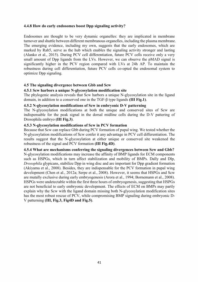
41
4.4.8 How do early endosomes boost Dpp signaling activity?
Endosomes are thought to be very dynamic organelles: they are implicated in membrane turnover and shuttle between different membranous organelles, including the plasma membrane. The emerging evidence, including my own, suggests that the early endosomes, which are marked by Rab5, serve as the hub which enables the signaling activity stronger and lasting (Alanko et al., 2015). During PCV cell differentiation, future PCV cells receive only a very small amount of Dpp ligands from the LVs. However, we can observe the pMAD signal is significantly higher in the PCV region compared with LVs at 24h AP. To maintain the robustness during cell differentiation, future PCV cells co-opted the endosomal system to optimize Dpp signaling. 4.5 The signaling divergence between Gbb and Scw 4.5.1 Scw harbors a unique N-glycosylation modification site The phylogenic analysis reveals that Scw harbors a unique N-glycosylation site in the ligand domain, in addition to a conserved one in the TGF-β type ligands (III Fig.1). 4.5.2 N-glycosylation modifications of Scw in embryonic D-V patterning The N-glycosylation modifications at both the unique and conserved sites of Scw are indispensable for the peak signal in the dorsal midline cells during the D-V pattering of Drosophila embryo (III Fig.3). 4.5.3 N-glycosylation modifications of Scw in PCV formation Because that Scw can replace Gbb during PCV formation of pupal wing. We tested whether the N-glycosylation modifications of Scw confer it any advantage in PCV cell differentiation. The results suggest that the N-glycosylation at either unique or conserved site weakened the robustness of the signal and PCV formation (III Fig.4D). 4.5.4 What are mechanisms conferring the signaling divergences between Scw and Gbb? N-glycosylation modifications may increase the affinity of BMP ligands for ECM components such as HSPGs, which in turn affect stabilization and mobility of BMPs. Dally and Dlp, Drosophila glypicans, stabilize Dpp in wing disc and are important for Dpp gradient formation (Akiyama et al., 2008). Besides, they are indispensable for the PCV formation in pupal wing development (Chen et al., 2012a; Serpe et al., 2008). However, it seems that HSPGs and Scw are mutally exclusive during early embryogenesis (Arora et al., 1994; Bornemann et al., 2008). HSPGs were undetectable within the first three hours of embryogenesis, suggesting that HSPGs are not beneficial to early embryonic development. The effects of ECM on BMPs may partly explain why the Scw with the ligand domain missing both N-glycosylation modification sites has the most robust rescue of PCV, while compromising BMP signaling during embryonic D-V patterning (III, Fig.3, Fig4D and Fig.5).
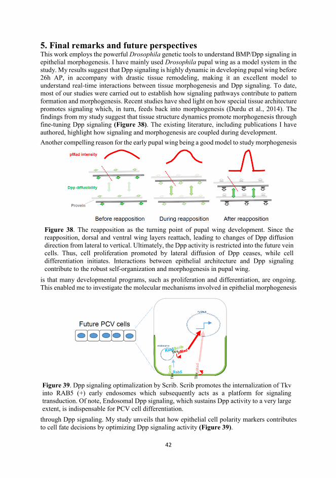
42
5. Final remarks and future perspectives This work employs the powerful Drosophila genetic tools to understand BMP/Dpp signaling in epithelial morphogenesis. I have mainly used Drosophila pupal wing as a model system in the study. My results suggest that Dpp signaling is highly dynamic in developing pupal wing before 26h AP, in accompany with drastic tissue remodeling, making it an excellent model to understand real-time interactions between tissue morphogenesis and Dpp signaling. To date, most of our studies were carried out to establish how signaling pathways contribute to pattern formation and morphogenesis. Recent studies have shed light on how special tissue architecture promotes signaling which, in turn, feeds back into morphogenesis (Durdu et al., 2014). The findings from my study suggest that tissue structure dynamics promote morphogenesis through fine-tuning Dpp signaling (Figure 38). The existing literature, including publications I have authored, highlight how signaling and morphogenesis are coupled during development. Another compelling reason for the early pupal wing being a good model to study morphogenesis
is that many developmental programs, such as proliferation and differentiation, are ongoing. This enabled me to investigate the molecular mechanisms involved in epithelial morphogenesis
through Dpp signaling. My study unveils that how epithelial cell polarity markers contributes to cell fate decisions by optimizing Dpp signaling activity (Figure 39).
Figure 38. The reapposition as the turning point of pupal wing development. Since the reapposition, dorsal and ventral wing layers reattach, leading to changes of Dpp diffusion direction from lateral to vertical. Ultimately, the Dpp activity is restricted into the future vein cells. Thus, cell proliferation promoted by lateral diffusion of Dpp ceases, while cell differentiation initiates. Interactions between epithelial architecture and Dpp signaling contribute to the robust self-organization and morphogenesis in pupal wing.
Figure 39. Dpp signaling optimalization by Scrib. Scrib promotes the internalization of Tkv into RAB5 (+) early endosomes which subsequently acts as a platform for signaling transduction. Of note, Endosomal Dpp signaling, which sustains Dpp activity to a very large extent, is indispensable for PCV cell differentiation.

43
This work broadens my understandings of Dpp signaling and morphogenesis, but also raises further questions, for instance, how the dorsal and ventral wing epitheila are mutually coordinated to align perfectly. They are separated during inflation stages, and eventually reach the same size and pattern. Further studies are needed to elucidate the underlying mechanisms. The studies conducted in this work should benefit research in many aspects, including development and cancer biology.
44
AAcknowledgements All work in this thesis was conducted at the Institute of Biotechnology (BI), and at the Department of Biosciences, Division of Genetics, the University of Helsinki. I am grateful to BI for providing state-of-the-art facilities and specialists. I want to thank BI director Prof. Howy Jacobs, developmental biology program directors, Prof. Irma Thesleff and Dr. Marja Mikkola, for creating a great research atmosphere. During my thesis work, I was financially supported by VGSB/ILS doctoral program, BI and the Academy of Finland. I am sincerely grateful to Dr. Osamu Shimmi, my supervisor, who created the opportunities for me to study in Finland, my second homeland. Words are not enough to express my gratitude to him. I still remember clearly the moment when Osamu had the interview with me over the telephone, and the exciting moment when I got the offer. I have experienced these trying years with the supervision, company and encouragement of Osamu. I have shared happiness, worry, anger and anxiety with him. I have borrowed cash from him . To me, Osamu is a friend and brother, and much more than a mere supervisor. He helped me polish projects and build scientific insight. He is so frank, honest and patient that I have come to trust him very much. He tolerated my mistakes and my attempts at independence during my studies. Optimally, I would continue working with him indefinitely. I also want to thank my thesis committee members, Drs. Marja Mikkola and Ville Hietakangas. They spent lots of time organizing committee meetings and giving me comments and feedback, ensuring my graduation and continued success. I really appreciate Prof. Mattias Mannervik and Dr. Giorgos Pyrowolakis for taking the responsibility of being my pre-examiners. Also, I am very grateful to Prof. Juha Partanen for being the custodian and for handling with the matters of my graduation. It has been my honor to have all of you on my path to receiving my doctoral degree. In addition, I thank Dr. Martin Kracklauer, one of my postdoctoral colleagues in the Shimmi group, for proofreading my thesis writing and giving constructive comments and inspiration. During my studies, I benefitted from the devoted expertise of the LMU specialists, especially Marco Crivaro and Mika Molin. Many documentation issues were handled by Pekka Heino and Erkki Raulo. My studies have gained lots of help from my colleagues in the Shimmi Hietakangas groups. I would like to express deep gratitude to all of them. It has been my pleasure to work with my dear colleagues, Lisa, Jorge, Shinya, Jaana, Petra, Yunxian, Martin and Daniel. Thanks, Lisa, for celebrating everybody’s birthday with hand-made cake and cards. I have made many friendships in the last few years, and would like to express my gratitude to all the people who helped and encouraged me. In the beginning of my Finnish life, Ying gave me many useful tips to survive in Finland and inducted me into the basketball team. Then I met Yan and Yajing, who helped my family a lot, especially when my wife followed me to Finland and our family began to grow. Weixian and Biyun have also always been there for us when we needed them. It’s been my good fortune to have all of you taking care of me and my family. I also want to present the special gratitude to my friend’s family members including Akari and Hikari, they have been caring for my wife and children so much. Ultimately, I want to thank my family. My parents, including my in-laws, were supportive of my studying abroad, and helped me considerably as my wife and I established our family. My wife, my darling, is the most important person to me in this world. She has been through every moment of my frustration and success, and I will also be there for you for the rest of my life. Thank you also for bearing and mothering our two wonderful children. You are my soul mate, the source of my diligence and inspiration. I am so lucky to know you and to have you in my life. Let’s look forward to a bright future together!
45
RReferences Aegerter-Wilmsen, T., Aegerter, C.M., Hafen, E., and Basler, K. (2007). Model for the regulation of size in the wing imaginal disc of Drosophila. Mechanisms of development 124, 318-326. Aigouy, B., Farhadifar, R., Staple, D.B., Sagner, A., Roper, J.C., Julicher, F., and Eaton, S. (2010). Cell Flow Reorients the Axis of Planar Polarity in the Wing Epithelium of Drosophila. Cell 142, 773-786. Akiyama, T., and Gibson, M.C. (2015). Decapentaplegic and growth control in the developing Drosophila wing. Nature 527, 375-+. Akiyama, T., Kamimura, K., Firkus, C., Takeo, S., Shimmi, O., and Nakato, H. (2008). Dally regulates Dpp morphogen gradient formation by stabilizing Dpp on the cell surface. Developmental biology 313, 408-419. Alanko, J., Mai, A., Jacquemet, G., Schauer, K., Kaukonen, R., Saari, M., Goud, B., and Ivaska, J. (2015). Integrin endosomal signalling suppresses anoikis. Nature Cell Biology 17, 1412-1421. Aldaz, S., Escudero, L.M., and Freeman, M. (2010). Live imaging of Drosophila imaginal disc development. Proceedings of the National Academy of Sciences of the United States of America 107, 14217-14222. Araujo, H., Negreiros, E., and Bier, E. (2003). Integrins modulate Sog activity in the Drosophila wing. Development 130, 3851-3864. Arora, K., Levine, M.S., and Oconnor, M.B. (1994). The Screw Gene Encodes a Ubiquitously Expressed Member of the Tgf-Beta Family Required for Specification of Dorsal Cell Fates in the Drosophila Embryo. Genes & development 8, 2588-2601. Azim, A.C., Knoll, J.H., Marfatia, S.M., Peel, D.J., Bryant, P.J., and Chishti, A.H. (1995). DLG1: chromosome location of the closest human homologue of the Drosophila discs large tumor suppressor gene. Genomics 30, 613-616. Baum, B., Settleman, J., and Quinlan, M.P. (2008). Transitions between epithelial and mesenchymal states in development and disease. Seminars in Cell & Developmental Biology 19, 294-308. Belenkaya, T.Y., Han, C., Yan, D., Opoka, R.J., Khodoun, M., Liu, H., and Lin, X. (2004). Drosophila Dpp morphogen movement is independent of dynamin-mediated endocytosis but regulated by the glypican members of heparan sulfate proteoglycans. Cell 119, 231-244. Bertet, C., Sulak, L., and Lecuit, T. (2004). Myosin-dependent junction remodelling controls planar cell intercalation and axis elongation. Nature 429, 667-671. Bilder, D., and Perrimon, N. (2000). Localization of apical epithelial determinants by the basolateral PDZ protein Scribble. Nature 403, 676-680. Boggiano, J.C., and Fehon, R.G. (2012). Growth control by committee: intercellular junctions, cell polarity, and the cytoskeleton regulate Hippo signaling. Dev Cell 22, 695-702. Bornemann, D.J., Park, S., Phin, S., and Warrior, R. (2008). A translational block to HSPG synthesis permits BMP signaling in the early Drosophila embryo. Development 135, 1039-1047. Brown, N.H. (2000). Cell-cell adhesion via the ECM: integrin genetics in fly and worm. Matrix Biol 19, 191-201. Bryant, D.M., and Mostov, K.E. (2008). From cells to organs: building polarized tissue. Nat Rev Mol Cell Bio 9, 887-901. Butler, M.T., and Wallingford, J.B. (2017). Planar cell polarity in development and disease. Nat Rev Mol Cell Biol 18, 375-388. Cao, X., Surma, M.A., and Simons, K. (2012). Polarized sorting and trafficking in epithelial cells. Cell research 22, 793-805. Chalcroft, J.P., and Bullivant, S. (1970). An interpretation of liver cell membrane and junction structure based on observation of freeze-fracture replicas of both sides of the fracture. The Journal of cell biology 47, 49-60. Chen, J., Honeyager, S.M., Schleede, J., Avanesov, A., Laughon, A., and Blair, S.S. (2012a). Crossveinless d is a vitellogenin-like lipoprotein that binds BMPs and HSPGs, and is required for normal BMP signaling in the Drosophila wing. Development 139, 2170-2176.
46
Chen, J., Honeyager, S.M., Schleede, J., Avanesov, A., Laughon, A., and Blair, S.S. (2012b). Crossveinless d is a vitellogenin-like lipoprotein that binds BMPs and HSPGs, and is required for normal BMP signaling in the Drosophila wing. Development 139, 2170-2176. Chen, Y.J., Spence, H.J., Cameron, J.M., Jess, T., Ilsley, J.L., and Winder, S.J. (2003). Direct interaction of beta-dystroglycan with F-actin. The Biochemical journal 375, 329-337. Christoforou, C.P., Greer, C.E., Challoner, B.R., Charizanos, D., and Ray, R.P. (2008). The detached locus encodes Drosophila Dystrophin, which acts with other components of the Dystrophin Associated Protein Complex to influence intercellular signalling in developing wing veins. Developmental biology 313, 519-532. Classen, A.K., Aigouy, B., Giangrande, A., and Eaton, S. (2008). Imaging Drosophila pupal wing morphogenesis. Methods in molecular biology 420, 265-275. Classen, A.K., Anderson, K.I., Marois, E., and Eaton, S. (2005). Hexagonal packing of Drosophila wing epithelial cells by the planar cell polarity pathway. Developmental Cell 9, 805-817. Colombani, J., Andersen, D.S., and Leopold, P. (2012). Secreted peptide Dilp8 coordinates Drosophila tissue growth with developmental timing. Science 336, 582-585. Costa, M., Wilson, E.T., and Wieschaus, E. (1994). A putative cell signal encoded by the folded gastrulation gene coordinates cell shape changes during Drosophila gastrulation. Cell 76, 1075-1089. Dai, J.D., and Gilbert, L.I. (1991). Metamorphosis of the Corpus Allatum and Degeneration of the Prothoracic Glands during the Larval Pupal Adult Transformation of Drosophila-Melanogaster - a Cytophysiological Analysis of the Ring Gland. Developmental biology 144, 309-326. Dawes-Hoang, R.E., Parmar, K.M., Christiansen, A.E., Phelps, C.B., Brand, A.H., and Wieschaus, E.F. (2005). folded gastrulation, cell shape change and the control of myosin localization. Development 132, 4165-4178. de Vreede, G., Schoenfeld, J.D., Windler, S.L., Morrison, H., Lu, H., and Bilder, D. (2014). The Scribble module regulates retromer-dependent endocytic trafficking during epithelial polarization. Development 141, 2796-2802. Denholm, B., Brown, S., Ray, R.P., Ruiz-Gomez, M., Skaer, H., and Hombria, J.C.G. (2005). crossveinless-c is a RhoGAP required for actin reorganisation during morphogenesis. Development 132, 2389-2400. Devenport, D. (2014). The cell biology of planar cell polarity. The Journal of cell biology 207, 171-179. Diz-Munoz, A., Fletcher, D.A., and Weiner, O.D. (2013). Use the force: membrane tension as an organizer of cell shape and motility. Trends in Cell Biology 23, 47-53. Dow, L.E., Brumby, A.M., Muratore, R., Coombe, M.L., Sedelies, K.A., Trapani, J.A., Russell, S.M., Richardson, H.E., and Humbert, P.O. (2003). hScrib is a functional homologue of the Drosophila tumour suppressor Scribble. Oncogene 22, 9225-9230. Drees, F., Pokutta, S., Yamada, S., Nelson, W.J., and Weis, W.I. (2005). alpha-catenin is a molecular switch that binds E-cadherin-beta-catenin and regulates actin-filament assembly. Cell 123, 903-915. Duband, J.L., and Thiery, J.P. (1982). Appearance and distribution of fibronectin during chick embryo gastrulation and neurulation. Developmental biology 94, 337-350. Ducuing, A., and Vincent, S. (2016). The actin cable is dispensable in directing dorsal closure dynamics but neutralizes mechanical stress to prevent scarring in the Drosophila embryo. Nature Cell Biology 18, 1149-+. Durdu, S., Iskar, M., Revenu, C., Schieber, N., Kunze, A., Bork, P., Schwab, Y., and Gilmour, D. (2014). Luminal signalling links cell communication to tissue architecture during organogenesis. Nature 515, 120-+. Elsum, I., Yates, L., Humbert, P.O., and Richardson, H.E. (2012). The Scribble-Dlg-Lgl polarity module in development and cancer: from flies to man. Essays Biochem 53, 141-168. Etournay, R., Merkel, M., Popovic, M., Brandl, H., Dye, N.A., Aigouy, B., Salbreux, G., Eaton, S., and Julicher, F. (2016). TissueMiner: A multiscale analysis toolkit to quantify how cellular processes create tissue dynamics. eLife 5.
47
Etournay, R., Popovic, M., Merkel, M., Nandi, A., Blasse, C., Aigouy, B., Brandi, H., Myers, G., Salbreux, G., Julicher, F., et al. (2015). Interplay of cell dynamics and epithelial tension during morphogenesis of the Drosophila pupal wing. eLife 4. Friedl, P., and Gilmour, D. (2009). Collective cell migration in morphogenesis, regeneration and cancer. Nat Rev Mol Cell Bio 10, 445-457. Fujise, M., Takeo, S., Kamimura, K., Matsuo, T., Aigaki, T., Izumi, S., and Nakato, H. (2003). Dally regulates Dpp morphogen gradient formation in the Drosophila wing. Development 130, 1515-1522. Gelbart, M.A., He, B., Martin, A.C., Thiberge, S.Y., Wieschaus, E.F., and Kaschube, M. (2012). Volume conservation principle involved in cell lengthening and nucleus movement during tissue morphogenesis. Proceedings of the National Academy of Sciences of the United States of America 109, 19298-19303. Gonzalez-Gaitan, M., and Jackle, H. (1999). The range of spalt-activating Dpp signalling is reduced in endocytosis-defective Drosophila wing discs. Mechanisms of development 87, 143-151. Gorfinkiel, N. (2016). From actomyosin oscillations to tissue-level deformations. Dev Dynam 245, 268-275. Goto, H., Tomono, Y., Ajiro, K., Kosako, H., Fujita, M., Sakurai, M., Okawa, K., Iwamatsu, A., Okigaki, T., Takahashi, T., et al. (1999). Identification of a novel phosphorylation site on histone H3 coupled with mitotic chromosome condensation. Journal of Biological Chemistry 274, 25543-25549. Green, K.J., Getsios, S., Troyanovsky, S., and Godsel, L.M. (2010). Intercellular junction assembly, dynamics, and homeostasis. Cold Spring Harbor perspectives in biology 2, a000125. Gui, J.H., Huang, Y.X., and Shimmi, O. (2016). Scribbled Optimizes BMP Signaling through Its Receptor Internalization to the Rab5 Endosome and Promote Robust Epithelial Morphogenesis. PLoS genetics 12. Guichard, A., Biehs, B., Sturtevant, M.A., Wickline, L., Chacko, J., Howard, K., and Bier, E. (1999). rhomboid and Star interact synergistically to promote EGFR MAPK signaling during Drosophila wing vein development. Development 126, 2663-2676. Hacker, U., Nybakken, K., and Perrimon, N. (2005). Heparan sulphate proteoglycans: the sweet side of development. Nat Rev Mol Cell Biol 6, 530-541. Haeger, A., Wolf, K., Zegers, M.M., and Friedl, P. (2015). Collective cell migration: guidance principles and hierarchies. Trends in Cell Biology 25, 556-566. Haigo, S.L., Hildebrand, J.D., Harland, R.M., and Wallingford, J.B. (2003). Shroom induces apical constriction and is required for hingepoint formation during neural tube closure. Current Biology 13, 2125-2137. Hamaratoglu, F., Affolter, M., and Pyrowolakis, G. (2014). Dpp/BMP signaling in flies: from molecules to biology. Semin Cell Dev Biol 32, 128-136. Han, C., Belenkaya, T.Y., Wang, B., and Lin, X. (2004). Drosophila glypicans control the cell-to-cell movement of Hedgehog by a dynamin-independent process. Development 131, 601-611. Han, C., Yan, D., Belenkaya, T.Y., and Lin, X. (2005). Drosophila glypicans Dally and Dally-like shape the extracellular Wingless morphogen gradient in the wing disc. Development 132, 667-679. Hay, E.D. (1995). An overview of epithelio-mesenchymal transformation. Acta Anat (Basel) 154, 8-20. He, B., Doubrovinski, K., Polyakov, O., and Wieschaus, E. (2014). Apical constriction drives tissue-scale hydrodynamic flow to mediate cell elongation. Nature 508, 392-+. He, L., Wang, X.B., Tang, H.L., and Montell, D.J. (2010). Tissue elongation requires oscillating contractions of a basal actomyosin network. Nature Cell Biology 12, 1133-U1140. Holley, S.A., Jackson, P.D., Sasai, Y., Lu, B., De Robertis, E.M., Hoffmann, F.M., and Ferguson, E.L. (1995). A conserved system for dorsal-ventral patterning in insects and vertebrates involving sog and chordin. Nature 376, 249-253. Humbert, P., Russell, S., and Richardson, H. (2003). Dlg, Scribble and Lgl in cell polarity, cell proliferation and cancer. Bioessays 25, 542-553. Humbert, P.O., Grzeschik, N.A., Brumby, A.M., Galea, R., Elsum, I., and Richardson, H.E. (2008a). Control of tumourigenesis by the Scribble/Dlg/Lgl polarity module. Oncogene 27, 6888-6907.
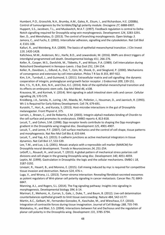
48
Humbert, P.O., Grzeschik, N.A., Brumby, A.M., Galea, R., Elsum, I., and Richardson, H.E. (2008b). Control of tumourigenesis by the Scribble/Dlg/Lgl polarity module. Oncogene 27, 6888-6907. Huppert, S.S., Jacobsen, T.L., and Muskavitch, M.A.T. (1997). Feedback regulation is central to Delta-Notch signalling required for Drosophila wing vein morphogenesis. Development 124, 3283-3291. Iber, D., and Menshykau, D. (2013). The control of branching morphogenesis. Open biology 3. Jamora, C., and Fuchs, E. (2002). Intercellular adhesion, signalling and the cytoskeleton. Nat Cell Biol 4, E101-108. Kalluri, R., and Weinberg, R.A. (2009). The basics of epithelial-mesenchymal transition. J Clin Invest 119, 1420-1428. Kaltcheva, M.M., Anderson, M.J., Harfe, B.D., and Lewandoski, M. (2016). BMPs are direct triggers of interdigital programmed cell death. Developmental biology 411, 266-276. Keller, R., Cooper, M.S., Danilchik, M., Tibbetts, P., and Wilson, P.A. (1989). Cell Intercalation during Notochord Development in Xenopus-Laevis. J Exp Zool 251, 134-154. Keller, R., Davidson, L., Edlund, A., Elul, T., Ezin, M., Shook, D., and Skoglund, P. (2000). Mechanisms of convergence and extension by cell intercalation. Philos T R Soc B 355, 897-922. Kim, S.H., Turnbull, J., and Guimond, S. (2011). Extracellular matrix and cell signalling: the dynamic cooperation of integrin, proteoglycan and growth factor receptor. J Endocrinol 209, 139-151. Kim, Y.S., Yi, B.R., Kim, N.H., and Choi, K.C. (2014). Role of the epithelial-mesenchymal transition and its effects on embryonic stem cells. Exp Mol Med 46, e108. Krausova, M., and Korinek, V. (2014). Wnt signaling in adult intestinal stem cells and cancer. Cellular signalling 26, 570-579. Kreidberg, J.A., Sariola, H., Loring, J.M., Maeda, M., Pelletier, J., Housman, D., and Jaenisch, R. (1993). Wt-1 Is Required for Early Kidney Development. Cell 74, 679-691. Kuraishi, T., Hori, A., and Kurata, S. (2013). Host-microbe interactions in the gut of Drosophila melanogaster. Front Physiol 4, 375. Larrain, J., Brown, C., and De Robertis, E.M. (2003). Integrin-alpha3 mediates binding of Chordin to the cell surface and promotes its endocytosis. EMBO reports 4, 813-818. Lecuit, T., and Cohen, S.M. (1998). Dpp receptor levels contribute to shaping the Dpp morphogen gradient in the Drosophila wing imaginal disc. Development 125, 4901-4907. Lecuit, T., and Lenne, P.F. (2007). Cell surface mechanics and the control of cell shape, tissue patterns and morphogenesis. Nat Rev Mol Cell Bio 8, 633-644. Lecuit, T., and Yap, A.S. (2015). E-cadherin junctions as active mechanical integrators in tissue dynamics. Nat Cell Biol 17, 533-539. Lee, T.M., and Luo, L.Q. (2001). Mosaic analysis with a repressible cell marker (MARCM) for Drosophila neural development. Trends in Neurosciences 24, 251-254. LeGoff, L., Rouault, H., and Lecuit, T. (2013). A global pattern of mechanical stress polarizes cell divisions and cell shape in the growing Drosophila wing disc. Development 140, 4051-4059. Leptin, M. (1999). Gastrulation in Drosophila: the logic and the cellular mechanisms. EMBO J 18, 3187-3192. Levayer, R., Hauert, B., and Moreno, E. (2015). Cell mixing induced by myc is required for competitive tissue invasion and destruction. Nature 524, 476-+. Luga, V., and Wrana, J.L. (2013). Tumor-stroma interaction: Revealing fibroblast-secreted exosomes as potent regulators of Wnt-planar cell polarity signaling in cancer metastasis. Cancer Res 73, 6843-6847. Manning, A.J., and Rogers, S.L. (2014). The Fog signaling pathway: Insights into signaling in morphogenesis. Developmental biology 394, 6-14. Marinari, E., Mehonic, A., Curran, S., Gale, J., Duke, T., and Baum, B. (2012). Live-cell delamination counterbalances epithelial growth to limit tissue overcrowding. Nature 484, 542-U177. Martin, A.C., Gelbart, M., Fernandez-Gonzalez, R., Kaschube, M., and Wieschaus, E.F. (2010). Integration of contractile forces during tissue invagination. Journal of Cell Biology 188, 735-749. Matakatsu, H., and Blair, S.S. (2004). Interactions between Fat and Dachsous and the regulation of planar cell polarity in the Drosophila wing. Development 131, 3785-3794.
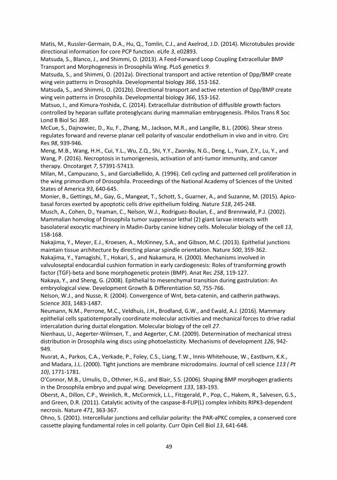
49
Matis, M., Russler-Germain, D.A., Hu, Q., Tomlin, C.J., and Axelrod, J.D. (2014). Microtubules provide directional information for core PCP function. eLife 3, e02893. Matsuda, S., Blanco, J., and Shimmi, O. (2013). A Feed-Forward Loop Coupling Extracellular BMP Transport and Morphogenesis in Drosophila Wing. PLoS genetics 9. Matsuda, S., and Shimmi, O. (2012a). Directional transport and active retention of Dpp/BMP create wing vein patterns in Drosophila. Developmental biology 366, 153-162. Matsuda, S., and Shimmi, O. (2012b). Directional transport and active retention of Dpp/BMP create wing vein patterns in Drosophila. Developmental biology 366, 153-162. Matsuo, I., and Kimura-Yoshida, C. (2014). Extracellular distribution of diffusible growth factors controlled by heparan sulfate proteoglycans during mammalian embryogenesis. Philos Trans R Soc Lond B Biol Sci 369. McCue, S., Dajnowiec, D., Xu, F., Zhang, M., Jackson, M.R., and Langille, B.L. (2006). Shear stress regulates forward and reverse planar cell polarity of vascular endothelium in vivo and in vitro. Circ Res 98, 939-946. Meng, M.B., Wang, H.H., Cui, Y.L., Wu, Z.Q., Shi, Y.Y., Zaorsky, N.G., Deng, L., Yuan, Z.Y., Lu, Y., and Wang, P. (2016). Necroptosis in tumorigenesis, activation of anti-tumor immunity, and cancer therapy. Oncotarget 7, 57391-57413. Milan, M., Campuzano, S., and GarciaBellido, A. (1996). Cell cycling and patterned cell proliferation in the wing primordium of Drosophila. Proceedings of the National Academy of Sciences of the United States of America 93, 640-645. Monier, B., Gettings, M., Gay, G., Mangeat, T., Schott, S., Guarner, A., and Suzanne, M. (2015). Apico-basal forces exerted by apoptotic cells drive epithelium folding. Nature 518, 245-248. Musch, A., Cohen, D., Yeaman, C., Nelson, W.J., Rodriguez-Boulan, E., and Brennwald, P.J. (2002). Mammalian homolog of Drosophila tumor suppressor lethal (2) giant larvae interacts with basolateral exocytic machinery in Madin-Darby canine kidney cells. Molecular biology of the cell 13, 158-168. Nakajima, Y., Meyer, E.J., Kroesen, A., McKinney, S.A., and Gibson, M.C. (2013). Epithelial junctions maintain tissue architecture by directing planar spindle orientation. Nature 500, 359-362. Nakajima, Y., Yamagishi, T., Hokari, S., and Nakamura, H. (2000). Mechanisms involved in valvuloseptal endocardial cushion formation in early cardiogenesis: Roles of transforming growth factor (TGF)-beta and bone morphogenetic protein (BMP). Anat Rec 258, 119-127. Nakaya, Y., and Sheng, G. (2008). Epithelial to mesenchymal transition during gastrulation: An embryological view. Development Growth & Differentiation 50, 755-766. Nelson, W.J., and Nusse, R. (2004). Convergence of Wnt, beta-catenin, and cadherin pathways. Science 303, 1483-1487. Neumann, N.M., Perrone, M.C., Veldhuis, J.H., Brodland, G.W., and Ewald, A.J. (2016). Mammary epithelial cells spatiotemporally coordinate molecular activities and mechanical forces to drive radial intercalation during ductal elongation. Molecular biology of the cell 27. Nienhaus, U., Aegerter-Wilmsen, T., and Aegerter, C.M. (2009). Determination of mechanical stress distribution in Drosophila wing discs using photoelasticity. Mechanisms of development 126, 942-949. Nusrat, A., Parkos, C.A., Verkade, P., Foley, C.S., Liang, T.W., Innis-Whitehouse, W., Eastburn, K.K., and Madara, J.L. (2000). Tight junctions are membrane microdomains. Journal of cell science 113 ( Pt 10), 1771-1781. O'Connor, M.B., Umulis, D., Othmer, H.G., and Blair, S.S. (2006). Shaping BMP morphogen gradients in the Drosophila embryo and pupal wing. Development 133, 183-193. Oberst, A., Dillon, C.P., Weinlich, R., McCormick, L.L., Fitzgerald, P., Pop, C., Hakem, R., Salvesen, G.S., and Green, D.R. (2011). Catalytic activity of the caspase-8-FLIP(L) complex inhibits RIPK3-dependent necrosis. Nature 471, 363-367. Ohno, S. (2001). Intercellular junctions and cellular polarity: the PAR-aPKC complex, a conserved core cassette playing fundamental roles in cell polarity. Curr Opin Cell Biol 13, 641-648.
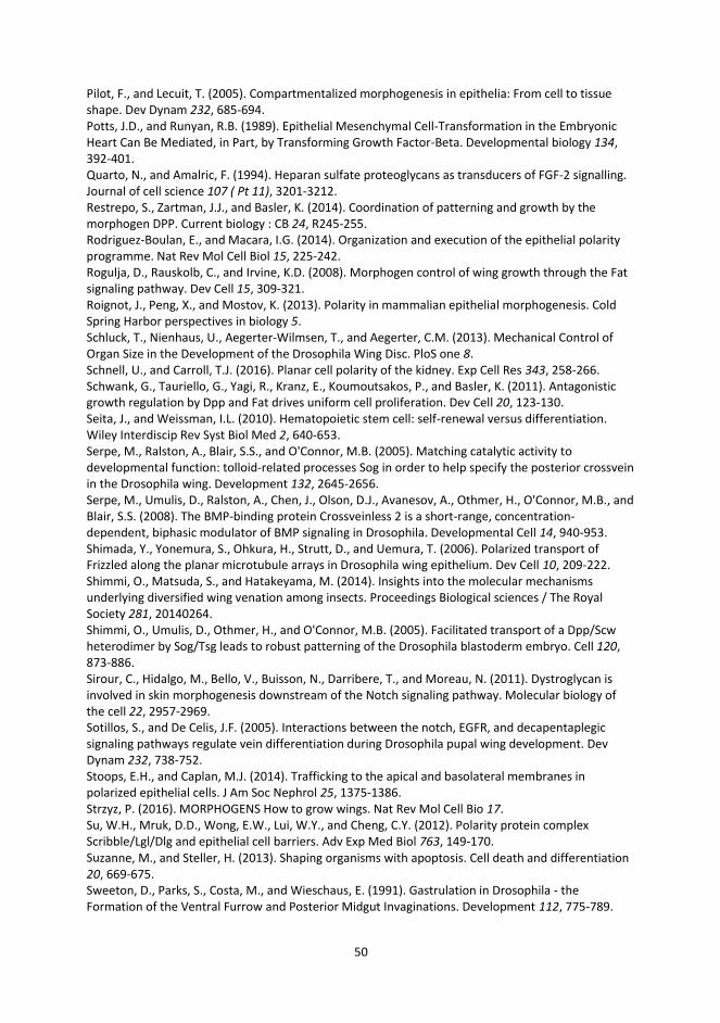
50
Pilot, F., and Lecuit, T. (2005). Compartmentalized morphogenesis in epithelia: From cell to tissue shape. Dev Dynam 232, 685-694. Potts, J.D., and Runyan, R.B. (1989). Epithelial Mesenchymal Cell-Transformation in the Embryonic Heart Can Be Mediated, in Part, by Transforming Growth Factor-Beta. Developmental biology 134, 392-401. Quarto, N., and Amalric, F. (1994). Heparan sulfate proteoglycans as transducers of FGF-2 signalling. Journal of cell science 107 ( Pt 11), 3201-3212. Restrepo, S., Zartman, J.J., and Basler, K. (2014). Coordination of patterning and growth by the morphogen DPP. Current biology : CB 24, R245-255. Rodriguez-Boulan, E., and Macara, I.G. (2014). Organization and execution of the epithelial polarity programme. Nat Rev Mol Cell Biol 15, 225-242. Rogulja, D., Rauskolb, C., and Irvine, K.D. (2008). Morphogen control of wing growth through the Fat signaling pathway. Dev Cell 15, 309-321. Roignot, J., Peng, X., and Mostov, K. (2013). Polarity in mammalian epithelial morphogenesis. Cold Spring Harbor perspectives in biology 5. Schluck, T., Nienhaus, U., Aegerter-Wilmsen, T., and Aegerter, C.M. (2013). Mechanical Control of Organ Size in the Development of the Drosophila Wing Disc. PloS one 8. Schnell, U., and Carroll, T.J. (2016). Planar cell polarity of the kidney. Exp Cell Res 343, 258-266. Schwank, G., Tauriello, G., Yagi, R., Kranz, E., Koumoutsakos, P., and Basler, K. (2011). Antagonistic growth regulation by Dpp and Fat drives uniform cell proliferation. Dev Cell 20, 123-130. Seita, J., and Weissman, I.L. (2010). Hematopoietic stem cell: self-renewal versus differentiation. Wiley Interdiscip Rev Syst Biol Med 2, 640-653. Serpe, M., Ralston, A., Blair, S.S., and O'Connor, M.B. (2005). Matching catalytic activity to developmental function: tolloid-related processes Sog in order to help specify the posterior crossvein in the Drosophila wing. Development 132, 2645-2656. Serpe, M., Umulis, D., Ralston, A., Chen, J., Olson, D.J., Avanesov, A., Othmer, H., O'Connor, M.B., and Blair, S.S. (2008). The BMP-binding protein Crossveinless 2 is a short-range, concentration-dependent, biphasic modulator of BMP signaling in Drosophila. Developmental Cell 14, 940-953. Shimada, Y., Yonemura, S., Ohkura, H., Strutt, D., and Uemura, T. (2006). Polarized transport of Frizzled along the planar microtubule arrays in Drosophila wing epithelium. Dev Cell 10, 209-222. Shimmi, O., Matsuda, S., and Hatakeyama, M. (2014). Insights into the molecular mechanisms underlying diversified wing venation among insects. Proceedings Biological sciences / The Royal Society 281, 20140264. Shimmi, O., Umulis, D., Othmer, H., and O'Connor, M.B. (2005). Facilitated transport of a Dpp/Scw heterodimer by Sog/Tsg leads to robust patterning of the Drosophila blastoderm embryo. Cell 120, 873-886. Sirour, C., Hidalgo, M., Bello, V., Buisson, N., Darribere, T., and Moreau, N. (2011). Dystroglycan is involved in skin morphogenesis downstream of the Notch signaling pathway. Molecular biology of the cell 22, 2957-2969. Sotillos, S., and De Celis, J.F. (2005). Interactions between the notch, EGFR, and decapentaplegic signaling pathways regulate vein differentiation during Drosophila pupal wing development. Dev Dynam 232, 738-752. Stoops, E.H., and Caplan, M.J. (2014). Trafficking to the apical and basolateral membranes in polarized epithelial cells. J Am Soc Nephrol 25, 1375-1386. Strzyz, P. (2016). MORPHOGENS How to grow wings. Nat Rev Mol Cell Bio 17. Su, W.H., Mruk, D.D., Wong, E.W., Lui, W.Y., and Cheng, C.Y. (2012). Polarity protein complex Scribble/Lgl/Dlg and epithelial cell barriers. Adv Exp Med Biol 763, 149-170. Suzanne, M., and Steller, H. (2013). Shaping organisms with apoptosis. Cell death and differentiation 20, 669-675. Sweeton, D., Parks, S., Costa, M., and Wieschaus, E. (1991). Gastrulation in Drosophila - the Formation of the Ventral Furrow and Posterior Midgut Invaginations. Development 112, 775-789.
51
Tabata, T., and Takei, Y. (2004). Morphogens, their identification and regulation. Development 131, 703-712. Tauscher, P.M., Gui, J.H., and Shimmi, O. (2016). Adaptive protein divergence of BMP ligands takes place under developmental and evolutionary constraints. Development 143, 3742-3750. Teleman, A.A., and Cohen, S.M. (2000). Dpp gradient formation in the Drosophila wing imaginal disc. Cell 103, 971-980. Turing, A.M. (1990). The chemical basis of morphogenesis. 1953. Bull Math Biol 52, 153-197; discussion 119-152. Walch, L. (2013). Emerging role of the scaffolding protein Dlg1 in vesicle trafficking. Traffic 14, 964-973. Wang, X., Harris, R.E., Bayston, L.J., and Ashe, H.L. (2008). Type IV collagens regulate BMP signalling in Drosophila. Nature 455, 72-77. Varner, V.D., and Nelson, C.M. (2014). Cellular and physical mechanisms of branching morphogenesis. Development 141, 2750-2759. Vladar, E.K., Bayly, R.D., Sangoram, A.M., Scott, M.P., and Axelrod, J.D. (2012). Microtubules enable the planar cell polarity of airway cilia. Current biology : CB 22, 2203-2212. Wu, J., Roman, A.C., Carvajal-Gonzalez, J.M., and Mlodzik, M. (2013). Wg and Wnt4 provide long-range directional input to planar cell polarity orientation in Drosophila. Nature Cell Biology 15, 1045-+. Wu, S.K., Budnar, S., Yap, A.S., and Gomez, G.A. (2014). Pulsatile contractility of actomyosin networks organizes the cellular cortex at lateral cadherin junctions. European journal of cell biology 93, 396-404. Yan, D., and Lin, X. (2009). Shaping morphogen gradients by proteoglycans. Cold Spring Harbor perspectives in biology 1, a002493. Yeaman, C., Grindstaff, K.K., and Nelson, W.J. (1999). New perspectives on mechanisms involved in generating epithelial cell polarity. Physiol Rev 79, 73-98. Young, P.E., Richman, A.M., Ketchum, A.S., and Kiehart, D.P. (1993). Morphogenesis in Drosophila Requires Nonmuscle Myosin Heavy-Chain Function. Genes & development 7, 29-41. Zeitler, J., Hsu, C.P., Dionne, H., and Bilder, D. (2004). Domains controlling cell polarity and proliferation in the Drosophila tumor suppressor Scribble. The Journal of cell biology 167, 1137-1146. Zhu, J.W., Shang, Y., Wan, Q.W., Xia, Y.T., Chen, J., Du, Q.S., and Zhang, M.J. (2014). Phosphorylation-dependent interaction between tumor suppressors Dlg and Lgl. Cell research 24, 451-463.