Embed Size (px)
Citation preview

The Body’s “Slow but Sure” Endocrine Message System The endocrine system
sends molecules asmessages, just like the nervous system, but it sends them through the bloodstream instead of across synapses.
These molecules, called hormones, are produced in various glands around the body.
The messages go to the brain and other tissues.
Hormones hang out for a bit, so the effect of the message lasts longer. 1
The endocrine system refers to a set of glands that produce chemical messengers called hormones.

1. The sympathetic “fight or flight” nervous system responds to stress by sending a message to adrenal glands to release the hormones listed above.
2. Effect: increased heart rate, blood pressure, and blood sugar. These provide ENERGY for the fight or flight!
Adrenal Glandsproduce hormones such as adrenaline/epinephrine, noradrenaline/norepinephrine, and cortisol.
Pancreas
Adrenal Glands
2

The Pituitary Gland The pituitary gland is the “master gland” of the endocrine system.
It is controlled through the nervous system by the nearby brain area‐‐the hypothalamus.
The pituitary gland produces hormones that regulate other glands such as the thyroid.
It also produces growth hormone (especially during sleep) and oxytocin, the “bonding” hormone.
Pituitary gland
3

Areas of the brain and their functions
The brainstem and cerebellum:
• ensure basic survival
• coordinates the body
The limbic (border) system:
•manages emotions, and connects thought to body
The cortex (the outer covering):
• integrates information
4

The medulla controls the most basic functions such as heartbeat and respiration.
The pons (“bridge”) helps coordinate automatic and unconscious movements. It relays signals
from the forebrain to the cerebellum, along with nuclei that deal primarily with sleep,
respiration, swallowing, bladder control, hearing, equilibrium, taste, eye movement, facial
expressions, facial sensation, and posture. The pons is implicated in sleep paralysis, and also
plays a role in generating dreams 5
The Brainstem: Pons and Medulla

Reticular (“Netlike”) Formation The reticular formation is a nerve network in the brainstem. It enables alertness, (arousal) from deep sleep to wide awake when stimulated and produces permanent coma when lesioned (as demonstrated in the cat experiments). It also filters incoming sensory information and controls selective awareness. 6

The Thalamus (“Inner Chamber”) The thalamus is the “sensory switchboard” or “router.”
All sensory messages, except smell, are routed through the thalamus on the way to the cortex (higher, outer brain).
The thalamus also sends messages from the cortex to the medulla and cerebellum.
Damage to the thalamus can cause blindness and other loss of the senses, even if the sensory organ is fine.
7

The cerebellum helps coordinate voluntary movement and balance, such as playing a sport.
Cerebellum (“little brain”)
The cerebellum also is the area where implicit memories and conditioning are stored. It also helps us judge time, modulate emotions, and integrate multiple sources of sensory input.
8

emotions such as fear and aggression.
basic drives such as hunger and sex.
the formation of episodic memories.
The hippocampus (“seahorse”) processes conscious, episodic
memories. works with the amygdala to
form emotionally charged memories.
one of the few places in the brain in which neurogenesis is known to take place.
The Amygdala (“almond”) consists of two lima bean‐
sized neural clusters. helps process emotions,
especially fear and aggression.
The Limbic (“Border”) SystemThe limbic system coordinates:
9

The Amygdala Electrical stimulation of a cat’s amygdalaprovokes aggressive reactions. If you move the electrode very slightly and cage the cat with a mouse, the cat will cower in terror. Destruction of part of the amygdala can apparently eliminate both emotions.
10

Hypothalamus
DUDE!!! WHAT HAPPENED???

lies below (“hypo”) the thalamus.
regulates body temperature and ensures adequate food and water intake (homeostasis), and is involved in sex drive.
directs the endocrine system via messages to the pituitary gland.
The Hypothalamus:Thalamus
Riddle: Why did the rat cross the grid? Why did the rat want to get to the other side?
The Hypothalamus as a Reward Center
Pushing the pedal that stimulated the electrode placed in the hypothalamus was much more rewarding than food pellets. 12

Review of Brain Structures
13

The Cerebral CortexThe lobes consist of:
300 billion synaptic connections
The brain has left and right hemispheres
outer grey “bark” structure that is wrinkled in order to create more surface area for 20+ billion neurons.
inner white stuff—axons linking parts of the brain. 180+ billion glial cells, which feed and protect neurons
and assist neural transmission.
14

15
The Lobes of the Cerebral Cortex: Preview
Frontal Lobes
Parietal Lobes
Occipital Lobes
Temporal Lobes
involved in speaking and muscle movements and in making plans and judgments
include the sensory cortex
include the visual areas; they receive visual information from the opposite visual field
include the auditory processing areas

Input: Sensory cortex (Left hemisphere section receives input from the body’s right side)
Output: Motor cortex (Left hemisphere section controls the body’s right side)
Functions of the Brain: The Motor and Sensory Strips
Axons receiving motor signals FROMthe cortex
Axons sending sensory
information TO the cortex16

Sensory Functions of the Cortex The sensory strip deals with information from touch stimuli. The occipital lobe deals with visual information. Auditory information is sent to the temporal lobe. Auditory areas are also active when someone in a psychotic state is experiencing “voices” or auditory hallucinations
17

The Visual Cortex
This fMRI scan shows increased activity in the visual cortex when a person looks at a photograph.
18
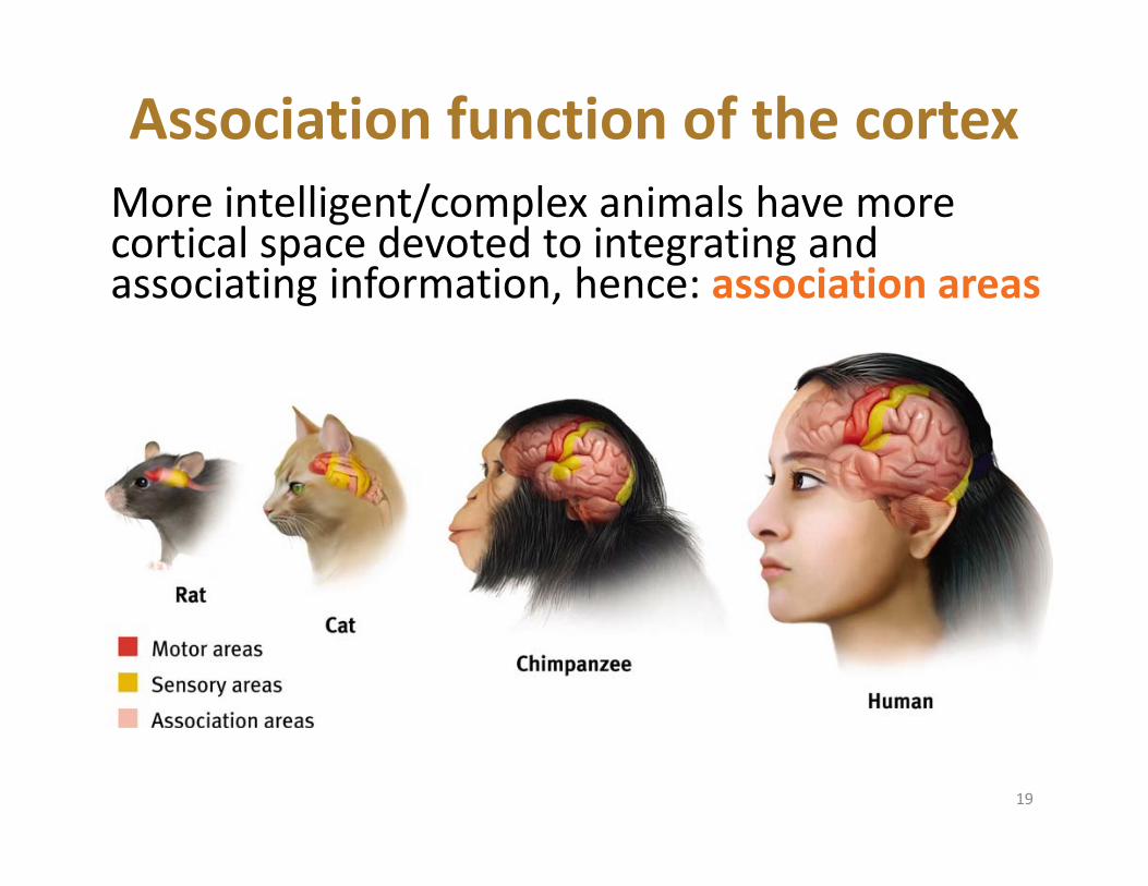
Association function of the cortexMore intelligent/complex animals have more cortical space devoted to integrating and associating information, hence: association areas
19

Association Areas:Frontal Lobes
The frontal lobes are active in “executive functions” such as judgment, planning, and inhibition of impulses. The frontal lobes are also active in the use of working memory and the processing of new memories. They contribute largely to making us uniquely “human.” 20

Phineas Gage (1823‐1860)
Case study: In a work accident, a metal rod shot up through Phineas Gage’s skull, destroying his eye and part of his frontal lobes. After healing, he was able to function in many ways, but his personality changed; he was rude, odd, irritable, and unpredictable.
Possible explanation: Damage to the frontal lobes could result in loss of the ability to suppress impulses and to modulate emotions.
21
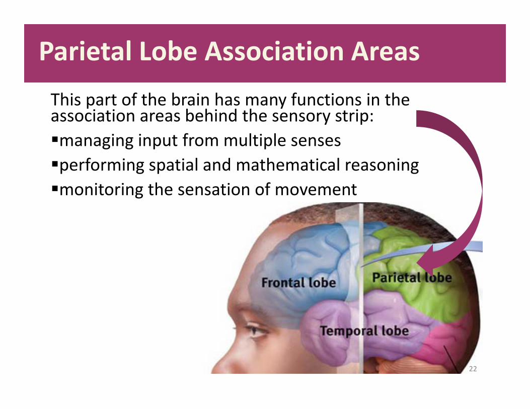
Parietal Lobe Association AreasThis part of the brain has many functions in the association areas behind the sensory strip:managing input from multiple sensesperforming spatial and mathematical reasoningmonitoring the sensation of movement
22

Temporal Lobe Association Areas
Some abilities managed by association areas in this “by the temples” lobe: recognizing specific facesmanaging sensory input related to sound, which helps the understanding of spoken words 23

Whole‐brain Association Activity
Whole‐brain association activity involves complex activities which require communication among association areas across the brain such as:memory language attentionmeditation and spirituality consciousness
24

Specialization and Integration Five steps in reading a word aloud:
25

Language – A Lateralized Function
Broca’s Area an area of the left frontal
lobe that directs the muscle movements involved in speech
Expressive Aphasia difficulty initiating and
executing voluntary movement patterns necessary to produce speech when there is no paralysis or weakness of speech muscles.
Signs: Producing the desired speech sound.
Using the correct rhythm and rate of speaking.

Language – A Lateralized Function
Wernicke’s Area an area of the left temporal
lobe involved in language comprehension and expression
Receptive Aphasia impairment in the ability to
use or comprehend words Signs:
Understanding words. Finding the word to express a thought
Understanding grammatical sentences.
Reading or writing words or sentences.

This 6‐year‐old had a hemispherectomy to end life‐threatening seizures; her remaining hemisphere compensated for the damage.
Plasticity: The Brain is Flexible
If the brain is damaged, especially in the general association areas of the cortex: the brain does not repair damaged neurons, BUT it can restore some functionsit can form new connections, reassign existing networks, and insert new neurons, some grown from stem cells
28

Cerebral Cortex – Neural Organization Experiment
• Some rats are housed alone in empty cages
• Their littermate twins are group‐housed in cages with toys, which are changed frequently
• Richer environments led to heavier, thicker brains, more synapses, and better learning

Our Two Hemispheres
Lateralization (“going to one side”)The two hemispheres serve some different functions.How do we know about these differences? Brain damage studies revealed many functions of the left hemisphere. Brain scans and split brain studies show more about the functions of the two hemispheres, and how they coordinate with each other.
30

Our Divided Brain – The Two Hemispheres
• Left Hemisphere– Verbal– Math– Analytic– Boring
• Right Hemisphere– Spatial– Holistic– Nonverbal– Fun Party
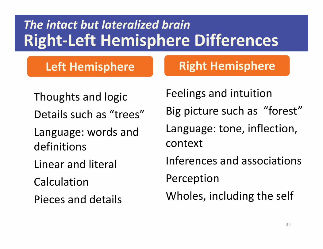
Thoughts and logicDetails such as “trees”Language: words and definitionsLinear and literal CalculationPieces and details
Feelings and intuitionBig picture such as “forest”Language: tone, inflection, contextInferences and associationsPerceptionWholes, including the self
The intact but lateralized brainRight‐Left Hemisphere Differences
Left Hemisphere Right Hemisphere
32

Brain Studies
Researchers have studied the impact of this surgery on patients’ functioning.
Split‐
To end severe whole‐brain seizures, some people have had surgery to cut the corpus callosum, a band of axons connecting the hemispheres.
33

Split visual field
Each hemisphere does not perceive what each EYE sees. Instead, it perceives the half of the view in front of you that goes with the half of the body that is controlled by that hemisphere.
34

35
Divided Awareness in the Split BrainTry to explain the following result:

The divided brain in action
Talent: people are able to follow two instructions and draw two different shapes simultaneously
Drawback: people can be frustrated that the right and left sides do different things
36