Embed Size (px)
Citation preview
Bone regeneration and stem cells
K. Arvidson a, *, B. M. Abdallah b, L. A. Applegate c, N. Baldini d, E. Cenni d, E. Gomez-Barrena e, D. Granchi d, M. Kassem b, f, Y. T. Konttinen g, h, i, K. Mustafa a, D. P. Pioletti c, j,
T. Sillat g, A. Finne-Wistrand k
a Department of Clinical Dentistry, Center for Clinical Resarch, Faculty of Medicine and Dentistry, University of Bergen, Bergen, Norwayb Department of Endocrinology, Molecular Endocrinology Laboratory, Odense University Hospital &
Medical Biotechnology Centre, University of Southern Denmark, Odense, Denmarkc Department of Musculoskeletal Medicine, Service of Plastic and Reconstructive Surgery,
Cellular Therapy Unit, University Hospital of Lausanne, Lausanne, Switzerlandd Laboratory for Orthopaedic Pathophysiology and Regenerative Medicine, Istituto Ortopedico Rizzoli, Bologna, Italy
e Servicio de Cirugía Ortopédica y Traumatología, Hospital, Universitario ‘La Paz’, Universidad Autónoma de Madrid, Madrid, Spain
f Department of Anatomy, Stem Cell Unit, College of Medicine, King Saud University, Riyadh, Saudi Arabiag Department of Medicine, Helsinki University Central Hospital, Helsinki, Finland
h ORTON Orthopaedic Hospital, ORTON Foundation, Helsinki, Finlandi COXA Hospital for Joint Replacement, Tampere, Finland
j Laboratory of Biomechanical Orthopedics, Institute of Bioengineering, Ecole Polytechnique Fédérale Lausanne, Lausanne, Switzerlandk Royal Institute of Technology, Fibre and Polymer Technology, School of Chemical Science and Engineering, Stockholm, Sweden
Received: August 7, 2010; Accepted: November 2, 2010
J. Cell. Mol. Med. Vol 15, No 4, 2011 pp. 718-746
© 2011 The AuthorsJournal of Cellular and Molecular Medicine © 2011 Foundation for Cellular and Molecular Medicine/Blackwell Publishing Ltd
doi:10.1111/j.1582-4934.2010.01224.x
Guest Editor: N.I. Moldovan
*Correspondence to: Prof. Kristina ARVIDSON, Department of Clinical Dentistry, Årstadsv.17, 5009 Bergen, Norway.
Tel.: !46707368988Fax: !468858140E-mail: [email protected]
Regenerative Medicine Review Series
• Introduction• Bone fracture healing and healing problems• Biomaterial scaffolds and tissue engineering in bone formation
- Bone tissue engineering- Biomaterial scaffolds- Synthetic scaffolds- Micro- and nanostructural properties of scaffolds- Conclusion
• Mesenchymal stem cells and osteogenesis- Bone tissue- Origin of osteoblasts- Isolation and characterization of bone marrow derived MSC- In vitro differentiation of MSC into osteoblast lineage cells- In vivo differentiation of MSC into bone- Factors and pathways controlling osteoblast differentiation
of hMSC- Defining the relationship between osteoblast and adipocyte
differentiation from MSC- MSC and sex hormones- Effect of aging on osteoblastogenesis- Conclusion
• Embryonic, foetal and adult stem cells in osteogenesis- Cell-based therapies for bone- Specific features of bone cells needed to be advantageous
for clinical use- Development of therapeutic biological agents- Clinical application concerns- Conclusion
• Platelet-rich plasma (PRP), growth factors and osteogenesis- PRP effects in vitro on the cells involved in bone repair
- PRP effects on osteoblasts- PRP effects on osteoclasts - PRP effects on endothelial cells
- PRP effects in vivo on experimental animals- The clinical use of PRP for bone repair
- Non-union- Distraction osteogenesis- Spinal fusion- Foot and ankle surgery- Total knee arthroplasty- Odontostomatology and maxillofacial surgery
- Conclusion• Molecular control of osteogenesis
- TGF-"" signalling- FGF signalling- IGF signalling- PDGF signalling- MAPK signalling pathway- Wnt signalling pathway- Hedgehog signalling- Notch signalling- Ephrin signalling- Transcription factors regulating osteoblast
differentiation- Conclusion
• Summary

J. Cell. Mol. Med. Vol 15, No 4, 2011
719© 2011 The AuthorsJournal of Cellular and Molecular Medicine © 2011 Foundation for Cellular and Molecular Medicine/Blackwell Publishing Ltd
AbstractThis invited review covers research areas of central importance for orthopaedic and maxillofacial bone tissue repair, including normalfracture healing and healing problems, biomaterial scaffolds for tissue engineering, mesenchymal and foetal stem cells, effects of sexsteroids on mesenchymal stem cells, use of platelet-rich plasma for tissue repair, osteogenesis and its molecular markers. A variety ofcells in addition to stem cells, as well as advances in materials science to meet specific requirements for bone and soft tissue regener-ation by addition of bioactive molecules, are discussed.
Keywords: bone regeneration • stem cells • biomaterials • polymers • regenerative medicine
Introduction
New approaches to clinical problems based on translational med-icine start with basic research and progress ‘hand-in-hand’ withclinical observations. Scientists are increasingly aware that this‘bench-to-bedside’ approach to translational research is really atwo-way street which can strengthen and accelerate critical pointsof the research process.
Bone defects due to trauma and to pathological and physio-logical bone resorption represent a major challenge and are aglobal health problem. The need for bone regeneration in cranial,oral and maxillo-facial and orthopaedic surgery is one of the central clinical issues in regenerative and rehabilitation medicine.It is difficult to convey the enormous social and psychologicalhandicap of persons with bone defects and the significant reduc-tion in their quality of life. In addition to trauma, bone healingproblems may be related to age, sex and infection as exemplifiedby diagnoses such as osteoporosis, osteopenia and severe dental problems related to loss of teeth. The aim of this review isto describe the current state of the art in our understanding ofbone healing and bone regeneration.
Bone fracture healing and healingproblems
Bone repair after fracture is a special process where sequentialcellular and molecular events take place to generate new bone,rather than a fibrous scar like other connective tissues. The pre-cise series of ordered events required to produce new bone aremodulated by systemic and local factors, and disruption of theseorderly events may cause healing problems. Thus, a clear under-standing of the sequence of events and their regulation is neededto decide when and how an intervention is required to promotehealing and to avoid complications.
Since early histological descriptions of fracture healing in man[1], the general pattern of indirect fracture healing, based on endo-chondral ossification, has included the chronological phases ofhaematoma, inflammation, angiogenesis, chondrogenesis toosteogenesis and finally bone remodelling [2]. Direct healingbased on membranous ossification, with no periosteal reaction or
visible callus formation, is seldom seen. The well-establishedcharacteristics of the above mentioned phases require differentprocesses of cell migration and differentiation, extracellular matrixformation and organization towards calcification, as well as bothlocal and systemic modulation. Apart from the classical histologi-cal phases of fracture healing, much remains to be understoodabout the regulation of these processes both at the molecular andthe cellular level.
Formation of a haematoma related to blood vessel damage is accompanied by an inflammatory response [3], where pro-inflammatory cytokines such as interleukin-1 (IL-1), IL-6, and particularly tumour necrosis factor-#, initiate the bone healingcascade and push it towards endochondral bone formation andremodelling [4]. Secondarily, apart from ischemia, growth and differentiation factors and particularly the transforming growthfactor-" (TGF-") superfamily, including bone morphogenetic protein (BMPs), as well as platelet-derived growth factors (PDGF),fibroblast growth factors (FGFs) and insulin-like growth factors(IGFs), orchestrate crucial events for chondro-osteogenesis,including chemotaxis, mesenchymal and osteoprogenitor cell pro-liferation and differentiation, and extracellular matrix ossification[5]. Finally, angiogenesis, a key aspect of fracture healing, is alsoregulated at the molecular level. An angiopoietin pathway has beendescribed in the early stages of the healing process [6], as has avascular endothelial growth factor (VEGF)-dependent pathwayrelated to endochondral bone formation, where BMPs stimulatethe expression of VEGF by osteoblasts and osteoblast-like cells[7]. Inhibitory molecules are also needed to control growth factors(GFs), and various BMP antagonists [8] are released into theextracellular compartment (noggin, sclerostatin, follistatin, etc.).Other inhibitory mechanisms include receptor inhibition of somemembers of the TGF-" superfamily that has been related to apseudo-receptor defined as BAMBI (BMP and activin membranebound inhibitor) [9], and intracellular inhibition by the activation ofI-Smads [10, 11], among other mechanisms. Many aspects of thisbone healing cascade of molecules, cells and events have beenidentified, but the complex interactions and processes are stillonly partially understood.
Interestingly, adult bone healing reproduces normal develop-ment during embryogenesis, except for the associated inflamma-tion, despite the limited number of osteoprogenitor cells available
720 © 2011 The AuthorsJournal of Cellular and Molecular Medicine © 2011 Foundation for Cellular and Molecular Medicine/Blackwell Publishing Ltd
in the adult, and the strong influence of external and internalmechanics. This fact offers researchers an invaluable clue towardsbiological events in the developmental processes of skeletogene-sis [12]. Bone formation during embryogenesis is initiated bymesenchymal stem cells (MSC) aggregation and condensation,which then progresses to endochondral ossification through car-tilage formation, or to membranous ossification throughosteoblast differentiation. Although MSC are scarce in adult bone,both committed osteoprogenitor cells from the periostium andundifferentiated multipotent MSC from bone marrow are involvedin callus formation, which is important in the structural progressof fracture healing. In addition to the classical triad of bone heal-ing (cells, extracellular matrix and osteoinductive factors), a fourthmajor factor (the biomechanical properties of the callus) has beenstressed recently [13, 14]. Mechanical influences on biologicalprocesses, known as mechanobiology, significantly affect allphases of bone formation. Not only may major external forces dis-rupt the healing process, but mechanical loading influences onendochondral ossification are also important as compressionenhances bone apposition [13], empirically defined since the 19thcentury as Wolff’s law. However, earlier phases of osteogenesisshow increased cell proliferation if cyclic motion with associatedshear stresses occurs, although intramembranous ossificationmay be permitted in areas with low stress and strain. Mechanicalsignalling at the cellular level may modulate molecular changes incytoskeleton, integrins and ion channel activities with conse-quences to the differentiation and gene expression of cellsinvolved in the healing process. Transduction mechanisms rangefrom direct mechanical stimulation of cells [15, 16], to fluid shearstresses and various matrix effects that indirectly affect cells [17].Many of these experimental studies were developed after clinicalstudies showed the beneficial influence of interfragmentarymotion (of about 0.6 mm) in the early stages of the healingprocess [18], and the role of mechanical stimulus in general as aninfluence on the rate of healing.
Despite this well developed natural mechanism of fracturehealing, alteration of local and systemic factors may lead toimpaired healing. Non-union (or pseudoarthrosis), due to the falsearticulation generated in the area of the unhealed fracture, anddelayed union, are the main problems in bone repair and are significant health care issues. Systemic factors such as use of non-steroidal anti-inflammatory drugs (which inhibit cyclooxygenaseand therefore prostaglandins required in the inflammation phase),age (with decreased expression of mediators, hormonal changes,impaired osteoblast function), or smoking [19], and local factorsboth influence fracture healing. Mechanical stability is required forcallus formation, and surgical techniques focusing on stabilitygreatly improve the chances of fracture healing, as excess motionimpairs it. Angiogenesis and appropriate tissue differentiation, keyfactors required for callus formation, are also improved with fracture stability [20]. However, the rate of differentiation of osteo-progenitors is modulated by the mechanical environment, thusosteogenic differentiation may be promoted or impaired by move-ment. When tensile strains occur in the interfragmentary gap, theylead to ‘cleavage’ of the callus, and hydrostatic pressure and
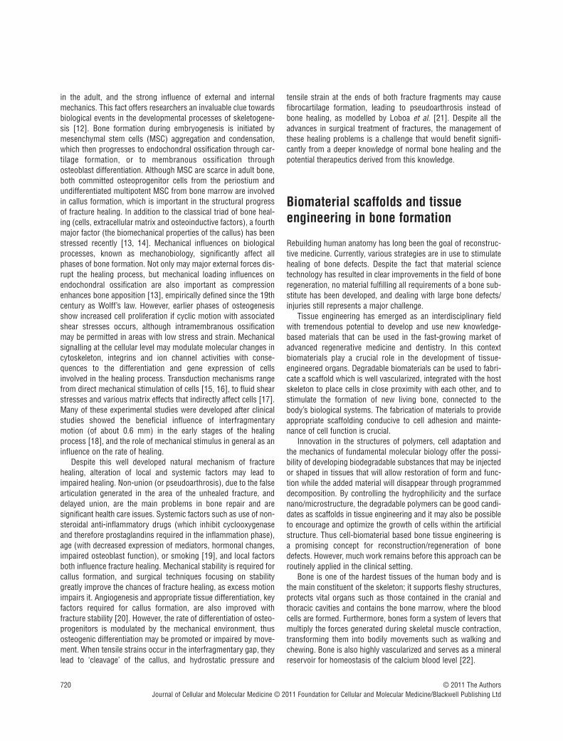
tensile strain at the ends of both fracture fragments may causefibrocartilage formation, leading to pseudoarthrosis instead ofbone healing, as modelled by Loboa et al. [21]. Despite all theadvances in surgical treatment of fractures, the management ofthese healing problems is a challenge that would benefit signifi-cantly from a deeper knowledge of normal bone healing and thepotential therapeutics derived from this knowledge.
Biomaterial scaffolds and tissue engineering in bone formation
Rebuilding human anatomy has long been the goal of reconstruc-tive medicine. Currently, various strategies are in use to stimulatehealing of bone defects. Despite the fact that material sciencetechnology has resulted in clear improvements in the field of boneregeneration, no material fulfilling all requirements of a bone sub-stitute has been developed, and dealing with large bone defects/injuries still represents a major challenge.
Tissue engineering has emerged as an interdisciplinary fieldwith tremendous potential to develop and use new knowledge-based materials that can be used in the fast-growing market ofadvanced regenerative medicine and dentistry. In this context biomaterials play a crucial role in the development of tissue-engineered organs. Degradable biomaterials can be used to fabri-cate a scaffold which is well vascularized, integrated with the hostskeleton to place cells in close proximity with each other, and tostimulate the formation of new living bone, connected to thebody’s biological systems. The fabrication of materials to provideappropriate scaffolding conducive to cell adhesion and mainte-nance of cell function is crucial.
Innovation in the structures of polymers, cell adaptation andthe mechanics of fundamental molecular biology offer the possi-bility of developing biodegradable substances that may be injectedor shaped in tissues that will allow restoration of form and func-tion while the added material will disappear through programmeddecomposition. By controlling the hydrophilicity and the surfacenano/microstructure, the degradable polymers can be good candi-dates as scaffolds in tissue engineering and it may also be possibleto encourage and optimize the growth of cells within the artificialstructure. Thus cell-biomaterial based bone tissue engineering isa promising concept for reconstruction/regeneration of bonedefects. However, much work remains before this approach can beroutinely applied in the clinical setting.
Bone is one of the hardest tissues of the human body and isthe main constituent of the skeleton; it supports fleshy structures,protects vital organs such as those contained in the cranial andthoracic cavities and contains the bone marrow, where the bloodcells are formed. Furthermore, bones form a system of levers thatmultiply the forces generated during skeletal muscle contraction,transforming them into bodily movements such as walking andchewing. Bone is also highly vascularized and serves as a mineralreservoir for homeostasis of the calcium blood level [22].

J. Cell. Mol. Med. Vol 15, No 4, 2011
721© 2011 The AuthorsJournal of Cellular and Molecular Medicine © 2011 Foundation for Cellular and Molecular Medicine/Blackwell Publishing Ltd
Bone tissue develops either by intramembranous or endochon-dral ossification and the embryonic development starts as early asthe fourth to sixth week. Membranous or direct ossification occursin parts of the skull or craniofacial skeleton, clavicles and scapu-lae when mesenchymal cells proliferate and gradually change theirshape, which occurs within a layer of connective tissue.Endochondral or indirect ossification takes place within a cartilagi-nous model in the long bones and the rest of the skeleton, whichin addition to the growth of the length also increase in width viaappositional bone growth. This cartilage model is graduallydestroyed and replaced by bone formed by incoming cells fromadjacent periosteal connective tissues. In both processes the bonetissue appears first as primary or immature tissue and grows rapidly from the first postnatal year to the end of adolescence. Inthe adult skeleton bone tissue is either arranged in a trabecularpattern or in a compact pattern [23]. A tentative third mode ofbone formation has been described in distraction osteogenesisstudies. Transchondroid ossification produces bone via chondroidbone. It is speculated that hypertrophic and/or early stage chon-drocytes undergo differentiation into osteoblast-like cells, whichlay done bone without capillary invasion.
As the development of bone is very complicated, birth bonedefects can occur in any bone although the bones of the skull andface, spine, hips, legs and feet are affected most often resulting inabnormal appearance and function [24]. The most commondefects of the skull and face are cleft lip, cleft palate and jaw defor-mities. Most of these defects can be repaired surgically. Often, thesurgery is complex and involves reconstructing deformed orabsent body parts [25–27].
Bone defects due to trauma, and to pathological and physiolog-ical bone resorption still represent a major challenge. The need forbone regeneration in cranial, oral maxillo-facial and orthopaedicsurgery is a major clinical issue. Most fractures heal well usingstandard treatments, mostly without any scar tissue formation.However, bone defects due to tumour resections, unhealed frac-tures, major trauma and bone resorption of edentulous jaws or oftooth-supporting alveolar bone are candidates for bone recon-struction and cause significant handicap without adequate treat-ment. In neurosurgery, spinal fusion is performed in manypatients suffering from intervertebral disc degeneration, and thisprocedure also requires bone grafting.
Plastic and reconstructive surgeons conventionally fill andrepair bony defects using autologous bone transplantation as thegold standard [28]. Current clinical treatments to repair bonydefects can be problematic and often yield poor healing due to theanatomy and physiology of bone tissue, as well as the limitationsof knowledge of the process. Because of the major problems asso-ciated with autograft transplantation, such as insufficient tissue,donor-site injury and surgical risks as such as bleeding, infectionand chronic pain, alternative approaches are needed. Skeletaldefects may require volumes of bone often not available. The donorsite for bone harvesting is usually the iliac crest, which requires asecond surgical intervention and has some surgical morbidity.Allografts can be used, especially in prosthetic reconstruction, butmay still not solve many problems of bone deficiency.
Because of the disadvantages associated with both autograftsand allografts, scientists have long searched for biocompatiblematerials that could be substituted for the transplanted bone.Although most of the available synthetic bone substitutes havesome of the positive properties of an autograft, to date no singlesynthetic material offers all the benefits of the patient’s own bone.For instance, calcium phosphate bioceramics do not possess sufficient osteogenic properties to allow reconstruction of largedefects [29, 30]. Thus despite the commercial availability of manybone substitute materials for clinical application, the use of alloplastic materials and autologous bone grafting remains thepreferred approach to treatment of bone defects [31]. Thus, inbone tissue engineering, it may be of importance to primarilyspecify the area and the function for regeneration or implantationof bone. In those cases, orthopaedics, neurosurgery and maxillo-facial surgery and implant dentistry may have different treatmentmodalities and may ask for materials to be used either in a solidor an injectable phase for the different targets.
Bone tissue engineering
In view of the above limitations and the increasing demand forbone grafting procedures, surgeons are looking for a betterapproach. Tissue engineering combines bone marrow cells orMSC, synthetic scaffolds and molecular signals (growth or differ-entiating factors) to form hybrid constructs. In a classicalapproach, bone tissue engineering consists of harvesting bonemarrow from a patient, isolating MSC by adherence to tissue cul-ture plastic, expanding and differentiating MSC in culture and thenseeding them onto a suitable synthetic scaffold prior to implanta-tion into the same patient [32]. It has been shown that from asmall volume (0.1–3 ml) aspirate, alveolar bone marrow stromalcells (BMSC) expand by 70% [33].
Although this raises few ethical issues, harvesting of cells frombone marrow is still an invasive procedure, and in addition stemcell numbers may decrease significantly with the age of the individual. The search for more readily accessible sources ofpluripotent cells has led to investigation of other tissues, includingmobilized peripheral blood, umbilical cord blood and morerecently, periodontal ligament and deciduous and permanentteeth. Dental pulp tissue is also a readily accessible source ofpulp-derived MSC (PDSC) and can be easily isolated. PDSCexpress the endothelial and smooth muscle marker STRO-1 [34]and display a pericyte phenotype, with expression of the pericyte-associated antigen 3G5 [34]. It is therefore assumed, but not confirmed, that the perivascular region in the pulp is the niche forPDSC and that pericytes give rise to dental pulp stem cells.Isolated dental pulp stem cells have been shown to be plastic-adherent and express the MSC markers STRO-1, CD90, CD29,CD44, CD166, CD105, CD106, CD146, CD13 and are also negativefor CD14 and CD34 [35, 36]. In vitro, PDSC are capable of self-renewal, display plasticity and mutilineage potential (adipocytes,chondrocytes, osteoblasts, neural cell progenitors and myotubes)and can therefore be defined as stem cells [37].
722 © 2011 The AuthorsJournal of Cellular and Molecular Medicine © 2011 Foundation for Cellular and Molecular Medicine/Blackwell Publishing Ltd
To date, these approaches have been evaluated in both animalexperiments and clinical studies involving low numbers ofpatients, with inconclusive outcome and low bone regenerationefficacy [38–40]. Overall, the clinical outcome have been disap-pointing, especially with respect to restoration of large bonedefects. The limited success of clinical studies to date may beattributable to a number of issues, related primarily to translationof laboratory procedures to clinical application. Translation of laboratory processes into clinically effective, reproducible, safe,economically viable and competitive products is generallyacknowledged to be a complicated and challenging phase in thedevelopment of new clinical techniques [32]. In the context of bonetissue engineering, the difficulties reflect the multidisciplinarynature of the concept.
Biomaterial scaffolds
Bone tissue engineering utilizes scaffolds to deliver biofactorsincluding cells, genes and proteins to generate bone and assess-ment of blood vessel formation and maturation into the construct.The scaffold itself must fulfil three primary functions to ensure suc-cessful treatment of bone defects. First, the scaffold must providethe correct anatomic geometry to define and maintain the space fortissue regeneration. Second, the scaffold must provide temporarymechanical load bearing within the tissue defect and third, the scaffold should enhance the regenerative capability of the chosenbiofactor; a balance to a regenerative capacity. For load-bearing purposes, achieving stiffness and strength equivalent to bone tissuerequires minimally porous scaffolds. Conversely, enhanced deliveryof biofactors requires more highly connected porous scaffolds toallow cell migration, vascularization and connective tissue formationwithin scaffolds. In clinical applications, it is obvious that scaffolds,matching complex anatomic defects require computational methodsand control of anatomic shape and architecture.
Many different materials have been proposed as synthetic bonesubstitutes. Hydroxyapatite (HA), Ca10(PO4)6(OH)2, is regarded asone of the most bioactive bone substitute ceramics because of its
superior osteoconductivity. This applies to HA of synthetic or biological origin, "-tricalcium phosphate ("-TCP) and biphasiccalcium phosphate. Calcium phosphate biomaterials are currentlyused, for example, for bone repair, substitution, augmentation andregeneration. As synthetic bone substitutes, the main advantagesof the ceramics are excellent biocompatibility and better stabilitythan polymers and metals in the living body. Synthetic octacalciumphosphate has been shown to be a good precursor of biologicalapatite in bone and tooth and has shown better bone regenerativeand biodegradable characteristics than other calcium phosphatebone substitute materials [41]. A disadvantage is that it is some-times difficult to achieve close apposition of the material to thesurrounding bone, especially in complicated defects and this isessential for complete and successful bone regeneration. Anothergeneral disadvantage of the ceramics is inherent brittleness. Thiscan be overcome by blending ceramics with, for example, poly-esters to form a composite that combines the mechanical strengthand osteoconductivity of calcium phosphates with the high affinityfor cells and good biodegradability of polyesters.
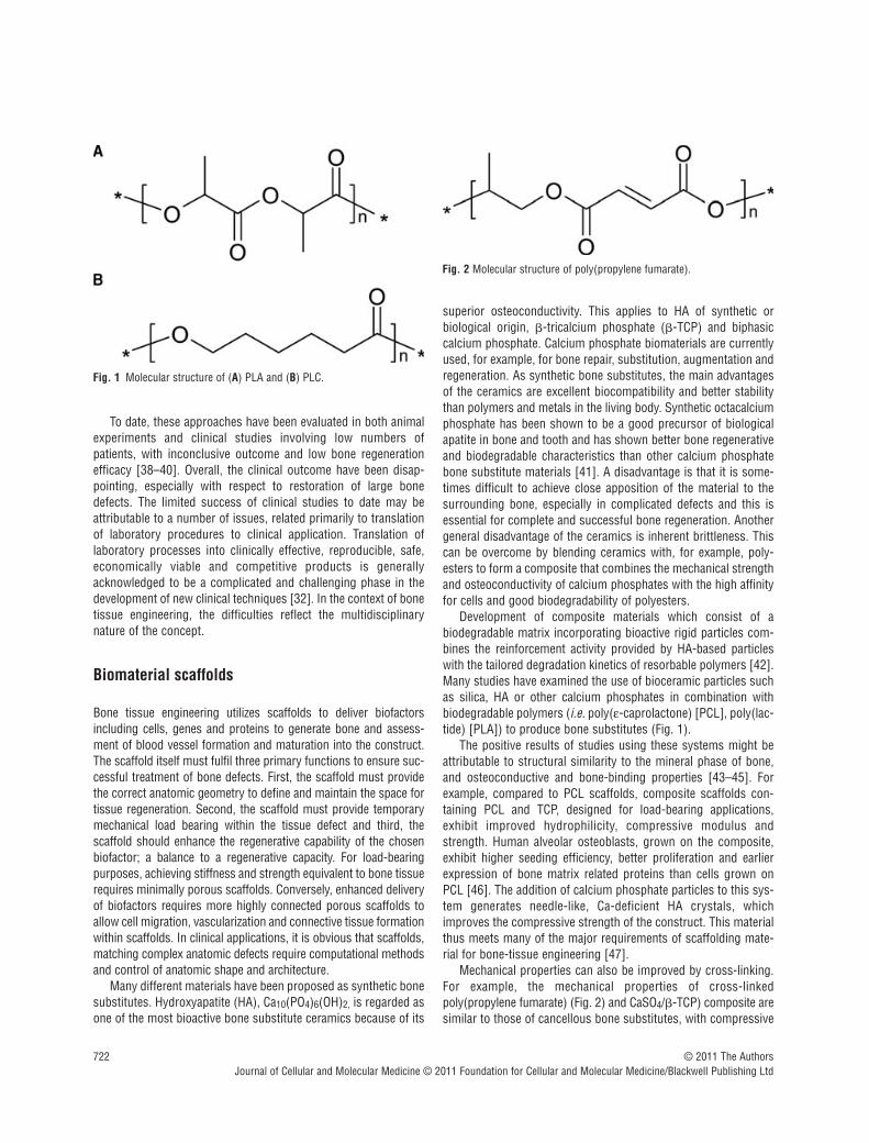
Development of composite materials which consist of abiodegradable matrix incorporating bioactive rigid particles com-bines the reinforcement activity provided by HA-based particleswith the tailored degradation kinetics of resorbable polymers [42].Many studies have examined the use of bioceramic particles suchas silica, HA or other calcium phosphates in combination withbiodegradable polymers (i.e. poly(!-caprolactone) [PCL], poly(lac-tide) [PLA]) to produce bone substitutes (Fig. 1).
The positive results of studies using these systems might beattributable to structural similarity to the mineral phase of bone,and osteoconductive and bone-binding properties [43–45]. Forexample, compared to PCL scaffolds, composite scaffolds con-taining PCL and TCP, designed for load-bearing applications,exhibit improved hydrophilicity, compressive modulus andstrength. Human alveolar osteoblasts, grown on the composite,exhibit higher seeding efficiency, better proliferation and earlierexpression of bone matrix related proteins than cells grown onPCL [46]. The addition of calcium phosphate particles to this sys-tem generates needle-like, Ca-deficient HA crystals, whichimproves the compressive strength of the construct. This materialthus meets many of the major requirements of scaffolding mate-rial for bone-tissue engineering [47].
Mechanical properties can also be improved by cross-linking.For example, the mechanical properties of cross-linkedpoly(propylene fumarate) (Fig. 2) and CaSO4/"-TCP) composite aresimilar to those of cancellous bone substitutes, with compressive
Fig. 1 Molecular structure of (A) PLA and (B) PLC.
Fig. 2 Molecular structure of poly(propylene fumarate).
J. Cell. Mol. Med. Vol 15, No 4, 2011
723© 2011 The AuthorsJournal of Cellular and Molecular Medicine © 2011 Foundation for Cellular and Molecular Medicine/Blackwell Publishing Ltd
strengths of 5 MPa and a compressive modulus of 50 MPa duringdegradation [48]. Composite bone substitutes are also available ininjectable form, an obvious advantage in the clinical setting. Forexample, HA-atelocollagen composites have proved suitable pre-cursors for osseous regeneration [49]. However, despite manypositive outcome, the results of most studies to date show that theincorporation of a ceramic phase improves bioactivity, but themechanical properties of the composites are inadequate [50].Another observed disadvantage is that the HA particles within aPCL matrix tend to migrate to the surface and form clusters, cre-ating inhomogeneous scaffolds [51].
Another fundamental component of bone tissue engineeringis the addition of GFs. BMP-2, a member of a class of proteinsand TGF-"s are regarded as the most important regulators ofbone repair and regeneration. Addition of GFs to the scaffolds isintended to create an osteogenic microenvironment and variousstrategies are commonly used for controlled delivery of GFs,such as hydrogels, direct loading, electrostatic interaction andcovalent binding.
Synthetic scaffolds
There are a wide variety of synthetic polymers that have beeninvestigated for biomaterial and tissue engineering applications.Scaffolds comprised solely of degradable polymers have alsobeen successfully used to aid bone regeneration in vivo [52]. Thebiggest advantage of polymers is that properties such ashydrophilicity, degradation rate and mechanical properties can beoptimized. These properties can be manipulated in various ways,most commonly by copolymerization or the introduction of differ-ent architecture. Control of the rate and extent of degradability of a polymeric biomaterial is critical. Many factors influencedegradability, such as chemical structure, copolymer composition,architecture, molecular weight, morphology, surface area andmedium character.
Among the various families of degradable polymers, aliphaticpolyesters are prominent, because hydrolytic and/or enzymaticchain cleavage yields $-hydroxyacids. Aliphatic polyesters can besynthesized through either polycondensation of functional acidsand alcohols, or ring-opening polymerization of cyclic esters.PLLA belongs to the group of poly(#-hydroxy acids) but PLLA isof limited application because it is hydrophobic, with no reactive-chain group. It is difficult to chemically attach active moleculeslike drugs and recognition agents onto these and other polyesters:the high hydrophobicity sometimes interferes with the intendedmedical application.
A scaffold made of resorbable material is intended primarilyas a temporary support during the regeneration phase. Ideally,once the damaged tissue has been sufficiently regenerated, thescaffolding should start to degrade and lose its mechanicalproperties. The material needs good mechanical properties tobe able to withstand complex forces and for this reason crys-talline materials are often used. A potential disadvantage ofcrystalline materials is the risk of unfavourable tissue
responses: amorphous polymers are preferable, but theirmechanical properties may be inadequate.
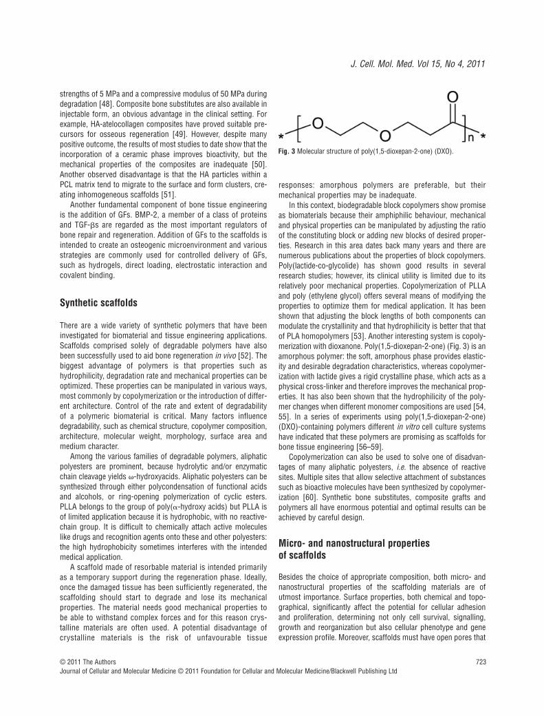
In this context, biodegradable block copolymers show promiseas biomaterials because their amphiphilic behaviour, mechanicaland physical properties can be manipulated by adjusting the ratioof the constituting block or adding new blocks of desired proper-ties. Research in this area dates back many years and there arenumerous publications about the properties of block copolymers.Poly(lactide-co-glycolide) has shown good results in severalresearch studies; however, its clinical utility is limited due to itsrelatively poor mechanical properties. Copolymerization of PLLAand poly (ethylene glycol) offers several means of modifying theproperties to optimize them for medical application. It has beenshown that adjusting the block lengths of both components canmodulate the crystallinity and that hydrophilicity is better that thatof PLA homopolymers [53]. Another interesting system is copoly-merization with dioxanone. Poly(1,5-dioxepan-2-one) (Fig. 3) is anamorphous polymer: the soft, amorphous phase provides elastic-ity and desirable degradation characteristics, whereas copolymer-ization with lactide gives a rigid crystalline phase, which acts as aphysical cross-linker and therefore improves the mechanical prop-erties. It has also been shown that the hydrophilicity of the poly-mer changes when different monomer compositions are used [54,55]. In a series of experiments using poly(1,5-dioxepan-2-one)(DXO)-containing polymers different in vitro cell culture systemshave indicated that these polymers are promising as scaffolds forbone tissue engineering [56–59].
Copolymerization can also be used to solve one of disadvan-tages of many aliphatic polyesters, i.e. the absence of reactivesites. Multiple sites that allow selective attachment of substancessuch as bioactive molecules have been synthesized by copolymer-ization [60]. Synthetic bone substitutes, composite grafts andpolymers all have enormous potential and optimal results can beachieved by careful design.
Micro- and nanostructural properties of scaffolds
Besides the choice of appropriate composition, both micro- andnanostructural properties of the scaffolding materials are ofutmost importance. Surface properties, both chemical and topo-graphical, significantly affect the potential for cellular adhesionand proliferation, determining not only cell survival, signalling,growth and reorganization but also cellular phenotype and geneexpression profile. Moreover, scaffolds must have open pores that
Fig. 3 Molecular structure of poly(1,5-dioxepan-2-one) (DXO).

724 © 2011 The AuthorsJournal of Cellular and Molecular Medicine © 2011 Foundation for Cellular and Molecular Medicine/Blackwell Publishing Ltd
allow cell in-growth and even cell distribution throughout theporous structure. The porosity should also facilitate neovascular-ization from the surrounding host tissue. Furthermore, the scaffolds should have adequate microporosity in order to allowcapillary in-growth. However, the degree of porosity influencesother important properties of the scaffolds such as degradationcharacteristics and mechanical stability, which need to be consid-ered in the context of the mechanical demands of the particulartissue to be replaced.
Conclusion
Despite major advances in material science technology, a materialfulfilling all requirements of a bone and cartilage substitute has yetto be developed. For the repair of bone defects, tissue engineeringshould combine cells capable of osteogenic activity with an appro-priate scaffolding material, in order to stimulate physiologicalbone regeneration and repair processes.
MSC derived from bone marrow are multipotential: dependingon culture conditions, such cells have the potential to differentiateinto many different cell types, including osteoblasts and chondro-cytes. Thus, MSC comprise a readily available and abundantsource of cells for tissue engineering applications. The lack ofimmunogenicity of MSC has opened up the potential of usingthese cells in tissue repair.
The goal of these strategies is to exploit the body’s natural abil-ity to repair injured bone with new bone cells, and to remodel thenew bone tissue in response to local stress factors. The primaryfunction of an optimal biomaterial scaffold is to support the areaundergoing reconstruction, providing adequate initial mechanicalstrength. It should also stimulate new bone formation in theregion, and then gradually degrade without causing a pronouncedinflammatory response, thus allowing the new bone to remodeland assume the mechanical support function.
Selection of the most appropriate scaffolding material is crucialin a tissue-engineered construct. Preferable to ceramics arebiodegradable scaffolds based on polyesters and hydrogels, whichcan be chemically modified with a controlled degradation time andreadily processed into three dimensional, porous structures, withdesired pore morphology. Tissue engineering has thus emergedas an interdisciplinary field with tremendous potential to developand apply new, knowledge-based materials in advanced regenera-tive medicine and dentistry. In this context, biomaterials play acrucial role in the development of tissue-engineered organs.Degradable biomaterials can be used to fabricate a three-dimensional construct or scaffold which is well vascularized, inte-grated with the host skeleton to maintain cells in close proximityto one another, and to stimulate the formation of new living bone, functioning as part of the body’s biological systems. Thus the fabrication of materials which provide appropriate scaffoldingconducive to cell adhesion and maintenance of cell function is offundamental importance to successful bone regeneration.
To fulfil the prerequisites of biocompatibility, as well as to meetthe increasing demands of functional and tailor-made surfaces of
the future, much research effort is focused on surface modifica-tion, especially the design and development of functional andnano-structured surfaces. Whenever a polymeric biomaterial isused, the surface chemistry and structure are of fundamentalimportance, especially in the early stages after implantation.Biological interactions between the device and the host tissueoccur mainly on the surface of the scaffold. For this reason, thereis a strong demand for viable techniques for covalent surfacemodification of polymers, in order to enhance surface wettabilityand thus circumvent an important limitation of current polymericbiomaterials: their hydrophobicity. Surface modification alsooffers the potential to functionalize the surface, so that bioactivemoieties may be attached and the biocompatibility improved.Depending on the nature of the substrate, there are several differenttechniques by which the surfaces of both synthetic and naturalmaterials can be covalently modified.
Mesenchymal stem cells and osteogenesis
Bone tissue
Bone tissue is composed of bone matrix and bone cells. Bonematrix is primarily built of type I collagen (90%), with the remain-ing 10% being composed of a large number of non-collagenousproteins (e.g. osteocalcin [OC], osteonectin, bone sialoproteinsand various proteoglycans). Non-collagenous proteins participatein the process of matrix maturation, mineralization and may regu-late the functional activity of bone cells. Two main types of bonecells have been identified. Osteoblasts (bone-forming cells) andosteoclasts (bone resorbing cells) that together with their precur-sor cells and associated cells (e.g. endothelial cells, nerve cells)are organized in specialized units called bone multicellular units(analogous to the organization of kidney cells into nephrons) [61].The main function of the bone multicellular units in the adult skele-ton is to mediate a bone ‘rejuvenation’ mechanism called ‘boneremodelling’ aimed at the maintenance of the integrity of the skele-ton by removing old bone of high mineral density and high preva-lence of fatigue micro-fractures through repetitive cycles of boneresorption and bone formation [61, 62]. During bone formationphase, the osteoblasts are recruited from MSC present in bonemarrow [63]. On the other hand, osteoclasts are derived fromhaematopoietic stem cells through committed osteoclast progen-itors that fuse to form mature multinucleated cells [64].Understanding the mechanisms that control the differentiation ofosteoblastic cells from MSC is thus of one of the fundamentalareas of research of bone biology. The ability of MSC to differenti-ate into non-osteoblastic cells has been recognized recently andhere we attempt to provide an update regarding the trans-differen-tiation potential of MSC with regard to the relationship betweenosteoblast and adipocyte differentiation.

J. Cell. Mol. Med. Vol 15, No 4, 2011
725© 2011 The AuthorsJournal of Cellular and Molecular Medicine © 2011 Foundation for Cellular and Molecular Medicine/Blackwell Publishing Ltd
Origin of osteoblasts
One current dogma of bone biology is that mature osteoblasts aredifferentiated from precursor cells present in the bone marrow.Based on the pioneer work of Friedenstein and coworkers [65, 66],it has been recognized that the non-hematopoietic compartmentof bone marrow (known as bone marrow stroma) contains agroup of fibroblast-like stem cells with osteogenic differentiationpotential [referred to as MSC, bone marrow stromal cells (BMSC),or skeletal stem cells]. The exact location of these cells in vivois not known but recent work suggests that MSC are located in the perivascular spaces as sub-endothelial cells surrounding thevascular sinusoids in the bone marrow [67].
Isolation and characterization of bone marrowderived MSC
MSC have been isolated from the low-density mononuclearfraction of bone marrow aspirates by their selective capacityfor adherence to plastic surfaces compared to haematopoieticcells [68–71]. However, MSC cultures established by thismethod are heterogeneous and contain both stem cells andprogenitor cell populations. This has been demonstrated byclonal analysis of MSC where ~30% of the clones representedtrue stem cells [72, 73]. Stemness of MSC is defined as theability of the clonal cells to form ectopic bone and bone mar-row organ upon in vivo implantation with an osteo-conductivecarrier such as HA/TCP in immune deficient SCID mice [74].Thus, describing the whole plastic-adherent MSC population as‘stem cells’ is not appropriate.
The human MSC (hMSC) phenotype has been defined throughexpression of many CD markers. hMSC are negative forhaematopoietic surface markers (CD34, CD45, CD14) and positivefor STRO-1, CD29, CD73, CD90, CD105, CD106, CD166, CD146and CD44 [74–76]. Several attempts have been made to isolate ahomogeneous population of the true stem cells among MSCbased on the expression of one or more of these specific surfacemarkers. STRO-1 alone or in combination with CD106 (VCAM-1)or CD146 (MUC18) [77], CD271 (low-affinity nerve GF receptor)[78], CD18 ("2 integrin) [79] or the embryonic stem cell markerSSEA-4 [80] have been tried. A global plasma membrane pro-teomic signature has been established in order to identify novelsurface markers for ‘stemness’ [75]. However, there is still a needto identify both sensitive and specific markers that allow prospec-tive isolation of the true multipotent MSC population.
Interestingly, MSC with similar biological characteristics tothose derived from bone marrow have been isolated from othersources including peripheral blood [81], umbilical cord blood[82], synovial membrane [83], deciduous teeth [84] and amnioticfluid [85]. These various MSC populations share some commonproperties and surface phenotypes but differ in their differentiationpotential and their gene expression profile in ways that reflect theirtissue of origin [86].
In vitro differentiation of MSC into osteoblast lineage cells
MSC are capable of differentiation under appropriate in vitro con-ditions to mesoderm-type cells, e.g. osteoblasts, adipocytes andchondrocytes [74, 87]. Differentiation is induced through additionof ‘cocktails’ of morphogens and chemicals important for differen-tiation of a particular cell type. For example, for osteoblast differ-entiation a mixture of dexamethasone, calcitriol, ascorbic acid and"-glycerophosphate is usually added. In vitro differentiation isverified by demonstrating the induction of osteoblast-specificgene expression and proteins. Similar procedures are used fordemonstrating differentiation to other cell types. For example, theability of hMSC to differentiate into adipocytes has been reportedby several investigators using a cocktail of dexamethasone,isobutyl methyl xanthine, insulin and rosiglitazone (a peroxisomeproliferator-activated receptor %2 (PPAR-%2) agonist) [71, 88, 89].
In vivo differentiation of MSC into bone
There is increasing recognition that in vitro osteoblast differentia-tion assays have limitations and thus there is a need to verifyosteoblast differentiation potential of MSC based on an in vivoassay. Friedenstein employed ‘diffusion chambers’ to determinethe differentiation potential of MSC [66]. Ectopic implantation ofMSC with a carrier (HA/TCP) in an open system (subcutaneousimplantation) in SCID mice has evolved as the ‘gold standard’assay for osteoblast differentiation and stemness characteristicsof MSC [87, 90, 91]. Under these conditions, MSC can form boneand a bone marrow microenvironment supporting haematopoesis,whereas osteoprogenitor cells can only form bone [91]. Thisassay has also been employed to demonstrate the ability of themultipotential MSC cell to exhibit self-renewal and maintenance ofstemness capacity during serial implantations [92].
The importance of testing the in vivo phenotype of culturedMSC is shown by the recent demonstration of the limited value ofstandard in vitro criteria for identification of hMSC clones with invivo bone-forming capacity [93]. In this study, using DNAmicroarray analysis, a predictive molecular signature was identi-fied for in vivo bone formation of MSC that can be employed toselect for an MSC population with high in vivo osteogenic capac-ity for bone regeneration [93].
Factors and pathways controlling osteoblast differentiation of hMSC
Several approaches have been employed in order to identify factors and pathways important for lineage-specific differentiationof hMSC. Using a genetic approach, several lineage-specific transcription factors have been identified and demonstrated to induce MSC differentiation. For example, core-binding factor1(CBFA1/Runx2; Runx & Runt-related factors) [94], osterix [95]

726 © 2011 The AuthorsJournal of Cellular and Molecular Medicine © 2011 Foundation for Cellular and Molecular Medicine/Blackwell Publishing Ltd
(Osx) and lipoprotein related receptor 5 (Lrp5) [96] overexpres-sion lead to osteoblast differentiation, whereas PPAR-%2 [97] andSox9 [98] induce adipocyte or chondrocyte lineages, respectively.Alternatively, a ‘micro-environmental’ approach has beenemployed where MSC are exposed to different mixtures of GFs,hormones and extracellular matrix components to induce their differentiation. These experiments identified several factors thatinduce osteoblast differentiation, e.g. BMPs [99], Wnt [100] orthat inhibit osteoblast differentiation, e.g. Dlk1/Pref-1 [101] andNoggin [102].
We have employed novel proteomic approaches to identify theintracellular signalling pathways that determine the osteoblast differentiation fate of MSC [103]. A newly developed quantitativeproteomic method called stable isotope labelling in cell culture hasalso been employed to compare the phosphotyrosine signallingpathway initiated by epidermal growth factor (EGF) which resultedin osteoblast differentiation, with that of PDGF which did not.Interestingly, more than 90% of the signalling proteins were utilized by EGF and PDGF, whereas the phosphatidylinositol 3-kinase (PI3K) pathway was exclusively activated by PDGF, impli-cating PI3K as a possible control point. Indeed, chemical inhibitionof PI3K in PDGF-stimulated cells rescued the osteoblast differen-tiation phenotype [103]. These studies demonstrate the ability ofstate-of-the-art quantitative proteomic approaches to identify targets for pharmacological intervention in order to control MSCdifferentiation.
Defining the relationship between osteoblast and adipocyte differentiation from MSC
Recent studies have suggested that bone mass and fat mass arestrongly associated processes mediated by the same hormonalfactors, including insulin [104], growth hormone [105] andrecently leptin [106].
The majority of clinical conditions associated with bone loss,including aging, osteoporosis, anti-diabetic drugs (thiazolidine-diones) treatment and increased cortisol production, are known tobe accompanied by increasing marrow adipocytes, due to a shift inthe differentiation decision of MSC to favour the adipocyte lineageover the osteoblast lineage [107, 108]. Several lines of evidencesupport this relationship between osteoblastogensis and adipoge-nesis. At a cellular level, an inverse relationship and phenotypicplasticity exists between osteoblast and adipocyte differentiation ofbone marrow MSC. Several transcriptional factors and pathwayshave been demonstrated to control the balance between osteoblastand adipocyte differentiation of MSC, for example, transcriptionalcoactivator with PDZ-binding motif [109], PPAR-%2 [110], 'FosB[111], canonical Wnt-"-catenin and non-canonical Wnt signallingpathways [112]. We have also reported that Wnt co-receptor, Lrp5with active mutation (T253I) and Wnt activation control the differ-entiation of hMSC into osteoblast versus adipocyte in patients withhigh bone mass due to this mutation [96].
On the other hand, bone marrow adipocytes secret severalinflammatory factors, including leptin, adipsin, adiponectin and
resistin, that act in a paracrine manner to suppress osteoblastfunction and differentiation [113, 114]. Very recently, Lee et al.[115] provided the first evidence that bone can act as an endocrineorgan to regulate glucose and fat metabolism through the secretion of OC (osteoblast-specific protein), which acts as a pro-hormone to decrease fat mass and promotes expression ofadiponectin, an insulin-sensitizing adipokine [115, 116]. Thisnovel finding established a cross-talk between bone formation andenergy metabolism through the secretion of systemic factors. Inthis context, we have recently identified dlk1/FA1 protein (( like1/foetal antigen 1) as a novel pre-adipocyte derived factor that hasregulatory effects on both osteoblast and adipocyte differentiationof hMSC [101]. dlk1/FA1 is an imprinted paternally expressedgene that encodes for a trans-membrane protein. It has six EGF-like repeats in its extracellular domain, which is similar to othermembers of the Notch/Delta/Serrate family. Also, we showed thatthe extracellular domain of the molecule FA1 which circulates inblood and tissue fluids, acts in an endocrine fashion to controlbone and fat mass in vivo [117]. Using DNA microarray technol-ogy, we found that dlk1/FA1 modulates the expression of severalpro-inflammatory cytokines by MSC and thus may influencehMSC differentiation by controlling the composition of themicroenvironment ‘niche’ [118]. Thus, identifying novel secretedfactors that regulate osteoblast/adipocyte cross-talk is an important research area that can potentially lead to identificationof therapeutic targets for enhancing bone formation.
MSC and sex hormones
Sex hormones, especially oestrogens, have a critical role in devel-opment and maintenance of healthy skeleton [119, 120], but it isclear that sex steroids influence not only mature differentiated bonecells, but also the behaviour of stem and different stage progenitorcells [121]. The phytoestrogen genistein enhances osteogenesisand represses adipogenic differentiation of hMSCs [122].
hMSC express oestrogen receptor (ER)-# and possibly alsoseveral ER-" isoforms [122–125]. Interestingly, osteogenic differentiation seems to increase the mRNA expression of all ERspresent in MSC with the exception of ER-"4 isoform that seems tobe mainly expressed in undifferentiated MSC [122]. Such changesin ER profile during differentiation of MSC suggest differenteffects of and sensitivity to oestrogens depending of the differen-tiation lineage and stage. ER gene polymorphism may alsoaccount to certain interindividual variablity in responses to estradiol stimulations [125].
17"-estradiol enhances dose dependent osteogenesis fromhMSC as shown by up-regulation of OC and increased calciumdeposition [124]. In other work, where MSC from both male andfemale donors were stimulated with estradiol and testosterone,estradiol also increased calcium deposition, but alkaline phos-phatase (ALP) activity increased only in cells isolated from maledonors [125]. Testosterone, on the other hand, had no effect oncalcium deposition in either sex. Resveratrol, a polyphenolic phy-toestrogen, has been shown to enhance osteoblastic maturation of
J. Cell. Mol. Med. Vol 15, No 4, 2011
727© 2011 The AuthorsJournal of Cellular and Molecular Medicine © 2011 Foundation for Cellular and Molecular Medicine/Blackwell Publishing Ltd
hMSC, possibly through extracellular signal-regulated kinase 1/2(ERK1/2) pathway [126, 127].
More recent studies have revealed that hMSC can express several key enzymes needed for intracrine conversion of dehy-droepiandrosterone to oestrogens or testosterone [122, 128,129]. This suggests the interesting possibility that hMSC maythemselves in certain conditions make the needed sex hormonesor adjust their levels using serum dehydroepiandrosterone as aprecursor, and participate therefore in an intra-, auto- or paracrinemanner in their own differentiation processes and in overall morecomplex local sex hormone regulation in bone [130].
Effect of aging on osteoblastogenesis
One of the most consistent histomorphometric findings in bonebiopsies obtained from elderly persons is the presence ofdecreased mean wall thickness in both trabecular and corticalbone indicating decreased osteoblastic bone-forming capacityduring bone remodelling [131]. Mean wall thickness is dependenton both the number of recruited osteoblasts at the beginning ofbone formation phase and the activity of individual osteoblasts.Recruitment of an adequate number of osteoblasts is dependenton the availability of stem and precursor cells and their properresponse to growth, differentiation and chemotactic signals in thebone microenvironment. Matrix production and mineralizationfunctions of mature osteoblasts are dependent on the properresponse of osteoblasts to hormones, GFs and cytokines as wellas the availability of nutrients and ions necessary for accomplishingthese processes. Age-related impairment of osteoblast functionscan thus be the result of any mechanism interfering with theseprocesses. It has been shown that the number of MSC does notchange with aging or in osteoporosis [132] and that MSC exhibita limited lifespan in vitro, with all the characteristics of the in vitroreplicative senescence phenotype [133]. Interestingly, an age-related decline in the maximal lifespan of hMSC has beenobserved, from 41 ) 10 population doublings in young donors to24 ) 11 population doublings in old donors [133]. In addition toimpairment of cell proliferation of MSC with aging, it was alsoshown that the senescent microenvironment affects MSC functions. As a surrogate condition for senescent microenviron-ment experienced by hMSC, the effects of sera from young and old persons on MSC function were tested, and it was found that aged-sera inhibited osteoblast differentiation and functions of MSC [134]. Thus, intrinsic aging of MSC and the negative effects of the senescent microenvironment are possiblemechanisms for the observed defective osteoblast function andbone formation in vivo in aged human beings. The senescent phenotype of MSC can be ‘rescued’ through overexpression ofhuman telomerase reverse transcriptase gene [135]. The telomerized hMSC exhibit enhanced cell proliferation and in vivobone formation capacity [135]. Telomerization also enhances geneexpression of some osteoblastic genes [136]. Other approaches torejuvenation of senescent MSC need to be explored for theirpotential use in therapy.
Conclusion
During the last decade, enormous amounts of data have beengathered related to the mechanisms of osteoblast differentiationfrom stem cells. It is expected that this knowledge will be trans-lated into novel approaches to enhance bone regeneration andbone formation, a much needed task to manage the major healthcare problem of bone fractures and osteoporosis.
Embryonic, foetal and adult stem cellsin osteogenesis
Cellular therapy is becoming a useful addition to medical therapiesfor repairing, restoring or ameliorating function of tissues. Severalcell types and tissues have been proposed as starting materialincluding autologous cells, adult stem cells (including specific tissue populations and also bone marrow and adipose MSC),embryonic stem cells, foetal cells and tissues from placental and amniotic fluid [137–143]. Some cell choices are more adaptable to cellular therapy in patients [144]. Tissue from animals and human beings at all stages of development must beevaluated for the advantages and disadvantages of each cell type(Fig. 4). The terminology of embryonic, foetal and adult stem cellsis complicated. Legally, the term ‘embryo’ denotes the earlieststages following fertilization of an ovum by a sperm. Zygote wouldinclude early stage cleavage embryos produced by cell division upto 50–60 cell-stages (each cell of which is a blastomere) and theblastocyte for the 60 cell stage to the point of implantation atabout 2 weeks after fertilization. Pathology would classify theembryonic stage as up to 9 weeks of gestation and thereafter asthe foetal stage from 9 weeks to until birth. Different cell lines canbe established from each of these tissues, but with different com-plications encountered in tissue culture techniques needed foreach. Embryonic stem cells are harvested from pre-implantationembryos from the inner-cell mass before the first 2 weeks ofdevelopment. These cells are frequently obtained from extraembryos developed by in vitro fertilization techniques to aid cou-ples with fertility problems. There has been a moratorium on useof embryonic stem cells since 1975 with new laws in 1993 permit-ting use under certain circumstances. Because these particularcells have created an ethical debate, other researchers have begunusing embryonic or foetal cells derived from voluntary interruptionof pregnancy between 5 and 8 weeks [145]. Cell lines are normallydeveloped from the genital ridge of the foetus but specific tissuescan be isolated from immature tissue with the necessity to addGFs to assure particular cellular differentiation. Most foetal cellresearch has developed from research using specific tissues at thelatter end of the first trimester (11–14 weeks) following voluntaryinterruption of pregnancies. As this tissue (*9 weeks) is consid-ered an organ donation in most countries, it bypasses the majorproblems that have been raised by embryonic stem cells. Cell lines

728 © 2011 The AuthorsJournal of Cellular and Molecular Medicine © 2011 Foundation for Cellular and Molecular Medicine/Blackwell Publishing Ltd
at this stage are tissue specific and therefore cells are differenti-ated and have specific functions [142, 146–148].
Cell-based therapies for bone
The first cell-based strategy used for repair of bone tissue wasautologous connective tissue progenitors harvested from the iliaccrest and immediately transplanted to sites for skeletal repair inthe same patient [149, 150]. This autograft procedure can beaccomplished without any outside manipulation of the tissue (i.e.cell separation, culture or expansion) and is thus considered partof a surgical procedure and is not regulated as a complex cell ther-apy. If extensive manipulation has to be done, then the procedure
falls into a specific regulatory process which requires a certaininfrastructure for cell culture. Although the auto-grafting strategyis used by many surgeons due to the ‘supplemental’ biologicalactivity and low risk involved with use of autologous bone marrow,only a very small fraction of the cells actually survive and only 1of 103 or 104 cells are potentially active [151]. The success ofmarrow grafting is therefore entirely dependent on transfer of suf-ficient numbers of progenitor cells, and therefore this approachmay be least applicable in those situations where it is mostneeded, because ageing or disease are accompanied by a reduc-tion of healthy marrow elements and especially osteogenic pro-genitors. Alternative cell sources are therefore of great interest, inparticular normal allogeneic sources, which would provide ‘ready-for-use’ products for bone repair strategies.
Fig. 4 Development stage, cell type and source of cells currently used in cellular therapy. Cellular source can come from human and animals at differentstages of development including embryonic, embryonic-foetal, foetal and adult involving different beginning tissue sources ranging from zygotes to spe-cific tissues (bone marrow, adipose, amniotic fluid, skin, liver, bone, cartilage etc.). Embryonic stem cells are derived from embryos at early stage whereless than 100 cells are present, followed by foetal stem cells that are taken from the genital ridge section from 5 to 8 weeks of gestation (drawing mod-ified from http://commons.wikimedia.org/wiki/Image:10_weeks_pregnant.jpg donated by [email protected] with a free license). Tissue-specificfoetal cells are taken following 9 weeks of gestation usually up to 14–16 weeks from normal tissue. Adult stem cells can be isolated from most tissuesources but are rare with only 1 in every 104 to 105 of total cell volume.

J. Cell. Mol. Med. Vol 15, No 4, 2011
729© 2011 The AuthorsJournal of Cellular and Molecular Medicine © 2011 Foundation for Cellular and Molecular Medicine/Blackwell Publishing Ltd
Other adult stem cells, frequently referred to as MSC, have raisedhopes for new treatments because they a have high self-renewalcapacity and can generate multiple cell lineages. They can be isolatednot only from bone marrow but also from many tissues such asamniotic fluid, adipose tissue, brain, skin, heart, kidneys and liver.Although widely distributed, adult stem cells represent only a smallfraction of a tissue cell population (many times only 1 in every 104 to105 cells), thus requiring extensive in vitro separation and expansionsteps [152]. Stem cell cultures are technically demanding.Maintenance and expansion of stem cells in an undifferentiated staterequires the addition of many specific GFs [153]. Culture of thesecells without feeder layers (which are usually formed by animalcells), is difficult and feeder layers are responsible for some aspectsof inconsistent colony cell growth. The necessity of using exogenousGFs as well as animal products is a limiting factor for the scale up ofstem cell cultures for clinical applications [138].
Unlike stem cells, foetal cells are differentiated cells with highexpansion and regeneration and low immunogenic properties[154]. They can be isolated from foetal tissues, which followembryonic stage after 9 weeks of development. Foetal cells have extensive expansion capabilities and cell culture require-ments are minimal compared to stem or mesenchymal cell types.As the foetal cells are already differentiated and do not need to bedirected or altered, the vast number of additional GFs normallynecessary are not needed for cell culture and expansion [146]. Cellchoice is of utmost importance and each element of processingnecessitates special attention in order to produce a safe, consis-tent and successful cell therapy for clinical trials [155].
Specific features of bone cells needed to beadvantageous for clinical use
As whole bone marrow transplantation may not be optimal biologically, the expansion of bone marrow osteoprogenitors hasbeen evaluated. The procedure is laborious as their isolation andcharacterization has been difficult due to their low numbers in theoriginal tissue and lack of specific reactive antibodies to assureseparation of active cellular components. Patient treatment is onlypossible following 4–6 weeks of expansion in cell culture.Demonstration of cell function is of particular importance so thatseparated cells are shown to be capable of matrix deposition, mineralization, ossification, nodule formation and bone formationin vivo without fibrous tissue formation. Although these cells havebeen shown to fulfil the required needs, it is labour intensive touse these procedures for each individual patient.
More recently, femurs from foetal tissue of ~8 weeks were evalu-ated for isolation of bone progenitors and thus these cell populationswould be formally considered as foetal stem cells (5–8 weeks)[156]. Mirmalek-Sani et al. have shown that these cells histologi-cally resemble cartilage and this was confirmed by the high plastic-ity seen with these cells when compared to studies using foetalbone cell populations derived from 12- to 14 week gestationfemurs. In vivo mineralization of 8 week stem cells is seen withosteoinductive media but with 12–14 week foetal bone cells such
media are not necessary because spontaneous minerialization isseen without external GFs [157]. Spontaneous behaviour of cells isthus important to eliminate the use of multiple GFs and increasebiosafety while reducing the cost of cell production. It is advanta-geous to use differentiated cells with lower plasticity as theydepend less than undifferentiated cells on the many environmentalfactors such as nutrition, mechanical loading, inflammation, angio-genesis and interaction with endogenous cells. In addition, foetalbone cells from 12 to 14 weeks have been extensively evaluated in vivo for immunological privilege, safety and osteoinduction.
Montjovent et al. [158] have demonstrated that a compositemade of poly-L-lactic acid and "-TCP particles used as scaffoldsto seed foetal bone cells can offer suitable conditions forosteoblasts to achieve full differentiation due to the highosteogenic potential of these cells. Furthermore, in vivo studiesusing dedicated foetal bone cell banks and a solid matrix haveshown significant promotion of bone growth using two differentmodel systems [158]. Safety studies in a large animal model usingsheep have also shown that 12 week foetal bone cells derived froma dedicated cell bank are immunologically accepted, do not formfibrous tissue and promote biological activity with bone growth(personal communication by Applegate and Pioletti).
Development of therapeutic biological agents
Organ donation, whole cell bioprocessing and procedures adapt-able to good manufacturing processes (GMP) make it possible todevelop extensive master cell banks and working cell banks tofacilitate thorough testing (Fig. 5) [159]. Once master cell bankscan be produced, working cell banks can be produced to establishindividual batches for treatment of large numbers patients (thou-sands from one cell bank). Further, these cell banks can be testedcompletely for safety regarding sterility, pathogens and adventi-tious agents and tumorigenicity. Once safety can be assured, effi-cient cell presentation with biocompatible delivery systems can beassessed for specific tissues. For delivery systems, biocompatiblebiomaterials need to be available in order to provide an extracellu-lar matrix environment for cell differentiation, delivery and release.Cells and materials need to be tested together to not only assurebiocompatibility but also their interactions, cellular stability,possible degraded by-products of combination and degradation orabsorption. Ease of applicability of the final product will be ofimportance for clinical use.
All cellular products must be in compliance with GMP guidelineswith respect to medicinal products and investigational medicinalproducts for human use. The European Union (EU) regulation onadvanced therapy medicinal products was adopted by all EuropeanMember States on December 30, 2008 and the United States Foodand Drug Administration (FDA) recently also proposed regulationson human cells, tissues and cellular and tissue-based products[160–164]. The main scope of these regulations is to establish clearclassification criteria for many new cell-based medicinal products.For the EU, it makes reference to the 2004/23/EC directive on dona-tion, procurement and testing of human cells and tissues and also

730 © 2011 The AuthorsJournal of Cellular and Molecular Medicine © 2011 Foundation for Cellular and Molecular Medicine/Blackwell Publishing Ltd
with directive 2002/98/EC on human blood and blood components.These directives dictate that human cells used for therapeutic pur-poses must be in compliance with the quality requirements theydescribe and that all advanced therapy medicinal products must beprepared under GMP conditions [161, 165, 166]. Key elements forcellular-based products include identity, purity, sterility, stability,safety and efficacy are recommended. In all, these new regulationsimpose strict criteria for the production and the environment usedfor the production of cell-based products to be used in clinical tri-als and treatments [167, 168].
Clinical application concerns
Risk assessment of final cellular products for human use is ofutmost concern. Self-renewal of undifferentiated cells representspotential for tumour formation. Certain techniques such as cellu-lar cloning or encapsulation of cellular products are alternatives to
assure safety. Many cell-based therapies will not consist of a uni-form cell population. Associated accessory cells raise additionalquestions for potential risk as well as their physiological role afteradministration. Cellular death of transplanted cell populations,ectopic tissue formation or migration from the site of administra-tion could be a problem when cellular therapies are used withinanatomically sensitive areas such as the central nervous system,joints or myocardium. Pre-clinical animal models for specificpathologies or tissue repair are an important step and special caremust be taken in the interpretation of pertinent safety and biolog-ical activity. A thorough appreciation of relevant advantages andlimitations must be made regarding an animal model choice.
Conclusions
Cell-based therapies are being developed and introduced for alltypes of tissue repair including skin, bone, cartilage, muscle and
Fig. 5 Organization of a cell therapy platform. Director of technical and logistics coordinates the program with essential legal and ethical advisors and amedical director for interpretation of medical quality assurance (serology and pathology reports). The separation of hospitals for the organ donation andall other aspects of the platform including serology, pathology and the GLP cell culture laboratory can assure complete anonymous and coded organ dona-tions. Importantly, the Director of Technical and Logistics is not involved in any manner in the organ donation process as required by law. Final approvalfor use of validated cell banks for human therapy is coordinated and approved with both Hospital Ethics Committees and National regulatory agencies(i.e. European Medicines Association, Food and Drug Administration, SwissMedic [32]).

J. Cell. Mol. Med. Vol 15, No 4, 2011
731© 2011 The AuthorsJournal of Cellular and Molecular Medicine © 2011 Foundation for Cellular and Molecular Medicine/Blackwell Publishing Ltd
spine. They offer promise for repairing and/or replacing damagedtissue and restoring lost functionality. One of the major challengesfor assuring that more patients will benefit from cell-based thera-pies in the future will be the optimization of the choice of cell typeas well as methods for their isolation and expansion. Today, it isstill unknown what type of osteogenic cell will be the most suitablefor engineering bone tissue. MSC, foetal-stem and foetal cells,bone marrow stromal cells, periosteal cells and osteoblasts havebeen successfully used for generation of bone tissue. Isolationand expansion efficiency, stability of osteoblastic phenotype, in vivobone formation capacity and long-term safety are essentialrequirements that have to be met by any type of osteogenic cell forsuccessful clinical application.
Equally important is the delivery system for the cell of choiceand their interaction with scaffolds to assure biocompatibility. Thedevelopment of master cell banks from the cells of choice providesa major advantage for the creation of a therapeutic biologicalagent. Careful selection of donors and extensive screening of boththe donor and cultured cells avoids transmissible viral, fungal orbacterial disease and therefore can provide a safe and secure utilization of cells for therapeutic purposes. Clear regulatory over-sight of cellular use, particularly for organ donation, embryonicand foetal cells will be necessary. Overall, cooperative interdisci-plinary efforts and cooperation to form successful translationalmedicine platforms in universities and hospitals will serve toensure further patient safety.
Platelet-rich plasma (PRP), growthfactors and osteogenesis
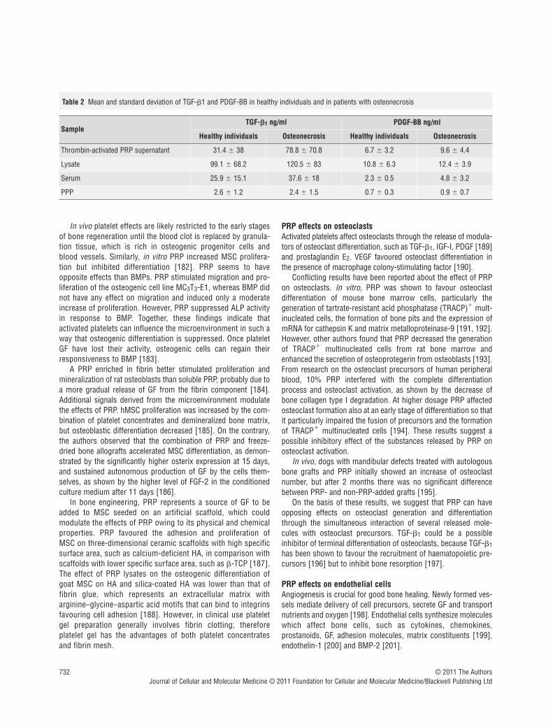
PRP is a concentrate of platelets in a small volume of plasma fromfreshly drawn whole blood activated with a mixture of thrombinand calcium [169]. PRP is used to facilitate wound and bone healing, thanks to the molecules released by platelets during acti-vation. A number of these substances affect osteogenesis, such asTGF-"1, PDGF-BB, VEGF-A and IGF-I (Table 1). These moleculesare more concentrated in PRP than in a normal clot as a result ofthe higher platelet number. After release, they are trapped withinthe fibrin mesh, then slowly pass into the microenvironment andbind to receptors on the cell membrane. The molecules releasedby platelets regulate key processes involved in tissue repair,including chemotaxis, cell proliferation, differentiation and extra-cellular matrix synthesis (Table 1). The fibrin mesh itself can actas a conductive matrix or ‘scaffold’ for cell adhesion [170].
The combination of GFs in PRP is in an optimal level and ratio,and seems to be more efficient than the single rhGF, owing to theirsynergistic effects [87]. The technique for obtaining GF from PRPis relatively simple and less expensive than rhGF. Because of itsautologous origin, PRP does not hold any risk of immunologicalreactions and transmissible diseases. The drawbacks of PRP arethe high variability of GF concentrations, due both to individualfactors and to different preparation methods [171]. We found a
high variability in PDGF-BB and TGF-"1 levels assayed in thesupernatant of thrombin-activated PRP, on lysates of platelet con-centrates, on serum and platelet poor plasma from 11 healthyindividuals and from 11 patients with osteonecrosis (Table 2).
The original Marx protocol obtained PRP by the addition of calcium and thrombin to a platelet concentrate [169]. Recently,other protocols for PRP preparation have been proposed, whichdiffer for anticoagulant [172], leucocyte depletion and platelet activators [173, 174]. Leucocyte depletion avoids the release ofpro-inflammatory cytokines [175]. However, some authors suggestthat leucocytes represent an additional source of GF and have animportant role in host immune defence [176]. Myeloperoxidasecontained in neutrophils and monocytes generates reactive oxygenspecies, that act as potent bactericidal and may be helpful to pre-vent post-surgical infections and in the prophylaxis and treatmentof infection-related delayed healing and non-union [177].
PRP effects in vitro on the cells involved in bone repair
PRP effects on osteoblastsThe use of PRP was proposed in order to provide a microenviron-ment for the orchestration of the sequential process of boneregeneration involving migration, proliferation and differentiationof osteogenic cells. The mitogenic effect of PRP was demon-strated by in vitro studies on hMSC [178, 179] and human trabec-ular osteoblasts [180]. Among the GF released by platelets, thehighest contribution to the proliferation of osteoblasts was fromPDGF and TGF-"1 [181].
Table 1 The role in bone remodelling of the GFs and other moleculesreleased by platelets
Molecule Role in bone remodelling
TGF-"
Mesenchymal stromal cell proliferation; osteoblastprecursor recruitment; osteoblast and chondrocytedifferentiation (but inhibition of terminal differenti-ation); bone matrix production; recruitment ofosteoclast precursors but inhibition of terminaldifferentiation and induction of apoptosis
PDGFOsteoprogenitor migration, proliferation and differentiation; osteoclastogenesis
VEGFConversion of cartilage into bone; osteoblast proliferation and differentiation
IGFOsteoblast proliferation; bone matrix synthesis;bone resorption
EGF Osteoblast recruitment and proliferation
Fibronectin Osteoblast migration and adhesion
Vitronectin Osteoblast migration and adhesion
Prostaglandin E2 Bone resorption
732 © 2011 The AuthorsJournal of Cellular and Molecular Medicine © 2011 Foundation for Cellular and Molecular Medicine/Blackwell Publishing Ltd
In vivo platelet effects are likely restricted to the early stagesof bone regeneration until the blood clot is replaced by granula-tion tissue, which is rich in osteogenic progenitor cells andblood vessels. Similarly, in vitro PRP increased MSC prolifera-tion but inhibited differentiation [182]. PRP seems to have opposite effects than BMPs. PRP stimulated migration and pro-liferation of the osteogenic cell line MC3T3-E1, whereas BMP didnot have any effect on migration and induced only a moderateincrease of proliferation. However, PRP suppressed ALP activityin response to BMP. Together, these findings indicate that activated platelets can influence the microenvironment in such away that osteogenic differentiation is suppressed. Once plateletGF have lost their activity, osteogenic cells can regain theirresponsiveness to BMP [183].
A PRP enriched in fibrin better stimulated proliferation andmineralization of rat osteoblasts than soluble PRP, probably due toa more gradual release of GF from the fibrin component [184].Additional signals derived from the microenvironment modulatethe effects of PRP. hMSC proliferation was increased by the com-bination of platelet concentrates and demineralized bone matrix,but osteoblastic differentiation decreased [185]. On the contrary,the authors observed that the combination of PRP and freeze-dried bone allografts accelerated MSC differentiation, as demon-strated by the significantly higher osterix expression at 15 days,and sustained autonomous production of GF by the cells them-selves, as shown by the higher level of FGF-2 in the conditionedculture medium after 11 days [186].
In bone engineering, PRP represents a source of GF to beadded to MSC seeded on an artificial scaffold, which couldmodulate the effects of PRP owing to its physical and chemicalproperties. PRP favoured the adhesion and proliferation of MSC on three-dimensional ceramic scaffolds with high specificsurface area, such as calcium-deficient HA, in comparison withscaffolds with lower specific surface area, such as "-TCP [187].The effect of PRP lysates on the osteogenic differentiation ofgoat MSC on HA and silica-coated HA was lower than that of fibrin glue, which represents an extracellular matrix with arginine–glycine–aspartic acid motifs that can bind to integrinsfavouring cell adhesion [188]. However, in clinical use plateletgel preparation generally involves fibrin clotting; thereforeplatelet gel has the advantages of both platelet concentrates and fibrin mesh.
PRP effects on osteoclasts Activated platelets affect osteoclasts through the release of modula-tors of osteoclast differentiation, such as TGF-"1, IGF-I, PDGF [189]and prostaglandin E2. VEGF favoured osteoclast differentiation inthe presence of macrophage colony-stimulating factor [190].
Conflicting results have been reported about the effect of PRPon osteoclasts. In vitro, PRP was shown to favour osteoclast differentiation of mouse bone marrow cells, particularly the generation of tartrate-resistant acid phosphatase (TRACP)! mult-inucleated cells, the formation of bone pits and the expression ofmRNA for cathepsin K and matrix metalloproteinase-9 [191, 192].However, other authors found that PRP decreased the generationof TRACP! multinucleated cells from rat bone marrow andenhanced the secretion of osteoprotegerin from osteoblasts [193].From research on the osteoclast precursors of human peripheralblood, 10% PRP interfered with the complete differentiationprocess and osteoclast activation, as shown by the decrease ofbone collagen type I degradation. At higher dosage PRP affectedosteoclast formation also at an early stage of differentiation so thatit particularly impaired the fusion of precursors and the formationof TRACP! multinucleated cells [194]. These results suggest apossible inhibitory effect of the substances released by PRP onosteoclast activation.
In vivo, dogs with mandibular defects treated with autologousbone grafts and PRP initially showed an increase of osteoclastnumber, but after 2 months there was no significant differencebetween PRP- and non-PRP-added grafts [195].
On the basis of these results, we suggest that PRP can haveopposing effects on osteoclast generation and differentiationthrough the simultaneous interaction of several released mole-cules with osteoclast precursors. TGF-"1 could be a possibleinhibitor of terminal differentiation of osteoclasts, because TGF-"1has been shown to favour the recruitment of haematopoietic pre-cursors [196] but to inhibit bone resorption [197].
PRP effects on endothelial cellsAngiogenesis is crucial for good bone healing. Newly formed ves-sels mediate delivery of cell precursors, secrete GF and transportnutrients and oxygen [198]. Endothelial cells synthesize moleculeswhich affect bone cells, such as cytokines, chemokines,prostanoids, GF, adhesion molecules, matrix constituents [199],endothelin-1 [200] and BMP-2 [201].
Table 2 Mean and standard deviation of TGF-"1 and PDGF-BB in healthy individuals and in patients with osteonecrosis
SampleTGF-""1 ng/ml PDGF-BB ng/ml
Healthy individuals Osteonecrosis Healthy individuals Osteonecrosis
Thrombin-activated PRP supernatant 31.4 ) 38 78.8 ) 70.8 6.7 ) 3.2 9.6 ) 4.4
Lysate 99.1 ) 68.2 120.5 ) 83 10.8 ) 6.3 12.4 ) 3.9
Serum 25.9 ) 15.1 37.6 ) 18 2.3 ) 0.5 4.8 ) 3.2
PPP 2.6 ) 1.2 2.4 ) 1.5 0.7 ) 0.3 0.9 ) 0.7

J. Cell. Mol. Med. Vol 15, No 4, 2011
733© 2011 The AuthorsJournal of Cellular and Molecular Medicine © 2011 Foundation for Cellular and Molecular Medicine/Blackwell Publishing Ltd
The GF released by platelets, such as VEGF, TGF-", PDGF andEGF [202], not only have pro-angiogenic properties [203–205],but also induce the endothelial cells to express a pro-osteogenicphenotype. PRP induced human umbilical endothelial cells(HUVEC) to express PDGF-", which in turn has chemotacticeffects [206], recruiting MSC to the site of bone repair. MSCmigration was higher in HUVEC incubated with PRP in compari-son with HUVEC incubated with low serum medium or with PRP[207]. PRP favours monocyte adhesion to the vessel wall, throughthe stimulation of endothelial cell expression of intercellular adhesion molecule 1 [207], which is important because the monocyte/macrophage response is essential for bone repair.
PRP effects in vivo on experimental animals
The results of research on osteogenic activity of PRP in animalsare conflicting. In combination with bovine cancellous bone, PRPincreased bone formation in a non-critical-size bone defect in rab-bit cranium [208]. However, other authors noticed that the use ofbone graft material in combination with PRP was not superior tothat of bone allograft alone [209–211]. These different results maydepend on the animal species used, the method of gel preparation,type of bone defect, outcome parameters measured, the presenceof MSC and of scaffolds or bone substitutes. An important reasonfor the discordant results from different authors is the fact thatcritical-size bone defects were not assayed. When critical-sizebone defects were tested, the combination of PRP, bone substitutesand MSC induced the greatest improvement in healing comparedto a single component assayed alone [212]. Another variableseems to be the ratio of bone graft to PRP [213].
When PRP was used in combination with artificial scaffolds,the physical and chemical properties of the scaffold materialshould be considered because they can affect the performance ofPRP [187]. When PRP was used in association with "-TCP [214]or triphasic ceramic- (calcium silicate, HA and TCP) coated HA[215], more intense bone regeneration was observed.
Even though it is generally assumed that PRP might supportosteogenesis in the presence of precursor cells, sometimes theaddition of PRP to MSC did not further increase the rate ofbone formation in comparison to MSC alone. In goats no effectwas shown when PRP was added to MSC seeded on scaffoldsmade of the combination of HA and "-TCP implanted at anorthotopic or ectopic location [216]. MSC added to the criticalsize defect were likely already induced towards osteogenesisand PRP could not significantly increase this. Moreover,osteotomy might recruit osteoblast precursors from the bonemarrow [217].
The effect of PRP combined with BMSC was also tested inexperimental dental implant models. In a dog mandible model ofbone defects, the combination of canine MSC and PRP favouredwell-formed mature bone formation and neo-vascularization,comparable with that induced by autologous cancellous bone,whereas PRP alone performed poorly [218]. In other experiments,the combination of MSC-PRP-fibrin gel, used as grafting material
for alveolar augmentation in adult dogs, lead to the highest degreeof direct bone-implant contact [219].
From the analysis of the results of in vivo research, it can beconcluded that PRP promotes bone healing [220], particularly inthe early phase [221] and in combination with MSC and bonegrafts [222, 223] or an artificial scaffold. In the later course ofbone healing, the PRP effects were less evident [224, 225].However, a beneficial effect of PRP on bone healing was notdemonstrated in every animal model and with every bone substi-tute, probably owing to the differences in the protocol design andthe method for measuring bone formation [226].
The clinical use of PRP for bone repair
In orthopaedics, PRP is generally considered a tool for promotingbone repair, even though there have been discordant or inconsis-tent results [227]. PRP, sometimes combined with MSC, has beenproposed for the treatment of non-unions [228], in femoral andtibial lengthenings [229], in distraction osteogenesis, in spinalfusions, in foot and ankle surgery and in joint arthroplasties. PRPis also used in odontostomatology and maxillo-facial surgery, toimprove bone regeneration.
Non-unionThe scientific basis for the use of PRP in non-unions andpseudoarthrosis is the significant reduction of GF which has beenobserved at the non-union sites, in comparison with the site offresh fractures [230]. In non-unions, both PRP administrationduring surgery together with bone allograft and percutaneousadministration of PRP have been proposed. PRP with autologousbone graft gave resolution of non-unions of the foot and ankle at a mean of 60 days [230]. With percutaneous administrationhealing was improved only if the treatment was performed within11 months after the initial surgery [231]. In a prospective study,PRP, injected into atrophic non-unions, treated with percutaneousstabilization, failed to improve consolidation [232]. Therefore, atpresent there is no clinical evidence of benefit of PRP in non-unions in comparison with other treatments. An important issue isthe option to treat with PRP or recombinant BMP. The preliminaryresults of a recent prospective randomized clinical study on 120 patients, where PRP was compared with rhBMP-7 for thetreatment of persistent long bone non-unions, supported the viewthat the application of rhBMP-7 was superior to PRP [233].
Distraction osteogenesisIn distraction osteogenesis of the femur or tibia, for patients withachondroplasia, hypochondroplasia or congenital pseudoarthrosis,PRP in combination with MSC resulted in acceleration of newbone regeneration [234].
Spinal fusionIn a prospective study on lumbar interbody fusion, PRP with anallogeneic bone graft gave a clinical and radiological outcome at
734 © 2011 The AuthorsJournal of Cellular and Molecular Medicine © 2011 Foundation for Cellular and Molecular Medicine/Blackwell Publishing Ltd
12 and 24 months comparable to an autologous graft [235].However, other researchers failed to show an enhancement in lumbarfusion using autologous bone graft with or without PRP [236].
Foot and ankle surgeryIn total ankle arthroplasty PRP improved the syndesmosis unionrate [237, 238]. PRP may help to achieve an acceptable time forfusion also in high-risk patients with diabetes, immunosuppres-sion, malnutrition, alcohol abuse, osteomyelitis and history ofimpaired osseous healing or multiple same site surgeries [239].
Total knee arthroplastyThe primary indication for use of PRP in total knee arthroplasty isto promote wound healing and to decrease blood loss. A study of71 patients treated with PRP fibrin sealant, which was sprayed in theknee just prior to closure, showed not only higher post-operativehaemoglobin levels, but also a significantly greater knee range ofmotion compared with the untreated group after 6 weeks [240].
Odontostomatology and maxillofacial surgeryPRP has been largely applied to promote bone regeneration inodontostomatology and maxillofacial surgery. MSC and PRPapplied simultaneously with titanium implants provided stable andpredictable results in terms of implant success [241]. Implants of"-TCP seeded with MSC and PRP for maxillary sinus floor aug-mentation were clinically stable 12 months after loading [242].Recently, PRP associated with cancellous marrow grafting hasbeen proposed in selected cases of osteonecrosis of the jaws[243]. A systematic review of human controlled clinical trialsdesigned to treat maxillofacial bony defects with the application ofPRP found evidence for beneficial effects in the treatment of peri-odontal defects, but minimal effects in sinus elevation [244].
There are few randomized, controlled studies of the clinicaleffects of PRP, and the apparent discrepancies reported in clinicaleffects and the diverse success rates among the differentorthopaedic and maxillofacial applications could be a function ofthe different PRP preparation methods, poor characterization ofthe platelet concentrate used, the presence or absence of fibringlue, the presence or absence of leucocytes, the presence orabsence of MSC, the presence or absence of bone allografts orscaffolds (and what scaffold) and differences in the primary outcome measured. In addition, the power of different studiesvaries considerably. Although there are a number of studies inorthopaedic patients, the majority of them are anecdotal and onlya few are well-designed controlled clinical trials. A recent system-atic literature review on the use of PRP in orthopaedic surgery,after the exclusion of the applications in periodontal and maxillo-facial surgery, identified 39 articles concerning clinical trials [245].Only one was a randomized controlled study [246], which demon-strated that PRP, particularly if combined with MSC, increased theosteogenic potential of lyophilized bone chips. The strength of thisclinical research was the randomization and the comparisonamong different treatments (freeze-dried bone allograft alone or in
combination with PRP with or without MSC); its weakness was thelack of the evaluation of PRP effect in alone and of a clear primaryoutcome. More well-designed, prospective, rigorous clinical trialsusing consistent techniques may lead to more conclusive generalrecommendations [247].
Conclusion
PRP has been evaluated through in vitro, in vivo and clinical modelsof bone regeneration, with conflicting results. PRP use appears tobe safe and feasible and may have a beneficial therapeutic effect,particularly in the early phases of bone formation. In vitro data havedemonstrated that PRP, in addition to the stimulation of cellgrowth, induces target cells to synthesize GF and other substancesable to sustain bone healing. The best in vivo and clinical resultsare obtained when PRP is added to MSC and osteogenic scaffoldsor bone allografts. Further basic science and clinical research isneeded to define the clinical indications, the methods of prepara-tion and administration and the primary outcome with PRP.
Molecular control of osteogenesis
The timeline of molecular events that regulate osteogenesis hasbeen described by Li et al., who analysed the ‘fracture healingtranscriptoma’ and the ‘fracture healing proteoma’ by comparingnon-fractured and post-fractured rat femurs during early and latestages of bone repair [248, 249]. In early phases after the boneinjury, they observed an up-regulation of genes related to cell-cycle and cell-to-cell signalling, supporting the assumption thatboth cell division and cell communication are essential to initiatefracture healing. Many of the genes controlling cell growth andsurvival are constantly up-regulated, whereas those functionallyassociated with differentiation of osteogenic precursors and bonematrix formation undergo temporary modulation over time. In particular, expression of IGF-I and IGF-II, PDGF, FGFR, fibronectin,matrix metalloproteinases, glypican, byglican, osteomodulin,osteonectin, tenascin C, cartilage collagen (types VI and XI) andbone collagen (types I, V, VI and XII) increase until the immatureosteoid synthesized by osteoblast progenitors is histologicallydetectable. The start of endochondral ossification is marked by anumber of molecular pathways, but p38/MAPK (mitogen-activatedprotein kinase) signalling plays a pivotal role in advancing theprocess to this phase. Even though recent studies clearly demon-strated that native MSC are phenotypically and functionally differentfrom cultured MSC [250], the time course and extent of geneexpression in in vitro expanded osteogenic precursors is not significantly different from that which has been observed in vivo[251]. The gene expression profile of hMSC undergoingosteogenic induction has been analysed at different time-pointsuntil MSC were able to form mineral nodules [252]. As expected,in the early stage of differentiation most of the up-regulated genes
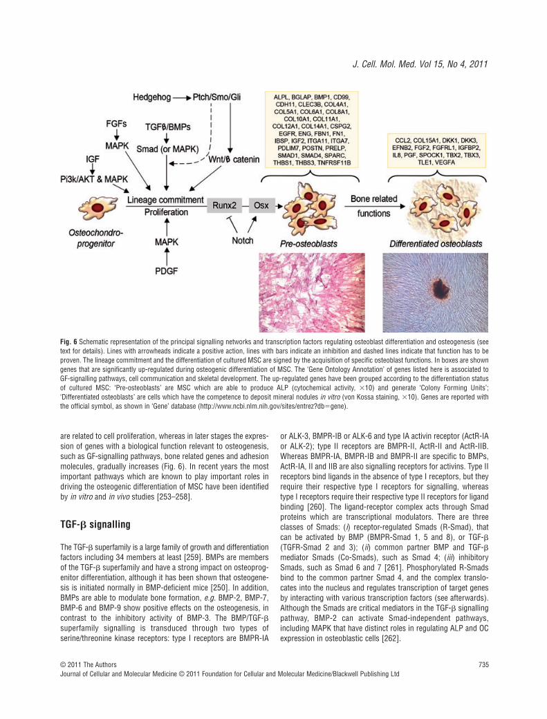
J. Cell. Mol. Med. Vol 15, No 4, 2011
735© 2011 The AuthorsJournal of Cellular and Molecular Medicine © 2011 Foundation for Cellular and Molecular Medicine/Blackwell Publishing Ltd
are related to cell proliferation, whereas in later stages the expres-sion of genes with a biological function relevant to osteogenesis,such as GF-signalling pathways, bone related genes and adhesionmolecules, gradually increases (Fig. 6). In recent years the mostimportant pathways which are known to play important roles indriving the osteogenic differentiation of MSC have been identifiedby in vitro and in vivo studies [253–258].
TGF-! signalling
The TGF-" superfamily is a large family of growth and differentiationfactors including 34 members at least [259]. BMPs are membersof the TGF-" superfamily and have a strong impact on osteoprog-enitor differentiation, although it has been shown that osteogene-sis is initiated normally in BMP-deficient mice [250]. In addition,BMPs are able to modulate bone formation, e.g. BMP-2, BMP-7,BMP-6 and BMP-9 show positive effects on the osteogenesis, incontrast to the inhibitory activity of BMP-3. The BMP/TGF-"superfamily signalling is transduced through two types ofserine/threonine kinase receptors: type I receptors are BMPR-IA
or ALK-3, BMPR-IB or ALK-6 and type IA activin receptor (ActR-IAor ALK-2); type II receptors are BMPR-II, ActR-II and ActR-IIB.Whereas BMPR-IA, BMPR-IB and BMPR-II are specific to BMPs,ActR-IA, II and IIB are also signalling receptors for activins. Type IIreceptors bind ligands in the absence of type I receptors, but theyrequire their respective type I receptors for signalling, whereastype I receptors require their respective type II receptors for ligandbinding [260]. The ligand-receptor complex acts through Smadproteins which are transcriptional modulators. There are threeclasses of Smads: (i) receptor-regulated Smads (R-Smad), thatcan be activated by BMP (BMPR-Smad 1, 5 and 8), or TGF-"(TGFR-Smad 2 and 3); (ii) common partner BMP and TGF-"mediator Smads (Co-Smads), such as Smad 4; (iii) inhibitorySmads, such as Smad 6 and 7 [261]. Phosphorylated R-Smadsbind to the common partner Smad 4, and the complex translo-cates into the nucleus and regulates transcription of target genesby interacting with various transcription factors (see afterwards).Although the Smads are critical mediators in the TGF-" signallingpathway, BMP-2 can activate Smad-independent pathways,including MAPK that have distinct roles in regulating ALP and OCexpression in osteoblastic cells [262].
Fig. 6 Schematic representation of the principal signalling networks and transcription factors regulating osteoblast differentiation and osteogenesis (seetext for details). Lines with arrowheads indicate a positive action, lines with bars indicate an inhibition and dashed lines indicate that function has to beproven. The lineage commitment and the differentiation of cultured MSC are signed by the acquisition of specific osteoblast functions. In boxes are showngenes that are significantly up-regulated during osteogenic differentiation of MSC. The ‘Gene Ontology Annotation’ of genes listed here is associated toGF-signalling pathways, cell communication and skeletal development. The up-regulated genes have been grouped according to the differentiation statusof cultured MSC: ‘Pre-osteoblasts’ are MSC which are able to produce ALP (cytochemical activity, +10) and generate ‘Colony Forming Units’;‘Differentiated osteoblasts’ are cells which have the competence to deposit mineral nodules in vitro (von Kossa staining, +10). Genes are reported withthe official symbol, as shown in ‘Gene’ database (http://www.ncbi.nlm.nih.gov/sites/entrez?db&gene).
736 © 2011 The AuthorsJournal of Cellular and Molecular Medicine © 2011 Foundation for Cellular and Molecular Medicine/Blackwell Publishing Ltd
FGF signalling
The FGF gene family is composed of 23 members that variouslybind to seven FGF tyrosine kinase receptor isoforms (Fgfr) [263].The FGF-family polypeptides play a critical role in regulating endo-chondral and intramembranous ossification. Fgfr1 is expressed inhypertrophic chondrocytes, and Fgfr1 signalling has stage-specific effects on osteoblast maturation, because it acts either tostimulate differentiation in osteoprogenitor cell or to arrest thematuration of differentiated osteoblasts [264]. FGFs 2, 9 and 18probably bind Fgfr1 in osteoblasts. FGF9 and FGF18 are expressedin the perichondrium/periosteum, and seem to play a predominantrole during the embryonic skeletogenesis [255]. FGF2 isexpressed in periosteal cells and osteoblasts, and its activityshould be more relevant during postnatal stages, as proven by thedecreased ability to mineralize in vitro which has been observed inMSC cultures from FGF2-null mice [265]. Fgfr2 is expressed inreserve chondrocytes and appears to be down-regulated in prolif-erating chondrocytes. Fgf2r shows tissue-specific alternativesplicing and the mesenchymal variants are the Fgfr2c forms,which are activated by FGF2, 4, 6, 8 and 9. FGF18 acts as a phys-iological ligand for Fgfr3, which regulates cell growth and differ-entiation of proliferating chondrocytes, whereas in differentiatedosteoblasts it regulates bone density and cortical thickness [266].
IGF signalling
The IGF family consists of two secreted GFs, namely IGF-I andIGF-II, two receptors (IGF1R and IGF2R) and six high-affinity bind-ing proteins. Both IGFs are expressed by osteoblasts and showsimilar biological activities, even though IGF-I is more potent thanIGF-II. They stimulate osteoblast function and bone matrix depo-sition favouring the synthesis of collagens and non-collagenousproteins [267]. IGF-I does not influence the differentiation of MSCinto osteoblasts and cell proliferation, which instead is stimulatedby IGF-II [5]. Actions of both IGFs are mediated by the IGF-I recep-tor, a ligand-activated tyrosine protein kinase that uses a series ofintracellular adaptor molecules, including the insulin receptor sub-strate proteins IRS1 and IRS2, to engage downstream signallingpathways [268]. IGF-I utilizes both the P13K, which induces theactivation of Akt, and the MAPK pathway, which activates p38,Jun-N-terminal kinases and ERK1/2. The use of each pathway isdependent on culture conditions and the stage of cell differentia-tion [267]. Studies in experimental animals have concluded that action of IGF is essential for normal bone formation, growthand maintenance.
PDGF signalling
PDGF is an extracellular factor which exists as three biologicallyactive isoforms composed of PDGF-A and PDGF-B polypeptidechain. Binding of PDGF requires and results in the dimerization of
the two distinct receptors (#-PDGFR and "-PDGFR) that formthree distinct receptor patterns (##, #", ""). MSCs express #and " receptors. Although PDGF signalling has been reported toplay an important role in the control of various cell functions ofskeletal cells, the network of PDGF signalling for MSCs has notbeen fully clarified. PDGF-BB, but not PDGF-AA, inhibitedosteogenic differentiation accompanied by decreased ALP activityand mRNA levels The depletion of PDGFR-" gene in MSCdecrease the mitogenic and migratory responses and enhancedosteogenic differentiation, as evaluated by increased ALP activityand mRNA levels, OC, BMP-2, Runx2 and Osx [269]. More recentdata show that the pharmacological inhibition of PDGFRdecreases the MSC proliferation induced by PDGF-BB, but doesnot affect the osteoblastic differentiation, as evaluated byanalysing the expression of other osteogenic marker genes andthe mineralized matrix production [270].
MAPK signalling pathway
MAPK pathway is activated by a variety of GFs with a role inosteogenesis, including FGF, PDGF, TGF-" and IGFs. The extracellular stimuli lead to the activation of a signalling cascadecomposed of MAP kinase, MAP kinase kinase (MKK or MAP2K)and MAP kinase kinase kinase (MKKK or MAP3K) [271]. Thephysiological functions of this complex cascade are summarizedin Table 3.
Wnt signalling pathway
Wnts (for wingless) forms a large family of secreted molecules,which are ligands for the membrane-spanning frizzled (FZD)receptors involved in various aspects of cellular biology, includ-ing cell growth, differentiation, function and death. Wnts cantransduce their signals through two different pathways, namely‘canonical’ and ‘non-canonical’ Wnt signalling. The first oneinvolves the formation of a complex among Wnt proteins, FZDand low density Lrp5 or Lrp6 receptors. There is considerableevidence indicating that Wnt canonical pathway plays a criticalrole in bone formation resulting in the expression of osteoblast-specific markers [272, 273]. The binding of Wnt proteins to the FZD/Lrp5/6 complex generates a signal, which inhibits theactivity of glycogen synthase kinase 3 (GSK-3). GSK-3 inactivityblocks the phosphorylation of "-catenin, and prevents its degra-dation in the cytoplasm by the ubiquitin-pathway, leading totranslocation of "-catenin into the nucleus where it cooperateswith transcription factors of the T-cell factor/lymphoid enhancerfactor family [274]. The "-catenin activity seems to be a require-ment to direct the bipotential osteochondroprogenitor towards aspecific lineage. Indeed, the "-catenin/T-cell factor 1 complexenhances expression and activity of the osteogenic promoterRunx2, whereas low levels of "-catenin direct the mesenchymalprecursors toward chondrogenesis [275].

J. Cell. Mol. Med. Vol 15, No 4, 2011
737© 2011 The AuthorsJournal of Cellular and Molecular Medicine © 2011 Foundation for Cellular and Molecular Medicine/Blackwell Publishing Ltd
Hedgehog signalling
Hedgehog signalling (Hh) family members provide positionalinformation during the skeletogenesis, and initiate or maintain cel-lular differentiation programs regulating the formation of cartilageand bone. Indian hedgehog (Ihh) is produced by pre-hypertrophicchondrocytes and its signalling appears to act directly onosteoblast progenitors located in the perichondrium [276, 277].The pathway is triggered by binding Hh protein to its receptorPatched thus activating the seven-transmembrane proteinsmoothened (Smo). The Hh signal transduction cascade activatesthe transcription factor GliA, a member of the Kruppel family ofzinc finger proteins, which is translocated into the nucleus, regu-lates stem cell proliferation and activates gene targets. Severalpieces of data show that Ihh signalling and Wnt signalling cooper-ate in controlling various aspects of skeletal development.Interestingly, Smo receptors have considerable amino acidsequence similarity with the Wnt receptor FZD, and GSK-3" isable to inhibit the Hh signalling [250]. Experimental data provethat Ihh signalling acts upstream of Wnt signalling and its activityseems to be limited to the early stage of osteoblast commitment[278]. In fact, when Smo activity is removed in Osx! osteoprog-enitors, normal osteoblasts are generated and the endochondralskeleton at birth is indistinguishable from wild-type [279].
Notch signalling
Notch signalling is well known for determining cell fate. BecauseNotch receptors and their ligands ((1, 3, 4 and Jagged 1, 2) aretransmembrane proteins, a cell–cell interaction is required foractivating signalling cascade [280]. Notch 1 and Notch 2 are gen-erally expressed in osteoblasts, whereas Notch 3 and Notch 4have been found in subsets of the osteogenic lineage [281].Experimental data support a dimorphic function of Notch sig-nalling, which is able to up-regulate bone related genes, to favour
the proliferation of immature osteoblasts, and to induce a severeosteosclerosis, but also to repress the BMP-induced osteoblastmaturation by inhibiting the Runx2 transactivation function [282].
Ephrin signalling
There are two classes of ephrins: the B class (B1 to B3) are ligandsfor EphB1–6 tyrosine kinase receptors, whereas the A class (A1 toA5) are ligands for GPI-anchored EphA receptors (A1 to A10).Ephrins have the capacity of bidirectional signalling. Thus, when acell expressing an Eph receptor contacts a cell expressing anephrin ligand, signals are transduced into both cells by forward andreverse signalling, respectively [283]. The reverse signalling fromEphB4 in osteoblasts to ephrinB2 in osteoclast progenitors leads tothe inhibition of osteoclast differentiation, whereas the forward sig-nalling through EphB4 induces osteogenic transcription factors[284]. However, ephrinA2–EphA2 interaction facilitates the initia-tion phase of bone remodelling by enhancing osteoclast differenti-ation and suppressing osteoblast differentiation [285].
Transcription factors regulating osteoblast differentiation
The signalling pathways described above lead to the activation oftranscription factors that exert positive and negative regulatoryeffects on the expression of genes controlling the acquisition ofthe osteoblast phenotype [255, 286]. Runx2 is a member of theRunx family of transcription factors abundantly expressed in cal-cified cartilage and bone, and it is considered as a master regula-tory switch essential for osteoblast differentiation [287]. Runx2may be expressed in early osteoprogenitors, but is also requiredfor osteoblast function beyond differentiation. Targeted disruptionof Runx2 results in the complete inhibition of bone formation,revealing that Runx2 is essential for both endochondral and
Table 3 Families of MAPKs
Family Name Function
Extracellular signal-regulated kinases
ERK1, ERK2The ERKs (also known as classical MAPK) signalling pathways are activated inresponse to GFs, and regulate cell proliferation and cell differentiation
ERK5 (MAPK7) Activated both by GFs and by stress stimuli; participates in cell proliferation
ERK3 (MAPK6) ERK4 (MAPK4)
Atypical MAPKs. ERK3/4 are mostly cytoplasmic proteins which bind, translocate and activate the MK5 (PRAK, MAP2K5). ERK3 is known as anunstable unlike ERK4 which is relatively stable
ERK7/8 (MAPK15) New members of MAPKs structurally related to ERK3/4
c-Jun N-terminal kinases (JNKs) MAPK8, MAPK9, MAPK10 Stress-activated protein kinases
p38 isoformsMAPK11, MAPK12 (ERK6),MAPK13, MAPK14
Both JNK and p38 signalling pathways are responsive to stress stimuli, such ascytokines, ultraviolet irradiation, heat shock and osmotic shock, and areinvolved in cell differentiation and apoptosis

738 © 2011 The AuthorsJournal of Cellular and Molecular Medicine © 2011 Foundation for Cellular and Molecular Medicine/Blackwell Publishing Ltd
intramembranous bone formation. Runx2 acts in nuclear microen-vironments as a scaffolding protein that controls gene expressionin response to physiological signals. Many transcription factorsinteract with Runx2: some provide costimulatory signals, whereasothers repress Runx2 function by affecting its DNA binding activ-ity and/or transactivation potential [255]. The second transcrip-tional regulator for the final stages of bone tissue formation is thehuman homologue of the mouse Osx i.e. specificity protein-7. Thisis a zinc finger transcription factor which contains specificdomains responsible for the activation of OC and Col1a1 genes.Osx-null osteoblast precursors in the periosteum of membranousbones express chondrocyte markers, such as Sox9 and Col2a1.This suggests that Runx2-expressing preosteoblasts are still bipo-tential cells. Nevertheless, Osx-null osteoblast precursors cannotdifferentiate into osteoblasts and deposit bone matrix, so thatendochondral or intramembranous bone formation do not occur[95]. These data prove that Osx acts downstream of Runx2 toinduce osteoblastic differentiation in osteochondroprogenitorcells. Osx may be induced by additional signalling pathways act-ing in parallel to, or independent of, Runx2. It has been shown thatRunx2 is required but not sufficient for the BMP-2-mediated Osxinduction, because MAPK signalling pathways serve as points ofconvergence to mediate the BMP-2 effect on Osx expression.Cooperation between Osx and nuclear factor of activated T cellsactivates the Col1a1 and OC promoters and accelerates osteoblastdifferentiation and bone formation in a Runx2-independent man-ner [288]. Another important transcription factor controllingosteoblast differentiation is ATF4 (activating transcription factor 4)or cAMP response elements binding protein 2. ATF4 is requiredfor terminal differentiation of osteoblasts, and it interacts withRunx2 to regulate bone sialoprotein and OC expression. The cru-cial role of ATF4 has been shown by blocking its activity andobtaining a severe decrease in bone formation [289].
Conclusion
The sequential phases of osteoblast commitment and differentiationare regulated by a variety of complex activities, including hormones,GF, mechanical stimuli, cell–cell and cell–matrix interaction, that actvia interconnected signalling networks, resulting in the activation ofspecific transcription factors and, in turn, their target genes.
Summary
Indirect fracture healing is usually clinically uncomplicated, isbased on endochondral ossification, and proceeds by stages fromhaematoma through inflammation, angiogenesis, chondrogenesisand osteogenesis, to bone remodelling. Non-union, delayed unionand various congenital or acquired bone defects can cause prob-lems. Instead of traditional internal or external fracture fixation orosteosynthetic devices, regenerative medicine attempts to recruit,
guide endogenous or to implant various exogenous mesenchymalor foetal stem cells to support active healing or to replace missingbone. These attempts often include composites of biodegradablepolymers, e.g. PCL, poly(propylene fumarate) or PLA, with silica,HA or other calcium phosphate filler particles. Scaffold/matrix-cellinteractions will be better utilized once the biological effects ofaging, hormonal, intracrine, transcriptional and the legal regula-tion of adult and foetal stem cells are more clearly understood.PRP provides an inexpensive autologous source of potent biofac-tors, which together with other signals contribute to osteogenesisvia TGF-", FGF, IGF, PDGF, MAPK, Wnt, hedgehog, Notch andephrin signalling via transcriptional regulation, which also providethe tools to monitor regeneration and healing.
The European Science Foundation was established in 1974 tocreate a common European platform for cross-border cooperationin all aspects of scientific research. With its emphasis on a multidis-ciplinary and pan-European approach, the Foundation provides theleadership necessary to open new frontiers in European science.Today the frontiers of regenerative medicine are rapidly expanding.Regenerative medicine has already provided new insight into cellu-lar proliferation, effects of humoural and matrix signalling on cells,angiogenesis and tissue remodelling as well as basic insights intocell biology. Nevertheless, regenerative medicine is in its infancyand to facilitate the process of growth, 14 EU countries funded byEuropean Science Foundation have joined forces in an attempt toimprove networking and clarify where the frontiers and future needsare in this complex multidisciplinary field. Proof of principle experi-ments and randomized clinical trials remain in the future for regen-erative medicine. In the most visionary view, scientists hope to seethe human body undergo repeated and well timed maintenance offailing body parts through cycles of healing cell therapies.
Acknowledgements
D.P. and L.A. were financially supported by several grants from the SwissNational Science Foundation and in part from the Foundation S.A.N.T.E andSandoz Family Foundation. M.K., B.M.A. and the study were support bygrants from the Danish Medical Research Council, the Novo NordiskFoundation, the Vellux Foundation and Danish Center for Stem CellResearch (DASC) and by a local grant from the University Hospital ofOdense, Denmark. E.G.B. was financially supported by The EU by means ofthe 7th Frame Program in the REBORNE project (‘Regenerating Bonedefects using New biomedical Engineering approaches’, EC-GA n,241879,HEALTH-2009–1.4.2). N.B., E.C. and D.G. were supported by grants fromItalian Ministry of the Health for research activities of the RizzoliOrthopaedic Institute, and from ‘Regione Emilia-Romagna’ for the researchprogram on ‘Regenerative Medicine in Osteo-articular Diseases’. Y.T.K. andT.S. were financially supported by the EU MATERA project ‘BioactiveNanocomposite Constructs for Regeneration of Articular Cartilage’, EUMNT ERA Net project ‘New Generation Titania’, Danish Council forStrategic Research ‘Individualized Musculoskeletal Medicine’, SigridJusélius Foundation, Finska Läkaresällskapet, evo grants, HelsinkiUniversity Central Hospital and ORTON Orthopedic Hospital of the OrtonFoundation. A.F.W., K.M., K.A. were financially supported by The EU

J. Cell. Mol. Med. Vol 15, No 4, 2011
739© 2011 The AuthorsJournal of Cellular and Molecular Medicine © 2011 Foundation for Cellular and Molecular Medicine/Blackwell Publishing Ltd
support, the 7th Frame Program, VascuBone project (‘Construction kit for tailor-made vascularized boneimplants’, EC-GA n, 242175 HEALTH-2009–1.4.2) and by The Research Council of Norway ([FRIMED,17734/V50], [156744], [Stem Cell 180383/V40]).
Conflict of interest
The authors report that there are no conflicts of interest.
References
1. Urist MR, Johnson RW Jr. Calcificationand ossification. IV The healing of frac-tures in man under clinical conditions. JBone Joint Surg Am. 1943; 25: 375–426.
2. Phillips AM. Overview of the fracture heal-ing cascade. Injury. 2005; 36: S5–7.
3. Einhorn TA, Majeska RJ, Rush EB, et al. The expression of cytokine activityby fracture callus. J Bone Miner Res. 1995;10: 1272–81.
4. Gerstenfeld LC, Cho TJ, Kon T, et al.Impaired fracture healing in the absence ofTNF-alpha signaling: the role of TNF-alphain endochondral cartilage resorption. J Bone Miner Res. 2003a; 18: 1584–92.
5. Tsiridis E, Upadhyay N, Giannoudis P.Molecular aspects of fracture healing:which are the important molecules? Injury.2007; 38: S11–25.
6. Gerstenfeld LC, Cullinane DM, BarnesGL, et al. Fracture healing as a post-nataldevelopmental process: molecular, spatial,and temporal aspects of its regulation. J Cell Biochem. 2003b; 88: 873–84.
7. Deckers MM, van Bezooijen RL, van derHorst G, et al. Bone morphogenetic proteins stimulate angiogenesis throughosteoblast-derived vascular endothelialgrowth factor A. Endocrinology. 2002;143: 1545–53.
8. Yoshimura Y, Nomura S, Kawasaki S, et al. Colocalization of noggin and bone mor-phogenetic protein-4 during fracture healing.J Bone Miner Res. 2001; 16: 876–84.
9. Onichtchouk D, Chen YG, Dosch R, et al.Silencing of TGF-beta signalling by thepseudoreceptor BAMBI. Nature. 1999;401: 480–5.
10. Nakao A, Afrakhte M, Morén A, et al.Identification of Smad7, a TGFbeta-inducible antagonist of TGF-beta sig-nalling. Nature. 1997; 389: 631–5.
11. Bai S, Shi X, Yang X, Cao X. Smad6 as atranscriptional corepressor. J Biol Chem.2000; 275: 8267–70.
12. Ferguson C, Alpern E, Miclau T, HelmsJA. Does adult fracture repair recapitulateembryonic skeletal formation? Mech Dev.1999; 87: 57–66.
13. Carter DR, Beaupre GS, Giori NJ, HelmsJA. Mechanobiology of skeletal regenera-
tion. Clin Orthop Relat Res. 1998; 355:S41–55.
14. Giannoudis PV, Einhorn TA, Marsh D.Fracture healing: the diamond concept.Injury. 2007; 38: S3–6.
15. Zhuang H, Wang W, Tahernia AD, et al.Mechanical strain-induced proliferation ofosteoblastic cells parallels increased TGF-beta 1 mRNA. Biochem Biophys ResCommun. 1996; 229: 449–53.
16. Sasaki K, Takagi M, Konttinen Y, et al.Upregulation of matrix metalloproteinase(MMP)-1 and its activator MMP-3 ofhuman osteoblasts by uniaxial cyclic stim-ulation. J Biomed Mater Res B ApplBiomater. 2007; 80B: 491–8.
17. Chiquet M. Regulation of extracellularmatrix gene expression by mechanicalstress. Matrix Biol. 1999; 18: 417–26.
18. Kershaw CJ, Cunningham JL, KenwrightJ. Tibial external fixation, weight bearing,and fracture movement. Clin Orthop RelatRes. 1993; 293: 28–36.
19. Kwong FN, Harris MB. Recent develop-ments in the biology of fracture repair. J AmAcad Orthop Surg. 2008; 16: 619–25.
20. Claes L, Eckert-Hübner K, Augat P. Theeffect of mechanical stability on local vas-cularization and tissue differentiation incallus healing. J Orthop Res. 2002; 20:1099–105.
21. Loboa EG, Beaupre GS, Carter DR.Mechanobiology of initial pseudarthrosisformation with oblique fractures. J OrthopRes. 2001; 19: 1067–72.
22. Gartner LP, Hiatt JL. Color textbook of histology. 3rd ed. Philadelphia, PA:Saunders Elsevier; 2007. pp. 19103–2899.
23. Langman J. Medical embryology. humandevelopment-normal and abnormal. 3rded. Baltimore: The Williams & WilkinsCompany; 1975.
24. ten Cate AR. Oral histology. Development,structure and function. St. Louis, MO: TheC.V. Mosby Company; 1980.
25. Glenny AM, Hooper L, Shaw WC, et al.Feeding interventions for growth anddevelopment in infants with cleft lip, cleftpalate or cleft lip and palate. Review.Cochrane Database Syst Rev. 2004;CD003315.
26. Mossey PA, Little J, Munger RG, et al.Cleft lip and palate. Review. Lancet. 2009;9703: 1773–85.
27. Pena WA, Vargervik K, Sharma A, OberoiS. The role of endosseous implants in themanagement of alveolar clefts. Review.Pediatr Dent. 2009; 31: 329–33.
28. Esposito M, Grusovin MG, Coulthard P, et al. The efficacy of various bone aug-mentation procedures for dental implants:a cochrane systematic review of random-ized controlled clinical trials. Int J OralMaxillofac Implants. 2006; 21: 696–710.
29. Kelly CM, Wilkins RM, Gitelis S, et al.The use of a surgical grade calcium sulfateas a bone graftsubstitute: results of a mul-ticenter trial. Clin Orthop Relat Res. 2001;382: 42–50.
30. Schindler OS, Cannon SR, Briggs TWR, et al. Composite ceramic bone graft sub-stitute in the treatment of locally aggres-sive benign bone tumours. J Orthop Surg.2008; 16: 66–74.
31. Schieker M, Seitz H, Drosse I, et al.Biomaterials as scaffold for bone tissueengineering. Eur J Trauma. 2006; 2:114–24.
32. Petite H, Viateau V, Bensaid W, et al.Tissue-engineered bone regeneration. NatBiotechnol. 2000; 18: 959–63.
33. Matsubara T, Suardita K, Ishii M, et al.Alveolar bone marrow as a cell source for regenerative medicine: differencesbetween alveolar and iliac bone marrowstromal cells. J Bone Miner Res. 2005; 20:399–409.
34. Shi S, Gronthos S. Perivascular niche ofpostnatal mesenchymal stem cells inhuman bone marrow and dental pulp. J Bone Miner Res. 2003; 8: 696–704.
35. Shi S, Bartold PM, Miura M, et al. Theefficacy of mesenchymal stem cells toregenerate and repair dental structures.Orthod Craniofac Res. 2005; 8: 191–9.
36. Ikeda E, Hirose M, Kotobuki N, et al.Osteogenic differentiation of human dentalpapilla mesenchymal cells. Biochem BiophysRes Commun. 2006; 342: 1257–62.
37. Gronthos S, Brahim J, Li W, et al. Stemcell properties of human dental pulp stemcells. J Dent Res. 2002; 81: 531–5.
740 © 2011 The AuthorsJournal of Cellular and Molecular Medicine © 2011 Foundation for Cellular and Molecular Medicine/Blackwell Publishing Ltd
38. Derubeis AR, Cancedda R. Bone marrowstromal cells (BMSCs) in bone engineer-ing: limitations and recent advances. AnnBiomed Eng. 2004; 32: 160–5.
39. Caplan AI. Mesenchymal stem cells. J Orthop Res. 1991; 9: 641–50.
40. Khan Y, Yaszemski MJ, Mikos AG, et al.Tissue engineering of bone: material andmatrix consideration. J Bone Joint SurgAm. 2008; 90: 36–42.
41. Suzuki O, Imaizumi H, Kamakura S, et al.Bone regeneration by synthetic octacal-cium phosphate and its role in biologicalmineralization. Curr Med Chem. 2008; 15:305–13.
42. Guarino V, Causa F, Ambrosio L.Bioactive scaffolds for connective tissueregeneration. Exp Rev Med Dev. 2007; 4:406–18.
43. Coombes AGA, Rizzi SC, Williamson M,et al. Precipitation casting of polycapro-lactone for applica- tions in tissue engi-neering and drug delivery. Biomaterials.2004; 25: 315–25.
44. Shikinami Y, Okazaki K, Saito M, et al.Bioactive and bioresorbable cellular cubic-composite scaffolds for use in bone recon-struction. J R Soc Interface. 2006; 3:805–21.
45. Mondrinos MJ, Dembzinsky R, Lu L, et al. Porogen-based solid freeform fabri-cation of polycaprolactone–calcium phos-phate scaffold for tissue engineering.Biomaterials. 2006; 27: 4399–408.
46. Zhou Y, Hutmacher DW, Varawan S-L, et al. In vitro bone engineering based onpolycaprolactone and polycaprolactone-tricalcium phosphate composites. PolymInt. 2007; 56: 333–42.
47. Guarino V, Ambrosio L. The synergiceffect of polylactide fiber and calciumphosphate particle reinforcement in poly !-caprolactone-based composite scaffolds.Acta Biomaterialia. 2008; 4: 1778–87.
48. Cai Z-Y, Yang D-A, Zhang N, et al.Poly(propylene fumarate)/(calcium sul-phate/"-tricalcium phosphate) compos-ites: preparation, characterization and invitro degradation. Acta Biomaterialia.2009; 5: 628–35.
49. Irina M, Pelin, Stelian S. Maier, et al.Preparation and characterization of ahydroxyapatite–collagen composite ascomponent for injectable bone substitute.Mater Sci Engin. 2009; 29: 2188–94.
50. Azevedo MC, Reis RL, Claase MB, et al.Development and properties of polycapro-lactone/hydroxyapatite composite bioma-terials. J Mater Sci Mat Med. 2003; 14:103–7.
51. Guarino V, Causa F, NettiPA, et al. Therole of hydroxyapatite as solid signal onperformance of PCL porous scaffolds forbone tissue regeneration. J Biomed MaterRes B. 2008; 86: 548–57.
52. Kroeze RJ, Helder MN, Govaert LE, et al.Biodegradable polymers in bone tissueengineering. Materials. 2009; 2: 833–56.
53. Li F, Li S, Ghzaoui AE, et al. Synthesisand gelation properties of PEG-PLA-PEGtriblock copolymers obtained by couplingmonohydroxylated PEG-PLA with adipoylchloride. Langmuir. 2007a; 27: 2778–83.
54. Ryner M, Albertsson A-C. Resorbable and highly elastic block copolymers from 1,5-dioxepan-2-one and L-lactidewith controlled tensile properties andhydrophilicity. Biomacromolecules. 2002;3: 601–8.
55. Ryner M, Valdre A, Albertsson A-C. Star-shaped and photo-crosslinked poly(1,5-dioxepan-2-one) – synthesis and charac-terization. J Polym Sci A: Polym Sci. 2002;40: 2049–54.
56. Idris S, Arvidson K, Plikk P, et al.Polyester copolymer scaffolds enhanceexpression of bone markers in osteoblast-like cells. J Biomed Mater Res A. 2010; 94:631–9.
57. Dånmark S, Finne-Wistrand A, AlbertssonA-C, et al. Osteogenic differentiation in rat bone rarrow derived stromal cells on refined biodegradable polymer scaf-folds. J Bioact Compat Pol. 2010; 25:207–23.
58. Schander K, Arvidson K, Mustafa M, et al. Response of Bone and PeriodontalLigament Cells to Biodegradable PolymerScaffolds in vitro. J Bioact Compat Pol.2010; 25: 584–602.
59. Idris S, Danmark S, Finne-Wistrand A, et al. Biocompatibility of PolyesterScaffolds with Fibroblasts and Osteoblast-like Cells for Bone TissueEngineering. J Bioact Compat Pol. 2010;25: 567–83.
60. Wang S, Cui W, Bei J. Bulk and surfacemodifications of polylactide. Anal BioanalChem. 2005; 381: 547–56.
61. Frost HM. Why should many skeletal scien-tists and clinicians learn the Utah paradigmof skeletal physiology? J MusculoskelNeuron Interact. 2001; 2: 121–30.
62. Parfitt AM. Bone forming cells in clinicalconditions. In: Hall BK, editor. Bone, theosteoblast and osteocytes. London, UK:The Telford Press; 1991. pp. 351–426.
63. Harada S, Rodan GA. Control ofosteoblast function and regulation of bonemass. Nature. 2003; 423: 349–55.
64. Blair HC, Robinson LJ, Zaidi M.Osteoclast signalling pathways. BiochemBiophys Res Commun. 2005; 328: 728–38.
65. Friedenstein AJ, Ivanov-Smolenski AA,Chajlakjan RK, et al. Origin of bone marrowstromal mechanocytes in radiochimerasand heterotopic transplants. Exp Hematol.1978; 6: 440–4.
66. Friedenstein AJ. Osteogenic stem cells inthe bone marrow. Bone Miner. 1991; 7:243–72.
67. Benayahu D, Akavia UD, Shur I.Differentiation of bone marrow stroma-derived mesenchymal cells. Curr MedChem. 2007; 14: 173–9.
68. Friedenstein AJ, Chailakhjan RK,Lalykina KS. The development of fibrob-last colonies in monolayer cultures ofguinea- pig bone marrow and spleen cells.Cell Tissue Kinet. 1970; 3: 393–403.
69. Luria EA, Panasyuk AF, Friedenstein AY.Fibroblast colony formation from mono-layer cultures of blood cells. Transfusion.1971; 11: 345–9.
70. Kassem M, Mosekilde L, Eriksen EF.1,25-dihydroxyvitamin D3 potentiates flu-oride-stimulated collagen type I produc-tion in cultures of human bone marrowstromal osteoblast-like cells. J Bone MinerRes. 1993; 8: 1453–8.
71. Rickard DJ, Kassem M, Hefferan TE, et al. Isolation and characterization ofosteoblast precursor cells from humanbone marrow. J Bone Miner Res. 1996; 11:312–24.
72. Kuznetsov SA, Krebsbach PH, SatomuraK, et al. Single-colony derived strains ofhuman marrow stromal fibroblasts formbone after transplantation in vivo. J BoneMiner Res. 1997; 12: 1335–47.
73. Post S, Abdallah BM, Bentzon JF,Kassem M. Demonstration of the pres-ence of independent pre-osteoblastic andpre-adipocytic cell populations in bonemarrow-derived mesenchymal stem cells.Bone. 2008; 43: 32–9.
74. Abdallah BM, Haack-Sorensen M, BurnsJS, et al. Maintenance of differentiationpotential of human bone marrow mes-enchymal stem cells immortalized byhuman telomerase reverse transcriptasegene despite [corrected] extensive prolif-eration. Biochem Biophys Res Commun.2005; 326: 527–38.
75. Foster LJ, Zeemann PA, Li C, et al.Differential expression profiling of mem-brane proteins by quantitative proteomicsin a human mesenchymal stem cell lineundergoing osteoblast differentiation.Stem Cells. 2005; 23: 1367–77.
J. Cell. Mol. Med. Vol 15, No 4, 2011
741© 2011 The AuthorsJournal of Cellular and Molecular Medicine © 2011 Foundation for Cellular and Molecular Medicine/Blackwell Publishing Ltd
76. Dominici M, Le BK, Mueller I, et al.Minimal criteria for defining multipotentmesenchymal stromal cells. TheInternational Society for Cellular Therapyposition statement. Cytotherapy. 2006; 8:315–7.
77. Gronthos S, Zannettino AC, Hay SJ, et al.Molecular and cellular characterisation ofhighly purified stromal stem cells derivedfrom human bone marrow. J Cell Sci.2003a; 116: 1827–35.
78. Quirici N, Soligo D, Bossolasco P, et al.Isolation of bone marrow mesenchymalstem cells by anti-nerve growth factorreceptor antibodies. Exp Hematol. 2002;30: 783–91.
79. Miura Y, Miura M, Gronthos S, et al.Defective osteogenesis of the stromalstem cells predisposes CD18-null mice toosteoporosis. Proc Natl Acad Sci USA.2005; 102: 14022–7.
80. Gang EJ, Bosnakovski D, Figueiredo CA,et al. SSEA-4 identifies mesenchymalstem cells from bone marrow. Blood.2007; 109: 1743–51.
81. Kuznetsov SA, Mankani MH, Gronthos S,et al. Circulating skeletal stem cells. J CellBiol. 2001; 153: 1133–40.
82. Rosada C, Justesen J, Melsvik D, et al.The human umbilical cord blood: a poten-tial source for osteoblast progenitor cells.Calcif Tissue Int. 2003; 72: 135–42.
83. De Bari C, Dell’Accio F, Tylzanowski P, et al. Multipotent mesenchymal stem cellsfrom adult human synovial membrane.Arthritis Rheum. 2001; 44: 1928–42.
84. Miura M, Gronthos S, Zhao MR, et al.SHED: stem cells from human exfoliateddeciduous teeth. Proc Natl Acad Sci USA.2003; 100: 5807–12.
85. De Coppi P, Bartsch G Jr, Siddiqui MM,et al. Isolation of amniotic stem cell lineswith potential for therapy. Nat Biotechnol.2007: 25: 100–6.
86. Wagner W, Wein F, Seckinger A, et al.Comparative characteristics of mesenchy-mal stem cells from human bone marrow,adipose tissue, and umbilical cord blood.Exp Hematol. 2005; 33: 1402–16.
87. Bianco P, Riminucci M, Gronthos S, et al.Bone marrow stromal stem cells: nature,biology, and potential applications. StemCells. 2001; 19: 180–92.
88. Pittenger MF, Mackay AM, Beck SC, et al. Multilineage potential of adulthuman mesenchymal stem cells. Science.1999; 284: 143–7.
89. Justesen J, Stenderup K, Eriksen EF, et al. Maintenance of osteoblastic andadipocytic differentiation potential with age
and osteoporosis in human marrow stro-mal cell cultures. Calcif Tissue Int. 2002;71: 36–44.
90. Abdallah BM, Kassem M. Human mes-enchymal stem cells: from basic biology toclinical applications. Gene Ther. 2008; 15:109–16.
91. Kuznetsov SA, Krebsbach PH, SatomuraK, et al. Single-colony derived strains ofhuman marrow stromal fibroblasts formbone after transplantation in vivo. J BoneMiner Res. 1997; 12: 1335–47.
92. Piersanti S, Sacchetti B, Funari A, et al.Lentiviral transduction of human postnatalskeletal (stromal, mesenchymal) stemcells: in vivo transplantation and genesilencing. Calcif Tissue Int. 2006; 78:372–84.
93. Larsen KH, Frederiksen CM, Burns JS, et al. Identifying a molecular phenotypefor bone marrow stromal cells with in vivobone-forming capacity. J Bone Miner Res.2010; 25: 796–808.
94. Komori T, Yagi H, Nomura S, et al.Targeted disruption of Cbfa1 results in acomplete lack of bone formation owing tomaturational arrest of osteoblasts. Cell.1997; 89: 755–64.
95. Nakashima K, Zhou X, Kunkel G, et al.The novel zinc finger-containing tran-scription factor osterix is required forosteoblast differentiation and bone forma-tion. Cell. 2002; 108: 17–29.
96. Qiu W, Andersen TE, Bollerslev J, et al.Patients with high bone mass phenotypeexhibit enhanced osteoblast differentiationand inhibition of adipogenesis of humanmesenchymal stem cells. J Bone MinerRes. 2007; 22: 1720–31.
97. Lecka-Czernik B, Moerman EJ, Grant DF,et al. Divergent effects of selective peroxi-some proliferator-activated receptor-gamma 2 ligands on adipocyte versusosteoblast differentiation. Endocrinology.2002; 143: 2376–84.
98. Ng LJ, Wheatley S, Muscat GE, et al.SOX9 binds DNA, activates transcription,and coexpresses with type II collagen dur-ing chondrogenesis in the mouse. DevBiol. 1997; 183: 108–21.
99. Luu HH, Song WG, Luo X, et al. Distinctroles of bone morphogenetic proteins inosteogenic differentiation of mesenchymalstem cells. J Orthop Res. 2007; 25:665–77.
100. Gaur T, Lengner CJ, Hovhannisyan H, et al. Canonical WNT signaling promotesosteogenesis by directly stimulatingRunx2 gene expression. J Biol Chem.2005; 280: 33132–40.
101. Abdallah BM, Jensen CH, Gutierrez G, et al. Regulation of human skeletal stemcells differentiation by Dlk1/Pref-1. J BoneMiner Res. 2004; 19: 841–52.
102. Rifas L. The role of noggin in human mes-enchymal stem cell differentiation. J CellBiochem. 2007; 100: 824–34.
103. Kratchmarova I, Blagoev B, Haack-Sorensen M, et al. Mechanism of diver-gent growth factor effects in mesenchymalstem cell differentiation. Science. 2005;308: 1472–7.
104. Barrett-Connor E, Kritz-Silverstein D.Does hyperinsulinemia preserve bone?Diabetes Care. 1996; 19: 1388–92.
105. Mukherjee A, Murray RD, Shalet SM.Impact of growth hormone status on bodycomposition and the skeleton. Horm Res.2004; 62: 35–41.
106. Takeda S, Elefteriou F, Levasseur R, et al. Leptin regulates bone formation viathe sympathetic nervous system. Cell.2002; 111: 305–17.
107. Chan GK, Duque G. Age-related bone loss:old bone, new facts. Gerontology. 2002;48: 62–71.
108. Tchkonia T, Giorgadze N, Pirtskhalava T,et al. Fat depot origin affects adipogenesisin primary cultured and cloned humanpreadipocytes. Am J Physiol Regul IntegrComp Physiol. 2002; 282: R1286–96.
109. Hong JH, Hwang ES, McManus MT, et al.TAZ, a transcriptional modulator of mes-enchymal stem cell differentiation.Science. 2005; 309: 1074–8.
110. Akune T, Ohba S, Kamekura S, et al. PPARgamma insufficiency enhancesosteogenesis through osteoblast forma-tion from bone marrow progenitors. J ClinInvest. 2004; 113: 846–55.
111. Sabatakos G, Sims NA, Chen J, et al.Overexpression of DeltaFosB transcriptionfactor(s) increases bone formation andinhibits adipogenesis. Nat Med. 2000; 6:985–90.
112. Takada I, Kouzmenko AP, Kato S. Wnt andPPARgamma signaling in osteoblastogenesisand adipogenesis. Nat Rev Rheumatol.2009; 5: 442–7.
113. Gimble JM, Zvonic S, Floyd ZE, et al.Playing with bone and fat. J Cell Biochem.2006; 98: 251–66.
114. Rosen CJ, Bouxsein ML. Mechanisms ofdisease: is osteoporosis the obesity of bone?Nat Clin Pract Rheumatol. 2006; 2: 35–43.
115. Lee NK, Sowa H, Hinoi E, et al. Endocrineregulation of energy metabolism by theskeleton. Cell. 2007; 130: 456–69.
116. Ferron M, Hinoi E, Karsenty G, et al.Osteocalcin differentially regulates beta
742 © 2011 The AuthorsJournal of Cellular and Molecular Medicine © 2011 Foundation for Cellular and Molecular Medicine/Blackwell Publishing Ltd
cell and adipocyte gene expression andaffects the development of metabolic dis-eases in wild-type mice. Proc Natl AcadSci USA. 2008; 105: 5266–70.
117. Abdallah BM, Ding M, Jensen CH, et al.Dlk1/FA1 is a novel endocrine regulator of bone and fat mass and its serum level is modulated by growth hormone.Endocrinology. 2007a; 148: 3111–21.
118. Abdallah BM, Boissy P, Tan Q, et al.dlk1/FA1 regulates the function of humanbone marrow mesenchymal stem cells bymodulating gene expression of pro-inflam-matory cytokines and immune response-related factors. J Biol Chem. 2007b; 282:7339–51.
119. Compston JE. Sex steroids and bone.Physiol Rev. 2001; 81: 419–47.
120. Douchi T, Oki T, Nakamura S, et al. Theeffect of body composition on bone den-sity in pre- and postmenopausal women.Maturitas. 1997; 27: 55–60.
121. Ray R, Novotny NM, Crisostomo PR, et al. Sex steroids and stem cell function.Mol Med. 2008; 14: 493–501.
122. Heim M, Frank O, Kampmann G, et al.The phytoestrogen genistein enhancesosteogenesis and represses adipogenicdifferentiation of human primary bonemarrow stromal cells. Endocrinology.2004; 145: 848–59.
123. Ramalho AC, Jullienne A, Couttet P, et al. Effect of oestradiol on cytokine pro-duction in immortalized human marrowstromal cell lines. Cytokine. 2001; 16:126–30.
124. Hong L, Colpan A, Peptan IA.Modulations of 17-beta estradiol onosteogenic and adipogenic differentiationsof human mesenchymal stem cells. TissueEng. 2006; 12: 2747–53.
125. Leskelä HV, Olkku A, Lehtonen S, et al.Estrogen receptor alpha genotype confersinterindividual variability of response toestrogen and testosterone in mesenchy-mal-stem-cell-derived osteoblasts. Bone.2006; 39: 1026–34.
126. Boissy P, Andersen TL, Abdallah BM, et al. Resveratrol inhibits myeloma cellgrowth, prevents osteoclast formation,and promotes osteoblast differentiation.Cancer Res. 2005; 65: 9943–52.
127. Dai Z, Li Y, Quarles LD, et al. Resveratrolenhances proliferation and osteoblasticdifferentiation in human mesenchymalstem cells via ER-dependent ERK1/2 acti-vation. Phytomedicine. 2007; 14: 806–14.
128. Pino AM, Rodriguez JM, Rios S, et al.Aromatase activity of human mesenchy-mal stem cells is stimulated by early differ-
entiation, vitamin D and leptin. JEndocrinol. 2006; 191: 715–25.
129. Sillat T, Pöllänen R, Lopes JR, et al.Intracrine androgenic apparatus in humanbone marrow stromal cells. J Cell MolMed. 2009; 13: 3296–302.
130. Compston J. Local biosynthesis of sexsteroids in bone. J Clin Endocrinol Metab.2002; 87: 5398–400.
131. Brockstedt H, Kassem M, Eriksen EF, et al. Age- and sex-related changes in iliaccortical bone mass and remodelling. Bone.1993; 14: 681–91.
132. Stenderup K, Justesen J, Eriksen EF, et al. Number and proliferative capacity ofosteogenic stem cells are maintained dur-ing aging and in patients with osteoporo-sis. J Bone Miner Res. 2001; 16: 1120–9.
133. Stenderup K, Justesen J, Clausen C, et al. Aging is associated with decreasedmaximal life span and accelerated senes-cence of bone marrow stromal cells. Bone.2003; 33: 919–26.
134. Abdallah BM, Haack-Sorensen M, Fink T,et al. Inhibition of osteoblast differentia-tion but not adipocyte differentiation ofmesenchymal stem cells by sera obtainedfrom aged females. Bone. 2006; 39:181–8.
135. Simonsen JL, Rosada C, Serakinci N, et al. Telomerase expression extends theproliferative life-span and maintains theosteogenic potential of human bone mar-row stromal cells. Nat Biotechnol. 2002;20: 592–6.
136. Gronthos S, Chen SQ, Wang CY, et al.Telomerase accelerates osteogenesis ofbone marrow stromal stem cells by upreg-ulation of CBFA1, osterix, and osteocalcin.J Bone Miner Res. 2003b; 18: 716–22.
137. Bianco P, Robey PG. Stem cells in tissueengineering. Nature. 2001; 414: 118–21.
138. Guillot PV, Cui W, Fisk NM, et al. Stemcell differentiation and expansion for clini-cal applications of tissue engineering. JCell Mol Med. 2007; 11: 935–44.
139. Kaviani A, Perry TE, Dzakovic A, et al.The amniotic fluid as a source of cells forfetal tissue engineering. J Pediatr Surg.2001; 36: 1662–5.
140. Kaviani A, Perry TE, Barnes CM, et al.The placenta as a cell source in fetal tissueengineering. J Pediatr Surg. 2002; 37:995–9.
141. Murry CE, Keller G. Differentiation ofembryonic stem cells to clinically relevantpopulations: lessons from embryonicdevelopment. Cell. 2008; 132: 661–80.
142. Quintin A, Schizas C, Scaletta C, et al.Isolation and in vitro chondrogenic poten-
tial of human foetal spine cells. J Cell MolMed. 2009; 13: 2559–69.
143. Tobita M, Uysal AC, Ogawa R, et al.Periodontal tissue regeneration with adi-pose-derived stem cells. Tissue Eng PartA. 2008; 14: 945–53.
144. Hohlfeld J, de Buys Roessingh A, Hirt-Burri N, et al. Tissue engineered fetal skinconstructs for paediatric burns. Lancet.2005; 366: 840–2.
145. Kent J, Pfeffer N. Regulating the collec-tion and use of fetal stem cells. BMJ. 2006;332: 866–7.
146. Applegate LA, Scaletta C, Hirt-Burri N, et al. Whole-cell bioprocessing of humanfetal cells for tissue engineering of skin.Skin Pharmacol Physiol. 2009; 22: 63–73.
147. Hirt-Burri N, de Buys Roessingh AS,Scaletta C, et al. Human muscular fetalcells: a potential cell source for musculartherapies. Pediatr Surg Int. 2008; 24:37–47.
148. Pioletti DP, Montjovent MO, Zambelli PY,et al. Bone tissue engineering using foetalcell therapy. Swiss Med Wkly. 2006; 136:557–60.
149. Bruder SP, Fink DJ, Caplan AI.Mesenchymal stem cells in bone develop-ment, bone repair, and skeletal regenera-tion therapy. J Cell Biochem. 1994; 56:283–94.
150. Connolly JF, Guse R, Tiedeman J, et al.Autologous marrow injection as a substi-tute for operative grafting of tibialnonunions. Clin Orthop Relat Res. 1991;266: 259–70.
151. Kruyt M, De Bruijn J, Rouwkema J Van Bliterswijk C, et al. Analysis of the dynamics of bone formation, effect ofcell seeding density, and potential of allogeneic cells in cell-based bone tissueengineering in goats. Tissue Eng A. 2008;14: 1081–8.
152. Connolly J, Guse R, Lippiello L, et al.Development of an osteogenic bone-mar-row preparation. J Bone Joint Surg Am.1989; 71: 684–91.
153. Barry FP, Murphy JM. Mesenchymal stemcells: clinical applications and biologicalcharacterization. Int J Biochem Cell Biol.2004; 36: 568–84.
154. Montjovent MO, Bocelli-Tyndall C,Scaletta C, et al. In vitro characterizationof immune-related properties of humanfetal bone cells for potential tissue engi-neering applications. Tissue Eng A. 2009;15: 1523–32.
155. Quintin A, Hirt-Burri N, Scaletta C, et al.Consistency and safety of cell banks forresearch and clinical use: preliminary
J. Cell. Mol. Med. Vol 15, No 4, 2011
743© 2011 The AuthorsJournal of Cellular and Molecular Medicine © 2011 Foundation for Cellular and Molecular Medicine/Blackwell Publishing Ltd
analysis of fetal skin banks. CellTransplant. 2007; 16: 675–84.
156. Mirmalek-Sani SH, Tare RS, Morgan SM,et al. Characterization and multipotential-ity of human fetal femur-derived cells:implications for skeletal tissue regenera-tion. Stem Cells. 2006; 24: 1042–53.
157. Montjovent MO, Burri N, Mark S, et al.Fetal bone cells for tissue engineering.Bone. 2004; 35: 1323–33.
158. Montjovent MO, Mathieu L, Hinz B, et al.Biocompatibility of bioresorbable poly(L-lactic acid) composite scaffolds obtainedby supercritical gas foaming with humanfetal bone cells. Tissue Eng. 2005; 11:1640–9.
159. SwissMedics. Swiss Federal CouncilTransplantation Law. TxL;SR 810.21,2007.
160. EU. Community code relating to medicinalproducts for hum use. In: Parliament E,editor. Directive 2001/83/EC, 2001.
161. EU. Implementing Directive 2004/23/EC ofthe European Parliament and of theCouncil as regards certain technicalrequirements for the donation, procure-ment and testing of human tissues andcells. In: Parliament E, editor. Directive2006/17/EC, 2006.
162. EU. Implementing Directive 2004/23/EC ofthe European Parliament and of theCouncil as regards traceability require-ments, notification of serious adversereactions and events and certain technicalrequirements for the coding, processing,preservation, storage and distribution ofhuman tissues and cells. In: Parliament E,editor. Directive 2006/86/EC, 2006.
163. EU. Advanced therapy medicinal productsamending Directive 2001/83/EC andRegulation (EC) No 726/2004. In:Parliament, editor. Regulation (EC) No1394/2007, 2007.
164. FDA. Human cells, tissues, and cellularand tissue-based products. 21 CFR 1271,2006.
165. EU. Setting standards of quality and safetyfor the collection, testing, processing,storage and distribution of human bloodand blood components, Directive 2002/98/EC, 2002.
166. EU. Setting standards of quality and safetyfor the donation, procurement, testing,processing, preservation, storage and dis-tribution of human tissues and cells. In:Parliament E, editor. Directive 2004/23/EC,2004.
167. Heinonen M, Oila O, Nordstrom K.Current issues in the regulation of humantissue-engineering products in the
European Union. Tissue Eng. 2005; 11:1905–11.
168. Trommelmans L, Selling J, Dierickx K. Acritical assessment of the directive on tis-sue engineering of the European union.Tissue Eng. 2007; 13: 667–72.
169. Marx RE, Carlson ER, Eichstaedt RM, et al. Platelet-rich plasma: growth factorenhancement for bone grafts. Oral SurgOral Med Oral Pathol Oral Radiol Endod.1998; 85: 638–46.
170. Foster TE, Puskas BL, Mandelbaum BR,et al. Platelet-rich plasma: from basic sci-ence to clinical applications. Am J SportsMed. 2009; 37: 2259–72.
171. Mazzucco L, Balbo V, Cattana E, et al.Not every PRP-gel is born equal.Evaluation of growth factor availability fortissues through four PRP-gel prepara-tions: fibrinet, RegenPRP-Kit, Plateltex andone manual procedure. Vox Sang. 2009;97: 110–8.
172. Dohan DM, Choukrou J, Diss A, et al.Platelet-rich fibrin (PRF): a second-gener-ation platelet concentrate. Part I: techno-logical concepts and evolution. Oral SurgOral Med Oral Pathol Oral Radiol Endod.2006a; 101: e37–44.
173. Anitua E, Sánchez M, Orive G, et al. Thepotential impact of the preparation rich ingrowth factors (PRGF) in different medicalfields. Biomaterials. 2007; 28: 4551–60.
174. Mazzucco L, Balbo V, Cattana E, et al.Platelet-rich plasma and platelet gel prepa-ration using Plateltex. Vox Sang. 2008; 94:202–8.
175. Anitua E, Aguirre JJ, Algorta J, et al.Effectiveness of autologous preparationrich in growth factors for the treatment ofchronic cutaneous ulcers. J Biomed MaterRes B Appl Biomater. 2008; 84: 415–21.
176. Moojen DJ, Everts PA, Schure RM, et al.Antimicrobial activity of platelet-leukocytegel against Staphylococcus aureus. J Orthop Res. 2008; 26: 404–10.
177. Dohan DM, Choukroun J, Diss A, et al.Platelet-rich fibrin (PRF): a second-gener-ation platelet concentrate. Part III: leuco-cyte activation: a new feature for plateletconcentrates? Oral Surg Oral Med OralPathol Oral Radiol Endod. 2006b; 101:e51–55.
178. Vogel JP, Szalay K, Geiger F, et al.Platelet-rich plasma improves expansionof human mesenchymal stem cells andretains differentiation capacity and in vivobone formation in calcium phosphate.Platelets. 2006; 17: 462–9.
179. Lucarelli E, Beccheroni A, Donati D, et al. Platelet-derived growth factors
enhance proliferation of human stromalstem cells. Biomaterials. 2003; 24:3095–100.
180. Gruber R, Varga F, Fischer MB, et al.Platelets stimulate proliferation of bonecells: involvement of platelet-derivedgrowth factor, microparticles and mem-branes. Clin Oral Implants Res. 2002a; 13:529–35.
181. Ogino Y, Ayukawa Y, Kukita T, et al. Thecontribution of platelet-derived growth fac-tor, transforming growth factor-beta1, andinsulin-like growth factor-I in platelet-richplasma to the proliferation of osteoblast-like cells. Oral Surg Oral Med Oral PatholOral Radiol Endod. 2006; 101: 724–9.
182. Gruber R, Karreth F, Kandler B, et al.Platelet-released supernatants increasemigration and proliferation, and decreaseosteogenic differentiation of bone marrow-derived mesenchymal progenitor cellsunder in vitro conditions. Platelets. 2004;15: 29–35.
183. Gruber R, Kandler B, Fischer MB, et al.Osteogenic differentiation induced by bonemorphogenetic proteins can be sup-pressed by platelet-released supernatantin vitro. Clin Oral Implants Res. 2006; 17:188–93.
184. He L, Lin Y, Hu X, et al. A comparativestudy of platelet-rich fibrin (PRF) andplatelet-rich plasma (PRP) on the effect ofproliferation and differentiation of ratosteoblasts in vitro. Oral Surg Oral MedOral Pathol Oral Radiol Endod. 2009; 108:707–13.
185. Butcher A, Milner R, Ellis K, et al.Interaction of platelet-rich concentratewith bone graft materials: an in vitro study.J Orthop Trauma. 2009; 23: 195–200.
186. Cenni E, Perut F, Ciapetti G, et al. In vitroevaluation of freeze-dried bone allograftscombined with platelet rich plasma andhuman bone marrow stromal cells for tis-sue engineering. J Mater Sci Mater Med.2009a; 20: 45–50.
187. Kasten P, Vogel J, Geiger F, et al.The effect of platelet-rich plasma on healing in critical-size long-bone defects.Biomaterials. 2008; 29: 3983–92.
188. Nair MB, Varma HK, John A. Platelet-richplasma and fibrin glue-coated bioactiveceramics enhance growth and differentia-tion of goat bone marrow-derived stemcells. Tissue Eng A. 2009a; 15: 1619–31.
189. Zhang Z, Chen J, Jin D. Platelet-derivedgrowth factor (PDGF)-BB stimulatesosteoclastic bone resorption directly: therole of receptor beta. Biochem BiophysRes Commun. 1998; 251: 190–4.
744 © 2011 The AuthorsJournal of Cellular and Molecular Medicine © 2011 Foundation for Cellular and Molecular Medicine/Blackwell Publishing Ltd
190. Niida S, Kaku M, Amano H, et al.Vascular endothelial growth factor cansubstitute for macrophage colony-stimu-lating factor in the support of osteoclasticbone resorption. J Exp Med. 1999; 190:293–8.
191. Weicht B, Maitz P, Kandler B, et al.Activated platelets positively regulateRANKL-mediated osteoclast differentia-tion. J Cell Biochem. 2007; 102: 1300–7.
192. Gruber R, Karreth F, Fischer MB, et al.Platelet-released supernatants stimulateformation of osteoclast-like cells through aprostaglandin/RANKL-dependent mecha-nism. Bone. 2002b; 30: 726–32.
193. Ogino Y, Ayukawa Y, Kukita T, et al.Platelet-rich plasma suppresses osteoclas-togenesis by promoting the secretion ofosteoprotegerin. J Periodontal Res. 2009;44: 217–24.
194. Cenni E, Avnet S, Fotia C, et al. Platelet-rich plasma impairs osteoclast generationfrom human precursors of peripheralblood. J Orthop Res. 2010; 28: 792–7.
195. Gerard D, Carlson ER, Gotcher JE, et al.Effects of platelet-rich plasma at the cellu-lar level on healing of autologous bone-grafted mandibular defects in dogs. J OralMaxillofac Surg. 2007; 65: 721–7.
196. Pilkington MF, Sims SM, Dixon SJ.Transforming growth factor-" inducesosteoclast ruffling and chemotaxis: poten-tial role in osteoclast recruitment. J BoneMiner Res. 2001; 16: 1237–47.
197. Takai H, Kanematsu M, Yano K, et al.Transforming growth factor-" stimulatesthe production of osteoprotegerin/osteo-clastogenesis inhibitory factor by bonemarrow stromal cells. J Biol Chem. 1998;273: 27091–6.
198. Towler DA. The osteogenic-angiogenicinterface: novel insights into the biology ofbone formation and fracture repair. CurrOsteoporos Rep. 2008; 6: 67–71.
199. Brandi ML, Collin-Osdoby P. Vascularbiology and the skeleton. J Bone MinerRes. 2006; 21: 183–92.
200. von Schroeder HP, Veillette CJ,Payandeh J, et al. Endothelin-1 promotesosteoprogenitor proliferation and differen-tiation in fetal rat calvarial cell cultures.Bone. 2003; 33: 673–84.
201. Bouletreau PJ, Warren SM, Spector JA,et al. Hypoxia and VEGF up-regulate BMP-2 mRNA and protein expression inmicrovascular endothelial cells: implica-tions for fracture healing. Plast ReconstrSurg. 2002, 109: 2384–97.
202. Bertrand-Duchesne MP, Grenier D,Gagnon G. Epidermal growth factor
released from platelet-rich plasma pro-motes endothelial cell proliferation in vitro.J Periodontal Res. 2009; 45: 87–93.
203. Kilian O, Flesch I, Wenisch S, et al.Effects of platelet growth factors onhuman mesenchymal stem cells andhuman endothelial cells in vitro. Eur J MedRes. 2004; 9: 337–44.
204. Kandler B, Fischer MB, Watzek G, et al.Platelet-released supernatant increasesmatrix metalloproteinase-2 production,migration, proliferation and tube formationof human umbilical vascular endothelialcells. J Periodontol. 2004; 75: 1255–61.
205. Bir SC, Esaki J, Marui A, et al.Angiogenic properties of sustained releaseplatelet-rich plasma: characterization in-vitro and in the ischemic hind limb of themouse. J Vasc Surg. 2009; 50: 870–9.
206. Fiedler J, Etzel N, Brenner RE. To go ornot to go: migration of human mesenchy-mal progenitor cells stimulated by iso-forms of PDGF. J Cell Biochem. 2004; 93:990–8.
207. Cenni E, Ciapetti G, Granchi D, et al.Endothelial cells incubated with platelet-rich plasma express PDGF-B and ICAM-1and induce bone marrow stromal cellmigration. J Orthop Res. 2009b; 27:1493–8.
208. Aghaloo TL, Moy PK, Freymiller EG.Evaluation of platelet-rich plasma in com-bination with anorganic bovine bone in therabbit cranium: a pilot study. Int J OralMaxillofac Implants. 2004; 19: 59–65.
209. Jensen TB, Rahbek O, Overgaard S, et al. Platelet rich plasma and fresh frozenbone allograft as enhancement of implantfixation. An experimental study in dogs. JOrthop Res. 2004; 22: 653–8.
210. Roldán JC, Jepsen S, Miller J, et al.Bone formation in the presence of platelet-rich plasma versus bone morphogeneticprotein-7. Bone. 2004: 34, 80–90.
211. Yamada Y, Ueda M, Hibi H, et al.Translational research for injectable tissue-engineered bone regeneration using mes-enchymal stem cells and platelet-richplasma: from basic research to clinicalcase study. Cell Transplant. 2004a; 4:343–55.
212. Dallari D, Fini M, Stagni C, et al. In vivostudy on the healing of bone defectstreated with bone marrow stromal cells,platelet-rich plasma, and freeze-dried boneallografts, alone and in combination. J Orthop Res. 2006; 24: 877–88.
213. Nagata M, Messora M, Okamoto R, et al.Influence of the proportion of particulateautogenous bone graft/platelet-rich
plasma on bone healing in critical-sizedefects: an immunohistochemical analysisin rat calvaria. Bone. 2009a; 45: 339–45.
214. Suba Z, Takacs D, Gyulai-Gaal S, et al.Facilitation of "-tricalcium phosphate-induced alveolar bone regeneration byplatelet-rich plasma in beagle dogs: a his-tologic and histomorphometric study. Int JOral Maxillofac Implants. 2004; 19: 832–8.
215. Nair MB, Varma HK, Menon KV, et al.Reconstruction of goat femur segmentaldefects using triphasic ceramic-coatedhydroxyapatite in combination with autolo-gous cells and platelet-rich plasma. ActaBiomater. 2009b; 5: 1742–55.
216. Geuze RE, Everts PA, Kruyt MC.Orthotopic location has limited benefitfrom allogeneic or autologous multipotentstromal cells seeded on ceramic scaffolds.Tissue Engineering A. 2009; 15: 3231–9.
217. Lieberman JR, Daluiski A, Einhorn TA.The role of growth factors in the repair ofbone. Biology and clinical applications. JBone Joint Surg Am. 2002; 84-A:1032–44.
218. Yamada Y, Ueda M, Naiki T, et al.Autogenous injectable bone for regenera-tion with mesenchymal stem cells andplatelet-rich plasma: tissue-engineeredbone regeneration. Tissue Eng. 2004b; 10:955–64.
219. Ito K, Yamada Y, Naiki T, et al.Simultaneous implant placement and boneregeneration around dental implants usingtissue-engineered bone with fibrin glue,mesenchymal stem cells and platelet-richplasma. Clin Oral Implants Res. 2006; 5:579–86.
220. Fuerst G, Gruber R, Tangl S, et al.Enhanced bone-to-implant contact byplatelet-released growth factors inmandibular cortical bone: a histomorpho-metric study in minipigs. Int J OralMaxillofac Implants. 2003, 18, 685–90.
221. Nagata MJ, Melo LG, Messora MR, et al.Effect of platelet-rich plasma on bone heal-ing of autogenous bone grafts in critical-size defects. J Clin Periodontol. 2009b; 36:775–83.
222. Wiltfang J, Kloss FR, Kessler P, et al.Effects of platelet-rich plasma on bonehealing in combination with autogenousbone and bone substitutes in critical-sizedefects. Clin Oral Implants Res. 2004; 15:187–93.
223. Fennis JP, Stoelinga PJ, Jansen JA.Mandibular reconstruction: a histologicaland histomorphometric study on the useof autogenous scaffolds, particulate cor-tico-cancellous bone grafts and platelet

J. Cell. Mol. Med. Vol 15, No 4, 2011
745© 2011 The AuthorsJournal of Cellular and Molecular Medicine © 2011 Foundation for Cellular and Molecular Medicine/Blackwell Publishing Ltd
rich plasma in goats. Int J Oral MaxillofacSurg. 2004; 33: 48–55.
224. Choi BH, Im CJ, Huh JY, et al. Effect ofplatelet-rich plasma on bone regenerationin autogenous bone graft. Int J OralMaxillofac Surg. 2004; 33: 56–9.
225. Thor A, Franke-Stenport V, JohanssonCB, et al. Early bone formation in humanbone grafts treated with platelet-richplasma: preliminary histomorphometricresults. Int J Oral Maxillofac Surg. 2007;36: 1164–71.
226. Intini G. The use of platelet-rich plasma inbone reconstruction therapy. Biomaterials.2009; 30: 4956–66.
227. Alsousou J, Thompson M, Hulley P, et al.The biology of platelet-rich plasma and itsapplication in trauma and orthopaedic sur-gery: a review of the literature. J BoneJoint Surg Br. 2009; 91: 987–96.
228. Galasso O, Mariconda M, Romano G, et al. Expandable intramedullary nailingand platelet rich plasma to treat long bonenon-unions. J Orthop Traumatol. 2008; 9:129–34.
229. Kitoh H, Kawasumi M, Kaneko H, et al.Differential effects of culture-expandedbone marrow cells on the regeneration ofbone between the femoral and the tibiallengthenings. J Pediatr Orthop. 2009; 29:643–9.
230. Gandhi A, Bibbo C, Pinzur M, et al. Therole of platelet-rich plasma in foot andankle surgery. Foot Ankle Clin. 2005; 10:621–37.
231. Bielecki T, Gazdzik TS, Szczepanski T.Benefit of percutaneous injection of autol-ogous platelet-leukocyte-rich gel inpatients with delayed union and nounion.Eur Surg Res. 2008: 40: 289–96.
232. Mariconda M, Cozzolino F, Cozzolino A, et al. Platelet gel supplementation inlong bone nonunions treated by externalfixation. J Orthop Trauma. 2008; 22:342–5.
233. Calori GM, Tagliabue L, Gala L, et al.Application of rhBMP-7 and platelet-richplasma in the treatment of long bone non-unions: a prospective randomised clinicalstudy on 120 patients. Injury. 2008; 39:1391–402.
234. Kitoh H, Kitakoji T, Tsuchiya H, et al.Transplantation of culture expanded bonemarrow cells and platelet rich plasma indistraction osteogenesis of the long bones.Bone. 2007; 40: 522–8.
235. Jenis LG, Banco RJ, Kwon B. A prospec-tive study of autologous growth factors(AGF) in lumbar interbody. Spine J. 2006;6: 14–20.
236. Carreon LY, Glassman SD, Anekstein Y,et al. Platelet gel (AGF) fails to increasefusion rates in instrumented posterolateralfusions. Spine. 2005; 30: E243–6.
237. Coetzee JC, Pomeroy GC, Watts JD, et al. The use of autologous concentratedgrowth factors to promote syndesmosisfusion in the Agility total ankle replace-ment. A preliminary study. Foot Ankle Int.2005; 26: 840–6.
238. Barrow CR, Pomeroy GC. Enhancement ofsyndesmotic fusion rates in total anklearthroplasty with the use of autologousplatelet concentrate. Foot Ankle Int. 2005;26: 458–61.
239. Bibbo C, Bono CM, Lin SS. Union ratesusing autologous platelet concentratealone and with bone graft in high-risk footand ankle surgery patients. J Surg OrthopAdv. 2005; 14: 17–22.
240. Berghoff W, Pietrzak W, Rhodes R.Platelet-rich plasma application during clo-sure following total knee arthroplasty.Orthopedics. 2006; 29: 590–8.
241. Yamada Y, Ueda M, Naiki T, et al. Tissue-engineered injectable bone regenerationfor osseointegrated dental implants. ClinOral Implants Res. 2004c; 5: 589–597.
242. Ueda M, Yamada Y, Ozawa R, et al.Clinical case reports of injectable tissue-engineered bone for alveolar augmentationwith simultaneous implant placement. IntJ Periodont Restorative Dent. 2005; 2:129–37.
243. Marx RE. Reconstruction of defectscaused by bisphosphonate-inducedosteonecrosis of the jaws. J OralMaxillofac Surg. 2009; 67: 107–19.
244. Plachokova AS, Nikolidakis D, Mulder J,et al. Effect of platelet-rich plasma onbone regeneration in dentistry: a system-atic review. Clin Oral Implants Res. 2008;19: 539–45.
245. Griffin XL, Smith CM, Costa ML. The clin-ical use of platelet-rich plasma in the pro-motion of bone healing: a systematicreview. Injury. 2009; 40: 158–62.
246. Dallari D, Savarino L, Stagni C, et al.Enhanced tibial osteotomy healing withuse of bone grafts supplemented withplatelet gel or platelet gel and bone mar-row stromal cells. J Bone Joint Surg Am.2007; 89: 2413–20.
247. Smith SE, Roukis TS. Bone and woundhealing augmentation with platelet-richplasma. Clin Podiatr Med Surg. 2009; 26:559–88.
248. Li X, Quigg RJ, Zhou J, et al. Early signalsfor fracture healing. J Cell Biochem. 2005;95: 189–205.
249. Li X, Wang H, Touma E, et al. Geneticnetwork and pathway analysis of differen-tially expressed proteins during critical cel-lular events in fracture repair. J CellBiochem. 2007; 100: 527–43.
250. Deschaseaux F, Sensébé L, Heymann D.Mechanisms of bone repair and regenera-tion. Trends Mol Med. 2009; 15: 417–29.
251. Kulterer B, Friedl G, Jandrositz A, et al.Gene expression profiling of human mes-enchymal stem cells derived from bonemarrow during expansion and osteoblastdifferentiation. BMC Genomics. 2007; 8:70–85.
252. Granchi D, Ochoa G, Leonardi E, et al.Gene expression patterns related toosteogenic differentiation of bone marrow-derived mesenchymal stem cells during exvivo expansion. Tissue Eng Part CMethods. 2010; 16: 511–24.
253. Gronthos S, Simmons PJ, Graves SE, et al. Integrin-mediated interactionsbetween human bone marrow stromal pre-cursor cells and the extracellular matrix.Bone. 2001; 28: 174–81.
254. Hadjiargyrou M, Lombardo F, Zhao S, et al. Transcriptional profiling of boneregeneration. Insight into the molecularcomplexity of wound repair. J Biol Chem.2002; 277: 30177–82.
255. Huang W, Yang S, Shao J, et al. Signalingand transcriptional regulation in osteoblastcommitment and differentiation. FrontBiosci. 2007; 12: 3068–92.
256. Civitelli R. Cell-cell communication in the osteoblast/osteocyte lineage. ArchBiochem Biophys. 2008; 473: 188–92.
257. Deng ZL, Sharff KA, Tang N, et al.Regulation of osteogenic differentiationduring skeletal development. Front Biosci.2008; 13: 2001–21.
258. Papachroni KK, Karatzas DN, PapavassiliouKA, et al. Mechanotransduction in osteoblastregulation and bone disease. Trends MolMed. 2009; 15: 208–16.
259. Dimitriou R, Tsiridis, E, Giannoudis PV. Current concepts of molecular aspects of bone healing. Injury. 2005; 36: 1392–404.
260. Chen D, Zhao M, Mundy GR. Bone mor-phogenetic proteins. Growth Factors.2004; 22: 233–41.
261. Wan M, Cao X. BMP signaling in skeletaldevelopment. Biochem Biophys ResCommun. 2005; 328: 651–7.
262. Lai CF, Cheng SL. Signal transductionsinduced by bone morphogenetic protein-2and transforming growth factor-beta innormal human osteoblastic cells. J BiolChem. 2002; 277; 15514–22.

746 © 2011 The AuthorsJournal of Cellular and Molecular Medicine © 2011 Foundation for Cellular and Molecular Medicine/Blackwell Publishing Ltd
263. Su N, Du X, Chen L. FGF signaling: its rolein bone development and human skeletondiseases. Front Biosci. 2008; 13: 2842–65.
264. Jacob AL, Smith C, Partanen J, et al.Fibroblast growth factor receptor 1 signal-ing in the osteochondrogenic cell lineageregulates sequential steps of osteoblastmaturation. Dev Biol. 2006; 296: 315–28.
265. Montero A, Okada Y, Tomita M, et al.Disruption of the fibroblast growth factor-2 gene results in decreased bone mass andbone formation. J Clin Invest. 2000; 105:1085–93.
266. Ornitz DM. FGF signaling in the developingendochondral skeleton. Cytokine GrowthFactor Rev. 2005; 16: 205–13.
267. Giustina A, Mazziotti G, Canalis E.Growth hormone, insulin-like growth fac-tors, and the skeleton. Endocr Rev. 2008;29: 535–59.
268. Nakae J, Yoshiaki Kido Y, Accili D.Distinct and overlapping functions ofInsulin and IGF-I receptors. EndocrineReviews. 2001; 22: 818–35.
269. Tokunaga A, Oya T, Ishii Y, et al. PDGFreceptor beta is a potent regulator of mes-enchymal stromal cell function. J BoneMiner Res. 2008; 23: 1519–28.
270. Kumar A, Salimath BP, Stark GB, et al.Platelet-derived growth factor receptorsignaling is not involved in osteogenic dif-ferentiation of human mesenchymal stemcells. Tissue Eng A. 2010; 16: 983–93.
271. Chang L, Karin M. Mammalian MAP kinasesignaling cascades. Nature. 2001; 410: 37–40.
272. Westendorf JJ, Kahler RA, SchroederTM. Wnt signaling in osteoblasts and bonediseases. Gene. 2004: 341; 19–39.
273. Jackson A, Vayssiere B, Garcia T, et al.Gene array analysis of Wnt-regulatedgenes in C3H10T1/2 cells. Bone. 2005; 36:585–98.
274. Fujita K, Janz S. Attenuation of WNT sig-naling by DKK-1 and -2 regulates BMP2-induced osteoblast differentiation andexpression of OPG, RANKL and M-CSF.Mol Cancer. 2007; 6: 71–83.
275. Day TF, Guo X, Garrett-Beal L, Yang Y.Wnt/beta-catenin signaling in mesenchy-mal progenitors controls osteoblast andchondrocyte differentiation during verte-brate skeletogenesis. Dev Cell. 2005; 8:739–50.
276. Chung UI, Schipani E, McMahon AP, etal. Indian hedgehog couples chondrogen-esis to osteogenesis in endochondral bonedevelopment. J Clin Invest. 2001; 107:295–304.
277. Long F, Chung UI, Ohba S, et al. Ihh sig-naling is directly required for the osteoblastlineage in the endochondral skeleton.Development. 2004; 131: 1309–18.
278. Day TF, Yang Y. Wnt and hedgehog signal-ing pathways in bone development. J BoneJoint Surg Am. 2008; 90: 19–24.
279. Rodda SJ, McMahon AP. Distinct roles forHedgehog and canonical Wnt signaling inspecification, differentiation and maintenanceof osteoblast progenitors. Development.2006; 133: 3231–44.
280. Canalis E. Notch signaling in osteoblasts.Sci Signal. 2008; 1: 1–4.
281. Chau JF, Leong WF, Li B. Signaling path-ways governing osteoblast proliferation,differentiation and function. HistolHistopathol. 2009; 24: 1593–606.
282. Engin F, Yao Z, Yang T, et al. Dimorphiceffects of Notch signaling in bone home-ostasis. Nat Med. 2008; 14: 299–305.
283. Mundy GR, Elefteriou F. Boning up onephrin signaling. Cell. 2006; 126: 441–3.
284. Zhao C, Irie N, Takada Y, et al.Bidirectional ephrinB2-EphB4 signalingcontrols bone homeostasis. Cell Metab.2006; 4: 111–21.
285. Irie N, Takada Y, Watanabe Y, et al.Bidirectional signaling through ephrinA2-EphA2 enhances osteoclastogenesis andsuppresses osteoblastogenesis. J BiolChem. 2009; 284: 14637–44.
286. Marie PJ. Transcription factors control-ling osteoblastogenesis. Arch BiochemBiophys. 2008; 473: 98–105.
287. Lian JB, Javed A, Zaidi SK, et al.Regulatory controls for osteoblast growthand differentiation: role of Runx/Cbfa/AMLfactors. Crit Rev Eukaryot Gene Expr.2004; 14: 1–41.
288. Koga T, Matsui Y, Asagiri M, et al.NFAT and Osterix cooperatively regulatebone formation. Nat Med. 2005; 11:880–5.
289. Hartmann C. Transcriptional networkscontrolling skeletal development. CurrOpin Genet Dev. 2009; 19: 437–43.


![Review The neural system regulates bone homeostasis via ... · stem-cell-based regeneration of bone with bone tissue engineering grafts and growth factors have been successful [34]](https://img.pdfslide.net/doc/110x75/5ec8ff66b1da2435da4a0563/review-the-neural-system-regulates-bone-homeostasis-via-stem-cell-based-regeneration.jpg)