Embed Size (px)
Citation preview
BONE REGENERATION USING b-TRICALCIUMPHOSPHATE IN A CALCIUM SULFATE MATRIXLeonidas Podaropoulos, MSc; Alexander A. Veis, PhD; Serafim Papadimitriou, PhD; Constantinos Alexandridis, PhD;Demos Kalyvas, PhD
The aim of the study was the histomorphometric comparison of the osteogenic potential of b-tricalcium phosphate (b-TCP) alone or in a calcium sulfate matrix. Three round defects, 10 mm
(diameter) 3 5 mm (depth), were created on each iliac crest of 4 dogs. The defects were divided into 3groups. Ten defects were filled with b-TCP in a calcium sulfate (CS) matrix (Fortoss Vital; group A), 10
defects were filled with b-TCP alone (Fortoss Resorb; group B), and 4 defects were left ungrafted toheal spontaneously (group C). All defects were left to heal for 4 months without the use of a barriermembrane. Histologic evaluation and morphometric analysis of undecalcified slides was performed
using the areas of regenerated bone and graft remnants. All sites exhibited uneventful healing. Ingroup A sites (b-TCP/CS), complete bone formation was observed in all specimens, graft granules
dominated the area, and a thin bridge of cortical bone was covering the defect. Group B (b-TCP)defects were partially filled with new bone, the graft particles still dominated the area, while the outer
cortex was not restored. In the ungrafted sites (group C), incomplete new bone formation wasobserved. The outer dense cortical layer was restored in a lower level, near the base of the defect. The
statistical analysis revealed that the mean percentage of new bone regeneration in group A was higherthan in group B (49.38% and 40.31%, respectively). A statistically significant difference existed between
the 2 groups. The beta-TCP/CS group exhibited significantly higher new bone regeneration accordingto a marginal probability value (P¼ .004 , .05). The use of b-TCP in a CS matrix produced significantlymore vital new bone fill and preserved bone dimensions compared with the use of b-TCP alone.
Key Words: bone regeneration, b-tricalcium phosphate, calcium sulphate
INTRODUCTION
Aprerequisite for achieving a successful
outcome using dental implants is theadequate bone volume and quality atthe recipient site.1 However, this is notusually the case due to postextractiontrauma, bone resorption, or periodontal
defects. Guided bone regeneration (GBR) is a well-established method to exclude soft-tissue cells bymeans of barrier membranes.2–4 One of the alterna-tives to overcome membrane collapse is the use of agraft material to support the membrane by filling thespace beneath, which may also act as a scaffold ofbone ingrowth.5–9 Nowadays, a large number of fillingmaterials are available, among which autogenousbone is still considered to be the gold standard.However, harvesting autogenous bone has its disad-vantages: secondary donor site surgery, extendedoperating time, risk of complications, as well as limitedamount of graft material.10,11 Furthermore, one of themain advantages of using autogenous bone, that is, itsosteogenic and osteoinductive potential, has beenquestioned lately, since studies have shown that itundergoes necrosis.12–14 As an alternative, bone graftsubstitutes such as xenografts, allografts, or alloplasticmaterials have been proposed. Among the most
Leonidas Podaropoulos, MMedSci, MSc, is a scientific collabo-rator, Constantinos Alexandridis, PhD, is a professor, andDemos Kalyvas, PhD, is an assistant professor at the Departmentof Oral and Maxillofacial Surgery, Dental School, University ofAthens (EKPA), Greece. Address correspondence to Dr Podaropoulosat 25th Martiou St. No 17, Holargos, Athens 155 61, Greece. (e-mail:[email protected])
Alexander A. Veis, PhD, is a lecturer at the Department ofSurgery, Implantology and Radiology, Dental School, Aristotle’sUniversity of Thessaloniki, Thessaloniki, Greece.
Serafim Papadimitriou, PhD, is an assistant professor at theFaculty of Veterinary Medicine, University of Thessaly, Karditsa,Greece.
28 Vol. XXXV / No. One / 2009
CLINICAL
promising is the tricalcium phosphate (TCP), analloplastic ceramic material studied and used exten-sively in the past decades.15–19 It is considered to bebioactive (by means of inducing specific biologicreactions) and biocompatible (not stimulating inflam-matory or foreign-body giant cell activity).16,20,21 Thisis mainly because TCP is composed of Ca and P ions,which are the most commonly found elements inbone. However, TCP cements have a slower resorptionrate than bone and are usually too dense to allowbone tissue to grow into the defect in a limited periodof time.22–24 By adding a faster resorbing material,pores may be created, ensuring new bone tissuegrowing into the defect.
Calcium sulfate (CS) has been used as a bone fillerfor many decades25,26 and is considered to be highlybiocompatible and bioresorbable.27,28 However, CSalone is not an effective material as bone filler since itsresorption rate is considerably faster than bonegrowth, resulting in an absence of the appropriatescaffold within the defect. This means that CS hastime-limited osteoconductive properties, as docu-mented by many studies.29,30 By mixing CS with otherbone graft materials, the osteogenesis is accelerated,by accomplishing increased calcification and quantityof new bone in a shorter period of time.31,32
The aim of the present study is the histologicalevaluation of the osteogenic potential of b-TCP (b-TCP) alone or in combination with a CS matrix withoutusing a membrane, in bony defects of a canine model.
MATERIALS AND METHODS
Graft material
Two types of bone substitutes were tested.Fortoss Resorb (Biocomposites Ltd, Keele, Stafford-
shire, England) is a porous b-TCP synthetic graft in agranular form with a particle size of 250 to 500 lm.
Fortoss Vital (Biocomposites Ltd) is a syntheticcomposite biomaterial based on a porous b-TCP in amatrix of calcium sulfate.
Animal model
The protocol of the study was approved by thestanding committee on Animal Research at VeterinaryHeadquarters of Karditsa Prefecture, Thessalia, Greece.
Four adult Beagle dogs were used. The dogs werehoused in Surgery Clinic (Faculty of VeterinaryMedicine, University of Thessaly, Karditsa, Greece)and maintained according to E.U.–Guide for the Careand Use of Laboratory Animals.
Surgical procedure and experimental design
The dogs were not fed for 12 hours before generalanesthesia to prevent aspiration of stomach contents.
All operating procedures were performed undergeneral anesthesia and sterile conditions in an animaloperating theatre. The dogs were premedicated with0.7 mg/kg xylazine (Rompun; Bayer, Leverkusen,Germany) intramuscularly. Anesthesia was inducedwith 5 mg/kg sodium thiopentone (Pentothal; AbbottLaboratories, Chicago, Ill) intravenously and main-tained with a mixture of isoflurane (Forenium; AbbottLaboratories) and oxygen in a semiclosed breathingcircuit.
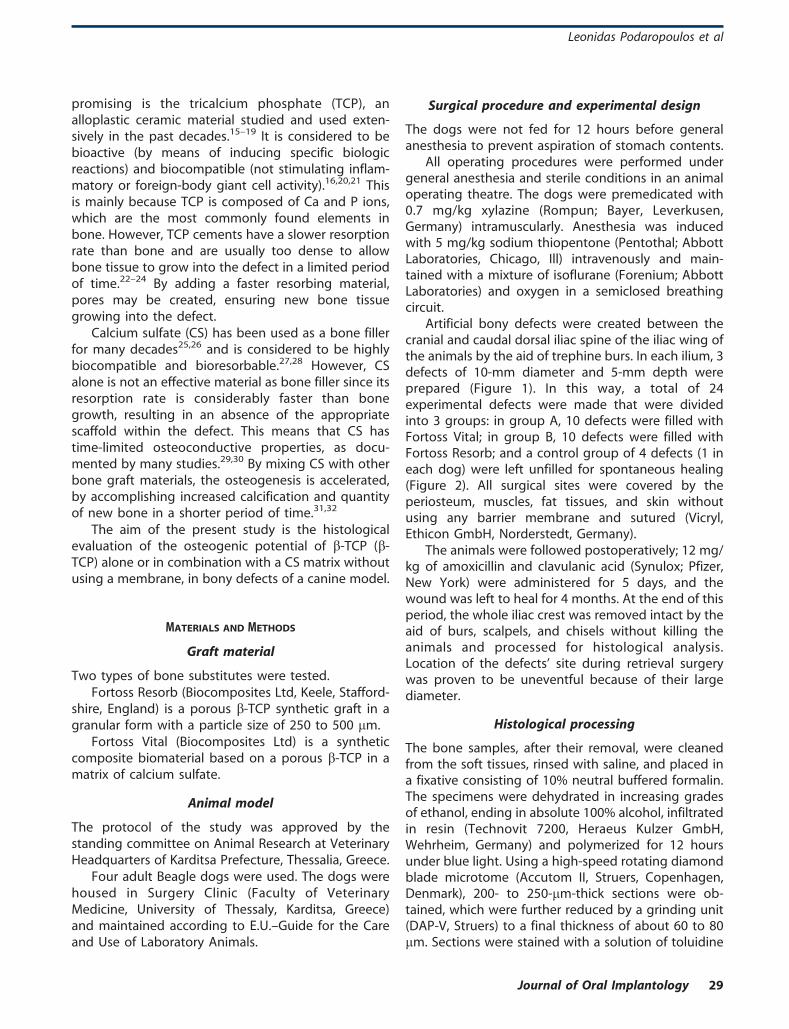
Artificial bony defects were created between thecranial and caudal dorsal iliac spine of the iliac wing ofthe animals by the aid of trephine burs. In each ilium, 3defects of 10-mm diameter and 5-mm depth wereprepared (Figure 1). In this way, a total of 24experimental defects were made that were dividedinto 3 groups: in group A, 10 defects were filled withFortoss Vital; in group B, 10 defects were filled withFortoss Resorb; and a control group of 4 defects (1 ineach dog) were left unfilled for spontaneous healing(Figure 2). All surgical sites were covered by theperiosteum, muscles, fat tissues, and skin withoutusing any barrier membrane and sutured (Vicryl,Ethicon GmbH, Norderstedt, Germany).
The animals were followed postoperatively; 12 mg/kg of amoxicillin and clavulanic acid (Synulox; Pfizer,New York) were administered for 5 days, and thewound was left to heal for 4 months. At the end of thisperiod, the whole iliac crest was removed intact by theaid of burs, scalpels, and chisels without killing theanimals and processed for histological analysis.Location of the defects’ site during retrieval surgerywas proven to be uneventful because of their largediameter.
Histological processing
The bone samples, after their removal, were cleanedfrom the soft tissues, rinsed with saline, and placed ina fixative consisting of 10% neutral buffered formalin.The specimens were dehydrated in increasing gradesof ethanol, ending in absolute 100% alcohol, infiltratedin resin (Technovit 7200, Heraeus Kulzer GmbH,Wehrheim, Germany) and polymerized for 12 hoursunder blue light. Using a high-speed rotating diamondblade microtome (Accutom II, Struers, Copenhagen,Denmark), 200- to 250-lm-thick sections were ob-tained, which were further reduced by a grinding unit(DAP-V, Struers) to a final thickness of about 60 to 80lm. Sections were stained with a solution of toluidine
Journal of Oral Implantology 29
Leonidas Podaropoulos et al
blue and pyronin G. The histological sections wereevaluated using a transmission light microscope(Axiostar Plus, Zeiss, Gottingen, Germany) with anintegrated color video camera (DC88AP, Sony, Tokyo,Japan) and a frame grabber. The ActioVisio (Zeiss,Gottingen, Germany) image analysis software wasused to digitize the selected images for the histo-metric analysis. Bone graft area (BGA) and the totalvolume of the regenerated bone (BV) were measuredand expressed as a percentage of the total defect area.The statistical comparison of the measurementsbetween the groups was made using the Student ttest. The level of significance was set at P � .05.
RESULTS
Histological evaluation
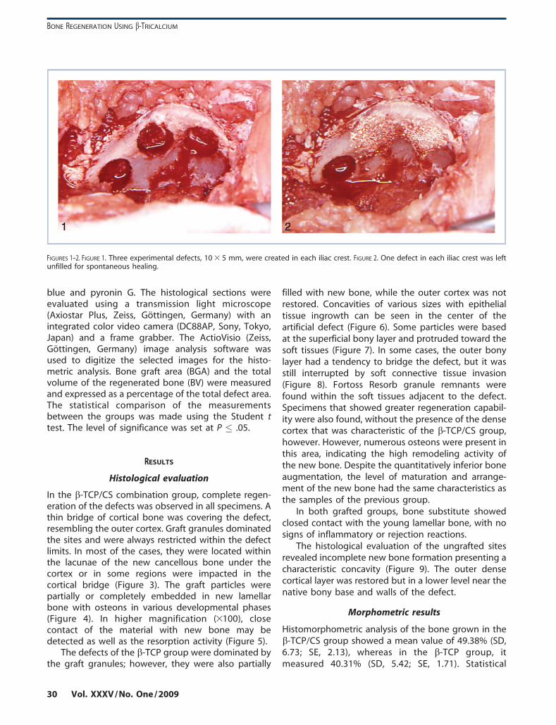
In the b-TCP/CS combination group, complete regen-eration of the defects was observed in all specimens. Athin bridge of cortical bone was covering the defect,resembling the outer cortex. Graft granules dominatedthe sites and were always restricted within the defectlimits. In most of the cases, they were located withinthe lacunae of the new cancellous bone under thecortex or in some regions were impacted in thecortical bridge (Figure 3). The graft particles werepartially or completely embedded in new lamellarbone with osteons in various developmental phases(Figure 4). In higher magnification (3100), closecontact of the material with new bone may bedetected as well as the resorption activity (Figure 5).
The defects of the b-TCP group were dominated bythe graft granules; however, they were also partially
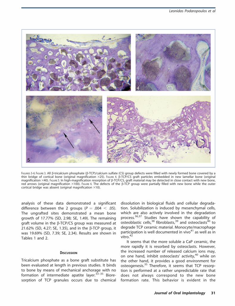
filled with new bone, while the outer cortex was notrestored. Concavities of various sizes with epithelialtissue ingrowth can be seen in the center of theartificial defect (Figure 6). Some particles were basedat the superficial bony layer and protruded toward thesoft tissues (Figure 7). In some cases, the outer bonylayer had a tendency to bridge the defect, but it wasstill interrupted by soft connective tissue invasion(Figure 8). Fortoss Resorb granule remnants werefound within the soft tissues adjacent to the defect.Specimens that showed greater regeneration capabil-ity were also found, without the presence of the densecortex that was characteristic of the b-TCP/CS group,however. However, numerous osteons were present inthis area, indicating the high remodeling activity ofthe new bone. Despite the quantitatively inferior boneaugmentation, the level of maturation and arrange-ment of the new bone had the same characteristics asthe samples of the previous group.
In both grafted groups, bone substitute showedclosed contact with the young lamellar bone, with nosigns of inflammatory or rejection reactions.
The histological evaluation of the ungrafted sitesrevealed incomplete new bone formation presenting acharacteristic concavity (Figure 9). The outer densecortical layer was restored but in a lower level near thenative bony base and walls of the defect.
Morphometric results
Histomorphometric analysis of the bone grown in theb-TCP/CS group showed a mean value of 49.38% (SD,6.73; SE, 2.13), whereas in the b-TCP group, itmeasured 40.31% (SD, 5.42; SE, 1.71). Statistical
FIGURES 1–2. FIGURE 1. Three experimental defects, 10 3 5 mm, were created in each iliac crest. FIGURE 2. One defect in each iliac crest was leftunfilled for spontaneous healing.
30 Vol. XXXV / No. One / 2009
BONE REGENERATION USING b-TRICALCIUM
analysis of these data demonstrated a significantdifference between the 2 groups (P ¼ .004 , .05).The ungrafted sites demonstrated a mean bonegrowth of 17.77% (SD, 2.98; SE, 1.49). The remaininggraft volume in the b-TCP/CS group was measured at21.62% (SD, 4.27; SE, 1.35), and in the b-TCP group, itwas 19.69% (SD, 7.39; SE, 2.34). Results are shown inTables 1 and 2.
DISCUSSION
Tricalcium phosphate as a bone graft substitute hasbeen evaluated at length in previous studies. It bindsto bone by means of mechanical anchorage with noformation of intermediate apatite layer.33–35 Biore-sorption of TCP granules occurs due to chemical
dissolution in biological fluids and cellular degrada-tion. Solubilization is induced by mesenchymal cells,which are also actively involved in the degradationprocess.36,37 Studies have shown the capability ofosteoblastic cells,38 fibroblasts,39 and osteoclasts40 todegrade TCP ceramic material. Monocyte/macrophageparticipation is well documented in vivo41 as well as invitro.42
It seems that the more soluble a CaP ceramic, themore rapidly it is resorbed by osteoclasts. However,the increased number of released calcium ions may,on one hand, inhibit osteoclasts’ activity,40 while onthe other hand, it provides a good environment forosteogenesis.35 Therefore, it seems that TCP resorp-tion is performed at a rather unpredictable rate thatdoes not always correspond to the new boneformation rate. This behavior is evident in the
FIGURES 3–6. FIGURE 3. All b-tricalcium phosphate (b-TCP)/calcium sulfate (CS) group defects were filled with newly formed bone covered by athin bridge of cortical bone (original magnification 325). FIGURE 4. b-TCP/CS graft particles embedded in new lamellar bone (originalmagnification 340). FIGURE 5. In high-magnification resorption of b-TCP/CS, graft material may be detected in close contact with new bone,red arrows (original magnification 3100). FIGURE 6. The defects of the b-TCP group were partially filled with new bone while the outercortical bridge was absent (original magnification 310).
Journal of Oral Implantology 31
Leonidas Podaropoulos et al
conflicting results of many studies on the bioresorp-tion of TCP.43–47 The b-phase isomer of TCP (b-TCP),however, is characterized by physiologic pH, homog-enous microporosity, increased solubility, and a morepredictable resorption rate that resembles the newbone remodeling rate. Variations in composition orimpurities may affect solubility, whereas the purephase seems to be resorbed in 5 to 6 months.21,48 It
should be noted that a faster resorbable materialmight allow soft-tissue cells to prematurely intrudeinto the defect, while nonresorbable or slowlyresorbable materials that remain for a long time mayinhibit new bone deposition.30
Material microporosity seems to regulate itsdegradation rate and provides the right environmentfor the deposition of new bone by the adjacent livingbone.49,50 The presence of CS increases the porosity ofthe grafting material by its early resorption, while itfacilitates the circulation of biological fluids andgrowth factors. Nevertheless, the exact period of timethat CS remains in a bony defect without beingresorbed has not yet been estimated. It is reported,however, to be approximately 4 to 5 weeks28,51,52;however, other studies report 4 to 10 weeks,53 16weeks,54 6 months,55 or even 9 months.56 In any case,the CS degradation rate depends on many factorssuch as the vascularity and the size and shape of thedefect.
Schenk57 stated that a stable material surface playsFIGURES 7–9. FIGURE 7. Occasionally b-tricalcium phosphate (b-TCP) graftparticles protruded superficially toward the soft tissues (originalmagnification 340). FIGURE 8. Numerous soft connective tissueinvasions from the surface toward the center of the defect wereobserved in the b-TCP group specimens, red arrows (originalmagnification 325). FIGURE 9. Ungrafted site: incomplete boneformation near the native bony walls with restoration of the outercortex (original magnification 310).
TABLE 1
New bone volume values (%)*
b-TCP/CS b-TCP Without Graft
50.94 40.46 17.6048.67 40.15 16.7061.67 33.96 21.8939.45 38.87 14.8746.56 49.9847.47 39.7955.3 44.6640.57 30.3548.37 41.8954.78 43.03
Mean 49.38 40.31 17.77SD 6.73 5.42 2.98
*b-TCP indicates b-tricalcium phosphate; CS, calcium sulfate.
TABLE 2
Remaining graft volume values (%)*
b-TCP/CS b-TCP
30.44 6.9823.76 12.5318.46 20.4422.67 18.9620.53 29.5617.89 28.8925.67 17.4519.48 26.9815.76 21.3621.56 13.78
Mean 21.62 19.69SD 4.27 7.39
*b-TCP indicates b-tricalcium phosphate; CS, calcium sulfate.
32 Vol. XXXV / No. One / 2009
BONE REGENERATION USING b-TRICALCIUM
an important role in GBR procedures. That is, the moresolid the scaffold of the graft, the more successful theoutcome. Covering the defect by a barrier membrane,specially a reinforced one, increases immobilization ofthe bone substitute, avoids its displacement, andimproves its osteoconductive properties.58 In contrast,several studies suggest that a membrane is notabsolutely necessary and may even interfere withbone regeneration because it compromises bloodsupply from the periosteum and impedes its osteo-genic effect, which is attributed to the inner cambiallayer.59,60 Salata et al61 found that the use of GBRmembrane in combination with bone substitutes didnot significantly improve bone formation comparedwith the use of bone substitutes alone. The findings ofthe present study do not support the opinion that amembrane in bone regeneration procedures is super-fluous. This would ignore many significant studies thatclearly show the benefit of using a barrier membraneeven without using bone filler.2,4,5,62 In any case, amembrane group was not used for comparison. Theresults should be interpreted only as an out-perfor-mance of the b-TCP/CS group compared with the b-TCP alone.
Research data suggest that the occlusive proper-ties of barrier membranes may be achieved by otherbiomaterials such as CS. Calcium sulfate acts as abinder and enhances graft containment, making themixture more stable and pressure resistant.27 In aseries of studies, CS barrier properties were tested inbone or periodontal defects in conjunction with avariety of grafts. These studies showed that the CSbarrier increases the vital bone volume,63 promotesperiodontal regeneration,64 excludes epithelial andconnective tissue cells, and preserves the alveolarridge dimensions after tooth extraction.65–67 Payne etal,68 in an interesting in vitro study, compared themigration ability of human gingival fibroblasts stimu-lated by chemotactic substances on 3 differentbarriers: CS, e-PTFE membrane, and polylactic acidmembrane. Calcium sulfate proved to be the mostcompatible, showing the least interference to cellmigration. The problem that seems to be related tothe use of a CS barrier is the possibility of the earlymaterial resorption and the fractures that may occuron the material surface during the initial postoperativeperiod by any kind of pressure exercised on it. Both ofthese parameters may allow epithelial ingrowth in thedefect area. These latter disadvantages may besurpassed by the use of a b-TCP/CS combination. Thismixture solidifies in a few minutes’ time after mixingand creates a stable mass with a surface that is notvulnerable to fractures. Whether epithelial ingrowth
takes place after CS is resorbed is questionablebecause the main scaffold of the material is preservedand the pores that are left are relatively small.
It should be noted that bone regeneration seemsto vary widely between the different species or evenbetween individual animals of the same species.Furthermore, it is differentiated by the type of bone,the age of the individual, and the presence of theperiosteum.69,70 Mainly, however, healing is largelydependent on wound size and shape, which meansthat a small 5-wall defect may heal spontaneouslywithout the aid of a graft material or a membrane. Onthe contrary, a critical size defect (CSD) is defined asthe smallest intraosseous wound that does not healspontaneously by bone formation during the lifetimeof the animal or human being.71 In a later study, a CSDwas defined as a defect that has less than 10% bonyregeneration during the lifetime of the animal.72 In thecase of the canine ilium, the CSD has not yet beenidentified.
The number of walls of the host bone defect iscritical and should always be taken into considerationwhen comparing study results. In the present study,cylindrical monocortical defects were created. Thisshape may be compared with an extraction socket,that is, a 5-wall defect model, a situation quitecommon in everyday clinical practice. A 10-mm-diameter defect was chosen as it was estimated thatthis would be similar to a CSD for the dog’s ilium.These defects failed to heal spontaneously, and, in anycase, a defect of that size would be a challenge toregenerate in clinical practice.
In the present study, the b-TCP/CS combinationdemonstrated complete regeneration up to the cortexin all 10-mm specimens tested, while b-TCP alone didnot succeed in regenerating these large-diameterdefects. It is not the first time that CS was used incombination with other biomaterials.27,51,73 However,differences in powder processing lead to changes inelements’ ratios, that is, in the specific case, the Ca/Pratio, which alters the surface chemistry. This leads todifferences in the surface Z-potential of the graft. Themineral scaffold of Fortoss Vital is a stoichiometric b-TCP with a Ca/P molar ratio of 1.5. The Z-potentialassesses the degree of ionic activity of a material’ssurface, which is considered to be one of the mainphysical factors that interfere in the biologicalbehavior of a tissue around an implanted material.74
This potential depends on a variety of factors, amongwhich is the composition of the implanted materialand the surrounding biological fluids, the inflamma-tory situation, and the environmental pH.75 Thedegree to which hydroxyl or carboxyl ion groups alter
Journal of Oral Implantology 33
Leonidas Podaropoulos et al
the ceramic to osteoblast attachment is not wellunderstood.76 The link between the Z-potential ofbioceramics and their resulting attraction to bone andosteoblasts has been tested in previous studies as wellas the relation between the modifications in theprocessing method of CaP powders and their resultingZ-potential and, hence, their suitability for use as bonetissue engineering scaffolds.77,78 It is well known thatprotein adsorption plays an important role in graftbehavior and implant integration. The relation be-tween Z-potential and protein adsorption has beenconfirmed in previous studies.79 This means that bycontrolling the Z-potential, by means of special graftprocessing, host proteins may be attracted into thesurgical site, and a positive osteoblast activity iscreated. This shifting of the isoelectric potential of thesurface of Fortoss Vital may be an explanation of itspositive regenerative behavior that has been demon-strated in the present study.
CONCLUSION
This study demonstrated complete bone regenerationof critical-size cylindrical bone defects 10 mm indiameter using a composite alloplastic graft of b-TCPin a CS matrix, without a membrane barrier. Use of b-TCP alone resulted in partial bone formation in a 4-month control period. The safety of the testedmaterial was demonstrated as well. Further researchshould follow to define the critical-size defect in thecanine ilium and the necessary period of time for thiscomposite material to be resorbed.
ACKNOWLEDGMENTS
The authors thank Biocomposites Ltd, Keele, Stafford-shire, England, and Lazarelis Biomaterials, Athens,Greece, for providing the bone substitutes used inthe study.
REFERENCES
1. Breine U, Branemark P-I. Reconstruction of alveolar jawbone: an experimental and clinical study of immediate andpreformed autogenous bone grafts in combination with osseointe-grated implants. Scand J Plastic Reconstr Surg Hand Surg. 1980;14:23–48.
2. Dahlin C, Linde A, Gottlow J, Nyman S. Healing of bonedefects by guided tissue regeneration. J Plastic Reconstr Surg. 1988;81:672–676.
3. Dahlin C, Linde A, Gottlow J, Nyman S. Healing of maxillaryand mandibular bone defects using a membrane technique: anexperimental study in monkeys. Scand J Plastic Reconstr Surg. 1990;24:13–19.
4. Schenk RK, Buser D, Hardwick WR, Dahlin C. Healing patternof bone regeneration in membrane-protected defects: a histologicstudy in the canine mandible. Int J Oral Maxillofac Implants. 1994;9:13–29.
5. Buser D, Bragger U, Lang NP, Nyman S. Regeneration andenlargement of jaw bone using guided tissue regeneration. ClinOral Implant Res. 1990;1:22–32.
6. Nevins M, Melonig JT. Enhancement of the damagededentulous ridge to receive dental implants: a combination ofallograft and the Gore-Tex membrane. Int J Periodont Restor Dent.1992;12:97–111.
7. Buser D, Dula K, Hirt HP, Schenk RK. Lateral ridgeaugmentation using autografts and barrier membranes: a clinicalstudy in 40 partially edentulous patients. J Oral Maxillofac Surg.1996;54:420–432.
8. Buser D, Hoffmann B, Bernard P, Lussi A, Mettler D, SchenkRK. Evaluation of filling materials in membrane-protected bonedefects: a comparative histomorphometric study in the mandible ofminiature pigs. Clin Oral Implant Res. 1998;9:137–150.
9. von Arx T, Cochran DL, Hermann JS, Schenk RK, Buser D.Lateral ridge augmentation using different bone fillers and barriermembrane application: a histologic and histomorphometric studyin the canine mandible. Clin Oral Implant Res. 2001;12:260–269.
10. Kalk WWI, Raghoebar GM, Jansma J, Boering G. Morbidityfrom iliac crest bone harvesting. J Oral Maxillofac Surg. 1996;54:1424–1429.
11. Goulet JA, Senunas LE, De Silva GL, Greenfield ML.Autogenous iliac crest bone graft: complications and functionalassessment. Clin Orthop Rel Res. 1997;337:76–81.
12. Albrektsson T, Linder L. Intravital long-term follow-up ofautologous experimental bone grafts. Arch Orthop Trauma Surg.1981;98:189–193.
13. Younger EM, Chapman MW. Morbidity at bone graft donorsites. J Orthop Trauma. 1989;3:192–195
14. Nystrom E, Kahnberg KE, Albrektsson T. Treatment of theseverely resorbed maxillae with bone graft and titanium implants: ahistologic review of autopsy specimens. Int J Oral MaxillofacImplants. 1993;8:167–172.
15. Cameron HU. Evaluation of biodegradable ceramic. JBiomed Mater Res. 1977;11:179–186.
16. Jarcho M. Biomaterial aspects of calcium phosphates:properties and applications. Dent Clin North Am. 1986;30:25–47.
17. Stahl S, Froum S. Histological evaluation of humanintraosseous healing responses to the placement of tricalciumphosphate ceramic implants. I. Three to eight months. JPeriodontol. 1986;57:211–217.
18. Driessens FCM, Ramselaar MMA, Schaeken HG, Stols ALH,Van Mullem PJ. Chemical reactions of calcium phosphate implantsafter implantation in vivo. J Mater Science Mater Med. 1992;3:413–417.
19. LeGeros RZ. Properties of osteoconductive materials:calcium phosphates. Clin Orthop Rel Res. 2002;395:81–98.
20. Metsger DS, Driskell TD, Paulsrud JR. Tricalcium phosphateceramic, a resorbable bone implant: Review and current status. JAm Dent Assoc. 1982;105:1035–1038.
21. Trisi P, Rao W, Rebaudi A, Fiore P. Histologic effect of pure-phase beta-tricalcium phosphate on bone regeneration in humanartificial jawbone defects. Int J Periodont Restor Dent. 2003;23:69–77.
22. Nilsson M, Fernandez E, Sarda S, Lidgren L, Planell JA.Characterization of a novel calcium phosphate/sulphate bonecement. J Biomed Mater Res. 2002;61:600–607.
23. Wiltfang J, Merten HA, Schlegel KA, et al. Degradation
34 Vol. XXXV / No. One / 2009
BONE REGENERATION USING b-TRICALCIUM
characteristics of alpha and beta tri-calcium phosphate in minipigs.J Biomed Mater Res (Appl Biomater). 2002;63:115–121.
24. Artzi Z, Weinreb M, Givol N, et al. Biomaterial resorptionrate and healing site morphology of inorganic bovine bone andbeta-tricalcium phosphate in the canine: a 24-month longitudinalhistologic study and morphometric analysis. Int J Oral MaxillofacImplants. 2004;19:357–368.
25. Peltier LF. The use of plaster of Paris to fill defects in bone.Clin Orthop. 1961;21:1–31.
26. Alderman N. Sterile plaster of Paris as an implant in theintrabony environment: a preliminary study. J Periodontol. 1969;40:11–13.
27. Aichelmann-Reidy ME, Heath CD, Reynolds MA. Clinicalevaluation of calcium sulphate in combination with demineralisedfreeze-dried bone allograft for the treatment of human intraosse-ous defects. J Periodontol. 2004;75:340–347.
28. Sbordone L, Bortolaia C, Perrotti V, Pasquantonio G,Petrone G. Clinical and histologic analysis of calcium sulfate intreatment of a post-extraction defect: a case report. Implant Dent.2005;14:82–87.
29. Bell WH. Resorption characteristics of bone and bonesubstitutes. Oral Surg Oral Med Oral Pathol. 1964;17:650–657.
30. Ricci JL, Alexander H, Nadkarni P, et al. Biologicalmechanisms of calcium-sulfate replacement by bone. In: DaviesEJ, ed. Bone Engineering. Toronto, Canada: Em Squared; 2000:332–344.
31. Al Ruhaimi KA. Effect of calcium sulphate on the rate ofosteogenesis in distracted bone. Int J Ora lMaxillofac Surg. 2001;30:228–233.
32. Orsini M, Orsini G, Benlloch D, et al. Comparison of calciumsulphate and autologous bone graft to bioabsorbable membranesplus autogenous bone graft in the treatment of intrabonyperiodontal defects: a split-mouth study. J Periodontol. 2001;72:296–302.
33. Kotani S, Fujita Y, Kitsugi T, et al. Bone bonding mechanismof beta-tricalcium phosphate. J Biomed Mater Res. 1991;25:1303–1315.
34. Neo M, Kotani S, Nakamura T, et al. A comparative study ofultrastructures of the interfaces between four kinds of surface-active ceramic and bone. J Biomed Mater Res. 1992;26:1419–1432.
35. Fujita R, Yokoyama A, Nodasaka Y, Kohgo T, Kawasaki T.Ultrastructure of ceramic-bone interface using hydroxyapatite andb-tricalcium phosphate ceramics and replacement mechanism of b-tricalcium phosphate in bone. Tissue Cell. 2003;35:427–440.
36. Evans RW, Cheung HS, McCarty DJ. Cultured humanmonocytes and fibroblatsts solubilize calcium phosphate crystals.Calcif Tissue Int. 1984;36:645–650.
37. Owens JL, Cheung HS, McCarty DJ. Endocytosis precedesdissolution of basic calcium phosphate crystals by murinemacrophages. Calcif Tissue Int. 1986;38:170–174.
38. Gregoire MM, Orly II, Menanteau J. The influence ofcalcium phosphate biomaterials on human bone cell activities: anin vitro approach. J Biomed Mater Res. 1990;24:165–177.
39. Gregoire MM, Orly II, Kerebel LM, Kerebel BB. In vitroeffects of calcium phosphate biomaterials on fibroblastic cellbehavior. Biol Cell. 1987;59:255–260.
40. Yamada S, Heymann D, Bouler J-M, Daculsi G. Osteoclasticresorption of calcium phosphate ceramics with different hydroxy-apatite/b-tricalcium phosphate ratios. Biomaterials. 1997;18:1037–1041.
41. Gaasbeek RD, Toonen HG, van Heerwaarden RJ, Buma P.Mechanism of bone incorporation of beta-TCP bone substitute inopen wedge tibia osteotomy in patients. Biomaterials. 2005;26:6713–6719.
42. Benahmed M, Bouler JM, Heymann D, Gan O, Daculsi G.Biodegradation of synthetic calcium phosphate by human mono-cytes in vitro: a morphological study. Biomaterials. 1996;17:2173–2178.
43. Bowers CM, Vargo JN, Lery B, Emerson JR, Gergquist JJ.Histologic observations following the placement of tricalciumphosphate implants in human intrabony defects. J Periodontol.1986;57:286–287.
44. Jarcho M. Calcium phosphate ceramics as chard tissueprosthetics. Clin Orthop Rel Res. 1981;157:259–278.
45. Kent JN. Reconstruction of the alveolar ridge withhydroxyapatite. Dent Clin North Am. 1986;30:231–257.
46. Eggli PS, Miller W, Schenk RK. Porous hydroxyapatite andtricalcium phosphate cylinders with two different pore size rangesimplanted in the cancellous bone of rabbits. Clin Orthop Rel Res.1988;232:127–138.
47. Saffar JF, Colombier ML, Datenville R. Bone formation intricalcium phosphate filled periodontal lesions: histological obser-vations in humans. J Periodontol. 1990;61:209–216.
48. Rey C. Calcium phosphate biomaterials and bone mineral:differences in composition, structures and properties. Biomaterials.1990;11:13–15.
49. De Groot K. Bioceramics consisting of calcium phosphatesalts. Biomaterials. 1980;1:47–50.
50. Zerbo IR, Bronckers ALJJ, de Lange GL, van Beek GJ, BurgerEH. Histology of human alveolar bone regeneration with a poroustricalcium phosphate: a report of two cases. Clin Oral Implant Res.2001;12:379–384.
51. Al Ruhaimi KA. Effect of adding resorbable calcium sulfateto grafting materials on early bone regeneration in osseous defectsin rabbits. Int J Oral Maxillofac Implants. 2000;15:859–864.
52. Orsini G, Ricci J, Scarano A, et al. Bone-defect healing withcalcium-sulfate particles and cement: an experimental study in therabbit. J Biomed Mater Res (Appl Biomater). 2004;68B:199–208.
53. Maragos P, Bissada NF, Wang R, Cole RP. Comparison ofthree methods using calcium sulfate as a graft barrier materil forthe treatment of Class II mandibular molar furcation defects. Int JPeriodont Restor Dent. 2002;22:493–501.
54. Yoshikawa G, Murashima Y, Wadachi R, Sawada N, Suda H.Guided bone regeneration (GBR) using membranes and calciumsulphate after apicectomy: a comparative histomorphometricalstudy. Int Endodont J. 2002;35:255–263.
55. Kelly CM, Wilkins RM, Gitelis G, Hartjen C, Watson JT, KimPT. The use of a surgical grade calcium sulfate as a bone graftsubstitute: results of a multicenter study. Clin Orthop Rel Res. 2001;382:42–50.
56. Pecora GE, De Leonardis D, Della Rocca C, Cornelini R,Cortesini C. Short-term healing following the use of calcium sulfateas a grafting material for sinus augmentation: a clinical report. Int JOral Maxillofac Implants. 1998;13:866–873.
57. Schenk RK. Bone regeneration: biologic basis. In: Buser D,Dahlin C, Schenk RK, eds. Guided Bone Regeneration in ImplantDentistry. London, UK: Quintessence; 1995:49–100.
58. Donath K, Rohrer MD, Hormann K. Mobile and immobilehydroxyapatite integration and resorption and its influence onbone. J Oral Implantol. 1987;13:120–127.
59. Burchardt H. The biology of bone graft repair. Clin OrthopRel Res. 1983;174:28–42.
60. Spagnioli DB, Mazzonetto R, Marchena JM. Clinicalprocedures currently using bone grafting with guided tissueregeneration techniques. Oral Maxillofac Surg Clin North Am.2001;13:423–436.
61. Salata LA, Craig GT, Brook IM. Bone healing following theuse of hydroxyapatite or ionomeric bone substitutes alone or
Journal of Oral Implantology 35
Leonidas Podaropoulos et al
combined with a guided bone regeneration technique: an animalstudy. Int J Oral Maxillofac Implants. 1998;13:44–51.
62. Stavropoulos F, Dahlin C, Ruskin JD, Johansson C. Acomparative study of barrier membranes as grafted protectors inthe treatment of localized bone defects. Clin Oral Implant Res. 2004;15:435–442.
63. Vance GS, Greenwell H, Miller RL, Hill M, Johnston H,Scheetz JP. Comparison of an allograft in an experimental puttycarrier and a bovine-derived xenografts used in ridge preservation:a clinical and histologic study in humans. Int J Oral MaxillofacImplants. 2004;19:491–497.
64. Kim CK, Kim HY, Chai JK, et al. Effect of a calcium sulfateimplant with calcium sulfate barrier on periodontal healing in 3-wall intrabony defects in dogs. J Periodontol. 1998;69:982–988.
65. Sottosanti J. Calcium sulfate: a biodegradable and bio-compatible barrier for guided tissue regeneration. Compend. 1992;13:226–228, 230, 232–234.
66. Anson D. Calcium sulfate: a 4-year observation of its use asa resorbable barrier in guided tissue regeneration of periodontaldefects. Compend Cont Ed Dent. 1996;17:859–899.
67. Pecora G, Andreana S, Margarone JE III, Covani U,Sottosanti JS. Bone regeneration with a calcium sulfate barrier.Oral Surg Oral Med Oral Pathol Oral Radiol Endodontol. 1997;84:424–429.
68. Payne JM, Cobb CM, Rapley JW, Killoy WJ, Spencer P.Migration of human gingival fibroblasts over guided tissueregeneration barrier materials. J Periodontol. 1996;67:236–244.
69. Enneking WF, Burchardt H, Puhl JJ, Piotrowski G. Physicaland biological aspects of repair in dog cortical bone transplants. JBone Joint Surg. 1975;57A:237–252.
70. Prolo DJ, Pedrotti PW, Burres KP, Oklund S. Superiorosteogenesis in transplanted allogeneic canine skull followingchemical sterilization. Clin Orthop Relat Res. 1982;168:230–242.
71. Schmitz JP, Hollinger JH. The critical size defect as anexperimental model for craniomandibulofacial nonunions. ClinOrthop Rel Res. 1986;105:299–308.
72. Hollinger JO, Kleinschmidt JC. The critical size defect as anexperimental model to test bone repair materials. J Craniofac Surg.1990;1:60–68.
73. Kim SG, Yeo HH, Kim YK. Grafting of large defects of thejaws with a particulate dentin-plaster of Paris combination. OralSurg Oral Med Oral Pathol Oral Radiol Endodontol. 1999;88:22–25.
74. Krajewski A, Piancastelli A, Malavolti R. Albumin adhesionon ceramics and correlation with their Z-potential. Biomaterials.1998;19:637–641.
75. Clark AE, Hench LL, Paschall HA. The influence of surfacechemistry on implant interface histology: a theoretical basis forimplant materials selection. J Biomed Mater Res. 1976;10:161–174.
76. Bagambisa FB, Joos U, Schilli W. Interaction of osteogeniccells with hydroxylapatite implant materials in vitro and in vivo. IntJ Oral Maxillofac Implants. 1990;5:217–226.
77. Bagambisa FB, Joos U, Schilli W. Mechanisms and structureof the bond between bone and hydroxyapatite ceramics. J BiomedMater Res. 1993;27:1047–1055.
78. Oppermann DA, Crimp MJ, Bement DM. In vitro stabilitypredictions for the bone/hydroxyapatite composite system. JBiomed Mater Res. 1998;42:412–416.
79. Krajewski A, Malavolti R, Piancastelli A. Albumin adhesionon some biological and non-biological glasses and connection withtheir Z-potentials. Biomaterials. 1996;17:53–60.
36 Vol. XXXV / No. One / 2009
BONE REGENERATION USING b-TRICALCIUM


![Science Manuscript Template · Web viewbone formation with autologous adipose stem cells and β-tricalcium phosphate granules [36]. It has been demonstrated that BMMSCs promote cartilage](https://img.pdfslide.net/doc/110x75/5eaf134ba3fe5a5ff51cd9c6/science-manuscript-web-view-bone-formation-with-autologous-adipose-stem-cells-and.jpg)