Embed Size (px)
Citation preview

BOOK OF ABSTRACTS
Firenze, 26 Giugno 2015

Giornata dedicata alla memoria del Prof. Marco Mascini
1
INDICE
PROGRAMMA SCIENTIFICO .............................................................................................................................. 4
INVITED LECTURES .......................................................................................................................................... 10
AMBIENT MASS SPECTROMETRY AND ION MOBILITY: NEW APPROACHES IN THE STUDY OF BIOMOLECULES ......................................................................................................................................................................... 10
INNOVATION IN DIAGNOSTICS; CASE STUDY: NEW TECHNIQUES IMPROVING CURRENT APPLICATIONS ..... 10
COMUNICAZIONI ORALI.................................................................................................................................. 11
DETERMINATION OF NEW SYNTHETIC DRUGS IN BIOLOGICAL MATRIX BY HPLC-MS/MS ............................. 12
PEPTIDOMIC AND BIOACTIVITY STUDY ON THE PEPTIDES ISOLATED IN COMMERCIAL DONKEY MILK ......... 14
MULTIDRUG RESISTANCE INHIBITORS IN PLASMA SAMPLES: THE POWER OF LC-ENERGY RESOLVED MS/MS METHODS FOR STABILITY INVESTIGATION ...................................................................................................... 16
MODERN TRENDS IN DRUG ANALYSIS: VISUALIZING DESIGN SPACE IN THE QUALITY CONTROL OF PHARMACEUTICALS BY CAPILLARY ELECTROPHORESIS .................................................................................. 18
ARTIFICIAL ANTIBODIES: WHERE DO WE STAND? ........................................................................................... 19
A NEW SMARTPHONE-BASED CHEMILUMINESCENT LATERAL FLOW IMMUNOSENSOR FORMAT FOR POINT OF CARE TESTING ............................................................................................................................................ 20
ORGANIC BIOELECTRONIC SENSORS: COMPARATIVE STUDY OF CRP DETECTION USING DIFFERENT ORGANIC THIN FILM TRANSISTORS CONFIGURATIONS .................................................................................................. 22
MICRORNA DETECTION BY SPR IMAGING AND PNA PROBES: NANOPARTICLE AND ENZYMATIC AMPLIFICATION METHODS.............................................................................................................................. 23
A DNA NANO PH-METER BASED ON TRIPLEX FORMATION............................................................................. 24
A NEW IMMUNOSENSOR FOR THE DETERMINATION OF VALPROIC ACID IN SERUM USING FUNCTIONALIZED SILICA NANOPARTICLES DOPED WITH A THERMOCHEMILUMINESCENT 1,2–DIOXETANE DERIVATIVE AS LABEL ............................................................................................................................................................... 25
SPECTROPHOTOMETRIC CELL-FREE ASSAYS FOR MEASUREMENT OF THE OXIDATIVE POTENTIAL OF ATMOSPHERIC AESOSOL ................................................................................................................................. 27
DETERMINATION OF WARFARIN AND WARFARIN ALCOHOLS IN ORAL FLUID AND PLASMA SAMPLES FOR MONITORING PATIENTS UNDERGOING ANTICOAGULANT THERAPY ............................................................. 29
NEW BIOANALYTICAL APPROCH FOR EARLY DETECTION OF ß-THALASSEMIA COUPLING TGA AND CHEMOMETRICS .............................................................................................................................................. 31
DETECTION OF OCHRATOXIN A IN FOOD SAMPLES BY A NOVEL APTAMER BASED SENSOR ASSAY INTEGRATED IN A MICROFLUIDIC CHIP ........................................................................................................... 32
THERAPEUTIC DRUG MONITORING OF PROTEIN-UNBOUND IMMUNOSUPPRESSANTS BY A NOVEL POCT OPTICAL DEVICE ............................................................................................................................................... 34
2
ACETAMIPRID DETECTION BY DNA TECHNOLOGY SENSING FOR ENVIRONMENTAL ANALYSIS ..................... 36
COMUNICAZIONI POSTER ............................................................................................................................... 37
P.1. MICROBIOLOGICAL SCREENING TEST FOR VETERINARY DRUGS IN FOOD AND FEED: FEASIBILITY AND RELIABILITY VERIFICATION ANALYSING REAL SAMPLES .................................................................................. 38
P2. TOWARDS THE IDENTIFICATION OF NEUROPROTECTIVE AGENTS. PHARMACOKINETIC EVALUATION AND CNS DISTRIBUTION OF (R)-RC-33, A PROMISING SIGMA1 RECEPTOR AGONIST ............................................. 39
P3. DEVELOPMENT OF AN ELIME ASSAY AND A REAL-TIME PCR FOR SALMONELLA ENTERICA DETECTION: APPLICATION IN IRRIGATION WATERS ............................................................................................................ 40
P4. SCREENING OF MICROCYSTINS AND OKADAIC ACID IN DRINKING, FRESH AND SEA WATER SAMPLES USING AN OPTIMIZED COLORIMETRIC PHOSPHATASE INHIBITION ASSAY ..................................................... 42
P5. SMARTPHONE-BASED COLORIMETRIC ASSAY FOR CA125 CANCER BIOMARKER DETECTION .................. 43
P6. MOLECULARLY IMPRINTED MONOLITHS FOR EFFICIENT RECOGNITION OF PROTEINS ........................... 44
P7. OCCUPATIONAL EXPOSURE MONITORING TO ACTIVE PHARMACEUTICAL INGREDIENTS: DETERMINATION OF CHEMICAL TRACERS ON MEMBRANE FILTERS BY NIR/PLS METHOD ............................ 45
P8. SIMULTANEOUS QUANTIFICATION OF INTACT GLUCOSINOLATES AND ISOTHIOCYANATES BY HPLC-ES-MS/MS IN BRASSICACEAE SEEDS AND FUNCTIONAL FOODS .......................................................................... 46
P9. A VISUAL SEMI-QUANTITATIVE IMMUNOCHROMATOGRAPHIC STRIP TEST IN COMPETITIVE FORMAT . 48
P10. SENSITIVE THERMOCHEMILUMINESCENT-BASED IMMUNOSENSOR USING NEW 1,2-DIOXETANE ANALOGUES AS LABELS ................................................................................................................................... 50
P11. DEVELOPMENT OF AN AFFINITY SENSOR FOR ORGANIC CONTAMINANTS DETECTION IN FOOD ......... 52
P12. NOVEL APPROACHES FOR ALZHEIMER’S DISEASE BIOMOLECULAR DIAGNOSIS ..................................... 53
P13. GREEN COFFEE BEAN EXTRACTS AS POTENTIAL NEUROPROTECTIVE AND CHEMOPROTECTIVE DIETARY SUPPLEMENTS: A MOLECULAR POINT OF VIEW ............................................................................................. 54
P14. NATURAL COMPOUNDS AGAINST ALZHEIMER’S DISEASE: EFFECTS OF HOP EXTRACTS ON AΒ PEPTIDE INDUCED TOXCICITY ON NEURONAL CELL LINES ............................................................................................ 55
P15. APTAMER-BASED OPTICAL SENSOR FOR THE DETECTION OF SAXITOXIN IN MARINE WATER ............... 56
P16. MICROEXTRACTION ON PACKED SORBENT (MEPS) FOR THE DETERMINATION OF PESTICIDES IN WHEAT FLOUR BY HPLC-MS/MS ................................................................................................................................... 57
P17. SELECTIVE SOLID PHASE EXTRACTION OF SYNTHETIC CANNABINOIDS BY USING COMPUTATIONALLY DESIGNED PEPTIDES ........................................................................................................................................ 59
P18. CARBON BLACK-CHITOSAN FILM FOR SCREEN PRINTED ELECTRODE AS NOVEL PLATFORMS FOR BIOSENSORS .................................................................................................................................................... 60
P19. MAGNETIC BEADS-BASED ELECTROCHEMICAL IMMUNOASSAY FOR SCREENING OF CELIAC DISEASE IN SALIVA .............................................................................................................................................................. 62

3
P20. DIRECT-EI-UPLC-MS: A NEW TOOL FOR THE ANALYSIS OF STEROLS IN PHARMACEUTICALS AND COMPLEX MATRICES ....................................................................................................................................... 64
P21. HOMOGENOUS FLUOROIMMUNOASSAYS BASED ON THE QUENCHING OF QUANTUM DOTS FLUORESCENCE BY GRAPHENE ........................................................................................................................ 66
P22. INFLUENCE OF THE SAMPLING PROCEDURE ON THE MEASURED CONCENTRATION OF URIC ACID IN ORAL FLUID ...................................................................................................................................................... 67
4
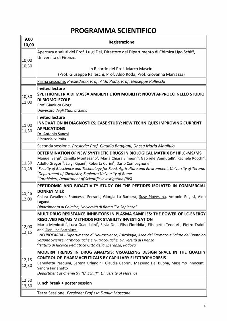
PROGRAMMA SCIENTIFICO 9,00
10,00 Registrazione
10,00 10,30
Apertura e saluti del Prof. Luigi Dei, Direttore del Dipartimento di Chimica Ugo Schiff, Università di Firenze.
In Ricordo del Prof. Marco Mascini (Prof. Giuseppe Palleschi, Prof. Aldo Roda, Prof. Giovanna Marrazza)
Prima sessione. Presiedono: Prof. Aldo Roda, Prof. Giuseppe Palleschi
10,30 11,00
Invited lecture SPETTROMETRIA DI MASSA AMBIENT E ION MOBILITY: NUOVI APPROCCI NELLO STUDIO DI BIOMOLECOLE Prof. Gianluca Giorgi
Università degli Studi di Siena
11,00 11,30
Invited lecture INNOVATION IN DIAGNOSTICS; CASE STUDY: NEW TECHNIQUES IMPROVING CURRENT APPLICATIONS Dr. Antonio Sanesi Biomerieux Italia
Seconda sessione. Presiede: Prof. Claudio Baggiani, Dr.ssa Maria Magliulo
11,30 11,45
DETERMINATION OF NEW SYNTHETIC DRUGS IN BIOLOGICAL MATRIX BY HPLC-MS/MS
Manuel Sergi1, Camilla Montesano2, Maria Chiara Simeoni1, Gabriele Vannutelli2, Rachele Rocchi2, Adolfo Gregori3, Luigi Ripani3, Roberta Curini2, Dario Compagnone1 1Faculty of Bioscience and Technology for Food, Agriculture and Environment, University of Teramo 2Department of Chemistry, Sapienza University of Rome 3Carabinieri, Department of Scientific Investigation (RIS)
11,45 12,00
PEPTIDOMIC AND BIOACTIVITY STUDY ON THE PEPTIDES ISOLATED IN COMMERCIAL DONKEY MILK
Chiara Cavaliere, Francesca Ferraris, Giorgia La Barbera, Susy Piovesana, Antonio Puglisi, Aldo Laganà Dipartimento di Chimica, Università di Roma “La Sapienza”
12,00 12,15
MULTIDRUG RESISTANCE INHIBITORS IN PLASMA SAMPLES: THE POWER OF LC-ENERGY RESOLVED MS/MS METHODS FOR STABILITY INVESTIGATION Marta Menicatti1, Luca Guandalini1, Silvia Dei1, Elisa Floriddia1, Elisabetta Teodori1, Pietro Traldi2 and Gianluca Bartolucci1 1 NEUROFARBA - Dipartimento di Neuroscienze, Psicologia, Area del Farmaco e Salute del Bambino Sezione Scienze Farmaceutiche e Nutraceutiche, Università di Firenze 2Istituto di Ricerca Pediatrica Città della Speranza, Padova
12,15 12,30
MODERN TRENDS IN DRUG ANALYSIS: VISUALIZING DESIGN SPACE IN THE QUALITY CONTROL OF PHARMACEUTICALS BY CAPILLARY ELECTROPHORESIS
Benedetta Pasquini, Serena Orlandini, Claudia Caprini, Massimo Del Bubba, Massimo Innocenti, Sandra Furlanetto Department of Chemistry “U. Schiff”, University of Florence
12,30 13,50
Lunch break + poster session
Terza Sessione. Presiede: Prof.ssa Danila Moscone
5
13,50 14,05
ARTIFICIAL ANTIBODIES: WHERE DO WE STAND? Claudio Baggiani, Laura Anfossi, Cristina Giovannoli Dipartimento di Chimica, Università di Torino
14,05 14,20
A NEW SMARTPHONE-BASED CHEMILUMINESCENT LATERAL FLOW IMMUNOSENSOR FORMAT FOR POINT OF CARE TESTING Martina Zangheri1, Luca Cevenini1,Laura Anfossi2, Claudio Baggiani2, Patrizia Simoni3, Fabio Di Nardo2, Aldo Roda1 1 Dipartimento di Chimica “Giacomo Ciamician”, Alma Mater Studiorum, Università di Bologna 2Dipartimento di Chimica, Università di Torino 3Dipartimento di Medicina e Chirurgia, Università di Bologna
14,20 14,35
ORGANIC BIOELECTRONIC SENSORS: COMPARATIVE STUDY OF CRP DETECTION USING DIFFERENT ORGANIC THIN FILM TRANSISTORS CONFIGURATIONS
Kyriaki Manoli, Maria Magliulo, Mohammad Yusuf Mulla, Donato De Tullio, Preethi Seshadri, Gerardo Palazzo, Luisa Torsi Dipartimento di Chimica, Università degli studi di Bari Aldo Moro
14,35 14,50
MICRORNA DETECTION BY SPR IMAGING AND PNA PROBES: NANOPARTICLE AND ENZYMATIC AMPLIFICATION METHODS
Roberta D’Agata1, Alex Manicardi2, Alessia Finotti3, Roberto Corradini2, Roberto Gambari3, Giuseppe Spoto1,4 1Department of Chemical Science, University of Catania, 2Department of Chemical Science, University of Parma, 3 Department of Life Sciences and Biotechnology, University of Ferrara 4Consortium INBB
14,50 15,05
A DNA NANO PH-METER BASED ON TRIPLEX FORMATION Andrea Idili1, A e a e- e2, Giuseppe Palleschi1, Francesco Ricci1
1Dipartimento di Scienze e Tecnologie Chimiche, University of Rome, Tor Vergata 2La oratory of Biosensors and anomachines, D partement de Chimie, Universit de Montr al, Canada
15,05 15,20
A NEW IMMUNOSENSOR FOR THE DETERMINATION OF VALPROIC ACID IN SERUM USING FUNCTIONALIZED SILICA NANOPARTICLES DOPED WITH A THERMOCHEMILUMINESCENT 1,2–DIOXETANE DERIVATIVE AS LABEL Massimo Di Fusco1,2, Arianna Quintavalla2, Marco Lombardo2, Massimo Guardigli2, Mara Mirasoli1,2, Luca Alfio Andronico2, Claudio Trombini2, Aldo Roda2 1Advanced Applications in Mechanical Engineering and Materials Technology, Interdepartmental Center for Industrial Research, Alma Mater Studiorum, University of Bologna 2Department of Chemistry ‘‘Giacomo Ciamician’’, Alma Mater Studiorum, University of Bologna
15,20 15,50
Coffee break + poster session
Quarta sessione. Presiede: Prof.ssa Mara Mirasoli, Dott.ssa Chiara Cavaliere
15,50 16,05
SPECTROPHOTOMETRIC CELL-FREE ASSAYS FOR MEASUREMENT OF THE OXIDATIVE POTENTIAL OF ATMOSPFERIC AEREOSOSOL Maria Chiara Pietrogrande, Marco Visentin Dipartimento di Scienze Chimiche e Farmaceutiche, Università di Ferrara
16,05 16,20
DETERMINATION OF WARFARIN AND WARFARIN ALCOHOLS IN ORAL FLUID AND PLASMA SAMPLES FOR MONITORING PATIENTS UNDERGOING ANTICOAGULANT THERAPY Tommaso Lomonaco1, Silvia Ghimenti1, Isabella Piga1, Denise Biagini1, Massimo Onor2, Aldo Paolicchi3, Lucia Ruocco4, Giovanni Pellegrini4, Maria Giovanna Trivella5, Roger Fuoco1, Fabio Di
6
Francesco1,5 1 Department of Chemistry and Industrial Chemistry, University of Pisa 2 Institute of Chemistry of Organometallic Compounds, CNR 3 Department of Translational Research and New Technologies in Medicine and Surgery, University of Pisa 4 Chemical-Clinical Analysis Laboratory, AOUP 5 Institute of Clinical Physiology, CNR
16,20 16,35
NEW BIOANALYTICAL APPROCH FOR EARLY DETECTION OF ß-THALASSEMIA COUPLING TGA AND CHEMOMETRICS Roberta Risoluti1, Stefano Materazzi1, Giuseppina Gullifa1 Francesco Sorrentino2, Patrizia Caprari3 1 Dept. of Chemistry, "Sapienza" - University of Rome 2 UOS DH- Thalassemia, S. Eugenio Hospital 3 Dept. of Hematology, Oncology and Molecular Medicine, Istituto Superiore di Sanità
16,35 16,50
DETECTION OF OCHRATOXIN A IN FOOD SAMPLES BY A NOVEL APTAMER BASED SENSOR ASSAY INTEGRATED IN A MICROFLUIDIC CHIP F. Costantini1, C. Sberna1, G. Petrucci2, G. de Cesare2, C. Manetti1, D. Caputo2 and A.Nascetti3
1 Department of Chemistry, University of Rome “La Sapienza” 2 D.I.E.T., University of Rome “La Sapienza”, 3D.I.A.E.E., University of Rome “La Sapienza”
16,50 17,05
THERAPEUTIC DRUG MONITORING OF PROTEIN-UNBOUND IMMUNOSUPPRESSANTS BY A NOVEL POCT OPTICAL DEVICE Sara Tombelli1, Barbara Adinolfi1, Simone Berneschi1, Romeo Bernini2, Chiara Berrettoni1,3, Heike Bittersohl4, Ambra Giannetti1, Peter Bruno Luppa4, Mark O’Conne 5, Cosimo Trono1, Francesco Baldini1 1Istituto di Fisica Applicata Nello Carrara, CNR, Sesto Fiorentino 2Institute for Electromagnetic Sensing of the Environment, CNR, Napoli 2Dept. Information Engineering and Mathematics, Siena University; 3Institute of Clinical Chemistry and Pathobiochemistry, Klinikum rechts der Isar, TU München, Munich, Germany; 4Probe Scientific Ltd, Coventry, UK
17,05 17,20
ACETAMIPRID DETECTION BY DNA TECHNOLOGY SENSING FOR ENVIRONMENTAL ANALYSIS Riccardo Rapini, Giovanna Marrazza Department of Chemistry “Ugo Schiff”, University of Firenze
17,20 17,30
Conclusioni e saluti
7
POSTER Titolo
P1 MICROBIOLOGICAL SCREENING TEST FOR VETERINARY DRUGS IN FOOD AND FEED: FEASIBILITY AND RELIABILITY VERIFICATION ANALYSING REAL SAMPLES Maria Campaniello, Antonio Armentano, Annalisa Conticelli, Marilena Muscarella Istituto Zooprofilattico della Puglia e della Basilicata
P2 TOWARDS THE IDENTIFICATION OF NEUROPROTECTIVE AGENTS. PHARMACOKINETIC EVALUATION AND CNS DISTRIBUTION OF (R)-RC-33, A PROMISING SIGMA1 RECEPTOR AGONIST Annamaria Marra,1 Daniela Rossi,1 Giulio Dondio,2 Chiara Bigogno,2 Annalisa Canta,3 Norberto Oggioni,3 Guido Cavaletti,3 Daniela Curti,4 Simona Collina1 1Department of Drug Sciences, University of Pavia, 2Aphad S.r.l., Via della Resistenza 65, 20090 Buccinasco (Milan-I), 3Department of Neuroscience and Biomedical Technologies, University of Milan Bicocca, 4Department of Biology and Biotechnology “L. Spallanzani”, University of Pavia
P3 DEVELOPMENT OF AN ELIME ASSAY AND A REAL-TIME PCR FOR SALMONELLA ENTERICA DETECTION: APPLICATION IN IRRIGATION WATERS Laura Fabiani1, Giulia Volpe1, Elisabetta Delibato2, Eleonora Pucci2, Silvia Piermarini1, Annamar a D’Ange o4, Federico Capuano3, Giuseppe Palleschi1
1 Dipartimento di Scienze e Tecnologie Chimiche, Università degli Studi di Roma Tor Vergata 2Dipartimento di Sanità Pubblica Veterinaria e Sicurezza Alimentare, Istituto Superiore di Sanità, 3Dipartimento Ispezione Alimenti, Istituto Zooprofilattico Sperimentale del Mezzogiorno, 4Dipartimento di Ambiente e Connessa Prevenzione Primaria, Istituto Superiore di Sanità
P4 SCREENING OF MICROCYSTINS AND OKADAIC ACID IN DRINKING, FRESH AND SEA WATER SAMPLES USING AN OPTIMIZED COLORIMETRIC PHOSPHATASE INHIBITION ASSAY Konstantinos Petropoulos, Giulia Volpe, Laura Micheli, Danila Moscone, Giuseppe Palleschi Dipartimento di Scienze e Tecnologie Chimiche, Università di Roma Tor Vergata
P5 SMARTPHONE-BASED COLORIMETRIC ASSAY FOR CA125 CANCER BIOMARKER DETECTION O. Hosu1,2, A. Ravalli2, C. Cristea1, R. Săndu e cu1, G. Marrazza2 1 Department of Analytical Chemistry, Faculty of Pharmacy, University of Medicine and Pharmacy, Iuliu Hatieganu”, Cluj-Napoca, Romania 2 Department of Chemistry ”Ugo Schiff”, University of Florence
P6 MOLECULARLY IMPRINTED MONOLITHS FOR EFFICIENT RECOGNITION OF PROTEINS
Cinzia Passini, Cristina Giovannoli, Laura Anfossi, Claudio Baggiani Dipartimento di Chimica, Università di Torino,
P7 OCCUPATIONAL EXPOSURE MONITORING TO ACTIVE PHARMACEUTICAL INGREDIENTS: DETERMINATION OF CHEMICAL TRACERS ON MEMBRANE FILTERS BY NIR/PLS METHOD Jessica Finamore1, Federico Marini1, Remo Bucci1, Maria Aurora Fabiano1, Stefano Materazzi1
1Department of Chemistry, “Sapienza” University of Roma
P8 SIMULTANEOUS QUANTIFICATION OF INTACT GLUCOSINOLATES AND ISOTHIOCYANATES BY HPLC-ES-MS/MS IN BRASSICACEAE

8
SEEDS AND FUNCTIONAL FOODS P. Franco1, S. Spinozzi1, E.Pagnotta2, L. Lazzeri2, L.Ugolini2, C. Camborata1, A.Roda1
1Department of Chemistry G. Ciamician, University of Bologna, 2 Council for Agricultural Research and Economic Analysis, Research Centre for Industrial Crops (CRA-CIN), Bologna
P9 A VISUAL SEMI-QUANTITATIVE IMMUNOCHROMATOGRAPHIC STRIP TEST IN COMPETITIVE FORMAT
Fabio Di Nardo1, Laura Anfossi1, Cristina Giovannoli1, Cinzia Passini1, Claudio Baggiani1 1 Dipartimento di Chimica, Università degli Studi di Torino
P10 SENSITIVE THERMOCHEMILUMINESCENT-BASED IMMUNOSENSOR USING NEW 1,2-DIOXETANE ANALOGUES AS LABELS Luca Alfio Andronico1, Massimo Di Fusco1,2, Arianna Quintavalla1, Marco Lombardo1, Massimo Guardigli1, Mara Mirasoli1,2, Claudio Trombini1, Aldo Roda1 1Department of Chemistry ‘‘G. Ciamician’’, Alma Mater Studiorum, University of Bologna, 2CIRI-MAM, Alma Mater Studiorum, University of Bologna,
P11 DEVELOPMENT OF AN AFFINITY SENSOR FOR ORGANIC CONTAMINANTS DETECTION IN FOOD Sara Romanelli1,2, Francesca Bettazzi2, Tania Martellini2, Alessandra Cincinelli2, Roberta Galarini1, Ilaria Palchetti2 1Istituto Zooprofilattico Sperimentale dell’Um ria e delle Marche, Via G. Salvemini 1, 06126 Perugia, Italy; 2Dipartimento di Chimica “Ugo Schiff”, Università degli Studi di Firenze
P12 NOVEL APPROACHES FOR ALZHEIMER’S DISEASE BIOMOLECULAR DIAGNOSIS Samuele Lisi1,2, Simona Scarano1, Corinne Ravelet2, Eric Peyrin2, Maria Minunni*1
1Dipartimento di Chimica “Ugo Schiff”, Università di Firenze, 2Département de pharmacochimie moléculaire, Université Grenoble alpes, France
P13 GREEN COFFEE BEAN EXTRACTS AS POTENTIAL NEUROPROTECTIVE AND CHEMOPROTECTIVE DIETARY SUPPLEMENTS: A MOLECULAR POINT OF VIEW Alessandro Palmioli1, Carlotta Ciaramelli1, Milda Stuknytė2, Laura Colombo,3 Ada De Luigi,3 Michela Spinelli1, Elena Sacco1, Ilaria Bruni1, Maria-Elena Regonesi1, Ivano De Noni2, Massimo Labra1, Cristina Airoldi1 1 Dipartimento di Biotecnologie e Bioscienze, Università degli studi di Milano-Bicocca, 2 Dipartimento di Scienze per gli Alimenti, la Nutrizione e l'Ambiente, Università degli studi di Milano, 3 Dipartimento di Biochimica e Farmacologia Molecolare, Istituto di Ricerche Farmacologiche Mario Negri, Milano
P14 NATURAL COMPOUNDS AGAINST ALZHEIMER’S DISEASE: EFFECTS OF HOP EXTRACTS ON Aβ PEPTIDE INDUCED TOXCICITY ON NEURONAL CELL LINES Valeria Mazzoni,1 Alessandro Palmioli,1 Carlotta Ciaramelli,1 Laura Colombo,2 Ada De Luigi,2 Cristina Airoldi1 1 Dipartimento di Biotecnologie e Bioscienze, Università degli studi di Milano-Bicocca, 2 Dipartimento di Biochimica e Farmacologia Molecolare, Istituto di Ricerche Farmacologiche Mario Negri, Milano
P15 APTAMER-BASED OPTICAL SENSOR FOR THE DETECTION OF SAXITOXIN IN MARINE WATER Marianna Rossetti1, Alessandro Porchetta1, Francesco Ricci1, Giuseppe Palleschi1 1 Dipartimento di Scienze e Tecnologie Chimiche, Università di Roma Tor Vergata
P16 MICROEXTRACTION ON PACKED SORBENT (MEPS) FOR THE DETERMINATION OF
9
PESTICIDES IN WHEAT FLOUR BY HPLC-MS/MS Manuel Sergi1, Francesca Di Ottavio1, Camilla Montesano2, Rossana Scarpone3, Roberta Curini2, Dario Compagnone1 1Faculty of Bioscience and Technology for Food, Agriculture and Environment, University of Teramo 2Department of Chemistry, Sapienza University of Rome, 3Istituto Zooprofilattico Dell’A ruzzo e del Molise
P17 SELECTIVE SOLID PHASE EXTRACTION OF SYNTHETIC CANNABINOIDS BY USING COMPUTATIONALLY DESIGNED PEPTIDES Marcello Mascini1, Manuel Sergi1, Camilla Montesano2, German Perez3, Roberta Curini2, Dario Compagnone1 1Faculty of Bioscience and Technology for Food, Agriculture and Environment, University of Teramo 2 Department of Chemistry, Sapienza University of Rome
3Laboratory of Computational and Theoretical Chemistry, Faculty of Chemistry, University of Havana
P18 CARBON BLACK-CHITOSAN FILM FOR SCREEN PRINTED ELECTRODE AS NOVEL PLATFORMS FOR BIOSENSORS Daria Talarico1, Aziz Amine3, Fabiana Arduini1,2, Danila Moscone1,2, Giuseppe Palleschi1,2 1 1Dipartimento di Scienze e Tecnologie Chimiche, Università di Roma Tor Vergata, 2Consorzio Interuniversitario Biostrutture e Biosistemi “I BB”, 3Université Hassan II-Mohammedia, Faculté de Sciences et Techniques Laboratoire Génie des Procédés et Environnement, Mohammadia, Morocco
P19 MAGNETIC BEADS-BASED ELECTROCHEMICAL IMMUNOASSAY FOR SCREENING OF CELIAC DISEASE IN SALIVA Gianluca Adornetto, Laura Fabiani, Giulia Volpe, Danila Moscone Dipartimento di Scienze e Tecnologie Chimiche, Università di Roma Tor Vergata,
P20 DIRECT-EI-UPLC-MS: A NEW TOOL FOR THE ANALYSIS OF STEROLS IN PHARMACEUTICALS AND COMPLEX MATRICES
Cappiello Achille1, Termopoli Veronica1, Famiglini Giorgio1, Palma Pierangela1, Spinozzi Silvia2, Franco Placido2, Camborata Cecilia2, Roda Aldo2 1LC-MS Laboratory, DiSTeVA, University of Urbino, 2 Department of Chemistry “G. Ciamician”, University of Bologna
P21 HOMOGENOUS FLUOROIMMUNOASSAYS BASED ON THE QUENCHING OF QUANTUM DOTS FLUORESCENCE BY GRAPHENE
Laura Anfossi1, Cristina Giovannoli1, Fabio Di Nardo1, Paola Calza1, Marco Sarro1, Fabrizio Sordello1, Marta Cerruti2, Irina Goryacheva, Elena Speranskaja3, Claudio Baggiani1 1 Dipartimento di Chimica, Università di Torino, 2Materials Engineering, McGill University, 3610 University St., Montreal, Canada 3Department of General and Inorganic Chemistry, Chemistry Institute, Saratov State University, Russia
P22 INFLUENCE OF THE SAMPLING PROCEDURE ON THE MEASURED CONCENTRATION OF URIC ACID IN ORAL FLUID Silvia Ghimenti1, Tommaso Lomonaco1, Francesca Bellagambi1, Massimo Onor2, Maria Giovanna Trivella3, Roger Fuoco1, Fabio Di Francesco1,3 1 Department of Chemistry and Industrial Chemistry, University of Pisa, 2 Institute of Chemistry of Organometallic Compounds, CNR, 3 Institute of Clinical Physiology, CNR

10
INVITED LECTURES
AMBIENT MASS SPECTROMETRY AND ION MOBILITY: NEW APPROACHES IN THE STUDY OF BIOMOLECULES Prof. Gianluca Giorgi
Università degli Studi di Siena
INNOVATION IN DIAGNOSTICS; CASE STUDY: NEW TECHNIQUES IMPROVING CURRENT APPLICATIONS Dr. Antonio Sanesi
Biomerieux Italia

11
COMUNICAZIONI ORALI
12
DETERMINATION OF NEW SYNTHETIC DRUGS IN BIOLOGICAL MATRIX BY HPLC-MS/MS
Manuel Sergi1, Camilla Montesano2, Maria Chiara Simeoni1, Gabriele Vannutelli2, Rachele Rocchi2, Adolfo
Gregori3, Luigi Ripani3, Roberta Curini2, Dario Compagnone1
1Faculty of Bioscience and Technology for Food, Agriculture and Environment, University of Teramo, 64023 Teramo, Italy, [email protected]
2 Department of Chemistry, Sapienza University of Rome, 00185, Rome, Italy 3Carabinieri, Department of Scientific Investigation (RIS), 00191 Rome, Italy
Drug abuse is a growing global problem that affects people of all ages. Often the consumers are not aware
of what substances they are using and of the correlated risks. In recent years new psychoactive substances
(NPS), often o d a “ ega -h gh ”, appeared in the illicit market. These substances are new molecules,
naturals or synthetic, which are sold in smart shops as incense, bath salts or standard not for human use.
This term refers to a different group of compounds such as synthetic cannabinoids, synthetic cathinones,
tryptamine, and piperazine derivatives. The United Nations and the European Union have repeatedly
reported the presence of NPS which have been reported to be pharmacologically and toxicologically
hazardous. The number of NPS (251 in mid-2012) has already exceeded the total number of substances
under international control (234) [1].
These NPS, or smart drugs, are psychoactive substances that may not be controlled by law and can be
purchased in special shops also online. In some cases these substances allow increased brain power,
learning ability and memory as well as improve the physical performance, but also they could provide
psychedelic effects or hallucinogenic visions and sensory details, perceptions, sensations and mental
processes in general, in some cases with harmful side effects.
In our laboratories, we have developed different analytical methods for the identification and
quantification of several NPS in different biological matrices: plasma, oral fluids (OF), urine and hair.
The development of such methods, based on liquid chromatography-tandem mass spectrometry (HPLC-
MS/MS), was focused to the simultaneous identification and quantification of 48 NPS, including cathinones,
phenethylamines, synthetic cannabinoids and several metabolites. A part of the research activities was
carried out in RIS-Carabinieri laboratories.
Plasma
A rapid sample preparation is performed on 250 µL of human plasma: the sample was mixed with
ACN/MeOH protein precipitation and the analysis is carried out by means of liquid chromatography-high
resolution mass spectrometry (UHPLC-HRMS/MS) by Orbitrap mass spectrometer.
The validation was performed on spiked plasma samples: the variability of matrix effect, evaluated with 5
different plasma samples, was <15% for all the analytes; accuracy and precision were measured at three
concentration values using fortified plasma samples and resulted always within the limits of 15%. The limits
of quantification (LOQs) ranged from 0.1 pg/mg to 0.5 pg/mg; linearity was investigated in the range from
LOQ to 50 pg/mg, for each compound (R2 from 0.995 to 0.999). After validation, the procedure was applied
to real samples.
Hair
The extraction of analytes from hair is based on pressurized liquid extraction (PLE) followed by SPE in order
to obtain both reduction of matrix effect and enrichment of the analytes [2]. The chromatographic
conditions obtained with a fused-core column allowed a good separation of the analytes in less than 5 min.
Validation was performed on both spiked and soaked hair samples. The matrix effect was <15% for all the
analytes. Accuracy and precision were measured at three concentration values using fortified samples and
resulted always within the limits of 15%. The limits of quantification (LOQs) ranged from 0.1 pg/mg to 0.5
pg/mg. Linearity was investigated in the range from LOQ to 50 pg/mg, for each compound (R2 from 0.993 to
13
0.999). Good recoveries were obtained by means of PLE with water–methanol 80:20 (v/v) as extracting
medium, for different compounds belonging to different chemical classes.
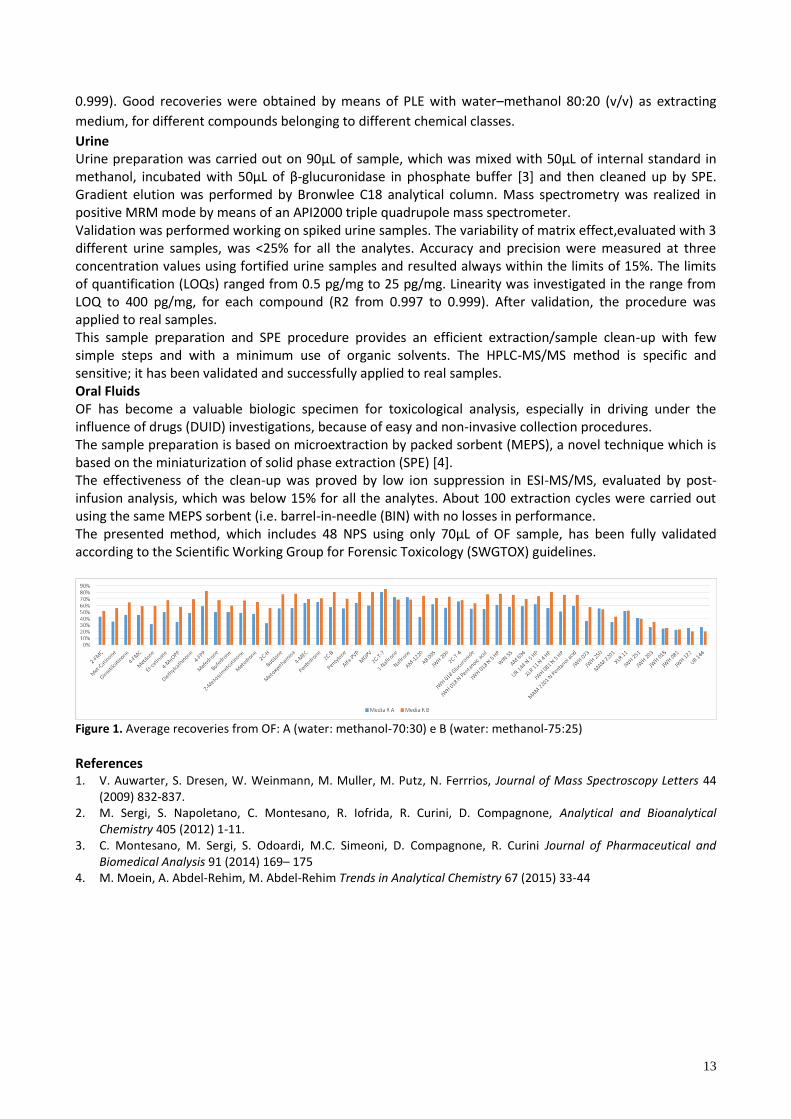
Urine Urine preparation was carried out on 90µL of sample, which was mixed with 50µL of internal standard in methanol, incubated with 50µL of β-glucuronidase in phosphate buffer [3] and then cleaned up by SPE. Gradient elution was performed by Bronwlee C18 analytical column. Mass spectrometry was realized in positive MRM mode by means of an API2000 triple quadrupole mass spectrometer. Validation was performed working on spiked urine samples. The variability of matrix effect,evaluated with 3 different urine samples, was <25% for all the analytes. Accuracy and precision were measured at three concentration values using fortified urine samples and resulted always within the limits of 15%. The limits of quantification (LOQs) ranged from 0.5 pg/mg to 25 pg/mg. Linearity was investigated in the range from LOQ to 400 pg/mg, for each compound (R2 from 0.997 to 0.999). After validation, the procedure was applied to real samples. This sample preparation and SPE procedure provides an efficient extraction/sample clean-up with few simple steps and with a minimum use of organic solvents. The HPLC-MS/MS method is specific and sensitive; it has been validated and successfully applied to real samples. Oral Fluids OF has become a valuable biologic specimen for toxicological analysis, especially in driving under the influence of drugs (DUID) investigations, because of easy and non-invasive collection procedures. The sample preparation is based on microextraction by packed sorbent (MEPS), a novel technique which is based on the miniaturization of solid phase extraction (SPE) [4]. The effectiveness of the clean-up was proved by low ion suppression in ESI-MS/MS, evaluated by post-infusion analysis, which was below 15% for all the analytes. About 100 extraction cycles were carried out using the same MEPS sorbent (i.e. barrel-in-needle (BIN) with no losses in performance. The presented method, which includes 48 NPS using only 70µL of OF sample, has been fully validated according to the Scientific Working Group for Forensic Toxicology (SWGTOX) guidelines.
Figure 1. Average recoveries from OF: A (water: methanol-70:30) e B (water: methanol-75:25)
References 1. V. Auwarter, S. Dresen, W. Weinmann, M. Muller, M. Putz, N. Ferrrios, Journal of Mass Spectroscopy Letters 44
(2009) 832-837. 2. M. Sergi, S. Napoletano, C. Montesano, R. Iofrida, R. Curini, D. Compagnone, Analytical and Bioanalytical
Chemistry 405 (2012) 1-11. 3. C. Montesano, M. Sergi, S. Odoardi, M.C. Simeoni, D. Compagnone, R. Curini Journal of Pharmaceutical and
Biomedical Analysis 91 (2014) 169– 175 4. M. Moein, A. Abdel-Rehim, M. Abdel-Rehim Trends in Analytical Chemistry 67 (2015) 33-44
14
PEPTIDOMIC AND BIOACTIVITY STUDY ON THE PEPTIDES ISOLATED IN COMMERCIAL DONKEY MILK
Chiara Cavaliere, Francesca Ferraris, Giorgia La Barbera, Susy Piovesana, Antonio Puglisi, Aldo Laganà
Dipartimento di Chimica, Università di Roma “La Sapienza”, Piazzale A. Moro, 5 – 00185 Roma,
Due to the strong correlation between nutrition and health, the characterization of the main constituents
of food is of fundamental importance. In this context proteins are key nutrients, but they are also
responsible of food bioactivities. Some proteins display a bioactivity in their native form, however, more
frequently, the bioactivity is cryptic and latent until a proteolytic event which releases the active peptides
encoded in the parent protein. These peptides can be part of the endogenous peptidome of food or they
can be produced by enzymatic activity during food modifications (gastrointestinal digestion, ripening and
fermentation). Milk, together with other dairy products, is one of the major sources of biologically active
peptides1.
Commercial donkey milk is an interesting valuable product and can be used in multiple applications,
spanning from food to cosmetics production. The main reasons for donkey milk scientific interest are
related to its nutritional values: donkey milk composition make it the most suitable mammalian milk for
infant consumption, also in case of cow milk allergies, and the best substitution to human milk2; moreover,
several bioactivities have also been associated with donkey milk and derivative products, among which
antioxidant activity3, anti-inflammatory activity4 and antimicrobial properties5.
Provided the importance and the interest for donkey milk for human consumption, the characterization of
the protein and peptide content of this food matrix is significant. The proteomic profile of donkey milk has
already been elucidated6; peptidomic studies were also performed, but dealt with the analysis of the
potentially bioactive peptides released after simulated hydrolysis in gastrointestinal conditions7.
However, previous studies never focused on the peptide profile of commercial unmodified donkey milk but
this type of information would be useful to provide a more comprehensive description of the nutritional
potential of this food. Peptides in food matrices often play an important biological role but they remain
poorly characterized in typical proteomics studies. In proteomics workflows the limiting point is the
isolation process, specific for the purification of proteins, which does not allow to isolate the endogenous
peptides, which are not effectively precipitated as well as proteins. Moreover, endogenous peptides do
often originate from precursor proteins, but in phenomena which are independent of the shotgun digestion
protocol, thus they can be obtained from cleavage specificities other than trypsin's, therefore they will not
be successfully identified during database search. Thus the need to develop different workflows for peptide
analysis.
In the work here presented this issue has been considered for the analysis of the peptides in commercial
donkey milk, to further mine its characterization and provide an analytical workflow for the analysis of such
peptides, which would otherwise not be investigated. An extensive peptidomic study was performed,
starting from the development of a suitable analytical protocol for the efficient isolation and purification of
peptides in donkey milk, assessing the effect of the purification protocol on the final identifications. Two
peptide purification strategies were tested and then compared. In fact the removal of interfering
compounds, such as whole proteins and lipids, is fundamental for the analysis of the peptide fraction. The
first purification strategy is based on the precipitation of all proteins in the sample and was performed by
organic solvent using cold acetone. In the other case only the most abundant milk proteins were
precipitated (i.e. caseins) at their isoelectric point, treating milk with acetic acid at pH 4.6. After protein
removal, the supernatants containing the peptides were purified by C18 solid phase extraction and samples
15
analyzed by reversed phase nanoHPLC and high resolution Orbitrap mass spectrometry. Data were then
processed with bioinformatic software, to retrieve peptide identifications from raw mass spectra, using
MaxQuant for peptide sequencing.
In this work two bioactivities were also investigated on the different peptide extracts. Two of the most
important biological activities were chosen, the angiotensin-converting-enzyme (ACE) inhibition and the
antioxidant activity, which were tested on the purified peptides. After the in-vitro biological activity assays,
a bioinformatic driven approach was employed to identify the peptides responsible of such bioactivities.
The list of identified peptides were searched in databases, including known bioactive peptides (BIOPEP,
http://www.uwm.edu.pl/biochemia/index.php/pl/biopep/) and PeptideDB (http://peptides.be/).
The procedures were compared and proved to be partially complementary. Considered together they
provided 1330 peptide identifications for donkey milk, mainly coming from the most abundant proteins in
milk.
Riferimenti 1. L. Sánchez-Rivera, D. Martínez-Maqueda, E. Cruz-Huerta, B. Miralles, I. Recio, Food Res. Int. 63 (2014) 170. 2. E. Salimei,F. N. Fantuz, Int. Dairy J. 24 (2012) 130. 3. . učev ć-Popov ć, I. De aš, S. Međugorac, M. Pave a- ranč ć, T. Ku š ć- uš ć, Int. J. Dairy Technol. 67 (2014)
394. 4. A. Tafaro, T. Magrone, F. Jirillo, G. Martemucci, A. G. D'Alessandro, L. Amati, E. Jirillo, Cur.r Pharm. Des. 13 (2007)
3711. 5. F. Tidona, C. Sekse, A. Criscione, M. Jacobsen, S. Bordonaro, D. Marletta, G. E. Vegarud, Int. Dairy J. 21 (2011) 158. 6. V. Cunsolo, V. Muccilli, E. Fasoli, R. Saletti, P. G. Righetti, S. Foti, J. Proteomics 74 (2011) 2083. 7. I. B. Bidasolo, M. Ramos, J. A. Gomez-Ruiz, Int. Dairy J. 24 (2012) 146.
16
MULTIDRUG RESISTANCE INHIBITORS IN PLASMA SAMPLES: THE POWER OF LC-ENERGY RESOLVED MS/MS METHODS FOR STABILITY INVESTIGATION
Marta Menicatti1, Luca Guandalini1, Silvia Dei1, Elisa Floriddia1, Elisabetta Teodori1, Pietro Traldi2 and Gianluca Bartolucci1
1 NEUROFARBA - Dipartimento di Neuroscienze, Psicologia, Area del Farmaco e Salute del Bambino Sezione Scienze Farmaceutiche e Nutraceutiche, Università di Firenze, Via Schiff, 6 50019 Sesto Fiorentino (FI), Italy
2Istituto di Ricerca Pediatrica Città della Speranza, Corso Stati Uniti 4, 35100 Padova, Italy
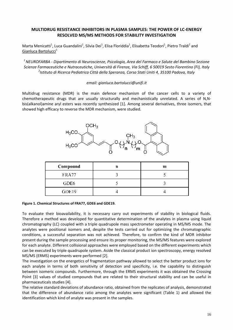
email: [email protected] Multidrug resistance (MDR) is the main defence mechanism of the cancer cells to a variety of chemotherapeutic drugs that are usually structurally and mechanistically unrelated. A series of N,N-bis(alkanol)amine aryl esters was recently synthesized [1]. Among several derivatives, three isomers, that showed high efficacy to reverse the MDR mechanism, were studied.
Figure 1. Chemical Structures of FRA77, GDE6 and GDE19.
To evaluate their bioavailability, it is necessary carry out experiments of stability in biological fluids. Therefore a method was developed for quantitative determination of the analytes in plasma using liquid chromatography (LC) coupled with a triple quadrupole mass spectrometer operating in MS/MS mode. The analytes were positional isomers and, despite the tests carried out for optimizing the chromatographic conditions, a successful separation was not achieved. Therefore, to confirm the kind of MDR inhibitor present during the sample processing and ensure its proper monitoring, the MS/MS features were explored for each analyte. Different collisional approaches were employed based on the different experiments which can be executed by triple quadrupole system. Aside the classical product ion spectroscopy, energy resolved MS/MS (ERMS) experiments were performed [2]. The investigation on the energetics of fragmentation pathway allowed to select the better product ions for each analyte in terms of both sensitivity of detection and specificity, i.e. the capability to distinguish between isomeric compounds. Furthermore, through the ERMS experiments it was obtained the Crossing Point [3] values of studied compounds that are related to their structural stability and can be useful in pharmaceuticals studies [4]. The relative standard deviations of abundance ratio, obtained from the replicates of analysis, demonstrated that the difference of abundance ratio among the analytes were significant (Table 1) and allowed the identification which kind of analyte was present in the samples.
17
Table 1: Results of relative abundance of product ions and their standard deviations of quantification and qualification
ions.
Compound 205 (m/z) SD 221 (m/z) SD
FRA77 39.6% 2.9% 100.0% n.d
GDE6 100.0% n.d 4.0% 0.7%
GDE19 100.0% n.d 25.7% 3.1%
The developed LC-MS/MS method showed a precision (between 1.8 % and 7.9 %), an accuracy (between 92.8 % and 99.9 %) and LOD values (between 1.0 and 2.7 ng mL-1), for all the analytes. Furthermore, the evaluation of matrix effects demonstrated that the procedure of preparation of samples did not affected to the ionization efficiency (ME greater than 95 %) or recovery (RE greater than 88 %). The obtained results demonstrated that the developed LC-MS/MS method was suitable for analyzing MDR inhibitors in PBS or plasma samples and for describing their degradation profiles. The degradation profiles experiments for each analyte, were carried out by comparing the variation of analyte concentration at different incubation times in PBS and plasma samples. The proposed LC-MS/MS method was applicable for evaluating the bioavailability of the analytes in plasma samples, although small concentration variations occur. Furthermore, the investigation on the energetics of fragmentation pathways, allowed to select the better product ions for distinguish between isomeric compounds and achieve information on their structural stability. Riferimenti 1. S. Dei, M. Coronnello, E. Floriddia, G. Bartolucci, C. Bellucci, L. Guandalini, D. Manetti, M. N. Romanelli, M.
Salerno, I. Bello, E. Mini, E. Teodori. Multidrug resistance (MDR) reversers: High activity and efficacy in a series of asymmetrical N,N-bis(alkanol)amine aryl esters. Eur J Med Chem. 2014, 30 , 398.
2. Y. X. J ang, K. . Wood, R. G. Cook . Character zat on of and energy depo t on n [C2H4O2]+• on u ng energy-resolved mass spectrometry. Org. Mass Spectrom. 1986, 21 (3) , 101.
3. G. Giorgi, L. Salvini, F. Ponticelli. Gas Phase Reactivity of Isomeric Arylglycosides Towards Amines. A Chemical Ionization Mass Spectrometry and Tandem Mass Spectrometry Study. J Am Soc Mass Spectrom. 2004, 15 (2) , 244.
4. E. Basso, A. Duranti,M. Mor, D. Piomelli, A. Tontini, G. Tarzia, P. Traldi. Tandem mass spectrometric data–FAAH inhibitory activity relationships of some carbamic acid O-arylesters. J. Mass Spectrom. 2004, 39 , 1450.
18
MODERN TRENDS IN DRUG ANALYSIS: VISUALIZING DESIGN SPACE IN THE QUALITY CONTROL OF PHARMACEUTICALS BY CAPILLARY ELECTROPHORESIS
Benedetta Pasquini, Serena Orlandini, Claudia Caprini, Massimo Del Bubba, Massimo Innocenti, Sandra
Furlanetto
Department of Chemistry “U. Schiff”, University of Florence, Via U. Schiff 6-Via della Lastruccia 3, 50019 Sesto F.no (FI), [email protected]
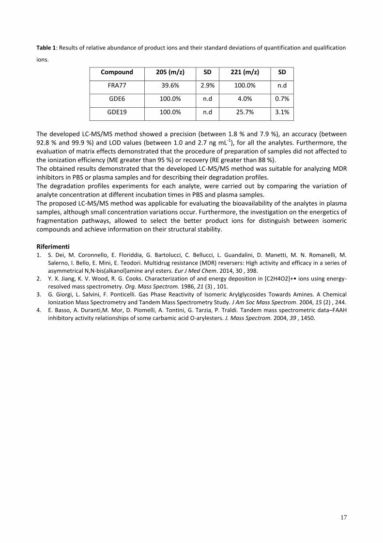
In the pharmaceutical field, Quality by Design (QbD) has been recently introduced [1] as a fundamental quality model with the aim of demonstrating both understanding and control of pharmaceutical processes to deliver high quality pharmaceutical products. The analytical procedures are inseparable components of the global pharmaceutical process, and as such are required to answer quality demands postulated by the regulatory documents. Therefore, the concept of QbD should also be implemented in analytical method development, because these methods are intended to be used for quality control of both the active pharmaceutical ingredients and drug products. The key of QbD approach is the definition of the design space (DS), which corresponds to the multidimensional region of knowledge space where satisfactory values of all defined critical quality attributes (CQAs) are computed with a desired probability level. In this study, QbD workflow [2] has been applied for the set up of a capillary electrophoresis (CE) method for the quality control and impurity profiling of the antimigraine drug zolmitriptan in its pharmaceutical product. In CE several chemical, physical and instrumental parameters should be controlled in order to obtain good analysis performances in terms of minimum analysis time and high resolution, efficiency and sensitivity; moreover, these parameters may be often interacting in nature. Thus, it was essential to implement and strengthen the CE method development by means of a systematic strategy based on QbD principles. Preliminary scouting experiments led to select Capillary Zone Electrophoresis based on phosphate buffer as operative mode. Afterwards, in a screening phase the effect of critical process parameters (CPPs), both instrumental and related to the background electrolyte, on CQAs (critical resolution values, analysis time and peak efficiency) was evaluated by a symmetric screening matrix. Response surface methodology was then carried out by a Box-Behnken design and contour plots were drawn highlighting significant interactions between some of the CPPs. Probability surfaces were calculated by employing Monte-Carlo simulations, making it possible to consider the propagation of the predictive errors of the model. By setting a r k of error equa to 1% (π≥99%), the probab ty map reported n F g. 1 were obta ned, where the DS visualized in green. Additional verification points at the edges of DS were selected by a Plackett-Burman matrix and then tested to verify the requirements for CQAs to be fulfilled. A control strategy was finally implemented based on robustness test and system suitability limits, which corresponded to the lower and the higher CQAs values observed during system repeatability studies. The developed method was validated and applied to a real sample of zolmitriptan tablets.
Figure 1. Design space definition by probability maps. Acceptance limits: Rs4≥3.0; Rs5≥0.5; t≤5 min; Log N≥5.5. Design space is colored in green and is included in the line corresponding to 1% risk of failure.
References 1. ICH Harmonised Tripartite Guideline. Pharmaceutical development Q8(R2) (2009) International Conference on
Harmonisation of technical requirements for registration of pharmaceuticals for human use. 2. S. Orlandini, S. Pinzauti, S. Furlanetto, Anal. Bioanal. Chem. 405 (2013) 443-450.
19
ARTIFICIAL ANTIBODIES: WHERE DO WE STAND?
Claudio Baggiani, Laura Anfossi, Cristina Giovannoli
Dipartimento di Chimica, Università di Torino,Via Giuria 5 – 10125 Torino, [email protected]
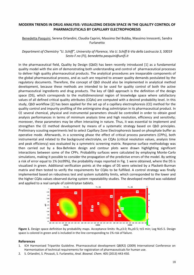
Nature has, through billions of years of evolution, assembled a multitude of polymeric macromolecules characterized by marked molecular recognition properties towards well defined targets. This property arises from the precise control exerted over their biosynthesis, resulting in key residues being anchored in the appropriate positions to interact with target substrates. Among such macromolecules, antibodies are large proteins produced and used by the immunosystem of vertebrate to identify and neutralize foreign substances introduced in the body. In the last fifty years such biopolymers have been increasingly used in analytical applications as efficient and selective recognition elements in immunoassays, immunosensors and immunoextraction materials. Despite the undoubted success of antibodies in widespread analytical applications, there are several shortcomings that may limit their practical applications. High costs of productions, low batch-to-batch reproducibility and unstability in non-aqueous environments push toward the development of alternative binding systems based on man-made receptors. Deve op ng ‘who y ynthet c’ macromo ecu ar tructures that can mimic natural antibodies presents a considerable challenge for chemists, who lack the biological machinery used in nature to assemble biomacromolecules with high precision. In addressing this challenge, molecularly imprinted polymers (MIP) represent the most successful achievment, and in the last twenty years have been frequently described by many author a “art f c a ant bod e ” or e common y “p a t bod e ”.
However, despite the rapid development of MIP-based technology as a research hotspot and the undeniable success of these polymers in certain analytical applications such as solid phase extraction, there are serious limitations to the use of MIPs as an efficient alternative to antibody-based technology in application fields such as sensoristics and immunoassay. We briefly review the current status of MIP technology, with particular emphasis on present challenges involving dimensional downscaling, difficult biomacromolecule imprinting and incompatibility with aqueous media, and the possible strategies to be implemented in order to overcome these technological bottlenecks.

20
A NEW SMARTPHONE-BASED CHEMILUMINESCENT LATERAL FLOW IMMUNOSENSOR FORMAT FOR POINT OF CARE TESTING
Martina Zangheri1, Luca Cevenini1,Laura Anfossi2, Claudio Baggiani2, Patrizia Simoni3, Fabio Di Nardo2, Aldo
Roda1
1 Dipartimento di Chimica, Università di Bologna, Via Selmi 2, 40126 Bologna 2Dipartimento di Chimica, Università di Torino, Via P. Giuria 5, 10125 Torino
3Dipartimento di Medicina e Chirurgia, Università di Bologna, Via Massarenti 9, 40138 Bologna
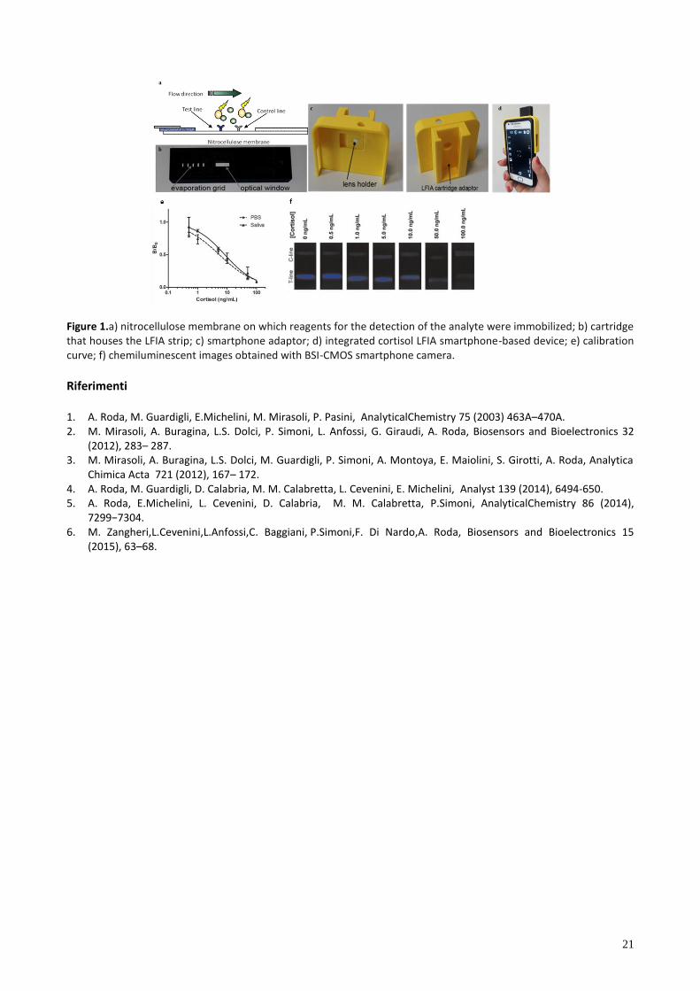
Development of rapid, accurate and sensitive diagnostic device for point-of-care-testing (POCT) is one of the major issuein the field of analytical chemistry. For this purpose, several studies have focused on the development of self-standing devices suitable for clinical biomarker monitoring. To date, lateral flow immunoassays (LFIAs) integrated with handled sensor devices (pregnant test) have been among the most popular point-of-care applications. However, their diffusion has been limited by difficulties in achieving sensitive and quantitative information using conventional colorimetric or visual reading systems. Optical detection based on chemiluminescence (CL) can be an ideal alternative method for miniaturized biosensor development because of its inherent sensitivity and simplicity [1]. We recently described biosensors based on a chemiluminescence Lateral Flow ImmunoAssay (CL-LFIA) coupled with a portable ultrasensitive CCD as a CL detector [2,3]. Recently smartphones have attracted considerable attention in diagnostics as a promising tool for the development of POCT device owing to their peculiarities, such as embedded physical sensors and the possibility to use specific Applications. Improved image-processing technology based on back side illuminated CMOS (BSI-CMOS) sensors of the martphone’ camera, makes it possible to develop fast and accurate point-of-care diagnosis, combining paper technology and bio-chemilumiscent detection [4,5]. This will allow ana y d rect y at home. The martphone’ advanced connect v ty cou d then be u ed to end the data to an appropriate diagnostic center for therapeutic action. Here, we report on the development of a simple, rapid, and accurate biosensor based on a chemiluminescent (CL)-LFIA method for quantitative detection of cortisol in saliva, using a smartphone camera as a light detector [6]. The biosensor is based on a direct competitive immunoassay using peroxidase (HRP)-cortisol conjugate, which is detected by adding the chemiluminescent substrate luminol/enhancer/hydrogen peroxide (Fig. 1a) and by using a smartphone camera for the image acquisition and data handling via a specific application. Using a 3D printer, we made simple accessories to turn a smartphone into a biosensing device. The system comprises a cartridge (Fig. 1b), which houses the LFIA strip, and a smartphone adaptor equipped with a plano-convex lens and a narrow slot for inserting the cartridge (Fig. 1c). This provides an aligned optical interface between the camera and the LFIA membrane. When the cartridge is inserted, it creates a minidarkbox making it possible to acquire the CL signals (Fig 1d). The developed method is simple and fast with a detection limit of 0.3 ng/mL.Calibration curve is reported in Fig.1e and chemiluminescent images are shown in Fig.1f. The method provides quantitative analysis in the range from 0.3 to 60 ng/mL, which is adequate for detecting salivary cortisol in the clinically accepted normal range and in different pathophysiological conditions. Finally the performances of the method were evaluated by analysing some real saliva samples founding a good agreement between our results and those obtained with a commercial ELISA kit. In the future, this concept can pave the way for a new generation of portable analytical devices even based on multiplex capability. These kind of biosensors will be useful not only in the medical diagnostic field but in all situations where a decentralized and fast detection is required such as bioterrorism attack, critical medicine, space station, environmental toxicity and analyses in developing countries taking advantages of the peculiar properties of a low cost mobile phone in term of connectivity, location (GPS), long distance transfer of data via wireless.
21
Figure 1.a) nitrocellulose membrane on which reagents for the detection of the analyte were immobilized; b) cartridge that houses the LFIA strip; c) smartphone adaptor; d) integrated cortisol LFIA smartphone-based device; e) calibration curve; f) chemiluminescent images obtained with BSI-CMOS smartphone camera.
Riferimenti 1. A. Roda, M. Guardigli, E.Michelini, M. Mirasoli, P. Pasini, AnalyticalChemistry 75 (2003) 463A–470A. 2. M. Mirasoli, A. Buragina, L.S. Dolci, P. Simoni, L. Anfossi, G. Giraudi, A. Roda, Biosensors and Bioelectronics 32
(2012), 283– 287. 3. M. Mirasoli, A. Buragina, L.S. Dolci, M. Guardigli, P. Simoni, A. Montoya, E. Maiolini, S. Girotti, A. Roda, Analytica
Chimica Acta 721 (2012), 167– 172. 4. A. Roda, M. Guardigli, D. Calabria, M. M. Calabretta, L. Cevenini, E. Michelini, Analyst 139 (2014), 6494-650. 5. A. Roda, E.Michelini, L. Cevenini, D. Calabria, M. M. Calabretta, P.Simoni, AnalyticalChemistry 86 (2014),
7299−7304. 6. M. Zangheri,L.Cevenini,L.Anfossi,C. Baggiani, P.Simoni,F. Di Nardo,A. Roda, Biosensors and Bioelectronics 15
(2015), 63–68.
22
ORGANIC BIOELECTRONIC SENSORS: COMPARATIVE STUDY OF CRP DETECTION USING DIFFERENT ORGANIC THIN FILM TRANSISTORS CONFIGURATIONS
Kyriaki Manoli, Maria Magliulo, Mohammad Yusuf Mulla, Donato De Tullio, Preethi Seshadri, Gerardo
Palazzo, Luisa Torsi
1 Dipartimento di Chimica, Università degli studi di Bari Aldo Moro, via Orabona 4 70126 Bari, [email protected]
The possibility to develop low cost organic electronic biosensors in order to replace conventional analytical
techniques is of great scientific and commercial interest. The coupling between biosystems and organic
electronic devices allows for realizing label-free biosensing platforms, since biomolecular interactions (e.g.
antibody-antigen binding, hybridization of nucleic acids) can be converted to an electrical signal. Among
others, organic thin film transistors (OTFTs) hold great potential as electronic ultrasensitive biosensing
platforms. Bioelectronic sensors based on OTFT have been proposed for label-free detection of several
chemical and biological species1. Owing to the diverse nature of the OTFT structural materials and to their
operation principle, transistors are devices that are governed by interfacial effects. Therefore, different
strategies have been developed for efficient immobilization of bioreceptors on each of the device
interfaces.
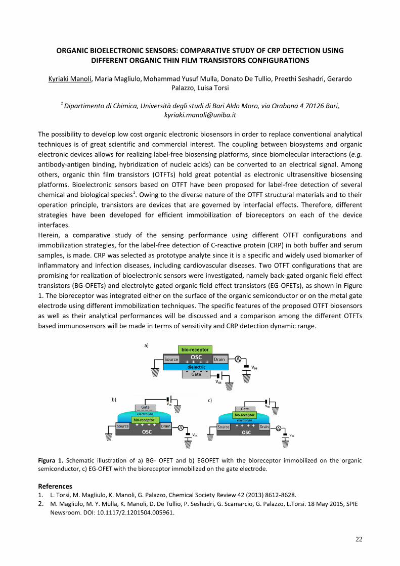
Herein, a comparative study of the sensing performance using different OTFT configurations and
immobilization strategies, for the label-free detection of C-reactive protein (CRP) in both buffer and serum
samples, is made. CRP was selected as prototype analyte since it is a specific and widely used biomarker of
inflammatory and infection diseases, including cardiovascular diseases. Two OTFT configurations that are
promising for realization of bioelectronic sensors were investigated, namely back-gated organic field effect
transistors (BG-OFETs) and electrolyte gated organic field effect transistors (EG-OFETs), as shown in Figure
1. The bioreceptor was integrated either on the surface of the organic semiconductor or on the metal gate
electrode using different immobilization techniques. The specific features of the proposed OTFT biosensors
as well as their analytical performances will be discussed and a comparison among the different OTFTs
based immunosensors will be made in terms of sensitivity and CRP detection dynamic range.
Figura 1. Schematic illustration of a) BG- OFET and b) EGOFET with the bioreceptor immobilized on the organic semiconductor, c) EG-OFET with the bioreceptor immobilized on the gate electrode.
References 1. L. Torsi, M. Magliulo, K. Manoli, G. Palazzo, Chemical Society Review 42 (2013) 8612-8628.
2. M. Magliulo, M. Y. Mulla, K. Manoli, D. De Tullio, P. Seshadri, G. Scamarcio, G. Palazzo, L.Torsi. 18 May 2015, SPIE
Newsroom. DOI: 10.1117/2.1201504.005961.
23
MICRORNA DETECTION BY SPR IMAGING AND PNA PROBES: NANOPARTICLE AND ENZYMATIC AMPLIFICATION METHODS
Roberta D’Agata1, Alex Manicardi2, Alessia Finotti3, Roberto Corradini2, Roberto Gambari3,
Giuseppe Spoto1,4
1Department of Chemical Science, University of Catania, Viale A. Doria 6, 95124 Catania, [email protected]
2Department of Chemical Science, University of Parma, Parco Area delle Scienze 17/A, 43124 Parma 3
Department of Life Sciences and Biotechnology, University of Ferrara, Via L. Borsari 46, 44121 Ferrara 4Consortium INBB, Viale delle Medaglie d'Oro 305, 00136 Roma
MicroRNAs (miRNAs) are recognized as key players in gene regulatory networks and have been shown to be implicated in many fundamental biological processes, such us cell proliferation and apoptosis.1 Their prominent role in the outcome of many human disorders makes miRNAs very promising diagnostic and prognostic biomarkers. However, several features, including miRNA small size, low abundance and close sequence similarity, make their detection2 a challenging task. Accurate and robust methods able to detect miRNA are today strongly needed. Here we describe a novel approach which combines flexibility and sensitivity in targeting miRNAs based on the combined use of surface plasmon resonance imaging (SPRi)3 and surface-oriented orthogonally PNA probes.4 The immobilization of a PNA tethered to the gold surface through lysine-modified backbone, so that the probe had both termini accessible, allows to modify target m croRNA d rect y at e ther 3’ hydro y or 5’-monophosphate terminus after the hybridization. An excellent SPRi signal amplification is then achieved by using the subsequent adsorption of gold nanoparticles (AuNPs) functionalized with poly(T) or streptavidin (SA), which respectively bind to the appended poly(A) or biotin tails on modified microRNA. To demonstrate the versatility and the efficiency of the approach, we applied it to two methods, established in-solution, for the enzymat c abe ng of o gonuc eot de at 3’ or 5’ term n . The e atter have been optimized for the highly sensitive detection of miRNA directly onto the SPRi surface. Our results show that the coupling of the nanoparticle amplified SPRi detection and the surface-oriented orthogonally PNA probes allows the detection of miRNAs in the low subpicomolar concentrations with high specificity. Furthermore this approach has been shown to operate with biological samples. The detection of microRNA-210 tested from transfected human K562 cells,5 the sensitivity and versatility of the detection protocol may be a convenient tool for biomedical research and clinical diagnostic applications not requiring target amplification or manipulation. References 1. C.M. Croce. Nat Rev Genet 10 (2009) 704. 2. H. Dong, J. Lei, L. Ding, Y. Wen, H. Ju, X. Zhang, Chem Rev (2013) 113, 6207. 3. R. D’Agata, G. Spoto, Anal Bioanal Chem 405 (2013) 573. 4. A. Bertucci, A. Manicardi, R. Corradini, in Detection of Non-Amplified Genomic DNA, Soft and Biological Matter,
Spoto, G., Corradini, R. (Eds.) 2012, 89. 5. M.C. Giuffrida, L.M. Zanoli, R. D’Agata, A. Finotti, R. Gambari, G. Spoto, Anal Bioanal Chem 407, (2015) 1533.
24
A DNA NANO PH-METER BASED ON TRIPLEX FORMATION
Andrea Idili1, A e a e- e2, Giuseppe Palleschi1, Francesco Ricci1
1Dipartimento di Scienze e Tecnologie Chimiche, University of Rome, Tor Vergata, 00133, Rome, Italy
2La oratory of Biosensors and anomachines, D partement de Chimie, Universit de Montr al, Canada
Nature often employs finely pH-regulated biomolecules to modulate and tune a number of biological
activities. For this reason, developing nanoprobes, nanoswitches, or nanomaterials that are able to respond
to specific pH changes should prove useful for several applications in the fields of in vivo imaging, clinical
diagnostics, and drug delivery. We have designed programmable DNA-based nanoswitches whose
closing/opening can be triggered over specific different pH windows. These nanoswitches form an
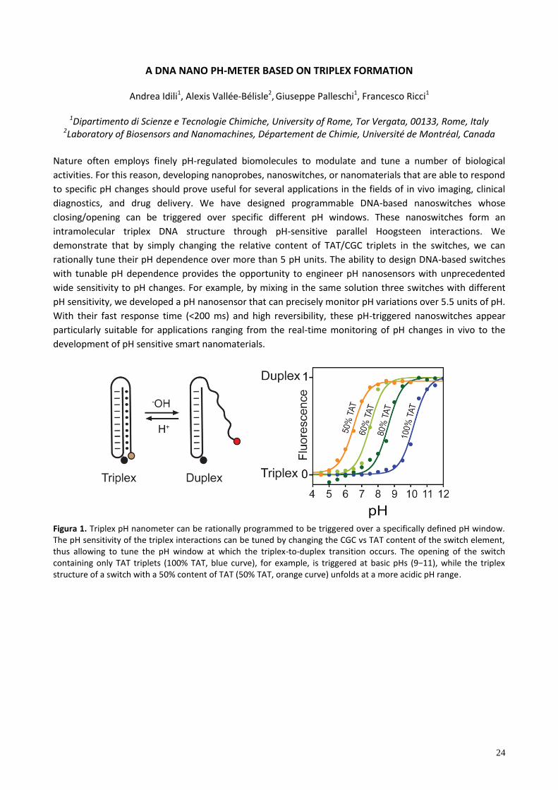
intramolecular triplex DNA structure through pH-sensitive parallel Hoogsteen interactions. We
demonstrate that by simply changing the relative content of TAT/CGC triplets in the switches, we can
rationally tune their pH dependence over more than 5 pH units. The ability to design DNA-based switches
with tunable pH dependence provides the opportunity to engineer pH nanosensors with unprecedented
wide sensitivity to pH changes. For example, by mixing in the same solution three switches with different
pH sensitivity, we developed a pH nanosensor that can precisely monitor pH variations over 5.5 units of pH.
With their fast response time (<200 ms) and high reversibility, these pH-triggered nanoswitches appear
particularly suitable for applications ranging from the real-time monitoring of pH changes in vivo to the
development of pH sensitive smart nanomaterials.
Figura 1. Triplex pH nanometer can be rationally programmed to be triggered over a specifically defined pH window. The pH sensitivity of the triplex interactions can be tuned by changing the CGC vs TAT content of the switch element, thus allowing to tune the pH window at which the triplex-to-duplex transition occurs. The opening of the switch containing only TAT triplets (100% TAT, b ue curve), for e amp e, tr ggered at ba c pH (9−11), wh e the tr p e structure of a switch with a 50% content of TAT (50% TAT, orange curve) unfolds at a more acidic pH range.
25
A NEW IMMUNOSENSOR FOR THE DETERMINATION OF VALPROIC ACID IN SERUM USING FUNCTIONALIZED SILICA NANOPARTICLES DOPED WITH A THERMOCHEMILUMINESCENT 1,2–
DIOXETANE DERIVATIVE AS LABEL
Massimo Di Fusco1,2, Arianna Quintavalla2, Marco Lombardo2, Massimo Guardigli2, Mara Mirasoli1,2, Luca
Alfio Andronico2, Claudio Trombini2, Aldo Roda2 1Advanced Applications in Mechanical Engineering and Materials Technology, Interdepartmental Center for
Industrial Research, Alma Mater Studiorum, University of Bologna, Viale Risorgimento 2, 40136 Bologna 2Department of Chemistry ‘‘G. Ciamician’’, Alma Mater Studiorum, University of Bologna, Via Francesco
Selmi 2, 40126 Bologna
We recently demonstrated that the use of labels based on thermochemiluminescence (TCL), i.e., the light
emission originating from a product in the singlet excited state after the thermolysis of a 1,2–dioxetane, is a
powerful tool for biosensors development. Indeed, the process enables a reagentless chemical
luminescence–based detection technique, thus simplifying the microfluidic network in miniaturized
analytical devices and biosensors based on the use of conventional chemiluminescence (CL). The main
problems of TCL detection are the h gh operat ng temperature (200−250 °C) required to decompose the
molecule and to produce the singlet excited state, and the lower detectability in comparison with other CL
labels, due to the low efficiency of the luminescence process. Recently, we overcome these limitations by synthesizing a library of new TCL acridine–1,2–dioxetane derivatives proposed as new TCL labels [1−3]. Suitable structural modifications were introduced to decrease the em on tr gger ng temperature down to 80−100 °C and to produce h gh y eff c ent f uorophore n the
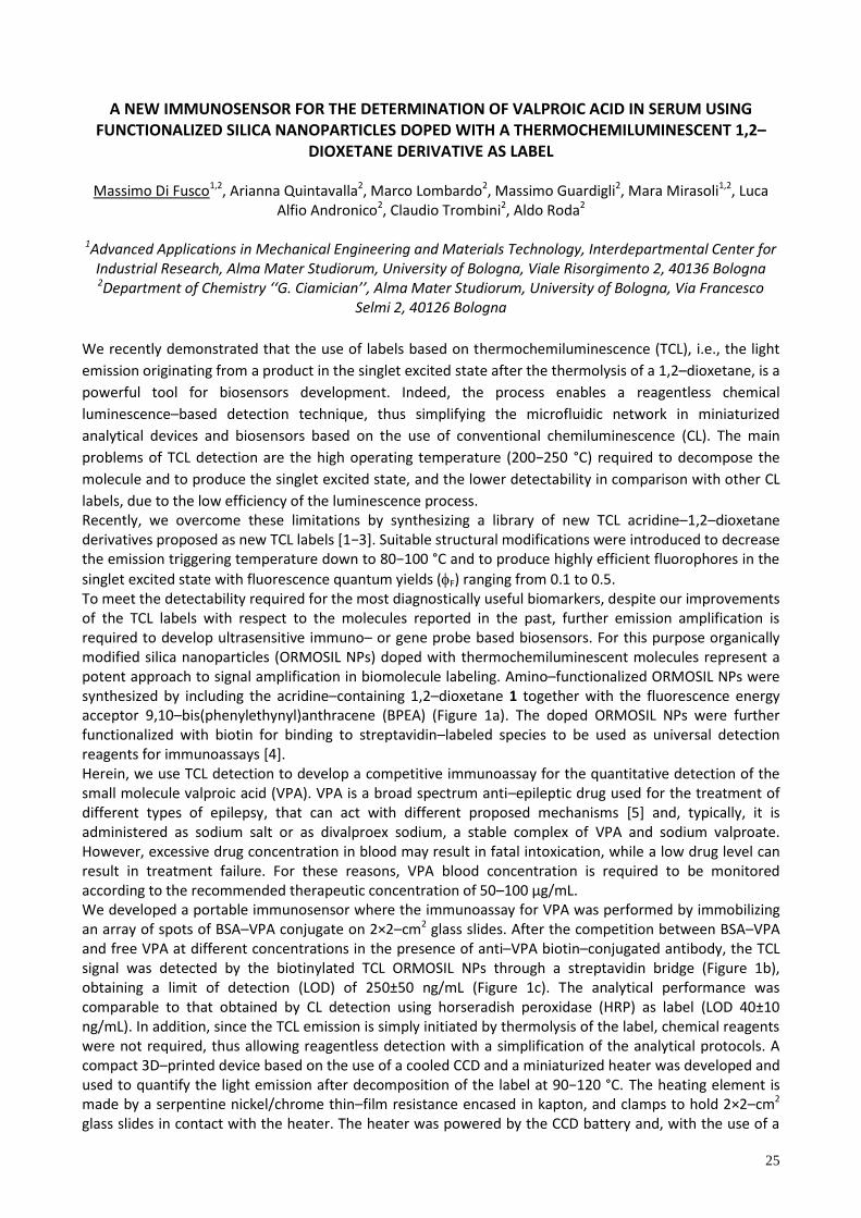
singlet excited state with fluorescence quantum yields (F) ranging from 0.1 to 0.5. To meet the detectability required for the most diagnostically useful biomarkers, despite our improvements of the TCL labels with respect to the molecules reported in the past, further emission amplification is required to develop ultrasensitive immuno– or gene probe based biosensors. For this purpose organically modified silica nanoparticles (ORMOSIL NPs) doped with thermochemiluminescent molecules represent a potent approach to signal amplification in biomolecule labeling. Amino–functionalized ORMOSIL NPs were synthesized by including the acridine–containing 1,2–dioxetane 1 together with the fluorescence energy acceptor 9,10–bis(phenylethynyl)anthracene (BPEA) (Figure 1a). The doped ORMOSIL NPs were further functionalized with biotin for binding to streptavidin–labeled species to be used as universal detection reagents for immunoassays [4]. Herein, we use TCL detection to develop a competitive immunoassay for the quantitative detection of the small molecule valproic acid (VPA). VPA is a broad spectrum anti–epileptic drug used for the treatment of different types of epilepsy, that can act with different proposed mechanisms [5] and, typically, it is administered as sodium salt or as divalproex sodium, a stable complex of VPA and sodium valproate. However, excessive drug concentration in blood may result in fatal intoxication, while a low drug level can result in treatment failure. For these reasons, VPA blood concentration is required to be monitored according to the recommended therapeutic concentration of 50–100 μg/mL. We developed a portable immunosensor where the immunoassay for VPA was performed by immobilizing an array of spots of BSA–VPA conjugate on 2×2–cm2 glass slides. After the competition between BSA–VPA and free VPA at different concentrations in the presence of anti–VPA biotin–conjugated antibody, the TCL signal was detected by the biotinylated TCL ORMOSIL NPs through a streptavidin bridge (Figure 1b), obtaining a limit of detection (LOD) of 250±50 ng/mL (Figure 1c). The analytical performance was comparable to that obtained by CL detection using horseradish peroxidase (HRP) as label (LOD 40±10 ng/mL). In addition, since the TCL emission is simply initiated by thermolysis of the label, chemical reagents were not required, thus allowing reagentless detection with a simplification of the analytical protocols. A compact 3D–printed device based on the use of a cooled CCD and a miniaturized heater was developed and u ed to quant fy the ght em on after decompo t on of the abe at 90−120 °C. The heating element is made by a serpentine nickel/chrome thin–film resistance encased in kapton, and clamps to hold 2×2–cm2 glass slides in contact with the heater. The heater was powered by the CCD battery and, with the use of a
26
manually regulated resistor, the appropriate voltage was applied to reach the required temperature. These characteristics make TCL doped ORMOSIL NPs ideal universal nanoprobes for ultrasensitive bioassays such as immuno– and DNA–based assays in a compact and simple biosensor format.
Figure 1. a) Chemical structures of the 1,2–dioxetane derivative 1 and the fluorescence energy acceptor 9,10–bis(phenylethynyl)anthracene (BPEA) used as dopants for ORMOSIL NPs; b) images of the TCL emission from an array of spots incubated with different concentrations of VPA; c) calibration curve of the TCL–based competitive immunoassay for VPA.
More recently, we developed a smartphone–based TCL device comprising a 3D–printed cover, easily snapped onto any smartphone model, and a battery powered mini–heater, obtaining a further miniaturization of TCL biosensors. This device is under investigation to develop a TCL–based immunoassay for VPA, taking the advantages of the smartphone connectivity that will facilitate the use of such format for point of need biosensor. In addition, the synthesis of new TCL molecules containing different fluorophores in the backbone, i.e., fluorenone, fluorene, xanthone and flavone, or different substituents on the acridine moiety or endocyclic nitrogen atom was performed to obtain more efficient TCL molecules and thus increasing the detectability of TCL–based biosensors. References 1. A. Roda, M. Di Fusco, A. Quintavalla, M. Guardigli, M. Mirasoli, M. Lombardo, C. Trombini Anal. Chem. 84 (2012)
9913−9919. 2. M. Di Fusco, A. Quintavalla, C. Trombini, M. Lombardo, A. Roda, M. Guardigli, M. Mirasoli J. Org. Chem. 78 (2013)
11238–11246. 3. M. Di Fusco, M. Guardigli, M. Lombardo, M. Mirasoli, A. Quintavalla, A. Roda, C. Trombini Patent WO2014024106
A1 (2014). 4. M. Di Fusco, A. Quintavalla, M. Lombardo, M. Guardigli, M. Mirasoli, C. Trombini, A. Roda Anal. Bioanal. Chem.
407 (2015) 1567–1576. 5. C. U. Johannessen, S.I. Johannessen CNS Drug Rev. 9 (2003) 199–216.
27
SPECTROPHOTOMETRIC CELL-FREE ASSAYS FOR MEASUREMENT OF THE OXIDATIVE POTENTIAL OF ATMOSPHERIC AESOSOL
Maria Chiara Pietrogrande, Marco Visentin
1 Dipartimento di Scienze Chimiche e Farmaceutiche, Università di Ferrara via Fossato di Mortara 17, 44121 Ferrara, e-mail: [email protected]
Oxidative stress has been suggested as an important underlying mechanism of action by which exposure to
ambient particulate matter (PM) may lead to adverse health effects in humans [1]. Oxidative stress results
when the generations of reactive oxygen species (ROS), or free radicals, exceed the available antioxidant
defenses. For assessment of the capacity of a PM sample to catalyze ROS generation, the oxidative
potential (OP) has been proposed, as a measure of the ability of PM to oxidize target molecules, i.e. by
generating ROS in environments without living cells [2].
Among the various assays developed for measuring OP, in this study two common methods are
investigated and compared in terms of different sensitivity to the ROS generating compounds.
The dithiothreitol (DTT) assay measures the presence of reactive oxygen species via consumption of DTT to
form the DTT-disulfide due to transfer electrons from DTT to ROS by recycling chemicals such as quinones.
The reaction was stopped at designated time points by addition of trichloroacetic acid. The subsequent loss
of DTT is followed by its reaction with 5,5’-dithiobis-2-nitrobenzoic acid (DTNB) to form 2-nitro-5-
mercaptobenzoic acid, which is monitored spectrophotometrically at 412nm. The linear rate of DTT loss is
measured (expressed as μmol DTT min-1) [3].
The ascorbate (AA) depletion assay measures the ability of PM to deplete ascorbic acid: the reaction kinetic
is followed by measuring AA absorption at 265 nm. The re u t are e pre ed a μmo m n-1 of AA depletion
[4].
The performances of the two assays were investigated with standard solutions of individual redox-active
species that are common in ambient PM, such as quinones and transition metals.
Quinones – mainly phenanthrenequinone and 1,2-naphthoquinone – were found more efficient at oxidizing
DTT compared to the less reactive transition metals, i.e., copper, manganese, nickel, chromium, iron.
On the contrary, the AA assay is most sensitive to transition metals – mainly copper, chromium, iron and
nickel – in comparison to quinones. The obtained information is very relevant to understand the relative
importance of metals and organics towards ROS generation from ambient particles.
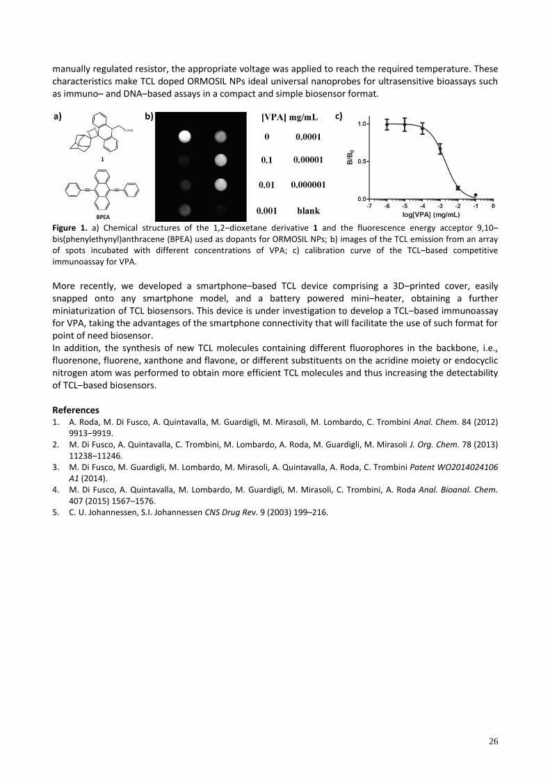
Figure 1. Results of DDT assay for a PM2.5 filter collected at Bologna in winter 2013.
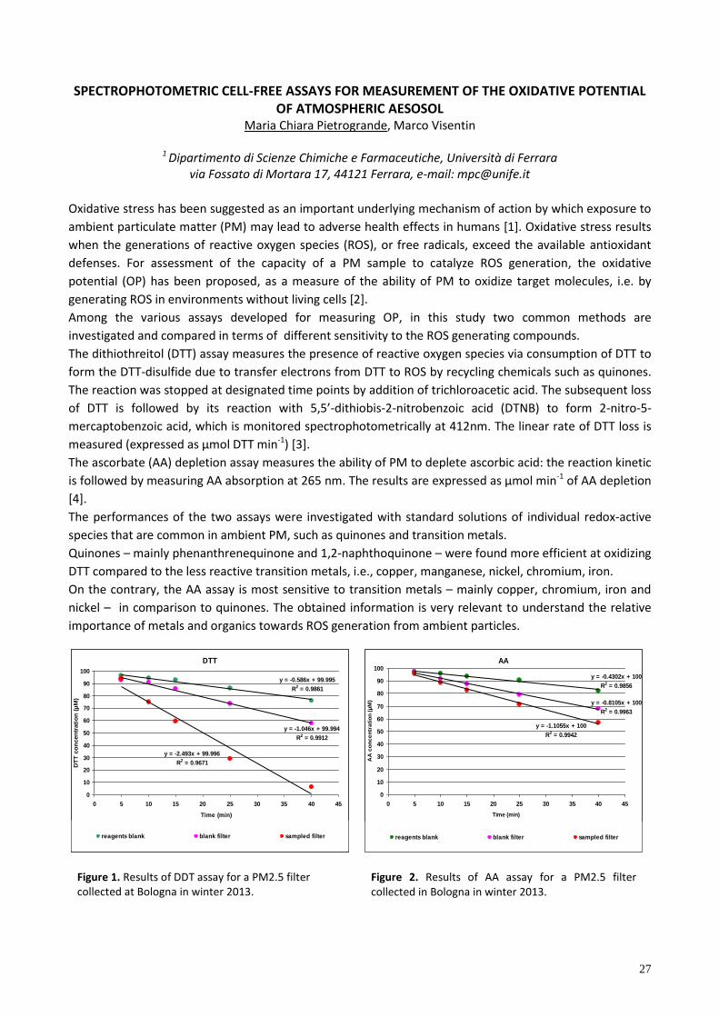
Figure 2. Results of AA assay for a PM2.5 filter collected in Bologna in winter 2013.
DTT
y = -0.586x + 99.995
R2 = 0.9861
y = -1.046x + 99.994
R2 = 0.9912
y = -2.493x + 99.996
R2 = 0.9671
0
10
20
30
40
50
60
70
80
90
100
0 5 10 15 20 25 30 35 40 45
Time (min)
DT
T c
on
cen
trati
on
(µ
M)
reagents blank blank filter sampled filter
AA
y = -0.4302x + 100
R2 = 0.9856
y = -0.8105x + 100
R2 = 0.9963
y = -1.1055x + 100
R2 = 0.9942
0
10
20
30
40
50
60
70
80
90
100
0 5 10 15 20 25 30 35 40 45
Time (min)
AA
co
nc
en
tra
tio
n (
µM
)
reagents blank blank filter sampled filter
28
Both methods were applied to real ambient PM2.5 samples collected at urban sites in winter (Figures 1 and
2). The preliminary results show that the two OP assays can provide complementary data to be used as a
good quantitative chemical assay for oxidant generation and toxicity measurement of PM. Future studies
will focus on relationship with chemical characterization of PM samples to assess the role of organic and
inorganic species in generating redox activity. References 1. A.J. Ghio, M.S. Carraway, M. Madden Composition of air pollution particles and oxidative stress in cells, tissues,
and living systems, J. Toxicol. Environ. Health-Pt b-Crit. Rev. 15 (2012) 1–21. 2. P.J.A. Borm, F. Kelly, N. Künzli, R.P.F. Schins, K. Donaldson Oxidant generation by particulate matter: from
biologically effective dose to a promising, novel metric. Occup. Environ. Med. 64 (2007) 73-74. 3. J. G. Charrier and C. Anastasio, Atmos. Chem. Phys. 12 (2012) 9321–9333. 4. I.S. Mudway, N. Stenfors, S.T. Duggan, H. Roxborough, H. Zielinski, S.L. Marklund, A. Blomberg, A.J. Frew, T.
Sandström, , F.J. Kelly An in vitro and in vivo investigation of the effects of diesel exhaust on human airway lining fluid antioxidants. Arch. Biochem. Biophys. 423 (2004) 200-212.
29
DETERMINATION OF WARFARIN AND WARFARIN ALCOHOLS IN ORAL FLUID AND PLASMA SAMPLES FOR MONITORING PATIENTS UNDERGOING ANTICOAGULANT THERAPY
Tommaso Lomonaco1, Silvia Ghimenti1, Isabella Piga1, Denise Biagini1, Massimo Onor2, Aldo Paolicchi3, Lucia
Ruocco4, Giovanni Pellegrini4, Maria Giovanna Trivella5, Roger Fuoco1, Fabio Di Francesco1,5
1 Department of Chemistry and Industrial Chemistry, University of Pisa, Via Moruzzi, 13 Pisa, Italy, [email protected].
2 Institute of Chemistry of Organometallic Compounds, CNR, Via Moruzzi, 1 Pisa, Italy. 3 Department of Translational Research and New Technologies in Medicine and Surgery, University of Pisa,
Via Risorgimento, 36 Pisa, Italy. 4 Chemical-Clinical Analysis Laboratory, AOUP, Via Paradisa, 2 Pisa, Italy.
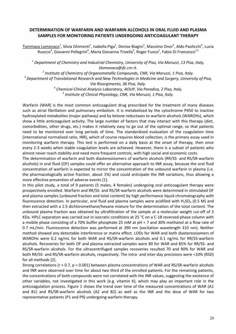
5 Institute of Clinical Physiology, CNR, Via Moruzzi, 1 Pisa, Italy.
Warfarin (WAR) is the most common anticoagulant drug prescribed for the treatment of many diseases such as atrial fibrillation and pulmonary embolism. It is metabolized by the cytochrome P450 to inactive hydroxylated metabolites (major pathway) and by ketone reductases to warfarin alcohols (WAROHs), which show a little anticoagulant activity. The large number of factors that may interact with this therapy (diet, comorbidities, other drugs, etc.) makes it relatively easy to go out of the optimal range, so that patients need to be monitored over long periods of time. The standardized evaluation of the coagulation time (international normalized ratio, INR), which of course requires blood collection, is the primary assay used in monitoring warfarin therapy. This test is performed on a daily basis at the onset of therapy, then once every 2-3 weeks when stable coagulation levels are achieved. However, there is a subset of patients who almost never reach stability and need more frequent controls, with high social and economic costs. The determination of warfarin and both diastereoisomers of warfarin alcohols (RR/SS- and RS/SR-warfarin alcohols) in oral fluid (OF) samples could offer an alternative approach to INR assay, because the oral fluid concentration of warfarin is expected to mirror the concentration of the unbound warfarin in plasma (i.e. the pharmacologically active fraction, about 1%) and could anticipate the INR variations, thus allowing a more effective prevention of adverse events [1]. In this pilot study, a total of 9 patients (5 males, 4 females) undergoing oral anticoagulant therapy were prospectively enrolled. Warfarin and RR/SS- and RS/SR-warfarin alcohols were determined in stimulated OF and plasma samples (unbound fraction and total content) by high performance liquid chromatography with fluorescence detection. In particular, oral fluid and plasma samples were acidified with H2SO4 (0.5 M) and then extracted with a 1:5 dichloromethane/hexane mixture for the determination of the total content. The unbound plasma fraction was obtained by ultrafiltration of the sample at a molecular weight cut-off of 3 KDa. HPLC separation was carried out in isocratic conditions at 25 °C on a C-18 reversed-phase column with a mobile phase consisting of a 70% buffer phosphate 25 mM at pH = 7 and 30% methanol at a flow rate of 0.7 mL/min. Fluorescence detection was performed at 390 nm (excitation wavelength 310 nm). Neither method showed any detectable interference or matrix effect. LODs for WAR and both diastereoisomers of WAROHs were 0.2 ng/mL for both WAR and RS/SR-warfarin alcohols and 0.1 ng/mL for RR/SS-warfarin alcohols. Recoveries for both OF and plasma extracted samples were 80 for WAR and 85% for RR/SS- and RS/SR-warfarin alcohols. For the ultracentrifuged samples recoveries resulted 70 and 90% for WAR and both RR/SS- and RS/SR-warfarin alcohols, respectively. The intra- and inter-day precisions were <10% (RSD) for all methods [2]. Strong correlations (r > 0.7, p = 0.001) between plasma concentrations of WAR and RS/SR-warfarin alcohols and INR were observed over time for about two third of the enrolled patients. For the remaining patients, the concentrations of both compounds were not correlated with the INR values, suggesting the existence of other variables, not investigated in this work (e.g. vitamin K), which may play an important role in the anticoagulation process. Figure 1 shows the trend over time of the measured concentrations of WAR (A1 and B1) and RS/SR-warfarin alcohols (A2 and B2) as well as the INR and the dose of WAR for two representative patients (P1 and P9) undergoing warfarin therapy.
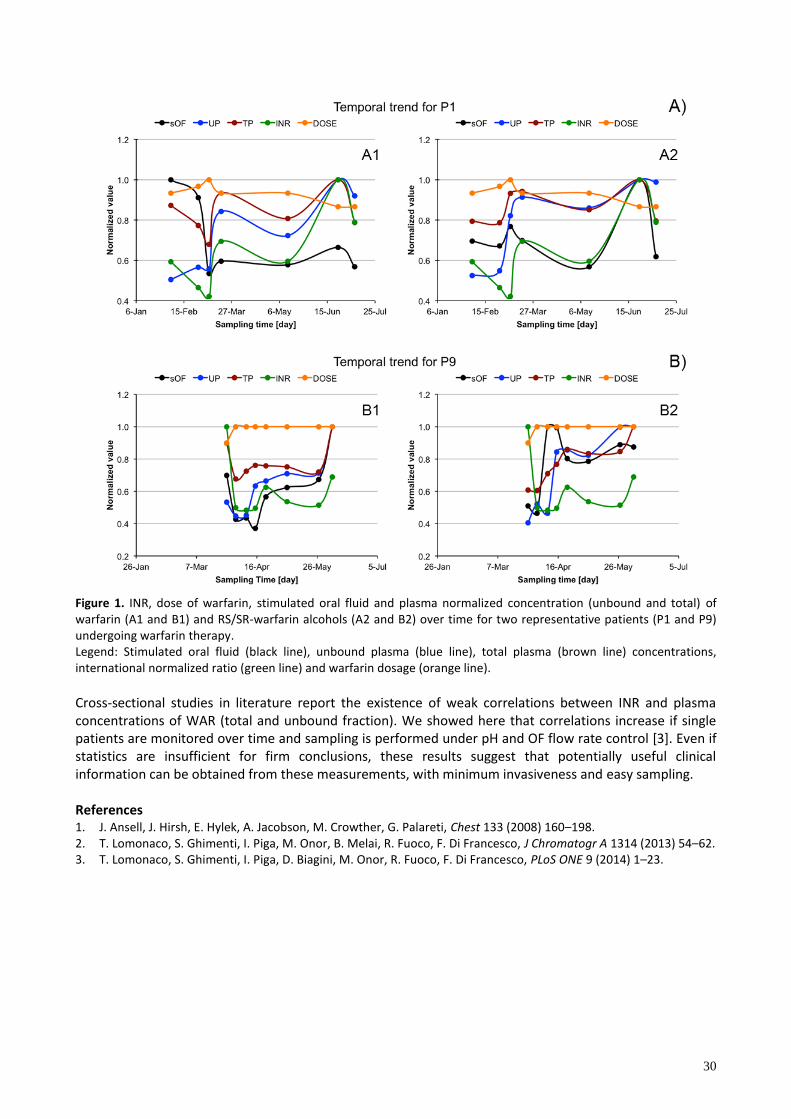
30
Figure 1. INR, dose of warfarin, stimulated oral fluid and plasma normalized concentration (unbound and total) of warfarin (A1 and B1) and RS/SR-warfarin alcohols (A2 and B2) over time for two representative patients (P1 and P9) undergoing warfarin therapy. Legend: Stimulated oral fluid (black line), unbound plasma (blue line), total plasma (brown line) concentrations, international normalized ratio (green line) and warfarin dosage (orange line).
Cross-sectional studies in literature report the existence of weak correlations between INR and plasma concentrations of WAR (total and unbound fraction). We showed here that correlations increase if single patients are monitored over time and sampling is performed under pH and OF flow rate control [3]. Even if statistics are insufficient for firm conclusions, these results suggest that potentially useful clinical information can be obtained from these measurements, with minimum invasiveness and easy sampling. References 1. J. Ansell, J. Hirsh, E. Hylek, A. Jacobson, M. Crowther, G. Palareti, Chest 133 (2008) 160–198. 2. T. Lomonaco, S. Ghimenti, I. Piga, M. Onor, B. Melai, R. Fuoco, F. Di Francesco, J Chromatogr A 1314 (2013) 54–62. 3. T. Lomonaco, S. Ghimenti, I. Piga, D. Biagini, M. Onor, R. Fuoco, F. Di Francesco, PLoS ONE 9 (2014) 1–23.

31
NEW BIOANALYTICAL APPROCH FOR EARLY DETECTION OF ß-THALASSEMIA COUPLING TGA AND CHEMOMETRICS
Roberta Risoluti1, Stefano Materazzi1, Giuseppina Gullifa1 Francesco Sorrentino2, Patrizia Caprari3
1 Dept. of Chemistry, "Sapienza" - University of Rome, P.le A.Moro 5 - 00185 Rome – Italy 2 UOS DH- Thalassemia, S. Eugenio Hospital, P.le dell'Umanesimo, 10 – 00144 Rome, Italy
3 Dept. of Hematology, Oncology and Molecular Medicine, Istituto Superiore di Sanità, Viale Regina Elena 299, 00161, Italy
β-Thalassemia is one of most common autosomal recessive disorders worldwide. β - Thalassemia is caused by the reduced (β+) or absent (β°) synthesis of the β globin chains of the hemoglobin tetramer.[1] Three clinical and hematological conditions of increasing severity are recognized, i.e. the β-thalassemia carrier state, thalassemia intermedia, and thalassemia major. The hematological features are microcytosis, hypochromia, and anemia.[2] For the first time, application of thermal analysis on whole blood samples from patients with β-thalassemia, without any pretreament, is proposed. Results show that TG and DTG curves of β-thalassemic patients are different from those of healthy individuals because of different amounts of water content and corpuscular fraction. The parallel hematological overview, determined by ADVIA 120 analyzer (Siemens), confirms significant decrease in Hb, Hct, MCH, and MCV values and increase in RBC counts and RDW values in thalassemic subjects as compared with healthy subjects. A chemometric approach based on Principal Components Analysis (PCA) has been exploited in order to enhance correlation among thermogravimetric profiles. Moreover, the implementation of a predictive classification model for β-thalassemia diagnosis has been also carried out by determining characteristic haematological features and comparing them with calculated weight losses. The promising preliminary achieved results allow to consider thermogravimetry followed by chemometrics as a fast, rapid and cost-effective diagnostic tool for early β-thalassemia detection. References 1. A. Cao, R. Galanello, R. Origa. Gene Reviews Seattle (WA);1993-2014, 2013 2. D.R. Higgs, J.D. Engel, G. Stamatoyannopoulos. Lancet. 2012, 28;379:373-83.
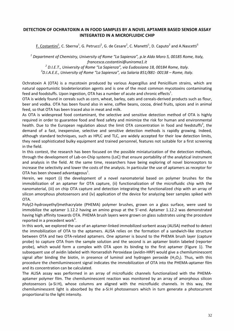
32
DETECTION OF OCHRATOXIN A IN FOOD SAMPLES BY A NOVEL APTAMER BASED SENSOR ASSAY INTEGRATED IN A MICROFLUIDIC CHIP
F. Costantini1, C. Sberna1, G. Petrucci2, G. de Cesare2, C. Manetti1, D. Caputo2 and A.Nascetti3
1 Department of Chemistry, University of Rome “La Sapienza”, p.le Aldo Moro 5, 00185 Rome, Italy, [email protected]
2 D.I.E.T., University of Rome “La Sapienza”, via Eudossiana 18, 00184 Rome, Italy. 3D.I.A.E.E., University of Rome “La Sapienza”, via Salaria 851/881- 00138 – Rome, Italy.
Ochratoxin A (OTA) is a mycotoxin produced by various Aspergillus and Penicillium strains, which are natural opportunistic biodeterioration agents and is one of the most common mycotoxins contaminating feed and foodstuffs. Upon ingestion, OTA has a number of acute and chronic effects1. OTA is widely found in cereals such as corn, wheat, barley, oats and cereals-derived products such as flour, beer and vodka. OTA has been found also in wine, coffee beans, cocoa, dried fruits, spices and in animal feed, so that OTA has been traced also in meat and milk. As OTA is widespread food contaminant, the selective and sensitive detection method of OTA is highly required in order to guarantee food and feed safety and minimize the risk for human and environmental health. Due to the European regulation about the limit OTA concentration in food and feedstuffs2, the demand of a fast, inexpensive, selective and sensitive detection methods is rapidly growing. Indeed, although standard techniques, such as HPLC and TLC, are widely accepted for their low detection limits, they need sophisticated bulky equipment and trained personnel, features not suitable for a first screening in the field. In this contest, the research has been focused on the possible miniaturization of the detection methods, through the development of Lab-on-Chip systems (LoC) that ensure portability of the analytical instrument and analysis in the field. At the same time, researchers have being exploring of novel bioreceptors to increase the selectivity and lower the costs of the analysis. In particular the use of aptamers as receptor for OTA has been showed advantageous1. Herein, we report (i) the development of a novel nanomaterial based on polymer brushes for the immobilization of an aptamer for OTA capture, (ii) functionalization of the microfluidic chip with the nanomaterial, (iii) on chip OTA capture and detection integrating the functionalized chip with an array of silicon amorphous photosensors and (iv) application of the device for analyzing beer samples spiked with OTA. Poly(2-hydroxyethyl)methacrylate (PHEMA) polymer brushes, grown on a glass surface, were used to immobilize the aptamer 1.12.2 having an amino group at the 5'-end. Aptamer 1.12.2 was demonstrated having high affinity towards OTA. PHEMA brush layers were grown on glass substrates using the procedure reported in a precedent work3. In this work, we explored the use of an aptamer-linked immobilized sorbent assay (ALISA) method to detect the immobilization of OTA to the aptamers. ALISA relies on the formation of a sandwich-like structure between OTA and two OTA-related aptamers. One aptamer is bound to the PHEMA brush layer (capture probe) to capture OTA from the sample solution and the second is an aptamer biotin labeled (reporter probe), which would form a complex with OTA upon its binding to the first aptamer (Figure 1). The subsequent use of avidin labeled with Horseradish Peroxidase (avidin-HRP) would give a chemiluminescent signal after binding the biotin, in presence of luminol and hydrogen peroxide (H2O2). Thus, with this procedure the chemiluminescent signal indicates the immobilization of OTA into the PHEMA-aptamer film and its concentration can be calculated. The ALISA assay was performed in an array of microfluidic channels functionalized with the PHEMA-aptamer polymer film. The chemiluminescent reaction was monitored by an array of amorphous silicon photosensors (a-Si:H), whose columns are aligned with the microfluidic channels. In this way, the chemiluminescent light is absorbed by the a-Si:H photosensors which in turn generate a photocurrent proportional to the light intensity.
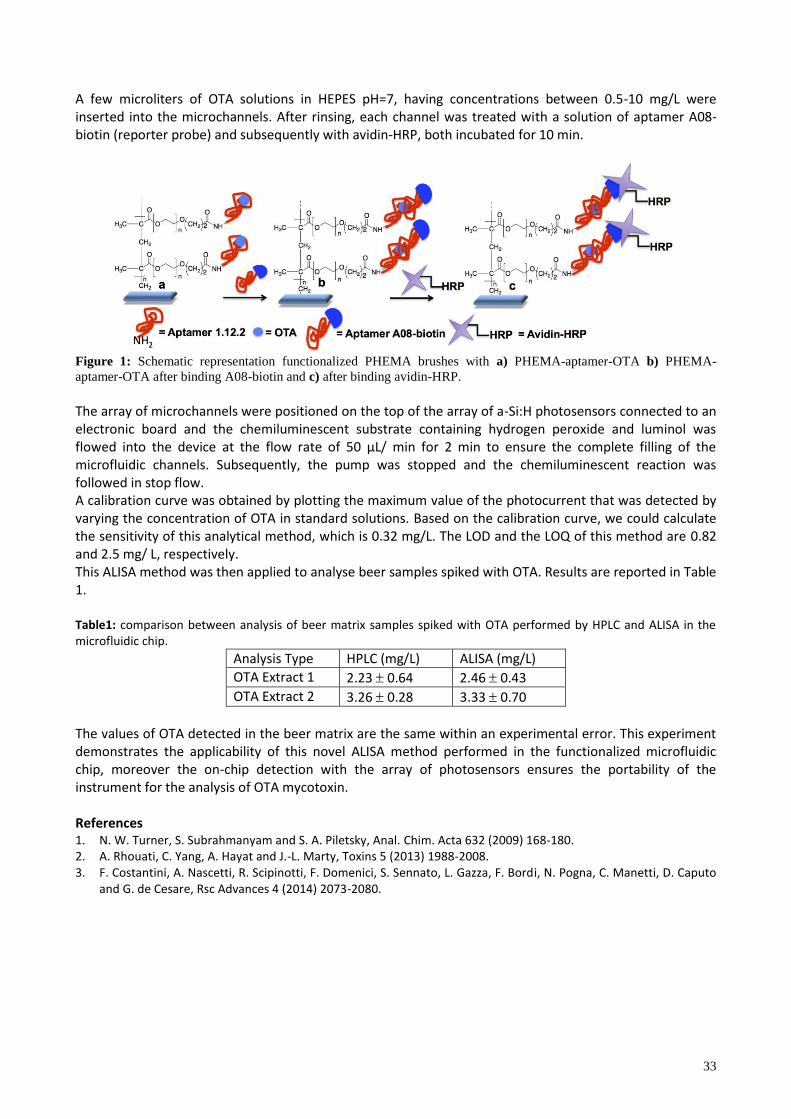
33
A few microliters of OTA solutions in HEPES pH=7, having concentrations between 0.5-10 mg/L were inserted into the microchannels. After rinsing, each channel was treated with a solution of aptamer A08-biotin (reporter probe) and subsequently with avidin-HRP, both incubated for 10 min.
Figure 1: Schematic representation functionalized PHEMA brushes with a) PHEMA-aptamer-OTA b) PHEMA-
aptamer-OTA after binding A08-biotin and c) after binding avidin-HRP. The array of microchannels were positioned on the top of the array of a-Si:H photosensors connected to an electronic board and the chemiluminescent substrate containing hydrogen peroxide and luminol was flowed into the device at the f ow rate of 50 μL/ m n for 2 m n to en ure the comp ete f ng of the microfluidic channels. Subsequently, the pump was stopped and the chemiluminescent reaction was followed in stop flow. A calibration curve was obtained by plotting the maximum value of the photocurrent that was detected by varying the concentration of OTA in standard solutions. Based on the calibration curve, we could calculate the sensitivity of this analytical method, which is 0.32 mg/L. The LOD and the LOQ of this method are 0.82 and 2.5 mg/ L, respectively. This ALISA method was then applied to analyse beer samples spiked with OTA. Results are reported in Table 1. Table1: comparison between analysis of beer matrix samples spiked with OTA performed by HPLC and ALISA in the microfluidic chip.
Analysis Type HPLC (mg/L) ALISA (mg/L)
OTA Extract 1 2.23 0.64 2.46 0.43
OTA Extract 2 3.26 0.28 3.33 0.70
The values of OTA detected in the beer matrix are the same within an experimental error. This experiment demonstrates the applicability of this novel ALISA method performed in the functionalized microfluidic chip, moreover the on-chip detection with the array of photosensors ensures the portability of the instrument for the analysis of OTA mycotoxin. References 1. N. W. Turner, S. Subrahmanyam and S. A. Piletsky, Anal. Chim. Acta 632 (2009) 168-180. 2. A. Rhouati, C. Yang, A. Hayat and J.-L. Marty, Toxins 5 (2013) 1988-2008. 3. F. Costantini, A. Nascetti, R. Scipinotti, F. Domenici, S. Sennato, L. Gazza, F. Bordi, N. Pogna, C. Manetti, D. Caputo
and G. de Cesare, Rsc Advances 4 (2014) 2073-2080.
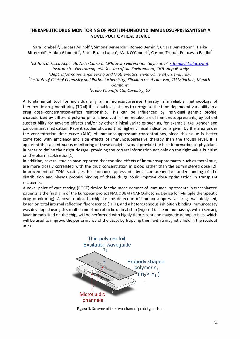
34
THERAPEUTIC DRUG MONITORING OF PROTEIN-UNBOUND IMMUNOSUPPRESSANTS BY A NOVEL POCT OPTICAL DEVICE
Sara Tombelli1, Barbara Adinolfi1, Simone Berneschi1, Romeo Bernini2, Chiara Berrettoni1,3, Heike
Bittersohl4, Ambra Giannetti1, Peter Bruno Luppa4, Mark O’Conne 5, Cosimo Trono1, Francesco Baldini1
1Istituto di Fisica Applicata Nello Carrara, CNR, Sesto Fiorentino, Italy, e-mail: [email protected]; 2Institute for Electromagnetic Sensing of the Environment, CNR, Napoli, Italy;
2Dept. Information Engineering and Mathematics, Siena University, Siena, Italy; 3Institute of Clinical Chemistry and Pathobiochemistry, Klinikum rechts der Isar, TU München, Munich,
Germany; 4Probe Scientific Ltd, Coventry, UK
A fundamental tool for individualizing an immunosuppressive therapy is a reliable methodology of therapeutic drug monitoring (TDM) that enables clinicians to recognize the time-dependent variability in a drug dose–concentration-effect relationship. This can be influenced by individual genetic profile, characterized by different polymorphisms involved in the metabolism of immunosuppressants, by patient susceptibility for adverse effects and/or by other clinical variables such as, for example age, gender and concomitant medication. Recent studies showed that higher clinical indication is given by the area under the concentration time curve (AUC) of immunosuppressant concentrations, since this value is better correlated with efficiency and side effects of immunosuppressive therapy than the trough level. It is apparent that a continuous monitoring of these analytes would provide the best information to physicians in order to define their right dosage, providing the correct information not only on the right value but also on the pharmacokinetics [1]. In addition, several studies have reported that the side effects of immunosuppressants, such as tacrolimus, are more closely correlated with the drug concentration in blood rather than the administered dose [2]. Improvement of TDM strategies for immunosuppressants by a comprehensive understanding of the distribution and plasma protein binding of these drugs could improve dose optimization in transplant recipients. A novel point-of-care-testing (POCT) device for the measurement of immunosuppressants in transplanted patients is the final aim of the European project NANODEM (NANOphotonic Device for Multiple therapeutic drug monitoring). A novel optical biochip for the detection of immunosuppressive drugs was designed, based on total internal reflection fluorescence (TIRF), and a heterogeneous inhibition binding immunoassay was developed using this multichannel microfluidic optical chip (Figure 1). The immunoassay, with a sensing layer immobilized on the chip, will be performed with highly fluorescent and magnetic nanoparticles, which will be used to improve the performance of the assay by trapping them with a magnetic field in the readout area.
Figura 1. Scheme of the two-channel prototype chip.
35
The multichannel chip will be integrated in a novel optical device where the biological sample will flow through a microfluidic section where it will be mixed with the reagents necessary to perform the bioassay and will be then pumped through the chip where the determination of the different immunosuppressants will take place for 48 hours. The sample will be drawn from blood by means of microdialysis, in order to collect the protein-unbound fraction of the different administrated immunosuppressants, mainly tacrolimus, mycophenolic acid and cyclosporine A. References 1. R.G. Morris, G.R. Russ, M.J. Cervelli, R. Juneja, S.P. McDonald, T.H. Mathew, Ther. Drug. Monit. 24 (2002) 479–
486. 2. H. Zahir, G. McCaughan, M. Gleeson, R. A. Nand, A. J. McLachlan, British Journal of Clinical Pharmacology 57
(2003) 298-309.
Acknowledgements This research study was supported by the European Community within the framework of the project Nanodem -NANOphotonic Device for Multiple therapeutic drug monitoring (FP7-ICT-2011 - 8318372).

36
ACETAMIPRID DETECTION BY DNA TECHNOLOGY SENSING FOR ENVIRONMENTAL ANALYSIS
R.Rapini, G.Marrazza Department of Chemistry “Ugo Schiff”, University of Firenze, via della Lastruccia, 3 – 50019 Sesto Fiorentino
(FI)
The use of pesticides is an essential necessity in modern agriculture and their detection remains one of the most important targets for environmental analytical chemistry. Most diffused methods for pesticide detection are based on laboratory techniques, like high pressure liquid chromatography, gas chromatography, mass spectrometry. These techniques are characterised by satisfactory analytical performances, but their use require high trained technicians, they are time consuming and have high costs: those reasons make them impractical for on field screening analysis and constant environment monitoring. Over the last few years, the use of DNA based biosensors has oftentimes been reported as a promising alternative to traditional methods for pesticide analysis and, in general, for environmental monitoring, thanks to the possibility to combine low costs for the realization of the artificial receptors with efficient analytical performances. DNA-based sensors can be used for the rapid detections of different kinds of pollutants and pesticides for environmental analysis. To our knowledge, few aptamers for the detection of pesticides have been selected and recently a DNA aptamer specific for acetamiprid has been described. In this work, an electrochemical DNA array for acetamiprid, a diffused neonicotinoid insecticide, is presented. The DNA array is based on a dual signal amplified strategy by employing a polyaniline film and gold nanoparticles as sensor platform and an enzyme-linked label for sensitive detection. Firstly, polyaniline film and gold nanoparticles were progressively grown on a graphite screen-printed electrode surface via electro-polymerization and electrochemical deposition, respectively. The polyaniline-gold modified surfaces were then modified with a mixed monolayer of a thiol-tethered DNA aptamer and a spacer thiol. The aptasensor was able to capture the pesticide from the sample solutions. An enzyme-amplified detection scheme, based on the coupling of a streptavidin-alkaline phosphatase conjugate and biotinylated secondary aptamer was then applied. The electro-active enzymatic product was detected by means of differential pulse voltammetry. The sensor coupled the strong advantages of the enzymatic amplification with the electrochemical properties of polyaniline and gold nanoparticles. Various experimental parameters of the realized DNA-based nanostructured sensor were studied and optimized using optical and electrochemical techniques. A calibration curve between 0-1000 nM acetamiprid concentration range was obtained.

37
COMUNICAZIONI POSTER
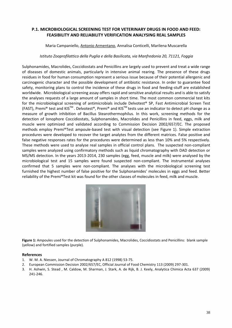
38
P.1. MICROBIOLOGICAL SCREENING TEST FOR VETERINARY DRUGS IN FOOD AND FEED: FEASIBILITY AND RELIABILITY VERIFICATION ANALYSING REAL SAMPLES
Maria Campaniello, Antonio Armentano, Annalisa Conticelli, Marilena Muscarella
Istituto Zooprofilattico della Puglia e della Basilicata, via Manfredonia 20, 71121, Foggia
Sulphonamides, Macrolides, Coccidiostats and Penicillins are largely used to prevent and treat a wide range of diseases of domestic animals, particularly in intensive animal rearing. The presence of these drugs residues in food for human consumption represent a serious issue because of their potential allergenic and carcinogenic character and the possible development of antibiotic resistance. In order to guarantee food safety, monitoring plans to control the incidence of these drugs in food and feeding-stuff are established worldwide. Microbiological screening assay offers rapid and sensitive analytical results and is able to satisfy the analyses requests of a large amount of samples in short time. The most common commercial test kits for the microbiological screening of antimicrobials include Delvotest® SP, Fast Antimicrobial Screen Test (FAST), Premi® test and KISTM . Delvotest®, Premi® and KISTM tests use an indicator to detect pH change as a measure of growth inhibition of Bacillus Stearothermophilus. In this work, screening methods for the detection of Ionophore Coccidiostats, Sulphonamides, Macrolides and Penicillins in feed, eggs, milk and muscle were optimized and validated according to Commission Decision 2002/657/EC. The proposed methods employ Premi®Test ampoule-based test with visual detection (see Figure 1). Simple extraction procedures were developed to recover the target analytes from the different matrices. False positive and false negative responses rates for the procedures were determined as less than 10% and 5% respectively. These methods were used to analyse real samples in official control plans. The suspected non-compliant samples were analyzed using confirmatory methods such as liquid chromatography with DAD detection or MS/MS detection. In the years 2013-2014, 230 samples (egg, feed, muscle and milk) were analysed by the microbiological test and 15 samples were found suspected non-compliant. The instrumental analyses confirmed that 5 samples were non-compliant. The analyses with the microbiological screening test furnished the highest number of fa e po t ve for the Su phonam de ’ mo ecu e n egg and feed. etter reliability of the Premi®Test kit was found for the other classes of molecules in feed, milk and muscle.
Figure 1: Ampoules used for the detection of Sulphonamides, Macrolides, Coccidiostats and Penicillins: blank sample (yellow) and fortified samples (purple).
References 1. W. M. A. Niessen, Journal of Chromatography A 812 (1998) 53-75. 2. European Commission Decision 2002/657/EC, Official Journal of Food Chemistry 113 (2009) 297-301. 3. H. Ashwin, S. Stead , M. Caldow, M. Sharman, J. Stark, A. de Rijk, B. J. Keely, Analytica Chimica Acta 637 (2009)
241-246.
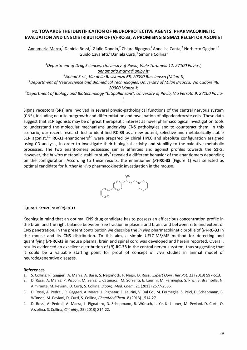
39
P2. TOWARDS THE IDENTIFICATION OF NEUROPROTECTIVE AGENTS. PHARMACOKINETIC EVALUATION AND CNS DISTRIBUTION OF (R)-RC-33, A PROMISING SIGMA1 RECEPTOR AGONIST
Annamaria Marra,1 Daniela Rossi,1 Giulio Dondio,2 Chiara Bigogno,2 Annalisa Canta,3 Norberto Oggioni,3
Guido Cavaletti,3 Daniela Curti,4 Simona Collina1
1Department of Drug Sciences, University of Pavia, Viale Taramelli 12, 27100 Pavia-I, [email protected];
2Aphad S.r.l., Via della Resistenza 65, 20090 Buccinasco (Milan-I); 3Department of Neuroscience and Biomedical Technologies, University of Milan Bicocca, Via Cadore 48,
20900 Monza-I; 4Department of Biology and Biotechnology “L. Spallanzani”, University of Pavia, Via Ferrata 9, 27100 Pavia-
I. Sigma receptors (SRs) are involved in several physio-pathological functions of the central nervous system (CNS), including neurite outgrowth and differentiation and myelination of oligodendrocyte cells. These data suggest that S1R agonists may be of great therapeutic interest as novel pharmacological investigation tools to understand the molecular mechanisms underlying CNS pathologies and to counteract them. In this scenario, our recent research led to identified RC-33 as a new potent, selective and metabolically stable S1R agonist.1,2 RC-33 enantiomers3,4 were prepared by chiral HPLC and absolute configuration assigned using CD analysis, in order to investigate their biological activity and stability to the oxidative metabolic processes. The two enantiomers possessed similar affinities and agonist profiles towards the S1Rs. However, the in vitro metabolic stability study3 revealed a different behavior of the enantiomers depending on the configuration. According to these results, the enantiomer (R)-RC-33 (Figure 1) was selected as optimal candidate for further in vivo pharmacokinetic investigation in the mouse.
CH3
N
Figure 1. Structure of (R)-RC33
Keeping in mind that an optimal CNS drug candidate has to possess an efficacious concentration profile in the brain and the right balance between free fraction in plasma and brain, and between rate and extent of CNS penetration, in the present contribution we describe the in vivo pharmacokinetic profile of (R)-RC-33 in the mouse and its CNS distribution. To this aim, a simple UFLC-MS/MS method for detecting and quantifying (R)-RC-33 in mouse plasma, brain and spinal cord was developed and herein reported. Overall, results evidenced an excellent distribution of (R)-RC-33 in the central nervous system, thus suggesting that it could be a valuable starting point for proof of concept in vivo studies in animal model of neurodegenerative diseases.
References 1. S. Collina, R. Gaggeri, A. Marra, A. Bassi, S. Negrinotti, F. Negri, D. Rossi, Expert Opin Ther Pat. 23 (2013) 597-613. 2. D. Rossi, A. Marra, P. Picconi, M. Serra, L. Catenacci, M. Sorrenti, E. Laurini, M. Fermeglia, S. Pricl, S. Brambilla, N.
Almirante, M. Peviani, D. Curti, S. Collina, Bioorg. Med. Chem. 21 (2013) 2577-2586.
3. D. Rossi, A. Pedrali, R. Gaggeri, A. Marra, L. Pignatar, E. Laurini, V. Dal Col, M. Fermeglia, S. Pricl, D. Schepmann, B.
Wünsch, M. Peviani, D. Curti, S. Collina, ChemMedChem. 8 (2013) 1514-27.
4. D. Rossi, A. Pedrali, A. Marra, L. Pignataro, D. Schepmann, B. Wünsch, L. Ye, K. Leuner, M. Peviani, D. Curti, O.
Azzolina, S. Collina, Chirality, 25 (2013) 814-22.

40
P3. DEVELOPMENT OF AN ELIME ASSAY AND A REAL-TIME PCR FOR SALMONELLA ENTERICA DETECTION: APPLICATION IN IRRIGATION WATERS
Laura Fabiani1, Giulia Volpe1, Elisabetta Delibato2, Eleonora Pucci2, Silvia Piermarini1, Annamar a D’Ange o4, Federico Capuano3, Giuseppe Palleschi1
1 Dipartimento di Scienze e Tecnologie Chimiche, Università degli Studi di Roma Tor Vergata, via della
Ricerca Scientifica 1 e-mail [email protected] 2Dipartimento di Sanità Pubblica Veterinaria e Sicurezza Alimentare, Istituto Superiore di Sanità, viale
Regina Elena 299, Roma 3Dipartimento Ispezione Alimenti, Istituto Zooprofilattico Sperimentale del Mezzogiorno, via della Salute 2,
Portici (NA) 4Dipartimento di Ambiente e Connessa Prevenzione Primaria, Istituto Superiore di Sanità, Roma
Traditionally foodborne illness were mainly caused by food of animal origin; however, recently outbreaks and infections are associated to the consumption of fruit and vegetables contaminated with pathogenic microorganisms. A potential source of contamination is the use of contaminated water to irrigate crops [1]. The European Rapid Alert System for Food and Feed (RASFF) has repeatedly drawn the attention of the member countries about the presence of Salmonella Napoli and Thompson in fresh vegetables produced in Italy [2-3]. The European legislation, dealing with the Microbiological Criteria, has established that this pathogen must be absent in a defined amount of a given food product. Despite the fact that at present no guidelines or regulations exist at the level of the European Union for the limit of Salmonella in irrigation waters, food business operators have to ensure the absence of this pathogen in a suitable water volume, to protect the health and safety of consumers. Although the standard cultural method for Salmonella detection (EN/ISO 6579) is very sensitive, and inexpensive, requires three to four days to obtain a negative result and up to five days to get a confirmed positive result. The aim of this work is the development and the evaluation of two different techniques, an ELIME (Enzyme-Linked-Immuno-Magnetic-Electrochemical) assay and a Real-Time PCR, for a more rapid analysis to assess the Salmonella presence in irrigation waters. In particular, the ELIME assay for Salmonella enterica uses magnetic beads (MBs), as a support of a sandwich immunological chain, coupled with a strip of eight-magnetized screen-printed electrodes localized at the bottom of eight wells (8-well/SPE strip). The product of the enzyme reaction is rapidly measured, at an applied potential of -100 mV, using a portable instrumentation. Two different kinds of MBs were tested: Dynabeads anti-salmonella (ready to use) and Dynabeads Pan Mouse IgG pre-coated with IgG anti-mouse and coated by us with monoclonal antibodies (MAb) anti-salmonella. With the final goal to develop a simple and rapid method able to detect this pathogen, we decided to block the surface of the MBs in a preliminary phase (to store them at 4 °C until use) and to merge the two conventional steps of incubation (the first with salmonella and the second one with PAb-HRP), interspersed by washings, in a single step. Great attention was focused on the selectivity test being salmonella, and in general pathogens, only a small fraction of a large population of non-target (NT) organisms present in real matrices, able to adhere to various surfaces, including MBs, giving false positive results. For this purpose different blocking agents were evaluated by analyzing various NT organisms. The best results in terms of sensitivity towards S. Napoli and S. Thompson and selectivity were obtained using Pan Mouse IgG MBs, coated with MAb. After that, an inclusivity test was carried out and the results show the ability of our method to detect different salmonella serovars, most commonly isolated from environmental sources. In the optimized conditions, the limit of detection (LOD) was found to be 104 and 105 CFU/ml for S. Napoli and S. Thompson, respectively. Sensitivity (IC50) was 105 CFU/mL for S. Napoli
and 106 for S. Thompson. The Real-time PCR employs primers and a specific LNA fluorescent probe able to amplify a region of the ttrRSBCA gene, conserved in all Salmonella serotypes, and an internal amplification control (IAC) to check false negative results. The standard curve obtained by plotting the mean Ct vs. log concentrations of S. Napoli, showed a good linearity from 10 to 108 CFU/mL. Experiments were conducted analysing 70 salmonella and 38 non-salmonella strains. All salmonella serotypes tested were ttr-positive and all other bacteria yielded only an amplification plot for the IAC.
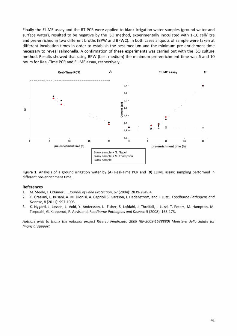
41
Finally the ELIME assay and the RT PCR were applied to blank irrigation water samples (ground water and surface water), resulted to be negative by the ISO method, experimentally inoculated with 1-10 cell/litre and pre-enriched in two different broths (BPW and BPWC). In both cases aliquots of sample were taken at different incubation times in order to establish the best medium and the minimum pre-enrichment time necessary to reveal salmonella. A confirmation of these experiments was carried out with the ISO culture method. Results showed that using BPW (best medium) the minimum pre-enrichment time was 6 and 10 hours for Real-Time PCR and ELIME assay, respectively.
Real-Time PCR
pre-enrichment time (h)
0 5 10 15 20
CT
A
ELIME assay
pre-enrichment time (h)
0 5 10 15 20
Cu
rre
nt
( A
)
0,0
0,2
0,4
0,6
0,8
1,0
1,2
1,4
B
Blank sample + S. Napoli
Blank sample + S. Thompson
Blank sample
Figure 1. Analysis of a ground irrigation water by (A) Real-Time PCR and (B) ELIME assay: sampling performed in different pre-enrichment time.
References 1. M. Steele, J. Odumeru, , Journal of Food Protection, 67 (2004): 2839-2849;4. 2. C. Graziani, L. Busani, A. M. Dionisi, A. Caprioli,S. Ivarsson, I. Hedenstrom, and I. Luzzi, Foodborne Pathogens and
Disease, 8 (2011): 997-1003. 3. K. Nygard, J. Lassen, L. Vold, Y. Andersson, I. Fisher, S. Lofdahl, J. Threlfall, I. Luzzi, T. Peters, M. Hampton, M.
Torpdahl, G. Kapperud, P. Aavisland, Foodborne Pathogens and Disease 5 (2008): 165-173. Authors wish to thank the national project Ricerca Finalizzata 2009 (RF-2009-1538880) Ministero della Salute for financial support.
42
P4. SCREENING OF MICROCYSTINS AND OKADAIC ACID IN DRINKING, FRESH AND SEA WATER SAMPLES USING AN OPTIMIZED COLORIMETRIC PHOSPHATASE INHIBITION ASSAY
Konstantinos Petropoulos, Giulia Volpe, Laura Micheli, Danila Moscone, Giuseppe Palleschi
Dipartimento di Scienze e Tecnologie Chimiche, Università di Roma Tor Vergata, Via della Ricerca Scientifica
1 - 00133 Roma.
e-mail: [email protected] Microcystins (MCs) are a class of cyclic natural peptide hepatotoxins produced by cyanobacteria such as Microcystis, Oscillatoria and Anabaena usually found in lakes, water reservoirs and recreational facilities.
MCs are also a drinking water public health issue with a provisional drinking water guideline of 1 g/l for microcystin-LR (WHO, 1998). On the basis of a widely conservative approach towards the protection of the
human ’ hea th the va ue of 1 g/l would be referred to the sum of the toxin concentrations present in the sample, considered as equivalents of MC-LR [1]. Okadaic acid (OA) is a lipophilic marine toxin produced by Dinophysis and Prorocentrum, and it is responsible for causing diarrheic shellfish poisoning (DSP) to humans after ingestion of contaminated shellfish. The contamination of bivalves has become a serious economic concern for the shellfish industry and the European Food Safety Authority, in order to protect consumer health, has established the maximum permitted level of OA as 45 μg/kg of mussels [2]. The mechanism of action of these toxins is based on the inhibition of protein phosphatase type 2A (PP2A) by the toxins. The degree of inhibition of the PP2A enzyme can be used as a measure of toxin concentration in aqueous solution sample. In this work we propose a colorimetric assay in which the activity of protein phospharase-2A is determined by measuring the rate of color production from the release of yellow p-nitrophenol using p-nitrophenyl phosphate as the substrate. In the presence of MCs or OA enzyme inhibition occurs and consequently the rate of color production decreases proportionally to the concentration of the toxin. Changes in absorbance at 405 nm are measured by a microtiter plate reader after incubation for 90 minutes in an appropriate microtiter stirrer at 30oC. In order to develop a very sensitive method for the screening test we focused our attention on the volume
of the standard/sample solution to add in the well: a large volume of 125L per well was chosen in a total
volume of 300L. In these conditions two typical sigmoidal inhibition curves were obtained for both toxins:
for MC-LR a detection limit of 0.08 ± 0.01 g/L, a working range between 0.20 and 0.80 g/L and an IC50
value of 0.33 ± 0.06 g/L were calculated, while for OA the detection limit, the working range and the IC50
value resulted to be 0.35 ± 0.03 g/L, 1.90 -15 g/L and 5.00 ± 0.08 g/L, respectively. Taking in consideration the results, mentioned above, different water samples were analyzed and evaluated, without a preconcentration step, in terms of recovery for both toxins. An average recovery of 98% and 99% were calculated for MC-LR and OA, respectively. Riferimenti 1. World Health Organization, Chemical fact sheets, 1, (2008), 407-408. 2. European Food Safety Authority, the EFSA Journal, 589, (2008), 1-62.
The authors wish to thank the SMS project (Sensing toxicants in Marine waters makes Sense using Biosensors) GA n. 613844 for financial support.
43
P5. SMARTPHONE-BASED COLORIMETRIC ASSAY FOR CA125 CANCER BIOMARKER DETECTION
O. Hosu1,2, A. Ravalli2, C. Cristea1, R. Săndu e cu1, G. Marrazza2
1 Department of Analytical Chemistry, Faculty of Pharmacy, University of Medicine and Pharmacy, Iuliu Hatieganu”, Pasteur 4, Cluj-Napoca, Romania
2 Department of Chemistry ”Ugo Schiff”, University of Florence, Via della Lastruccia 3, 50019, Sesto Fiorentino, Florence, Italy
There is an urgent need for cost-effective point-of-care (POC) instruments with homogenous technical
requirements as well as more flexible devices for biomarker diagnostics in clinical settings. Due to the
recent advances in smartphone features (such as capability, processing power, hardware and software), it
becomes a promising tool for mobile diagnostic and bio-analytical POC tools. Colorimetric detection is an
ideal method for miniaturization and POC biosensor development because of its inherent sensitivity and
simplicity.
In this work, we have developed a simple and accurate affisensor based on a colorimetric immunoassay
method coupled to a smartphone in order to detect quantitatively the ovarian cancer antigen 125 (CA125).
The affisensor is based on a sandwich immunoassay in which the primary antibody was immobilized by
spotting the antibody solution on nitrocellulose membrane. Subsequently, the spots were incubated with
CA125 antigen followed by affinity reaction with a secondary antibody conjugated to gold nanoparticles
(AuNPs). The silver enhancement reaction was introduced to magnify the signal detection. The
experimental data show that this reaction can be observed by the naked eye. The formation of gold-silver
nanoparticles results in a different grey colour, depending on CA125 concentration. The smartphone
camera was used as colour detector, for image acquisition and data handling via a specific application.
The parameters involved in each step of the affisensor design were optimized. The performance of the
immunoassay in terms of sensitivity, reproducibility and selectivity was studied.
Under optimal conditions, a linear response was obtained in the range of 60 – 1000 U/mL, which is also the
important range from clinical point of view. The method is simple, fast, and could be performed without
requiring highly skilled operating personnel and expensive instrumentation allowing point-of-care analysis
with reductions in cost and response time.
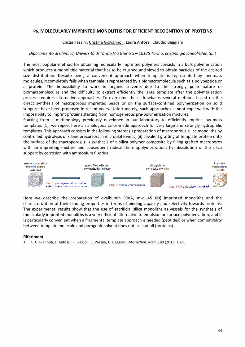
44
P6. MOLECULARLY IMPRINTED MONOLITHS FOR EFFICIENT RECOGNITION OF PROTEINS
Cinzia Passini, Cristina Giovannoli, Laura Anfossi, Claudio Baggiani
Dipartimento di Chimica, Università di Torino,Via Giuria 5 – 10125 Torino, [email protected]
The most popular method for obtaining molecularly imprinted polymers consists in a bulk polymerisation which produces a monolithic material that has to be crushed and sieved to obtain particles of the desired size distribution. Despite being a convenient approach when template is represented by low-mass molecules, it completely fails when tempale is represented by a biomacromolecule such as a polypeptide or a protein. The impossibility to work in organic solvents due to the strongly polar nature of biomacromolecules and the difficulty to extract efficiently the large template after the polymerization process requires alternative approaches. To overcome these drawbacks several methods based on the direct synthesis of macroporous imprinted beads or on the surface-confined polymerization on solid supports have been proposed in recent years. Unfortunately, such approaches cannot cope well with the impossibility to imprint proteins starting from homogeneous pre-polymerization mixtures. Starting from a methodology previously developed in our laboratory to efficiently imprint low-mass templates [1], we report here an analogous tailor-made approach for very large and strongly hydrophilic templates. This approach consists in the following steps: (i) preparation of macroporous silica monoliths by controlled hydrolysis of silane precursors in microplate wells; (ii) covalent grafting of template protein onto the surface of the macropores; (iii) synthesis of a silica-polymer composite by filling grafted macropores with an imprinting mixture and subsequent radical thermopolymerization; (iv) dissolution of the silica support by corrosion with ammonium fluoride.
Here we describe the preparation of ovalbumin (OVA, mw. 45 KD) imprinted monoliths and the characterization of their binding properties in terms of binding capacity and selectivity towards proteins. The experimental results show that the use of sacrificial silica monoliths as vessels for the synthesis of molecularly imprinted monoliths is a very efficient alternative to emulsion or surface polymerization, and it is particularly convenient when a fragmental template approach is needed (peptides) or when compatibility between template molecule and porogenic solvent does not exist at all (proteins). Riferimenti 1. C. Giovannoli, L. Anfossi, F. Biagioli, C. Passini, C. Baggiani, Microchim. Acta, 180 (2013) 1371
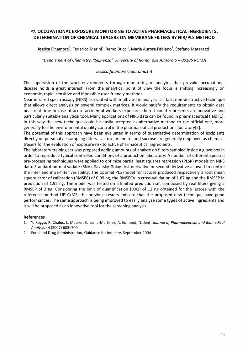
45
P7. OCCUPATIONAL EXPOSURE MONITORING TO ACTIVE PHARMACEUTICAL INGREDIENTS: DETERMINATION OF CHEMICAL TRACERS ON MEMBRANE FILTERS BY NIR/PLS METHOD
Jessica Finamore1, Federico Marini1, Remo Bucci1, Maria Aurora Fabiano1, Stefano Materazzi1
1Department of Chemistry, “Sapienza” University of Roma, p.le A.Moro 5 – 00185 ROMA
The supervision of the work environments through monitoring of analytes that provoke occupational disease holds a great interest. From the analytical point of view the focus is shifting increasingly on economic, rapid, sensitive and if possible user-friendly methods. Near infrared spectroscopy (NIRS) associated with multivariate analysis is a fast, non-destructive technique that allows direct analysis on several complex matrices. It would satisfy the requirements to obtain data near real time in case of acute accidental workers exposure, then it could represents an innovative and particularly suitable analytical tool. Many applications of NIRS data can be found in pharmaceutical field [1]. In this way the new technique could be easily accepted as alternative method to the official one, more generally for the environmental quality control in the pharmaceutical production laboratory[2]. The potential of this approach have been evaluated in terms of quantitative determination of excipients directly on personal air sampling filters. Lactose, mannitol and sucrose are generally employed as chemical tracers for the evaluation of exposure risk to active pharmaceutical ingredients. The laboratory training set was prepared adding amounts of analyte on filters sampled inside a glove box in order to reproduce typical controlled conditions of a production laboratory. A number of different spectral pre-processing techniques were applied to optimize partial least squares regression (PLSR) models on NIRS data. Standard normal variate (SNV), Savitsky-Golay first derivative or second derivative allowed to control the inter and intra-filter variability. The optimal PLS model for lactose produced respectively a root mean square error of calibration (RMSEC) of 0.99 ng, the RMSECV in cross-validation of 1.67 ng and the RMSEP in prediction of 1.92 ng. The model was tested on a limited prediction set composed by real filters giving a RMSEP of 2 ng. Considering the limit of quantification (LOQ) of 12 ng obtained for the lactose with the reference method UPLC/MS, the previous results indicate that the proposed new technique have good performances. The same approach is being improved to easily analyze some types of active ingredients and it will be proposed as an innovative tool for the screening analysis. References 1. Y. Roggo, P. Chalus, L. Maurer, C. Lema-Martinez, A. Edmond, N. Jent, Journal of Pharmaceutical and Biomedical
Analysis 44 (2007) 683–700 2. Food and Drug Administration, Guidance for Industry, September 2004

46
P8. SIMULTANEOUS QUANTIFICATION OF INTACT GLUCOSINOLATES AND ISOTHIOCYANATES BY HPLC-ES-MS/MS IN BRASSICACEAE SEEDS AND FUNCTIONAL FOODS
P. Franco1, S. Spinozzi1, E.Pagnotta2, L. Lazzeri2, L.Ugolini2, C. Camborata1, A.Roda1
1Department of Chemistry G. Ciamician, University of Bologna, Via Selmi 2, 40123- Bologna (Italy)
2 Council for Agricultural Research and Economic Analysis, Research Centre for Industrial Crops (CRA-CIN),
Via di Corticella 133, 40128 – Bologna (Italy) Glucosinolates (GLS) are natural polar molecules occurring in plant of the Brassica genus (broccoli, rocket a ad, cabbage and mu tard) [1]. They are c a f ed a (Z) β-thioglucosides N-hydroxysulfate characterized by a atera cha n (R) and a β-D-glucopyranosil moiety linked to the S-atom. During mechanic damage of the plant tissue, for example during chewing, Glucosinolates are hydrolyzed by myrosinase, a plant endogenous enzyme that cleaves off the glucose group, to produce a wide range of degradation products with different biological activities. Among the degradation products of GLS, isothiocyanates (ITC) have shown both in vitro and in vivo chemiopreventive activity, linked to the inducted expression of antioxidant phase 2 enzymes and by the simultaneous inhibition of proinflammatory signaling pathways [2]. Due to their beneficial effects, different GLS/ITC based functional foods were developed. Concerning GLS, it is important to control their stability during the whole industrial production process, in order to ensure the activity of functional food [3]. The quantification of GLS is regulated by the ISO 9167-1 [4] procedure, based on their preliminary conversion to desulphoglucosinolates and a successive HPLC-UV quantification, using desulphosinigrin as external standard and specific correction factors for each analyte. This method involves an indirect quantification and therefore the accuracy is strongly related to the conversion yield. For a complete conversion, in fact, reaction times up to three days may be required, so this kind of analysis is extremely long. Moreover UV detector is not very sensitive and selective being nowadays considered obsolete, and it could not provide an accurate quantification in less concentered samples. The present work describes the development and the validation of an analytical method based on high pressure liquid chromatography-electrospray ionization-triple quadrupole mass spectrometry (HPLC-ESI-MS/MS) [5] for the simultaneous detection and quantification of the glucosinolates Glucoraphanin (GRA), Glucoerucin (GER) and their relative isothiocyanates Sulforaphane (SFN) and Erucin (ERN) in rocket salad seeds and GLS based functional foods. SFN with ERN, represent a redox couple derived respectively from GRA and GER, which are the most abundant GLS in Eruca sativa seeds, used as starting material for the functional food production (Figure 1). The developed method proved to be suitable for GLS and ITC determination in nutraceutical bakery products and should be applied in food industry to follow the development of a functional food enriched in bioactive molecules, by the choice of the best biomasses to the various stages of the industrial process. In particular, an extraction procedure was developed and the HPLC-ESI-MS/MS method validated according to ICH Guidelines [6]. Chromatographic separation was achieved using a gradient mode (water with 0.5% formic acid/acetonitrile with 0.5% formic acid) mobile phase using a reverse phase C18 column. Detection was performed using an electrospray ion source, operating in negative and positive mode for the detection of GLSs and ITCs respectively, and the Multiple Reaction Monitoring (MRM) as acquisition mode. Calibration curves were validated over concentration ranges reflecting effective amounts into products analyzed usually from 0.2 to 30 µg/g. Precision and accuracy values obtained (SD% and Bias% less than 10%) are in accord with the followed international guidelines and detection limits and quantification limits are in the range of 0.5 – 400 ng/mL according to the analyte studied. The validated method was then successfully applied to the simultaneous determination of GLS and ITC in bakery product extracts, to evaluate the stability of GLS during all industrial process from cultivation to consumer processing.
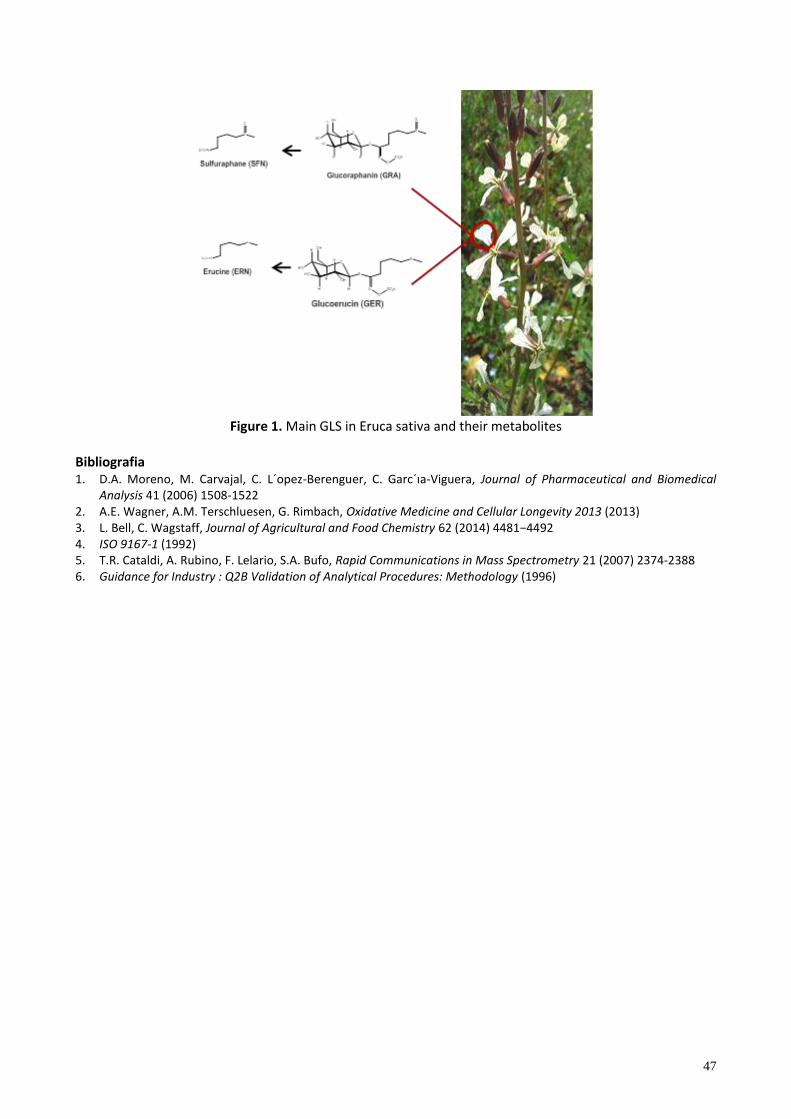
47
Figure 1. Main GLS in Eruca sativa and their metabolites
Bibliografia 1. D.A. Moreno, M. Carvajal, C. L´opez- erenguer, C. Garc´ıa-Viguera, Journal of Pharmaceutical and Biomedical
Analysis 41 (2006) 1508-1522 2. A.E. Wagner, A.M. Terschluesen, G. Rimbach, Oxidative Medicine and Cellular Longevity 2013 (2013) 3. L. Bell, C. Wagstaff, Journal of Agricultural and Food Chemistry 62 (2014) 4481−4492 4. ISO 9167-1 (1992) 5. T.R. Cataldi, A. Rubino, F. Lelario, S.A. Bufo, Rapid Communications in Mass Spectrometry 21 (2007) 2374-2388 6. Guidance for Industry : Q2B Validation of Analytical Procedures: Methodology (1996)
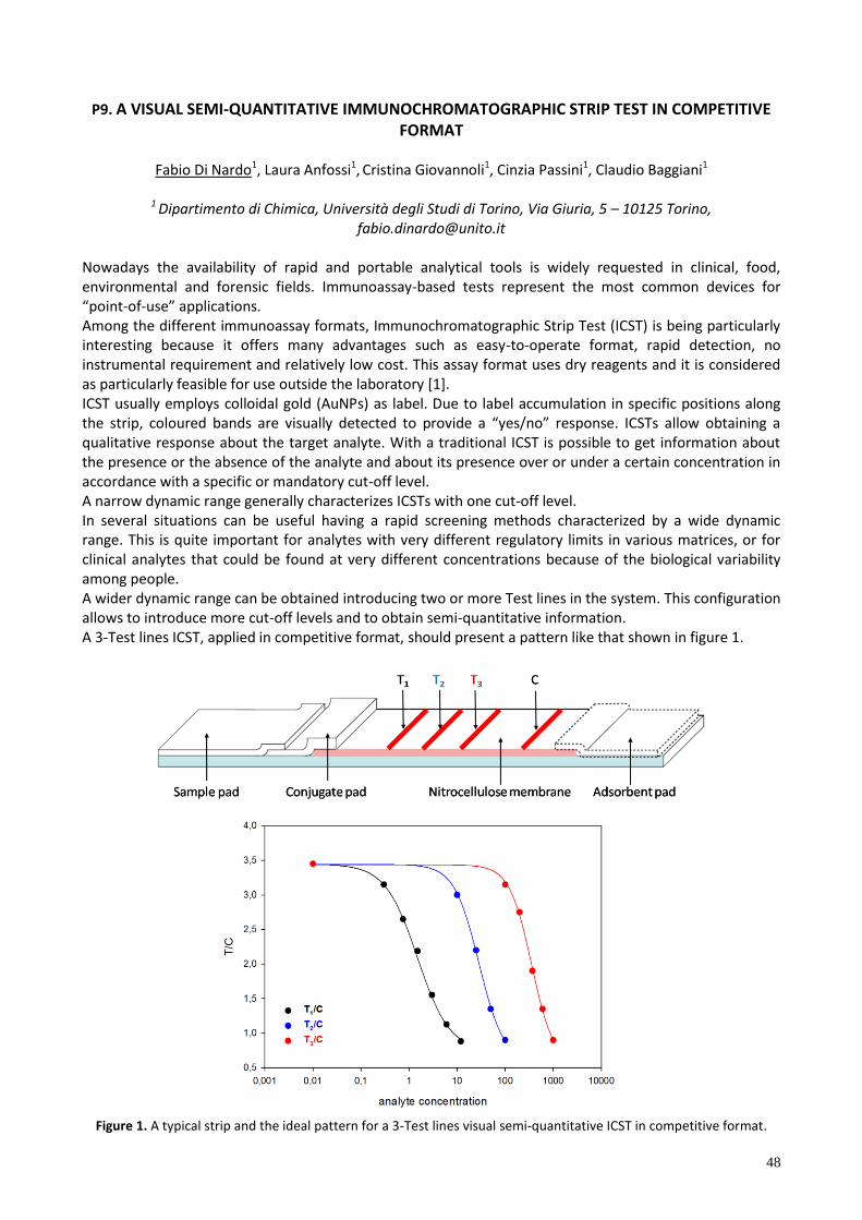
48
P9. A VISUAL SEMI-QUANTITATIVE IMMUNOCHROMATOGRAPHIC STRIP TEST IN COMPETITIVE FORMAT
Fabio Di Nardo1, Laura Anfossi1, Cristina Giovannoli1, Cinzia Passini1, Claudio Baggiani1
1 Dipartimento di Chimica, Università degli Studi di Torino, Via Giuria, 5 – 10125 Torino,
Nowadays the availability of rapid and portable analytical tools is widely requested in clinical, food, environmental and forensic fields. Immunoassay-based tests represent the most common devices for “po nt-of-u e” app cat on . Among the different immunoassay formats, Immunochromatographic Strip Test (ICST) is being particularly interesting because it offers many advantages such as easy-to-operate format, rapid detection, no instrumental requirement and relatively low cost. This assay format uses dry reagents and it is considered as particularly feasible for use outside the laboratory [1]. ICST usually employs colloidal gold (AuNPs) as label. Due to label accumulation in specific positions along the tr p, co oured band are v ua y detected to prov de a “ye /no” re pon e. ICST a ow obta n ng a qualitative response about the target analyte. With a traditional ICST is possible to get information about the presence or the absence of the analyte and about its presence over or under a certain concentration in accordance with a specific or mandatory cut-off level. A narrow dynamic range generally characterizes ICSTs with one cut-off level. In several situations can be useful having a rapid screening methods characterized by a wide dynamic range. This is quite important for analytes with very different regulatory limits in various matrices, or for clinical analytes that could be found at very different concentrations because of the biological variability among people. A wider dynamic range can be obtained introducing two or more Test lines in the system. This configuration allows to introduce more cut-off levels and to obtain semi-quantitative information. A 3-Test lines ICST, applied in competitive format, should present a pattern like that shown in figure 1.
Figure 1. A typical strip and the ideal pattern for a 3-Test lines visual semi-quantitative ICST in competitive format.
49
In this format, free antigen and the antigen deposited on the nitrocellulose compete for binding to the AuNPs-labelled antibody. A negative sample will be characterized by the presence of 4 coloured bands (3 Test lines and 1 Control line). As the analyte concentration increases, there will be a gradual disappearance of T1, T2 and, at higher concentrations, of T3. Depending on the number of visible T-lines is possible to obtain semi-quantitative information about the analyte concentration. To successfully develop a system, that works as required, it is necessary to study the concentration of the Test lines, the amount of antibody and the antibody-AuNPs OD, that are the most critical factors in the system optimization. The proposed approach has been applied in a model system to detect salivary cortisol, which is considered a biomarker of stress, anxiety, and depression [2,3]. Actually, the salivary cortisol levels, from basal to the highest, are included in un narrow range. However this system was used as a model because of our knowledge about its performances in the classic format (1-T line). The preliminary results of the case study will be discussed in this communication. References 1. G. A. Posthuma-Trumpie, J. Korf, A. van Amerongen, Anal Bioanal Chem 393 (2009) 569–582 2. D. H. Hellhammer, S. Wüs, B. M. Kudielka, Psychoneuroendocrinology 34 (2009) 163–171.
3. C. Tlili, N. V. Myung, V. Shetty, A. Mulchandani, Biosens.Bioelectron. 26 (2011) 4382–4386.
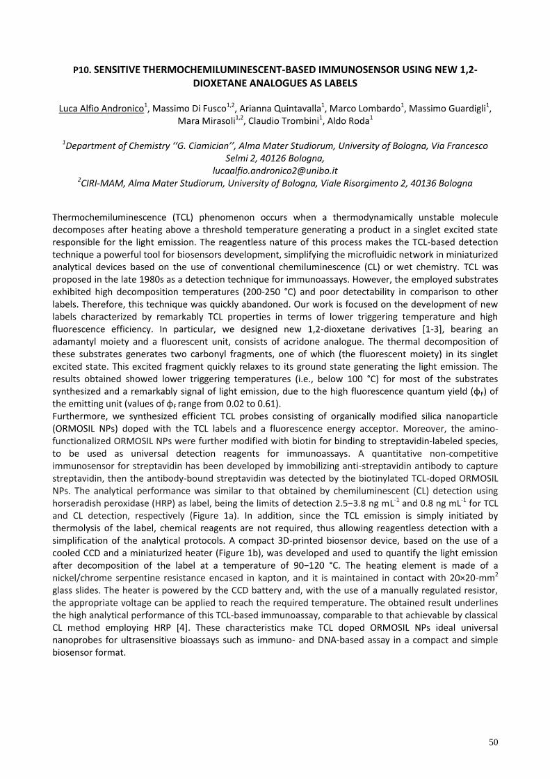
50
P10. SENSITIVE THERMOCHEMILUMINESCENT-BASED IMMUNOSENSOR USING NEW 1,2-DIOXETANE ANALOGUES AS LABELS
Luca Alfio Andronico1, Massimo Di Fusco1,2, Arianna Quintavalla1, Marco Lombardo1, Massimo Guardigli1,
Mara Mirasoli1,2, Claudio Trombini1, Aldo Roda1
1Department of Chemistry ‘‘G. Ciamician’’, Alma Mater Studiorum, University of Bologna, Via Francesco Selmi 2, 40126 Bologna,
[email protected] 2CIRI-MAM, Alma Mater Studiorum, University of Bologna, Viale Risorgimento 2, 40136 Bologna
Thermochemiluminescence (TCL) phenomenon occurs when a thermodynamically unstable molecule decomposes after heating above a threshold temperature generating a product in a singlet excited state responsible for the light emission. The reagentless nature of this process makes the TCL-based detection technique a powerful tool for biosensors development, simplifying the microfluidic network in miniaturized analytical devices based on the use of conventional chemiluminescence (CL) or wet chemistry. TCL was proposed in the late 1980s as a detection technique for immunoassays. However, the employed substrates exhibited high decomposition temperatures (200-250 °C) and poor detectability in comparison to other labels. Therefore, this technique was quickly abandoned. Our work is focused on the development of new labels characterized by remarkably TCL properties in terms of lower triggering temperature and high fluorescence efficiency. In particular, we designed new 1,2-dioxetane derivatives [1-3], bearing an adamantyl moiety and a fluorescent unit, consists of acridone analogue. The thermal decomposition of these substrates generates two carbonyl fragments, one of which (the fluorescent moiety) in its singlet excited state. This excited fragment quickly relaxes to its ground state generating the light emission. The results obtained showed lower triggering temperatures (i.e., below 100 °C) for most of the substrates ynthe zed and a remarkab y gna of ght em on, due to the h gh f uore cence quantum y e d (φF) of the em tt ng un t (va ue of φF range from 0.02 to 0.61). Furthermore, we synthesized efficient TCL probes consisting of organically modified silica nanoparticle (ORMOSIL NPs) doped with the TCL labels and a fluorescence energy acceptor. Moreover, the amino-functionalized ORMOSIL NPs were further modified with biotin for binding to streptavidin-labeled species, to be used as universal detection reagents for immunoassays. A quantitative non-competitive immunosensor for streptavidin has been developed by immobilizing anti-streptavidin antibody to capture streptavidin, then the antibody-bound streptavidin was detected by the biotinylated TCL-doped ORMOSIL NPs. The analytical performance was similar to that obtained by chemiluminescent (CL) detection using hor erad h pero da e (HRP) a abe , be ng the m t of detect on 2.5−3.8 ng mL-1 and 0.8 ng mL-1 for TCL and CL detection, respectively (Figure 1a). In addition, since the TCL emission is simply initiated by thermolysis of the label, chemical reagents are not required, thus allowing reagentless detection with a simplification of the analytical protocols. A compact 3D-printed biosensor device, based on the use of a cooled CCD and a miniaturized heater (Figure 1b), was developed and used to quantify the light emission after decompo t on of the abe at a temperature of 90−120 °C. The heating element is made of a nickel/chrome serpentine resistance encased in kapton, and it is maintained in contact with 20×20-mm2 glass slides. The heater is powered by the CCD battery and, with the use of a manually regulated resistor, the appropriate voltage can be applied to reach the required temperature. The obtained result underlines the high analytical performance of this TCL-based immunoassay, comparable to that achievable by classical CL method employing HRP [4]. These characteristics make TCL doped ORMOSIL NPs ideal universal nanoprobes for ultrasensitive bioassays such as immuno- and DNA-based assay in a compact and simple biosensor format.
51
Figure 1. a) Comparison of the calibration curves obtained for the model immunoassay using TCL and CL imaging detection (top) and images of CL and TCL emissions from an array of spots incubated with different concentrations of streptavidin (bottom); b) scheme of the portable imaging device comprising the heater-containing cover, the dark box and the CCD camera.
References 1. A. Roda, M. Di Fusco, A. Quintavalla, M. Guardigli, M. Mirasoli, M. Lombardo, C. Trombini Anal. Chem. 84 (2012),
9913−9919. 2. M. Di Fusco, A. Quintavalla, C. Trombini, M. Lombardo, A. Roda, M. Guardigli, M. Mirasoli J. Org. Chem. 78 (2013),
11238-11246. 3. M. Di Fusco, M. Guardigli, M. Lombardo, M. Mirasoli, A. Quintavalla, A. Roda, C. Trombini Patent WO2014024106
A1 (2014). 4. M. Di Fusco, A. Quintavalla, M. Lombardo, M. Guardigli, M. Mirasoli, C. Trombini, A. Roda Anal. Bioanal. Chem.
407 (2015), 1567-1576.
52
P11. DEVELOPMENT OF AN AFFINITY SENSOR FOR ORGANIC CONTAMINANTS DETECTION IN FOOD
Sara Romanelli1,2, Francesca Bettazzi2, Tania Martellini2, Alessandra Cincinelli2, Roberta Galarini1, Ilaria
Palchetti2
1Istituto Zooprofilattico Sperimentale dell’Um ria e delle Marche, Via G. Salvemini 1, 06126 Perugia, Italy; e-mail: [email protected]
2Dipartimento di Chimica “Ugo Schiff”, Università degli Studi di Firenze, Via della Lastruccia 3, 50019 Sesto Fiorentino (FI), Italy
Brominated flame retardants are compounds added to inhibit or slow down the ignition of combustible materials. Polybrominated diphenyl ethers (PBDEs) mixtures are a class of brominated flame retardants used worldwide in electric and electronic equipment, plastics, and textiles. There are theoretically 209 PBDE congeners, although the commercial products predominantly consist of penta-PBDEs (mainly the congeners 47, 99, 100, 153, 154), octa-PBDEs and deca-BDEs. While deca-BDE mixture remains on the list of authorized chemicals, penta-BDE and octa-BDE mixtures have been banned from the European market since 2003 (Directive 2003/11/EC) because of their persistence in the environment and their toxicological properties. European Commission has still not defined any maximum limit for PBDEs; thereby, in order to improve the knowledge about contamination patterns and levels and to evaluate human and wildlife exposure, in March 2014 the European Commission issued a Recommendation in which member states are requested to monitor brominated flame retardants in food. The PBDE analysis is usually carried out by gas-chromatography coupled to various mass spectrometric detectors applying the isotope dilution calibration. These techniques permit to achieve low detection limits (1-2 part for trillion) with high selectivity and accuracy, but they require long times, high costs and need for a well-trained staff, too. This kind of analysis is mainly suitable for confirmatory methods requiring quantitative results and high confidence in the compounds identification. On the other hand, immunochemical assays are rapid, simple, and sensitive; therefore they are more appropriate when only semi-quantitative screening results are required. The development of an electrochemical immunosensor for PBDEs detection based on magnetic beads is here reported. The immunological reaction is based on a direct competitive assay using alkaline phosphatase as enzymatic label. The antibody immobilized on the magnetic particles recognizes mainly BDE-47 and BDE-99 congeners. After the immunochemical recognition, the modified magnetic beads are captured by a magnet on the surface of the graphite working electrode. The electrochemical detection is thu ach eved through the add t on of the α-naphthyl-phosphate substrate using the differential pulse voltammetry technique. Under the optimized conditions, the Electrochemical Magneto-Immunosensor (EMI) show a near detect on range from 0.2 to 20 μg/mL of P DE-47 with an IC50 of about 1.7 μg/mL. Being water-filtering organisms, mussels (Mytilus galloprovincialis) are often used to detect persistent organic pollutants (POPs) in the marine environment where the light PBDE congeners 47, 99, 100, and 49 are the most abundant, as reported in literature. For this purpose, the developed EMI technique was successfully combined with a simple mussel sample preparation (QuEChERS-like extraction followed by an SPE purification). The complete procedure seems to offer a new screening test to be used in massive environmental campaigns, which must be able to rapidly reveal possible polluted coastal areas. Riferimenti 1. S. Centi, E. Silva, S. Laschi, I. Palchetti, M. Mascini, Analitica Chimica Acta 594 (2007) 9-16.
2. European Commission, 2014. Recommendation (EU) No 118/2014 of 3 March 2014 on the monitoring of traces of
brominated flame retardants in food. Official Journal of the European Union L 65/39.
3. C. Munschy, N. Olivier, B. Veyrand, P. Marchand, Chemosphere 118 (2015) 329-335.
4. W.L. Shelver, C.D. Parrotta, R. Slawecki, Q.X. Li, M.G. Ikonomou, D. Barcelo, S. Lacorte, F.M. Rubio, Chemosphere
73 (2008) 518-523.
53
P12. NOVEL APPROACHES FOR ALZHEIMER’S DISEASE BIOMOLECULAR DIAGNOSIS
Samuele Lisi1,2, Simona Scarano1, Corinne Ravelet2, Eric Peyrin2, Maria Minunni*1
1Dipartimento di Chimica “Ugo Schiff”, Università di Firenze, Via della Lastruccia 3,50019, Sesto F.no, Italy
[email protected], *[email protected] 2Département de pharmacochimie moléculaire, Université Grenoble alpes, 470 rue de la chimie, 38400, St
Martin d’ Heres, France
A zhe mer’ d ea e (AD) a w de pread pathogen c cond t on which cause memory and behaviour impairment in elderly people because of the accumulation of amyloid beta peptide and tau protein [1]. Since up to now therapeutic intervention is not able to stop the progression of the pathology, early diagnosis assumes crucial role to slow down cognitive decline in AD patients. After revising clinical criteria of AD diagnosis in 2010 [2], three core biomarkers have been accepted as supportive criteria for the identification of the pathology. Such biomarkers are quantified in cerebrospinal fluid by immunochemical methods (i.e. ELISA) and they are amyloid beta peptide (Aβ), protein tau (t-τ) and phosphorylated tau (p-τ181). A project ent t ed “Sviluppo di Biosensori di affinità a base di un nuovo recettore aptamerico, per la d agno t ca mo eco are de a ma att a d A zhe mer” within the Vinci program 2013 has been funded by the Università Italo-Francese. The aim of this project is the development of Surface Plasmon Resonance (SPR) aptasensing based on novel bioreceptors for AD biomarkers, i.e. tau protein, with application in AD molecular diagnostic. Here we will report preliminary results during the first year of collaborative work on the aptamer for tau protein development performed at the Université Grenoble alpes, which is thought as the innovative bioreceptor for SPR-based aptasensors. Behind this SPR immunosensing is set up to be used as reference approch. Concerning aptamer selection, after testing conventional SELEX process in which the selection is followed by PCR amplification that actually did not provided the aptamer with suitable characteristics an alternative procedure was tested, called non-SELEX approcach. Such idea has been developed by Berezovski and colleagues [3] who developed the non-SELEX method in 2006. Here, for the first time, we applied such method to an unfolded protein (protein tau). Results of the application of the non-SELEX are promising since after only four cycles a significant evolution in affinity constant of DNA strand toward the target was observed. Optimization of SPR platform is still in progress as well as sequencing of the selected DNA population. We will here report about the calibration curve for a direct assay using immobilized monoclonal antibodies (MAb) and tau in standadd solutions. References
1. Blennow, K., Hampel, H., Weiner, M., Zetterberg, H. Cerebrospinal fluid and plasma biomarkers in Alzheimer disease. Nat. Rev. Neurol. 6, (2010), 131–44.
2. Dubois, B., Feldman, H.H., Jacova, C., Cummings, J.L., Dekosky, S.T., Barberger-Gateau, P., Delacourte, A., Frisoni, G., Fo , N.C., Ga a ko, D., Gauth er, S., Hampe , H., J cha, G. a, Meguro, K., O’ r en, J., Pa qu er, F., Robert, P., Rossor, M., Salloway, S., Sarazin, M., de Souza, L.C., Stern, Y., Visser, P.J., Scheltens, P., 2010. Revising the def n t on of A zhe mer’ d ea e: a new e con. Lancet Neuro . 9, 1118–27
3. Berezovski, M., Musheev, M., Drabovich, A., Krylov, S.N., 2006. Non-SELEX selection of aptamers. J. Am. Chem. Soc. 128, 1410–1.
54
P13. GREEN COFFEE BEAN EXTRACTS AS POTENTIAL NEUROPROTECTIVE AND CHEMOPROTECTIVE DIETARY SUPPLEMENTS: A MOLECULAR POINT OF VIEW
Alessandro Palmioli1, Carlotta Ciaramelli1, Milda Stuknytė2, Laura Colombo,3 Ada De Luigi,3 Michela Spinelli1,
Elena Sacco1, Ilaria Bruni1, Maria-Elena Regonesi1, Ivano De Noni2, Massimo Labra1, Cristina Airoldi1
1 Dipartimento di Biotecnologie e Bioscienze, Università degli studi di Milano-Bicocca, Piazza della Scienza, 2, 20126 Milano, [email protected]; [email protected]
2 Dipartimento di Scienze per gli Alimenti, la Nutrizione e l'Ambiente, Università degli studi di Milano, Via G. Celoria, 2, 20133 Milano
3 Dipartimento di Biochimica e Farmacologia Molecolare, Istituto di Ricerche Farmacologiche Mario Negri, Via Giuseppe La Masa, 19, 20156 Milano
Recently, green coffee beans have gained a lot of interest as a source of metabolites with novel biological effects in drug discovery processes and indeed emerged as powerful nutraceutical dietary supplements.1 The interest is focused in particular on chlorogenic acids (CGA), esters formed between hydroxycinammic acids (mainly caffeic and ferulic) and quinic acid that represent the most abundant family of polyphenols in green coffee beans and occur ubiquitously in food.2 A number of beneficial biological effects, including anti-inflammatory activity, anticarcinogenic activity and protection in neurodegenerative diseases, have been described for CGA.3 However, the molecular mechanisms through which these biological activities manifest are not yet investigated in depth. Here we report preliminary results on quali-quantitative characterization of polyphenols in green coffee bean ’ e tract by mean of NMR and UPLC/HRMS and the r mo ecu ar nteract on w th protoncogen c human mode (hRAS) and neurodegenerat ve amy o d o gomer ’ mode (Aβ1-42) proteins exploiting in vitro and in ex-vivo techniques among which STD-NMR spectroscopy,4 cellular and biochemical assays to validate the molecular target directly correlated to the biological responses. References 1. P. Esquivel, V. M. Jiménez, Food Res. Int., 46 (2012), 488. 2. M. N. Clifford, J. Sci. Food Agric., 80 (2000), 1033. 3. a) R. Feng, Y. Lu, L. L. Bowman, Y. Qian, Castranova, V., M. Ding, Journal of Biological Chemistry, 280, (2005),
27888. b) H. S. Shin, H. Satsu, M.-J. Bae, Z. Zhao, H. Ogiwara, M.Totsuka, M. Shimizu, Food Chemistry, 168, (2015), 167. c) G. Oboh, O. Agunloye, A. Akinyemi, A. Ademiluyi, S. Adefegha Neurochem Res, 38, (2013), 413.
4. a) C. Airoldi, E. Sironi, C. Dias, F. Marcelo, A. Martins, A. P. Rauter, F. Nicotra, J. Jimenez-Barbero, Chem. Asian J., 8, (2013), 596; b) E. Sironi, L. Colombo, A. Lompo, M. Messa, M. Bonanomi, M. E. Regonesi, M. Salmona, C. Airoldi, Chem. Eur. J., 20, (2014), 13793.
55
P14. NATURAL COMPOUNDS AGAINST ALZHEIMER’S DISEASE: EFFECTS OF HOP EXTRACTS ON AΒ PEPTIDE INDUCED TOXCICITY ON NEURONAL CELL LINES
Valeria Mazzoni,1 Alessandro Palmioli,1 Carlotta Ciaramelli,1 Laura Colombo,2 Ada De Luigi,2 Cristina Airoldi1
1 Dipartimento di Biotecnologie e Bioscienze, Università degli studi di Milano-Bicocca, Piazza della Scienza 2,
20126 Milano, [email protected]; [email protected] 2 Dipartimento di Biochimica e Farmacologia Molecolare, Istituto di Ricerche Farmacologiche Mario Negri,
Via Giuseppe La Masa, 19 20156 Milano
In this communication we show the potential of NMR approaches for the metabolomics profiling and
screening of natural product mixtures obtained from plant extracts and food, aimed at the discovery of new
bioactive compounds.
In part cu ar we focu ed our ntere t on the dent f cat on of new gand of Aβ1-42 oligomers, involved in
A zhe mer’ 1 disease. Due to the severe impact of this pathology on the quality of life of the patients and
their families, its massive economic burden, and the lack of effective therapies and diagnostic tools, there is
an urgent need for effective molecules for its treatment and diagnosis. In this context, the availability of
new screening methods is strategic.
Recently, exploiting STD NMR and trNOESY experiments we were able to identify ligands of amyloid
peptides and proteins in Salvia sclareoides,2 Genista tenera3 and green tea extracts.4
Now we are investigating the presence of molecules able to interact with Aβ1-42 oligomers in hop extracts,
a o te t ng e tract act v ty aga n t Aβ-induced toxicity on neuronal cell lines. Our data provide important information for the rational design of new compounds with higher affinity for Aβ1-42, PrP106-126 and Ataxin-3, to generate new anti-amyloidogenic molecules and/or molecular tools for the specific targeting of amyloid aggregates in vivo.
References 1. G. G. Glenner, C. W. Wong, Biochem. Biophys. Res. Comm., 120, (1984) 885-890. 2. C. Airoldi, E. Sironi, C. Dias, F. Marcelo, A. Martins, A. P. Rauter, F. Nicotra, J. Jimenez-Barbero, Chem. Asian J. 8,
(2013) 596. 3. A. R. Jesus, C. Dias, A. M. Matos, R. F. M. de Almeida, A. S. Viana, F. Marcelo, R. T. Ribeiro, M. P. Macedo, C.
Airoldi, F. Nicotra, A. Martins, E. J. Cabrita, J. Jiménez-Barbero, A. J. Pilar Rauter Med. Chem., 57, (2014) 9463. 4. E. Sironi, L. Colombo, A. Lompo, M. Messa, M. Bonanomi, M. E. Regonesi, M. Salmona, C. Airoldi, Chem. Eur. J.
20, (2014) 13793.

56
P15. APTAMER-BASED OPTICAL SENSOR FOR THE DETECTION OF SAXITOXIN IN MARINE WATER
Marianna Rossetti1, Alessandro Porchetta1, Francesco Ricci1, Giuseppe Palleschi1
1 Dipartimento di Scienze e Tecnologie Chimiche, Università di Roma Tor Vergata, Via della Ricerca Scientifica 1, 00133 Roma, [email protected]
Saxitoxin (STX) is a low molecular weight neurotoxin mainly produced by certain marine Dinoflagellates and responsible for causing paralytic shellfish poisoning. STX contaminates seafood and freshwater, killing people and agricultural livestock as well as making drinking water unpotable. The presence of STX is often detected with a mouse lethality bioassay, the only method with regulatory standing for many years. More recently, HPLC analysis coupled with fluorescent detection of STX oxidation products has received official approval to be used for measurement of saxitoxins. We have developed an aptamer-based optical biosensors. In fact, optically labelled saxitoxin-binding aptamer can signal the target presence through a binding-induced conformational change which brings the fluorophore close to the quencher thus decreasing the fluorescence signal. The proposed method is simple, high specific and selective even if a preconcentration step is needed. The authors would like to acknow edge the f nanc a upport from the European Un on’ Seventh Framework Programme for research, technological development and demonstration under grant agreement no 613844. Riferimenti 1. S.M. Handy, et all., Toxicon 61 (2013) 30-37

57
P16. MICROEXTRACTION ON PACKED SORBENT (MEPS) FOR THE DETERMINATION OF PESTICIDES IN WHEAT FLOUR BY HPLC-MS/MS
Manuel Sergi1, Francesca Di Ottavio1, Camilla Montesano2, Rossana Scarpone3, Roberta Curini2, Dario
Compagnone1
1Faculty of Bioscience and Technology for Food, Agriculture and Environment, University of Teramo, 64023 Teramo, Italy, [email protected]
2Department of Chemistry, Sapienza University of Rome, 00185, Rome, Italy 3Istituto Zooprofilattico Dell’A ruzzo e del Molise G.Caporale, 64100 Teramo, Italy
The analysis of pesticide residues in food samples has received increasing attention in the last few decades, as can be deduced from the great number of published papers dealing with this subject. These compounds are usually determined by gas chromatography (GC), liquid chromatography (LC) or capillary electrophoresis (CE), depending on their polarity, volatility, and thermal stability. In recent years, the appearance and use of new, more polar pesticides has fostered the development of liquid chromatography/mass spectrometry (LC–MS) besides gas chromatography. A further feature of LC–MS for the analysis of pesticides is the possibility to perform multi-class methods even with the difficulties associated. Regulatory authorities provide assurance that any pesticide remaining in or on the food is within safe limits through monitoring programs or random sampling and analysis of raw or processed food on the market. In response to this requirement a number of methods have been developed and applied routinely for the control of pesticide residues in food [1]. In general, food samples cannot be analyzed without some preliminary sample preparation, because the matrix is rather complex. The pretreatment of the sample, especially in analysis of trace residues of pesticides in food, is the crucial point because of the matrix complexity. The aim of this work was the development of a sensitive, precise, accurate method for multiclass analysis of pesticide and fungicide residues in wheat flours; the attention was focused on 16 pesticides with different physico-chemical characteristics and different mechanism of action: acetylcholinesterase inhibitors like organophosphorus, carbamates and neonicotinoids, and inhibitors of ergosterol like imidazoles and triazoles. The wheat is exposed to phytosanitary treatment during planting, growing, harvesting and storage; to avoid serious effects on humans and the ecosystem, for these reasons the European Union by Regulation (EC) No. 396/2005 provided the restrictions on the use and applicability of these substances by laying down strict guidelines, imposing Maximum Residue Limit (MRL) values of pesticides in food and feed to protect human and animal health. The presented method involves a Micro Extraction by Packed Sorbent (MEPS) followed by High Performance Liquid Chromatography coupled to Tandem Mass Spectrometry (HPLC-MS/MS). 1 g of wheat flour sample was added with extracting solution (60% of acetonitrile and acetate buffer 10mM pH=5). The mixture was then sonicated for 5 minutes, and then placed in a water bath at the temperature of 40°C for 5minutes under constant stirring speed. Buffer solution was added to the eluate to bring the percentage of acetonitrile at 10%, and then it was subjected to a second centrifugation at 10000 rpm at 4°C for 5 minutes. The clean-up was carried out with syringes MEPS C18 [2]. The activation of the BIN (barrel insert and need e) wa conducted by tak ng 100 μL of methano for three t me . The cond t on ng tep wa carr ed out with a solution of water and acetonitrile 90:10 (v:v) for two times. 3mL of sample were loaded in extract-discart mode: each aliquot is discarded after passing through the MEPS syringe aspirating aliquots of 250 μL. The 100 μL of e uate were co ected n a v a and placed in the autosampler for the subsequent HPLC-MS/MS analysis. The chromatographic separation was conducted using a column Phenomenex Kinetex XB-C18, 100x2.1 mm, packed with particles of 2.6µm obtained with core-shell technology with its guard column from Phenomenex.
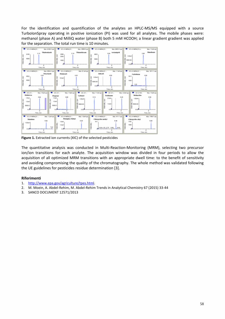
58
For the identification and quantification of the analytes an HPLC-MS/MS equipped with a source TurboIonSpray operating in positive ionization (PI) was used for all analytes. The mobile phases were: methanol (phase A) and MilliQ water (phase B) both 5 mM HCOOH; a linear gradient gradient was applied for the separation. The total run time is 10 minutes.
Figure 1. Extracted ion currents (XIC) of the selected pesticides
The quantitative analysis was conducted in Multi-Reaction-Monitoring (MRM), selecting two precursor ion/ion transitions for each analyte. The acquisition window was divided in four periods to allow the acquisition of all optimized MRM transitions with an appropriate dwell time: to the benefit of sensitivity and avoiding compromising the quality of the chromatography. The whole method was validated following the UE guidelines for pesticides residue determination [3]. Riferimenti 1. http://www.epa.gov/agriculture/tpes.html. 2. M. Moein, A. Abdel-Rehim, M. Abdel-Rehim Trends in Analytical Chemistry 67 (2015) 33-44 3. SANCO DOCUMENT 12571/2013
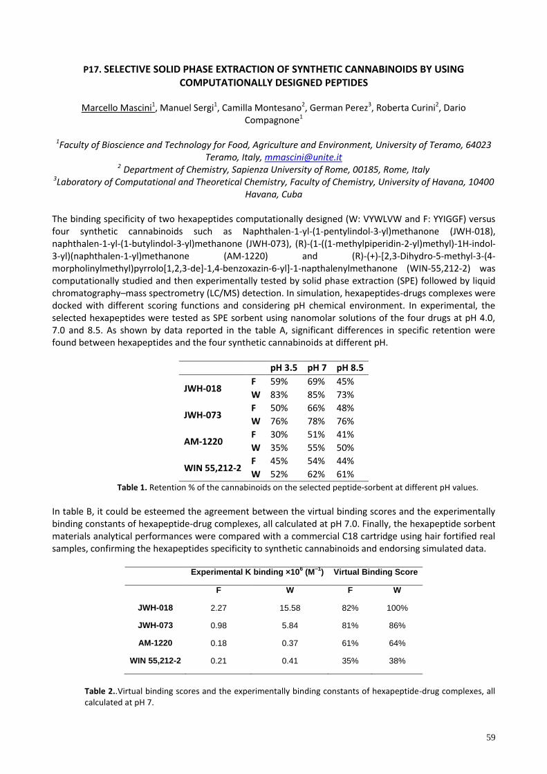
59
P17. SELECTIVE SOLID PHASE EXTRACTION OF SYNTHETIC CANNABINOIDS BY USING COMPUTATIONALLY DESIGNED PEPTIDES
Marcello Mascini1, Manuel Sergi1, Camilla Montesano2, German Perez3, Roberta Curini2, Dario
Compagnone1
1Faculty of Bioscience and Technology for Food, Agriculture and Environment, University of Teramo, 64023 Teramo, Italy, [email protected]
2 Department of Chemistry, Sapienza University of Rome, 00185, Rome, Italy
3Laboratory of Computational and Theoretical Chemistry, Faculty of Chemistry, University of Havana, 10400 Havana, Cuba
The binding specificity of two hexapeptides computationally designed (W: VYWLVW and F: YYIGGF) versus four synthetic cannabinoids such as Naphthalen-1-yl-(1-pentylindol-3-yl)methanone (JWH-018), naphthalen-1-yl-(1-butylindol-3-yl)methanone (JWH-073), (R)-(1-((1-methylpiperidin-2-yl)methyl)-1H-indol-3-yl)(naphthalen-1-yl)methanone (AM-1220) and (R)-(+)-[2,3-Dihydro-5-methyl-3-(4-morpholinylmethyl)pyrrolo[1,2,3-de]-1,4-benzoxazin-6-yl]-1-napthalenylmethanone (WIN-55,212-2) was computationally studied and then experimentally tested by solid phase extraction (SPE) followed by liquid chromatography–mass spectrometry (LC/MS) detection. In simulation, hexapeptides-drugs complexes were docked with different scoring functions and considering pH chemical environment. In experimental, the selected hexapeptides were tested as SPE sorbent using nanomolar solutions of the four drugs at pH 4.0, 7.0 and 8.5. As shown by data reported in the table A, significant differences in specific retention were found between hexapeptides and the four synthetic cannabinoids at different pH.
pH 3.5 pH 7 pH 8.5
JWH-018 F 59% 69% 45%
W 83% 85% 73%
JWH-073 F 50% 66% 48%
W 76% 78% 76%
AM-1220 F 30% 51% 41%
W 35% 55% 50%
WIN 55,212-2 F 45% 54% 44%
W 52% 62% 61%
Table 1. Retention % of the cannabinoids on the selected peptide-sorbent at different pH values.
In table B, it could be esteemed the agreement between the virtual binding scores and the experimentally binding constants of hexapeptide-drug complexes, all calculated at pH 7.0. Finally, the hexapeptide sorbent materials analytical performances were compared with a commercial C18 cartridge using hair fortified real samples, confirming the hexapeptides specificity to synthetic cannabinoids and endorsing simulated data.
Experimental K binding ×106 (M
–1) Virtual Binding Score
F W F W
JWH-018 2.27 15.58 82% 100%
JWH-073 0.98 5.84 81% 86%
AM-1220 0.18 0.37 61% 64%
WIN 55,212-2 0.21 0.41 35% 38%
Table 2..Virtual binding scores and the experimentally binding constants of hexapeptide-drug complexes, all calculated at pH 7.

60
P18. CARBON BLACK-CHITOSAN FILM FOR SCREEN PRINTED ELECTRODE AS NOVEL PLATFORMS FOR BIOSENSORS
Daria Talarico1, Aziz Amine3, Fabiana Arduini1,2, Danila Moscone1,2, Giuseppe Palleschi1,2
1 1Dipartimento di Scienze e Tecnologie Chimiche, Università di Roma Tor Vergata, Via della Ricerca
Scientifica, 00133 Rome, Italy, [email protected] 2Consorzio Interuniversitario Biostrutture e Biosistemi “I BB”, Viale Medaglie d'Oro, 305, Rome, Italy
3Université Hassan II-Mohammedia, Faculté de Sciences et Techniques Laboratoire Génie des Procédés et Environnement, B.P. 146, Mohammadia, Morocco
Il Traditionally, optical methods coupled with chromatographic techniques are used as reference methods
to detect several analytes in various types of matrix. Despite their sensitivity, they are time-consuming,
expensive and require of skilled-personnel. The bio-sensoristic field offers a good alternative to overcome
the disadvantages mentioned above by the use of bio-molecules (such as proteins, antibody, aptamers,
DNA, bacteria) as recognized elements immobilized on the sensors. Moreover the biosensors based on the
electrochemical techniques have been widely investigated, showing the suitability for in situ measurements
since the treatment of the sample usually is simple or not required. [1]
The coupling of bioelements with carbon nanomaterials (such as carbon nanotube, carbon nanodots,
Graphene) has been intensively studied because they can promote the electron transfer between the
bioelements and the working electrode surface. [2]
Among the carbon nanomaterials, the use of Carbon Black Nanoparticles (CBNPs) as modifying agent has
reported considerable success in the electroanalytical field; to this regard, our group has demonstrated the
enhancement of the electrochemical performances of screen-printed electrode (SPE) modified with CBNPs
for the detection of many analytes such as NADH, hydrogen peroxide, cysteine. [3]
The immobilization of the biomaterials have played a significant role because not only brings about the
intimate contact of the biologic catalysts with the transducer, but also helps in the stabilization of the
biologic system, thus enhancing its operational and storage stability. [4]
In this work, we present a novel biocompatible platform for enzymatic biosensors based on the use of
CBNPs and Chitosan (Chit) a polysaccharidic biopolymer, which displays excellent film-forming ability, good
adhesion and susceptibility to chemical modifications due to the presence of reactive amino and hydroxyl
functional groups. Moreover, using Chitosan, the insolubility in water of pristine CBNPs can be minimized,
allowing their integration with enzyme.
For this purpose, the working electrode of a screen-printed sensor was modified by drop casting with
CBNPs and a Chit dispersion (CBchit-SPEs). Its electrochemical performances was investigated and
compared with the unmodified SPE (Bare-SPE) using cyclic voltammetry (CV) in presence of ferro/ferri
(cyanide) as electrochemical probe. The enzymes Laccase (Lacc) and Hydrogen Peroxidase (HRP) were
immobilized by mixing directly these enzymes with the CBchit dispersion on the working electrode surface,
obtaining the CBchit/Enzyme-SPEs (CBchit/lacc-SPE and CBchit/HRP-SPE), and their responses towards
hydroquinone (HQ) and caffeic acid (CA) were studied by cyclic voltammetry. The amperometric detection
of the HQ in presence of hydrogen peroxide (H2O2) 1 mM at the applied potential of 0.0 V vs Ag/AgCl
shows a linear range between 0.10 and 30 µM and LOD equal to 0.05 µM.
Using CBchit/Lacc-SPE, the CA was detected at the applied potential of 0.150 V vs Ag/AgCl, reaching 1 µM
as LOD and with a linear range from 1 to 10 µM. The preliminary results show the suitability of CBchit-SPEs as platform for biosensor development with improved sensitivity and stability. Riferimenti 1. F.Arduini, A. Amine, Adv. Biochem. Eng. Biotechnol. 140 (2014) 299.

61
2. F. Arduini, M. Forchielli, A. Amine, D. Neagu, I. Cacciotti, F. Nanni, D. Moscone, G. Palleschi, Microch. Acta 182 (2015) 643.
3. F. Arduini, A. Amine, C. Majorani, F. Di Giorgio, D. De Felicis, F. Cataldo, D. Moscone, G. Palleschi Electrochem. Commun. 12 (2010) 346.
4. W. Putzbach, N. J. Ronkainen, Sensors (2013) 13 4811.

62
P19. MAGNETIC BEADS-BASED ELECTROCHEMICAL IMMUNOASSAY FOR SCREENING OF CELIAC DISEASE IN SALIVA
Gianluca Adornetto, Laura Fabiani, Giulia Volpe, Danila Moscone
Dipartimento di Scienze e Tecnologie Chimiche, Università di Roma Tor Vergata, via della Ricerca Scientifica, 00133 Roma; [email protected]
The gluten-induced autoimmune enteropathy Celiac Disease (CD) is one of the most common immune-mediated diseases in Europe and North America with a high number of undetected cases. This work investigates the development of a highly sensitive ELIME-assay (Enzyme Linked ImmunoMagnetic Electrochemical-assay) for the diagnosis of CD in saliva samples, avoiding the discomfort of blood sample collection. Currently, there is an unique method developed to date for the diagnosis of CD in saliva, the RadioImmunoAssay method, but it requires radioactive products and all the precautionary measures related to their use [1]. All other existing methods for CD diagnosis involve the analysis of blood serum samples to detect specific markers, such as anti-transglutaminase IgA antibodies (anti-tTG IgA). The system starts from our article already published concerning the CD diagnosis in serum samples [2], but the ELIME-assay was completely re-designed and re-optimized in order to have a system suitable for measuring anti-tTG IgA antibodies in a much more difficult matrix such as saliva. The new assay, in fact, solves the problems related to the low concentration of anti-tTG IgA antibodies in this medium and to its high viscosity using appropriate buffers and a different kind of Magnetic Beads covered with transglutaminase antigen (tTG), which reacts with the anti-tTG IgA present in positive saliva samples. Two different immunoassay approaches were optimized and compared analyzing 66 saliva samples, whose anti-tTG IgA levels were independently determined by a RIA method. The obtained results, expressed as Ab-Index:
A][ signal standard AU/ml 0 - A][ signal standard 5AU/ml
A] [ signal standard AU/ml 0 - A][ signal sampleIndexAb
were used to perform a diagnostic test evaluation through the construction of ROC curves. The selected approach, named ELIME Assay Plus and involving a pre-incubation between the anti-Human IgA-AP and saliva samples prior to the addition of MBs-tTG, showed a cut-off of 0.022 with 96% of clinical sensitivity and 95% of clinical specificity. The area under ROC curve resulted to be equal to 1, a result that c a f e our te t a “perfect”. Therefore, this study demonstrates that the our system, which employs saliva samples, could be considered an useful tool for preliminary screening diagnosis of CD in "Doctor office" or in non-hospital facilities, becoming a "point of care testing" (POCT). This aspect is of fundamental importance to screen a large number of subjects, especially in the paediatric age.
Figure 1. Assay scheme

63
References 1. R. Nenna, C. Tiberti, L. Petrarca, F. Lucantoni, M. Mennini, R. P. L. Luparia, F. Panimolle, G. Mastrogiorgio, N.
Pietropaoli, F. M. Magliocca, M. Bonamico, Journal of pediatric gastroenterology and nutrition, 56 (4), 2013, pp 416–421
2. G. Adornetto, G. Volpe, A. De Stefano, S. Martini, G. Gallucci, A. Manzoni, S. Bernardini, M. Mascini, D. Moscone, Anal. Bioanal. Chem. 403 (4), 2012, pp 1191–1194.
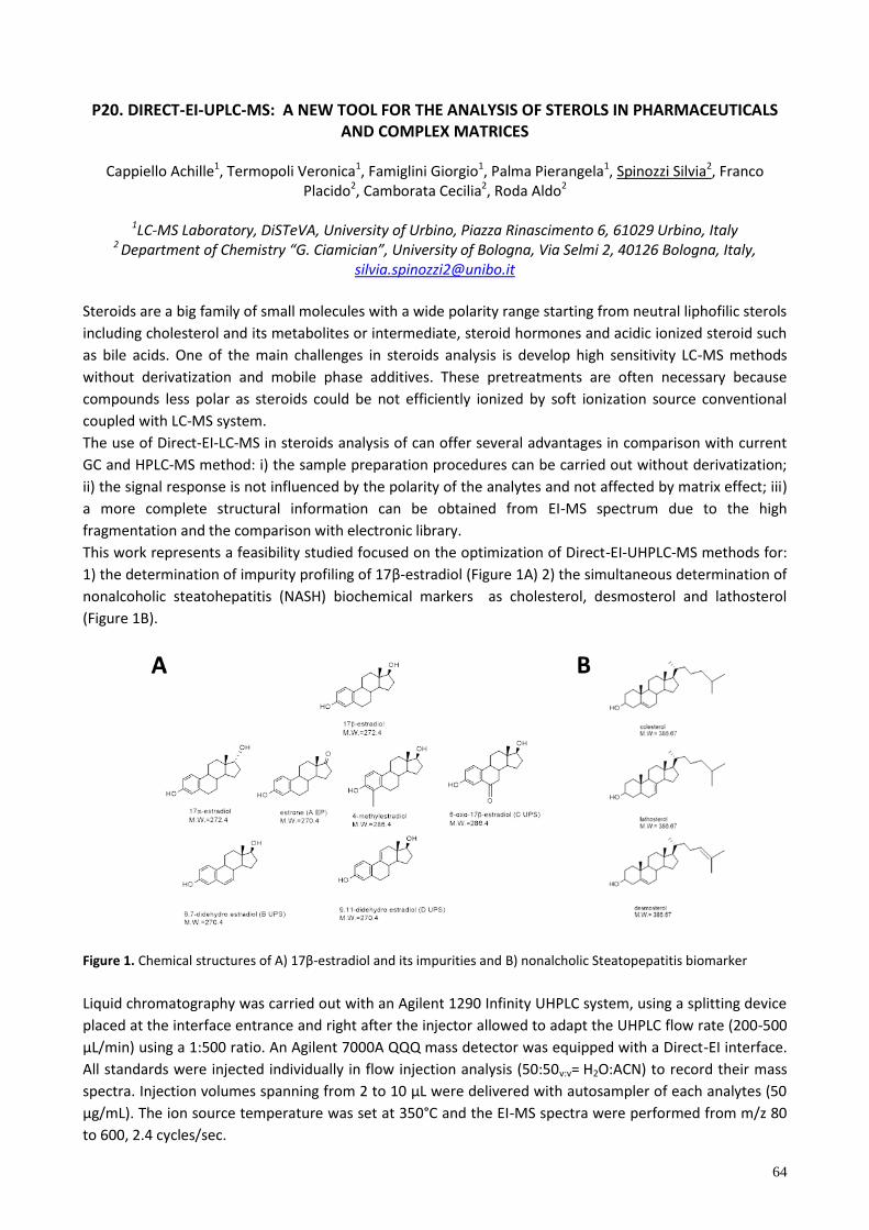
64
P20. DIRECT-EI-UPLC-MS: A NEW TOOL FOR THE ANALYSIS OF STEROLS IN PHARMACEUTICALS AND COMPLEX MATRICES
Cappiello Achille1, Termopoli Veronica1, Famiglini Giorgio1, Palma Pierangela1, Spinozzi Silvia2, Franco
Placido2, Camborata Cecilia2, Roda Aldo2
1LC-MS Laboratory, DiSTeVA, University of Urbino, Piazza Rinascimento 6, 61029 Urbino, Italy 2 Department of Chemistry “G. Ciamician”, University of Bologna, Via Selmi 2, 40126 Bologna, Italy,
Steroids are a big family of small molecules with a wide polarity range starting from neutral liphofilic sterols
including cholesterol and its metabolites or intermediate, steroid hormones and acidic ionized steroid such
as bile acids. One of the main challenges in steroids analysis is develop high sensitivity LC-MS methods
without derivatization and mobile phase additives. These pretreatments are often necessary because
compounds less polar as steroids could be not efficiently ionized by soft ionization source conventional
coupled with LC-MS system.
The use of Direct-EI-LC-MS in steroids analysis of can offer several advantages in comparison with current
GC and HPLC-MS method: i) the sample preparation procedures can be carried out without derivatization;
ii) the signal response is not influenced by the polarity of the analytes and not affected by matrix effect; iii)
a more complete structural information can be obtained from EI-MS spectrum due to the high
fragmentation and the comparison with electronic library.
This work represents a feasibility studied focused on the optimization of Direct-EI-UHPLC-MS methods for:
1) the determination of impurity profiling of 17β-estradiol (Figure 1A) 2) the simultaneous determination of
nonalcoholic steatohepatitis (NASH) biochemical markers as cholesterol, desmosterol and lathosterol
(Figure 1B).
Figure 1. Chemical structure of A) 17β-estradiol and its impurities and B) nonalcholic Steatopepatitis biomarker
Liquid chromatography was carried out with an Agilent 1290 Infinity UHPLC system, using a splitting device
placed at the interface entrance and right after the injector allowed to adapt the UHPLC flow rate (200-500
µL/min) using a 1:500 ratio. An Agilent 7000A QQQ mass detector was equipped with a Direct-EI interface.
All standards were injected individually in flow injection analysis (50:50v:v= H2O:ACN) to record their mass
spectra. Injection volumes spanning from 2 to 10 µL were delivered with autosampler of each analytes (50
µg/mL). The ion source temperature was set at 350°C and the EI-MS spectra were performed from m/z 80
to 600, 2.4 cycles/sec.
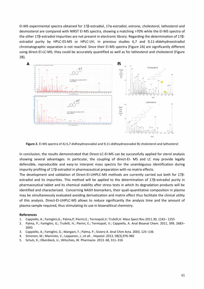
65
A
EI-MS experimental spectra obta ned for 17β-e trad o , 17α-estradiol, estrone, cholesterol, lathosterol and
desmosterol are compared with NIRST EI-MS spectra, showing a matching >70% while the EI-MS spectra of
the other 17β-estradiol impurities are not present in electronic l brary. Regard ng the determ nat on of 17β-
estradiol purity by HPLC-ES-MS or HPLC-UV, in previous studies 6,7 and 9,11-didehydroestradiol
chromatographic separation is not reached. Since their EI-MS spectra (Figure 2A) are significantly different
using direct-EI-LC-MS, they could be accurately quantified as well as for lathosterol and cholesterol (Figure
2B).
Figure 2. EI-MS spectra of A) 6,7-didheydroesradiol and 9,11-didheydroesradiol B) cholesterol and lathosterol
In conclusion, the results demonstrated that Direct-LC-EI-MS can be successfully applied for sterol analysis
showing several advantages. In particular, the coupling of direct-EI- MS and LC may provide legally
defensible, reproducible and easy-to interpret mass spectra for the unambiguous identification during
mpur ty prof ng of 17β-estradiol in pharmaceutical preparation with no matrix effects.
The development and validation of Direct-EI-UHPLC-MS method are current y carr ed out both for 17β-
estradiol and its impurities. Th method w be app ed to the determ nat on of 17β-estradiol purity in
pharmaceutical tablet and its chemical stability after stress tests in which its degradation products will be
identified and characterized. Concerning NASH biomarkers, their quali-quantitative composition in plasma
may be simultaneously evaluated avoiding derivatization and matrix effect thus facilitate the clinical utility
of this analysis. Direct-EI-UHPLC-MS allows to reduce significantly the analysis time and the amount of
plasma sample required, thus stimulating its use in bioanalitical chemistry. References 1. Cappiello, A.; Famiglini,G.; Palma,P; Pierini,E.; Termopoli,V; Trufelli,H. Mass Spect Rev.2011.30, 1242– 1255 2. Palma, P.; Famiglini, G.; Trufelli, H.; Pierini, E.; Termopoli, V.; Cappiello, A. Anal Bioanal Chem. 2011, 399, 2683–
2693 3. Cappiello, A.; Famiglini, G.; Mangani, F.; Palma, P.; Siviero A. Anal Chim Acta. 2003, 125–136 4. Simonen, M.; Mannisto, V.; Leppanen, J.; et all. , Hepatol. 2013, 58(3),976-982 5. Schulz, K.; Oberdieck, U.; Witschies, W. Pharmazie. 2013. 68, 311–316
Cholesterol
Lathosterol
Relative
intensity %
Relative intensity
%
m/z
m/z
B
m/z
66
P21. HOMOGENOUS FLUOROIMMUNOASSAYS BASED ON THE QUENCHING OF QUANTUM DOTS FLUORESCENCE BY GRAPHENE
Laura Anfossi1, Cristina Giovannoli1, Fabio Di Nardo1, Paola Calza1, Marco Sarro1, Fabrizio Sordello1, Marta
Cerruti2, Irina Goryacheva, Elena Speranskaja3, Claudio Baggiani1
1 Dipartimento di Chimica, Università di Torino, via Giuria 5,10125 Torino [email protected] 2Materials Engineering, McGill University, 3610 University St., Montreal, QC H3A 0C5, Canada
3Department of General and Inorganic Chemistry, Chemistry Institute, Saratov State University, Astrakhanskaya 83, 410012 Saratov, Russia
Indirect immunochemical methods of analysis require some probes to generate the signal and reveal immunocomplexes formation. Traditionally, radioisotopes had been employed for the purpose; however, nowadays they have been replaced by other probes, such as fluorophores, coloured nanoparticles, and mainly, enzymes, which catalyse the conversion of substrates into coloured, fluorescent, chemiluminescent, or electroactive species. Recently, new materials displaying unusual and attractive properties have been produced both from inorganic crystals (quantum dots, QDs) and from carbon (graphene). Although novel, they have already been exploited in disparate and numerous bio-sensing applications [1-2]. QDs are fluorescent semiconductor nanocrystals with extremely high quantum yields, narrow and tunable emission spectra, wide excitation range, high resistance to photobleaching, and simultaneous excitation of multiple fluorescence colors. These exceptional optical properties make QDs very attractive in a number of applications, including the development of highly-sensitive, multi-analyte immunoassays. Homogenous immunoassays combining optical properties of QDs and the fluorescence quenching ability of graphene (through resonance energy transfer) have been recently designed [3-5]. As an example, an elegant two-site immunoassay has been designed, in which one antibody is bound to graphene and the second to the fluorescent probe. The formation of the immunocomplex with the antigen determined the achievement of the Foster distance between QD and graphene thus quenching QD emission [3]. Further works were aimed at going over two major drawbacks of the proposed approach: applicability limited to very large analytes (e.g.: cells), and binding of antibodies/antigens to the graphene support. The presentation will briefly review existing literature on the argument, discuss possible future developments and report on examples of the experimental application of the above-mentioned approaches. In this regard, we confirmed attractiveness of the proposed strategy for developing homogenous immunoassays and we designed a competitive assay format, which expanded its applicability towards antigens of different complexity and dimension. Furthermore, we employed a functionalized graphene modified with carboxylic moieties that allows covalent binding through usual synthetic protocols. To demonstrate the feasibility of the assay, we used two QDs bound to antibodies selective for mouse or
chicken -globulins, respectively, and the functionalized graphene linked to both antigens. The antibody-antigen interaction led graphene close enough to QDs to quench their fluorescence by resonance energy tran fer. The add t on of free ant gen that competed w th tho e nked to graphene acted a a “turn on” effect on QD fluorescence. Fluorescence emitted by the two QDs could be recorded simultaneously since the QDs emitted light at different wavelengths while being excited at the same, and proved to be linearly correlated with free antigen concentration. The ame approach ha been further te ted u ng p,p’-DDT as a model for small analytes, however, in this case, it put in evidence additional drawbacks, due to the adsorption properties of graphene. Therefore, different quenchers will be proposed that could work in the assay format and possibly improve it.
1. W.R. Algar, U.J. Krull, Anal Bioanal Chem 391 (2008) 1609
2. T. Kuila, S. Bose, P, Khanra, A.K. Mishra, N.H. Kim, J.H. Lee, Recent advances in graphene-based biosensors. Biosens Bioelectron 26 (2011) 4637
3. M. Liu, H. Zhao, X. Quian, S. Chen, X. Fan, Chem Commun 46 (2010) 7909
4. L. Chen, X. Zhang, G. Zhou, X. Xiang, X. Ji, Z. Zheng, Z. He, H. Wang, Anal Chem 84 (2012)3200 5. H. Zhao, Y. Chang, M. Liu, S. Gao, H. Yu, X. Quan, Chem Commun 49 (2013) 234
67
P22. INFLUENCE OF THE SAMPLING PROCEDURE ON THE MEASURED CONCENTRATION OF URIC ACID IN ORAL FLUID
Silvia Ghimenti1, Tommaso Lomonaco1, Francesca Bellagambi1, Massimo Onor2, Maria Giovanna Trivella3,
Roger Fuoco1, Fabio Di Francesco1,3
1 Department of Chemistry and Industrial Chemistry, University of Pisa, Via Moruzzi, 13 Pisa, Italy, [email protected].
2 Institute of Chemistry of Organometallic Compounds, CNR, Via Moruzzi, 1 Pisa, Italy. 3 Institute of Clinical Physiology, CNR, Via Moruzzi, 1 Pisa, Italy.
Uric acid (UA), a weak acid (pKa = 5.4) distributed throughout the extracellular fluid compartment as sodium urate, is the final oxidation product of purine nucleotides catabolism in humans and is excreted in urine. Due to its role in the pathogenetic mechanism, the non-invasive measurement of uric acid in oral fluid may be a useful tool for monitoring patients affected by gout, hypertension, cardiovascular and neurodegenerative diseases. In this work, the influence of sampling parameters such as oral fluid pH and flow rate on the concentrations of UA in oral fluid (OF) was evaluated. Samples of non-stimulated and stimulated OF were collected from 10 healthy volunteers using a biocompatible roll-shaped polyester Salivette swab. In particular, the non-stimulated OF samples were collected by asking the subjects to place the swab in the mouth, between the gum and cheek, and to keep it steady for 5 min (no chewing or movements, whereas the stimulated OF samples were collected rolling a swab into the mouth for 1 min at different frequency of mastication (50, 100 and 150 chew/min).
The amount of the absorbed OF sample was calculated according to weight differences before and after
sampling. From these values, the OF flow rate in mL per minute was calculated. After OF sampling, the pH
was measured by two independent observers using a narrow range (resolution of 0.3 pH units) pH paper
strip (Pehanon, Macherey Nagel). The OF was recovered by centrifugation of the swabs at 3000 rpm for 5
min at room temperature. After a 50-fold dilution with the mobile phase (3% acetonitrile and 97% water with 0.1% of formic acid), the UA concentration in OF samples was measured by HPLC-UV. The chromatographic separation was carried out in isocratic conditions using a Zorbax SB-Aq reversed-phase column, and ultraviolet detection was performed at 290 nm.
Figure 1 shows the effect of different frequency of stimulation (50, 100 and 150 chew/min) on both the OF
pH (square symbol) and the concentration of UA (dot symbol) for subjects S4 and S8.
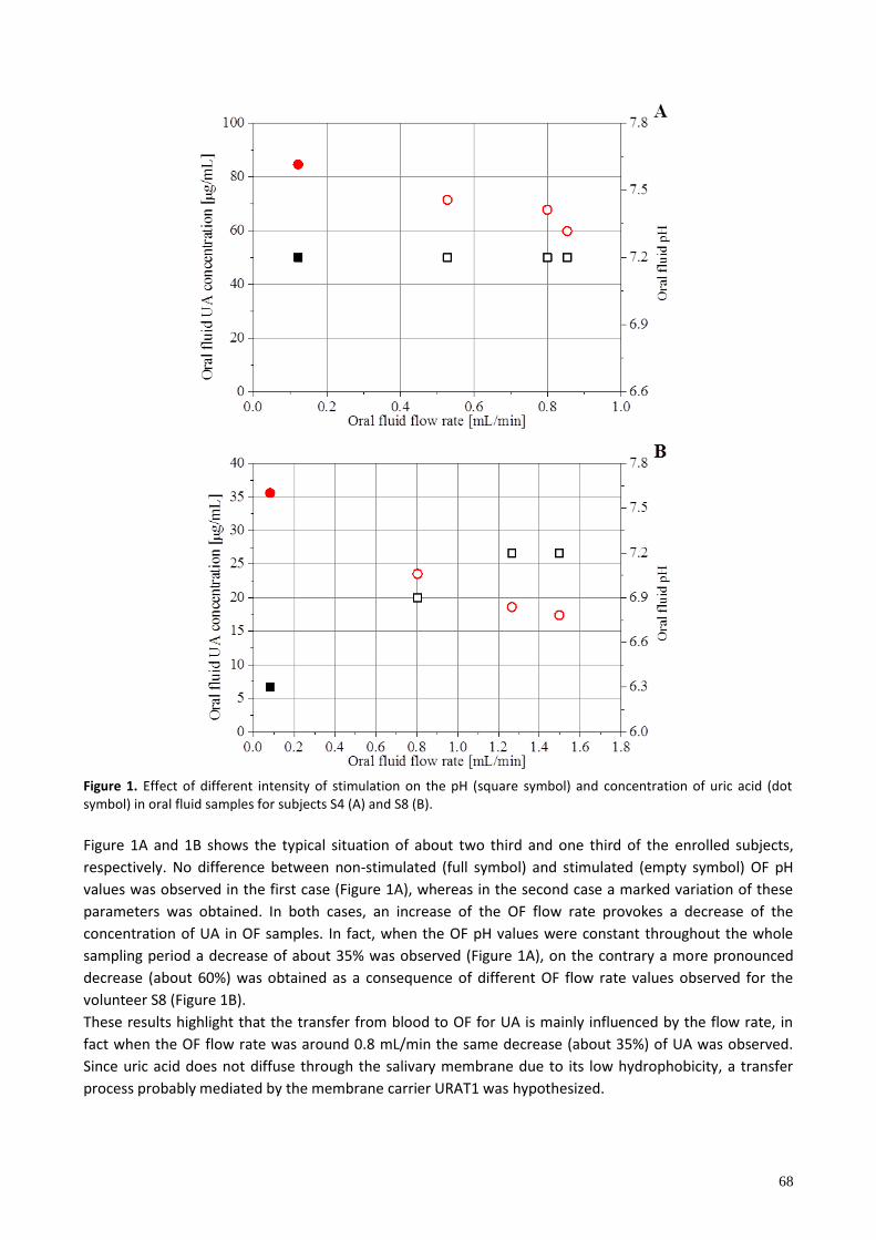
68
Figure 1. Effect of different intensity of stimulation on the pH (square symbol) and concentration of uric acid (dot symbol) in oral fluid samples for subjects S4 (A) and S8 (B).
Figure 1A and 1B shows the typical situation of about two third and one third of the enrolled subjects,
respectively. No difference between non-stimulated (full symbol) and stimulated (empty symbol) OF pH
values was observed in the first case (Figure 1A), whereas in the second case a marked variation of these
parameters was obtained. In both cases, an increase of the OF flow rate provokes a decrease of the
concentration of UA in OF samples. In fact, when the OF pH values were constant throughout the whole
sampling period a decrease of about 35% was observed (Figure 1A), on the contrary a more pronounced
decrease (about 60%) was obtained as a consequence of different OF flow rate values observed for the
volunteer S8 (Figure 1B).
These results highlight that the transfer from blood to OF for UA is mainly influenced by the flow rate, in
fact when the OF flow rate was around 0.8 mL/min the same decrease (about 35%) of UA was observed.
Since uric acid does not diffuse through the salivary membrane due to its low hydrophobicity, a transfer
process probably mediated by the membrane carrier URAT1 was hypothesized.