Embed Size (px)
Citation preview
4
Borderline and Malignant Surface Epithelial – Stromal Tumors of the Ovary
Susanna Syriac1, Faith Ough2 and Paulette Mhawech-Fauceglia3 1Roswell Park Cancer Institute, Department of Pathology,
Division of Surgical Oncologic Pathology Buffalo, 2University of Southern California, Department of Pathology,
Division of Cytopathology, Los Angeles, 3University of Southern California, Department of Pathology,
Division of Gynecologic Oncologic Pathology, Los Angeles USA
1. Introduction
Epithelial ovarian carcinoma (EOC) is the fourth leading cause of cancer mortality among women in western countries. The incidence of newly diagnosed EOC in the US is estimated to be 22,430 cases per year with 15,280 deaths (Jamel A et al., 2006). Surface epithelial-stromal tumors are the most common neoplasms of the ovary. Their origin is likely the epithelium lining the ovarian surface and/or invaginations of this lining into the superficial cortex of the ovary. They occur in women of reproductive age and older. They are usually subclassified as benign, borderline and malignant. Due to the numerous histologic types of ovarian neoplasms, we will limit our discussion to the most common epithelial stromal tumors. We will be discussing the gross appearances, microscopic patterns and differential diagnosis. Based on the 2002 World Health Organization (WHO) classification of ovarian tumors
(Tavassoli FA and Devilee P, 2003), Borderline and Malignant Surface-epithelial stromal
tumors are classified as:
1.1 WHO classification
Serous tumors
Malignant Adenocarcinoma Surface papillary adenocarcinoma Adenocarcinofibroma Borderline tumor Papillary cystic tumor Adenofibroma and cystadenofibroma
Mucinous tumors
Malignant Adenocarcinoma
www.intechopen.com
Ovarian Cancer – Clinical and Therapeutic Perspectives
56
Adenocarcinofibroma Borderline tumor Intestinal type Endocervical type Mucinous cystic tumor with mural nodules Mucinous cystic tumor with pseuodomyxoma peritonei
Endometrioid tumors including variants with squamous differentiation
Malignant Adenocarcinoma, NOS Adenocarcinofibroma Malignant mullerian mixed tumor (carcinosarcoma) Adenosarcoma Endometrial stromal sarcoma, low grade Undifferentiated sarcoma Borderline tumor Cystic tumor Adenofibroma and cystadenofibroma Clear cell tumors Malignant Adenocarcinoma Adenocarcinofibroma Borderline tumor Cystic tumor Adenofibroma and cystadenofibroma Transitional cell tumors Malignant
Transitional cell carcinoma (non-Brenner type)
Malignant Brenner tumor
Borderline
Borderline Brenner tumor Squamous cell tumors
Squamous cell carcinoma Mixed epithelial tumors Undifferentiated and unclassified tumors.
2. Serous tumors
2.1 Borderline tumors Serous borderline tumors (SBT) represent 25% to 30% of non benign serous tumors and
occur in women 30-50 years of age. In the majority of cases they are unilateral and usually
present at an early stage (stage I) (Prat J and de Nictolis M., 2002). The WHO defines SBT as
an “ovarian tumor of low malignant potential exhibiting an atypical epithelial proliferation
of serous type cells greater than that seen in its benign counterpart but without destructive
stromal invasion”.
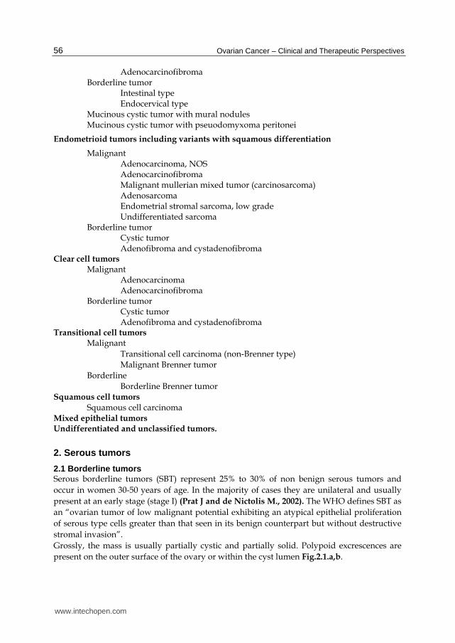
Grossly, the mass is usually partially cystic and partially solid. Polypoid excrescences are
present on the outer surface of the ovary or within the cyst lumen Fig.2.1.a,b.
www.intechopen.com
Borderline and Malignant Surface Epithelial –Stromal Tumors of the Ovary
57
Fig. 2.1.a. Borderline serous tumor (BST). The ovary shows polypoid excrescences on its outer surface.
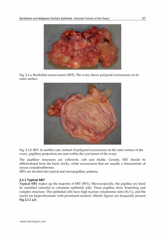
Fig. 2.1.b. BST. In another case, instead of polypoid excrescences on the outer surface of the ovary, papillary projections are seen within the cyst lumen of the ovary.
The papillary structures are yellowish, soft and friable. Grossly, SBT should be
differentiated from the hard, stocky, white excrescences that are usually a characteristic of
serous cystadenofibroma.
SBTs are divided into typical and micropapillary patterns.
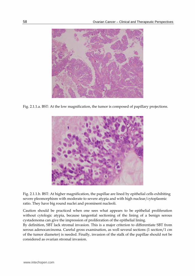
2.1.1 Typical SBT Typical SBT makes up the majority of SBT (90%). Microscopically, the papillae are lined
by stratified cuboidal to columnar epithelial cells. These papillae show branching and
complex structure. The epithelial cells have high nuclear cytoplasmic ratio (N/C), and the
nuclei are hyperchromatic with prominent nucleoli. Mitotic figures are frequently present
Fig 2.1.1 a,b.
www.intechopen.com
Ovarian Cancer – Clinical and Therapeutic Perspectives
58
Fig. 2.1.1.a. BST: At the low magnification, the tumor is composed of papillary projections.
Fig. 2.1.1.b. BST: At higher magnification, the papillae are lined by epithelial cells exhibiting
severe pleomorphism with moderate to severe atypia and with high nuclear/cytoplasmic
ratio. They have big round nuclei and prominent nucleoli.
Caution should be practiced when one sees what appears to be epithelial proliferation
without cytologic atypia, because tangential sectioning of the lining of a benign serous
cystadenoma can give the impression of proliferation of the epithelial lining.
By definition, SBT lack stromal invasion. This is a major criterion to differentiate SBT from
serous adenocarcinoma. Careful gross examination, as well several sections (1 section/1 cm
of the tumor diameter) is needed. Finally, invasion of the stalk of the papillae should not be
considered as ovarian stromal invasion.
www.intechopen.com
Borderline and Malignant Surface Epithelial –Stromal Tumors of the Ovary
59
2.1.2 SBT with micropapillary pa ttern or micropapillary SBT (MSBT ) SBT with micropapillary pattern or micropapillary SBT (MSBT) accounts 5-10% of all
SBTs. The significance of this subtype has generated a lot of debate in pathology. Some
authors have found a close association between MSBT and invasive implants and urged to
call this entity as “micropapillary serous carcinoma”. Yet others prefer the terminology of
MSBT, avoiding the use of the term of “carcinoma”, to minimize the possibility of over
treating patients (Chang SJ et al., 2008; Sehdev S et al., 2003). The general agreement on
the significance of micropapillary architecture in SBTs is that there is a significant increase
in incidence of invasive peritoneal implants (Burks R et al., 1996). Molecular studies show
that MSBT has a similar gene expression profile as low-grade serous carcinoma (LG-
serous carcinoma) and distinct from typical SBT [May T et al., 2010]. The underlying
genes involved in the pathogenesis of LG-serous carcinoma, and in MBST include
mutations in a number of different genes including KRAS and BRAF. Actually, MSBT is
the only surface-epithelial stromal tumor with a well defined adenoma-carcinoma
sequence, where LG serous is thought to arise in a stepwise fashion from a benign
cystadenoma through BST to an invasive LG-serous carcinoma (Kurman RJ et al., 2008).
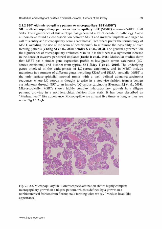
Microscopically, MSBTs shows highly complex micropapillary growth in a filigree
pattern, growing in a nonhierarchical fashion from stalk. It has been described as
“Medusa head” like appearance. Micropapillae are at least five times as long as they are
wide. Fig 2.1.2 a,b.
Fig. 2.1.2.a. Micropapillary SBT: Microscopic examination shows highly complex
micropapillary growth in a filigree pattern, which is defined by a growth in a
nonhierarchical fashion from fibrous stalk forming what we say “Medusa head’ like
appearance.
www.intechopen.com
Ovarian Cancer – Clinical and Therapeutic Perspectives
60
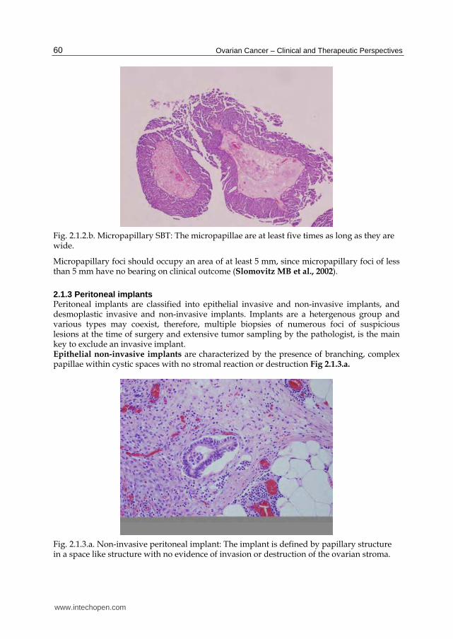
Fig. 2.1.2.b. Micropapillary SBT: The micropapillae are at least five times as long as they are wide.
Micropapillary foci should occupy an area of at least 5 mm, since micropapillary foci of less than 5 mm have no bearing on clinical outcome (Slomovitz MB et al., 2002).
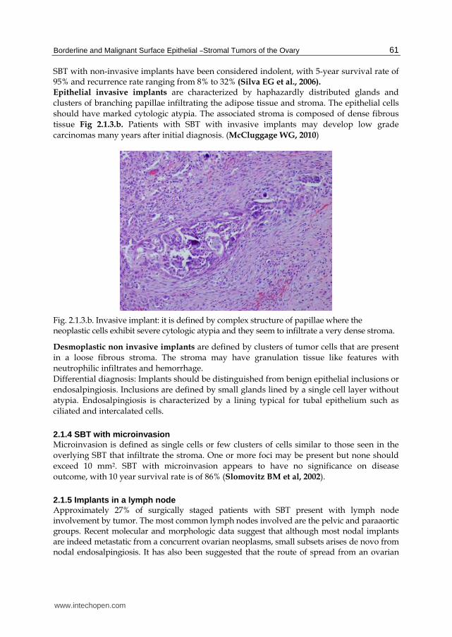
2.1.3 Peritoneal implants Peritoneal implants are classified into epithelial invasive and non-invasive implants, and desmoplastic invasive and non-invasive implants. Implants are a hetergenous group and various types may coexist, therefore, multiple biopsies of numerous foci of suspicious lesions at the time of surgery and extensive tumor sampling by the pathologist, is the main key to exclude an invasive implant. Epithelial non-invasive implants are characterized by the presence of branching, complex papillae within cystic spaces with no stromal reaction or destruction Fig 2.1.3.a.
Fig. 2.1.3.a. Non-invasive peritoneal implant: The implant is defined by papillary structure in a space like structure with no evidence of invasion or destruction of the ovarian stroma.
www.intechopen.com
Borderline and Malignant Surface Epithelial –Stromal Tumors of the Ovary
61
SBT with non-invasive implants have been considered indolent, with 5-year survival rate of 95% and recurrence rate ranging from 8% to 32% (Silva EG et al., 2006). Epithelial invasive implants are characterized by haphazardly distributed glands and
clusters of branching papillae infiltrating the adipose tissue and stroma. The epithelial cells
should have marked cytologic atypia. The associated stroma is composed of dense fibrous
tissue Fig 2.1.3.b. Patients with SBT with invasive implants may develop low grade
carcinomas many years after initial diagnosis. (McCluggage WG, 2010)
Fig. 2.1.3.b. Invasive implant: it is defined by complex structure of papillae where the neoplastic cells exhibit severe cytologic atypia and they seem to infiltrate a very dense stroma.
Desmoplastic non invasive implants are defined by clusters of tumor cells that are present
in a loose fibrous stroma. The stroma may have granulation tissue like features with
neutrophilic infiltrates and hemorrhage.
Differential diagnosis: Implants should be distinguished from benign epithelial inclusions or
endosalpingiosis. Inclusions are defined by small glands lined by a single cell layer without
atypia. Endosalpingiosis is characterized by a lining typical for tubal epithelium such as
ciliated and intercalated cells.
2.1.4 SBT with microinvasion Microinvasion is defined as single cells or few clusters of cells similar to those seen in the
overlying SBT that infiltrate the stroma. One or more foci may be present but none should
exceed 10 mm2. SBT with microinvasion appears to have no significance on disease
outcome, with 10 year survival rate is of 86% (Slomovitz BM et al, 2002).
2.1.5 Implants in a lymph node Approximately 27% of surgically staged patients with SBT present with lymph node involvement by tumor. The most common lymph nodes involved are the pelvic and paraaortic groups. Recent molecular and morphologic data suggest that although most nodal implants are indeed metastatic from a concurrent ovarian neoplasms, small subsets arises de novo from nodal endosalpingiosis. It has also been suggested that the route of spread from an ovarian
www.intechopen.com
Ovarian Cancer – Clinical and Therapeutic Perspectives
62
SBT to lymph nodes might be via a peritoneal route and not lymphatic. The morphology of the implant is similar to that occurring in the ovary. Lymph node involvement does not adversely impact the overall survival of patients with SBT of the ovary [Fadare O, 2009]. The major differential diagnosis is endosalpingiosis and the criteria are cited previously in the text.
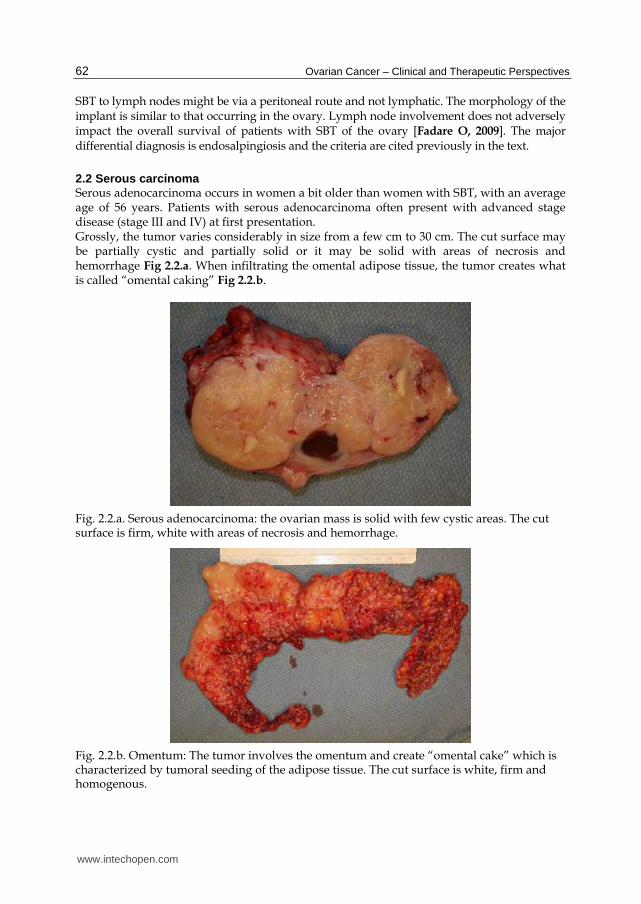
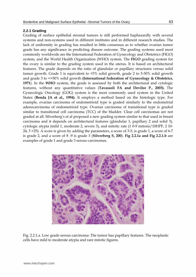
2.2 Serous carcinoma Serous adenocarcinoma occurs in women a bit older than women with SBT, with an average age of 56 years. Patients with serous adenocarcinoma often present with advanced stage disease (stage III and IV) at first presentation. Grossly, the tumor varies considerably in size from a few cm to 30 cm. The cut surface may be partially cystic and partially solid or it may be solid with areas of necrosis and hemorrhage Fig 2.2.a. When infiltrating the omental adipose tissue, the tumor creates what is called “omental caking” Fig 2.2.b.
Fig. 2.2.a. Serous adenocarcinoma: the ovarian mass is solid with few cystic areas. The cut surface is firm, white with areas of necrosis and hemorrhage.
Fig. 2.2.b. Omentum: The tumor involves the omentum and create “omental cake” which is characterized by tumoral seeding of the adipose tissue. The cut surface is white, firm and homogenous.
www.intechopen.com
Borderline and Malignant Surface Epithelial –Stromal Tumors of the Ovary
63
2.2.1 Grading Grading of surface epithelial stromal tumors is still performed haphazardly with several
systems and non-systems used in different institutes and in different research studies. The
lack of uniformity in grading has resulted in little consensus as to whether ovarian tumor
grade has any significance in predicting disease outcome. The grading systems used most
commonly worldwide are the International Federation of Gynecology and Obstetrics (FIGO)
system, and the World Health Organization (WHO) system. The FIGO grading system for
the ovary is similar to the grading system used in the uterus. It is based on architectural
features. The grade depends on the ratio of glandular or papillary structures versus solid
tumor growth. Grade 1 is equivalent to <5% solid growth, grade 2 to 5-50% solid growth
and grade 3 to =>50% solid growth (International federation of Gynecology & Obstetrics,
1971). In the WHO system, the grade is assessed by both the architectural and cytologic
features, without any quantitative values (Tavassoli FA and Devilee P., 2003). The
Gynecologic Oncology (GOG) system is the most commonly used system in the United
States (Benda JA et al., 1994). It employs a method based on the histologic type. For
example, ovarian carcinoma of endometrioid type is graded similarly to the endometrial
adenocarcinoma of endometrioid type. Ovarian carcinoma of transitional type is graded
similar to transitional cell carcinoma (TCC) of the bladder. Clear cell carcinomas are not
graded at all. Silverberg’s et al proposed a new grading system similar to that used in breast
carcinoma and it depends on architectural features (glandular 1, papillary 2 and solid 3),
cytologic atypia (mild 1, moderate 2, severe 3), and mitotic rate (1 0-9 mitosis/10HPF, 2 10-
24, 3 >25). A score is given by adding the parameters, a score of 3-5, is grade 1, a score of 6-7
is grade 2, and a score of 8 -9 is grade 3 (Silverberg S, 200). Fig 2.2.1a and Fig 2.2.1.b are
examples of grade 1 and grade 3 serous carcinomas.
Fig. 2.2.1.a. Low grade serous carcinoma: The tumor has papillary features. The neoplastic cells have mild to moderate atypia and rare mitotic figures.
www.intechopen.com
Ovarian Cancer – Clinical and Therapeutic Perspectives
64
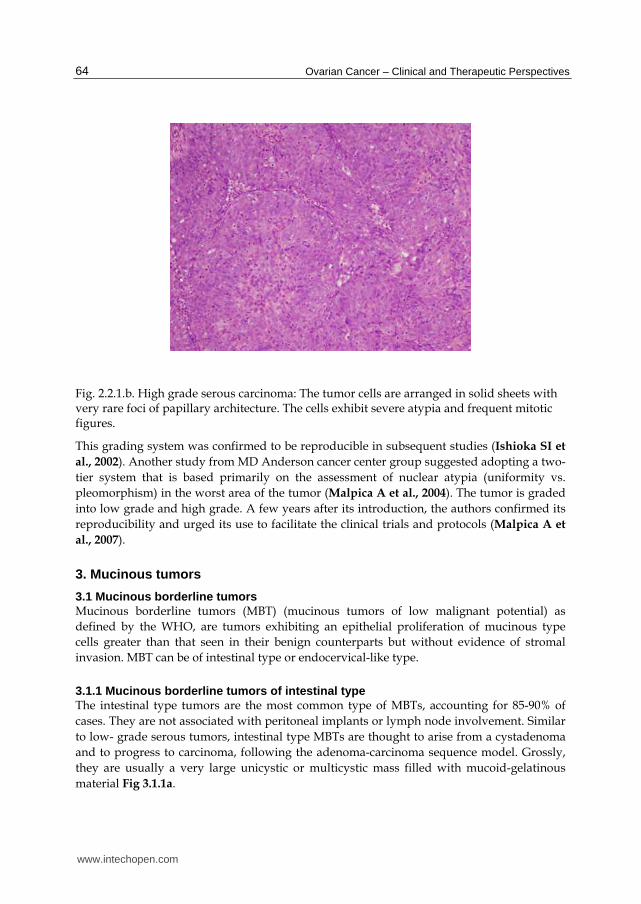
Fig. 2.2.1.b. High grade serous carcinoma: The tumor cells are arranged in solid sheets with very rare foci of papillary architecture. The cells exhibit severe atypia and frequent mitotic figures.
This grading system was confirmed to be reproducible in subsequent studies (Ishioka SI et
al., 2002). Another study from MD Anderson cancer center group suggested adopting a two-
tier system that is based primarily on the assessment of nuclear atypia (uniformity vs.
pleomorphism) in the worst area of the tumor (Malpica A et al., 2004). The tumor is graded
into low grade and high grade. A few years after its introduction, the authors confirmed its
reproducibility and urged its use to facilitate the clinical trials and protocols (Malpica A et
al., 2007).
3. Mucinous tumors
3.1 Mucinous borderline tumors Mucinous borderline tumors (MBT) (mucinous tumors of low malignant potential) as
defined by the WHO, are tumors exhibiting an epithelial proliferation of mucinous type
cells greater than that seen in their benign counterparts but without evidence of stromal
invasion. MBT can be of intestinal type or endocervical-like type.
3.1.1 Mucinous borderline tumors of intestinal type The intestinal type tumors are the most common type of MBTs, accounting for 85-90% of
cases. They are not associated with peritoneal implants or lymph node involvement. Similar
to low- grade serous tumors, intestinal type MBTs are thought to arise from a cystadenoma
and to progress to carcinoma, following the adenoma-carcinoma sequence model. Grossly,
they are usually a very large unicystic or multicystic mass filled with mucoid-gelatinous
material Fig 3.1.1a.
www.intechopen.com

Borderline and Malignant Surface Epithelial –Stromal Tumors of the Ovary
65
Fig. 3.1.1.a. Mucinous borderline tumor (MBT): The cut surface of the ovarian mass shows
multiple cysts filled with gelatinous material. However in some areas the wall of the cyst
seemed to be thickened.
Histologically, the lining of the cyst is composed of stratified lining of epithelial cells having
high N/C ratio and prominent nucleoli Fig 3.1.1.b,c. Goblet cells and Paneth cells are
present. No stromal invasion is seen.
Fig. 3.1.1.b. MBT: At low magnification, the tumor is composed of proliferation of glands which some are cystically dilated separated by abundant intervening stroma.
www.intechopen.com
Ovarian Cancer – Clinical and Therapeutic Perspectives
66
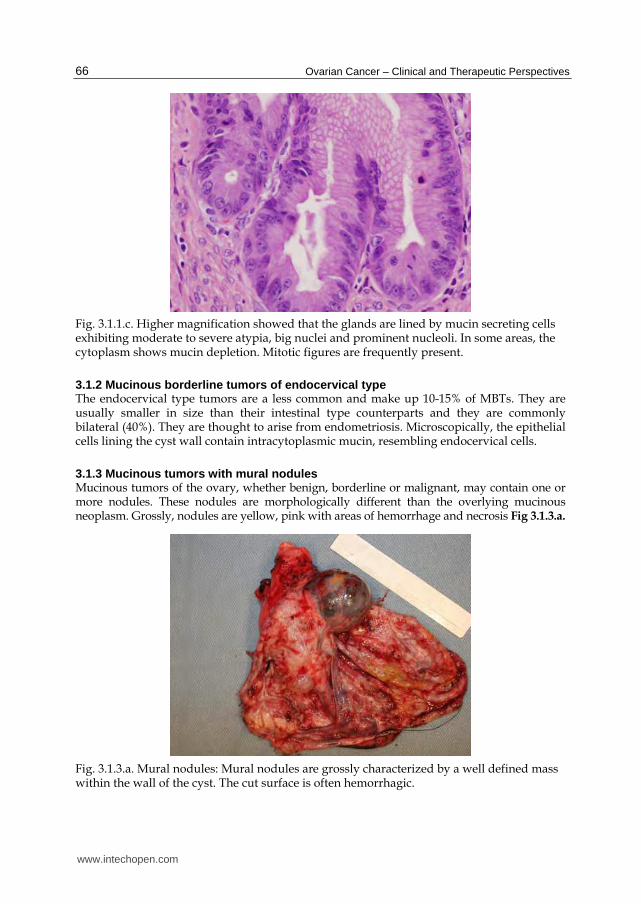
Fig. 3.1.1.c. Higher magnification showed that the glands are lined by mucin secreting cells exhibiting moderate to severe atypia, big nuclei and prominent nucleoli. In some areas, the cytoplasm shows mucin depletion. Mitotic figures are frequently present.
3.1.2 Mucinous borderline tumors of endocervical type The endocervical type tumors are a less common and make up 10-15% of MBTs. They are usually smaller in size than their intestinal type counterparts and they are commonly bilateral (40%). They are thought to arise from endometriosis. Microscopically, the epithelial cells lining the cyst wall contain intracytoplasmic mucin, resembling endocervical cells.
3.1.3 Mucinous tumors with mural nodules Mucinous tumors of the ovary, whether benign, borderline or malignant, may contain one or more nodules. These nodules are morphologically different than the overlying mucinous neoplasm. Grossly, nodules are yellow, pink with areas of hemorrhage and necrosis Fig 3.1.3.a.
Fig. 3.1.3.a. Mural nodules: Mural nodules are grossly characterized by a well defined mass within the wall of the cyst. The cut surface is often hemorrhagic.
www.intechopen.com

Borderline and Malignant Surface Epithelial –Stromal Tumors of the Ovary
67
Microscopically, the mural nodules may be malignant (anaplastic, sarcoma or carcinosarcoma) or benign (sarcoma-like). It is important to distinguish between benign and malignant mural nodules, because benign mural nodules are of no prognostic significance. Immunohistochemistry is a very helpful tool for this purpose. Sarcoma-like nodules are composed of a heterogenous cell population of cells including spindle cells, giant cells, mononuclear cells and inflammatory cells. The cells of the sarcoma- like nodules are negative or very weakly positive for cytokeratin Fig3.1.3.b.
Fig. 3.1.3.b. Sarcoma-like nodules: They are composed of heterogenous cell population including spindle cells, giant cells, mononuclear cells and inflammatory cells.
Anaplastic sarcoma mural nodules are composed of diffuse sheets of spindled or large rhabdoid-looking cells with abundant eosinophilic cytoplasm and prominent nucleoli fig 3.1.3.c,d. These cells are usually strongly positive for cytokeratin fig 3.1.3.e.
Fig. 3.1.3.c. Anaplastic nodules: They are characterized by proliferation of spindle cells.
www.intechopen.com
Ovarian Cancer – Clinical and Therapeutic Perspectives
68
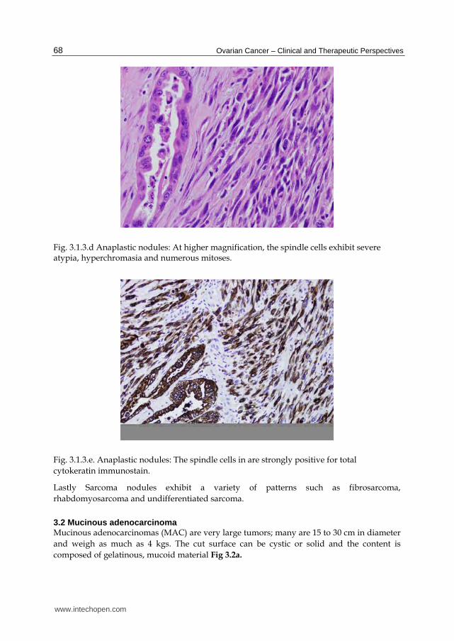
Fig. 3.1.3.d Anaplastic nodules: At higher magnification, the spindle cells exhibit severe atypia, hyperchromasia and numerous mitoses.
Fig. 3.1.3.e. Anaplastic nodules: The spindle cells in are strongly positive for total
cytokeratin immunostain.
Lastly Sarcoma nodules exhibit a variety of patterns such as fibrosarcoma,
rhabdomyosarcoma and undifferentiated sarcoma.
3.2 Mucinous adenocarcinoma Mucinous adenocarcinomas (MAC) are very large tumors; many are 15 to 30 cm in diameter
and weigh as much as 4 kgs. The cut surface can be cystic or solid and the content is
composed of gelatinous, mucoid material Fig 3.2a.
www.intechopen.com
Borderline and Malignant Surface Epithelial –Stromal Tumors of the Ovary
69
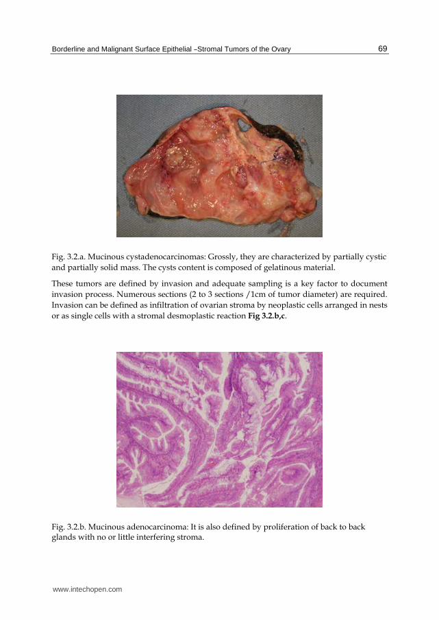
Fig. 3.2.a. Mucinous cystadenocarcinomas: Grossly, they are characterized by partially cystic
and partially solid mass. The cysts content is composed of gelatinous material.
These tumors are defined by invasion and adequate sampling is a key factor to document
invasion process. Numerous sections (2 to 3 sections /1cm of tumor diameter) are required.
Invasion can be defined as infiltration of ovarian stroma by neoplastic cells arranged in nests
or as single cells with a stromal desmoplastic reaction Fig 3.2.b,c.
Fig. 3.2.b. Mucinous adenocarcinoma: It is also defined by proliferation of back to back glands with no or little interfering stroma.
www.intechopen.com

Ovarian Cancer – Clinical and Therapeutic Perspectives
70
Fig. 3.2.c. Mucinous adenocarcinoma: These glands are cytologically malignant with severe atypia, large nucleoli, loss of cytoplasmic mucin and numerous mitosis.
However, one needs not to see typical stromal invasion with desmoplastic reaction to
diagnose MAC, because invasion can also be defined as neoplastic glands which are back to
back with no intervening stroma Fig 3.2d. Similar to MBTs, the epithelial lining in MAC can
be of intestinal or endocervical type. MAC should be distinguished from metastatic
adenocarcinoma from colonic origin. Metastatic colonic carcinomas are usually bilateral.
Morphologically, they are characterized by glandular proliferation with abundant dirty
necrosis and nuclear debris within amorphous necrotic tissue. The glands are lined by
stratified cells with prominent atypia and mitosis Fig 3.2.e
Fig. 3.2.d. Mucinous cystadenocarcinoma: It is defined by invasion of the ovarian stroma by small glands and nests of tumor cells . These glands are cytologically malignant and infiltrate the stroma in disorderly fashion.
www.intechopen.com
Borderline and Malignant Surface Epithelial –Stromal Tumors of the Ovary
71
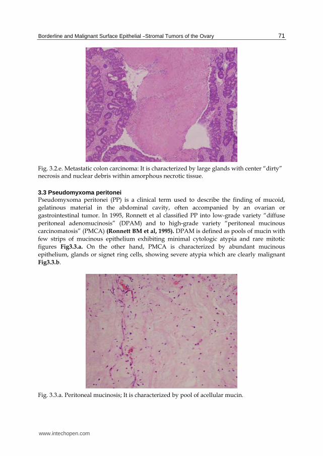
Fig. 3.2.e. Metastatic colon carcinoma: It is characterized by large glands with center “dirty” necrosis and nuclear debris within amorphous necrotic tissue.
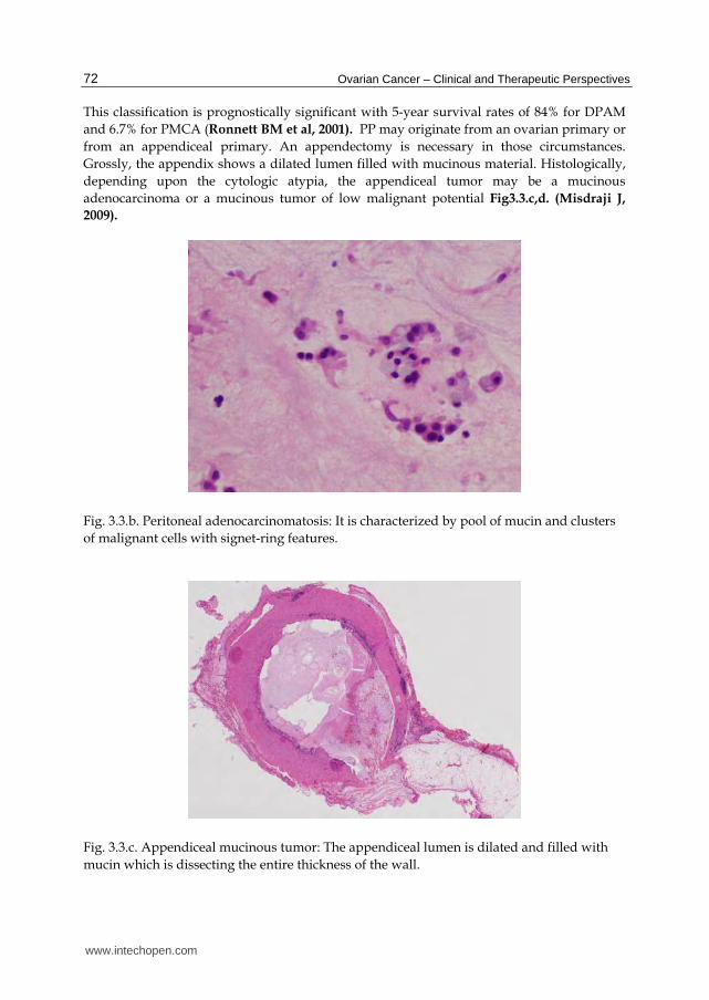
3.3 Pseudomyxoma peritonei Pseudomyxoma peritonei (PP) is a clinical term used to describe the finding of mucoid,
gelatinous material in the abdominal cavity, often accompanied by an ovarian or
gastrointestinal tumor. In 1995, Ronnett et al classified PP into low-grade variety “diffuse
peritoneal adenomucinosis” (DPAM) and to high-grade variety “peritoneal mucinous
carcinomatosis” (PMCA) (Ronnett BM et al, 1995). DPAM is defined as pools of mucin with
few strips of mucinous epithelium exhibiting minimal cytologic atypia and rare mitotic
figures Fig3.3.a. On the other hand, PMCA is characterized by abundant mucinous
epithelium, glands or signet ring cells, showing severe atypia which are clearly malignant
Fig3.3.b.
Fig. 3.3.a. Peritoneal mucinosis; It is characterized by pool of acellular mucin.
www.intechopen.com
Ovarian Cancer – Clinical and Therapeutic Perspectives
72
This classification is prognostically significant with 5-year survival rates of 84% for DPAM
and 6.7% for PMCA (Ronnett BM et al, 2001). PP may originate from an ovarian primary or
from an appendiceal primary. An appendectomy is necessary in those circumstances.
Grossly, the appendix shows a dilated lumen filled with mucinous material. Histologically,
depending upon the cytologic atypia, the appendiceal tumor may be a mucinous
adenocarcinoma or a mucinous tumor of low malignant potential Fig3.3.c,d. (Misdraji J,
2009).
Fig. 3.3.b. Peritoneal adenocarcinomatosis: It is characterized by pool of mucin and clusters
of malignant cells with signet-ring features.
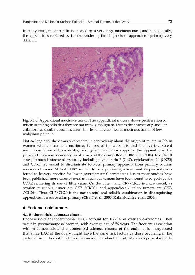
Fig. 3.3.c. Appendiceal mucinous tumor: The appendiceal lumen is dilated and filled with
mucin which is dissecting the entire thickness of the wall.
www.intechopen.com
Borderline and Malignant Surface Epithelial –Stromal Tumors of the Ovary
73
In many cases, the appendix is encased by a very large mucinous mass, and histologically,
the appendix is replaced by tumor, rendering the diagnosis of appendiceal primary very
difficult.
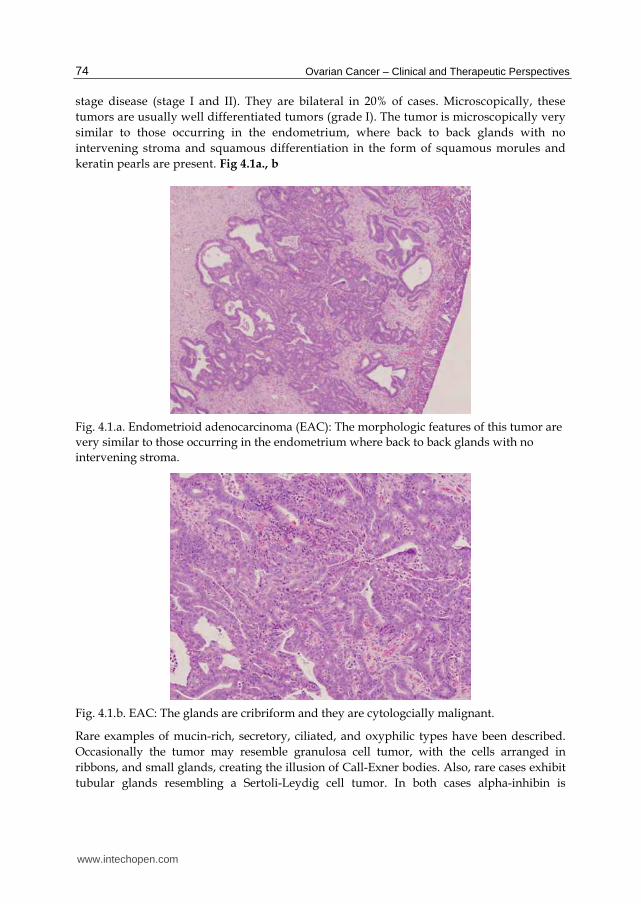
Fig. 3.3.d. Appendiceal mucinous tumor: The appendiceal mucosa shows proliferation of mucin-secreting cells that they are not frankly malignant. Due to the absence of glandular cribriform and submucosal invasion, this lesion is classified as mucinous tumor of low malignant potential.
Not so long ago, there was a considerable controversy about the origin of mucin in PP, in
women with concomitant mucinous tumors of the appendix and the ovaries. Recent
immunohistochemical, molecular, and genetic evidence supports the appendix as the
primary tumor and secondary involvement of the ovary (Ronnet BM et al, 2004) In difficult
cases, immunohistochemistry study including cytokeratin 7 (CK7), cytokeration 20 (CK20)
and CDX2 are useful to discriminate between primary appendix from primary ovarian
mucinous tumors. At first CDX2 seemed to be a promising marker and its positivity was
found to be very specific for lower gastrointestinal carcinomas but as more studies have
been published, more cases of ovarian mucinous tumors have been found to be positive for
CDX2 rendering its use of little value. On the other hand CK7/CK20 is more useful, as
ovarian mucinous tumor are CK7+/CK20+ and appendiceal/ colon tumors are CK7-
/CK20+. Thus, CK7/CK20 is the most useful and reliable combination in distinguishing
appendiceal versus ovarian primary (Chu P et al., 2000; Kaimaktchiev et al., 2004).
4. Endometrioid tumors
4.1 Endometrioid adenocarcinoma Endometrioid adenocarcinoma (EAC) account for 10-20% of ovarian carcinomas. They
occur in postmenauposal women, with average age of 56 years. The frequent association
with endometriosis and endometrioid adenocarcinoma of the endometrium suggested
that some EAC of the ovary might have the same risk factors as those occurring in the
endometrium. In contrary to serous carcinomas, about half of EAC cases present as early
www.intechopen.com
Ovarian Cancer – Clinical and Therapeutic Perspectives
74
stage disease (stage I and II). They are bilateral in 20% of cases. Microscopically, these
tumors are usually well differentiated tumors (grade I). The tumor is microscopically very
similar to those occurring in the endometrium, where back to back glands with no
intervening stroma and squamous differentiation in the form of squamous morules and
keratin pearls are present. Fig 4.1a., b
Fig. 4.1.a. Endometrioid adenocarcinoma (EAC): The morphologic features of this tumor are
very similar to those occurring in the endometrium where back to back glands with no
intervening stroma.
Fig. 4.1.b. EAC: The glands are cribriform and they are cytologcially malignant.
Rare examples of mucin-rich, secretory, ciliated, and oxyphilic types have been described.
Occasionally the tumor may resemble granulosa cell tumor, with the cells arranged in
ribbons, and small glands, creating the illusion of Call-Exner bodies. Also, rare cases exhibit
tubular glands resembling a Sertoli-Leydig cell tumor. In both cases alpha-inhibin is
www.intechopen.com
Borderline and Malignant Surface Epithelial –Stromal Tumors of the Ovary
75
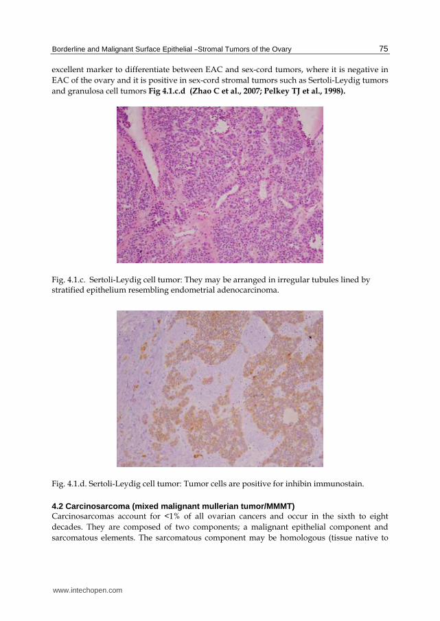
excellent marker to differentiate between EAC and sex-cord tumors, where it is negative in
EAC of the ovary and it is positive in sex-cord stromal tumors such as Sertoli-Leydig tumors
and granulosa cell tumors Fig 4.1.c.d (Zhao C et al., 2007; Pelkey TJ et al., 1998).
Fig. 4.1.c. Sertoli-Leydig cell tumor: They may be arranged in irregular tubules lined by stratified epithelium resembling endometrial adenocarcinoma.
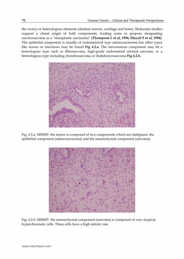
Fig. 4.1.d. Sertoli-Leydig cell tumor: Tumor cells are positive for inhibin immunostain.
4.2 Carcinosarcoma (mixed ma lignant mullerian tumor/MMMT) Carcinosarcomas account for <1% of all ovarian cancers and occur in the sixth to eight
decades. They are composed of two components; a malignant epithelial component and
sarcomatous elements. The sarcomatous component may be homologous (tissue native to
www.intechopen.com
Ovarian Cancer – Clinical and Therapeutic Perspectives
76
the ovary) or heterologous elements (skeletal muscle, cartilage and bone). Molecular studies
support a clonal origin of both components, leading some to propose designating
carcinosarcoma as a “metaplastic carcinoma” (Thompson L et al, 1996; Mayall F et al, 1994).
The epithelial component is usually of endometrioid type adenocarcinoma but other types
like serous or mucinous may be found Fig 4.2.a. The sarcomatous component may be a
homologous type such as fibrosarcoma, high-grade endometrial stromal sarcoma, or a
heterologous type including chondrosarcoma or rhabdomyosarcoma Fig 4.2.b.
Fig. 4.2.a. MMMT: the tumor is composed of two components which are malignant, the epithelial component (adenocarcinoma) and the mesenchymal component (sarcoma).
Fig. 4.2.b. MMMT: the mesenchymal component (sarcoma) is composed of very atypical,
hyperchromatic cells. These cells have a high mitotic rate.
www.intechopen.com
Borderline and Malignant Surface Epithelial –Stromal Tumors of the Ovary
77
5. Clear cell tumors
Clear cell carcinoma
Clear cell carcinomas (CCC) represent 6% of surface-epithelial tumors. They occur in
postmenopausal women, with a mean age of 57 years. CCC of the ovary has a few notable
characteristics 1- they are almost always unilateral, 2- they are admixed with
endometrioid type adenocarcinoma in 20-25% of cases, 3- they are often accompanied by
endometriosis of the same ovary, 4- they may be associated with paraneoplastic
hypercalcemia and 5- they have frequent mutations of ARID1A and PIK3CA genes
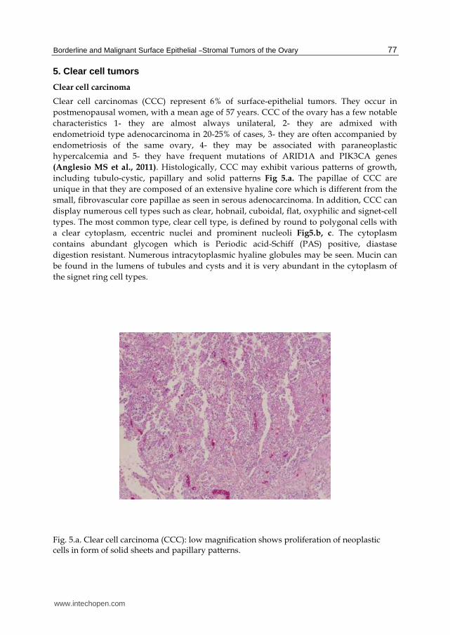
(Anglesio MS et al., 2011). Histologically, CCC may exhibit various patterns of growth,
including tubulo-cystic, papillary and solid patterns Fig 5.a. The papillae of CCC are
unique in that they are composed of an extensive hyaline core which is different from the
small, fibrovascular core papillae as seen in serous adenocarcinoma. In addition, CCC can
display numerous cell types such as clear, hobnail, cuboidal, flat, oxyphilic and signet-cell
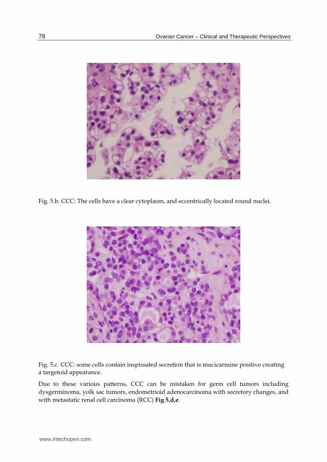
types. The most common type, clear cell type, is defined by round to polygonal cells with
a clear cytoplasm, eccentric nuclei and prominent nucleoli Fig5.b, c. The cytoplasm
contains abundant glycogen which is Periodic acid-Schiff (PAS) positive, diastase
digestion resistant. Numerous intracytoplasmic hyaline globules may be seen. Mucin can
be found in the lumens of tubules and cysts and it is very abundant in the cytoplasm of
the signet ring cell types.
Fig. 5.a. Clear cell carcinoma (CCC): low magnification shows proliferation of neoplastic cells in form of solid sheets and papillary patterns.
www.intechopen.com
Ovarian Cancer – Clinical and Therapeutic Perspectives
78
Fig. 5.b. CCC: The cells have a clear cytoplasm, and eccentrically located round nuclei.
Fig. 5.c. CCC: some cells contain inspissated secretion that is mucicarmine positive creating a targetoid appearance.
Due to these various patterns, CCC can be mistaken for germ cell tumors including
dysgerminoma, yolk sac tumors, endometrioid adenocarcinoma with secretory changes, and
with metastatic renal cell carcinoma (RCC) Fig 5.d,e.
www.intechopen.com
Borderline and Malignant Surface Epithelial –Stromal Tumors of the Ovary
79
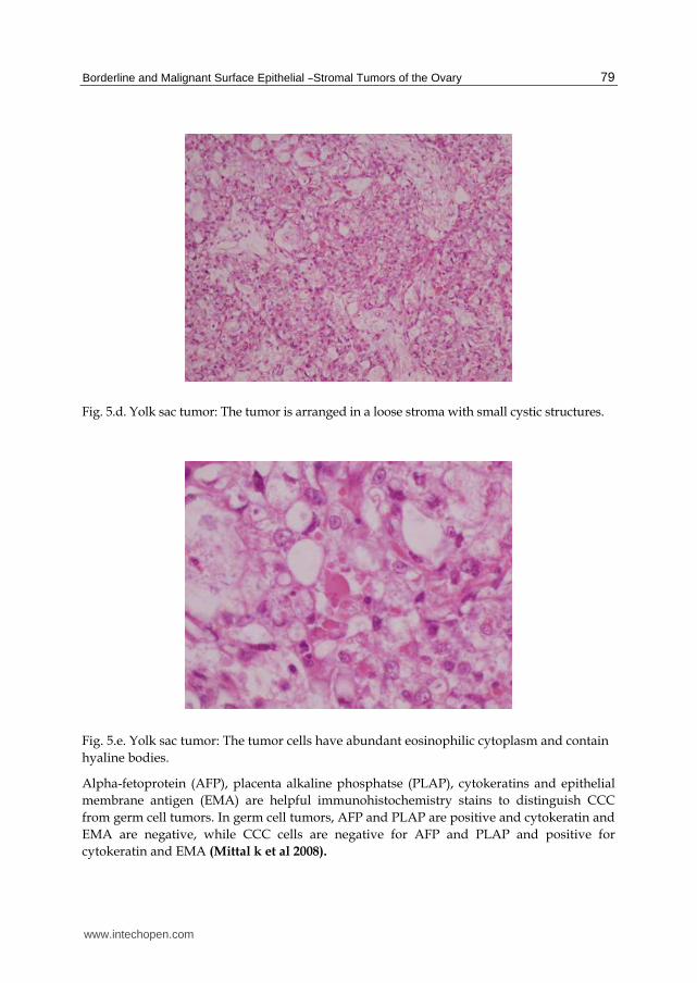
Fig. 5.d. Yolk sac tumor: The tumor is arranged in a loose stroma with small cystic structures.
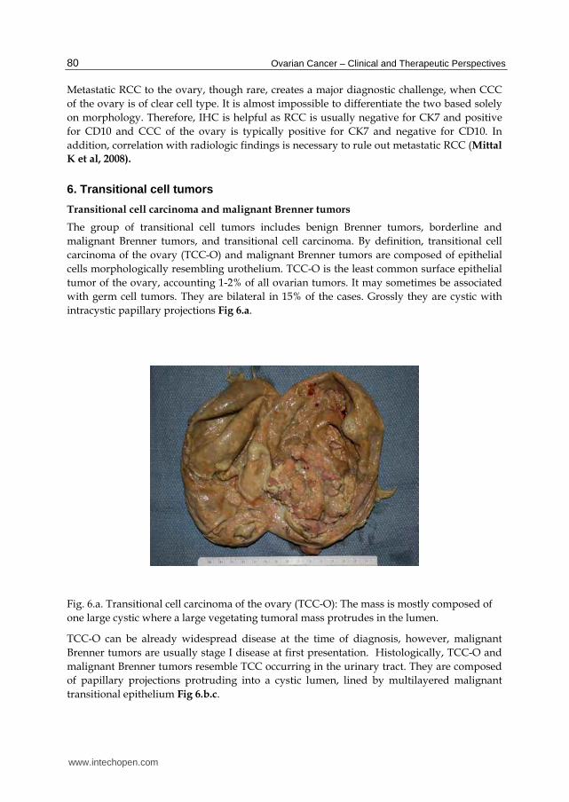
Fig. 5.e. Yolk sac tumor: The tumor cells have abundant eosinophilic cytoplasm and contain
hyaline bodies.
Alpha-fetoprotein (AFP), placenta alkaline phosphatse (PLAP), cytokeratins and epithelial
membrane antigen (EMA) are helpful immunohistochemistry stains to distinguish CCC
from germ cell tumors. In germ cell tumors, AFP and PLAP are positive and cytokeratin and
EMA are negative, while CCC cells are negative for AFP and PLAP and positive for
cytokeratin and EMA (Mittal k et al 2008).
www.intechopen.com
Ovarian Cancer – Clinical and Therapeutic Perspectives
80
Metastatic RCC to the ovary, though rare, creates a major diagnostic challenge, when CCC
of the ovary is of clear cell type. It is almost impossible to differentiate the two based solely
on morphology. Therefore, IHC is helpful as RCC is usually negative for CK7 and positive
for CD10 and CCC of the ovary is typically positive for CK7 and negative for CD10. In
addition, correlation with radiologic findings is necessary to rule out metastatic RCC (Mittal
K et al, 2008).
6. Transitional cell tumors
Transitional cell carcinoma and malignant Brenner tumors
The group of transitional cell tumors includes benign Brenner tumors, borderline and
malignant Brenner tumors, and transitional cell carcinoma. By definition, transitional cell
carcinoma of the ovary (TCC-O) and malignant Brenner tumors are composed of epithelial
cells morphologically resembling urothelium. TCC-O is the least common surface epithelial
tumor of the ovary, accounting 1-2% of all ovarian tumors. It may sometimes be associated
with germ cell tumors. They are bilateral in 15% of the cases. Grossly they are cystic with
intracystic papillary projections Fig 6.a.
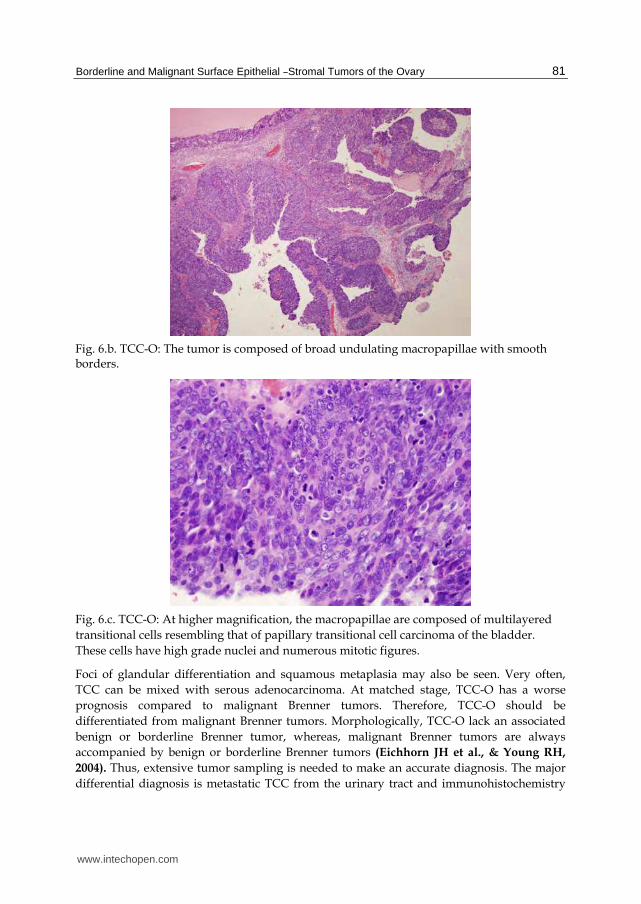
Fig. 6.a. Transitional cell carcinoma of the ovary (TCC-O): The mass is mostly composed of
one large cystic where a large vegetating tumoral mass protrudes in the lumen.
TCC-O can be already widespread disease at the time of diagnosis, however, malignant
Brenner tumors are usually stage I disease at first presentation. Histologically, TCC-O and
malignant Brenner tumors resemble TCC occurring in the urinary tract. They are composed
of papillary projections protruding into a cystic lumen, lined by multilayered malignant
transitional epithelium Fig 6.b.c.
www.intechopen.com
Borderline and Malignant Surface Epithelial –Stromal Tumors of the Ovary
81
Fig. 6.b. TCC-O: The tumor is composed of broad undulating macropapillae with smooth borders.
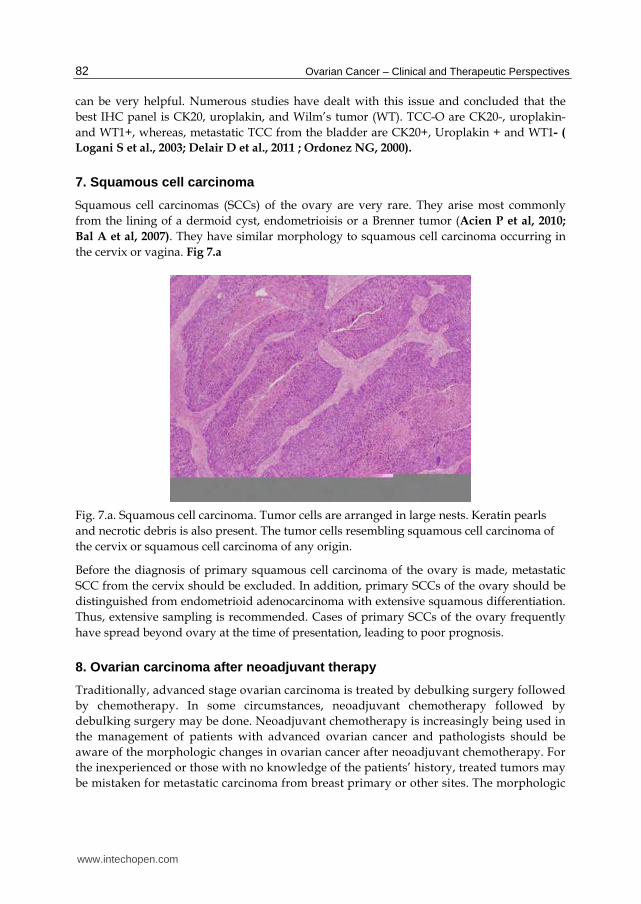
Fig. 6.c. TCC-O: At higher magnification, the macropapillae are composed of multilayered
transitional cells resembling that of papillary transitional cell carcinoma of the bladder.
These cells have high grade nuclei and numerous mitotic figures.
Foci of glandular differentiation and squamous metaplasia may also be seen. Very often,
TCC can be mixed with serous adenocarcinoma. At matched stage, TCC-O has a worse
prognosis compared to malignant Brenner tumors. Therefore, TCC-O should be
differentiated from malignant Brenner tumors. Morphologically, TCC-O lack an associated
benign or borderline Brenner tumor, whereas, malignant Brenner tumors are always
accompanied by benign or borderline Brenner tumors (Eichhorn JH et al., & Young RH,
2004). Thus, extensive tumor sampling is needed to make an accurate diagnosis. The major
differential diagnosis is metastatic TCC from the urinary tract and immunohistochemistry
www.intechopen.com
Ovarian Cancer – Clinical and Therapeutic Perspectives
82
can be very helpful. Numerous studies have dealt with this issue and concluded that the
best IHC panel is CK20, uroplakin, and Wilm’s tumor (WT). TCC-O are CK20-, uroplakin-
and WT1+, whereas, metastatic TCC from the bladder are CK20+, Uroplakin + and WT1- (
Logani S et al., 2003; Delair D et al., 2011 ; Ordonez NG, 2000).
7. Squamous cell carcinoma
Squamous cell carcinomas (SCCs) of the ovary are very rare. They arise most commonly
from the lining of a dermoid cyst, endometrioisis or a Brenner tumor (Acien P et al, 2010;
Bal A et al, 2007). They have similar morphology to squamous cell carcinoma occurring in
the cervix or vagina. Fig 7.a
Fig. 7.a. Squamous cell carcinoma. Tumor cells are arranged in large nests. Keratin pearls
and necrotic debris is also present. The tumor cells resembling squamous cell carcinoma of
the cervix or squamous cell carcinoma of any origin.
Before the diagnosis of primary squamous cell carcinoma of the ovary is made, metastatic
SCC from the cervix should be excluded. In addition, primary SCCs of the ovary should be
distinguished from endometrioid adenocarcinoma with extensive squamous differentiation.
Thus, extensive sampling is recommended. Cases of primary SCCs of the ovary frequently
have spread beyond ovary at the time of presentation, leading to poor prognosis.
8. Ovarian carcinoma after neoadjuvant therapy
Traditionally, advanced stage ovarian carcinoma is treated by debulking surgery followed
by chemotherapy. In some circumstances, neoadjuvant chemotherapy followed by
debulking surgery may be done. Neoadjuvant chemotherapy is increasingly being used in
the management of patients with advanced ovarian cancer and pathologists should be
aware of the morphologic changes in ovarian cancer after neoadjuvant chemotherapy. For
the inexperienced or those with no knowledge of the patients’ history, treated tumors may
be mistaken for metastatic carcinoma from breast primary or other sites. The morphologic
www.intechopen.com
Borderline and Malignant Surface Epithelial –Stromal Tumors of the Ovary
83
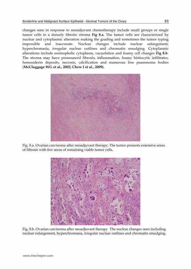
changes seen in response to neoadjuvant chemotherapy include small groups or single
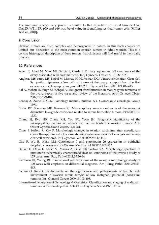
tumor cells in a densely fibrotic stroma Fig 8.a. The tumor cells are characterized by
nuclear and cytoplasmic alteration making the grading and sometimes the tumor typing
impossible and inaccurate. Nuclear changes include nuclear enlargement,
hyperchromasia, irregular nuclear outlines and chromatin smudging. Cytoplasmic
alterations include eosinopholic cytoplasm, vacuolation and foamy cell changes Fig 8.b.
The stroma may have pronounced fibrosis, inflammation, foamy histiocytic infiltrates,
hemosiderin deposits, necrosis, calcification and numerous free psammoma bodies
(McCluggage WG et al., 2002; Chew I et al., 2009).
Fig. 8.a. Ovarian carcinoma after neoadjuvant therapy: The tumor presents extensive areas of fibrosis with few areas of remaining viable tumor cells.
Fig. 8.b. Ovarian carcinoma after neoadjuvant therapy The nuclear changes seen including nuclear enlargement, hyperchromasia, irregular nuclear outlines and chromatin smudging.
www.intechopen.com
Ovarian Cancer – Clinical and Therapeutic Perspectives
84
The immunohistochemistry profile is similar to that of native untreated tumors. Ck7, CA125, WT1, ER, p53 and p16 may be of value in identifying residual tumor cells [Miller K et al., 2008].
9. Conclusion
Ovarian tumors are often complex and heterogenous in nature. In this book chapter we limited our discussion to the most common ovarian tumors in adult women. This is a concise histological description of these tumors that clinicians will find useful in their daily practice.
10. References
Acien P, Abad M, Maol MJ, Garcia S, Garde J. Primary squamous cell carcinoma of the ovary associated with endometriosis. Int J Gynaecol Obstet 2010;108:16-20.
Anglesio MS, carey MS, Kobel M, Mackay H, Huntsman DG; Vancouver Ovarian Clear Cell Symposium Speakers. Clear cell carcinoma of the ovary: a report from the first ovarian clear cell symposium, June 24th, 2010. Gynecol Oncol 2011;121:407-415.
Bal A, Mohan H, Singh SB, Sehgal A. Malignant transformation in mature cystic teratoma of the ovary: report of five cases and review of the literature. Arch Gynecol Obstet 2007;275:179-182.
Bendaj A, Zaino R. GOG Pathology manual, Buffalo, NY. Gynecologic Oncologic Group 1994.
Burks RT, Sherman ME, Kurman RJ. Micropapillary serous carcinoma of the ovary: A distinctive low-grade carcinoma related to serous borderline tumors. 1996;20:1319-1330.
Chang SJ, Ryu HS, Chang KH, Yoo SC, Yoon JH. Prognostic significance of the micropapillary pattern in patients with serous borderline ovarian tumors. Acta Obstet Gynecol Scand 2008;87:476-481.
Chew I, Soslow R, Kay P. Morphologic changes in ovarian carcinoma after neoadjuvant chemotherapy: Report of a case showing extensive clear cell changes mimicking clear cell carcinoma. Int J Gynecol Pathol 2009;28:442-446.
Chu P, Wu E, Weiss LM. Cytokeratin 7 and cytokeratin 20 expression in epithelial neoplasms: A survey of 435 cases. Mod Pathol 2000;13:962-972.
DeLair D, Oliva E, Kobel M, Macias A, Gilks CB, Soslow RA. Morphologic spectrum of immunohistochemically characterized clear cell carcinoma of the ovary: a study of 155 cases. Am J Surg Pathol 2011;35:36-44.
Eichhorn JH, Young RH. Transitional cell carcinoma of the ovary: a morphologic study of 100 cases with emphasis on differential diagnosis. Am J Surg Pathol 2004;28:453-463.
Fadare O. Recent developments on the significance and pathogenesis of lymph node involvement in ovarian serous tumors of low malignant potential (borderline tumors). Int j Gynecol Cancer 2009;19:103-108.
Internatioanl Federation of Gynecology & Obstetrics. Classification and staging of malignant tumours in the female pelvis. Acta Obstet Gynecol Scand 1971;50:1-7.
www.intechopen.com

Borderline and Malignant Surface Epithelial –Stromal Tumors of the Ovary
85
Ishioka S-I, Sagae S, Terasawa K, Sugimura M, Nishioka Y, Tsukada K, Kudo R. Comparison of the usefulness between a new universal grading system for epithelial ovarian cancer and the FIGO grading system. Gynecol Oncol 2003;89:447-452.
Kaimaktchiev V, Terracciano L, Tornillo L, Spichtin H, Stoios D, Bundi M, Korcheva V, Mirlacher M, Loda M, Sauter G, Corless CL. The homeobox intestinal differentiation factor CDX2 is selectively expressed in gastrointestinal adenocarcinomas. Mod Pathol 2004;17:1392-1399.
Kurman RJ, Shih LM. Pathogenesis of ovarian cancer. Lessons from morphology and molecular biology and their clinical implications. Int J Gynecol Pathol 2008;27:151-160.
Logani S, Oliva E, Amin MB, Folpe AL, Cohen C, Young RH. Immunoprofile of ovarian tumors with putative transitional cell (urothelial) differentiation using novel urothelial markers. Histogenetic and diagnostic implications. Am J surg pathol 2003;27:1434-1441.
Malpica A, Deavers MT, Lu K, Bodurka DC, Atkinson EN, Gershenson DM, Silva EG. Grading ovarian serous carcinoma using two-tier system. Am J Surg Pathol 2004;28:496-504.
Malpica A, Deavers MT, Tornos C, Kurman RJ, Soslow R, Seidman JD, Munsell MF, Gaertner E, Frishberg D, Silva EG. Interobserver and intraobserver variability of a two-tier system for grading serous carcinoma. Am J Surg Pathol 2007; 31: 1168-1174.
May T, Virtanen C, Sharma M, Milea A, Begley H, Rosen B, Murphy KJ, Brown TJ, Shaw PA. Low malignant potential tumors with micropapillary features are moleculary similar to low-grade serous carcinoma of the ovary. Gynecol Oncol 2020;117:9-17.
McCluggage WG, Lyness RW, Atkinson RJ, Dobbs SP, Harley I, McClelland HR, Price JH. Morphological effects of chemotherapy on ovarian carcinoma. J Clin Pathol 2002;55:27-31.
McCluggage WG. The pathology of and controversial aspects of ovarian borderline tumours. Curr Opin Oncol 2010; 22:462-472.
Miller K, Price JH, Dobbs SP, McClelland RH, Kennedy K, McCluggage WG. An immunohistochemical and morphological analysis of past-chemotherapy ovarian carcinoma. J clin Pathol 2008;61:652-657.
Misdraji J. Appendiceal mucinous neoplasms. Controversial issues. Arch Pathol Lab Med 2010;134:864-870.
Mittal K, Soslow R, McCluggage WG. Application of immunohistochemistry to Gynecologic Pathology. Arch Pathol Lab Med 2008;132:402-423.
Ordonez NG. Transitional cell carcinomas of the ovary and bladder are immunophenotypically. Histopathology 2000;36:433-438.Prat J, de Nictolis M. Serous borderline tumors of the ovary. A long-term follow-up study of 137 cases, including 18 with a micropapillary pattern and 20 with microinvasion. Am J Surg Pathol 2002;26:1128-1128.
Pelkey TJ, Frierson HF, Mills SE, Stoler MH. The diagnostic utility of inhibin staining in ovarian neoplasms. Int J Gynecol Pathol 1998;17:97-105.
Ronnett BM, Zahn CM, Kurman RJ, Kass ME, Sugarbaker PH, Shmookler BM. Disseminated peritoneal adenomucinosis and peritoneal mucinous carcinomatosis; a
www.intechopen.com
Ovarian Cancer – Clinical and Therapeutic Perspectives
86
clinicopathologic analysis of 109 caseswith emphasis on distinguishing pathologic features, site of origin, prognosis, and relationship to “pseudomyxoma peritonei”. Am J Surg Pathol 1995;19:1390-1408.
Ronnett BM, Yan H, Kurman RJ, Shmookler BM, Wu L, Sugarbaker PH. Patients with pseudomyxoma peritonei associated with disseminated peritoneal adenomucinosis have a significantly more favorable prognosis than patients with peritoneal mucinous carcinomatosis. Cancer 2001;92:85-91.
Ronnett BM, Kajdacsy-Balla A, Gilks CB, Merino MJ, Silva E, Werness BA, Young RH. Mucinous borderline ovarian tumors: pints of general agreement and persistent controversies regarding nomenclature, diagnostic criteria, and behavior. Hum pathol 2004;35:949-960.
Smith Sehdev AE, Sehdev P, Kurman RJ. Noninvasive and invasive micropapillary (low-grade) serous carcinoma of the ovary: A clinicopathologic analysis of 135 cases. Am J Surg Pathol 2003;27:725-736.
Silva EG, Greshenson DM, Malpica A, Deavers M. The recurrence and the overall survival rates of ovarian serous borderline neoplasms with noninvasive implants is time dependent. Am J Surg Pathol 2006;30:1367-1371.
Silverberg SG. Histopathologic grading of ovarian carcinomas: a review and proposal. Int J Gynecol Pathol 2000;19:7-15.
Slomovitz BM, Caputo TA, Gretz HF III, Economos K, Tortoriello DV, Schlosshauer PW, Baergen RN, Isacson C, Soslow RA. A comparative analysis of 57 serous borderline tumors with and without a noninvasive micropapillary component. Am J Surg Pathol 2002;26:592-600.
Zhao C, Bratthauer GL, Barner R, Vang R. Diagnostic utility of WT1 immunostianing in ovarian Sertoli cell tumor. Am J Surg Pathol 2007;31:1378-1386.
www.intechopen.com
Ovarian Cancer - Clinical and Therapeutic PerspectivesEdited by Dr. Samir Farghaly
ISBN 978-953-307-810-6Hard cover, 338 pagesPublisher InTechPublished online 15, February, 2012Published in print edition February, 2012
InTech EuropeUniversity Campus STeP Ri Slavka Krautzeka 83/A 51000 Rijeka, Croatia Phone: +385 (51) 770 447 Fax: +385 (51) 686 166www.intechopen.com
InTech ChinaUnit 405, Office Block, Hotel Equatorial Shanghai No.65, Yan An Road (West), Shanghai, 200040, China
Phone: +86-21-62489820 Fax: +86-21-62489821
Worldwide, Ovarian carcinoma continues to be responsible for more deaths than all other gynecologicmalignancies combined. International leaders in the field address the critical biologic and basic science issuesrelevant to the disease. The book details the molecular biological aspects of ovarian cancer. It providesmolecular biology techniques of understanding this cancer. The techniques are designed to determine tumorgenetics, expression, and protein function, and to elucidate the genetic mechanisms by which gene andimmunotherapies may be perfected. It provides an analysis of current research into aspects of malignanttransformation, growth control, and metastasis. A comprehensive spectrum of topics is covered providing up todate information on scientific discoveries and management considerations.
How to referenceIn order to correctly reference this scholarly work, feel free to copy and paste the following:
Susanna Syriac, Faith Ough and Paulette Mhawech-Fauceglia (2012). Borderline and Malignant SurfaceEpithelial – Stromal Tumors of the Ovary, Ovarian Cancer - Clinical and Therapeutic Perspectives, Dr. SamirFarghaly (Ed.), ISBN: 978-953-307-810-6, InTech, Available from: http://www.intechopen.com/books/ovarian-cancer-clinical-and-therapeutic-perspectives/malignant-and-borderline-surface-epithelial-stromal-tumors-of-the-ovary

© 2012 The Author(s). Licensee IntechOpen. This is an open access articledistributed under the terms of the Creative Commons Attribution 3.0License, which permits unrestricted use, distribution, and reproduction inany medium, provided the original work is properly cited.


![Borderline Epithelial Tumors of the Ovary · Borderline ovarian tumors represent 10-20% of epithelial ovarian neoplasm’s [5] with an incidence of 1.8-4.8 out of 100.000 women per](https://img.pdfslide.net/doc/110x75/5ebc0423c96cad7a96616a43/borderline-epithelial-tumors-of-the-ovary-borderline-ovarian-tumors-represent-10-20.jpg)