Embed Size (px)
Citation preview
Journal ofNeurology, Neurosurgery, and Psychiatry, 1976, 39, 481490
Brain abscess and subdural empyemaFactors influencing mortality and results of various surgical techniques
H. A. M. VAN ALPHEN' AND J. J. R. DREISSEN
From the Department ofNeurosurgery, University ofAmsterdam,Wilhelmina Gasthuis, Amsterdam, The Netherlands
SYNOP S IS The authors review the results of various surgical techniques in relation to mortality andmorbidity in 100 consecutive cases of brain abscess and subdural empyema. The mortality rate is thesame with total excision and fractional drainage of brain abscesses, although in acute and subacutecases slight differences between both techniques are seen. In terms of morbidity, fractional drainageappears to be more favourable than total excision. The authors believe that factors other than surgicalprocedure influence mortality in cases of brain abscess and subdural empyema. These factors aredefined in detail.
Untreated cases of brain abscess are lethal, but,even if treated with advanced surgical techniquescombined with antibiotic therapy, the mortalityrate remains high.
Since Macewen's series (published in 1893)with one death in 15 operative cases of brainabscess, less favourable series were frequentlyreported. In the pre-antibiotic era varioussurgical techniques were developed in an effort toreduce the mortality rate (Dandy, 1926, 1963;Vincent et al., 1937; Kahn, 1939) which neverthe-less remained at 70% in this period. Since thesecond world war, a significant reduction of themortality to 40% has been achieved by theintroduction of specific antibiotic therapy inaddition to surgery (Ballantine and White, 1953).Surgery, however, was as essential as before(Black et al., 1973). Notwithstanding moreadvanced surgical techniques and better diagnos-tic and postoperative care procedures, themortality rate has remained virtually unchangedduring the last three decades, which is disappoint-ing to many neurosurgeons. This is probably thereason why the most important techniques, suchas total excision and fractional drainage, have
I Address for reprint requests: Dr H. August M. van Alphen, Depart-ment of Neurosurgery, Wilhelmina Gasthuis, Amsterdam, TheNetherlands.(Accepted 16 December 1975.)
been in recent years alternately advocated as themethod of choice for the treatment of brainabscess (Van der Werf et al., 1960; Krayenbiihl,1967; Garfield, 1969; Carey et al., 1972; LeBeau et al., 1973; Morgan et al., 1973). A com-parison of the results, however, does not showessential differences.The question arises whether there are other
factors which influence mortality to a greaterextent than surgical technique, and which areunrelated to the specific surgical technique, inwhich case the method of choice should be thesimplest and less traumatic one. The aim of thispaper is to attempt to help answer this question.Since clinical course, diagnosis, and treatment ofsubdural empyema pose related problems, thisentity has been included.
METHODS
CLINICAL MATERIAL In the present study, a series of100 consecutive cases of proved brain abscess andsubdural empyema is reviewed. All patients wereadmitted to the neurosurgical department of theWilhelmina Gasthuis, University Hospital, from 1956through 1973 and treated by the neurosurgical staffand residents. All data were obtained from the casehistories. No special long-term follow-up was done,but all surviving patients were seen at our outpatientclinic at regular intervals during one to five years aftersurgery. A number of factors were considered which
481
Protected by copyright.
on 19 March 2019 by guest.
http://jnnp.bmj.com
/J N
eurol Neurosurg P
sychiatry: first published as 10.1136/jnnp.39.5.481 on 1 May 1976. D
ownloaded from
H. A. M. van Alphen and J. J. R. Dreissen
could be important in relation to mortality andmorbidity of brain abscess and subdural empyema.
This series includes 83 cases of intracerebral abscess(one patient had two brain abscesses within an
interval of four weeks) and 18 cases of subduralempyema. Eighty-nine patients underwent surgery:
73 for intracerebral abscess and 16 for subduralempyema.
In reviewing the case reports, there appeared to beno special system for the choice of operativeprocedure. In cases of subdural empyema, drainageof the subdural space was performed, either viamultiple burr holes or a large bone flap, both com-bined with specific antibiotic therapy. Varioussurgical techniques were used in cases of brainabscess: fractional drainage of the abscess cavity,total excision, in some cases burr hole drainagefollowed later by total excision, and in two cases
single aspiration. The choice of operation waslargely determined by the surgeon's personal prefer-ence and entirely unrelated to the patient's age,general condition, or neurological state. Sometimesan unspecified space occupying lesion rather than an
abscess was diagnosed and therefore excision of theabscess was performed during the intracerebralexploration, which was not a premeditated plan. Asthe choice of surgical treatment during the reviewedperiod was fairly arbitrary, it was possible to comparethe results of the different methods rather objectively.
SURGICAL TECHNIQUES Discouraged by the disas-trous results of the old efforts of wide open drainage,Dandy introduced the technique of tapping theabscess with a ventricular needle through a burr hole'as nearly over the most superficial part of the abscessas can be estimated' (Dandy, 1926, 1963). Thistapping could be repeated, ifnecessary, at increasinglylong intervals of time. Dandy advised againstaspiration or irrigation, because 'both do add verydefinite traumatic insults' (Dandy, 1963). Neverthe-less, the technique of 'repeated aspiration' has beenmentioned as a method of treatment in more recentliterature (Ballantine and White, 1953; Krayenbuihl,1967; Le Beau et al., 1973). In our opinion, bothtapping and aspiration have the following dis-advantages and hazards:
1. Aspiration of a larger amount of pus, especiallyin acute cases with a thin abscess wall, can easilylead to collapse of the abscess cavity and multilocu-lation. It will then be difficult, at subsequentpunctures, to drain all parts of the multiloculatedabscess; hence Dandy's advice to postpone tappinguntil a firm capsule has been formed. This courseof action, however, carries in itself a great risk andhas largely been abandoned (Botterell and Drake,1952).
2. A repeated aspiration is deemed necessary whenthe patient's neurological state deteriorates, whichis to say that his intracranial pressure rises, wherebyhis brain stem function is endangered intermittently.
3. Repeated punctures damage the overlyingcortex and give greater risk of contamination of thesubarachnoid and subdural space and cerebral tissuevia various puncture tracts.
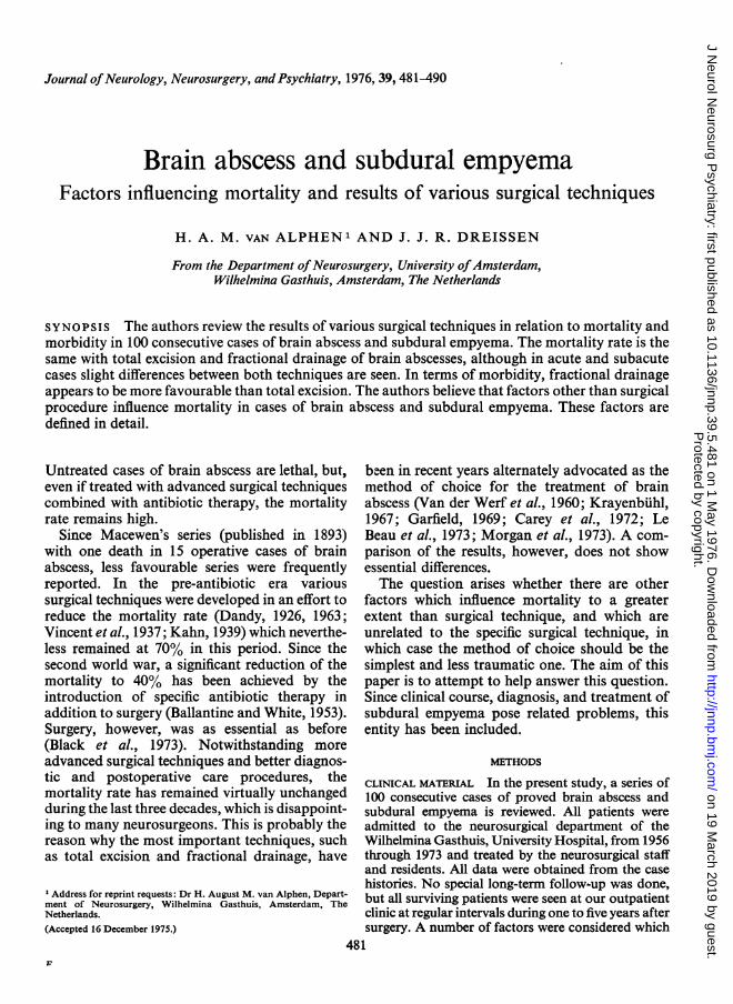
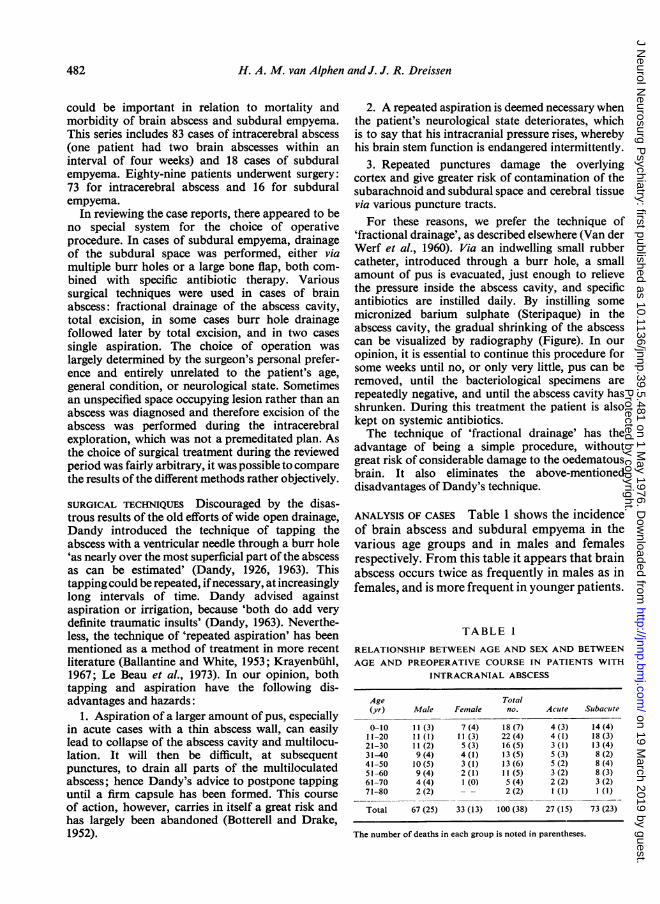
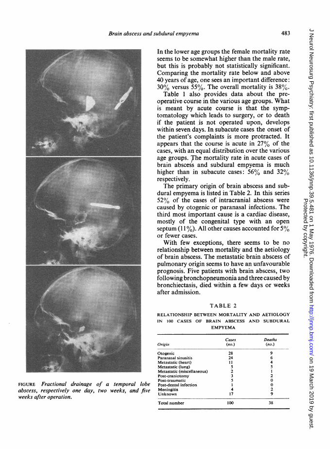
For these reasons, we prefer the technique of'fractional drainage', as described elsewhere (Van derWerf et al., 1960). Via an indwelling small rubbercatheter, introduced through a burr hole, a smallamount of pus is evacuated, just enough to relievethe pressure inside the abscess cavity, and specificantibiotics are instilled daily. By instilling somemicronized barium sulphate (Steripaque) in theabscess cavity, the gradual shrinking of the abscesscan be visualized by radiography (Figure). In our
opinion, it is essential to continue this procedure forsome weeks until no, or only very little, pus can beremoved, until the bacteriological specimens arerepeatedly negative, and until the abscess cavity hasshrunken. During this treatment the patient is alsokept on systemic antibiotics.The technique of 'fractional drainage' has the
advantage of being a simple procedure, withoutgreat risk of considerable damage to the oedematousbrain. It also eliminates the above-mentioneddisadvantages of Dandy's technique.
ANALYSIS OF CASES Table 1 shows the incidenceof brain abscess and subdural empyema in thevarious age groups and in males and femalesrespectively. From this table it appears that brainabscess occurs twice as frequently in males as infemales, and is more frequent in younger patients.
TABLE 1
RELATIONSHIP BETWEEN AGE AND SEX AND BETWEEN
AGE AND PREOPERATIVE COURSE IN PATIENTS WITH
INTRACRANIAL ABSCESS
Age Total(yr) Male Female no. Acute Subacute
0-10 11(3) 7 (4) 18 (7) 4 (3) 14 (4)11-20 11(1) 11(3) 22(4) 4(1) 18(3)21-30 11(2) 5 (3) 16 (5) 3 (1) 13 (4)31-40 9(4) 4(1) 13(5) 5(3) 8(2)41-50 10 (5) 3 (1) 13 (6) 5 (2) 8 (4)51-60 9(4) 2(1) 11(5) 3(2) 8(3)61-70 4 (4) 1 (0) 5 (4) 2 (2) 3 (2)71-80 2(2) - - 2(2) 1 (1) 1 (1)
Total 67 (25) 33 (13) 100 (38) 27 (15) 73 (23)
The number of deaths in each group is noted in parentheses.
482
Protected by copyright.
on 19 March 2019 by guest.
http://jnnp.bmj.com
/J N
eurol Neurosurg P
sychiatry: first published as 10.1136/jnnp.39.5.481 on 1 May 1976. D
ownloaded from
Brain abscess and subdural empyema
FIGURE Fractional drainage of a temporal lobeabscess, respectively one day, two weeks, and fiveweeks after operation.
In the lower age groups the female mortality rateseems to be somewhat higher than the male rate,but this is probably not statistically significant.Comparing the mortality rate below and above40 years of age, one sees an important difference:30% versus 55%. The overall mortality is 38%.
Table 1 also provides data about the pre-operative course in the various age groups. Whatis meant by acute course is that the symp-tomatology which leads to surgery, or to deathif the patient is not operated upon, developswithin seven days. In subacute cases the onset ofthe patient's complaints is more protracted. Itappears that the course is acute in 27% of thecases, with an equal distribution over the variousage groups. The mortality rate in acute cases ofbrain abscess and subdural empyema is muchhigher than in subacute cases: 56% and 32%respectively.The primary origin of brain abscess and sub-
dural empyema is listed in Table 2. In this series52% of the cases of intracranial abscess werecaused by otogenic or paranasal infections. Thethird most important cause is a cardiac disease,mostly of the congenital type with an openseptum (11%). All other causes accounted for 5%or fewer cases.With few exceptions, there seems to be no
relationship between mortality and the aetiologyof brain abscess. The metastatic brain abscess ofpulmonary origin seems to have an unfavourableprognosis. Five patients with brain abscess, twofollowing bronchopneumonia and three caused bybronchiectasis, died within a few days or weeksafter admission.
TABLE 2
RELATIONSHIP BETWEEN MORTALITY AND AETIOLOGYIN 100 CASES OF BRAIN ABSCESS AND SUBDURAL
EMPYEMA
Cases DeathsOrigini (no.) (no.)
Otogenic 28 9Paranasal sinusitis 24 6Metastatic (heart) 11 4Metastatic (lung) 5 5Metastatic (miscellaneous) 2 1Post-craniotomy 3 2Post-traumatic 5 0Post-dental infection 1 0Meningitis 4 2Unknown 17 9
Total number 100 38
483P
rotected by copyright. on 19 M
arch 2019 by guest.http://jnnp.bm
j.com/
J Neurol N
eurosurg Psychiatry: first published as 10.1136/jnnp.39.5.481 on 1 M
ay 1976. Dow
nloaded from
H. A. M. van Alphen andJ. J. R. Dreissen
One patient, a 14 year old boy who was suffering frombronchiectasis, was comatose and moribund uponadmission. He died before surgery could be under-taken. At necropsy a left temporal and a rightoccipital lobe abscess were found.Another patient, a 6 year old boy, had been
admitted to another hospital because of lympho-blastic leukaemia complicated by bronchopneu-monia and purulent meningitis. He was transferredto our hospital because of an incomplete transversemyelitis. He also had epileptic seizures. His cerebraldisease was, however, not diagnosed until afterdeath. At necropsy he appeared to have a rightfrontal lobe abscess with intraventricular rupture.A 28 year old female, suffering from a bronchiec-
tasis and pneumonia, was admitted because ofmeningitis and an acute grade 4 brain abscess whichwas treated by fractional drainage. She died somehours after surgery of bronchospasm and pulmonaryoedema.A 22 year old female, who was suffering from
bronchiectasis, was admitted to our hospital becauseof signs of meningitis and of a space occupying lesionof the left hemisphere. A deeply placed left temporallobe abscess was diagnosed and drained immediately.Some days after surgery she showed severe signs ofincreased intracranial pressure and died. At necropsysix differently located brain abscesses were found.A 72 year old male developed meningitis some
weeks after bronchopneumonia. He received anti-biotics and several lumbar punctures were done.After 10 days, he showed signs of a space occupyinglesion of the left hemisphere and became semi-comatose. A deeply placed left temporal lobe abscesswas diagnosed by angiography. The patient wastransferred to our clinic and the abscess was treatedwith fractional drainage and in the usual fashionwith antibiotics. In the following weeks he developedpneumonia, pleuritis, and uraemia. He died fourweeks after surgery. At necropsy, a large temporallobe abscess ruptured into the ventricle, basalmeningitis, and pulmonary oedema were found.
A more favourable group seems to be the brainabscesses due to trauma of the skull, mostlycompound skull fractures. There were five suchcases in our series. All these patients were young,between 6 and 35 years of age, healthy, and ina good overall condition before the accident.All were alive several years after surgery.The organisms responsible for brain abscess
and subdural empyema are summarized inTable 3. As in some of the other tables, the totalnumber of cases is 101, because one patient had
TABLE 3RELATIONSHIP BETWEEN MORTALITY AND RESPONSIBLEORGANISM IN 100 CASES OF BRAIN ABSCESS AND
SUBDURAL EMPYEMA
Cases DeathsOrganism (no.) (no.)
Streptococcus 30 10Staphylococcus 9 2B. proteus 5 2Pneumococcus 5 1Clostridium 3 0Klebsiella 1 1Haemophilus 1 1Escherichia coli 1 0B. tubercul. 2 2Actinomyces 1 1Mixed culture 10 5Mixed culture of anaerobic 8 2
organismsUnknown (no culture grown
in operated cases) 15 1Unknown (no surgery, nopostmortem examination) 10 10
Total number 101 38
two brain abscesses with a four weeks' intervalin between, with a different localization, adifferent organism, and a different surgicaltreatment. Both abscesses are therefore tabulatedseparately. The mortality rate ofthe streptococcalabscesses, compared with that of staphylococcalabscesses, seems to be fairly high. But in thegroup of streptococcal abscesses the abscess wasmissed at operation in three cases, which un-favourably influences the mortality rate of thisgroup. The difference between the two groupsmight, therefore, not be significant. Two casesof tuberculous brain abscesses were both lethal.
The first patient, an 11 year old girl, was found tohave a subacute abscess of unknown origin. At thetime of operation she was comatose and in very poorcondition. A large unencapsulated temporal lobeabscess was found and excised. This subsequentlyappeared to be of tuberculous nature. She died fourdays after surgery.The second patient, a male, had been treated five
years earlier for cavernous pulmonary tuberculosisand had had a more recent operation for intestinaltuberculosis. His subacute neurological deteriorationwas not diagnosed until after death. At necropsy aleft cerebellar tuberculous abscess was found.
484
Protected by copyright.
on 19 March 2019 by guest.
http://jnnp.bmj.com
/J N
eurol Neurosurg P
sychiatry: first published as 10.1136/jnnp.39.5.481 on 1 May 1976. D
ownloaded from
Brain abscess and subdural emnpyema
In these cases, the grade 4 neurological conditionand the diagnostic failure were probably respect-ively responsible for the lethal course, rather thanthe tuberculous origin.
In 10 cases, a mixed culture of two or threestrains, principally Staphylococcus, Strepto-coccus and Proteus, was obtained from either thesame abscess or from multiple abscesses con-
taining different organisms. The mortality rate oftwo cases of multiple abscesses in this group was
100% . The presence of more than one strain inthe same abscess does not seem to have any
specific influence upon mortality. Only one
patient died out of 15 in whom the bacteriologi-cal examination was negative after surgery. Thismortality rate is remarkably low and seems
to be significant.Ten other patients in whom the organism
responsible for brain abscess was unknown diedbecause they had no surgery. No postmortembacteriological examination was done in thesecases.
The possible influence of the localization on
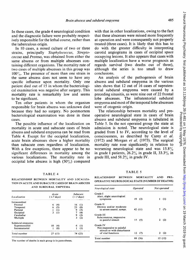
mortality in acute and subacute cases of brainabscess and subdural empyema can be read fromTable 4. Except for the occipital localization,acute brain abscesses show a higher mortalitythan subacute ones regardless of localization.With a few exceptions, there appear to be no
significant differences in mortality among thevarious localizations. The mortality rate inoccipital lobe abscess is high (50%) compared
TABLE 4
RELATIONSHIP BETWEEN MORTALITY AND LOCALIZA-
TION IN ACUTE AND SUBACUTE CASES OF BRAIN ABSCESS
AND SUBDURAL EMPYEMA
Acute SubacuteLocalization ( 7 days) (> 7 days)
IntracerebralFrontal 6 (4) 16 (1)Temporal 6 (5) 23 (8)Parietal I (1) 9 (3)Occipital 3 (1) 7 (4)Cerebellar 9 (3)Multiple 3 (2)
Subdural empyemaSupratentorial 10 (4) 6 (1)Intratentorial 1 (0) 1 (1)
Total number 27 (15) 74 (23)
The number of deaths in each group is in parentheses.
with that in other localizations, owing to the factthat these abscesses were missed more frequentlyat operation and were consequently not properlytreated (three cases). It is likely that this has todo with the greater difficulty in interpretingcarotid angiograms in cases of occipital spaceoccupying lesions. It also appears that cases withmultiple localization have a worse prognosis asregards survival (two deaths out of three),although the series is too small to drawconclusions.An analysis of the pathogenesis of brain
abscess and subdural empyema in the varioussites shows that 12 out of 16 cases of supraten-torial subdural empyema were caused by apreceding sinusitis, as were nine out of 22 frontallobe abscesses. The infratentorial subduralempyema and most ofthe temporal lobe abscesseswere of otogenic origin.The relationship between mortality and pre-
operative neurological state in cases of brainabscess and subdural empyema is tabulated inTable 5. In the not operated group the state atadmission is noted. The neurological state isgraded from I to IV, according to the level ofconsciousness, as described by Carey et al.(1972) and Morgan et al. (1973). The surgicalmortality rate rose significantly in relation toworsening neurological state and was 15.8%in grade I patients, 26.2% in grade II, 33.3% ingrade III, and 58.2% in grade IV.
TABLE 5
RELATIONSHIP BETWEEN MORTALITY AND PRE-
OPERATIVE NEUROLOGICAL STATE (NUMBER OF DEATHS)
Neurological state Operated Not operated
Grade IAlert, slight neurologicalsymptoms 19 (3) 1 (1)
Grade ItDrowsy and/or moderate
to severe neurol. sympt. 42 (11) 7 (7)
Grade IIISubcomatose, responsive
to painful stimuli 15 (5) 2 (2)
Grade IVNot responsive to painful
stimuli or with disturbanceof vital functions 12 (7) 2 (2)
Total number 88 (26) 12 (12)
485
Protected by copyright.
on 19 March 2019 by guest.
http://jnnp.bmj.com
/J N
eurol Neurosurg P
sychiatry: first published as 10.1136/jnnp.39.5.481 on 1 May 1976. D
ownloaded from
H. A. M. van Alphen and J. J. R. Dreissen
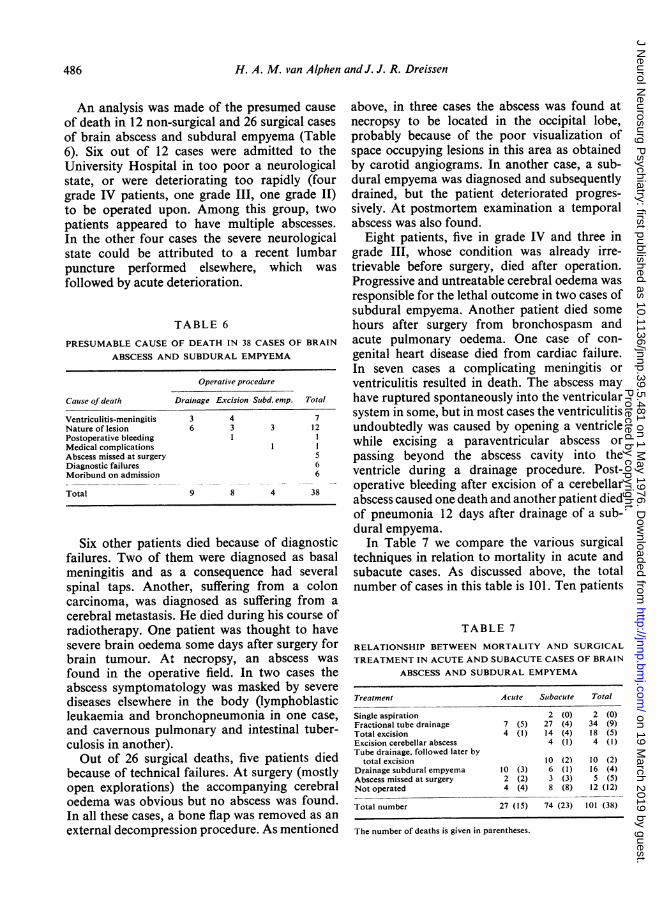
An analysis was made of the presumed causeof death in 12 non-surgical and 26 surgical casesof brain abscess and subdural empyema (Table6). Six out of 12 cases were admitted to theUniversity Hospital in too poor a neurologicalstate, or were deteriorating too rapidly (fourgrade IV patients, one grade III, one grade II)to be operated upon. Among this group, twopatients appeared to have multiple abscesses.In the other four cases the severe neurologicalstate could be attributed to a recent lumbarpuncture performed elsewhere, which wasfollowed by acute deterioration.
TABLE 6
PRESUMABLE CAUSE OF DEATH IN 38 CASES OF BRAIN
ABSCESS AND SUBDURAL EMPYEMA
Operative procedure
Cause ofdeath Drainage Excision Subd. enip. Total
Ventriculitis-meningitis 3 4 7Nature of lesion 6 3 3 12Postoperative bleeding 1 IMedical complications I IAbscess missed at surgery 5Diagnostic failures 6Moribund on admission 6
Total 9 8 4 38
Six other patients died because of diagnosticfailures. Two of them were diagnosed as basalmeningitis and as a consequence had severalspinal taps. Another, suffering from a coloncarcinoma, was diagnosed as suffering from a
cerebral metastasis. He died during his course ofradiotherapy. One patient was thought to havesevere brain oedema some days after surgery forbrain tumour. At necropsy, an abscess was
found in the operative field. In two cases theabscess symptomatology was masked by severe
diseases elsewhere in the body (lymphoblasticleukaemia and bronchopneumonia in one case,
and cavernous pulmonary and intestinal tuber-culosis in another).Out of 26 surgical deaths, five patients died
because of technical failures. At surgery (mostlyopen explorations) the accompanying cerebraloedema was obvious but no abscess was found.In all these cases, a bone flap was removed as an
external decompression procedure. As mentioned
above, in three cases the abscess was found atnecropsy to be located in the occipital lobe,probably because of the poor visualization ofspace occupying lesions in this area as obtainedby carotid angiograms. In another case, a sub-dural empyema was diagnosed and subsequentlydrained, but the patient deteriorated progres-sively. At postmortem examination a temporalabscess was also found.
Eight patients, five in grade IV and three ingrade III, whose condition was already irre-trievable before surgery, died after operation.Progressive and untreatable cerebral oedema wasresponsible for the lethal outcome in two cases ofsubdural empyema. Another patient died somehours after surgery from bronchospasm andacute pulmonary oedema. One case of con-genital heart disease died from cardiac failure.In seven cases a complicating meningitis orventriculitis resulted in death. The abscess mayhave ruptured spontaneously into the ventricularsystem in some, but in most cases the ventriculitisundoubtedly was caused by opening a ventriclewhile excising a paraventricular abscess orpassing beyond the abscess cavity into theventricle during a drainage procedure. Post-operative bleeding after excision of a cerebellarabscess caused one death and another patient diedof pneumonia 12 days after drainage of a sub-dural empyema.
In Table 7 we compare the various surgicaltechniques in relation to mortality in acute andsubacute cases. As discussed above, the totalnumber of cases in this table is 101. Ten patients
TABLE 7
RELATIONSHIP BETWEEN MORTALITY AND SURGICAL
TREATMENT IN ACUTE AND SUBACUTE CASES OF BRAIN
ABSCESS AND SUBDURAL EMPYEMA
Treatment Acute Suibacute Total
Single aspiration 2 (0) 2 (0)Fractional tube drainage 7 (5) 27 (4) 34 (9)Total excision 4 (1) 14 (4) 18 (5)Excision cerebellar abscess 4 (1) 4 (1)Tube drainage, followed later by
total excision 10 (2) 10 (2)Drainage subdural empyema 10 (3) 6 (1) 16 (4)Abscess missed at surgery 2 (2) 3 (3) 5 (5)Not operated 4 (4) 8 (8) 12 (12)
Total number 27 (15) 74 (23) 101 (38)
The number of deaths is given in parentheses.
486
Protected by copyright.
on 19 March 2019 by guest.
http://jnnp.bmj.com
/J N
eurol Neurosurg P
sychiatry: first published as 10.1136/jnnp.39.5.481 on 1 May 1976. D
ownloaded from
Brain abscess and subdural empyema
were treated by excision ofthe abscess cavity aftera period of fractional drainage or some timeafter completion of the same. In the latter, theshrunken abscess wall was excised in view of thepresumed epileptogenic hazard of a retainedabscess capsule. In the last six years this policywas abandoned without harmful consequences.In some cases, secondary excision was performedwhen fractional drainage-usually as a result ofcapsule collapse and subsequent multiloculation-proved to be insufficient to improve thepatient's neurological state.On the whole, there appears to be no significant
difference in mortality between the varioussurgical techniques. Looking at the acute andsubacute cases separately the mortality rateseems to be slightly higher for fractional drainagein acute cases, although the total number ofcases is too small for precise comparison. Insubacute cases, the fractional drainage wassomewhat more favourable than total excisionwith regard to mortality.The surgical mortality of the combined
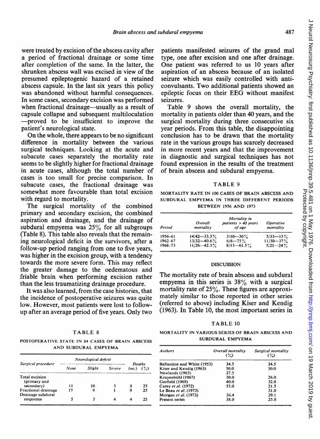
primary and secondary excision, the combinedaspiration and drainage, and the drainage ofsubdural empyema was 25% for all subgroups(Table 8). This table also reveals that the remain-ing neurological deficit in the survivors, after afollow-up period ranging from one to five years,was higher in the excision group, with a tendencytowards the more severe form. This may reflectthe greater damage to the oedematous andfriable brain when performing excision ratherthan the less traumatizing drainage procedure.
It was also learned, from the case histories, thatthe incidence of postoperative seizures was quitelow. However, most patients were lost to follow-up after an average period of five years. Only two
TABLE 8
POSTOPERATIVE STATE IN 84 CASES OF BRAIN ABSCESSAND SUBDURAL EMPYEMA
Neurological deficitSuirgical procedure Deaths
None Slight Sev ere (no.) (%
Total excision(primary andsecondary) 11 10 3 8 25
Fractional drainage 17 9 1 9 25Drainage subduralempyema 5 3 4 4 25
patients manifested seizures of the grand maltype, one after excision and one after drainage.One patient was referred to us 10 years afteraspiration of an abscess because of an isolatedseizure which was easily controlled with anti-convulsants. Two additional patients showed anepileptic focus on their EEG without manifestseizures.
Table 9 shows the overall mortality, themortality in patients older than 40 years, and thesurgical mortality during three consecutive sixyear periods. From this table, the disappointingconclusion has to be drawn that the mortalityrate in the various groups has scarcely decreasedin more recent years and that the improvementin diagnostic and surgical techniques has notfound expression in the results of the treatmentof brain abscess and subdural empyema.
TABLE 9
MORTALITY RATE IN 100 CASES OF BRAIN ABSCESS ANDSUBDURAL EMPYEMA IN THREE DIFFERENT PERIODS
BETWEEN 1956 AND 1973
Mortality inOverall patients > 40 years Operative
Period mortality ofage mortality
1956-61 14/42-33.3% 3/10-30% 5/33-15%1962-67 13/32-40.6% 6/8-75 % 11/30-37 %1968-73 11/26-42.3% 8/13-61.5%/ 5/21-24%
DISCUSSION
The mortality rate of brain abscess and subduralempyema in this series is 38% with a surgicalmortality rate of 25%. These figures are approxi-mately similar to those reported in other series(referred to above) including Kiser and Kendig(1963). In Table 10, the most important series in
TABLE 10
MORTALITY IN VARIOUS SERIES OF BRAIN ABSCESS ANDSUBDURAL EMPYEMA
Authors Overall mortality Surgical mortality(%) (%)
Ballantine and White (1953) 34.5 34.5Kiser and Kendig (1963) 50.0 30.0Newlands (1965) 27.5Krayenbuhl (1967) 30.0 26.0Garfield (1969) 40.0 32.0Carey et al. (1972) 53.0 21.5Le Beau et al. (1973) 31.0Morgan et al. (1973) 36.4 29.1Present series 38.0 25.0
487
Protected by copyright.
on 19 March 2019 by guest.
http://jnnp.bmj.com
/J N
eurol Neurosurg P
sychiatry: first published as 10.1136/jnnp.39.5.481 on 1 May 1976. D
ownloaded from

H. A. M. van Alphen and J. J. R. Dreissen
the period between 1953 and 1973 are listed. This be a negligible factor in the choice of surgicaltable shows that the overall results, as far as treatment.mortality is concerned, showed absolutely no Mortality in brain abscess and subduralchange during the last 20 years. In the individual empyema appears to be influenced by a number ofpublications, only Krayenbiihl (1967) reported an factors unrelated to the type of surgery:improvement of his results in the last six yearperiod compared with the previous 15 years. AGE Thabnechance of dying increases with risingOther authors (Garfield, 1969; Le Beau et al., age (Table 1), which probably reflects the lower1973), however, could not demonstrate such a resistance of the aging body to any noxiousprogressive improvement in their subsequent influenceperiods. Nor do our results show an improvementin the last six year period (Table 9). In cases of PREOPERATIVE COURSE In our series, the acutebrain athelastsixyea nyerperesultsabln caseso cases of brain abscess and subdural empyemabrain abscess the seemingly better results are had a more unfavourable prognosis with aattributed to the application of total excision by mortality rate of 56%, versus 32% in subacutesome authors (Krayenbiihl, 1967; Le Beau et at., cases. This is in accordance with the findings of1973). In Krayenbiihl's series the mortality rate Le Beau et al. (1973). The acute course sometimesin 14 cases of repeated aspiration was 21.4%, results in a too late diagnosis or in a too lateand in 90 cases of radical excision between 1945 transferral of the patient for surgery. On theand 1965 it was 22.2%. other hand, the surgical results in acute cases areLe Beau et a. (1973) compared the results in '240 cases treated by total excision (mortality27%) and in 32 cases treated by aspiration AETIOLOGY From various reports in the litera-(mortality 65%). Their aspiration techniques are ture (Carey et al., 1972; Le Beau et al., 1973), itnot further defined. Judging by these results, both can be deduced that the mortality rate in meta-authors prefer the total excision to the various static brain abscesses is relatively high. In ouraspiration techniques. Carey et al. (1972) found series, the metastatic abscesses from the lungthat abscess drainage was associated with were 100% lethal. This is even more strikingapproximately the same mortality as excision when compared with post-traumatic abscesses(19% vs 22%). They give some preference to (Table 2). These last patients were all young and'abscess drainage, as a form of therapy, since it healthy before the accident. Therefore, it iswas employed with no increased mortality in likely that in cases of metastatic abscess theneurological decompensated patients'. patients' resistance is greatly decreased by the
In our series the mortality rate in fractional general infectious condition.drainage as well as total excision was 25%. Thesame mortality was seen in cases of subdural PREOPERATIVE NEUROLOGICAL STATE Severalempyema. From these data, we can conclude that authors have mentioned the close correlationthe form of surgery is of only minor importance between survival and preoperative level ofas far as mortality is concerned. In terms of consciousness in patients with brain abscess ormorbidity, however, fractional drainage appears subdural empyema (Newlands, 1965; Garfield,to be more favourable than total excision (Table 1969; Carey et al., 1972; Morgan et al., 1973).8). Therefore, we prefer the use of fractional Our findings agree with this; the mortality ratedrainage, especially in subacute cases of brain ranging from 15.8% in grade I patients toabscess. In acute cases this procedure may not 58.2% in grade IV (Table 5). Further analysisalways be sufficient to relieve the high intracranial shows, however, that a relatively large numberpressure, because of the disproportional oedema of deceased grade IV patients had shown an(Botterell and Drake, 1952; Le Beau et al., 1973). acute course before surgery. Therefore, theIn these cases total excision may be life saving preoperative course and neurological state areat the cost of a greater morbidity. In contrast with not entirely unrelated with respect to mortalitysome reports in the literature (Carey et al., 1972; in grade IV patients. This correlation betweenLe Beau et al., 1973), the incidence of post- preoperative course and neurological state wasoperative epilepsy is very low (3%) and seems toianot seen in grade I to III patients.
488
:1
Protected by copyright.
on 19 March 2019 by guest.
http://jnnp.bmj.com
/J N
eurol Neurosurg P
sychiatry: first published as 10.1136/jnnp.39.5.481 on 1 May 1976. D
ownloaded from
Brain abscess and subdural empyema
LOCALIZATION OF BRAIN ABSCESS This alsoappeared to be a factor in relation to mortalityin some cases. The unfavourable prognosis ofmultiple abscesses has been pointed out by someauthors (Carey et al., 1972; Le Beau et al., 1973).The incidence of multiple abscesses was low inour series, but the mortality rate also seems to behigh: two deaths out of three. Also, in singleabscesses the localization may give rise todifficulties, as shown by an analysis of cases inwhich the abscess was missed at surgery. In allthese cases the presence of an abscess wassuspected before surgery. The carotid angio-grams showed signs of a space occupying lesionsomewhere in the posterior half of the cerebralhemisphere without precise localization. Atpostmortem examination, three abscesses ap-peared to be localized in the extreme posteriorpart of the occipital lobe, two were situateddeeply temporobasal. A brainscan would havebeen very helpful in these cases. If treatedadequately, either by total excision or by frac-tional drainage, no correlation was foundbetween mortality and localization.DIAGNOSTIC FAILURES The danger of lumbarpuncture in abscess or subdural empyema hasbeen frequently recognized (Dandy, 1963; Gar-field, 1969; Carey et al., 1972; Morgan et al.,1973). Spinal taps are not infrequently done inthose patients, who are suspected of havingmeningitis, which is, in fact, a sequela of un-suspected brain abscess or subdural empyema.When contaminated spinal fluid is found thediagnosis of meningitis seems to be confirmedand spinal taps are repeated for intrathecaladministration of antibiotics, resulting in acutedeterioration or demise. Therefore, in a patientsuspected of having meningitis, but also showinglateralizing symptoms, echoencephalographyshould be performed first. When a midline shiftis found the next step should be brainscanning orcarotid angiography rather than lumbar punc-ture. Once in a while, a severe infectious diseaseelsewhere in the body can cause a metastaticbrain abscess, but, as is seen in two of our casesin which the diagnosis was missed, it may alsodivert the attention from the neurologicalsymptoms caused by a brain abscess.
POSTOPERATIVE COMPLICATIONS Any type ofsurgery can be followed by lethal unspecific
complications such as pulmonary or thrombo-embolic processes, or postoperative haematomas.More directly related to brain abscess or subduralempyema are cerebral oedema and contaminationof spinal fluid compartments. In our series wehad a complicating ventriculitis or basal menin-gitis which led to death in some cases of excisionas well as in drainage procedures. A propertechnique and great care should be employed inpuncturing an abscess cavity to avoid thiscomplication, as already pointed out by Dandy(1963).
In conclusion, it can be stated from this studythat the overall mortality in brain abscess andsubdural empyema is determined considerablyby missing an abscess at surgery or by ruling outsurgery for whatever reason. Most other factorscontributing to mortality, as far as brain abscessis concerned, exist before surgery and areunrelated to the type of surgery, be it totalexcision or fractional drainage. Considering theresults in our drainage group both in regard tomortality and morbidity, we disagree with someother authors (Krayenbiihl, 1967; Le Beauet al., 1973), who have maintained that radicalremoval of a brain abscess is necessary.
REFERENCES
Ballantine, H. T., and White, J. C. (1953). Brain abscess,influence of the antibiotics on therapy and mortality.New England Journal ofMedicine, 248, 14-19.
Black, P., Graybill, R., and Charache, P. (1973). Penetra-tion of brain abscess by systemically administeredantibiotics. Journal ofNeurosurgery, 38, 705-709.
Botterell, E. H., and Drake, C. G. (1952). Localized ence-phalitis, brain abscess and subdural empyema. JournalofNeurosurgery, 9, 348-366.
Carey, M. E., Chou, S. N., and French, L. A. (1972).Experience with brain abscesses. Journal of Neuro-surgery, 36, 1-9.
Dandy, W. E. (1926). Treatment of chronic abscess ofthe brain by tapping. Journal of the American MedicalAssociation, 87, 1477-1478.
Dandy, W. E. (1963). Pyogenic abscesses of the brain.In Practice of Surgery, vol. 12, pp. 346-362. Edited byD. Lewis, W. Walters, and F. H. Ellis. Prior:Hagerstown,
Garfield, J. (1969). Management of supratentorial intra-cranial abscess: A review of 200 cases. British MedicalJournal, 2, 7-11.
Kahn, E. A. (1939). Treatment of encapsulated abscessof the brain: visualization by colloidal thorium dioxide.Archives of Neurology and Psychiatry (Chic.), 41, 158-165.
489
Protected by copyright.
on 19 March 2019 by guest.
http://jnnp.bmj.com
/J N
eurol Neurosurg P
sychiatry: first published as 10.1136/jnnp.39.5.481 on 1 May 1976. D
ownloaded from
H. A. M. van Alphen and J. J. R. Dreissent
Kiser, J. L., and Kendig, J. H. (1963). Intracranial sup-
puration: a review of 139 consecutive cases with elec-tron-microscopic observations on three. Journal ofNeurosurgery, 20, 494-511.
Krayenbiihl, H. A. (1967). Abscess of the brain. ClinicalNeurosurgery, 14, 25-44.
Le Beau, J., Creissard, P., Harispe, L., and Redondo A.(1973). Surgical treatment of brain abscess and subduralempyema. Journal ofNeurosurgery, 38, 198-203.
Macewen, W. (1893). Pyogenic Infective Diseases of theBrain and Spinal Cord. Maclehose: Glasgow.
Morgan, H., Wood, M. W., and Murphey, F. (1973).
Experience with 88 consecutive cases of brain abscess.Journal ofNeurosurgery, 38, 689-704.
Newlands, W. J. (1965). Otogenic brain abscess: a studyof eighty cases. Journal ofLaryngology and Otology, 79,120-130.
Vincent, C., David, M., and Askenasy, H. (1937). Sur une
m6thode de traitement des abces subaigus et chroniquesdes hemispheres cerebraux. Journal de Chirurgie (Paris),49, 1-46.
Van der Werf, A. J. M., Lie, T. A., and Noordenbos, W.(1960). Drainage fractionne des abces encephaliques.Neuro-chirurgie, 6, 325-33 1.
490
Protected by copyright.
on 19 March 2019 by guest.
http://jnnp.bmj.com
/J N
eurol Neurosurg P
sychiatry: first published as 10.1136/jnnp.39.5.481 on 1 May 1976. D
ownloaded from