Embed Size (px)
Citation preview

368
ORIGINAL ARTICLE SPINE SURGERY AND RELATED RESEARCH
Brain Activation in a Cynomolgus Macaque Model ofChymopapain-Induced Discogenic Low Back Pain:A Preliminary Study
Hiroki Ushirozako1), Go Yoshida1), Daisuke Togawa2), Takao Omura1), Tomohiko Hasegawa1), Yu Yamato1), Tomohiro Banno1),
Hideyuki Arima1), Shin Oe2), Yuki Mihara1), Tomohiro Yamada1), Takahiro Natsume3), Shinya Ogawa3), Yuji Awaga3),
Hiroyuki Takamatsu3) and Yukihiro Matsuyama1)
1) Department of Orthopedic Surgery, Hamamatsu University School of Medicine, Hamamatsu, Japan2) Department of Orthopedic Surgery and Division of Geriatric Musculoskeletal Health, Hamamatsu University School ofMedicine, Hamamatsu, Japan3) Pharmacology Group, Hamamatsu Pharma Research, Inc., Hamamatsu, Japan
Abstract:Introduction: There is currently a lack of translatable, preclinical models of low back pain (LBP). Chymopapain, a pro-
teolytic enzyme used to treat lumbar intervertebral disc (IVD) herniation, could induce discogenic LBP. The current study
developed a behavioral model of discogenic LBP in nonhuman primates. Significant brain activation is observed in clinical
LBP. Thus, the current study also sought to define brain activation over time in a macaque with discogenic LBP.
Methods: Responses to pressure applied to the back at L4/L5 were measured in eight adult male Macaca fasciculata us-
ing a pressure algometer. The nucleus pulpous of the IVD between L4 and L5 was aspirated and chymopapain (1 mg/mL)
was injected under fluoroscopic guidance (n = 2). In two macaques, the nucleus pulpous was only aspirated. Brain activa-
tion in response to pressure applied to the lower back was assessed using a 3.0T magnetic resonance imaging scanner in
four macaques before and 1, 3, 9, and 14 days after treatment.
Results: The mean (±SD) response pressure before treatment was 1.4 ± 0.1 kg. One day after chymopapain treatment,
the response pressure decreased to 0.6 ± 0.05 kg (P < 0.01), suggestive of pressure hypersensitivity. Over time, the pressure
thresholds following chymopapain treatment gradually returned to normal. Following aspiration only, the response pressure
was 1.4 ± 0.05 kg, which was not significantly different from the uninjured controls. There was activation of the secondary
somatosensory cortex and insular cortex one and three days after chymopapain treatment; there was no activation following
aspiration only.
Conclusions: Enzymatic treatment of the nucleus pulpous leads to acute LBP and pressure-evoked activation in pain-
related brain areas. The current model of discogenic LBP parallels clinical LBP and could be used to further elaborate the
mechanism of acute LBP.
Keywords:chymopapain, discogenic low back pain, nonhuman primate, pressure test, brain activation, functional magnetic resonance
imaging, secondary somatosensory cortex, insular cortex
Spine Surg Relat Res 2019; 3(4): 368-376
dx.doi.org/10.22603/ssrr.2018-0110
Introduction
Low back pain (LBP) is a common clinical problem that
leads to significant disability, which may ultimately result in
adverse socioeconomic impact1,2). Pain is inherently subjec-
tive, and visual analog scales are used to quantify pain in-
tensity. However, to better understand the mechanism and
standardize the measurement of treatment efficacy, objective
LBP measures are needed. There are a number of therapeu-
tic interventions, including pharmacological, psychological,
and behavioral, that may be useful in treating LBP patients3).
However, it is currently not known which of these treat-
Corresponding author: Hiroki Ushirozako, [email protected]
Received: December 11, 2018, Accepted: March 10, 2019, Advance Publication: April 5, 2019
Copyright Ⓒ 2019 The Japanese Society for Spine Surgery and Related Research

dx.doi.org/10.22603/ssrr.2018-0110 Spine Surg Relat Res 2019; 3(4): 368-376
369
ments has the greatest efficacy since currently used outcome
measures are subjective.
Neuroimaging has demonstrated morphological and func-
tional changes in the brains of chronic pain patients4-6). Brain
activation, as observed with functional magnetic resonance
imaging (fMRI), reflects activation of large ensembles of
neurons and local field potentials in response to stimulation.
While pain itself is a subjective experience, brain activity
could be useful as an objective marker to reflect the pres-
ence and intensity of pain7). Non-invasive neuroimaging has
been used to demonstrate the effect of analgesic treatment
on pain-related brain activation8). Thus, brain activation
could be used to uncover LBP mechanisms and suggest
treatment strategies5,9).
Chymopapain has been used to treat herniated lumbar in-
tervertebral discs (IVDs). A common adverse effect of chy-
mopapain treatment is LBP10,11). Nonhuman primates are
ideal species in which to model clinical LBP because they
have IVDs that are anatomically similar to those of humans
and undergo biomechanical stress comparable to that of hu-
man IVDs. In addition, nonhuman primates have pain proc-
essing brain areas similar to those of humans2). The current
study developed a behavioral model of chymopapain-
induced discogenic LBP in cynomolgus macaques and iden-
tified stimulation-evoked brain activation using fMRI.
Materials and Methods
Animals
This study was approved by the institutional review board
and followed the relevant guidelines and laws of Japan per-
taining to the care and use of laboratory animals. Eight
young adult male cynomolgus macaques (weight range: 3.7-
4.3 kg; EBS Co., Ltd., Hashimoto, Japan) were used. Ani-
mals were individually housed in stainless steel cages in an
AAALAC International-accredited primate unit where hous-
ing conditions and care were in accordance with the Guide
for the Care and Use of Laboratory Animals, Eighth Edition
(National Research Council). A 12-hour light-dark cycle was
used. Animals were fed a standard diet (Oriental Yeast Co.,
Ltd., Chiba, Japan) and had free access to water. While indi-
vidually housed, aural, visual, and olfactory contact with
conspecifics was maintained. Manipulanda were provided to
each animal. Animals were hand-fed fresh fruits, vegetables,
and treats by the animal care and research staff at least once
per week. Animals were divided into three groups (control:
n = 4; aspiration only: n = 2; chymopapain-treated: n = 2).
Behavioral testing
Pressure thresholds were quantified using a modified pres-
sure algometer (Matsumiya Medical Co., Ltd., Tokyo, Ja-
pan). The 9 mm rubber tip of the algometer was covered by
a flat plastic circular cap 2.5 cm in diameter. With the ma-
caque standing in a monkey walker, the algometer was
placed 2 cm to the right of back midline at the level of L4/L
5. The algometer was pushed on the back until the animal
responded with contraction of the muscles on the top of the
head and around the eyes. Three response pressures (in kg)
were measured at 1 min intervals by a blinded investigator,
and the mean response pressure was calculated. A cut-off
threshold of 3 kg was used.
Intervertebral treatment procedure and postoperative paintime course
Animals were anesthetized with an intramuscular injection
of ketamine (10 mg/kg; Daiichi Sankyo Co. Ltd., Tokyo, Ja-
pan). Body temperature during the procedure was main-
tained using a heating pad. Deep anesthesia was ensured
when animals did not respond to noxious digit pinch and
showed no corneal reflex. Hair on the lower back at the sur-
gical site was shaved and the skin was scrubbed with
povidone-iodine. The monkeys were placed in a left semi-
lateral decubitus position. Anteroposterior and lateral views
of the lumbar spine were obtained using fluoroscopy (SIRE-
MOBIL Compact L, Siemens Healthcare, Berlin, Germany).
Unlike humans, there are six lumbar IVDs in cynomolgus
macaques. It was safe and easy to perform percutaneous
puncture of the IVD between L4/L5 under fluoroscopy with-
out influence of pelvic position. The IVD between L4/L5
was identified and percutaneous puncture of the IVD, with
an 18-gauge needle attached to a syringe, was made via an
anterolateral retroperitoneal approach directly over the mid-
lateral aspect of the disc12). The needle tip was centered
within the nucleus pulposus, and the nucleus pulposus was
aspirated (n = 2). In macaques treated with chymopapain (n
= 2), the nucleus pulposus was aspirated and 0.3 mL chy-
mopapain (1.0 mg/mL in saline; Sigma Chemical Co., St.
Louis, MO) was injected. Following recovery from anesthe-
sia, all animals showed normal levels of activity and feeding
in their home cage. Response pressure was measured 1, 3,
9, and 14 days after treatment.
Lumbar IVD pathology and visualization of brain activa-tion
T1- and T2-weighted sagittal and axial images of the
lumbar vertebrae were obtained using an 3.0T MRI system
(Signa HDxt 3.0T MRI system [GE Healthcare, Milwaukee,
WI, USA]) (slice thickness, 3 mm; T1-weighted image, TR/
TE = 320/15 ms; T2-weighted image, TR/TE =2800/104
ms). The MRI signal intensities, and the IVDs’ structure and
height, were graded at each time point with the Pfirrmann
grading system13). Grading was performed on T2-weighted
mid-sagittal IVD images by three orthopedic surgeons who
were unaware of the treatment of each vertebra.
Brain activation, without and with low back stimulation,
was visualized in four sedated macaques before and 1, 3, 9,
and 14 days after treatment using fMRI. Anatomical MRI
protocols consisted of a T1-weighted fast spoiled gradient-
recalled sequence [repetition time (TR)/echo time (TE),
15.8/7.0 ms; number of averages, 1; flip angle, 12°; field of
view, 150 mm × 150 mm; matrix, 256 × 224; slice thick-

Spine Surg Relat Res 2019; 3(4): 368-376 dx.doi.org/10.22603/ssrr.2018-0110
370
Figure 1. Pressure sensitivity of the lumbar back following
chymopapain treatment. Response pressure thresholds (kg) were
measured over time after either chymopapain treatment or aspira-
tion of the intervertebral disc. Pressure thresholds of untreated
(“control”) macaques are expressed as mean±standard deviation.
Table 1. Change in Lumbar Intervertebral Disc Pathology Over
Time According to the Pfirrmann Grading System.
Case Before Day 1 Day 3 Day 9 Day 14
Aspiration 1 I III III III III
Aspiration 2 I III III III III
Chymopapain 1 I IV IV IV IV
Chymopapain 2 I III III III III
ness/interval, 1.0/0.5 mm; number of slices, 168]. Functional
scan sequences consisted of field-echo and echo-planar im-
aging (TR/TE, 3000/35 ms; flip angle, 90°; field of view,
140 mm × 140 mm; matrix, 64 × 64; slice thickness, 2.0
mm; number of slices, 30). The macaques were sedated by
continuous intravenous infusion of propofol (0.2 mg/kg/
min), which has little analgesic effect14).
To deliver pressure stimuli within the MRI, a disposable,
sealed, air-filled 10-mL syringe was applied by hand to the
back15). Manual compression on the syringe perpendicularly
against an object displaced the plunger, the force of which
was indicated on the side of the syringe sheath. In the MRI
scanner, macaques were placed in the prone position and
pressure was applied to the lower back - the same area that
was tested in awake macaques. During one fMRI scan, ani-
mals underwent two sets of low back pressure stimulations.
One set included ten cycles, with each cycle consisting of
30 s of 1.0 kg followed by 30 s of 0.1 kg. A 30-second in-
terval without stimulation separated each set.
MRI brain data analysis
All MRI analyses were conducted using SPM12 software
(Wellcome Trust Centre for Neuroimaging, London, UK).
The images were realigned and re-sliced onto mean echo-
planar images (EPIs) to correct for head motion. EPIs were
co-registered to the corresponding T1-weighted anatomical
image and normalized to a macaque brain template16). The
resulting images were smoothed with a 4 mm × 4 mm × 4
mm full-width at half-maximum Gaussian kernel. Voxel-
wise statistical analysis was based on a general linear
model. A fixed-effect model was used for group analysis of
data. Contrast (1.0 kg stimulation - 0.1 kg stimulation) was
defined to isolate regions responsive to 1.0 kg stimulation-
related signals of the whole brain. Brain regions with high
signals were selected for analysis. Peak voxels were consid-
ered significant at Z score > 1.96 (P < 0.05, uncorrected for
multiple comparisons, one-tailed t-test). Brian activation was
averaged from two macaques treated with chymopapain and
from two macaques with aspiration only of the IVD.
Statistical analysis
Statistical analyses were conducted using SPSS version
23.0 (IBM, Armonk, NY, USA). Continuous variables with
normal distributions were expressed as means ± standard de-
viations (SD) and analyzed using unpaired t-tests. P values
< 0.05 were considered statistically significant. While no
sample size calculation was used to determine the minimum
number of animals needed, the lowest possible number of
animals was used to obtain behavioral data. In a previous,
unrelated study, a significant decrease in the mean pressure
hypersensitivity of the knee joint, following unilateral me-
niscectomy, was observed between three experimental and
three sham-operated animals17).
Results
Following either aspiration or chymopapain treatment,
there were no general signs of chronic stress or pain, such
as appetite loss or decreased activity. Lumbar MRIs showed
slight bleeding in the muscles surrounding the IVD due to
the puncture. However, no leakage of chymopapain into the
vertebrae or muscles around the disc was observed.
Pressure sensitivity in macaques with chymopapain-induced LBP
Following treatment of the IVD with chymopapain, ma-
caques demonstrated decreased response pressure thresholds
(Fig. 1). One day following chymopapain treatment, the re-
sponse pressure was 0.6 ± 0.05 (mean ± SD) kg, which was
significantly less than that of the untreated controls (n = 4;
1.4 ± 0.1 kg; P < 0.01). The response pressure gradually in-
creased over time following chymopapain treatment - 14
days following chymopapain treatment, the response pres-
sure was 1.2 ± 0.04 kg (P < 0.05 vs. untreated controls). By
contrast, aspiration of the nucleus pulposus alone did not
significantly alter the response to pressure (Fig. 1; P = 0.297
vs. untreated controls).
dx.doi.org/10.22603/ssrr.2018-0110 Spine Surg Relat Res 2019; 3(4): 368-376
371
Figure 2. Lumbar magnetic resonance image from a macaque that underwent aspiration only. Beginning one day af-
ter aspiration, the distinction between the nucleus and anulus was unclear (Pfirrmann grade III). The disc pathology
appeared stable. Gray arrows show the intervertebral discs between L4/L5.
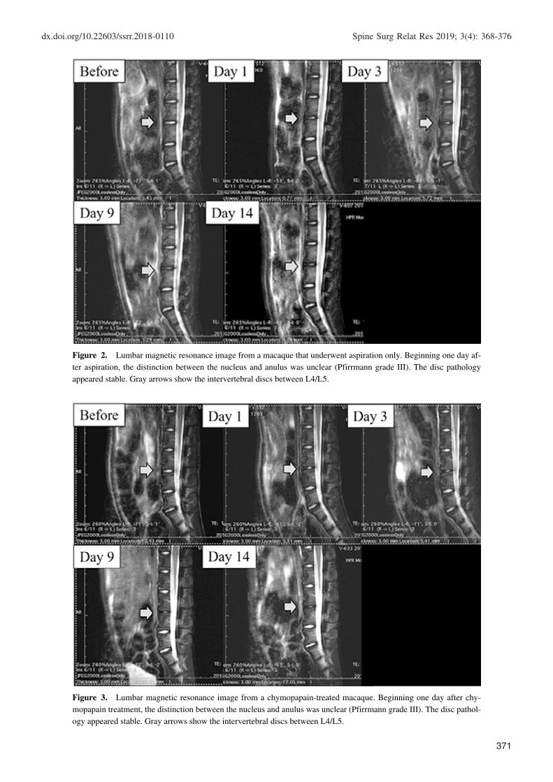
Figure 3. Lumbar magnetic resonance image from a chymopapain-treated macaque. Beginning one day after chy-
mopapain treatment, the distinction between the nucleus and anulus was unclear (Pfirrmann grade III). The disc pathol-
ogy appeared stable. Gray arrows show the intervertebral discs between L4/L5.

Spine Surg Relat Res 2019; 3(4): 368-376 dx.doi.org/10.22603/ssrr.2018-0110
372
Figure 4. Coronal brain sections arranged from rostral (upper left) to caudal (lower right). Before treatment, there was
no significant pressure-induced activation (A). One day after treatment, bilateral pressure-induced activation of the second-
ary somatosensory cortex and insular cortex was observed (B).
IVD pathology following treatment
Treatment with either chymopapain or aspiration alone
did not lead to significant changes to the vertebral body; pa-
thology was limited to the IVD. No lumbar disc degenera-
tion was noted in any macaque before treatment (Pfirrmann
grade I, Table 1). Beginning one day after nucleus pulposus
aspiration, the disc became inhomogeneous, the distinction
between the nucleus and annulus was not clear, and the disc
height was normal (Pfirrmann grade III; Table 1; Fig. 2).
IVD morphology stabilized over time; the Pfirrmann grades
3 to 14 days after aspiration were all III. A similar degrada-
tion of the IVD was observed in a macaque treated with
chymopapain beginning one day after treatment (Pfirrmann
grade III; Table 1; Fig. 3). In the other macaque treated with
chymopapain, beginning one day after treatment, the
Pfirrmann grade of the IVD was IV and there was a loss of
distinction between the nucleus and annulus and a slightly
decreased disc height. Disc pathology remained at grade IV
for the duration of the observational period.

dx.doi.org/10.22603/ssrr.2018-0110 Spine Surg Relat Res 2019; 3(4): 368-376
373
Figure 5. Low back pressure-induced brain activation over time in macaques following chymopapain treatment. Aver-
aged brain images from two macaques treated with chymopapain. Beginning one day after chymopapain treatment, activa-
tion of the secondary somatosensory cortex and insular cortex was observed.
Brain activation
Prior to chymopapain treatment, 1 kg of pressure applied
to the back did not evoke significant brain activation in the
macaques (Fig. 4A). One (Fig. 4B) and three days after chy-
mopapain treatment, pressure-evoked activation of the secon-
dary somatosensory cortex (SII) and insular cortex (Ins) was
observed (Fig. 5; Table 2). Thereafter, no significant
pressure-evoked activation of SII or Ins was observed. By
contrast, in macaques in which the IVD was aspirated, pres-
sure did not evoke significant brain activation (Fig. 6; Table
3).
Discussion
The current findings outlined a potential nonhuman pri-
mate model of LBP, characterized by low back hypersensi-
tivity to pressure, following IVD treatment with chymopa-
pain. In addition, increased SII and Ins activation during low
back stimulation was observed. Interestingly, despite notable
and persistent pathology of the IVD, neither low back hy-
persensitivity nor brain activation was observed beyond
three days after treatment. While aspiration of the nucleus
pulposus alone led to observable pathology of the IVD com-
parable to that following chymopapain treatment, there was
no change in low back pressure sensitivity or significant
brain activation during pressure stimulation. The current
findings suggest that chymopapain treatment activates a
pain-related mechanism that is not activated by loss of the
nucleus pulposus, which further suggests that mechanisms in
addition to, or other than, IVD degeneration underlie LBP.
Understanding the mechanism of LBP is hindered in part
by a lack of preclinical models that recapitulate clinical
LBP2). In general, other species that have been used to
model LBP, such as rodents, differ from humans with re-
spect to overall anatomy, movement biomechanics, and mo-
lecular and immunologic responses to injury2,18). Thus, the
mechanism mediating LBP in rodents may not entirely ap-
ply to clinical LBP. More specifically, models of IVD dam-
age in nonhuman primates, a species that is phylogenetically
closer to humans than rodents, demonstrated significant IVD
pathology but did not measure pain-related behavior2,12,18).
Thus, it is unknown whether IVD injury in nonhuman pri-
mates leads to LBP.
In order to address this, the current study utilized a pres-
sure algometer to assess pain-related behavior objectively.
Quantitative sensory testing (QST) is a clinical method of
objectively measuring pain based on a patient’s response to
graded stimuli19). A key strength of QST is that a particular
stimulus may be used to test the functioning of particular
aspects of the somatosensory system, thereby potentially
suggesting the mechanism. In addition, responsiveness to
standardized stimuli could be used to suggest a common
mechanism and treatment across patients. Pressure stimula-
tion has been used to assess clinical LPB and demonstrated
that the patients had an increased sensitivity to pressure20,21).
While other methods of assessing pain-related behavior have
been used in preclinical models of LBP, such as spontane-
ous activity, these tend to be subjective, indirect measures of
pain-related behavior22,23). A direct measure of pain was used

Spine Surg Relat Res 2019; 3(4): 368-376 dx.doi.org/10.22603/ssrr.2018-0110
374
Table 2. Brain Activation during Low Back Pressure Stimu-
lation in Macaques after Chymopapain Treatment Over Time.
AreaHemi-
sphereZ value
Coordinates (mm)
x y z
Pretreatment
SII and Ins Right 0.22 −16 18 4
Left 0.29 18 16 6
Thalamus Right 0.15 8 −2 8
Left 0.17 −6 −4 8
Cingulate cortex 0.24 0 −20 −10
Day 1 after chymopapain
SII and Ins Right 1.98* −14 16 4
Left 2.72* 16 16 6
Thalamus Right 0.32 4 −4 6
Left 0.22 −6 −2 4
Cingulate cortex 1.62 0 −22 −6
Day 3 after chymopapain
SII and Ins Right 1.32 −18 16 4
Left 2.41* 14 14 4
Thalamus Right 0.31 4 4 6
Left 0.28 −6 2 2
Cingulate cortex 0.76 2 −20 −4
Day 9 after chymopapain
SII and Ins Right 1.22 −14 14 4
Left 1.64 12 10 2
Thalamus Right 0.34 4 2 4
Left 0.36 −4 2 2
Cingulate cortex 0.43 2 −22 −6
Day 14 after chymopapain
SII and Ins Right 0.78 −10 14 4
Left 1.38 14 10 4
Thalamus Right 0.87 6 2 6
Left 0.56 −6 4 2
Cingulate cortex 0.46 0 −24 −6
1 kg of force was applied to the back of the macaque in a block design
during MR imaging.
SII, secondary somatosensory cortex; Ins, insular cortex.
Z values of peak voxels are shown. Stereotaxic coordinates according to
Horsley-Clarke’s stereotaxic coordinates. * Peak voxels were considered
significant (P <0.05) at Z score >1.96.
in the current study, but it is possible that other behavioral
indications of acute LBP could be obtained from the ma-
caques.
A further barrier in understanding the mechanism of LBP
and the development of effective treatments is elucidating
the relationship between pain-related behaviors and central
functioning24,25). In vivo neuroimaging has been suggested as
an objective method of observing pain by quantification of
brain activation with defined stimuli15). Previous fMRI stud-
ies involving pressure-induced pain in LBP patients demon-
strated activation of pain-related brain nuclei such as the pri-
mary somatosensory cortex (SI), SII, posterior cingulate cor-
tex, and Ins4,15). In the current study, activation was observed
only in SII and Ins. Some reports have shown that the use
of anesthesia could inhibit the detection of pain-related brain
activation26,27). Nakamura et al. have reported on the signifi-
cant differences of brain blood flow in patients with chronic
LBP and acute LBP detected by brain SPECT28). The lack of
activation of other nuclei in the current study could be due
to propofol sedation. The propofol dose used in the current
study was high enough to impair movement but not entirely
block cortical pain signaling14). Generally, the current ma-
caque model demonstrates activation of brain regions similar
to that of patients with LBP. The current findings are the
first to demonstrate stimulus-evoked brain activation in a
preclinical LBP model. Future studies could be performed
on trained awake macaques to uncover possible “resting
state” pain that was masked by propofol sedation.
In the current study, chymopapain-induced LBP, whereas
aspiration alone did not induce LBP, even though the extent
of IVD pathology was similar to that following chymopa-
pain treatment12). A possible explanation for this is that chy-
mopapain induces prolonged inflammation whereas aspira-
tion alone does not2,10,11). It is possible that chymopapain led
to an inflammation in tissues outside of the IVD, thereby
activating sensory and sympathetic nerves innervating the
IVD and vertebrae29). Previous preclinical models of LBP re-
ported a transient yet marked inflammation following in-
jury2,18), which appears in clinical LBP as well30). In the cur-
rent model, however, no signs of marked inflammation were
apparent. Alternatively, it is possible that chymopapain or its
breakdown products may have activated peripheral nerves.
Further study is needed to identify the mechanism of
chymopapain-induced LBP. Understanding why marked IVD
pathology alone did not lead to pain in the macaques could
assist in understanding a similarly observed clinical finding
of a lack of correlation between pathology and pain.
One limitation of the current study was that histopathol-
ogy was not performed, which may facilitate further under-
standing of the pain mechanisms in the current model. The
distribution of sensory and sympathetic nerves and possible
inflammatory mediators in the IVD and vertebral body,
which are beyond the detection capability of MRI, could be
important in the generation of LBP. Furthermore, a longitu-
dinal study may be needed as peak pain was observed three
days after chymopapain treatment but not thereafter, even
though marked IVD pathology was observed up to two
weeks after treatment. Examination of inflammatory sub-
stances during and after peak pain could reveal further clues
on the role of the immune system in the maintenance of
LBP in the current model and could suggest a mechanism of
LBP and potential treatment strategies. The second limita-
tion is usage of only male animal models. Young adult male
cynomolgus macaques for assessment of pain were used in
the previous study because young adult female cynomolgus
macaques might have menstrual pain. The final limitation
was that the number of the models was too small. This is a
preliminary study for development of LBP animal model,
and we hope to conduct future research.
Conflicts of Interest: There are no relevant financial in-

dx.doi.org/10.22603/ssrr.2018-0110 Spine Surg Relat Res 2019; 3(4): 368-376
375
Figure 6. Lack of low back pressure-induced brain activation over time in macaques following aspiration. Averaged
brain images over time from two macaques in which the nucleus pulposus was aspirated. There was a lack of significant
pressure-evoked brain activation in these macaques.
Table 3. Brain Activation during Low Back Pressure Stim-
ulation in Macaques before and 1 Day after Aspiration.
AreaHemi-
sphereZ value
Coordinates (mm)
x y z
Pretreatment
SII and Ins Right 0.29 −18 16 4
Left 0.31 16 16 4
Thalamus Right 0.18 6 −2 4
Left 0.22 −8 −2 6
Cingulate cortex 0.30 0 −22 −4
Day 1 after aspiration
SII and Ins Right 0.56 −16 16 4
Left 0.48 18 14 4
Thalamus Right 0.29 6 −2 6
Left 0.22 −6 −4 8
Cingulate cortex 0.28 2 −24 −4
1 kg of force was applied to the back of the macaque in a block design
during MR imaging.
SII, secondary somatosensory cortex; Ins, insular cortex.
Z values of peak voxels are shown. Stereotaxic coordinates according to
Horsley-Clarke’s stereotaxic coordinates. Peak voxels were considered
significant (P <0.05) at Z score >1.96.
terests outside of the current study. H.T., Y.A., S.O., and T.
N. are employees of Hamamatsu Pharma Research, Inc.
Sources of Funding: The current study was supported in
part by Hamamatsu University School of Medicine and Ha-
mamatsu Pharma Research, Inc.
Acknowledgement: The authors deeply appreciate the ex-
pert animal care provided by the Hamamatsu Pharma Re-
search, Inc. Animal Care Group during the course of the
current study.
Author Contributions: Dr. Matsuyama supervised the
study. Dr. Yoshida was responsible for the study’s concep-
tion and design. Dr. Ushirozako acquired, analyzed, and in-
terpreted data, drafted the article, and approved the final ver-
sion on behalf of all authors. All authors critically revised
the article and reviewed the submitted version.
References1. Takahashi N, Kikuchi S, Konno S, et al. Discrepancy between dis-
ability and the severity of low back pain: demographic, psy-
chologic, and employment-related factors. Spine (Phila Pa 1976).
2006;31(8):931-9; discussion 40.
2. Ohtori S, Inoue G, Miyagi M, et al. Pathomechanisms of disco-
genic low back pain in humans and animal models. Spine J. 2015;
15(6):1347-55.
3. deCharms RC, Maeda F, Glover GH, et al. Control over brain acti-
vation and pain learned by using real-time functional MRI. Proc
Natl Acad Sci U S A. 2005;102(51):18626-31.
4. Giesecke T, Gracely RH, Grant MA, et al. Evidence of augmented
central pain processing in idiopathic chronic low back pain. Ar-
thritis Rheum. 2004;50(2):613-23.
5. Apkarian AV, Sosa Y, Sonty S, et al. Chronic back pain is associ-
ated with decreased prefrontal and thalamic gray matter density. J
Neurosci. 2004;24(46):10410-5.
6. Konno SI, Sekiguchi M. Association between brain and low back
pain. J Orthop Sci. 2018;23(1):3-7.
7. Upadhyay J, Anderson J, Schwarz AJ, et al. Imaging drugs with
Spine Surg Relat Res 2019; 3(4): 368-376 dx.doi.org/10.22603/ssrr.2018-0110
376
and without clinical analgesic efficacy. Neuropsychopharmacology.
2011;36(13):2659-73.
8. Kregel J, Meeus M, Malfliet A, et al. Structural and functional
brain abnormalities in chronic low back pain: a systematic review.
Semin Arthritis Rheum. 2015;45(2):229-37.
9. Baliki MN, Chialvo DR, Geha PY, et al. Chronic pain and the
emotional brain: specific brain activity associated with spontane-
ous fluctuations of intensity of chronic back pain. J Neurosci.
2006;26(47):12165-73.
10. Benoist M. [20 years of lumbar chymonucleolysis]. Presse Med.
1996;25(16):743-5. French.
11. Benoist M, Bonneville JF, Lassale B, et al. A randomized, double-
blind study to compare low-dose with standard-dose chymopapain
in the treatment of herniated lumbar intervertebral discs. Spine
(Phila Pa 1976). 1993;18(1):28-34.
12. Xi Y, Kong J, Liu Y, et al. Minimally invasive induction of an
early lumbar disc degeneration model in rhesus monkeys. Spine
(Phila Pa 1976). 2013;38(10):E579-86.
13. Pfirrmann CW, Metzdorf A, Zanetti M, et al. Magnetic resonance
classification of lumbar intervertebral disc degeneration. Spine
(Phila Pa 1976). 2001;26(17):1873-8.
14. Steinbacher DM. Propofol: a sedative-hypnotic anesthetic agent for
use in ambulatory procedures. Anesth Prog. 2001;48(2):66-71.
15. Kobayashi Y, Kurata J, Sekiguchi M, et al. Augmented cerebral
activation by lumbar mechanical stimulus in chronic low back pain
patients: an FMRI study. Spine (Phila Pa 1976). 2009;34(22):
2431-6.
16. Black KJ, Koller JM, Snyder AZ, et al. Atlas template images for
nonhuman primate neuroimaging: baboon and macaque. Methods
Enzymol. 2004;385:91-102.
17. Ogawa S, Awaga Y, Takashima M, et al. Knee osteoarthritis pain
following medial meniscectomy in the nonhuman primate. Os-
teoarthritis Cartilage. 2016;24(7):1190-9.
18. Alini M, Eisenstein SM, Ito K, et al. Are animal models useful for
studying human disc disorders/degeneration? Eur Spine J. 2008;17
(1):2-19.
19. van Vliet J, Tieleman AA, Verrips A, et al. Qualitative and quanti-
tative aspects of pain in patients with myotonic dystrophy type 2.
J Pain. 2018;19(8):920-30.
20. Kaneko H, Zhang S, Sekiguchi M, et al. Dysfunction of nucleus
accumbens is associated with psychiatric problems in patients with
chronic low back pain: a functional magnetic resonance imaging
study. Spine (Phila Pa 1976). 2017;42(11):844-53.
21. Marcuzzi A, Wrigley PJ, Dean CM, et al. From acute to persistent
low back pain: a longitudinal investigation of somatosensory
changes using quantitative sensory testing-an exploratory study.
Pain Rep. 2018;3(2):e641.
22. Miyagi M, Ishikawa T, Kamoda H, et al. Assessment of pain be-
havior in a rat model of intervertebral disc injury using the Cat-
Walk gait analysis system. Spine (Phila Pa 1976). 2013;38(17):
1459-65.
23. Olmarker K. Puncture of a lumbar intervertebral disc induces
changes in spontaneous pain behavior: an experimental study in
rats. Spine (Phila Pa 1976). 2008;33(8):850-5.
24. Hama A, Natsume T, Ogawa S, et al. Pain-related behavior and
brain activation in a cynomolgus macaque model of postoperative
pain. CNS Neurol Disord Drug Targets. 2018;17(5):348-60.
25. Nagasaka K, Yamanaka K, Ogawa S, et al. Brain activity changes
in a macaque model of oxaliplatin-induced neuropathic cold hy-
persensitivity. Sci Rep. 2017;7(1):4305.
26. Seah S, Asad AB, Baumgartner R, et al. Investigation of cross-
species translatability of pharmacological MRI in awake nonhu-
man primate - a buprenorphine challenge study. PLoS One. 2014;9
(10):e110432.
27. Lahti KM, Ferris CF, Li F, et al. Comparison of evoked cortical
activity in conscious and propofol-anesthetized rats using func-
tional MRI. Magn Reson Med. 1999;41(2):412-6.
28. Nakamura Y, Nojiri K, Yoshihara H, et al. Significant differences
of brain blood flow in patients with chronic low back pain and
acute low back pain detected by brain SPECT. J Orthop Sci. 2014;
19(3):384-9.
29. Suseki K, Takahashi Y, Takahashi K, et al. Sensory nerve fibres
from lumbar intervertebral discs pass through rami communi-
cantes. A possible pathway for discogenic low back pain. J Bone
Joint Surg Br. 1998;80(4):737-42.
30. Burke JG, Watson RW, McCormack D, et al. Intervertebral discs
which cause low back pain secrete high levels of proinflammatory
mediators. J Bone Joint Surg Br. 2002;84(2):196-201.
Spine Surgery and Related Research is an Open Access journal distributed under
the Creative Commons Attribution-NonCommercial-NoDerivatives 4.0 Interna-
tional License. To view the details of this license, please visit (https://creativeco
mmons.org/licenses/by-nc-nd/4.0/).