Embed Size (px)
Citation preview

Brain and Behavior
Chapter 3
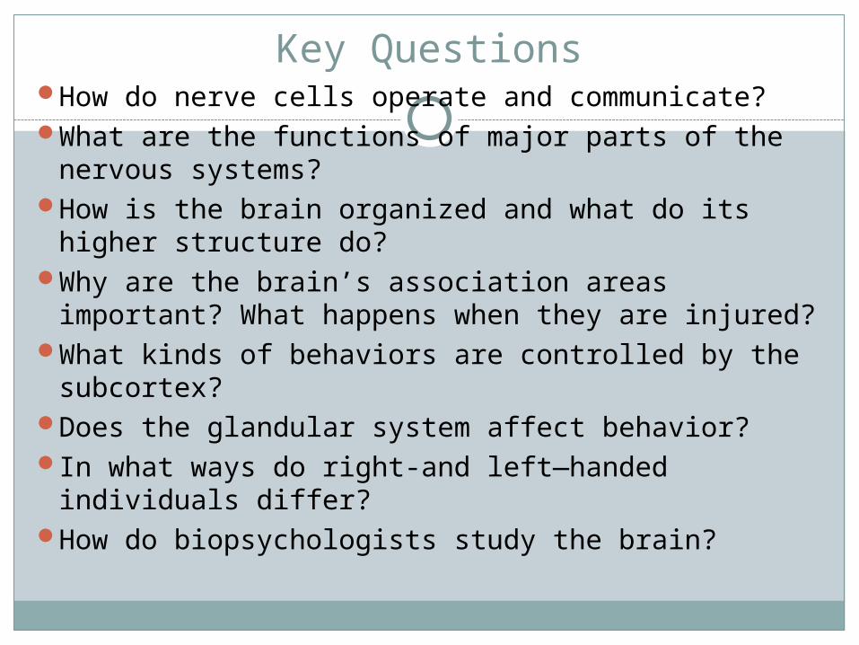
Key QuestionsHow do nerve cells operate and communicate?What are the functions of major parts of the nervous
systems?How is the brain organized and what do its higher
structure do?Why are the brain’s association areas important?
What happens when they are injured?What kinds of behaviors are controlled by the
subcortex?Does the glandular system affect behavior?In what ways do right-and left—handed individuals
differ?How do biopsychologists study the brain?
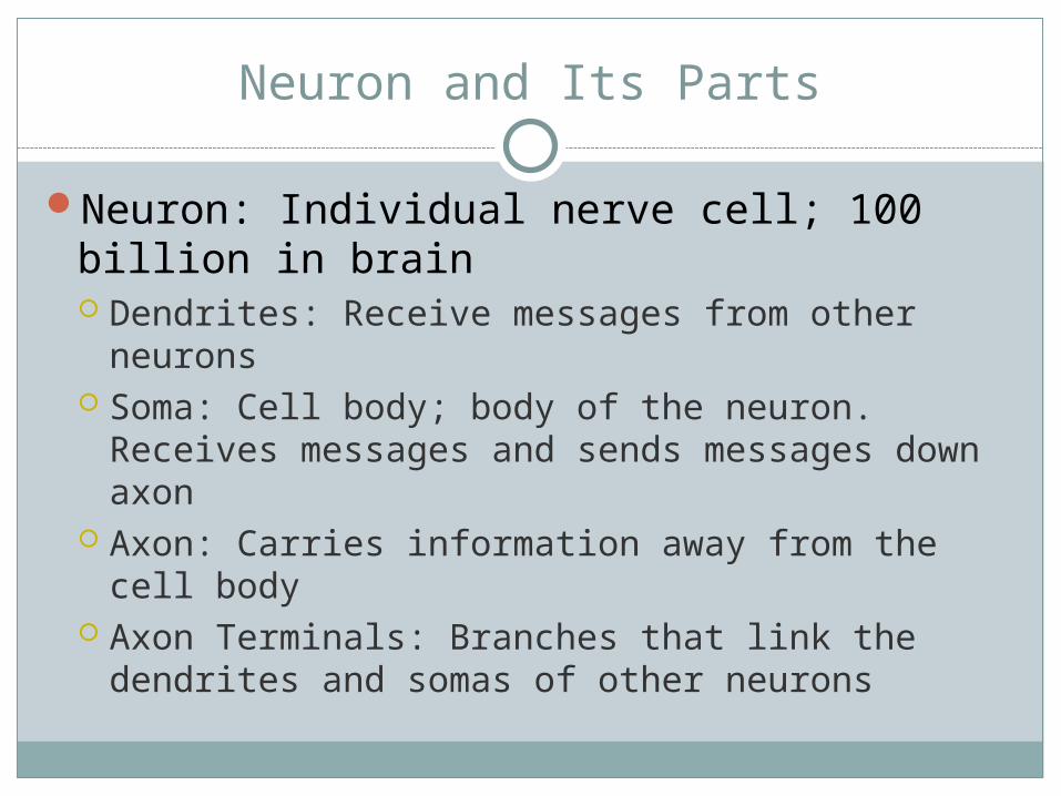
Neuron and Its Parts
Neuron: Individual nerve cell; 100 billion in brain Dendrites: Receive messages from other neurons Soma: Cell body; body of the neuron. Receives
messages and sends messages down axon Axon: Carries information away from the cell
body Axon Terminals: Branches that link the dendrites
and somas of other neurons
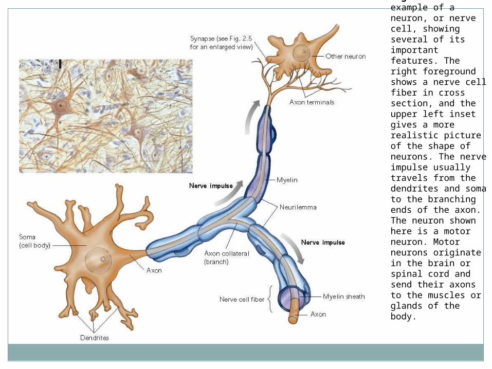
Fig. 2.1 An example of a neuron, or nerve cell, showing several of its important features. The right foreground shows a nerve cell fiber in cross section, and the upper left inset gives a more realistic picture of the shape of neurons. The nerve impulse usually travels from the dendrites and soma to the branching ends of the axon. The neuron shown here is a motor neuron. Motor neurons originate in the brain or spinal cord and send their axons to the muscles or glands of the body.
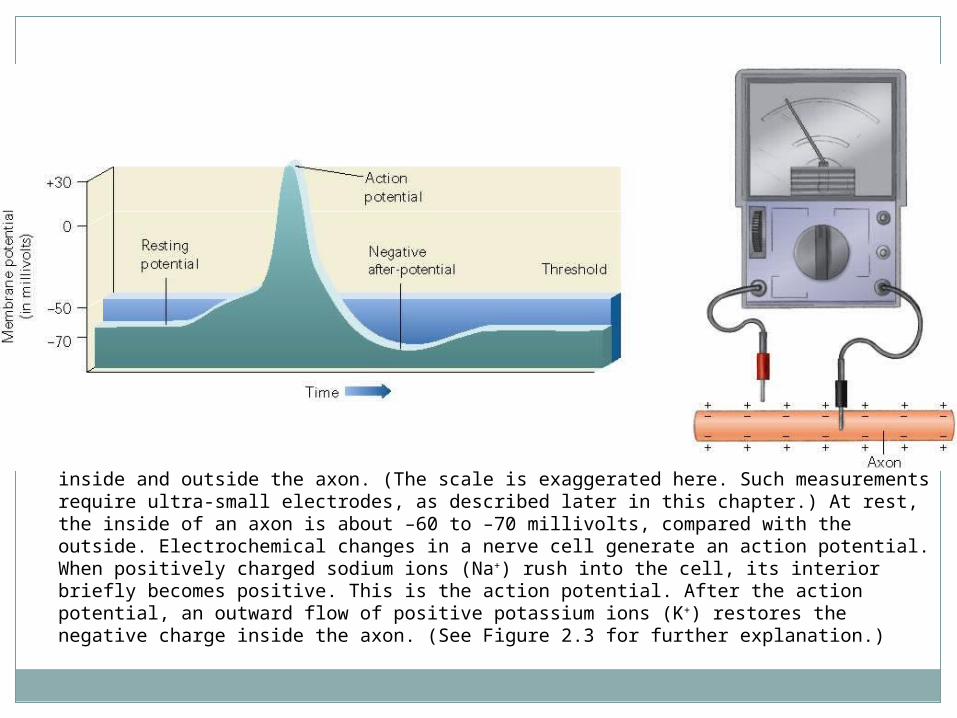
Fig. 2.2 Activity in an axon can be measured by placing electrical probes inside and outside the axon. (The scale is exaggerated here. Such measurements require ultra-small electrodes, as described later in this chapter.) At rest, the inside of an axon is about –60 to –70 millivolts, compared with the outside. Electrochemical changes in a nerve cell generate an action potential. When positively charged sodium ions (Na+) rush into the cell, its interior briefly becomes positive. This is the action potential. After the action potential, an outward flow of positive potassium ions (K+) restores the negative charge inside the axon. (See Figure 2.3 for further explanation.)
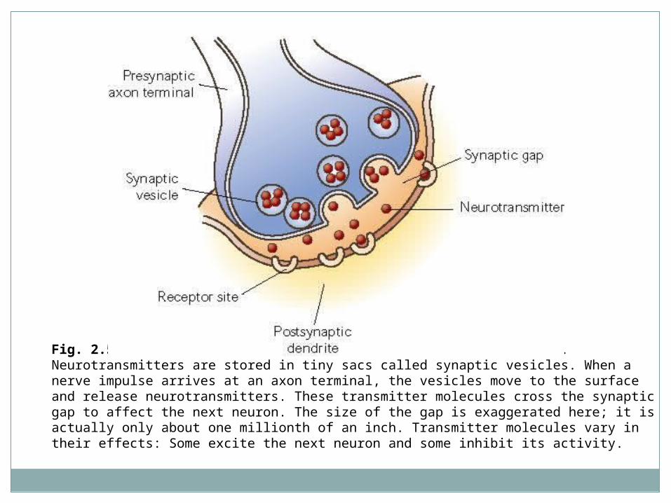
Fig. 2.5 A highly magnified view of the synapse shown in Fig. 2.1. Neurotransmitters are stored in tiny sacs called synaptic vesicles. When a nerve impulse arrives at an axon terminal, the vesicles move to the surface and release neurotransmitters. These transmitter molecules cross the synaptic gap to affect the next neuron. The size of the gap is exaggerated here; it is actually only about one millionth of an inch. Transmitter molecules vary in their effects: Some excite the next neuron and some inhibit its activity.
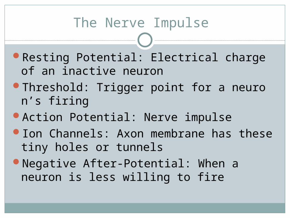
The Nerve Impulse
Resting Potential: Electrical charge of an inactive neuron
Threshold: Trigger point for a neuron’s firing
Action Potential: Nerve impulseIon Channels: Axon membrane has these
tiny holes or tunnelsNegative After-Potential: When a neuron is
less willing to fire
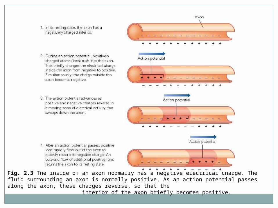
Fig. 2.3 The inside of an axon normally has a negative electrical charge. The fluid surrounding an axon is normally positive. As an action potential passes along the axon, these charges reverse, so that the interior of the axon briefly becomes positive.
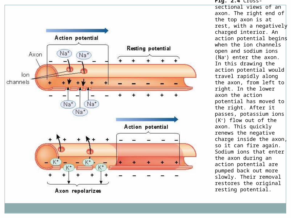
Fig. 2.4 Cross-sectional views of an axon. The right end of the top axon is at rest, with a negatively charged interior. An action potential begins when the ion channels open and sodium ions (Na+) enter the axon. In this drawing the action potential would travel rapidly along the axon, from left to right. In the lower axon the action potential has moved to the right. After it passes, potassium ions (K+) flow out of the axon. This quickly renews the negative charge inside the axon, so it can fire again. Sodium ions that enter the axon during an action potential are pumped back out more slowly. Their removal restores the original resting potential.
Animation: Neuron & Neural Impulse
Animation
Synapses and Neurotransmitters
Synapse: The microscopic space between two neurons, over with messages pass
Neurotransmitters: Chemicals that alter activity in neurons; brain chemicals Acetylcholine: Activates muscles Dopamine: Muscle control Serotonin: Mood and appetite control
Messages from one neuron to another pass over a microscopic gap called a synapse
Receptor Site: Areas on the surface of neurons and other cells that are sensitive to neurotransmitters
Animation: Synaptic Transmission
Animation
Neural Regulators
Neuropeptides: Regulate activity of other neurons Enkephalins: Relieve pain and stress; similar to
endorphins Endorphins: Released by pituitary gland; also help to
relieve pain Placebos raise endorphin levels
The Nervous System- Wired for Action
Nerves and Neurons Nerves: Large bundles of axons and dendritesMyelin: Fatty layer that coats some axons
Multiple Sclerosis (MS) occurs when myelin layer is destroyed; numbness, weakness, and paralysis occur
Neurilemma: Thin layer of cells wrapped around axons outside brain and spinal cord; forms a tunnel that damaged fibers follow as they repair themselves
Brain Grafts and Nerve Regeneration
Transplanting brain tissue
The Nervous System
Central Nervous System (CNS): Brain and spinal cord
Peripheral Nervous System: All parts of the nervous system outside of the brain and spinal cord Somatic System: Carries messages to and from
skeletal muscles and sense organs; controls voluntary behavior
Autonomic System: Serves internal organs and glands; controls automatic functions such as heart rate and blood pressure
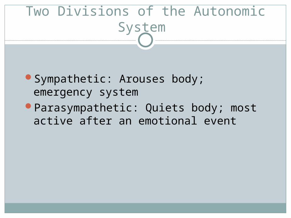
Two Divisions of the Autonomic System
Sympathetic: Arouses body; emergency system
Parasympathetic: Quiets body; most active after an emotional event
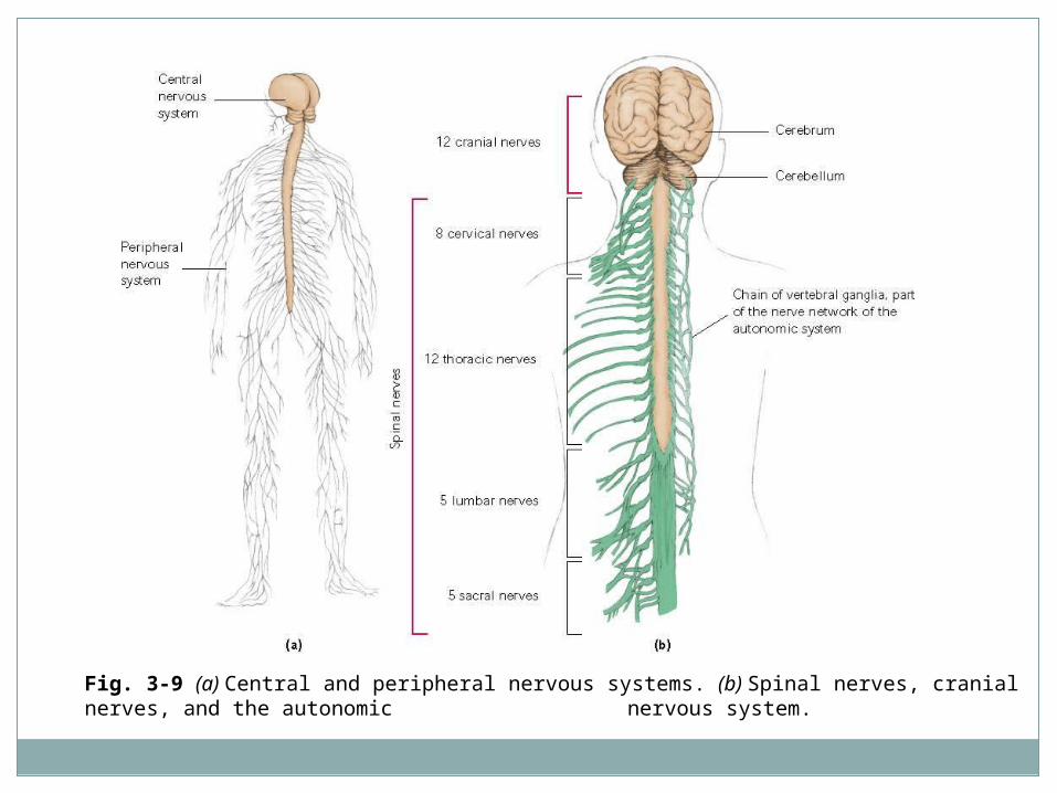
Fig. 3-9 (a) Central and peripheral nervous systems. (b) Spinal nerves, cranial nerves, and the autonomic nervous system.
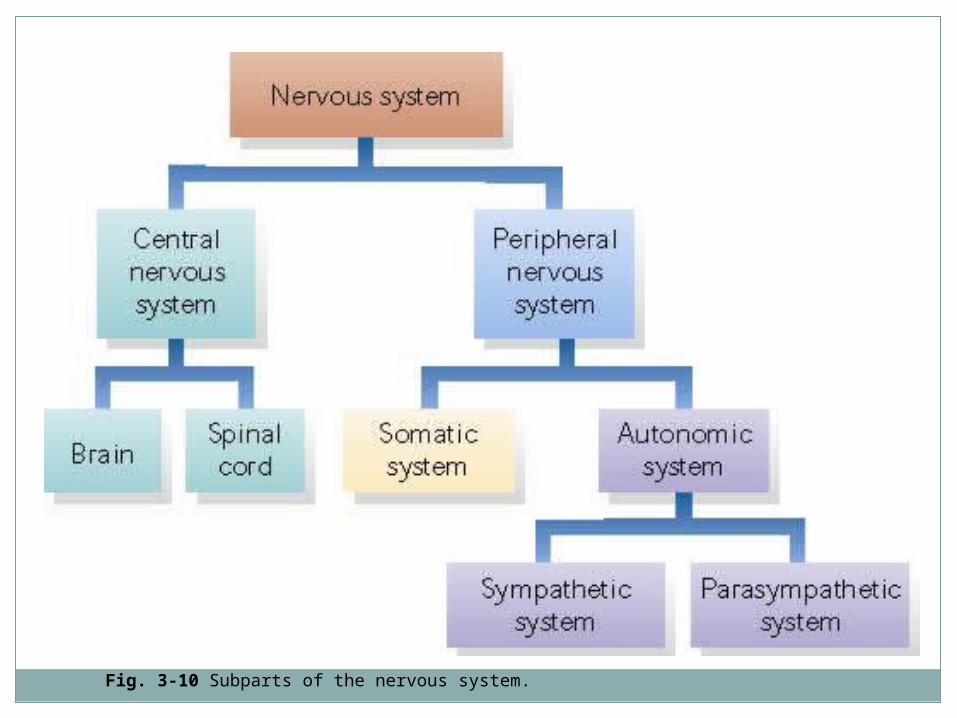
Fig. 3-10 Subparts of the nervous system.
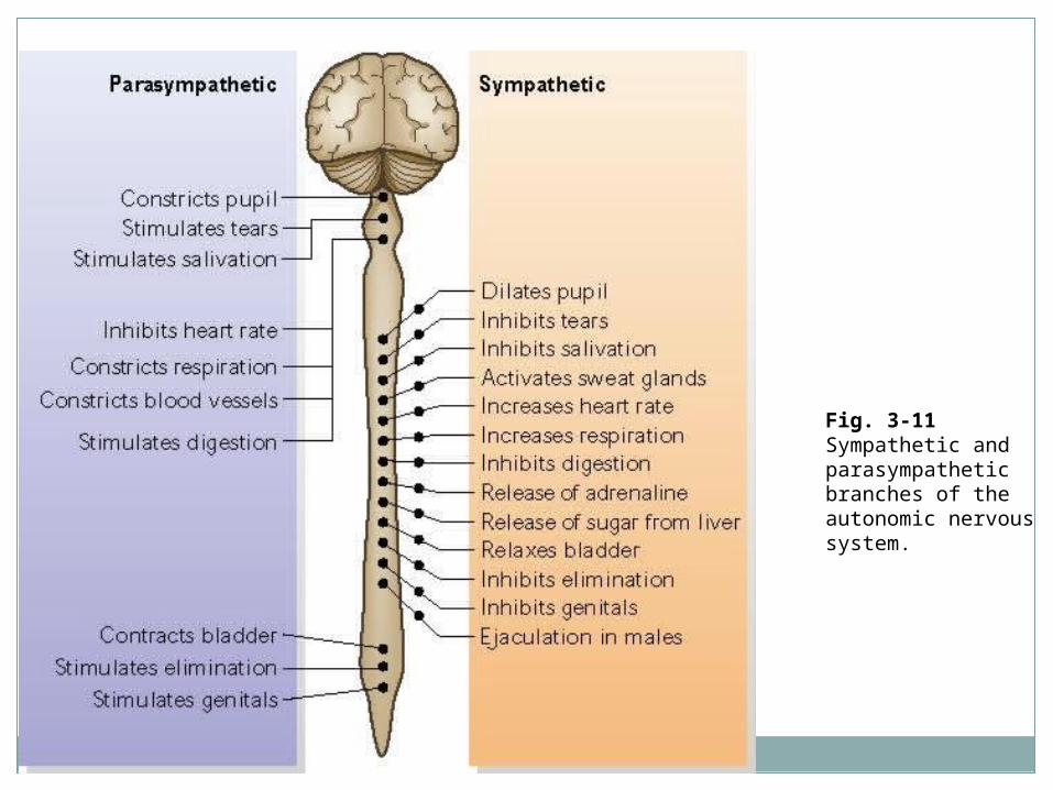
Fig. 3-11 Sympathetic and parasympathetic branches of the autonomic nervous system.
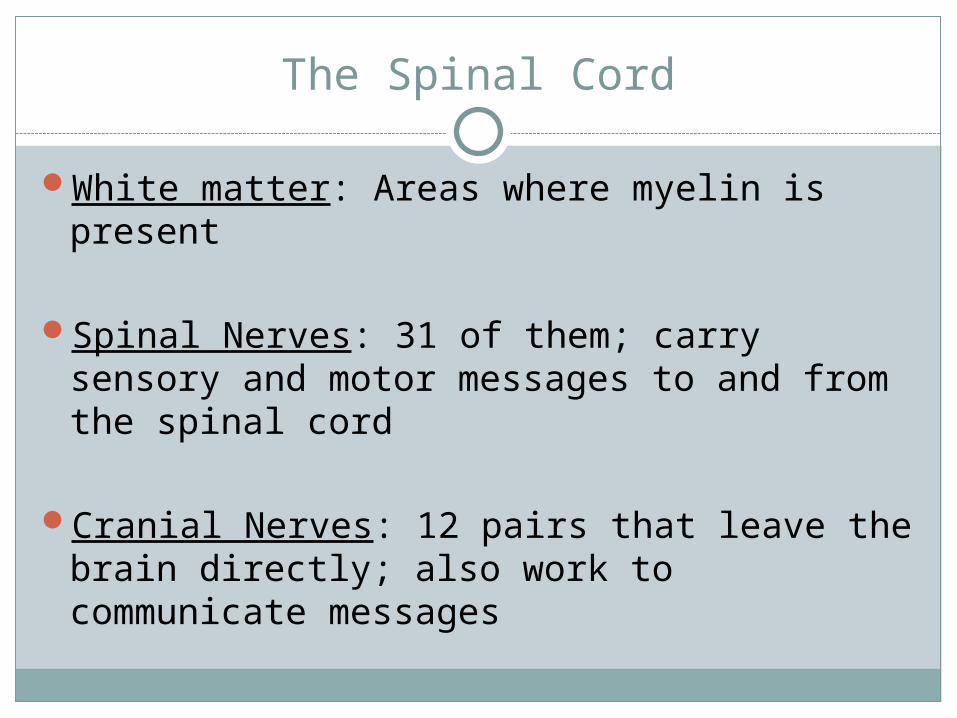
The Spinal Cord
White matter: Areas where myelin is present
Spinal Nerves: 31 of them; carry sensory and motor messages to and from the spinal cord
Cranial Nerves: 12 pairs that leave the brain directly; also work to communicate messages
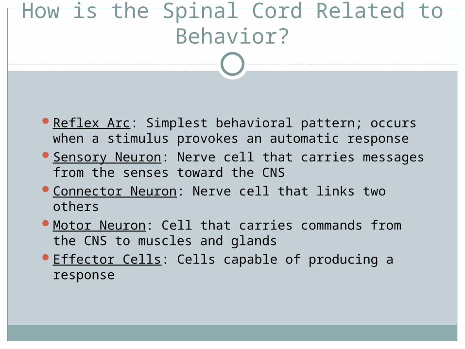
How is the Spinal Cord Related to Behavior?
Reflex Arc: Simplest behavioral pattern; occurs when a stimulus provokes an automatic response
Sensory Neuron: Nerve cell that carries messages from the senses toward the CNS
Connector Neuron: Nerve cell that links two othersMotor Neuron: Cell that carries commands from
the CNS to muscles and glandsEffector Cells: Cells capable of producing a
response
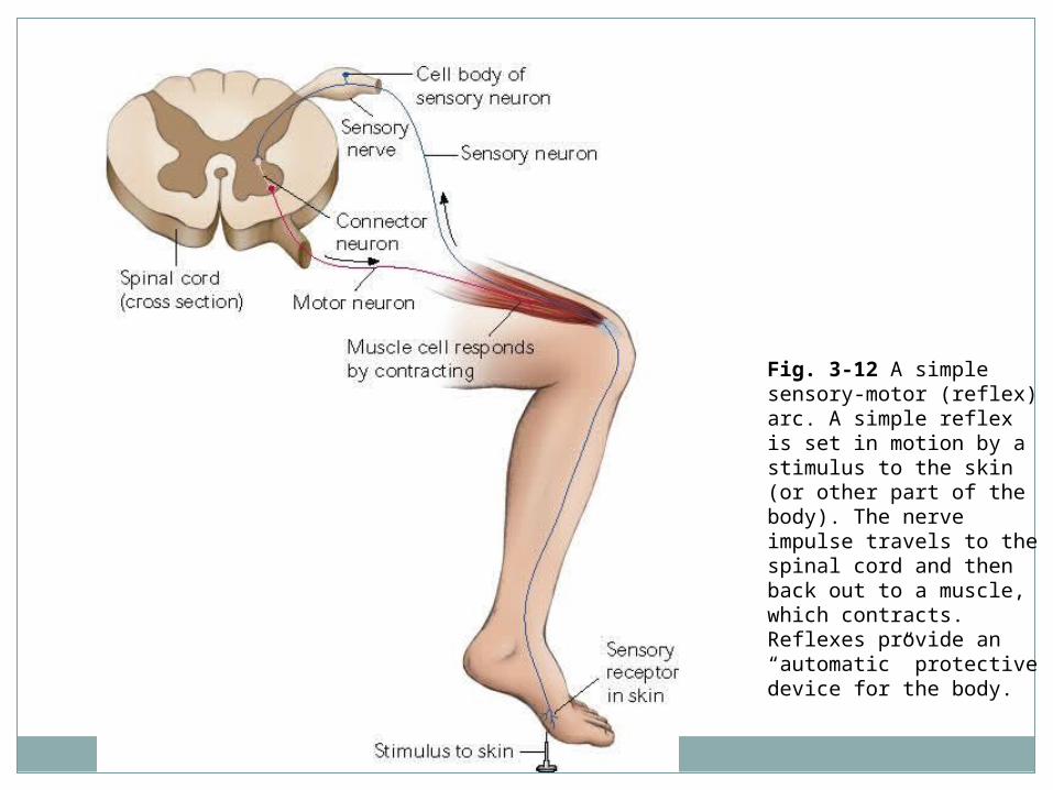
Fig. 3-12 A simple sensory-motor (reflex) arc. A simple reflex is set in motion by a stimulus to the skin (or other part of the body). The nerve impulse travels to the spinal cord and then back out to a muscle, which contracts. Reflexes provide an “automatic” protective device for the body.
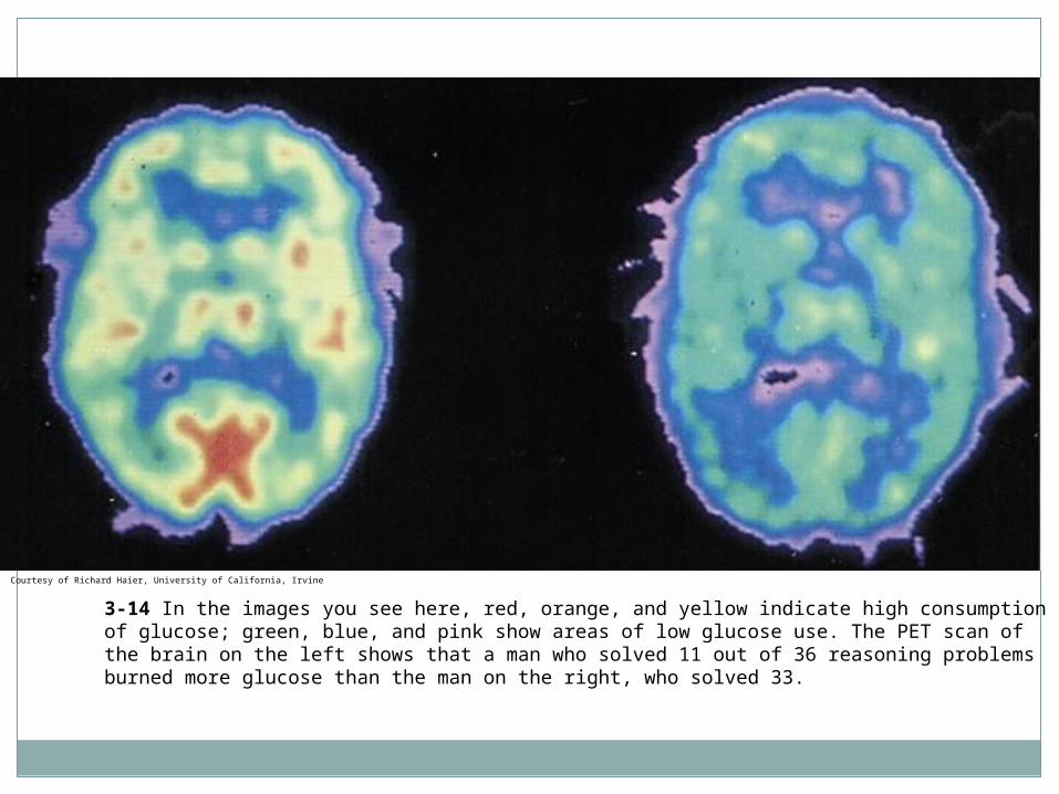
3-14 In the images you see here, red, orange, and yellow indicate high consumption of glucose; green, blue, and pink show areas of low glucose use. The PET scan of the brain on the left shows that a man who solved 11 out of 36 reasoning problems burned more glucose than the man on the right, who solved 33.
Courtesy of Richard Haier, University of California, Irvine
Brain Mapping
Read article from CNN
Project
Cerebral Cortex
Cerebral Cortex: Outer layer of the cerebrum; contains 70% of neurons in CNS
Cerebrum: Two large hemispheres that cover upper part of the brain
Corticalization: Increase in size and wrinkling of the cortex
Split Brains
Cerebral Hemispheres: Right and left halves of the cerebrum
Corpus Callosum: Bundle of fibers connecting cerebral hemispheres Corpus Callosum is cut; done to control severe epilepsy
(seizure disorder) Result: The person now has two brains in one body This operation is rare and is often used as a last resort
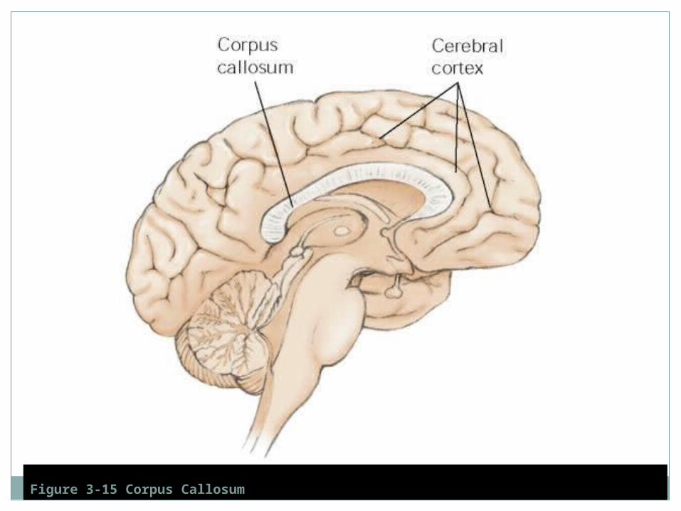
Figure 3-15 Corpus Callosum
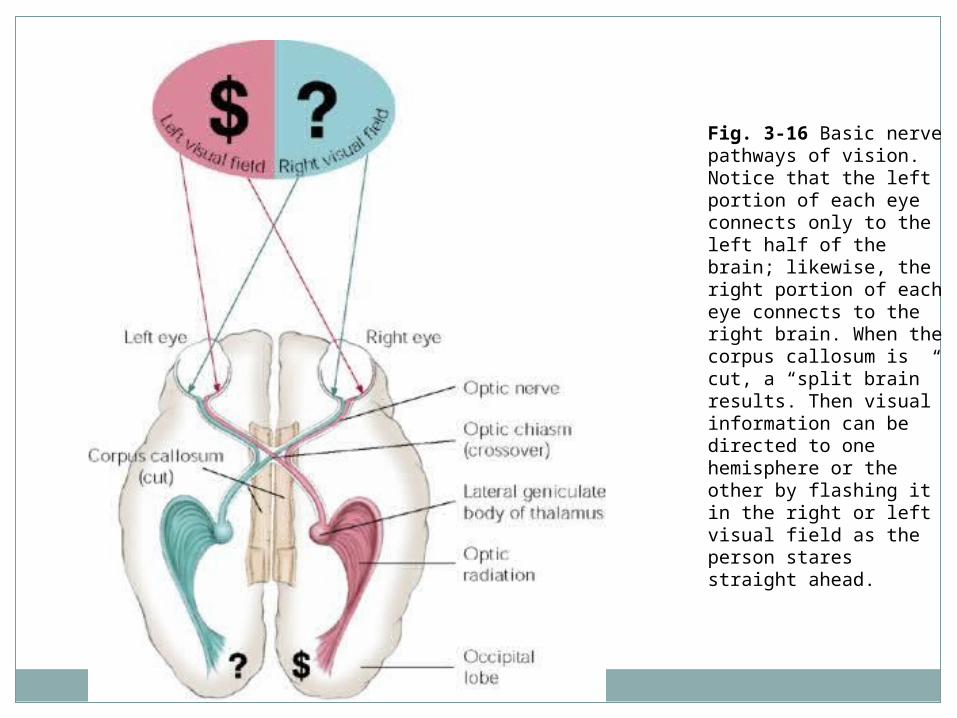
Fig. 3-16 Basic nerve pathways of vision. Notice that the left portion of each eye connects only to the left half of the brain; likewise, the right portion of each eye connects to the right brain. When the corpus callosum is cut, a “split brain” results. Then visual information can be directed to one hemisphere or the other by flashing it in the right or left visual field as the person stares straight ahead.
Right Brain/Left Brain
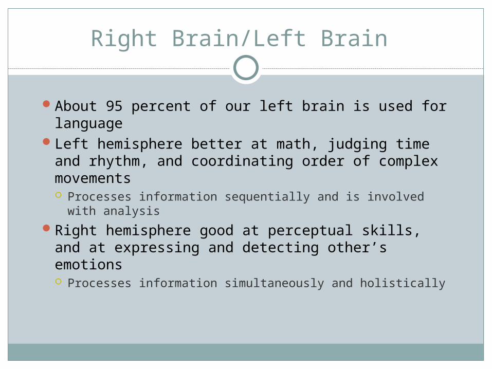
About 95 percent of our left brain is used for language
Left hemisphere better at math, judging time and rhythm, and coordinating order of complex movements Processes information sequentially and is involved
with analysisRight hemisphere good at perceptual skills, and
at expressing and detecting other’s emotions Processes information simultaneously and holistically
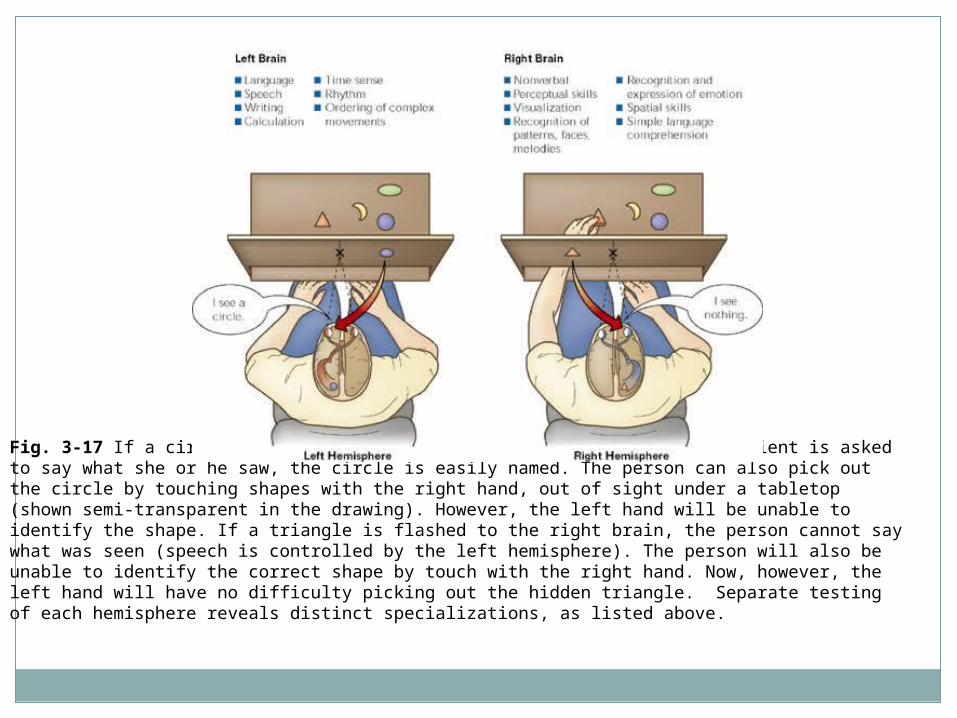
Fig. 3-17 If a circle is flashed to the left brain and a split-brain patient is asked to say what she or he saw, the circle is easily named. The person can also pick out the circle by touching shapes with the right hand, out of sight under a tabletop (shown semi-transparent in the drawing). However, the left hand will be unable to identify the shape. If a triangle is flashed to the right brain, the person cannot say what was seen (speech is controlled by the left hemisphere). The person will also be unable to identify the correct shape by touch with the right hand. Now, however, the left hand will have no difficulty picking out the hidden triangle. Separate testing of each hemisphere reveals distinct specializations, as listed above.
Corpus Callosum Cut
Video
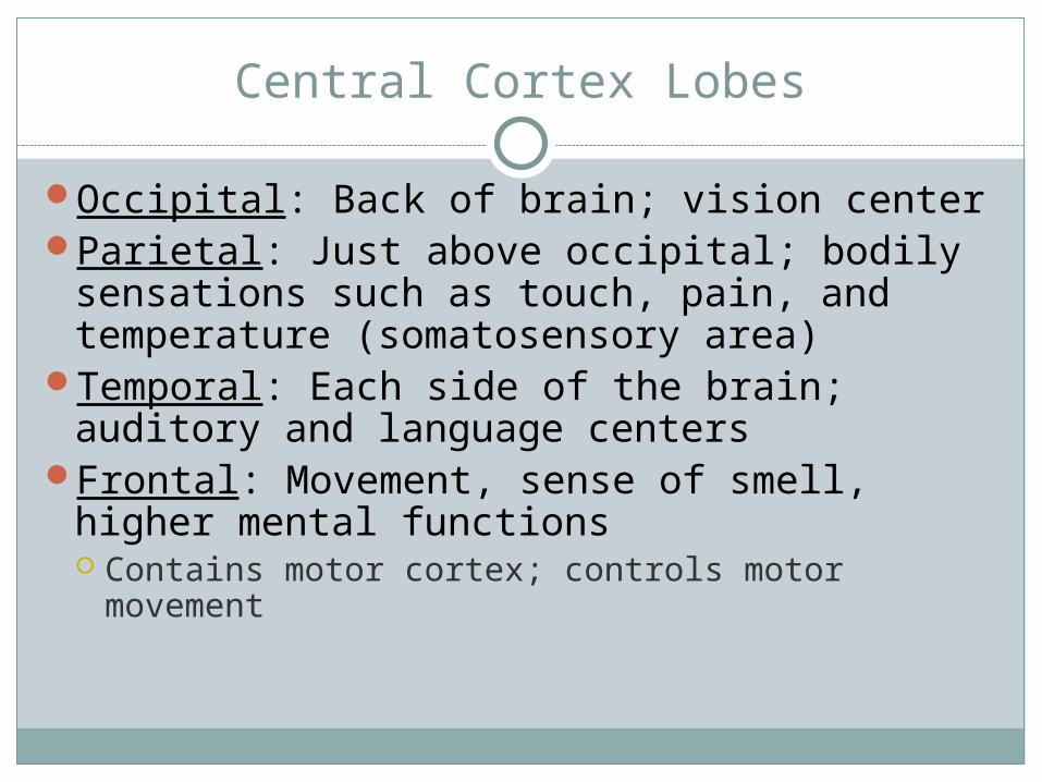
Central Cortex Lobes
Occipital: Back of brain; vision centerParietal: Just above occipital; bodily
sensations such as touch, pain, and temperature (somatosensory area)
Temporal: Each side of the brain; auditory and language centers
Frontal: Movement, sense of smell, higher mental functions Contains motor cortex; controls motor movement
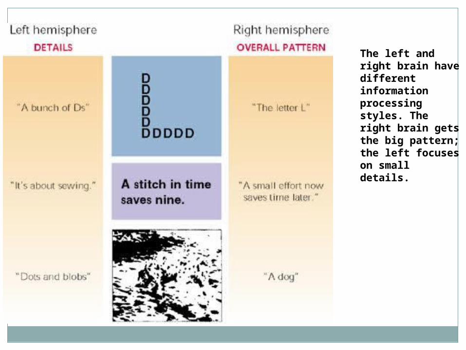
The left and right brain have different information processing styles. The right brain gets the big pattern; the left focuses on small details.
Brain Parts
Worksheet
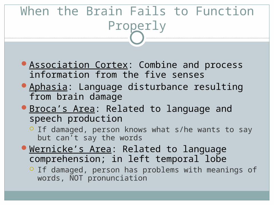
When the Brain Fails to Function Properly
Association Cortex: Combine and process information from the five senses
Aphasia: Language disturbance resulting from brain damage
Broca’s Area: Related to language and speech production If damaged, person knows what s/he wants to say but
can’t say the wordsWernicke’s Area: Related to language
comprehension; in left temporal lobe If damaged, person has problems with meanings of
words, NOT pronunciation
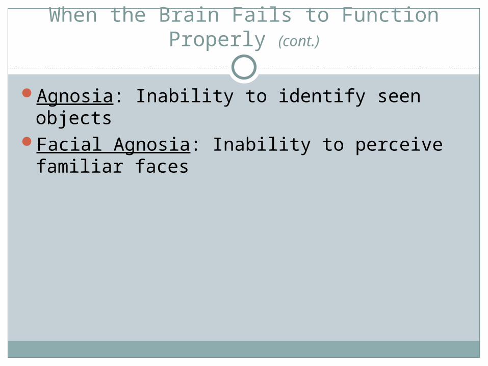
When the Brain Fails to Function Properly (cont.)
Agnosia: Inability to identify seen objectsFacial Agnosia: Inability to perceive familiar
faces

Brain Injury
Brain Injury Worksheet
His and Her Brains
Read page 66
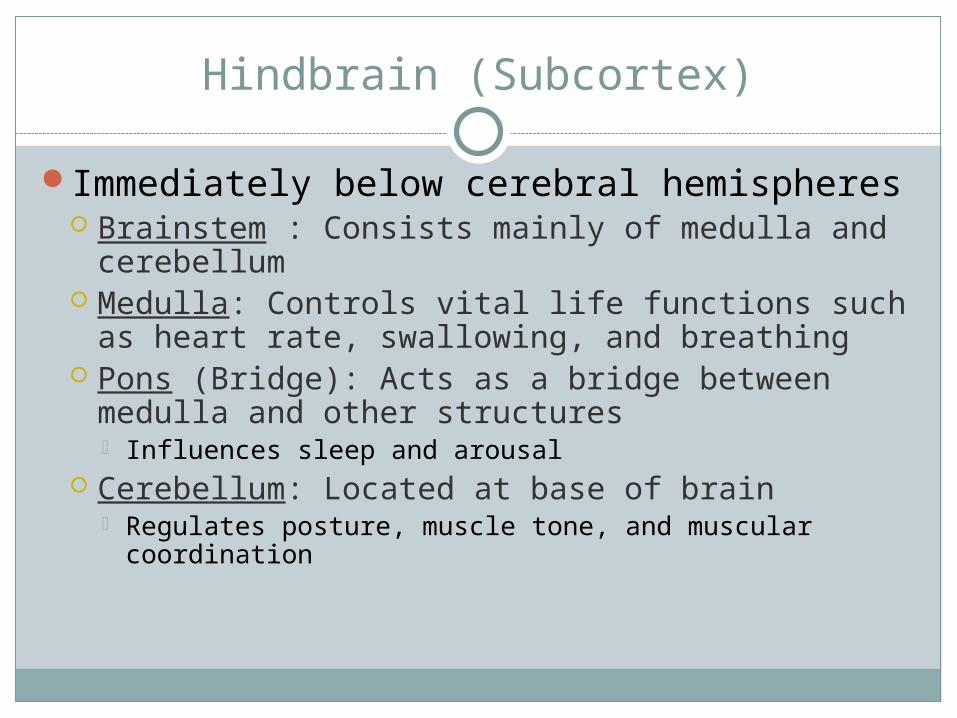
Hindbrain (Subcortex)
Immediately below cerebral hemispheres Brainstem : Consists mainly of medulla and
cerebellum Medulla: Controls vital life functions such as
heart rate, swallowing, and breathing Pons (Bridge): Acts as a bridge between medulla
and other structures Influences sleep and arousal
Cerebellum: Located at base of brain Regulates posture, muscle tone, and muscular
coordination

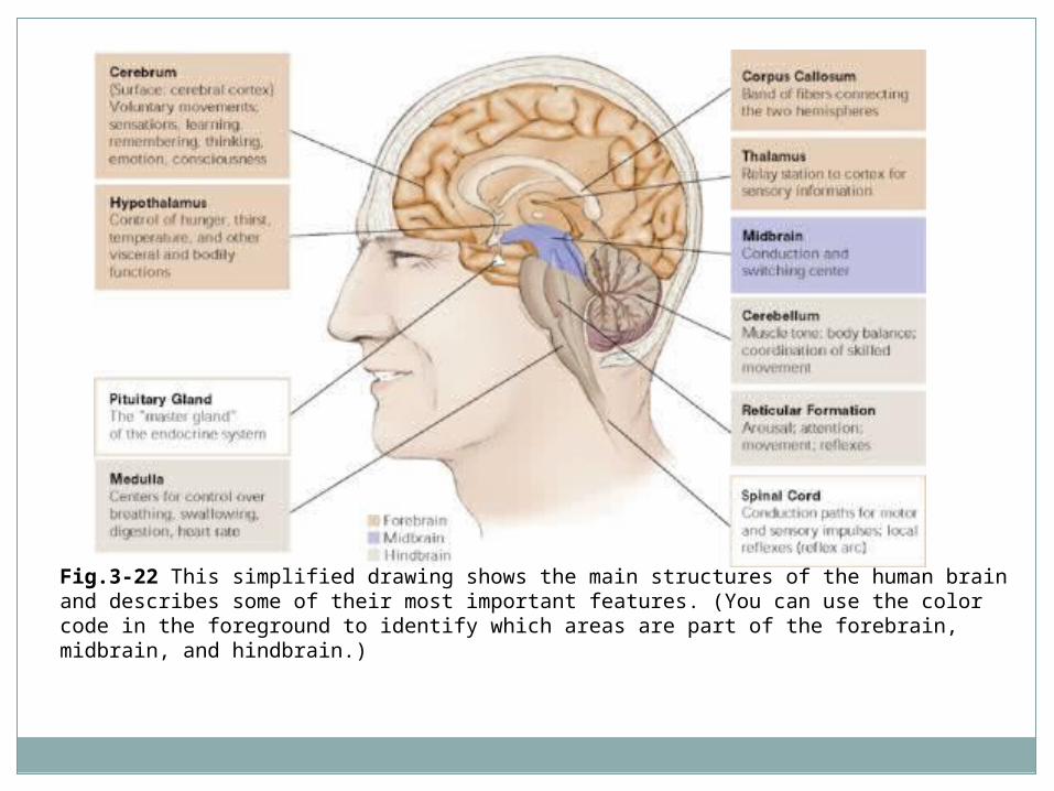
Fig.3-22 This simplified drawing shows the main structures of the human brain and describes some of their most important features. (You can use the color code in the foreground to identify which areas are part of the forebrain, midbrain, and hindbrain.)
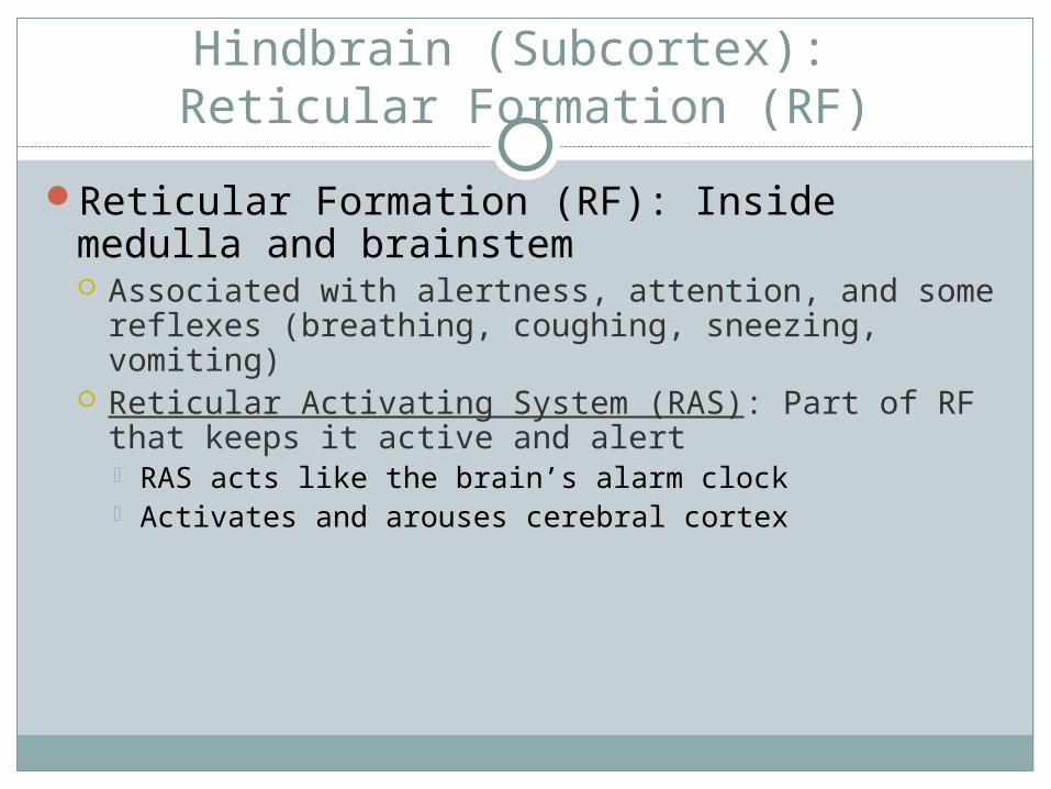
Hindbrain (Subcortex): Reticular Formation (RF)
Reticular Formation (RF): Inside medulla and brainstem Associated with alertness, attention, and some
reflexes (breathing, coughing, sneezing, vomiting) Reticular Activating System (RAS): Part of RF that
keeps it active and alert RAS acts like the brain’s alarm clock Activates and arouses cerebral cortex
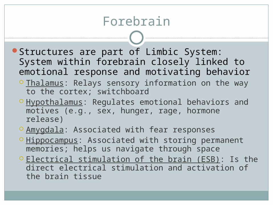
Forebrain
Structures are part of Limbic System: System within forebrain closely linked to emotional response and motivating behavior Thalamus: Relays sensory information on the way to
the cortex; switchboard Hypothalamus: Regulates emotional behaviors and
motives (e.g., sex, hunger, rage, hormone release) Amygdala: Associated with fear responses Hippocampus: Associated with storing permanent
memories; helps us navigate through space Electrical stimulation of the brain (ESB): Is the direct
electrical stimulation and activation of the brain tissue
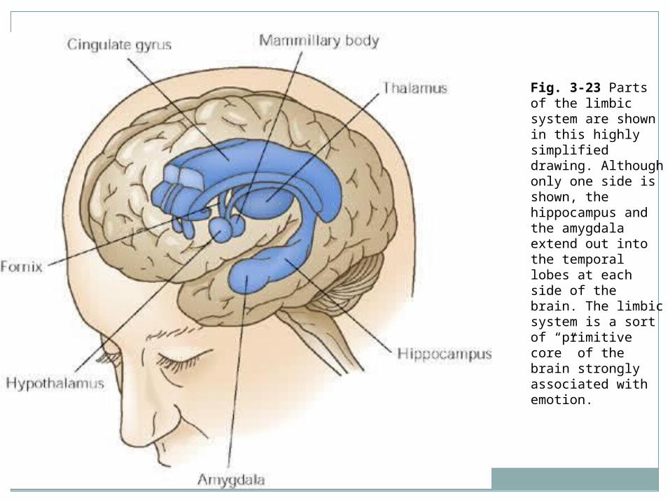
Fig. 3-23 Parts of the limbic system are shown in this highly simplified drawing. Although only one side is shown, the hippocampus and the amygdala extend out into the temporal lobes at each side of the brain. The limbic system is a sort of “primitive core” of the brain strongly associated with emotion.
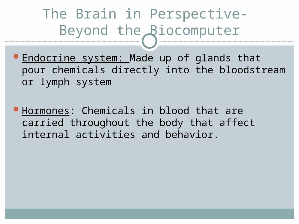
The Brain in Perspective- Beyond the Biocomputer
Endocrine system: Made up of glands that pour chemicals directly into the bloodstream or lymph system
Hormones: Chemicals in blood that are carried throughout the body that affect internal activities and behavior.
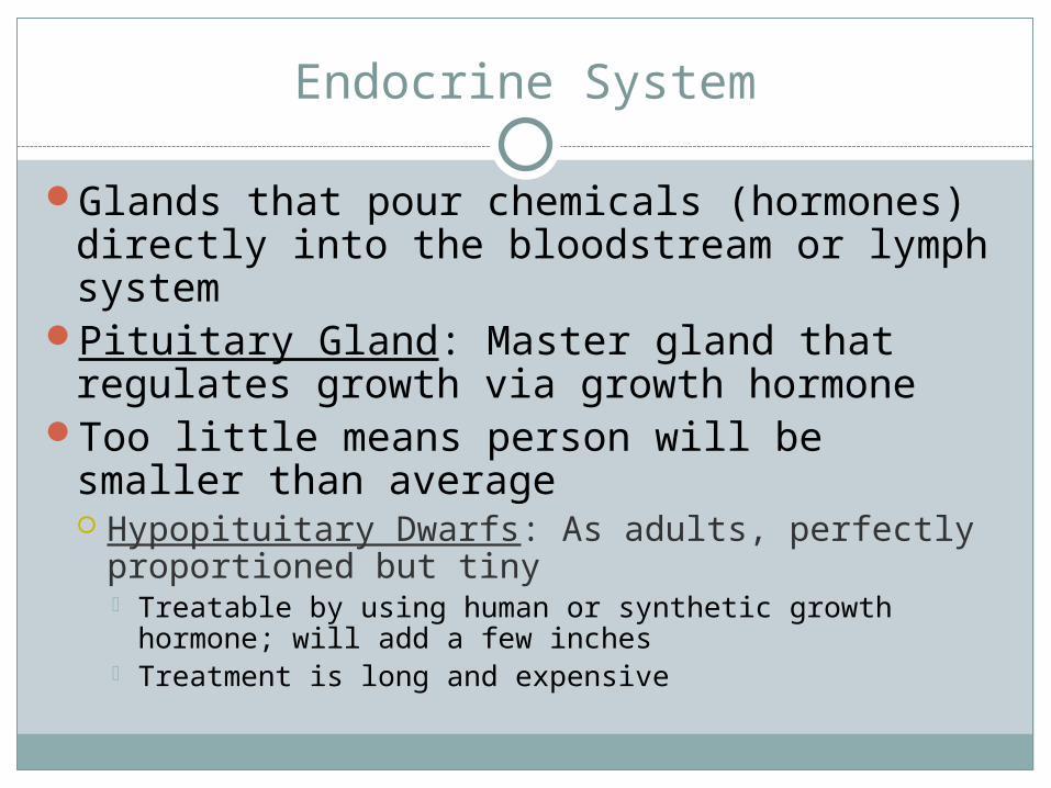
Endocrine System
Glands that pour chemicals (hormones) directly into the bloodstream or lymph system
Pituitary Gland: Master gland that regulates growth via growth hormone
Too little means person will be smaller than average Hypopituitary Dwarfs: As adults, perfectly
proportioned but tiny Treatable by using human or synthetic growth hormone;
will add a few inches Treatment is long and expensive

Endocrine System (cont.)
Too much growth hormone leads to giantism Excessive body growth
Acromegaly: Enlargement of arms, hands, feet, and facial bones Caused by too much growth hormone secreted
late in growth period Andre the Giant
Pituitary also governs functioning of other glands, especially thyroid, adrenals, and gonads

PRINCESS BRIDE
Endocrine System (cont.)
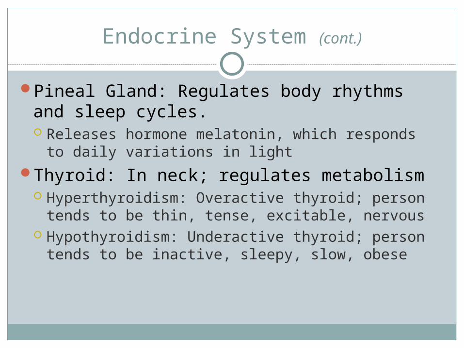
Pineal Gland: Regulates body rhythms and sleep cycles. Releases hormone melatonin, which responds to
daily variations in lightThyroid: In neck; regulates metabolism
Hyperthyroidism: Overactive thyroid; person tends to be thin, tense, excitable, nervous
Hypothyroidism: Underactive thyroid; person tends to be inactive, sleepy, slow, obese
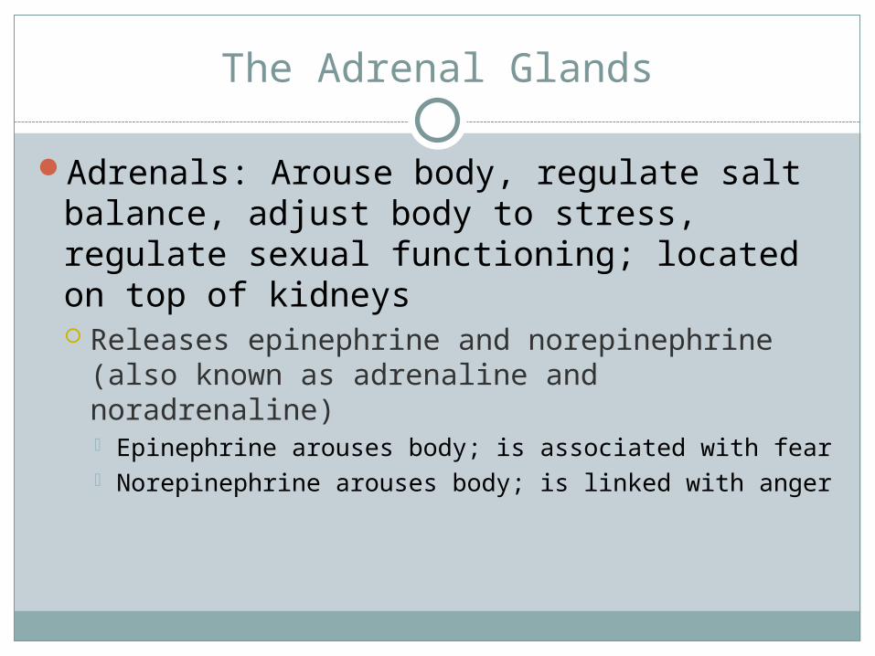
The Adrenal Glands
Adrenals: Arouse body, regulate salt balance, adjust body to stress, regulate sexual functioning; located on top of kidneys Releases epinephrine and norepinephrine (also
known as adrenaline and noradrenaline) Epinephrine arouses body; is associated with fear Norepinephrine arouses body; is linked with anger
The Adrenal Glands (cont.)
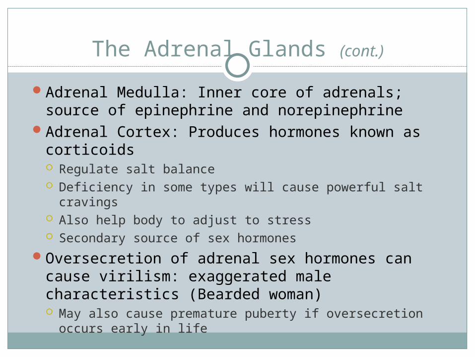
Adrenal Medulla: Inner core of adrenals; source of epinephrine and norepinephrine
Adrenal Cortex: Produces hormones known as corticoids Regulate salt balance Deficiency in some types will cause powerful salt cravings Also help body to adjust to stress Secondary source of sex hormones
Oversecretion of adrenal sex hormones can cause virilism: exaggerated male characteristics (Bearded woman) May also cause premature puberty if oversecretion occurs
early in life
Redundancy
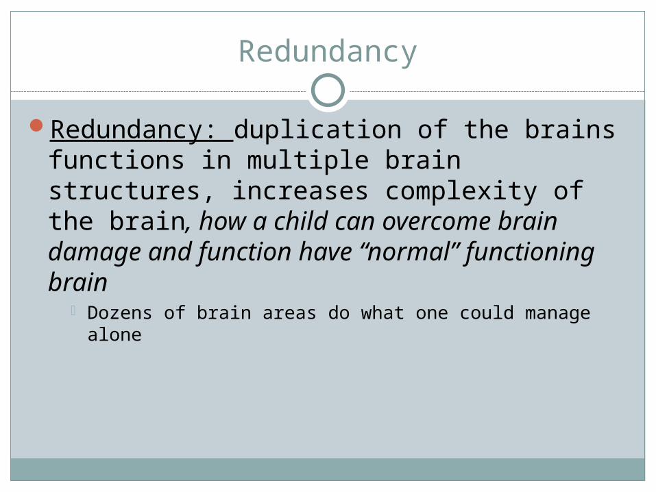
Redundancy: duplication of the brains functions in multiple brain structures, increases complexity of the brain, how a child can overcome brain damage and function have “normal” functioning brain
Dozens of brain areas do what one could manage alone
Plasticity
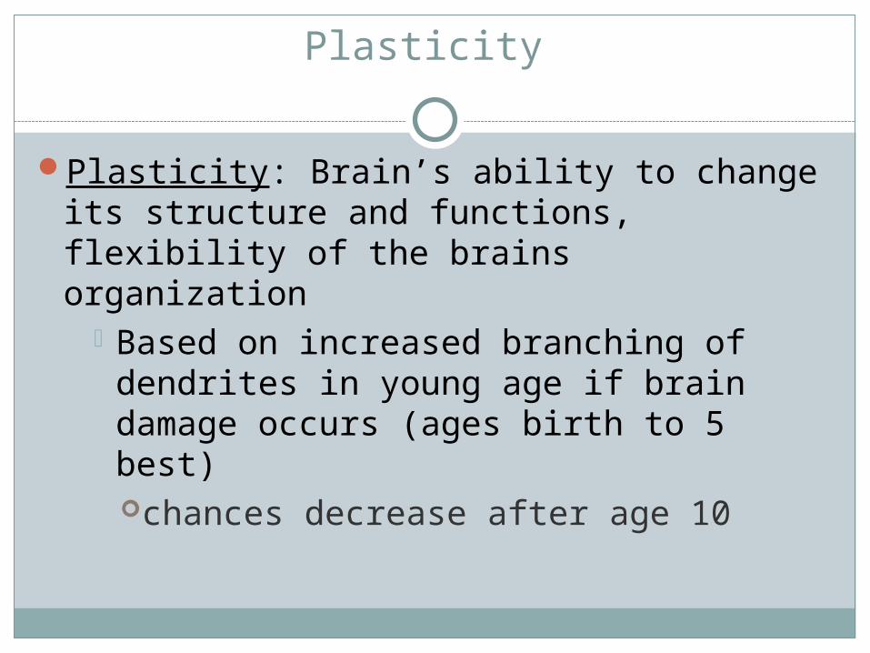
Plasticity: Brain’s ability to change its structure and functions, flexibility of the brains organization
Based on increased branching of dendrites in young age if brain damage occurs (ages birth to 5 best) chances decrease after age 10
Quiz #5, take homeGrade 41-70 Diagnostic AP testRead page 66, write one paragraph summary on “His & Her Brain”Read pages 75-78, write 2 paragraph summary on “Handedness”Define Follwing terms
EBS, pg 70use pgs. 79-82 for all below, or online power Chapter 3 point found on Bird websiteCT Scan,MRIEEGMANSCANPET ScanMEG Scan
Homework
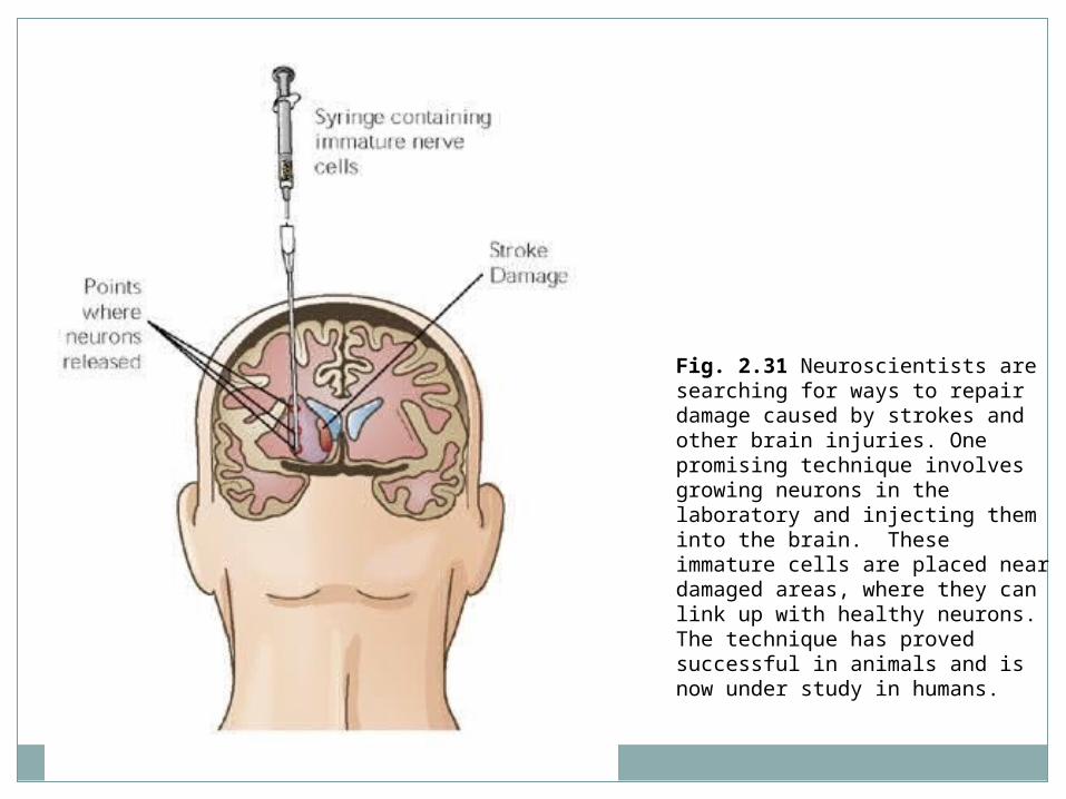
Fig. 2.31 Neuroscientists are searching for ways to repair damage caused by strokes and other brain injuries. One promising technique involves growing neurons in the laboratory and injecting them into the brain. These immature cells are placed near damaged areas, where they can link up with healthy neurons. The technique has proved successful in animals and is now under study in humans.
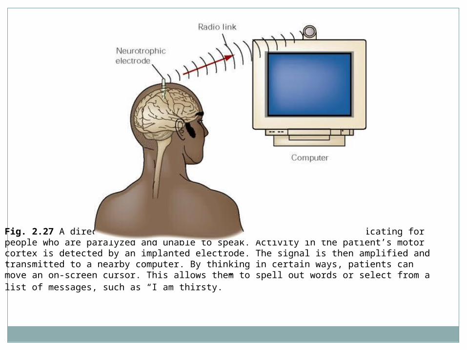
Fig. 2.27 A direct brain-computer link may provide a way of communicating for people who are paralyzed and unable to speak. Activity in the patient’s motor cortex is detected by an implanted electrode. The signal is then amplified and transmitted to a nearby computer. By thinking in certain ways, patients can move an on-screen cursor. This allows them to spell out words or select from a list of messages, such as “I am thirsty.”
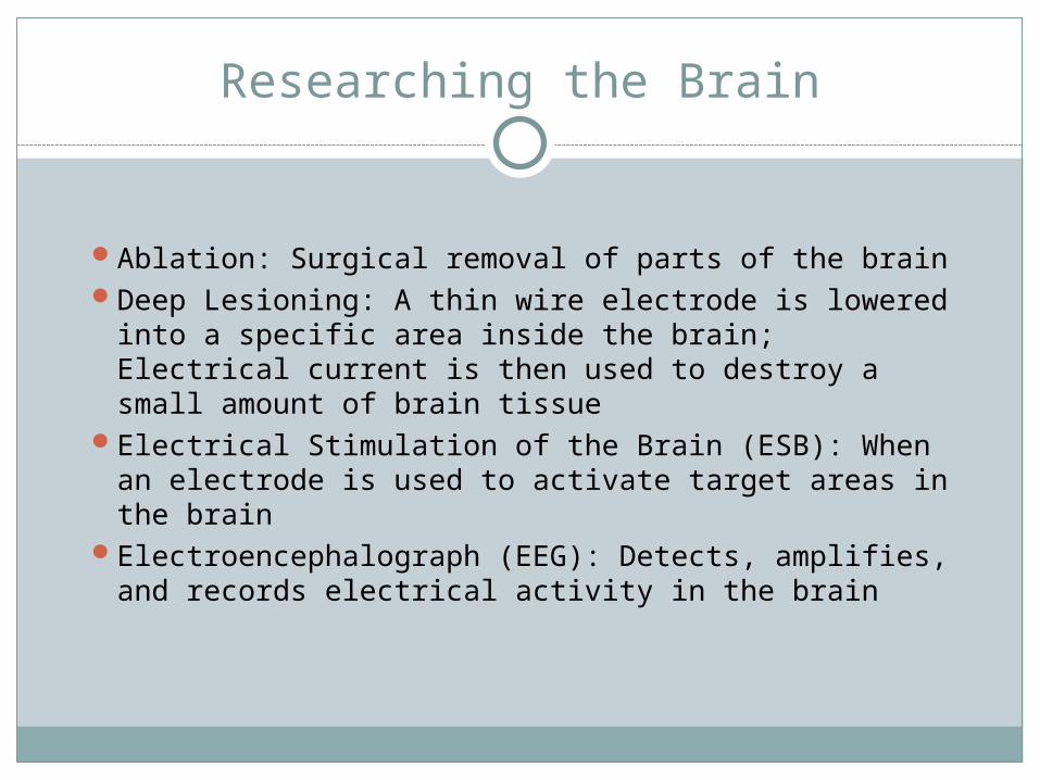
Researching the Brain
Ablation: Surgical removal of parts of the brainDeep Lesioning: A thin wire electrode is lowered into
a specific area inside the brain; Electrical current is then used to destroy a small amount of brain tissue
Electrical Stimulation of the Brain (ESB): When an electrode is used to activate target areas in the brain
Electroencephalograph (EEG): Detects, amplifies, and records electrical activity in the brain
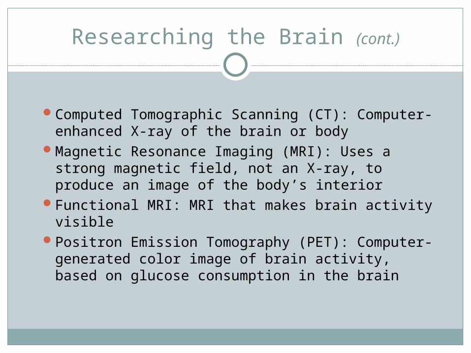
Researching the Brain (cont.)
Computed Tomographic Scanning (CT): Computer-enhanced X-ray of the brain or body
Magnetic Resonance Imaging (MRI): Uses a strong magnetic field, not an X-ray, to produce an image of the body’s interior
Functional MRI: MRI that makes brain activity visible
Positron Emission Tomography (PET): Computer-generated color image of brain activity, based on glucose consumption in the brain
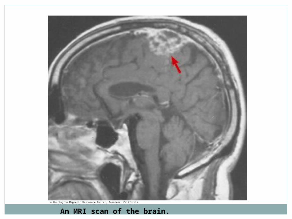
An MRI scan of the brain.
© Huntington Magnetic Resonance Center, Pasadena, California
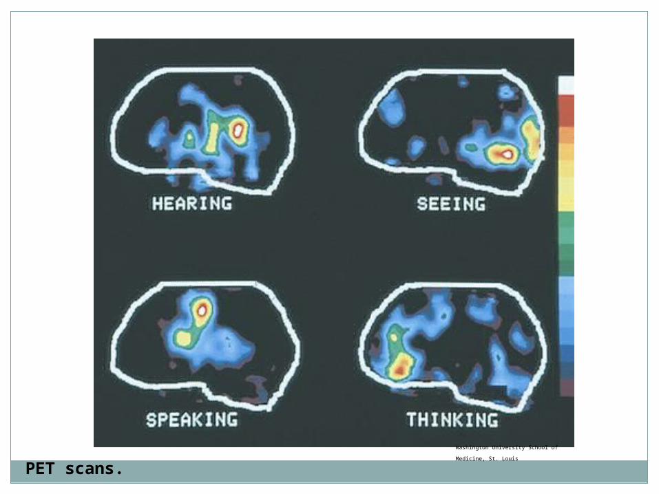
PET scans.
Washington University School of Medicine,
St. Louis
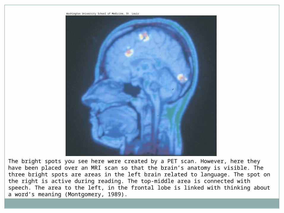
The bright spots you see here were created by a PET scan. However, here they have been placed over an MRI scan so that the brain’s anatomy is visible. The three bright spots are areas in the left brain related to language. The spot on the right is active during reading. The top-middle area is connected with speech. The area to the left, in the frontal lobe is linked with thinking about a word’s meaning (Montgomery, 1989).
Washington University School of Medicine, St. Louis
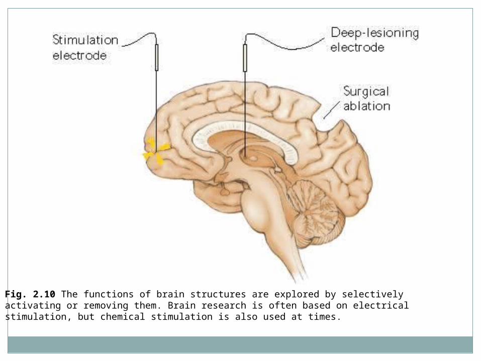
Fig. 2.10 The functions of brain structures are explored by selectively activating or removing them. Brain research is often based on electrical stimulation, but chemical stimulation is also used at times.
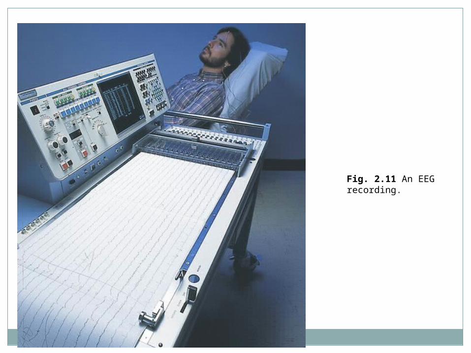
Fig. 2.11 An EEG recording.
Brain Drain
Great Brain Drain worksheet

Take Home Test!


![Introduction to the nervous system and nerve tissue[1]](https://img.pdfslide.net/doc/110x75/55b5a338bb61eba3108b4796/introduction-to-the-nervous-system-and-nerve-tissue1.jpg)