Embed Size (px)
Citation preview

Brain Development in Drosophila melanogaster
ADVANCES IN EXPERIMENTAL MEDICINE AND BIOLOGY
Editorial Board:NATHAN BACK, State University ofNew Yorkat BuffaloIRUN R. COHEN, The Weizmann InstituteofScienceABEL LAITHA, s.s. Kline Institutefor PsychiatricResearchJOHN D. LAMBRIS, University ofPennsylvaniaRODOLFO PAOLETII, University ofMilan
Recent Volumes in this Series
BIO-APPLICATIONS OF NANOPARTICLESEdited by Warren C.W. Chan
Volume 621AXON GROWTH AND GUIDANCE
Edited by Dominique Bagnard
Volume 622OVARIAN CANCER
Edited by George Coukos, Andrew Berchuck, and Robert Ozols
Volume 623ALTERNATIVE SPLICING IN THE POSTGENOMIC ERA
Edited by Benjamin 1. Blencowe and Brenton R. Graveley
Volume 624SUNLIGHT, VITAMIN D AND SKIN CANCER
Edited by Jorg Reichrath
Volume 625DRUG TARGETS IN KINETOPLASTID PARASITES
Edited by Hemanta K. Majumder
Volume 626GENOMIC IMPRINTING
Edited by Jon F.Wilkins
Volume 627TRANSGENESIS AND THE MANAGEMENT OF VECTOR-BORNE DISEASE
Edited by Serap Aksoy
Volume 628BRAIN DEVELOPMENT IN DROSOPHILA MELANOGASTER
Edited by Gerhard M. Technau
A Continuation Order Plan is available for this series. A continuation order will bring delivery of each newvolume immediately upon publication. Volumes are billed only upon actual shipment. For further informationplease contact the publisher.
Brain Developmentin Drosophila melanogasterEdited by
Gerhard M. Technau, PhD
Institute ofGenetics, University ofMainz, Mainz, Germany
Springer Science+Business Media, LLCLandes Bioscience
Springer Science+Business Media, LLCLandes Bioscience
Copyright ©2008 Landes Bioscience and Springer Science+Business Media, LLC
All rights reserved.No part ofthis book may be reproduced or transmitted in any form or by any means, electronic or mechanical, including photocopy, recording, or any information storage and retrieval system, without permissionin writing from the publisher, with the exception ofany material supplied specifically for the purpose ofbeing entered and executed on a computer system; for exclusive use by the Purchaser of the work.
Printed in the USA
Springer Science+Business Media, LLC, 233 Spring Street, New York, New York 10013, USAhttp://www.springer.com
Please address all inquiries to the publishers:Landes Bioscience, 1002 West Avenue, 2nd Floor, Austin, Texas 78701, USAPhone: 512/637 5060; FAX: 512/6376079http://www.1andesbioscience.com
Brain Development in Drosophila melanogaster, edited by Gerhard M. Technau, Landes BiosciencelSpringer Science+Business Media, LLC dual imprint I Springer series: Advances in ExperimentalMedicine and Biology.
ISBN: 978-0-387-78260-7
While the authors, editors and publisher believe that drug selection and dosage and the specifications andusage of equipment and devices, as set forth in this book, are in accord with current recommendationsand practice at the time of publication, they make no warranty, expressed or implied, with respect tomaterial described in this book. In view of the ongoing research, equipment development, changes ingovernmental regulations and the rapid accumulation ofinformation relating to the biomedical sciences,the reader is urged to carefully review and evaluate the information provided herein.
Library of Congress Cataioging-in-Publication Data
Brain development in Drosophila melanogaster I edited by Gerhard M. Technau.p. ;cm.
Includes bibliographical references.ISBN 978-0-387-78260-71. Drosophila melanogaster--Development. 2. Brain--Growth.3. Developmental neurobiology. I.
Technau, Gerhard M.[DNLM: 1. Brain--embryology. 2. Drosophila melanogaster--growth & development. QX 505
B8142008]QL537.D76B732008595.77'4--dc22
2008003551
PREFACE
The central nervous system (CNS) represents the organ with the highest structural and functional complexity. Accordingly, uncovering the mechanisms leadingto cell diversity, patterning and connectivity in the CNS is one of the major challenges in developmental biology. The developing CNS of the fruitfly Drosophilamelanogaster is an ideal model system to study these processes. Several principlequestions regarding neurogenesis (like stem cell formation, cell fate specification,axonal pathfinding) have been addressed in Drosophila by focusing on the relativelysimply structured truncal parts ofthe nervous system. However, information processing (e.g., vision, olfaction), behavior, learning and memory require highly specialized structures, which are located in the brain. Owing to much higher complexityand hidden segmental organisation, our understanding ofbrain development is stillquite rudimentary. Considerable advances have been made recently in bringingthe resolution of brain structures to the level of individual cells and their lineages,which significantly facilitates investigations into the mechanisms controlling braindevelopment.
This book provides an overview of some major facets of recent research onDrosophila brain development. The individual chapters were written by expertsin each field. V. Hartenstein et al survey the generic cell types that make up thedeveloping brain and describe the morphogenesis ofneural lineages and their relationship to neuropil compartments in the larval brain. Recent findings on anteroposteriorregionalization and on dorsoventral patterning in the embryonic brain are reviewedin the chapters by R. Lichtneckert and H. Reichert and by R. Urbach and G. Technau,respectively. Both processes show striking parallels between Drosophila and mouse.Photoactivated gene expression as a means for tracing cell fate through embryonic brain development is demonstrated in J. Minden's chapter. At present, the bestcharacterized neural network on the developmental, structural, and functional level isthe chemosensory system, to which three chapters are devoted: R. Stocker's chaptercovers the design of the larval chemosensory system and shows that it prefiguresthe adult system. V. Rodrigues and T. Hummel summarize recent findings on thespecification and connectivity development ofthe adult olfactory receptor neurons.P.Laissue and B. Vosshall review the molecular biology, neuroanatomy and functionof the adult olfactory system. A further focus of research is the visual system, withthe optic lobes comprising about half of the adult fly brain. The genetic and cellularprinciples which direct the assembly ofthe optic lobes are highlighted in the chapter
v

~ hifa~
by K. Fischbach and P. Hiesinger. The central brain harbors distinct neuropils likethe central complex and the mushroom bodies, as well as "diffused neuropils" whichlack clearly demarcated structures. K. Ito and T.Awasaki review the organization ofthe adult central brain and show how its complex architecture evolves from clonallyrelated neural circuits.
This book will be helpful to those who want to study brain development in thefly. As knowledge extracted from the Drosophila model has often proven to be ofmore general relevance, comparative aspects are included in most chapters. Therefore, this book should also be useful for researchers working on brain developmentin other organisms and on brain evolution, as well as for instructors and advancedstudents in the field of developmental neurobiology.
I would like to thank the authors for producing an excellent series of thoughtful reviews, Ronald Landes for encouraging me to edit this volume, and CynthiaConomos for continuous support.
Gerhard M Technau, PhD

ABOUT THE EDITOR...
GERHARD M. TECHNAU, PhD, is Professor and Head of the Institute ofGenetics at the University ofMainz, Germany. His main research interests includemechanisms controlling the generation of cell diversity and segmental patternin the nervous system using Drosophila as a model. He received his PhD fromWiirzburg Univers ity, and the venia legendi from Cologne University. Awarded aHeisenberg-Fellowship from the Deutsche Forschungsgemeinschaft, he worked atCologne University and University ofCalifornia (UCSF) before being recruited bythe University ofMainz.
vii
PARTICIPANTS
Takeshi AwasakiInstitute of Molecular
and Cellular BiosciencesThe University ofTokyoYayoi, Bunkyo-kuTokyoJapanandDepartment ofNeurobiologyUniversity of Massachusetts Medical
SchoolWorcester, MassachusettsUSA
Karl-Friedrich FischbachDepartment ofNeurobiologyAlbert-Ludwigs University
ofFreiburgFreiburgGermany
Siaumin FungDepartment of Molecular, Cell
and Developmental BiologyUniversity ofCalifornia, Los AngelesLos Angeles, CaliforniaUSA
Volker HartensteinDepartment of Molecular, Cell
and Developmental BiologyUniversity of California, Los AngelesLos Angeles, CaliforniaUSA
Peter Robin HiesingerDepartment of PhysiologyandGreen Center Division
for Systems BiologyUT Southwestern Medical CenterDallas, TexasUSA
Thomas HummelInstitut flier NeurobiologieUniversitaet MuensterMuensterGermany
Kei ItoInstitute of Molecular
and Cellular BiosciencesThe University ofTokyoYayoi, Bunkyo-kuTokyoJapan
Philippe P. LaissueKent Institute ofMedicine
and Health SciencesMedical Image ComputingUniversity ofKentCanterbury, KentUK
ix
x
Robert LichtneckertBiozentrumUniversity of BaselBaselSwitzerland
Jonathan MindenDepartment of Biological Sciences
and ScienceCarnegie Mellon UniversityPittsburgh, PennsylvaniaUSA
Wayne PereanuDepartment of Molecular, Cell
and Developmental BiologyUniversity ofCalifornia, Los AngelesLos Angeles, CaliforniaUSA
Heinrich ReichertBiozentrumUniversity of BaselBaselSwitzerland
Veronica RodriguesNational Centre for Biological
SciencesTata Institute of Fundamental
ResearchBangaloreIndia
Participants
Shana SpindlerDepartment of Molecular, Cell
and Developmental BiologyUniversity of California, Los AngelesLos Angeles, CaliforniaUSA
Reinhard F. StockerDepartment of BiologyUniversity ofFribourgFribourgSwitzerland
Gerhard M. TechnauInstitute of GeneticsUniversity ofMainzMainzGermany
RolfUrbachInstitute of GeneticsUniversity of MainzMainzGermany
Leslie B. VosshallLaboratory ofNeurogenetics
and BehaviorThe Rockefeller UniversityNew York, New YorkUSA
CONTENTS
1. THE DEVELOPMENT OF THE DROSOPHILA LARVAL BRAIN .••.•..... 1
Volker Hartenstein, Shana Spindler, Wayne Pereanu and Siaumin Fung
Abstract.•••••..•••••••••...•..•.•.......••.••••••..•••.•••••••••.....•..••••••••••.••.•.••••••••••..•••••••.••••••.••••.•••.•••••••••••• 1Synopsis of the Phases and Elements of Drosophila Brain Development .••••••••.••••••.•••••• 1Progenitors of the Drosophila Brain: Neuroblasts and Ganglion Mother Cells•.•..•.•...•• 3The Generic Cell Types of the Drosophila Brain.••••••••.•••••••••••••.••••••••.••••••••.••.•••.•••..••.•••••• 8Neuroanatomy of the Developing Drosophila Brain: The Systems of Lineages,
Tracts and Compartments.••••.•••.....•.•.•••.••••••••••••••...•....••....••..•••••...•••••.•....•.••.•.•••••.••• 17Outlook........•.••••••••.•.....•.....••.•......•••.••.....•.....••••••••••••••.•.•.••......•...•••.•.••....••••.....••••.••••.••••...• 29
2. ANTEROPOSTERIOR REGIONALIZATION OF THE BRAIN:GENETIC AND COMPARATIVE ASPECTS 32
Robert Lichtneckert and Heinrich Reichert
Abstract.•........•....•••...........•..••.•••••.•.............•.•••••.•.•••••••••..••••••.••••••••.•....••••••••••••••••••.•..•...•••• 32Introduction........••••••••...•....•.....••••••.••.•••....••..•.••••.••.••••••......•.•••••••.•.•.•...••.•••••••.•••••••••••..•.•.. 32The Cephalic Gap Genes Otd/Otx and Ems/Emx Control
Anterior Brain Development..•........•.••...•••••••......•..•••••..••••••.•.•..••...••......•....••.•.•••...••• 34The Box Genes Pattern the Posterior Brain........••••••••.••.••.•••.••••••.....•••.••......••.••••••.•••...•• 36Evidence for a Tripartite Organization of the Brain 37
3. DORSOVENTRAL PATTERNING OF THE BRAIN:A COMPARATIVE APPROACH ........................•...•...................................•••.. 42
Rolf Urbach and Gerhard M. Technau
Abstract .•.••.•.......••••..•..•.••••....•.•..•••••......•.•..••••...••••••.....••....••.•.•..••••.••.••..•...•••...••.•.•....••••..•. 42Introduction.•.....•••••••..•••••••••..•..•••••••.••••.•.•••••••.••••••.......••.•...••••••..•••••••••••.•••••••.••.••....••••••.•.• 42DV Patterning of the Truncal Part of the CNS in Drosophila and Vertebrates .••••••••.• 45DV Patterning of the Brain in Drosophila •••••••••••••••••••••••••••••••••••••••••••••••••••••••••••••••••••.••• 48A Comparison with DV Regionalization of the Vertebrate Telencephalon.....••••.•••••.••• 52Conclusions ..•••••••••••••.•••........•.••••••••••••.•........•...•.•••••••.••••.•.•.••••.••••.•...••••••••..••....•.••.••••.•••.•.. 53
xi
xii Contents
4. DISSECTION OF THE EMBRYONIC BRAIN USINGPHOTOACTIVATED GENE EXPRESSION 57
Jonathan Minden
Abstract••••.•..••.••••••..•.•••......•....••...........••...••••••.••.••...•....•.•......•.•.•.•..•.....•.••••.....•.••...•••..••..•.. 57Introduction..••••••.•••••••••....•.••....•..••••••.•••••••.•••••.•.••..•....•......••••.•......••..•••..•••••••••.•.•••••....•••... 57Procephalic Blastoderm Fate Map .•••••••••••••••••.••••••.......•••.•...•••.•..••••....•..•••.•.•.•..•........•.••. 58Brain-Forming Mitotic Domains Populate Distinct Brain Regions 58Conclusion ••••••.••.•••....••••.•••••••.•••••••••••••••...•••••••••.•••..••••••••••••••••••••••••••••.••...•.......•..•••••.••••..•• 67
5. DESIGN OF THE LARVAL CHEMOSENSORY SySTEM 69
Reinhard F. Stocker
Abstract .•..•.....•••••••.••..•..••••••••...•.••••..••••.•.••••••••••.•••••••••••.••••••...•••••..••.••••.••.•••..•.•.•.•..••.•••••••• 69Introduction••••••....••.•••••.•••••.•....••••••••••••••••••.••...•.•.•••••••••••••.•••••••••••.••••••••••••••.••.....••.••...••...• 69Chemosensory Organs of the Larval Head ••........•.••••••••.•.••••..•••.•••••.•••••••••••..•....••.••..•...• 70Olfactory System•••••••.•...••••••..•..•..••..••.••..••••.••••.•••••..••........•••..•.•.•........•.•.......••••••....••.•••••.. 71Gustatory System ••••••..••••••.••••••••••••••....••••••.••••••.•••••••••••...•.••...•..•••.••••••••..•........••••.•••.•••••••• 77The Drosophila Larva as a Model for Smell and Taste••.••...•••.......••.......••.•.....•....•....••••. 78
6. DEVELOPMENT OF THE DROSOPHILA OLFACTORY SYSTEM ..... 82
Veronica Rodrigues and Thomas Hummel
Abstract•..•......•.•...•..•••.••....•......•....•.••.•.•..•••.•••••••......••••.•.•..••.•.•••••••.•...•....•.............••••.•....••• 82Introduction.••.••••.•••••••••.•••..•••.•••.•••••••.••••...•..•.••..••.......•••.......••.•..••.••••.••••••••••••.•.•...........•..• 82Organization of the Drosophila Adult Olfactory System•••...•...•...••.•••••••••.••••••.......•..•.•.• 84Specification of the Olfactory Sense Organs •..••...•...•....•..•..•..••....••••.••••.••.•...•••.•............. 84Olfactory Lineages Revisited ..••••••......••..•...•••••.••..•....••..•..........•.•...••.......•••.•......•.•........... 87Origin of Glial Cells ..•.••••••.••••••••••••••••.••.......•..•..••••••...•.•.....•..•.....•.••••....•••.••..•.•••............... 89Development of ORN Connectivity ...........•......••••••..••.•.•....•.•...............••.•..•......•.............. 89Olfactory Map Organization ........•..•.....•••••••.•..•••••••......••.....•..•....•..•.•.............••...•••.•..••••• 90Cellular and Molecular Mechanisms of ORN Wiring Specificity......••....•.................••.. 91Specification of Projection Neurons ..•....•.•••••••..••••••......•.•......••••...•••••.•..•••••...•••.•...•..•...... 92Development of PN Dendrites.•.....••••..••••••••..•.....•....•••••..•••••...•.•........•..•.....•.•..•.•.•.••..•••••.• 95Concluding Remarks .••.......•...••.....••••...•••.••...•..........••.••.•..•...•.••..•...•.......••......•......•......•••• 96
7. THE OLFACTORY SENSORY MAP IN DROSOPHILA••..•...•••.•..........•. 102
Philippe P. Laissue and Leslie B. Vosshall
Abstract••..••••••.•••••••••.•.••••....•••.•••..•••••..•••..•...••••••.••••••••••••••..•.•.•.•..•••.••••••••••••••••••••••.••..•..••. 102Introduction•••••••••...••••••.••••••••..••...•...•.•..•...•••..........•••••••••••••••..•••....••.......................••.••••.. 102Olfactory Organs and Olfactory Sensory Neurons of Drosophila••••••••.••••.••••••••••••••••• 103Odorant Receptor Gene Expression......••••••••••.•.••.•••••.••••••••.•••••...•••••.•...•••...•••••..•.•••••..•. 104Ligand Tuning Profiles .••....•..••..••••.•...••.•.•....•.•..••.••..............•.•..•.........••.•.•.•..•..•.••........... 105A Receptor-Based Map of Glomerular Projections 108Sexual Dimorphism in the Drosophila Olfactory System••.••••.............•••...•.......•••..••••••.ll1Concluding Remarks ...•..................••..••.••••....................••........••••.....••••••.•••...••........••....•• 112
Contents xiii
8. OPTIC LOBE DEVELOPMENT ..............•.................•....•.•....•.•.•.....•.........115
Karl-Friedrich Fischbach and Peter Robin Hiesinger
Abstract•.•..•.••••••••••....•.....•..•.•••.•...•••..••••..............•..••..•.•••.••...•.•••••••.••..•.•••••••••.•••••••...••••.••• 115Introduction••.•••••••••••••••••••.•••.•••••••••••••••••••••••••••••••.•••••.••••••••••••••••••.••••••••.••••••••••••••••••••••••• 115The Adult Visual System Is Organized into Parallel Visuotoptic
Functional Pathways.•••••.••..•••.•••••••.•.....•..•..•....•..••....••...••.•••.•••••••••.•.•.•••...••••.•••••...•. 116Lamina Development •••••.•••••.....•.•••...••••.•••....•...•.........••••••••.•••..••••••••••.••••.•..•••..•••••••••••.••. 120Medulla and Lobula Complex Development ....••••..•.•..•••••••.•••••••••••••.•..•..•.....•••••••......••• 127Concluding Remarks ••••••••••••••••..••••••••••..••...•.••...........••.•••.....•••••••••••••••••••...••••••..••••••••.•.• 131
9. CLONAL UNIT ARCHITECTURE OF THE ADULT FLY BRAIN 137
Kei Ito and Takeshi Awasaki
Abstract...•••••••••......••••••••••••.•••..••...•..•.••...••.••••••.••••••••.•.•••.•..•.••.....••••••••••••••••.••••.••••••.••••.••. 137Introduction•••••••..•••••••••••••.•••.•.••...•.••..•••....••••.•.••••••••••••••••.....•••.••.•••••••••••••••••••••••••••••••..••. 137Structure of the Adult Brain ••••••••••••••••.••.••••••••••••••••••••••••••••••••••••.••••••••••••••••••••••••••••••.••• 137Techniques for Visualising Clonally Related Progeny 139Clonal Unit Architecture in the Adult Brain 147Formation of the Clonal Units During Development••..............•.•.••••••••••••••••••••••••••.•.••. 150Functional Importance of the Clonal Units ••.••••••••••...............•...•••••••••••••••••••.•••••••••••.••. 154Conclusion .•.•...••••••.•.....•...•••..•.•.•...•...•••..•............••.......................•.••.•••...•••••••.•.•••....••...... 156
INDEX.........•••......•....•••......•••.......••.••.•....••.........•.•....•••••..............•..•.....•..••........ 159
CHAPTER 1
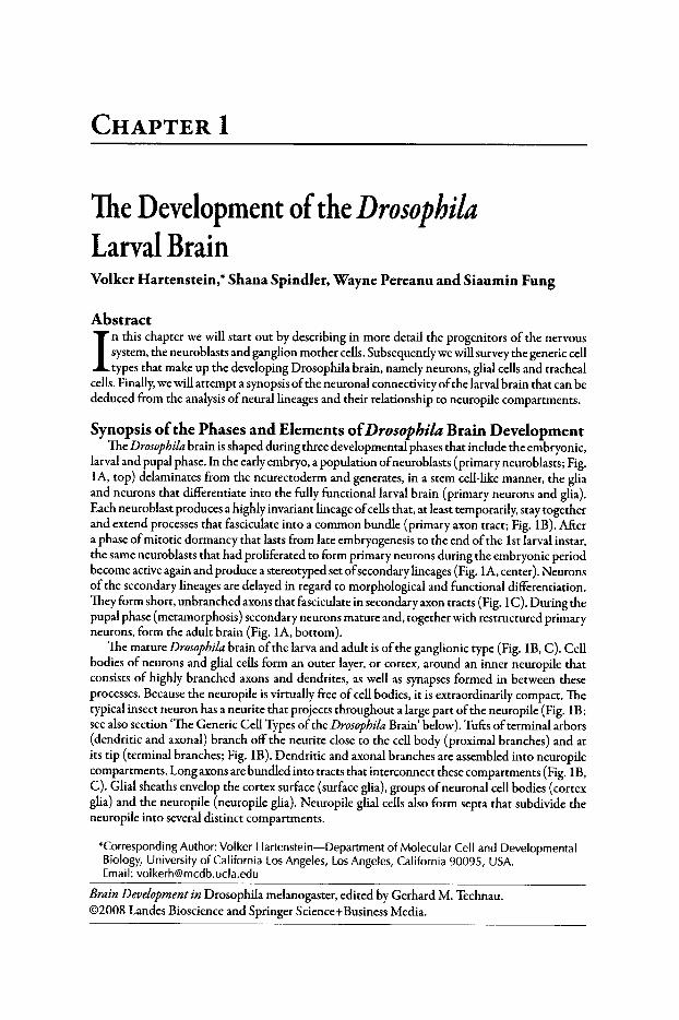
The Development ofthe DrosophilaLarval BrainVolker Hartenstein,* Shana Spindler, Wayne Pereanu and Siaumin Fung
Abstract
I n this chapter we will start out by describing in more detail the progenitors of the nervoussystem, the neuroblasts and ganglion mother cells.Subsequentlywe will survey the generic celltypes that make up the developing Drosophila brain, namely neurons, glial cells and tracheal
cells.Finally,we will attempt a synopsis ofthe neuronal connectivity ofthe larval brain that can bededuced from the analysis ofneural lineages and their relationship to neuropile compartments.
Synopsis ofthe Phases and Elements ofDrosophila Brain DevelopmentThe Drosophila brain is shaped during three developmental phases that include the embryonic,
larval and pupal phase. In the earlyembryo, a population ofneuroblasts (primary neuroblasts; Fig.lA, top) delaminates from the neurectoderm and generates, in a stem cell-like manner, the gliaand neurons that differentiate into the fully functional larval brain (primary neurons and glia).Each neuroblast produces a highly invariant lineage ofcells that, at least temporarily, stay togetherand extend processes that fasciculate into a common bundle (primary axon tract; Fig. IB). Aftera phase ofmitotic dormancy that lasts from late embryogenesis to the end ofthe Ist larval instar,the same neuroblasts that had proliferated to form primary neurons during the embryonic periodbecome active again and produce a stereotyped set ofsecondary lineages (Fig. lA, center). Neuronsof the secondary lineages are delayed in regard to morphological and functional differentiation.They form short, unbranched axons that fasciculate in secondary axon tracts (Fig. IC). During thepupal phase (metamorphosis) secondary neurons mature and, together with restructured primaryneurons, form the adult brain (Fig. lA, bottom).
The mature Drosophila brain ofthe larva and adult is ofthe ganglionic type (Fig. IB, C). Cellbodies of neurons and glial cells form an outer layer, or cortex, around an inner neuropile thatconsists of highly branched axons and dendrites, as well as synapses formed in between theseprocesses. Because the neuropile is virtually free ofcell bodies, it is extraordinarily compact. Thetypical insect neuron has a neurite that projects throughout a large part ofthe neuropile (Fig. IB;see also section 'The Generic Cell Types ofthe Drosophila Brain' below) . Tufts ofterminal arbors(dendritic and axonal) branch off the neurite close to the cell body (proximal branches) and atits tip (terminal branches; Fig. IB). Dendritic and axonal branches are assembled into neuropilecompartments. Long axons are bundled into tracts that interconnect these compartments (Fig. IB,C). Glial sheaths envelop the cortex surface (surface glia), groups ofneuronal cell bodies (cortexglia) and the neuropile (neuropile glia). Neuropile glial cells also form septa that subdivide theneuropile into several distinct compartments.
*Corresponding Author: Volker Hartenstein-Department of Molecular Cell and DevelopmentalBiology, University of California LosAngeles, LosAngeles, California 90095, USA.Email: volkerhwrncdb.ucla.edu
Brain Developmentin Drosophila melanogaster, edited by Gerhard M. Technau.©2008 Landes Bioscience and Springer Science+ Business Media.
2 Brain Development in Drosophila melanogaster
AdultBrain
B
Brain(. ~euroblasts I
primary \\.. secondary I
SecondaryLineage
IPrimaryLineage
__Optic LobeEpithelialAnlage
PrimaryNeurons
ProximalBranches
Optic LobeNeurons
~Optlc LobeNeuroblast
young-,/ Seconda ry/ '1l Neurons
• old~ Secon dary
AxonTract
f- NeuroplleCompartment
•
CortexGila
•
PrimaryAxonTract
c
Figure1. Drosophila brain development. A)Schematic drawings of head of early embryo (top),larval brain (center left), neuroblast lineage (center right) and adult brain (bottom). Primarybrain neuroblasts (dark lilac) delaminate from the head neurectoderm and produce primarylineages that form the larval brain (light lilac). In the late larva neuroblasts start producingsecondary lineages (orange) that are integrated with the primary neurons into the adult brain.(B, C) Schematic cross sect ions of one brain hemisphere of early larva (B) and late larva (e).Primary neuroblasts and neurons are shaded lilac; secondary neuroblasts and neurons arein orange. Two clusters of primary neurons are highlighted to show projection of neurites.Glia cells are colored green.

TheDevelopmentofthe DrosophilaLarval Brain 3
In this chapter we will start out by describing in more detail the progenitors of the nervoussystem, the neuroblasts and ganglion mother cells. Subsequently we will survey the generic celltypes that make up the developingDrosophila brain, namely neurons, glial cells and tracheal cells.Finally, we will attempt a synopsis of the neuronal connectivity of the larval brain that can bededuced from the analysis ofneural lineages and their relationship to neuropile compartments.
Progenitors ofthe Drosophila Brain: Neuroblastsand Ganglion Mother Cells
The Primary Phase ofNeuroblastActivity in the EmbryoThe brain ofinsects and some other arthropod taxa is formed by a unique type ofstem cell-like
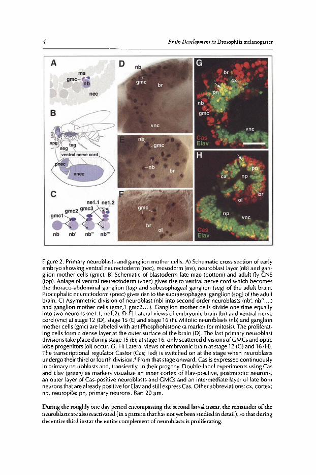
progenitor cell called a neuroblast. Neural progenitors ofthis type are not found in vertebrates or(as far as known to date) other invertebrate phyla. Neuroblasts delaminate from the embryonicneurectoderm and form a cell layer sandwiched in between the ectoderm and mesoderm (Fig.2A).The pattern ofneuroblasts is invariant. Thus, each neuroblast forms a uniquely identifiable cell thatappears at the same time and position in every individual ofa given species. Neuroblast patternsare very similar even when comparing different insect species such asDrosophila and grasshopper.Neuroblasts appear in two broad regions of the embryo. The head (procephalic) neurectoderm,located in the anterior-dorsal part of the ectoderm, gives rise to neuroblasts that form the brain(Fig. 2B). The ventral neurectoderm, stretching out along the trunk ectoderm, produces theneuroblasts of the ventral nerve cord. Neuroblasts are organized segmentally, with each segmentgivingrise to an identical segmental set, called neuromere, ofapproximately 25 neuroblasts per side.The brain, a composite structure formed by the fusion ofseveral modified neuromeres, containsapproximately 100 neuroblasts per side.1
,2
After delamination from the ectoderm, neuroblasts form a layer oflarge, rounded cells insidethe embryo. Soon these cells proliferate in what is known as a stem cell mode (Fig. 2C). Thus,whereas most cells in an embryo divide symmetrical, with both daughter cells being ofabout thesame size and fate, neuroblasts divide asymmetrically into one large and one small daughter cellwith very different fates. The large cell (still called a neuroblast) continues dividing in the stemcell mode for a variable number of rounds ofdivisions. Most primary neuroblasts in the embryodivide 5-8 times, with a cell cycle duration of45-60min;3 secondary neuroblasts may divide 50times or more (V.H., unpublished). The small cell resulting from a neuroblast mitosis, called aganglion mother cell (GMC), typically divides only one more time 60-90min after its birth.' Thetwo daughter cells of the GMC become postmitotic and differentiate into neurons or glial cells.Since the mitotic spindle of neuroblasts is typically directed perpendicular to the plane of theneuroblast layer,ganglion mother cellsand immature neurons form a stack on top ofthe neuroblastfrom which they originated (Fig. 2A,C). In this manner, all cells of a neuroblast lineage remainspatially close to each other and are arranged along a spatio-temporal gradient. Neuroblasts andganglion mother cells are situated externally at the brain surface (Fig. 2D,G), adjacent to the lastborn (youngest) neurons. Early born (old) neurons are the most remote from the neuroblast,bordering the nascent neuropile. The layered organization ofthe brain cortex can be analyzed indetail by using molecular markers that are expressed at different stages ofneuroblast proliferationand neuronal differentiation (Fig. 2G,H).4
Secondary Neuroblasts and GMCs ofthe LarvalBrainThe last rounds of primary neuroblast division occur at embryonic stages 14-15 (Fig.2E);
after that stage, only GMC divisions are recognizable for another 2-3 hours (Fig. 2F). Neuroblastsbecome mitotically inactive and shrink in size, so that they cannot be recognized in first instarbrains (Fig. 3A). A small set ofneuroblasts, including the four mushroom body neuroblasts andone ofthe basal anterior neuroblasts (for classificationofneuroblasts and their lineages, seesection'Neuroanatomy ofthe Developing Drosophila Brain' of this chapter), escape the general arrest ofneuroblast activity and continue to proliferate throughout the early larval period (Fig. 3A,B).s
4 Brain Development in Drosophila melanogaster
Figure 2. Primary neuroblasts and ganglion mother cells. A) Schematic cross section of earlyembryo showing ventral neurectoderm (nee), mesoderm (rns), neuroblast layer (nb) and ganglion mother cells (gmc). B) Schematic of blastoderm fate map (bottom) and adult fly CNS(top). Anlage of ventral neurectoderm (vnec) gives rise to ventral nerve cord wh ich becomesthe thoraco-abdominal ganglion (tag) and subesophageal ganglion (seg) of the adult brain.Procephalic neurectoderm (pnec) gives rise to the supraesophageal ganglion (spg)of the adultbrain . C) Asymmetric division of neuroblast (nb) into second order neuroblasts (nb', nb".... )and ganglion mother cells (grnc.t gmc2 ... .). Ganglion mother cells divide one time equallyinto two neurons (neLl , ne1.2). D-F) Lateral views of embryonic brain (br) and ventral nervecord (vnc) at stage 12 (D), stage 15 (E) and stage 16 (F). Mitotic neuroblasts (nb) and ganglionmother cells (grnc) are labeled with antiPhosphohistone (a marker for mitosis). The proliferating cells form a dense layer at the outer surface of the brain (D). The last primary neuroblastdivisions take place during stage 15 (E); at stage 16, only scattered divisions of GMCs and opticlobe progenitors (01) occur. G, H) Lateral views of embryonic brain at stage 12 (G) and 16 (H).The transcr iptional regulator Castor (Cas; red) is switched on at the stage when neuroblastsundergo their third or fourth division." From that stageonward, Cas is expressed continuouslyin primary neuroblasts and, transientl y, in their progen y. Double-label experiments using Casand Elav (green) as markers visualize an inner cortex of Elav-positive, postmitotic neurons,an outer layer of Cas-positive neuroblasts and GMCs and an intermediate layer of late bornneurons that are already positive for Elavand still expressCas. Other abbreviations: ex, cortex;np, neuropile; pn, primary neurons . Bar: 20 urn.
During the roughly one day period encompassing the second larval instar, the remainder of theneuroblasts are also reactivated (in a pattern that ha s not yet been studied in detail), so that duringthe entire third instar the entire complement ofneuroblasts is proliferating.
TheDevelopment ofthe Drosophila Larval Brain 5
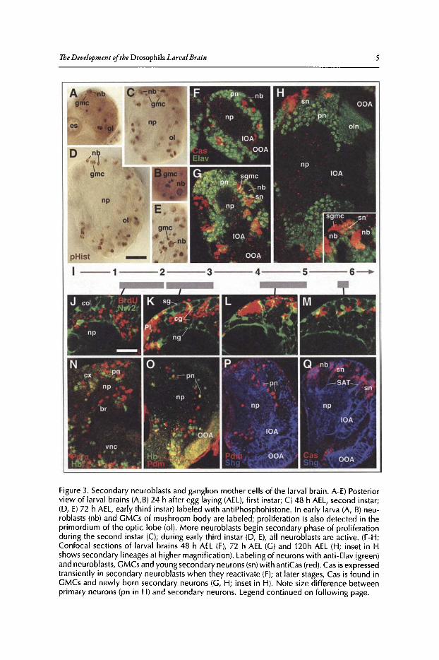
Figure 3. Secondary neuroblasts and ganglion mother cells of the larval brain . A-E) Posteriorview of larval brains (A,B) 24 h after egg laying (AEL), first instar; C) 48 h AEL, second instar;(D, E) 72 h AEL, early third instar) labeled with antiPhosphohistone. In early larva (A, B) neuroblasts (nb) and GMCs of mushroom body are labeled; proliferation is also detected in theprimordium of the optic lobe (01). More neuroblasts begin secondary phase of proliferationduring the second instar (C); during early third instar (D, E), all neuroblasts are active . (F-H :Confocal sections of larval brains 48 h AEL (F), 72 h AEL (G) and 120h AEL (H; inset in Hshows secondary lineages at higher magnification). Labeling of neurons with anti-Elav (green)and neuroblasts, GMCs and young secondary neurons (sn)with antiCas (red). Cas is expressedtransiently in secondary neuroblasts when they reactivate (F); at later stages, Cas is found inGMCs and newly born secondary neurons (G, H; inset in H). Note size difference betweenprimary neurons (pn in H) and secondary neurons. Legend continued on following page.
6 Brain Development in Drosophila melanogaster
Figure 3, continued from previous page. I-M: BrdU pulse chase experiments visualizing thecorrelation between birth date and neuron location. J-M show confocal cross sections ofdorsal brain hemisphere of 3rd instar larva. Glia cells (labeled green by Nrv2-Gal4 drivingUAS-GFP) outline the brain surface (sgsurface glia), the cortex (cg cortex glia; co cortex) andthe neuropile surface (ng neuropile glia; np neuropile). Larvae were fed with BrdU duringtime intervals indicated by gray bars in time line (panel I; numbers indicate days after fertilization). The location of BrdU labeled neurons (red in J-M) within the cortex correlates with thetime of BrdU incorporation: early born neurons occupy a deep location in the cortex, lateborn neurons are superficial. N) Confocal section of late embryonic brain showing expression of the transcription factors Hunchback (Hb; green) and Pdm (red) in primary neurons(pn) born during the first and third round of division of neuroblasts. 0) Confocal sectionof early third instar larval brain. Hb and Pdm are not reactivated in secondary neuroblasts,but stay expressed in primary neurons (presumably the same that had turned on expressionin the early embryo) located in the deep cortex. P, Q) Confocal section of early third instarlarval brain. Secondary lineages (sn), glia and optic lobe (IDA: inner optic anlage; OOAouter optic anlage) are labeled with antiShg (DEcadherin) antibody (blue). Cas expression(red in Q) overlaps with secondary lineages; Pdm (red in P) is restricted to primary neuronsnear neuropile. Other abbreviations: br, brain; es, esophagus; np, neuropile; oln, optic lobeneurons; sgmc, secondary GMC; PI, pars intercerebralis; SAT, secondary axon tract; vnc,ventral nerve cord Bar: 20 Jim
Labeling ofneuroblasts and GMCs ofthe larva reveals that these cells, just like their embryoniccounterparts, are located at the brain surface (Fig. 3A-E).6-S The orientation ofthe mitotic spindlein secondary neuroblasts appears to be much more variable than in primary neuroblasts, rangingfrom parallel to perpendicular relative to the brain surface.6.9 This could in part be due to thefact that the mechanism controlling spindle orientation could be quite different: in the embryo,neuroblasts are in contact with the epithelial neurectoderm and "inherit" from the neurectoderma protein complex, the Inscuteable complex, that remains apical and plays a role in tethering themitotic spindle to the membrane in such a way that results in a vertical orientation.P'" Secondaryneuroblasts in the larva have no contact with the ectoderm (or epidermis); rather, they are surrounded on all sides by a glial layer (see below). Thus, the mechanism that controls the mitoticspindle orientation, as well as the onset and frequency of mitosis, is likely to be controlled byglia-neuroblast inreracrions.I'" Within the secondary neuroblast, protein complexes orientingthe spindle appear to be the same as in the embryo. Thus, members ofthe Par complex, includingBaz, Par,6 and aPKC, localize to an apical crescent along with Inscuteable, while Miranda andProspero localize to the basal crescent (Fig.4E).6.9
Despite the variability of neuroblast mitotic spindle orientation, the larval brain cortex isorganized into concentric layers where the location of a neuron reflects its birth date. This correlation between birth date and location ofa neuron can be visualized by pulse chase experimentsin which BrdU is fed to larvae at different time intervals (Fig. 3I-M), or with the expression ofmolecular markers such as Cas, Pdm or Hb (Fig. 3F-H, N_Q).l4 Primary neurons are the deepestcells (Fig. 3H, 0, P), bordering the neuropile; late born secondary neurons are superficial, surrounding the neuroblasts (Fig. 3H, M, Q).
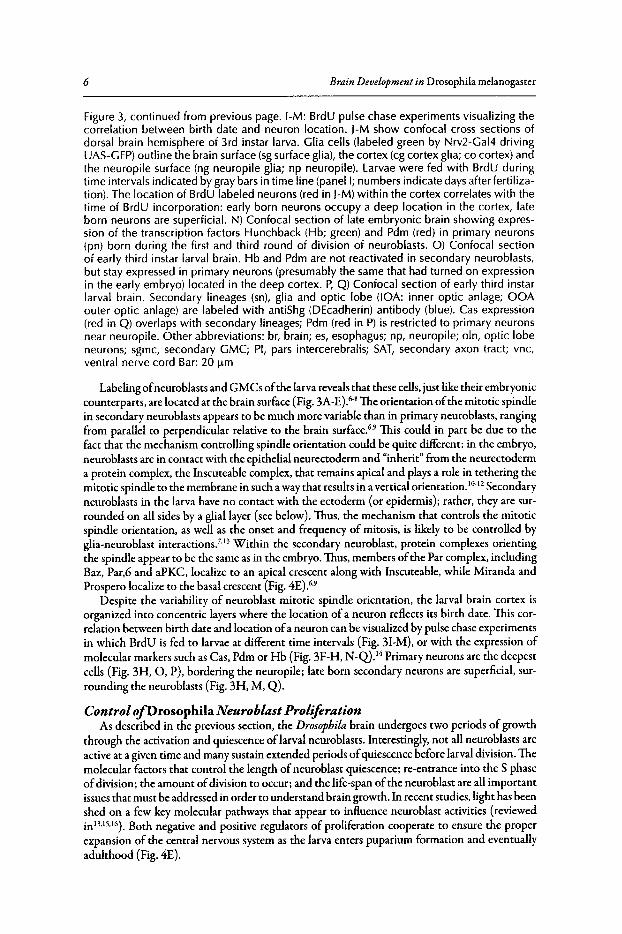
Control ofDrosophila Neuroblast ProliferationAs described in the previous section, the Drosophila brain undergoes two periods of growth
through the activation and quiescence oflarval neuroblasts. Interestingly, not all neuroblasts areactive at a given time and many sustain extended periods ofquiescence before larval division. Themolecular factors that control the length ofneuroblast quiescence; re-entrance into the S phaseofdivision; the amount ofdivision to occur; and the life-span ofthe neuroblast are all importantissues that must be addressed in order to understand brain growth. In recent studies, light has beenshed on a few key molecular pathways that appear to influence neuroblast activities (reviewedinl3.ls.l6). Both negative and positive regulators of proliferation cooperate to ensure the properexpansion of the central nervous system as the larva enters puparium formation and eventuallyadulthood (Fig. 4E).
t:;:l '" ~ 1t ~ :! s ... ~ So '" tl a ~ ., t-o l::
. ... iii ......
b:l
i::: ii'
Mira
nda
,P
rosp
ero
,AP
C2
EF
GF
&H
edg
eho
gs
ign
alin
g
Slri
rlli?
.-'"
perm
issi
ve
"'-
Pro
life
ratio
n
Cyc
linE
/
Qu
iesc
en
ce
,/
?N
EU
RO
BL
AS
T"G
rh
Ana-. •
I •• •
Fig
ure
4.M
ech
an
ism
ofN
eu
rob
last
Pro
lifer
atio
n.
A)
Mit
oti
cne
urob
last
sd
ivid
eto
self-
ren
ew
and
pro
du
cea
da
ug
hte
rg
an
glio
nm
oth
er
cell
,w
hic
hsu
bseq
uent
lyd
ivid
es
into
two
neur
ons
.Mit
oti
cne
urob
last
san
dG
MC
s(r
ed),
ne
uro
bla
stp
rog
en
y(g
reen
).B
)Sec
ond
inst
arla
rval
brai
nex
pres
sing
the
anac
hron
ism
(ana
)-la
cZm
arke
r.A
nais
only
expr
esse
dby
glia
(bro
wn
stai
n)an
dno
tby
the
neur
obla
sts
(mar
ked
wit
has
teris
k).
[from
Ebe
nset
alC
ell,
Vol
.74
,15
-27,
with
pe
rmis
sio
n].
C)
Baz
ooka
(Baz
),a
me
mb
er
ofth
eP
ar-c
ompl
ex,
loca
lize
sto
on
lyth
eap
ical
cres
cent
ofa
met
apha
sen
eu
rob
last
inth
isth
ird
inst
arbr
ain
.B
aza
nti
bo
dy
(red
,ar
row
head
);D
NA
lab
ele
dw
ith
syto
xgr
een
(gre
en,
arro
w).
D)
Mir
an
da
and
Pro
sper
oar
ese
greg
ated
toth
eba
sal
ne
uro
bla
stan
de
ven
tua
llyth
ebu
ddin
gG
MC
(arr
ow).
Mira
nda
(red
);P
rosp
ero
(gre
en).
E)M
ole
cula
rpa
thw
ays
invo
lved
inth
eco
ntr
ol
ofbr
ain
ne
uro
bla
stp
rolif
era
tion
.T
hen
eu
rob
last
isfo
und
inth
ree
stat
es:
quie
scen
ce,
pro
life
ratio
n,
oru
nd
erg
oin
ga
po
pto
sis
.Ana
allo
ws
for
aq
uie
sce
nts
tate
,w
hile
man
yp
ath
wa
ysac
tth
rou
gh
Tro
lto
initi
ate
pro
life
ratio
n.
Ina
dd
itio
n,
the
pres
ence
ofD
E-c
ad
he
rin
appe
ars
tobe
ape
rmis
sive
fact
or
inp
rolif
er
atio
n.D
uri
ng
mito
sis
,a
nu
mb
er
offa
ctor
s(e
.g.,
mem
bers
ofth
eP
arco
mp
lex)
loca
lize
toth
eap
ical
cort
ex
ofth
en
eu
rob
last
,w
hile
oth
er
fact
ors
(Mira
nda,
Pro
sper
oan
dA
PC
2)lo
caliz
eto
aba
salc
resc
ent
and
are
eve
ntu
ally
segr
egat
edin
toth
eb
ud
din
gG
MC
.La
stly
,in
som
en
eu
rob
last
sfa
cto
rssu
chas
the
ho
me
ob
ox
gene
Ab
dA
indu
ceap
opto
sis
tolim
itth
en
um
be
rof
mit
oti
cd
ivis
ion
s.'I
8 Brain Development in Drosophila melanogaster
Among the negative regulators ofneuroblast proliferation are the Hox genes and Anachronsism(Ana), a glycoprotein that is secreted from a subpopulation of surface glial cells and is requiredfor retaining neuroblasts in the G 1 phase of the cell cycle. In ana mutant larvae, both the opticlobe and central brain neuroblasts begin proliferation prematurely,'? Because Ana originates inadjacent glial cells, the idea that glial cells act similar to a stem-cell "niche" by mediating neuroblastre-entrance into the cell cycle is tempting. While Ana temporarily delays the onset ofdivision, theposterior Hox genes restrict neuroblast proliferation by inducing apoptosis in neuroblasts." Thus,once neuroblasts ofthe abdominal neuromeres (and, by inference, other neuroblasts as well) havereached the correct number of cell divisions, a pulse ofAbdA expression initiates programmedcell death, thereby delimiting the number ofneuroblast divisions.
A key player in the mechanism that initiates neuroblast division is Trol, the Drosophila homologue of mammalian Perlecan, a large multidomain heparan sulfate proteoglycan residingin the ECM.1
9 Like mammalian Perlecan, Drosophila Trol has been shown to mediate signalsthrough the FG F and Hedgehog pathways.20 In the larval central nervous system, Trol is requiredfor neuroblast re-entrance into S-phase. Cell division maintenance, however, does not appear tobe influenced by Trol expression. Epistasis experiments initially suggested that Trol acts downstream of Ana, by inhibiting Ana or members of an Ana pathway in the quiescent neuroblast."Later studies found, however, that induction of Cyclin E rescues the trolmutant phenotype, butdoes not phenocopy ana mutants.22.23Therefore, it is likely another mechanism exists, alone or inconjunction with the Trol pathway, to act as a negative regulator of ana-mediated repression ofneuroblast division (Fig. 4).
Given the importance of cell-cell interactions in regulating neuroblast proliferation it comesas no surprise that adhesion molecules and the molecular networks they form part ofplaya role(Fig. 4). Drosophila E-cadherin (DEcad) has a widespread expression in neuroblasts, secondaryneurons and glial cells and expression of a dominant negative D E-cadherin leads to reducedneuronal proliferation, resulting in the absence of neurons and axon tracts." Because this effectcan be phenocopied by expressing the dominant-negative construct in glial cells alone, DEcadmost likely mediates interactions between neuroblasts and the glial "niche" during neuroblastproliferation? Grainyhead, a transcription factor present in all post-embryonic neuroblasts,has been shown to directly increase DEcad expression in proliferating neuroblasts." APel andAPC2, a pair of cytoplasmic proteins that bind to the cadherin-catenin complex and playa rolein the context of Wg/Wnt signaling, have been found to be involved in Drosophila neuroblastproliferation as well."
The Generic Cell Types ofthe Drosophila Brain
NeuronsThe use of molecular markers or Dil injections reveals that the large majority ofDrosophila
larval brain neurons conform to the prototypical architecture which is typical for insect neurons(Fig. SA).26 Neurons are unipolar and project their single axon centripetally towards the neuropile.At or near the point where it crosses the boundary between cortex and neuropile, the neurite givesoffa collateral that forms a tuft ofhigher order branches (proximal branches). After continuing forvarious distances within the neuropile, the neurite ends in a tuft ofterminal branches. The neuritecan be bifurcated or trifurcated, in which case it produces multiple tufts ofterminal branches. Inmany cases where entire lineages of neurons were labeled, neurites of neurons of a given lineagebehave alike, traveling together in a cohesive axon bundle (the primary or secondary axon tract)and branching in the same or closely adjacent neuropile compartments.
The intrinsic neurons of the mushroom body (Kenyon cells) are a good example to illustratethese principles (Fig. SB).27-30 Initially, these cells,formed by four contiguous neuroblasts, send theiraxons in a tight bundle straight anteriorly. Subsequently, proximal (dendritic) branches form nearthe cell body. The distal tip ofKenyon cell axons trifurcate, forming the dorsal lobe, medial lobeand the spur. Larval Kenyon cells also exemplify the more general point (how general it is remainsto be seen through future anatomical studies) that neurons belonging to one lineage have a similar
The D evelopment ofthe Drosophila Larval Brain 9
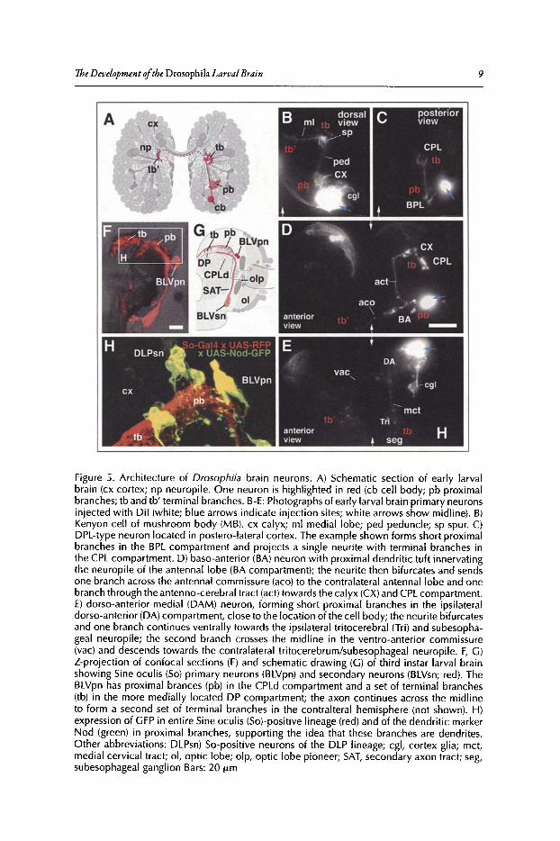
Figure 5. Architecture of Drosophila brain neurons. A) Schematic section of early larvalbrain (ex cortex; np neuropile. One neuron is highlighted in red (cb cell body ; pb proximalbranches; tb and tb' terminal branches. B-E: Photographs of early larval brain primary neuronsinjected with Dil (w hite; blue arrows indicate injection sites; white arrows show midline). B)Kenyon cell of mushroom bod y (M B). ex calyx ; ml medial lobe; ped peduncle; sp spur. C)DPL-type neuron located in postero-lateral cortex. The example shown forms short proximalbranches in the BPL compartment and projects a single neurite with terminal branches inthe CPL compartment. D) baso-anterior (BA) neuron with proximal dendritic tuft innervatingthe neuropile of the antennal lobe (BA compartment); the neurite then bifurcates and sendsone branch across the antennal commissure (aco) to the contralateral antennallobe and onebranch through the antenno-cerebral tract (act) towards the calyx (CX)and CPLcompartment.E) dorso-anterior-medial (DAM) neuron, forming short proximal branches in the ipsilateraldorso-anterior (DA) compartment, close to the location of the cell body; the neurite bifurcatesand one branch continues ventrally towards the ipsilateral tritocerebral (Tri) and subesophageal neuropile; the second branch crosses the midline in the ventro-anterior commissure(vac) and descends towards the contralateral tritocerebrum/subesophageal neuropile. F, G)Z-projection of confocal sections (F) and schematic drawing (G) of third instar larval brainshowing Sine ocul is (So) primar y neurons (BLVpn) and secondary neurons (BLVsn; red). TheBLVpn has proximal brances (pb) in the CPLd compartment and a set of terminal branches(tb) in the more medially located DP compartment; the axon continues across the midlineto form a second set of terminal branches in the contralteral hemisphere (not shown). H)expression of GFP in entire Sine oculis (So)-positive lineage (red) and of the dendritic markerNod (green) in proximal branches, supporting the idea that these branches are dendrites .Other abbrev iations: DLPsn) So-positive neurons of the DLP lineage; cgl, cortex glia; met,medial cervical tract ; 01, optic lobe; olp, opt ic lobe pioneer ; SAT, secondary axon tract ; seg,subesophageal ganglion Bars: 20 urn
10 Brain Development in Drosophila melanogaster
projection pattern. Thus, dendritic branches ofKenyon cells remain in close contact and, togetherwith axonal trees ofafferent fibers (mainly derived from the antennallobe), form a compact neuropile compartment, called the calyx.Tightly packed axonal branches ofKenyon cells, along withthe dendritic trees ofpostsynaptic cells," give rise to the peduncle, lobe compartments and spurofthe mushroom body. Figure SC - E show three additional examples ofDil filled neurons whosebranching pattern conforms to the same prototype. Preliminary data show that many secondarylineages that differentiate during the pupal period conform to the mushroom body lineages withregard to their proximal branching (V.H. and WP., unpublished). Thus, proximal arborizationsof most (if not all) neurons belonging to a given lineage appear to share in the same compartment. Terminal axonal arborization, on the other hand, are typically more diverse. Previous workon primary lineages ofthe ventral nerve cord had also shown that (terminal) arborizations in thispart of the CNS are also quite diverse within a given lineage.32.33
In very few instances, such as the Kenyon cells or some olfactory interneurons, has it actuallybeen shown that proximal branches of central neurons correspond to dendrites.tv" Moleculardifferences between dendrites and axons have been reported that in principle can be used todistinguish between the two. For example, the minus-end directed microtubule binding proteinNod 1 accumulates in the dendrites ofbipolar sensory neurons and the mushroom body's Kenyoncellsand therefore potentially represents a marker ofdendrites in the CNS.36As shown in Figure 5,Nod1-GFP driven in a small subset ofprimary neurons that belong to the sine oculis (so) expressingBLVllineage also accumulates in the subset ofneurite branches that are close to the cell bodies,indicating that these branches are dendrires.F'"
Figure 6 illustrates how the branching pattern ofneurons belonging to a lineage evolves overtime. To label lineages, the FLP /FRT technique was used." Each panel shows a member of theDAL lineages, a group oflineages located antero-Iaterally in the brain (for more detail see section'Neuroanatomy of the Developing Drosophila Brain' of this chapter), at different stages of itsdevelopment. A lineage at embryonic stage 14 (12-14h; Fig. SA,A') appears as a cluster ofcontiguous cell bodies capped by a neuroblast, sending a short, unbranched PAT towards the center ofthe brain primordium. A late embryonic clone (16h; Fig. 6B,B') still exhibits a compact PAT,butshort branches have appeared close to the cell body and, in many cases,at the PAT tip. In the earlylarva, branching ofaxons has increased dramatically (Fig. 6C,C'). Furthermore, the close packingofcell bodies and their axons has loosened up, although cells and neurites ofone lineage are stillclose to each other. A similar picture presents itselfif clones induced in the embryo are visualizedin late larvae. Primary neurons branch over much ofthe neuropile; in addition, secondary lineageshave now been added. Secondary neurons are alwaysexternally adjacent to the primary neurons.The secondary axon tract penetrates into the thicket ofprimary branches, suggesting that interactions between the primary axons and SAT exist. Clones induced in the early larva and visualizedin the late larva (Fig. 6E) contain exclusively secondary neurons, demonstrating the immature,unbranched nature ofsecondary axons. Proximal and terminal branches ofsecondary neurons areformed starting at 12h ofpupal development (Fig. 6G). Most lineages have proximal ("dendritic")branches restricted to one compartment, or part thereof By contrast, terminal branches are typically more widespread, but can also be fairly restricted, as in the case of the DAL lineage shownwhose terminal arbors are restricted to a layer of the ellipsoid body.
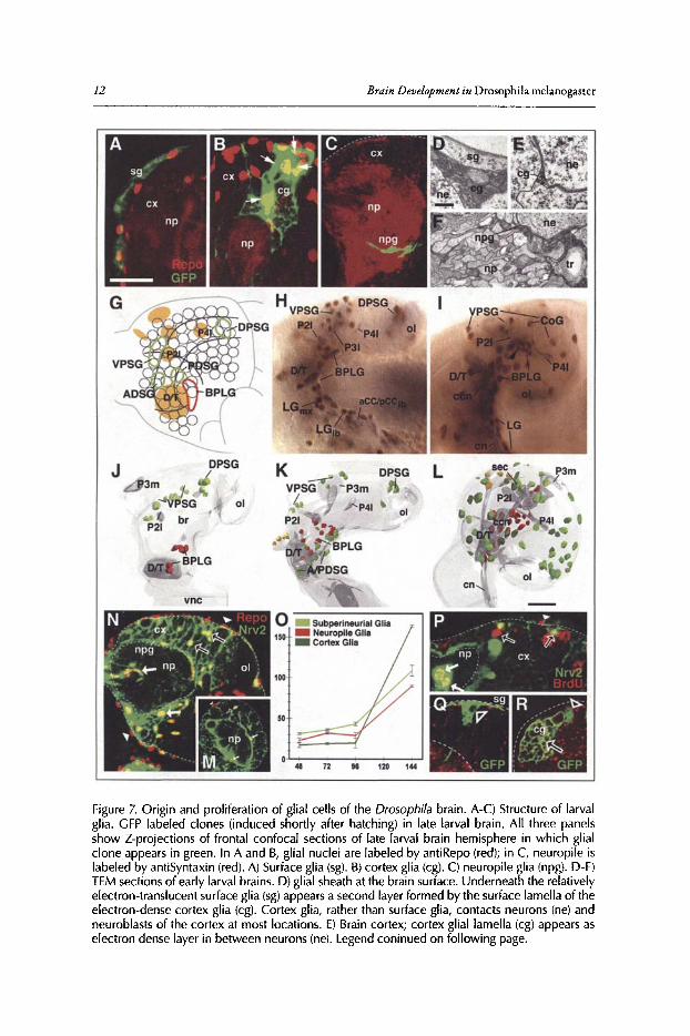
Glial CellsNeurons of the Drosophila brain are supported by a complex scaffold of glial cells that is es
tablished during late embryonic stages. Insect glial cells fall into three classes,39-42 each ofwhichis represented in the larval brain (Fig. 7A-F). Surface (subperineurial) glia form a sheath aroundthe surface ofthe brain (Fig. 7A). Cortex glia are located in the brain cortex and form a tightly-nitthree-dimensional scaffold that encapsulates neuronal cellbodies, ganglion mother cellsand neuroblasts (Fig. 7B). Neuropile glia surround the neuropile and form septa around individual neuropilecompartments, as well as major tracts ofneurites (Fig. 7C). Surface glial cells, interconnected byseptate junctions and covered by a thick basement membrane, act as the blood-brain barrier (Fig.7D).43 Cortex glia fulfill important trophic roles for neuronal cell bodies.f In the larval brain,

'IbeDevelopment ofthe Drosophila Larval Brain 11
Figure 6. Morphogenesis and branching of neural lineages. A-G are Z-projections of confocalstacks of brains in wh ich individual lineages were labeled by the FLP/FRT induced activationof tau-lacZ (green). For each panel (except G), a representative of the dorso-anterior-Iateral(DAL) group of lineages was selected. G shows the base-lateral-dorsal (BLD) lineage #5. Theneuropile is labeled with anti-DN-cadherin (red). In A-D, lineages were labeled by activating FLP in early embryo (primary lineages, A-C; pr imary plus secondary lineage, D). In E-G,activat ion of FLP occurred after hatching , resulting in labeling of secondary component oflineage only. A'-F' schematicall y depict one lineage at the stagecorresponding to the adjacentconfocal images. Primary neurons are in lilac, secondary neurons in orange. A, A': Stage 14embryo; B, B': Stage 16 embr yo; C, C : early larva; 0 ,0' and E: late larva; F, F' and G: pupaladult. Abbreviations: b neurite branches; BC baso-central compartment; CA centro-anteriorcompartment; cd cell death; cx cortex; eb ellipsoid body; lob lobula neurop ile; nb neuro blast; np neuropile; PATprimary axon tract ; pb proximal branches; PIBPprox imal interstitialbranchpoint; pn primary neuron; SATsecondary axon tract; sn secondary neuron; tb terminalbranches. Bar: 20!1m
12 Brain Development in Drosophila melanogascer
L
p " ' -'." ." " - ' -~- ' - --. » - • '.1-~ - "
...... .." ~ ~ -';~,"-p - .,' ex •
.1'..../ .. ~r~
01
DPSG
48 72 98 120
K•01
...
DPSG
vnc
.(...BPLG
P21
3mJ
Figure 7. Origin and proliferation of glial cells of the Drosophila brain. A-C) Structure of larvalglia . GFP labeled clones (induced shortly after hatching) in late larval brain. All three panelsshow Z-projections of frontal confocal sections of late larval brain hemisphere in which glialclone appears in green. In A and B, glial nuclei are labeled by antiRepo (red); in C, neuropile islabeled by antiSyntaxin (red). A) Surface glia (sg). B) cortex glia (cg). C) neuropile glia (npg). D-F)TEM sections of early larval brains. D) glial sheath at the brain surface. Underneath the relativelyelectron-translucent surface glia (sg)appears a second layer formed by the surface lamella of theelectron-dense cortex glia (cg). Cortex glia, rather than surface glia, contacts neurons (ne) andneuroblasts of the cortex at most locations. E) Brain cortex; cortex glial lamella (cg) appears aselectron dense layer in between neurons (ne). Legend coninued on following page.
TheDevelopment ofthe Drosophila LarvalBrain 13
Figure 7, continued from previous page. F) Cortex-neuropile boundary, showing prominent,electron-dense neuropile glial sheath (npg) separating neuronal somata (ne) from bundles ofneurites that constitute the neuropile (np). Tracheae and tracheoles (tr) penetrating the neuropileare always associated with glial sheaths. (G-L) Embryonic origin of brain glia. G) Schematic mapof stage 11 embryonic head showing approximate location of the clusters of glia progenitors(outlined in green and red, respectively) in relation to Fas-positive neuropile pioneer clusters(orange) and the brain neuroblast map.' Glia progenitors giving rise to surface and cortex gliacomprise a dorsal protocerebral cluster (DPSG), ventral protocerebral cluster (VPSG), anteriordeuterocerebral cluster (ADSG) and posterior deuterocerebral cluster (PDSG). Neuropile glia(longitudinal glia) is derived from a single cluster (BPLG) located in the deuterocerebral neuromere. H, I) lateral view of heads of embryos labeled with antiRepo expressed in glia cell nuclei(brown) and antiFas II expressed in pioneer neurons and their axons (P21, P31, P41, D/T, aCCIpCC; purple). H) late stage 12. Precursors of neuropile glia, forming the BPLG cluster, migratedorsally along the cervical connectives, pioneered by the D/T and P2 clusters. Ventrally, cellsof the BPLG have linked up with longitudinal glia cells of the ventral nerve cord (LGmx, LGlb:longitudinal glia derived from the maxillary and labial neuromere, respectively). Two majorclusters located in the ventral (VPSC) and dorsal (DPSC) part of the protocerebrum includeprecursors of surface glia and cortex glia. I) Late stage 14. Neuropile glia (LG and BPLG)form acontinuous covering of cervical connective (een)and connective of ventral nerve cord (en). Notegroup of small sized cells at dorsal front of BPLG (arrowhead); these cells most likely representearly postmitotic glia cells produced by the proliferating BPLG cluster. Surface glia precursorsderived from the VPSG cluster have spread over the lateral and dorsal brain hemisphere. At thisstagecortex glia cells (CoG), also derived from the VPSG and DPSG clusters, are seen separatelyfrom the more superficial surface glia. J-L) Digital 3D models of brain hemispheres of stage 11(J), late stage 12 (K) and late stage 14 (L)embryos, illustrating the pattern of different populationsof glia cell precursors in lateral view (seecolor key at top of panel 0) note that cortex glial cells(dark green) as entities different from subperineurial glia (light green) are indicated only in thelate stage 14 brain (L) because they cannot be distinguished earlier). Structures of the neuropile,including cervical connective (een), subesophageal commissure (sea), supraesophageal commissure (sec) and Fasll positive clusters [P1, P21, P3m, P31, P41, optic lobe (01); all shaded grey]are indicated as points of reference. (M-R) Glial proliferation during larval stages. M and Nshow confocal sections of larval brain hemispheres (M:48h AEL, first instar; N) 144h AEL, latethird instar) in which glial nuclei are labeled with antiRepo (red) and glial processes are markedwith Nrv2-GFP (green). Arrowheads point at representative surface glial nuclei; open arrowsat cortex glia, solid arrows at neuropile glia. Note dramatic increase in all three subclasses ofglia between first and third instar. 0) Plot of glial cell number against time (in hours after egglaying). P) Confocal section of brain of late larva that had been fed BrdU containing mediumfor 12h prior to dissection. BrdU incorporation appears in secondary neural lineages, as wellas in all three classes of glial cells (arrowheads: surface glia; open arrows: cortex glia; solidarrows: neuropile glia). (Q, R) GFP labeled clones of secondary lineages (neuroblasts indicatedby open arrowheads) with adjacent glial cells in third larval instar brains. Q shows surface glia(sg)forming part of secondary lineage (arrowhead). In R, cortex glia (cg; open arrow) is locateddi rectly adjacent to secondary Iineage (arrowhead). Other abbreviations: ex cortex; np neuropile;01 optic lobe Bars: 20,...,m (A-C); O.S,...,m (D-F); 10,...,m (G-L)
the meshwork ofcortex glial processes ("trophospongium") is required for stabilizing the positionof neurons in the cortex and for extension of secondary axon tracts? Neuropile and surface gliaplay numerous roles in axon pathfinding and targeting.45
-48 Glial septa formed by neuropile glia
are essential to establish and stabilize neuropile compartments, such as the glomeruli formed byneurites ofolfactory receptors and interneurons in the antennal Iobe."
The glial cellsofthe early larval brain (primary glia) arise from a small number ofneuro-glioblaststhat are active during the embryonic period. Neuro-glioblasts ofthe ventral nerve cord have beenidentified on a single cell basis,32,33 a feat not yet achieved for the brain. Here, precursors ofneuropileglia form a prominent cluster, the baso-posterior cluster, (BPLG)50 that consists ofapproximately20 cells and is located at the base ofthe brain primordium (Fig. 7G,]). During late embryogenesis,these cells spread out dorsally along the inner surface of the extending neuropile. Precursors of
14 Brain Development in Drosophila melanogaster
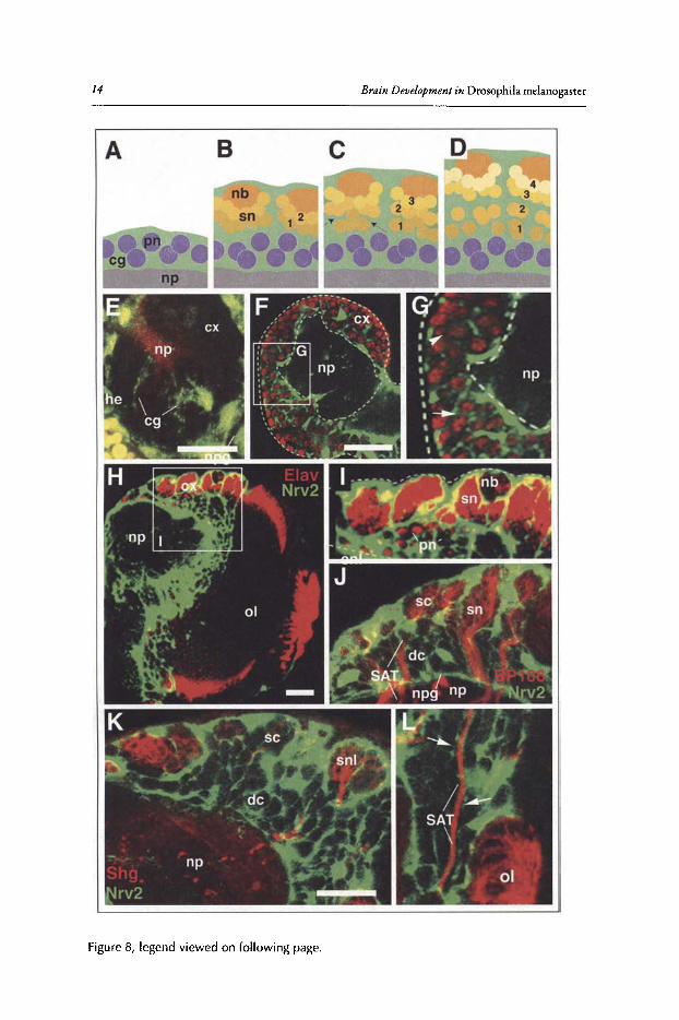
Figure 8, legend viewed on following page.
TheDevelopment ofthe Drosophila LarvalBrain 15
Figure 8, viewed on previous page. Formation of the trophospongium by cortex glia. A-D)Schematic cross section of brain cortex at different larval stages(A: 1st instar; B2nd instar; C early3rd instar; D late 3rd instar), illustrating formation of the trophospongium. Cortex glial cells (cg)are in green, neuroblasts (nb) and secondary neurons (sn) in shades of orange, primary neurons(pn) in lilac, neuropile (np) in gray. Numbers 1-4 indicate birth order of secondary neurons (1:early;4: late). E) Z-projection of serial horizontal confocal sections of embryonic brain hemisphere inwhich glial cells are labeled green by GFP reporter construct activated by the gem-Gal4 driver.Neuropile (np) is labeled with antiDN cadherin antibody (red. Cortex glia (cg) is represented byslender, radial cells extending throughout the cortex (cx) from the neuropile (np) to the brainsurface. Hemocytes (he) surrounding brain also express gem-gal. 4 (F-I) Confocal sections ofbrain hemispheres of first instar (F,G) and late third instar (H,I). Glial cells are labeled (green)by GFP reporter activated by the nrv2-Gal4 driver. Neurons are labeled by anti-Elav antibody(red). By the first instar, cortex glia have formed a meshwork of lamelliform processes that formmore (arrow) or less (arrowhead) complete sheaths around primary neurons. At later stages(H,I) all primary neurons (pn) and the first born secondary neurons (located deep in the cortex) areindividually surrounded by glial sheaths; secondary neuroblasts (nb) and their latest progeny(sn) located near the brain surface are enclosed within large glial chambers. J) Confocal section of cortex of late third instar brain labeled with antiBP106 (red, marks secondary lineages)and nrv2-Gal4 driving GFP (green; glia). Secondary lineages (sn) and their axon tracts (SAT)are encapsulated by cortex glia. (K,L) Confocal section of cortex of late third instar larval brainlabeled with antiShg (DE-cadherin; red) and nrv2-Gal4 driving GFP. DE-cadherin is expressedin secondary neuroblasts and the latest born neurons (snl in K), as well as the SAT formed bythese cells. Note enclosure of the Shg-positive cells in large, undivided glial chambers ("superficial chambers"; sc); earlier born neurons located in deep cortex are individually surroundedby glial septa ("deep chambers"; dc). Cortex glial septa (arrows in L) also flank the SAT in deepcortex. Other abbreviations: npg, neuropile glia; 01, optic lobe. Bars: 20 urn
surface glia (approximately 25-30 in the hatching larva) and cortex glia (approximately 10) alsooriginate in a small number ofdiscrete clusters which migrate outward to populate the entire brain(Fig. 7H,I,K,i).50 This pattern suggests that, similar to what has been found in the ventral nervecord, glial cells are produced by only few neuro-glioblasts.
Glial cell numbers increase slowly during the first halfoflarval development, but show a rapidincline in the third larval instar. Overall, glial cell numbers increases from about 30 to more than100 for surface glia, from 10 to 160 for cortex glia and from 20 to about 90 for neuropile glia(Fig. 7M-0).51 This increase in cell number is at least in part due to the mitotic divisions ofglialcells.Thus, feeding BrdU to larvae at different stages results in clusters oflabeled cells that includeall three types ofglial cells (Fig.7P). Moreover, a small fraction oflate larval glial cellscan alwaysbeseen in mitosis using a marker that labels phosphorylated histone H3. However, the low frequencyofphospho-histone positive glial cells, as well as the finding that glial cellslabeled by clonal induction were almost always in close contact with neural lineages (Fig. 7Q,R), indicates that the bulkofadded glial cells stems from the proliferation ofsecondary neuro-glioblasts located at the brainsurface. This is also supported by the shape of the glial growth curve, which is almost horizontalduring early larval life (when neuroblasts are mitotically quiescent) and becomes steep during thethird instar when neuroblasts divide (Fig. 70).
The trophospongium is formed by cortex glial cells, highly branched and lamellated cellswhose processes undergo extensive rearrangements during development (schematically shown inFig. 8A-D). Cortex glia appear in the stage 16 embryo as elongated, radially oriented cells mostofwhich extend from the brain surface to the neuropile (Fig. 8£).51,52 Subsequently lateral processes are formed, leading up to the three-dimensional, honey-combed structure revealed by thelarval clones shown in Figure 7B. Shortly after hatching these processes are still modest, formingrelatively large chambers that enclose multiple primary neurons (Fig. 8F,G). At subsequent stages,process density increases, so that by the second instar each primary neuron is completely enclosedby cortex glia. Cortex glia also form a superficial lamella that extends underneath the surface gliallayer. Thus, from the second instar onward, the glial layer covering the brain is composed of an
16 Brain Development in Drosophila melanogaster
outer, electron-light lamella ofsurface glia and an inner, extremely thin and electron-dense lamellaformed by cortex glia (Fig. 7D).
Beginning with the second instar, dividing neuroblasts produce secondary neurons that forman outer cortex of increasing thickness around the inner layer of primary neurons. During thisphase, the growth ofthe trophospongium and neuroblast proliferation must be coordinated in acomplex manner. Close to the brain surface, individual chambers ofthe trophospongium are large,containing a neuroblast, undivided ganglion mother cells and 20 to 40 neurons (Fig. 8C,D,H-K).Each superficial trophospongium chamber corresponds to part ofone secondarylineage, such thatthe neurons newly formed by one neuroblast over a certain period oftime are "received" into onechamber, thereby isolating them from other lineages. At deeper levels, chambers become smaller,such that older secondary neurons (like primary neurons before) become individually enclosedby glia. This implies that there is a dynamic rearrangement ofglia processes at the transition zonefrom large chambers to small chambers.
Tracheal System ofthe BrainGas exchange in the insect body is mediated by a branched network ofair-filled tubes called
tracheae. In the brain and ventral nerve cord, tracheae form an anastomosing plexus at the cortex-neuropile surface (perineuropilar plexus).53 From this plexus, several branches sprout into theneuropile and the cortex (see below). Tracheae develop from a bilateral set ofmetameric invaginations ofthe embryonic ectoderm.54 Each tracheal invagination subsequently forms a stereotypedset of primary branches (Fig. 9A). One branch, called ganglionic branch (GB), grows towardseach neuromere ofthe ventral nerve cord in the late embryo (Fig. 9B, arrowhead" I ").55 Advancingmedially, GBs pass underneath the neuropile of the ventral nerve cord (Fig. 9B, arrowhead "2")and then form a 180-degree turn around the medial and dorsal surface ofthe neuropile (arrowhead"3" and "4"). During larval stages, the advancing tips of the GBs close the circle and fuse with amore proximal part of the same or adjacent GBs. A similar pattern of ring- (or noose-) shapedtracheae is generated in the brain. Here, one main trachea, the cerebral trachea (CT), branchesoff the first tracheal invagination in segment T2 (Fig. 9A). After reaching the medial surface ofthe brain neuropile in the embryo (Fig. 9B, arrowhead "5") the CT gives off multiple branches(the primary tracheae ofthe brain) that grow laterally and medially around the neuropile surfaceto eventually meet and fuse.
Figure 9C-G show Z-projections ofconfocal sections that illustrate the growth ofthe trachealnetwork in the larval brain. Panels 8H-K show the tracheal system of an early third instar brain(when all primary and secondary branches are in place) in the form of 3D digital models. Inthe late embryo the cerebral trachea is visible as a thick, posteriorly directed branch of the firstsegmental trachea that belongs to the second thoracic segment (Fig. 9C,D). The CT follows themedial surface ofthe brain where the neuropile is covered by a layer ofsurface glia (Fig. 9C). Thecerebral trachea and all ofits branches are embedded in a glial layer. During the first larval instar,all ofthe primary brain tracheae become established. First, around the time ofhatching, the CTsplits into a laterally and a ventrally directed trunk (Fig. 9D,E). By the beginning of the secondinstar (48h after egg laying, AEL; Fig. 9F), the lateral trunk gives rise to the centro-medial trachea (CMT), centro-posterior trachea (CPT) and baso-Iateral trachea (BLT). The ventral trunkbifurcates into the baso-medial trachea (BMT) and the lateral and medial baso-central trachea(BCTl, BCTm).
By the early third instar two to three secondary tracheae enter the center ofthe neuropile. Theyinclude the trachea ofthe mushroom body (TMB), the trachea ofthe antennocerebral tract (TAC)and the internal dorsal transverse trachea (DT; not alwaysfound). The mushroom body trachea inmost casesbranches offthe BCT trachea. The trachea ofthe antennocerebral tract (TAC) typicallyconstitutes a branch ofthe CPTm trachea. In addition to the TAC and TMB tracheae which aredirected inward, into the center ofthe neuropile, a number ofsecondary tracheal branches projectoutward into the cortex and the optic lobe (Fig. 9).
TheDevelopment ofthe Drosophila LarvalBrain 17
Although the main, primary tracheal branches described above can be recognized faithfullyin all brains, the higher order branching pattern is highly variable. For example, the secondarytrachea towards the mushroom body (TMB) may branch off the BCTm in one hemisphere andthe CPT in the other hemisphere of the same brain. This is similar to the reported variability inhigher order branching patterns ofepidermal tracheae and strikingly contrasts with the invariantpattern ofneuroblasts, neurons and axon tracts in the brain.
Neuroanatomy ofthe Developing Drosophila Brain:The Systems ofLineages, Tracts and Compartments
Pattern ofPrimary Pioneer TractsAs described in section 'Secondary Neuroblasts and GMCs ofthe Larval Brain' of this chap
ter, the brain ofthe late embryo is formed by approximately 100 lineages per side whose neuronsadhere to each other and, at the onset of neuropile formation (stage 13), appear as cone shapedclusters distributed rather evenly over the periphery of the brain (Fig. lOA). Axons formed byneurons of the same lineage typically form one bundle, the primary axon tract (PAT; Fig. lOA,B, C). The pattern ofPATsappears highly invariant and provides essential information about thestructure of the evolving neuropile (see section 'Synopsis ofLineages, Compartments and FiberTracts of the Larval Brain' below). To describe the pattern ofprimary lineages and their PATs, ascaffold ofpioneer axon tracts laid down by early born neurons of a subset of lineages has beenutilized. 52,56,57 We will first introduce the pattern ofpioneer tracts, to then relate the primary andsecondary lineages to this pattern. Fig. 10D shows the FasII-positive pioneer neurons in relationship to neuromere boundaries, visualized by an engrailed-IacZ reporter construct; panell0E is aschematic map ofpioneer neurons in the late embryonic brain.
Longitudinal pioneer tracts: Three longitudinal tracts (connectives) pioneer the neuropileof the ventral nerve cord.56,58 By the end of embryogenesis, each of these three connectives hassplit into a dorsal and ventral component.v" The connectives of the ventral nerve cord continueanteriorly into the two preoral neuromeres that form the basal brain, the tritocerebrum anddeuterocerebrum (Fig. 10E).56.59.60 The medial connective continues as the medial cervical tract(MCT); the intermediate connective as the lateral cervical tracts (LCT) and the lateral connective as the posterior cervical tract (PCT), respectively.The MCT is organized by the large D/Tpioneer cluster, located in the deutero-tritocerebral boundary region. Ascending D/T axons reachthe P2 clusters, located in the antero-dorsal deuterocerebrum, that pioneer the ventral fascicleof the supraesophageal commissure (vSEC; Fig. IDE). The LCT is formed by axons ofD/T andPI that extend laterally adjacent of the MCT. Three tracts to and from the "corner points" ofthebasal brain converge upon Pl. The horizontally directed baso-medial protocerebral tract (BMPT)connects posterior and anterior realms ofthe basal brain (PI to/from P4m). The centro-anteriorprotocerebral tract (CAPT) and dorso-posterior protocerebral tract (DPPT) originate from theP3c and P3m clusters, respectively,both located in the boundary region between deuterocerebrumand protocerebrum (shaded light blue in Fig. IDE).
Transverse pioneer tract: The dorsal and lateral protocerebrum consists oflineages whose PATsform transverse (commissural) fiber systemsconnecting the two brain hemispheres. These transversesystems are quite separate from the longitudinally oriented MCT, LCT and PCT systems and arepioneered by the lateral protocerebral tract (LPT). The LPT is formed by several medio-Iaterallyarranged clusters ofpioneer neurons (PSI, P4I, P31) that extend from the optic lobe primordium(0 L) to the dorsal midline, where they establish the dorsal fascicle of the supraesophageal commissure (dSEC in Fig. 10E).
Mushroom body: The massive fiber tract formed by the mushroom body neurons (MB) interconnects the posterior protocerebrum with the proto-deuterocerebral boundary domain. Thistract (peduncle; indicated by gray hatched line in Fig. 10E) converges upon the PI cluster, butthen makes a sharp turn medially, pioneering the medial lobe ofthe mushroom body.
18
o " BtI-Gal,\x.UAS-GFI1
, BMT ' !
'I~ ~~ I')," I '\, ,:' ... CPT/BLT CT /./
Brain Development in Drosophila melanogaster
Figure 9. Development of the larval brain tracheal system. (A, B) Embryonic origin of the cerebral trachea and ganglionic tracheal branches. Both panels show Z-projections of confocalsections of embryos (lateral view, anterior to the left) labeled with antiCrb (green)to visualizetracheae . AntiDN-cadherin (red) labels neuropile and other embryonic structures. A) Stage14. Cerebral trachea (CT) and dorsal pharyngeal trachea (dPT) form a V-shaped, anteriorlydirected branch of the first segmental trachea (I) that grow around the posterior surface ofthe brain (br). Other branches of the first segmental trachea are the dorsal branches (DB) ofsegments T2 and T1 (formed later than stage 14), the ventral ganglionic branches (GB) of segments T1 and T2 and the ventral pharyngeal trachea. The location of the anterior spiracle isindicated by violet circle. B) Stage 15 late. Segmental tracheae have fused, primary brancheshave increased in length and some secondary branches have been initiated. Note positionof the cerebral trachea (CT) and dorsal pharyngeal trachea (dPT). The cerebral trachea hasreached the medial surface of the brain neuropile (arrowhead "5"). Legend continued onfollowing page.
TheDevelopment ofthe DrosophilaLarvalBrain 19
Figure 9, continued from previous page. Ventral ganglionic branches contact the ventralsurface of the neuropile of the ventral nerve cord (vc; arrowhead "1"). During later stagesganglionic branches will extend underneath the neuropile, turn dorsally (hatched blue line;arrowheads "2" and "3") and then laterally (solid blue line, arrowhead "4"). Anastomoses(ana; gray hatched line) will interconnect ganglionic branches of neighboring segments. C)Z-projection of a confocal stack of brain of late embryo in which tracheae are visualized by theexpression of btl-Gal4 driving UAS-GFP (green); the neuropile is labeled with antiDN-cadherin(red) and glial cells are labeled by antiRepo (blue). (D-G) Z-projections of confocal stacks ofbrains of stage 16 embryo (D), first instar larva (E), second instar larva (F) and early third instarlarva (G). Tracheae are visualized by the expression of btl-Gal4 driving UAS-GFP (green);the neuropile is labeled with antiDN-cadherin (red). (H-K) Digital 3D models of right brainhemisphere showing neuropile compartments in grey and major tracheae in different colors.Models of the top row (H, I) represent posterior view (dorsal up, lateral to the right); secondrow (I,K) shows dorsal view (dorsal up, lateral to the right). In models of left column (H, J),coloring indicates depth of tracheae: Tracheae forming the perineuropilar plexus (surrounding the neuropile surface) are depicted in green; secondary branches turning externally intothe cortex of the central brain are shown in light blue; optic lobe tracheae in purple. Twosecondary branches turning centrally into the neuropile are shown in red. In models of rightcolumn (H,K) neuropile is also semi-transparent and each primary brain trachea togetherwith its belonging secondary branches is depicted in its own color (see color key at bottomof panel), which allows one to follow the trajectories of tracheae. For description of patternof tracheae see text. Abbreviations: BA baso-anterior (antennal) neuropile compartment; BCbaso-central neuropile compartment; BAT baso-anterior trachea; BCT baso-central trachea;BCTI lateral baso-central trachea; BCTm medial baso-central trachea; BCvT baso-cervicaltrachea; BlT baso-Iateral trachea; BlTI lateral branch of baso-Iateral trachea; BPl baso-posterior lateral neuropile compartment; BMT baso-medial trachea; BPM baso-posterior medialneuropile compartment; CA centro-anterior neuropile compartment; CMT centro-medialtrachea; CMTa anterior branch of centro-medial trachea; CMTi intermediate branch ofcentro-medial trachea; CMTp posterior branch of centro-medial trachea; CPI centro-posteriorintermediate neuropile compartment; CPl centro-posterior lateral neuropile compartment;CPM centro-posterior medial compartment; CPT centro-posterior trachea; CPTm medialbranch of centroposterior trachea; CPTi intermediate branch of centro-posterior trachea;CPTI lateral branch of centro-posterior trachea; CT cerebral trachea; CX calyx of mushroombody; DA dorso-anterior neuropile compartment; dl dorsal lobe of mushroom body; DPdorso-posterior compartment; DOT dorsal oblique trachea; ml medial lobe of mushroombody; DTTe external dorsal transverse trachea; DTTi internal dorsal transverse trachea; INTintraneuropilat tracheae of ventral nerve cord; ngl neuropile glia; OlTa anterior optic lobetrachea; OlTI lateral optic lobe trachea; OlTp posterior optic lobe trachea; p peduncle ofmushroom body; ph pharynx; PNPbr perineuropilar plexus of brain; PNPvc perineuropilearplexus of ventral nerve cord; SEG subesophageal neuropile; SET subesophageal tracheae; spspur of mushroom body; TAC trachea of the antenno-cerebral tract; ThT thoracic trachea;TMB trachea of the mushroom body. Bars: 20 11m
Pattern ofLineages and Their Axon TractsPrimary lineages and PATs:Marking the evolving neuropile oflate embryos with global mark
ers such as Synaptobrevin-GFP fusion protein driven by elav-Gal4 reveals that PATs are ratheruniformly directed awayfrom the surface and extend centripetally towards the center ofthe brainprimordium (Figs. 10A,B; lIA).17.38,52 As a result, the direction ofmost PATscorrelates with thelocation of the corresponding primary lineage. PATsof lineages located at the posterior pole ofthe brain primordium project anteriorly, those ofdorsal lineages ventrally and so on. PATsline upwith the neuropile pioneer tracts in whose vicinity they are located. Thus, for example, lineagesgrouped around P3c align their PATswith the CAPT tract; posterior lineages close to P4m projectPATsclose to the pioneer tract (BMPT) formed by this cluster and so on. Figs. 11 and 12 showthe pattern ofprimary lineages and PATs.
Compartments: Up to stage 15, PATsconsist ofmostly short, unbranched axons that convergein the center ofthe brain primordium (Fig. lOA). The assembly ofPATsrepresents the "nucleus"
20 Brain Development in Drosophila melanogasrer
ventral ,--nerve cord
Figure 10. Patterning of the brain neuropile . A,B) Schematic horizontal sections of brainhemisphere illustrating neuropile formation . Neuroblasts (nb) produce lineages compris ingprecursors of primary neurons (pn) and glial cells (gl). At the stage shown in A (stage 13-15of embryonic development) primary neurons have formed short axon stumps organized inbundles, with each bundle belonging to one lineage (primary axon bundle, PAT). Early differentiating neurons (neuropile pioneers; ppn) establish scaffold of pioneer tracts. Primaryaxon tracts orient themselves around the pioneer tracts and form the early nucleus of theneuropile (np). Legend continued on following page.
TheDevelopmentofthe Drosophila Larval Brain 21
Figure 10, continued from previous page. Branching of primary axons (neb; stage 16-17; B)leads to an increase in neuropile volume. C) Confocal horizontal section of stage 13 embryonic brain hemisphere double labeled with anti-Acetylated tubulin (all differentiating neurons; yellow) and antiFasll (neuropile pioneers, green). Primary axon bundles (PAT) convergeradially towards the center of the brain. Note, at this confocal level, anteriorly directed axonbundles, emanating from lineages localized in the posterior cortex and extending parallelto the fasll-positive P41 pioneer neurons. 0) Z-projection of confocal parasagittal sectionsof stage 13 embryonic brain double-labeled with antiFasll (pioneer tracts; green) and anengrailed (rhx25)-lacZ reporter construct (red). Engrailed expression domains demarcate theposterior boundary of the deuterocerebrum (eO), the tritocerebrum (eT) and the mandibularsegment (eMO). The small engrailed-positive "head spot" (eP) indicates a point located on theotherwise undefined protocerebral-deuterocerebral boundary. E) Schematic lateral view ofembryonic brain (anterior to the left; dorsal to the top), showing Fasll positive pioneer tractsin relationship to dorso-ventral and antero-posterior axis and to neuromere boundaries. Otherabbreviations: BMPT baso-medial protocerebral tract; CAPT central anterior protocerebraltract; ex cortex; OPPTdorso-posterior protocerebral tract; dSEC dorsal supraesophageal commissure; dSEC dorsal supraesophageal commissure; O/T deuteroltritocerebral Fasll cluster;LCT lateral cervical tract; LPT lateral protocerebral tract; MB mushroom body; MCT medialcervical tract; OL optic lobe; Pl, P2, P3l1m/c, P4m/l, P5 Fasll positive pioneer clusters; PCTposterior cervical tract; vSEC ventral supraesophageal commissure. Bars: 20 urn
from which the brain neuropile is formed. Neuropile formation proceeds by branching of thePATs (Fig. lOB; see also Fig. 6B). These axonal and dendritic branches make up the content ofthe emerging neuropile compartments. Using the reproducible pattern ofPATsand FasII-positiveneuropile pioneer tracts as landmarks, the discrete neuropile compartments defined for the larvalbrain can be recognized already in the embryo (Fig. 13A-C; see also section'Synopsis ofLineages,Compartments and Fiber Tracts ofthe Larval Brain' below). 52 Between early and late larval instar,compartments grow substantially through additional branchingofprimary neurons, aswell as the"invasion" of the neuropile by SATs(Fig. 13D-I). Branching ofsecondary lineages, as well as themetamorphic reorganization ofprimary neurons, lead to changes in compartmental shape and theaddition ofnew (adult-specific) compartments; however, these changes notwithstanding, the basicpattern ofneuropile compartments ofthe larval brain can be followed throughout metamorphosisinto the adult brain (Fig. 13J-L).
Secondary lineages and SATs: Primary neurons of the brain and ventral nerve cord form thefunctional circuitry controlling larval behavior. During the early larval period, the brain growsonly slowly, mainly due to increased branching of primary neurons. Starting during the secondinstar, neuroblasts become reactivated and produce secondary lineages. Similar to primary axons,axons ofa given secondary lineage fasciculate with each other, thereby forming a discrete secondary axon tract (SAT) within the brain cortex and neuropile. SATs penetrate the neuropile glialsheath, or travel along the neuropile surface for variable distances (Figs. lIB; 12C).38In terms ofoverall number and trajectory, secondary and primary lineages show many similarities and wehave adopted a nomenclature that suggests correspondences between lineages.38,52 Thus, we assume that, for example, the primary BA lineages are generated by the same neuroblasts that laterform the secondary BA lineages. Likewise, PATsofprimary BA lineages show similar trajectoriesthan SATsofthe larval secondary BA lineages. However, one should point out that lineage tracingand the analysis of molecular markers that are continuously expressed from the embryonic tothe late larval period are needed to establish in detail the link between a primary lineage and itssecondary counterpart.
Secondary tract systems in the larval and pupal neuropile: Within the neuropile, tracts ofseveral neighboring lineages converge to form larger "secondary tract systems" (Fig. 11C,D).53 Somesecondary tract systems extend along the glial sheaths in between neuropile compartments; otherspenetrate the center ofcompartments, typically following the above described pioneer tracts laiddown by primary axons at an earlier stage (Fig. 11E).57 Each secondary lineage forms a tract withan invariant and characteristic trajectory within the neuropile. That is to say,a given SAT reaches

22
Figure 11 , legend viewed on following page .
Brain Development in Drosophila melanogaster
trPdLCOTd
crMBppMOT
__ exDLPliii.li.1i'-W MOT
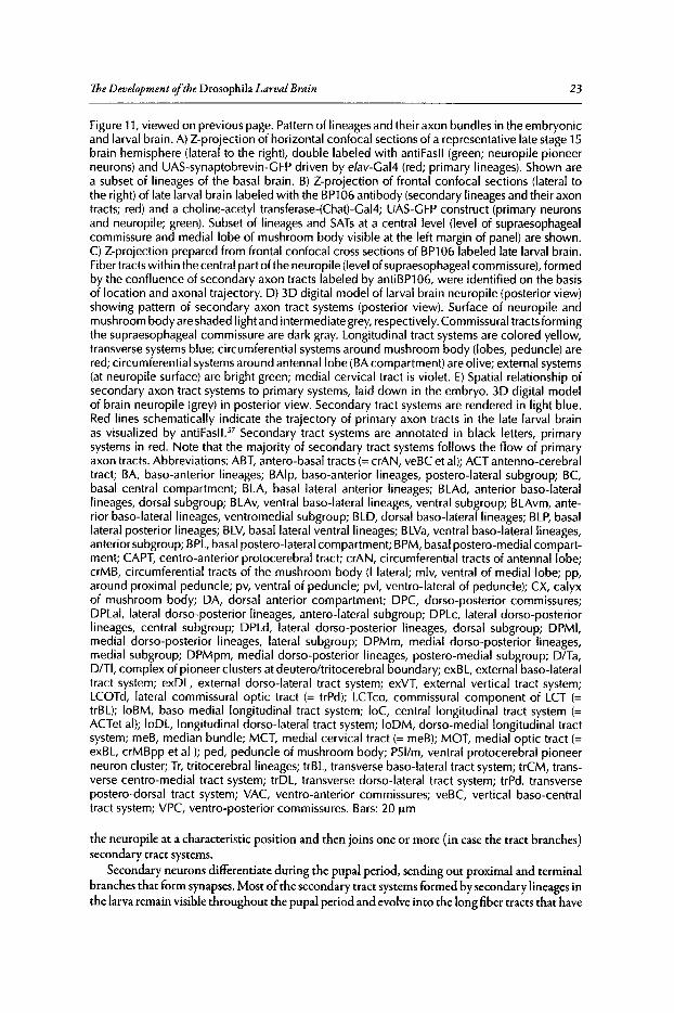
TheDevelopment ofthe Drosophila LarvalBrain 23
Figure 11,viewed on previous page. Pattern of lineages and their axon bundles in the embryonicand larval brain. A) Z-projection of horizontal confocal sections of a representative late stage 15brain hemisphere (lateral to the right), double labeled with antiFasl1 (green; neuropile pioneerneurons) and UAS-synaptobrevin-GFP driven by elav-Gal4 (red; primary lineages). Shown area subset of lineages of the basal brain. B) Z-projection of frontal confocal sections (lateral tothe right) of late larval brain labeled with the BP1 06 antibody (secondary lineages and their axontracts; red) and a choline-acetyl transferase-(Chat)-GaI4; UAS-GFP construct (primary neuronsand neuropile; green). Subset of lineages and SATs at a central level (level of supraesophagealcommissure and medial lobe of mushroom body visible at the left margin of panel) are shown.C) Z-projection prepared from frontal confocal cross sections of BP1 06 labeled late larval brain.Fiber tracts within the central part of the neuropile (level of supraesophageal commissure), formedby the confluence of secondary axon tracts labeled by antiBP106, were identified on the basisof location and axonal trajectory. 0) 30 digital model of larval brain neuropile (posterior view)showing pattern of secondary axon tract systems (posterior view). Surface of neuropile andmushroom body are shaded light and intermediate grey, respectively. Commissural tracts formingthe supraesophageal commissure are dark gray. Longitudinal tract systems are colored yellow,transverse systems blue; circumferential systems around mushroom body (lobes, peduncle) arered; circumferential systemsaround antennallobe (BA compartment) are olive; external systems(at neuropile surface) are bright green; medial cervical tract is violet. E) Spatial relationship ofsecondary axon tract systems to primary systems, laid down in the embryo. 30 digital modelof brain neuropile (grey) in posterior view. Secondary tract systems are rendered in light blue.Red lines schematically indicate the trajectory of primary axon tracts in the late larval brainas visualized by antifasll." Secondary tract systems are annotated in black letters, primarysystems in red. Note that the majority of secondary tract systems follows the flow of primaryaxon tracts. Abbreviations: ABT, antero-basal tracts (= crAN, veBC et al): ACT antenno-cerebraltract; BA, baso-anterior lineages; BAlp, baso-anterior lineages, postero-Iateral subgroup; BC,basal central compartment; BLA, basal lateral anterior lineages; BLAd, anterior baso-Iaterallineages, dorsal subgroup; BLAv, ventral baso-Iaterallineages, ventral subgroup; BLAvm, anterior baso-laterallineages, ventromedial subgroup; BLO, dorsal baso-laterallineages; BLP, basallateral posterior lineages; BLV,basal lateral ventral lineages; BLVa, ventral baso-Iaterallineages,anterior subgroup; BPL,basal postero-lateral compartment; BPM, basal postero-medial compartment; CAPT, centro-anterior protocerebral tract; crAN, circumferential tracts of antennal lobe;crMB, circumferential tracts of the mushroom body (I lateral; mlv, ventral of medial lobe; pp,around proximal peduncle; pv, ventral of peduncle; pvl, ventro-lateral of peduncle); CX, calyxof mushroom body; OA, dorsal anterior compartment; OPC, dorso-posterior commissures;OPLal, lateral dorso-posterior lineages, antero-Iateral subgroup; OPLc, lateral dorso-posteriorlineages, central subgroup; DPLd, lateral dorso-posterior lineages, dorsal subgroup; OPMI,medial dorso-posterior lineages, lateral subgroup; OPMm, medial dorso-posterior lineages,medial subgroup; OPMpm, medial dorso-posterior lineages, postero-medial subgroup; O/Ta,O/TI, complex of pioneer clusters at deuteroltritocerebral boundary; exBL, external baso-Iateraltract system; exOL, external dorso-Iateral tract system; exVT, external vertical tract system;LCOTd, lateral commissural optic tract (= trPd); LCTco, commissural component of LCT (=trBL); loBM, baso-medial longitudinal tract system; loC, central longitudinal tract system (=ACTet al): loOL, longitudinal dorso-lateral tract system; 100M, dorso-mediallongitudinal tractsystem; meB, median bundle; MCT, medial cervical tract (= meB); MOT, medial optic tract (=exBL, crMBpp et al ); ped, peduncle of mushroom body; P51/m, ventral protocerebral pioneerneuron cluster; Tr, tritocerebrallineages; trBL, transverse baso-Iateral tract system; trCM, transverse centro-medial tract system; trOL, transverse dorso-Iateral tract system; trPd, transversepostero-dorsal tract system; VAC, ventro-anterior commissures; veBC, vertical baso-centraltract system; VPC, ventro-posterior commissures. Bars: 20 urn
the neuropile at a characteristic position and then joins one or more (in case the tract branches)secondary tract systems.
Secondary neurons differentiate during the pupal period, sending out proximal and terminalbranches that form synapses. Most ofthe secondary tract systems formed by secondary lineages inthe larva remain visible throughout the pupal period and evolve into the long fiber tracts that have
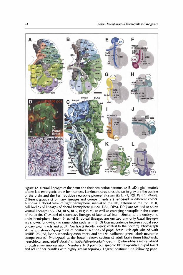
24 Brain Development in Drosophila melanogasrer
Figure 12. Neural lineagesof the brain and their projection patterns. (A,B) 3D digital modelsof one late embryonic brain hemisphere. Landmark structures shown in gray are the outlineof the brain and the Fasll-positive neuropile pioneer clusters (D/T, P1 , P21, P3m/l, P4m/I).Different groups of primary lineages and compartments are rendered in different colors.A shows a dorsal view of right hemisphere; medial to the left, anterior to the top. In B,cell bodies of lineages of dorsal hemisphere (DAM, DAL, DPM, DPL) are omitted to showventral lineages (BA, CM, BLA, BLD, BLP, BLV), as well as emerging neuropile in the centerof the brain. C) Model of secondary lineages of late larval brain. Similar to the embryonicbrain hemisphere shown in panel B, dorsal lineages are omitted and only basal lineagesare shown, following the same color code as in B. D) Correspondence between pupal secondary axon tracts and adult fiber tracts (frontal views; ventral to the bottom). Photographat the top shows Z-projection of confocal sections of pupal brain (72h apt) labeled withantiBP106 (red, labels secondary axon tracts) and antiDN-cadherin (green, labels neuropilecompartments). Photograph at the bottom shows section of adult brain (from http ://web .neurobio.arizona.edu/Flybrain/htm I/atlas/siIver/frontal/index.htmI)where f bersarevisualizedthrough silver impregnation. Numbers 1-10 point out specific BP106-positive pupal tractsand adult fiber bundles with highly similar topology. Legend cont inued on following page.
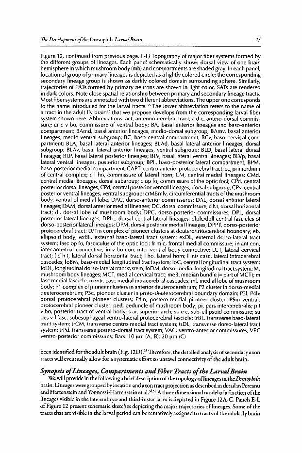
TheDevelopment ofthe Drosophila LarvalBrain 25
Figure 12, continued from previous page. E-L)Topography of major fiber systems formed bythe different groups of lineages. Each panel schematically shows dorsal view of one brainhemisphere in which mushroom body (mb) and compartments are shaded gray. In each panel,location of group of primary lineages is depicted as a lightly colored circle; the correspondingsecondary lineage group is shown as darkly colored domain surrounding sphere. Similarly,trajectories of PATs formed by primary neurons are shown in light color, SATs are renderedin dark colors. Note close spatial relationship between primary and secondary lineage tracts.Most fiber systemsare annotated with two different abbreviations. The upper one correspondsto the name introduced for the larval tracts." The lower abbreviation refers to the name ofa tract in the adult fly brain" that we propose develops from the corresponding larval fibersystem shown here. Abbreviations: act, antenno-cerebral tract; ad c, antero-dorsal commissure; ar c v bo, commissure of ventral body; BA, basal anterior lineages and baso-anteriorcompartment; BAmd, basal anterior lineages, medio-dorsal subgroup; BAmv, basal anteriorlineages, medio-ventral subgroup; BC, baso-central compartment; BCv, baso-cervical compartment; BLA, basal lateral anterior lineages; BLAd, basal lateral anterior lineages, dorsalsubgroup; BLAv, basal lateral anterior lineages, ventral subgroup; BLD, basal lateral dorsallineages; BLP, basal lateral posterior lineages; BLV, basal lateral ventral lineages; BLVp, basallateral ventral lineages, posterior subgroup; BPL, baso-posterior lateral compartment; BPM,baso-posterior medial compartment; CAPT,centro-anterior protocerebral tract; cc, primordiumof central complex; c I ho, commissure of lateral horn; CM, central medial lineages; CMd,central medial lineages, dorsal subgroup; c op fo, commissure of the optic foci; CPd, centralposterior dorsal lineages; CPd, central posterior ventral lineages, dorsal subgroup; CPv,centralposterior ventral lineages, ventral subgroup; crMBmlv, circumferential tracts of the mushroombody, ventral of medial lobe; DAC, dorso-anterior commissures; DAL, dorsal anterior laterallineages; DAM, dorsal anterior medial lineages; DC, dorsal commissure; d h t, dorsal horizontaltract; dl, dorsal lobe of mushroom body; DPC, dorso-posterior commissures; DPL, dorsalposterior lateral lineages; DPLc, dorsal central lateral lineages; dlplc/dpll central fascicles ofdorso-posterior lateral lineages; DPM, dorsal posterior medial lineages; DPPT,dorso-posteriorprotocerebral tract; D/Tm complex of pioneer clusters at deutero/tritocerebral boundary; eb,ellipsoid body; exBL, external baso-Iateral tract system; exDL, external dorso-Iateral tractsystem; fasc op fo, fasciculus of the optic foci; fr m c, frontal medial commissure; in ant con,inter antennal connective; in v bo con, inter ventral body connective; LCT, lateral cervicaltract; I d h t, lateral dorsal horizontal tract; I ho, lateral horn; I intr case, lateral intracerebralcascades; 10BM, baso-mediallongitudinal tract system; 10C, central longitudinal tract system;10DL,longitudinal dorso-Iateral tract system; 10DM, dorso-mediallongitudinal tract system;M,mushroom body lineages; MCT, medial cervical tract; meB, median bundle (= part of MeT); mfasc medial fascicle; m intr, case medial intracerebral cascades; ml, medial lobe of mushroombody; P1 complex of pioneer clusters in anterior deuterocerebrum; P2 cluster in dorso-medialdeuterocerebrum; P3c, pioneer cluster in proto-deuterocerebral boundary domain; P31, P41vdorsal protocerebral pioneer clusters; P4m, postero-medial pioneer cluster; PSm ventral,protocerebral pioneer cluster; ped, peduncle of mushroom body; pi, pars intercerebralis; p tv bo, posterior tract of ventral body; s ar, superior arch; su e c, sub-ellipsoid commissure; suoes v-I fasc, subesophageal ventro-lateral protocerebral fascicle; trBL, transverse baso-Iateraltract system; trCM, transverse centro-medial tract system; trDL, transverse dorso-Iateral tractsystem; trPd, transverse postero-dorsal tract system; VAC, ventro-anterior commissures; VPCventro-posterior commissures; Bars: 10 urn (A, B); 20 urn (C)
been identified for the adult brain (Fig. 12D).261herefore, the detailed analysis ofsecondary axontracts will eventually allow for a systematic effort to unravel connectivity of the adult brain.
Synopsis ofLineages, Compartments andFiber Tracts ofthe LarvalBrainWe will provide in the following a briefdescription ofthe topology oflineages in the Drosophila
brain. Lineages were grouped by location and axon tract projection as described in detail in Pereanuand Hartenstein and Younossi-Hartenstein et al.38,52 A three dimensional model ofa fraction ofthelineages visible in the late embryo and third-instar larva is depicted in Figure 12A-C. Panels E-LofFigure 12 present schematic sketches depicting the major trajectories of lineages. Some of thetracts that are visible in the larval period can be tentatively assigned to tracts ofthe adult fly brain
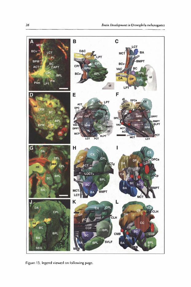
26 Brain Development in Drosophila melanogaster
Figure 13, legend viewed on following page.
TheDevelopment ofthe Drosophila LarvalBrain 27
Figure 13, viewed on previous page. Compartments of the brain neuropile. Each row of panelscorresponds to one developmental stage (A-C: Stage 15 embryo; D-F: first instar larva; G-H: latethird instar larva; J-L: adult). Panels of left column (A, D, G, J) show Z-projections of confocalsections (A, D: horizontal; G, J: frontal) in which neuropile is labeled green byantiDN-cadherin(A) or ChaT-GFP. The remainder of panels show 3D digital models of compartments in posteriorview (middle column; B, E, H, K; lateral to the right) and medial/sagittal view (right column; F,I, L); panel C of right column represents dorsal view (anterior to the top). Each compartmentis consistently shown in its own color. Fasll-positive tracts are rendered dark grey. In E and F,compartments are rendered semi-transparent to allow for clearer view of tracts. In H, I, K, L,the baso-cervical, baso-posterior medial and centro-posterior medial/centro-posterior intermediate compartments and their adult counterparts are omitted from the models for clarity. Inmedial views (F, I, L), red ovals demarcate position of commissural tracts. Note formation ofcentral complex compartments (CC in L)that expands within the space flanked by commissures.Abbreviations: ACO, antennal commissure; ACT, antenno-cerebral tract; BA, baso-anterior (antennal) compartment; BC, baso-central compartment; BCv, baso-cervical compartment; BLPT,baso-Iateral protocerebral tract; BMPT, baso-medial protocerebral tract; BPL, baso-posteriorlateral compartment; BPM, base-posterior medial compartment; CA, centro-anterior compartment; CAPT, centro-anterior protocerebral tract; CC, central complex; CLH, commissure ofthe lateral horn; CMB, chiasm of median bundle; COF, great commissures of optic foci; CPI,centro-posterior intermediate compartment; CPL, centro-posterior lateral compartment; CPM,centro-posterior medial compartment; CX, calyx compartment; DA, dorso-anterior compartment; DAC, dorso-anteriorcommissures; dl, dorsal lobe of mushroom body; DP,dorso-posteriorcompartment; DPCa, DPCp, anterior and posterior component of dorso-posterior commissures;DPPT, dorso-posterior protocerebral tract; LCT, lateral cervical tract; LOCTd, dorsal larval opticcommissural tract; LOCTw ventral larval optic commissural tract; LPT,lateral protocerebral tract;MCT, medial cervical tract; MEB, median bundle; ml, medial lobe of mushroom body; 01, opticlobe; P1, P4m/1 Fasll positive pioneer clusters; PCT, posterior cervical tract; ped, peduncle ofmushroom body; SEG, subesophageal ganglion; SVLF, sub-esophageal ventro-Iateral fascicle;VAC, ventro-anterior commissures; VPC, ventro-posterior commissures. Bars: 10 urn (A-F); 30urn (G-L)
(Fig. 12D),26and in these cases, the term for the larval tract shown in Figure 12E-L is followed bythe term describing the corresponding adult tract.
I>euterocerebrunnPrimary lineages falling into the realm of the deuterocerebrum, as defined by the expression
domain ofengrailed, comprise the BA group (Fig. 12A-C,E). It is possible that the CM lineages(Fig. 12A-C,F), located at a postero-medial-basal position, also have a deuterocerebral origin,although this needs to be confirmed by future lineage tracing. The axon bundles formed by thesetwo groups of lineages are oriented preferentially along the antero-posterior axis, BA bundlesgrowing posteriorly, CM bundles anteriorly (Fig. 12E,F). In addition, many BA and CM lineagesemit collateral transverse fiber tracts that cross in the brain commissures and/or contribute to theprimordium ofthe central complex (Fig. 12E,F). The BPM compartment appears as an elongatedneuropile domain surrounding these deuterocerebral axon tracts (Fig. 13A-F). The BA ( = antennal) compartment represents the anterior part ofthe deuterocerebral neuromere. It evolves as ananterior "alcove"ofthe BPM, pioneered by the PATsofthe medial BA lineages (Figs.12E, 13C,F).Branches offibers around the MCT tract develop into the BCv compartment (Fig. 13B,C) whichis located medial to the BPM.
Major axon tracts formed by BA lineages are the antennocerebral tract that connects the deuterocerebrum to the calyx of the mushroom body and the antennal commissure in between theantennallobes (BA compartments; Fig. 12E). Both of these connections grow into prominentfiber bundles of the adult brain. The prominent larvalloC fiber system, formed by CM lineagesas well as BA lineages, most likely gives rise to the posterior tract of the ventral body describedfor the adult flybrain (Fig. 12E,F).26 Commissural branches ofthe loC grow towards the primordium ofthe central complex (see below) and cross in the commissures closelyassociated with this
28 Brain Development in Drosophila melanogaster
structure, called VAC3/4 in the larva and sub-ellipsoid commissure/interventral body connectivein the adult.26
Lineages ofthe Deutero-Protocerebral Border RegionTwo engrailed expressing lineages derived from the engrailed "head spot" define the boundary
between deuterocerebrum and protocerebrum in the late embryo and larva (Fig. 12B,C).52,59 In thelate larva, one ofthese en-positive lineages corresponds to DALv;3 the other one shifts dorsally andforms the D PLam lineage." We define the region flanked by horizontal lines drawn through thesetwo lineages as the "deutero-protocerebral boundary region". Two groups oflineages, DAM andDAL, fall within this boundary region. Axon tracts of the DAM lineages are primarily orientedpostero-medially, crossing in the ventral part ofthe brain commissure; branching of these axonsform the major contribution of the CA and DA compartments ofthe brain (Figs.12G; 13A-C).Among the DAM neurons are some that have axons descending into the subesophageal ganglionvia the median bundle (Fig. 12G; see also Fig. 4 for Dil labeled DAM neuron). Larval SATsofthe DAM group also form part ofthe dorsomedian longitudinal tract (loDM) that interconnectsanterior and posterior domains within the medial brain (Fig. 12G). This fiber system may wellconstitute the forerunner ofthe dorsal horizontal tract ofthe adult brain."
Tracts ofthe DAL lineages are closelyassociated with the lobes and peduncle ofthe mushroombody (Fig. 12H); they form a major part ofthe "circumferential mushroom body systems" ofthelarval brain (Fig. IIB,D). Most SATsof the DAL lineages pass through and/or terminate in theBC compartment ( = ventral body in the adult brain) and the primordium ofthe central complex(trCM, DPCl; Fig. 12H). A number of tracts cross dorsal of the central complex primordium(DAC3, the future arched commissure ofthe ventral body) or ventral ofit (VAC3 /4; sub-ellipsoidcommissure/inter ventral body connective). These trajectories support the idea that many oftheneurons that innervate the central complex and interconnect this structure with the ventral bodyare derivatives of the DAL lineages.
Several DAL tracts have ventrally directed branches that follow the central anterior protocerebral tract (CAPT) which is established already in the late embryo.52,57 We surmise that this connection between brain (Le.,supraesophageal ganglion) and ventral nerve cord (i.e., subesophagealganglion) will develop into the fiber bundle called subesophageal-ventro-lateral tract in the adult."The contribution ofascending and descending fibers to this fiber system has not been investigatedin detail. It seems likely that many ofthe lateral deuterocerebral descending neurons identified byStrausfeld and collaborators are derivatives ofthe DAL lineages."
Lineages ofthe Dorsal ProtocerebrumTwo groups of lineages, DPM and DPL, occupy the dorsal surface of the brain. Their axon
tracts form the bulkofcommissural and longitudinal connections ofthe dorsal protocerebrum thatconstitute the D P and CPL compartments (differentiated into superior-medial and inferior-medialprotocerebrum, superior-lateral and inferior lateral protocerebrum and lateral horn in the adultbrain; Figs. lIB; 12A,I,J; 13C,G,J).26 The tracts formed by the DPLc and DPLllineages thatpenetrate into the protocerebrum in ventro-medial direction are likely the forerunners of the socalled "medial and lateral intracerebral cascades"ofthe adult brain (Fig. 12]).26 The longitudinallydirected fiber massesproduced by D PM and D PL lineages,Le., the loD M and loD L, foreshadow thedorsal horizontal tract and lateral dorsal horizontal tract ofthe adult protocerebrum (Figs.11B,D;121,J).26 The most conspicuous commissure ofthe adult dorsal protocerebrum is the commissure ofthe lateral horn, in addition to a number ofunnamed commissural tracts posterior to this system.These fiber systems are likely descended from the DPC2/3 commissures that are formed in thelarva by SATsof (among others) the DPM and DPL lineages (Fig. 13H,K).
Lateral Protocerebral LineagesLateral protocerebrallineages (BL) have tracts that radially converge towards each other and
form the BPL compartment (Figs.llA; 12K; 13A-C). In the larva, the SATsof the BL lineagesform the system ofexternal tracts (exBL,exDL, exVT; Figs. 11C, D; 12K). The BPL compartment
The Development oftheDrosophila LarvalBrain 29
undergoes enormous growth and diversification duringmetamorphosis, mainly due to the in-growthofaxons from the lobula of the optic lobe (compare BPL in Fig. 13G and]). BL lineage-derivedneurons along with the optic lobe afferents form the so called optic foci of the adult brain.P Theexternal fiber systemsofthe larva develop into the longitudinal and vertical connections in betweenoptic foci. Two transverse systems, the trDLand trBL, carry branches ofBL SATs.The trBL and itscontinuation, VPC2, can easilybe recognized as the forerunner ofthe large commissure ofthe opticfoci defined for the adult brain (Fig. 11C,D; 13K).26The fate ofthe trDL, a massive fiber systemofthe larval dorso-lateral protocerebrum (Fig. 11C), is unclear.
Lineages ofthe Posterior ProtocerebrumLineages located at the posterior pole of the brain, including the mushroom body (MB) and
CP lineages, project anteriorly and interconnect the dorsal protocerebrum with the deutero-protocerebral boundary region and the deuterocerebrum (Fig. 12A,B,L). The central compartmentsofthe neuropile, CPM, CPI and the mushroom body, arise as cylindrical domains around the axontracts ofthese posterior lineages (Figs. 12B; 13A-F). MB lineages give rise to the calyx, peduncleand lobes ofthe mushroom body whose development has been studied in detail in several recentpapers.27-30,62,63 CP lineages are close to the mushroom body and give rise to two main fiber systems.One is directed anteriorly, parallel to the peduncle and appears to end close to the BC compartment (ventral body). The other crosses over the proximal peduncle and then turns anteriorly andmedially towards the primordium ofthe central complex. While crossing, these axons (the trPd)follow the trajectory that is taken by the commissure ofthe lateral horn (Figs. lID; 12L).
OutlookAdressing fundamental problems ofneurobiology in Drosophila offers many advantages, aswell
as some disadvantages. Among the latter is the small size ofneurons and the fact that establishing functional relationships between neurons, recordings have to be made from often minusculeneurites within the neuropile, rather than nerve cell somata. Due to these anatomical features(shared among many invertebrate animals) we still know very little about synaptic relationshipsbetween neurons in the central nervous system. On the other hand, thanks to the abundance ofmolecular markers and genetic techniques, it is now possible to map individual cell types of theDrosophila nervous system throughout development with an a.ccuracythat is unparalleled in theanimal kingdom, with the possible exception of the nematode C. elegans. Recent work summarized in this chapter has started to lay the groundwork to map neurons and their connectionsrelative to discrete landmarks, such as neuropile compartments and lineage tracts. Given sufficiententhusiasm and support from the neurobiological community, the goal ofreconstructingneuronalcircuitry in its entirety, rather than in a few representative neuronal subsets like the mushroombody, is within reach.
AcknowledgementsThis work was supported by NIH Grant RO1 NS29367 to V.H.
References1. Urbach R, Technau GM. Molecular markers for identified neuroblasts in the developing brain of Dro
sophila. Development 2003; 130(16):3621-3637.2. Younossi-Hartenstein A, Nassif C, Green P et al. Early neurogenesis of the Drosophila brain. J Comp
Neurol 1996; 370(3):313-329.3. Hartenstein V, Rudloff E, Campos-Ortega JA. The pattern of proliferation of the neuroblasts in the
wild-type embryo of Drosophila melanogaster. Dev Genes Evo11987; 196:473-485.4. Brody T, Odenwald WE Cellular diversity in the developing nervous system: a temporal view from
Drosophila. Development 2002; 129(16):3763-3770.5. Ito K, Horta Y. Proliferation pattern of postembryonic neuroblasts in the brain of Drosophila melano
gaster. Dev BioI 1992; 149(1):134-148.6. Ceron J,Gonzalez C, Tejedor FJ. Patterns of cell division and expression of asymmetric cell fate deter
minants in postembryonic neuroblast lineages of Drosophila. Dev Bioi 2001; 230(2):125-138.
30 Brain Development in Drosophila melanogaster
7. Dumstrei K, Wang F, Hartenstein V. Role of DE-cadherin in neuroblast proliferation, neural morphogenesis and axon tract formation in Drosophila larval brain development. ] Neurosci 2003;23(8):3325-3335.
8. Dumstrei K, Wang F, Nassif C et al. Early development of the Drosophila brain: V. Pattern of postembryonic neuronal lineages expressing DE-cadherin.] Comp Neuro12003; 455(4):451-462.
9. Rolls MM, Albertson R, Shih HP et al. Drosophila aPKC regulates cell polarity and cell proliferationin neuroblasts and epithelia.] Cell Bioi 2003; 163(5):1089-1098.
10. Schaefer M, Shevchenko A, Knoblich ]A et al. A protein complex containing Inscuteable andthe Galpha-binding protein Pins orients asymmetric cell divisions in Drosophila. Curr Biol 2000;10(7):353-362.
11. Schober M, Schaefer M, Knoblich ]A. Bazooka recruits Inscuteable to orient asymmetric cell divisionsin Drosophila neuroblasts. Nature 1999; 402(6761):548-551.
12. Wodarz A, Ramrath A, Kuchinke U et al. Bazooka provides an apical cue for Inscuteable localizationin Drosophila neuroblasts. Nature 1999; 402(6761):544-547.
13. Maurange C, Gould AP. Brainy but not too brainy: starting and stopping neuroblast divisions in Drosophila. Trends Neurosci 2005; 28(1):30-36.
14. Isshiki T, Pearson B, Holbrook S et al. Drosophila neuroblasts sequentially express transcription factorswhich specify the temporal identity of their neuronal progeny. Cell 2001; 106(4):511-521.
15. Bilder D. Cell polarity: squaring the circle. Curr Bioi 2001; 11(4):R132-135.16. Doe CQ Bowerman B. Asymmetric cell division: fly neuroblast meets worm zygote. Cure Opin Cell
BioI 2001; 13(1):68-75.17. Ebens A], Garren H, Cheyette BN et al. The Drosophila anachronism locus: a glycoprotein secreted by
glia inhibits neuroblast proliferation. Cell 1993; 74(1):15-27.18. Bello BC, Hirth F, Gould AP. A pulse of the Drosophila Hox protein Abdominal-A schedules the end
of neural proliferation via neuroblast apoptosis. Neuron 2003; 37(2):209-219.19. Voigt A, Pflanz R, SchaferU et ale Perlecanparticipates in proliferation activation of quiescent Drosophila
neuroblasts. Dev Dyn 2002; 224(4):403-412.20. Park Y: Rangel C, Reynolds MM er al. Drosophila perlecan modulates FGF and hedgehog signals to
activate neural stem cell division. Dev Bioi 2003; 253(2):247-257.21. Datta S. Control of proliferation activation in quiescent neuroblasts of the Drosophila central nervous
system. Development 1995; 121(4):1173-1182.22. Caldwell MC, Datta S. Expression of cyclin E or DP/E2F rescues the G1 arrest of trol mutant neuro
blasts in the Drosophila larval central nervous system. Mech Dev 1998; 79(1-2):121-130.23. Park Y, Ng C, Datta S. Induction of string rescues the neuroblast proliferation defect in trol mutant
animals. Genesis 2003; 36(4):187-195.24. Almeida MS, Bray S]. Regulation of post-embryonic neuroblasts by Drosophila Grainyhead. Mech Dev
2005; 122(12):1282-1293.25. Akong K, Grevengoed EE, Price MH et ale Drosophila APC2 and APC1 play overlapping roles in
wingless signaling in the embryo and imaginal discs. Dev Biol 2002; 250(1):91-100.26. Strausfeld N]. Altas of an Insect Brain. Springer, 1976.27. Crittenden ]R, Skoulakis EM, Han KA et al. Tripartite mushroom body architecture revealed by anti
genic markers. Learn Mem 1998; 5(1-2):38-51.28. Ito K, Awano~ Suzuki K et al. The Drosophila mushroom body is a quadruple structure of clonal
units each of which contains a virtually identical set of neurones and glial cells. Development 1997;124(4):761-771.
29. Noveen A, Daniel A, Hartenstein V. Early development of the Drosophila mushroom body: the rolesof eyelessand dachshund. Development 2000; 127(16):3475-3488.
30. Tettamanti M, Armstrong ]D, Endo K et al. Early development of the Drosophila mushroom bodies,brain centres for associative learning and memory. Dev Genes Evo11997; 207(4):242-252.
31. Ito K, Suzuki K, Estes P et al. The organization of extrinsic neurons and their implications in thefunctional roles of the mushroom bodies in Drosophila melanogaster Meigen. Learn Mem 1998;5(1-2):52-77.
32. Bossing T, Udolph G, Doe CQ et al. The embryonic central nervous system lineages of Drosophilamelanogaster. I. Neuroblast lineagesderived from the ventral half of the neuroectoderm. Dev Bioi 1996;179(1):41-64.
33. Schmidt H, Rickert C, Bossing T et al. The embryonic central nervous system lineages of Drosophilamelanogaster. II. Neuroblast lineagesderived from the dorsal part of the neuroectoderm. Dev Biol 1997;189(2):186-204.
34. Prokop A, Meinertzhagen IA. Development and structure of synaptic contacts in Drosophila. SeminCell Dev BioI 2006; 17(1):20-30.
TheDevelopmentofthe Drosophila Larval Brain 31
35. Sanchez-Soriano N, Bottenberg w: Fiala A et al. Are dendrites in Drosophila homologous to vertebratedendrites? Dev Bioi 2005; 288(1):126-138.
36. Clark IE, Jan L~ Jan YN. Reciprocal localization of Nod and kinesin fusion proteins indicates microtubule polarity in the Drosophila oocyte, epithelium, neuron and muscle. Development 1997;124(2):461-470.
37. Chang T, Younossi-Hartenstein A, Hartenstein V. Development of neural lineages derived &om the sineoculis positive eye field of Drosophila. Arhtropod Stru Dev 2003; 32:303-317.
38. Pereanu w: Hartenstein V. Neural lineagesof the Drosophila brain: a three-dimensional digital atlas of thepattern of lineage location and projection at the late larval stage. J Neurosci 2006; 26(20):5534-5553.
39. Cantera R. Glial cells in adult and developing prothoracic ganglion of the hawk moth Manduca sexta,Cell Tiss Res 1993; 272:93-108.
40. Hoyle G. Glial cells of an insect ganglion. J Comp Neuro11986; 246(1):85-103.41. Ito K, Urbach R, Technau GM. Distribution, classification and development of Drosophila glia cells in
the late embryonic and early larval ventral nerve cord. Dev Genes Evol 1995; 204:284-307.42. Saint Marie RL, Carlson SD, Chi C. The Glial Cells of Insects. New York, NY: Plenum 1984.43. Lane N. Insect intercellular junctions: Their structure and development. New York: Plenum 1982.44. Hoyle G, Williams M, Phillips C. Functional morphology of insect neuronal cell-surface/glial contacts:
the trophospongium. J Comp Neurol 1986; 246( 1):113-128.45. Hidalgo A. Neuron-glia interactions during axon guidance in Drosophila. Biochem Soc Trans 2003;
31(Pt 1):50-55.46. Pielage J, Klambt C. Glial cells aid axonal target selection. Trends Neurosci 2001; 24(8):432-433.47. Sepp KJ, Auld VJ. Reciprocal interactions between neurons and glia are required for Drosophila peripheral
nervous system development. J Neurosci 2003; 23(23):8221-8230.48. Sepp KJ, Schulte J, Auld V]. Peripheral glia direct axon guidance across the CNS/PNS transition zone.
Dev BioI 2001; 238(1):47-63.49. Oland LA, Tolbert LP. Key interactions between neurons and glial cells during neural development in
insects. Annu Rev Entomol2003; 48:89-110.50. Hartenstein V, Nassif C, Lekven A. Embryonic development of the Drosophila brain. II. Pattern of
glial cells. J Comp Neuro11998; 402(1):32-47.51. Pereanu~ Shy D, Hartenstein V. Morphogenesis and proliferation of the larval brain glia in Drosophila.
Dev BioI 2005; 283(1):191-203.52. Younossi-Hartenstein A, Nguyen B, Shy D et al. Embryonic origin of the Drosophila brain neuropile.
J Comp Neuro12006; 497(6):981-998.53. Pereanu ~ Spindler S, Cruz L et al. Tracheal development in the Drosophila brain is constrained by
glial cells. Dev Biol 2006.54. Manning G, Krasnow MA. Development of the Drosophila tracheal system. New York: Cold Spring
Harbor, Laboratory Press 1993.55. Englund C, Uv AE, Cantera Ret al. adrift, a novel bnl-induced Drosophila gene, required for tracheal
pathfinding into the CNS. Development 1999; 126(7):1505-1514.56. Nassif C, Noveen A, Hartenstein V. Embryonic development of the Drosophila brain. I. Pattern of
pioneer tracts. J Comp Neuro11998; 402(1):10-31.57. Nassif C, Noveen A, Hartenstein V. Early development of the Drosophila brain: III. The pattern of
neuropile founder tracts during the larval period. J Comp Neuro12003; 455(4):417-434.58. Goodman CS, Doe CQ Embryonic development of the Drosophila central nervous system. New York:
Cold Spring Harbor Laboratory Press, 1993.59. Hirth F, Kammermeier L, Frei E et al. An urbilaterian origin of the tripartite brain: developmental
genetic insights from Drosophila. Development 2003; 130(11):2365-2373.60. Therianos S, Leuzinger S, Hirth F et al. Embryonic development of the Drosophila brain: formation
of commissural and descending pathways. Development 1995; 121(11):3849-3860.61. Strausfeld NJ, Bassemir U, Singh RN et al. Organizational principles of outputs from Dipteran brains.
J Ins Physiol1984; 30(1):73-93.62. ArmstrongJD, de BelleJS, Wang Z et al. Metamorphosis of the mushroom bodies; large-scalerearrange
ments of the neural substrates for associative learning and memory in Drosophila. Learn Mem 1998;5(1-2): 102-114.
63. Zhu S, Chiang AS, Lee T. Development of the Drosophila mushroom bodies: elaboration, remodelingand spatial organization of dendrites in the calyx. Development 2003; 130(12):2603-2610.
CHAPTER 2
Anteroposterior Regionalization ofthe Brain:Genetic and Comparative AspectsRobert Lichtneckert" and Heinrich Reichert
Abstract
Developmental genetic analyses of embryonic CNS development in Drosophila haveuncovered the role of key, high-order developmental control genes in anteroposteriorregionalization ofthe brain. The gene families that have been characterized include the
otd/Otx and ems/Emx genes which are involved in specification of the anterior brain, the Hoxgenes which are involved in the differentiation of the posterior brain and the Pax genes whichare involved in the development of the anterior/posterior brain boundary zone. Taken togetherwith work on the genetic control ofmammalian CNS development, these findings indicate thatall three gene sets have evolutionarily conserved roles in brain development, revealing a surprisingevolutionary conservation in the molecular mechanisms ofbrain regionalization.
IntroductionIn most animals, the central nervous system (CNS) is characterized by bilateral symmetry
and by an elongated anteroposterior axis, both ofwhich are established very early in embryonicdevelopment. During embryogenesis, regionalized anatomical subdivisions appear along the anteroposterior axis, also referred to as the neuraxis. These subdivisions are most prominent nearthe anterior pole, where the complex structures that comprise the brain are generated. As thebrain differentiates, the neuraxis often bends and species-specific flexures arise, which in laterstages tend to distort the original anteroposterior coordinates ofthe CNS. However, when this istaken into account and the neuraxis is reconstructed, remarkable similarities in anteroposteriorregionalization ofthe CNS in animals as diverse as arthropods and vertebrates become apparent.A full appreciation ofthese similarities comes from combined comparative neuroanatomical andmolecular genetic studies carried out in Drosophila and mouse, which reveal that comparable,evolutionarily conserved developmental patterning mechanisms operate in regionalization oftheembryonic CNS.1,2
Here we review recent findings on the developmental genetic control of anteroposterior regionalization in the embryonic CNS in Drosophila and compare these findings with investigationscarried out on regionalization of the embryonic murine CNS. The similarities in the expressionpatterns of key developmental control genes together with the comparable functions of thesegenes during CNS development in flies and mice suggest a common evolutionary origin of themechanism of embryonic CNS regionalization. Given the current molecular-based phylogenyofbilaterian animals, it seems likely that these features ofbrain development in arthropods andvertebrates were already present in the common bilaterian ancestor from which protostomes anddeuterostomes evolved (Fig. 1).3 This, in turn, challenges the classical view of an independentorigin ofprotostome and deuterostome brains.
*Corresponding Author: Robert Lichtneckert-Biozentrum, University of Basel,Klingelbergstrasse 50, CH-4056 Basel, Switzerland. Email: [email protected]
BrainDevelopment in Drosophila melanogaster, edited by Gerhard M. Technau.©2008 Landes Bioscience and Springer Science+Business Media.
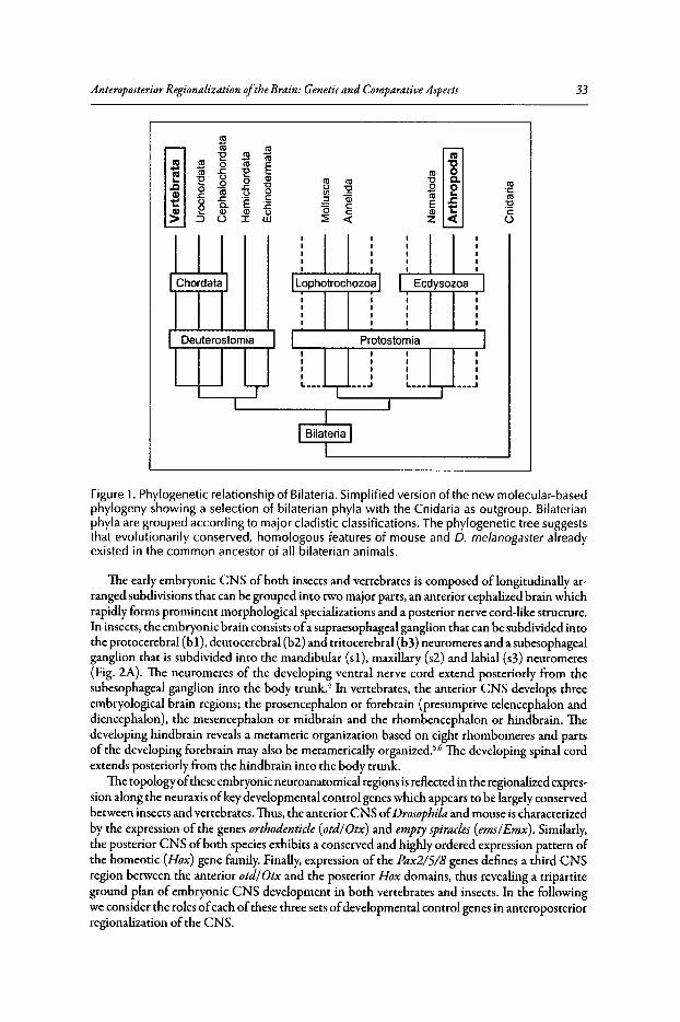
Anteroposterior Regionalization ofthe Brain: Genetic and Comparative Aspects
co16 co"E ~ 16 ca
ca ~ 0 co E "0.. co .J::. "E co 0ca Q5~ "E 0 0 co co "0 C..c 0 .J::. "0 0 0 0 coQ)
0 ro 0 0 en ~ 16 ~ s.J::. .J::. "E c~ Qi ..c::1:= 0 E "0
0 Q. :E (5 c 1:= "2~ :5 Q) Q) 0 c Q)
U :I: W ~ « z « o
33
Figure 1. Phylogenetic relationship of Bilateria. Simplified version of the new molecular-basedphylogeny showing a selection of bilaterian phyla with the Cnidaria as outgroup. Bilaterianphyla are grouped according to major cladistic classifications. The phylogenetic tree suggeststhat evolutionarily conserved, homologous features of mouse and 0" melanogaster alreadyexisted in the common ancestor of all bilaterian animals.
The early embryonic CNS of both insects and vertebrates is composed of longitudinally arranged subdivisions that can be grouped into two major parts, an anterior cephalized brain whichrapidly forms prominent morphological specializations and a posterior nerve cord-like structure.In insects, the embryonic brain consists ofa supraesophageal ganglion that can be subdivided intothe protocerebral (b1),deutocerebral (b2) and tritocerebral (b3) neuromeres and a subesophagealganglion that is subdivided into the mandibular (sL), maxillary (s2) and labial (s3) neuromeres(Fig. 2A). The neuromeres of the developing ventral nerve cord extend posteriorly from thesubesophageal ganglion into the body trunk." In vertebrates, the anterior CNS develops threeembryological brain regions; the prosencephalon or forebrain (presumptive telencephalon anddiencephalon), the mesencephalon or midbrain and the rhombencephalon or hindbrain. Thedeveloping hindbrain reveals a metameric organization based on eight rhombomeres and partsof the developing forebrain may also be metamerically organized.V The developing spinal cordextends posteriorly from the hindbrain into the body trunk.
The topologyofthese embryonic neuroanatomical regions is reflected in the regionalized expression along the neuraxis ofkey developmental control genes which appears to be largely conservedbetween insects and vertebrates. Thus, the anterior CNS ofDrosophila and mouse is characterizedby the expression of the genes ortbodenticle (otdIOtx) and emptyspiracles (ems/Emx). Similarly,the posterior CNS ofboth species exhibits a conserved and highly ordered expression pattern ofthe homeotic (Hox) gene family. Finally, expression of the Pax2/5/8 genes defines a third CNSregion between the anterior otdl Otx and the posterior Hox domains, thus revealing a tripartiteground plan of embryonic CNS development in both vertebrates and insects. In the followingwe consider the roles ofeach ofthese three sets ofdevelopmental control genes in anteroposteriorregionalization of the CNS.
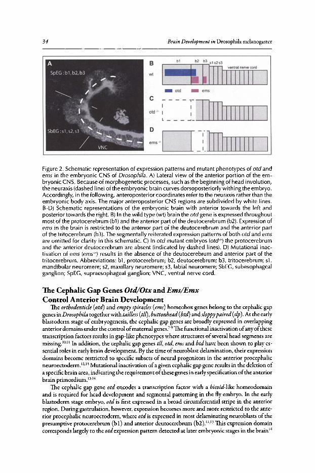
34 Brain Development in Drosophila melanogascec
C - - - - ~I__~Old " _~
DD--~erns" _ :.JJlWlWLJ
_ ems
b1
_ old
b2 b3 515 253
····MIIIbTIfIitJB
wt
Figure 2, Schematic representation of expression patterns and mutant phenotypes of otd andems in the embryonic CNS of Drosophila . A) Lateral view of the anterior portion of the embryonic CNS. Because of morphogenetic processes, such as the beginning of head involution,the neuraxis (dashed line) of the embryonic brain curves dorsoposteriorly withing the embryo.Accordingly, in the following, anteroposterior coordinates refer to the neuraxis rather than theembryonic body axis. The major anteroposterior CNS regions are subdivided by white lines.B-D) Schematic representations of the embryonic brain with anterior towards the left andposterior towards the right. B) In the wild type (wt) brain the otd gene is expressed throughoutmost of the protocerebrum (b1)and the anterior part of the deutocerebrum (b2). Expression ofems in the brain is restricted to the anterior part of the deutocerebrum and the anterior partof the tritocerebrum (b3). The segmentally reiterated expression patterns of both otd and emsare omitted for clarity in this schematic. C) In otd mutant embryos (otd '!') the protocerebrumand the anterior deutocerebrum are absent (indicated by dashed lines). D) Mutational inactivation of ems (ems'!') results in the absence of the deutocerebrum and anterior part of thetritocerebrum. Abbreviations: bl , protocerebrum; b2, deutocerebrum; b3, tritocerebrum; sl ,mandibular neuromere; 52, maxillary neuromere; s3, labial neuromere; SbEC, subesophagealganglion; SpEC, supraesophageal ganglion; VNC, ventral nerve cord.
The Cephalic Gap Genes Otd/Otx and Ems/EmxControl Anterior Brain Development
The ortbodenticle(otd) and emptyspiracles (ems) homeobox genes belong to the cephalic gapgenes in Drosophila together with tailless (tll),buttonhead (btd)and sloppypaired(sip) .At the earlyblastoderm stage of embryogenesis, the cephalic gap genes are broadly expressed in overlappinganterior domains under the control ofmaternal genes."?The functional inactivation ofanyofthesetranscription factors results in gap-like phenotypes where structures ofseveral head segments aremissing.'?'!' In addition, the cephalic gap genes tll, otd, emsand btdhave been shown to play essential roles in early brain development. By the time ofneuroblast delamination, their expressiondomains become restricted to specific subsets of neural progenitors in the anterior procephalicneuroectoderrn.P'!' Mutational inactivation ofa given cephalic gap gene results in the deletion ofa specificbrain area, indicating the requirement ofthese genes in earlyspecification ofthe anteriorbrain prirnordium.P'"
The cephalic gap gene otd encodes a transcription factor with a bicoid-like homeodomainand is required for head development and segmental patterning in the fly embryo . In the earlyblastoderm stage embryo, otd is first expressed in a broad circumferential stripe in the anteriorregion. During gastrulation, however, expression becomes more and more restricted to the anterior procephalic neuroectoderm, where otd is expressed in most delaminating neuroblasts of thepresumptive protocerebrum (b 1) and anterior deucocerebrum (b2 ).12.13 This expression domaincorresponds largely to the otdexpression pattern detected at later embryonic stages in the brain 14
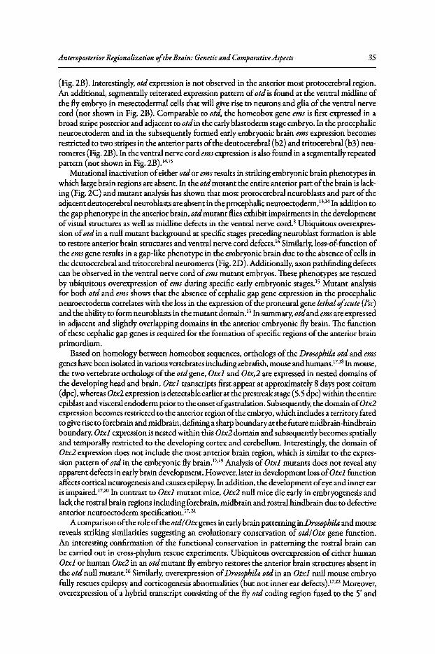
AnteroposteriorRegionalizationoftheBrain: Genetic and Comparative Aspects 35
(Fig. 2B). Interestingly, otd expression is not observed in the anterior most protocerebral region.An additional, segmentally reiterated expression pattern ofotd is found at the ventral midline ofthe fly embryo in mesectodermal cells that willgive rise to neurons and glia of the ventral nervecord (not shown in Fig. 2B). Comparable to otd, the homeobox gene ems is first expressed in abroad stripe posterior and adjacent to otd in the early blastoderm stage embryo. In the procephalicneuroectoderm and in the subsequently formed early embryonic brain ems expression becomesrestricted to two stripes in the anterior parts ofthe deutocerebral (b2) and tritocerebral (b3) neuromeres (Fig. 2B). In the ventral nerve cord emsexpression is also found in a segmentally repeatedpattern (not shown in Fig. 2B).14.1S
Mutational inactivation ofeither otd or ems results in striking embryonic brain phenotypes inwhich large brain regions are absent. In the otd mutant the entire anterior part ofthe brain is lacking (Fig. 2C) and mutant analysis has shown that most protocerebral neuroblasts and part oftheadjacent deutocerebral neuroblasts are absent in the procephalic neuroectoderm.P'" In addition tothe gap phenotype in the anterior brain, otd mutant flies exhibit impairments in the developmentofvisual structures as well as midline defects in the ventral nerve cord," Ubiquitous overexpression ofotd in a null mutant background at specific stages preceding neuroblast formation is ableto restore anterior brain structures and ventral nerve cord defects." Similarly, loss-of-function ofthe emsgene results in a gap-like phenotype in the embryonic brain due to the absence ofcells inthe deutocerebral and tritocerebral neuromeres (Fig. 2D). Additionally, axon pathfinding defectscan be observed in the ventral nerve cord ofems mutant embryos. These phenotypes are rescuedby ubiquitous overexpression of ems during specific early embryonic stages. IS Mutant analysisfor both otd and ems shows that the absence of cephalic gap gene expression in the procephalicneuroectoderm correlates with the loss in the expression ofthe proneural gene lethalofscute (lsc)and the ability to form neuroblasts in the mutant domain.13In summary, otd and emsare expressedin adjacent and slightly overlapping domains in the anterior embryonic fly brain. The functionof these cephalic gap genes is required for the formation of specific regions of the anterior brainprimordium.
Based on homology between homeobox sequences, orthologs of the Drosophilaotd and emsgenes have been isolated in various vertebrates includingzebrafish, mouse and humans.17.18 In mouse,the two vertebrate orthologs ofthe otd gene, Otxl and Otx,2 are expressed in nested domains ofthe developing head and brain. Otxl transcripts first appear at approximately 8 days post coitum(dpc), whereas Otx2 expression is detectable earlier at the prestreak stage (5.5 dpc) within the entireepiblast andvisceral endoderm prior to the onset ofgastrulation. Subsequently, the domain ofOtx2expression becomes restricted to the anterior region ofthe embryo, which includes a territory fatedto give rise to forebrain and midbrain, defining a sharp boundary at the future midbrain-hindbrainboundary. Otxl expression is nested within this Otx2 domain and subsequently becomes spatiallyand temporally restricted to the developing cortex and cerebellum. Interestingly, the domain ofOtx2 expression does not include the most anterior brain region, which is similar to the expression pattern ofotd in the embryonic fly brain.F'" Analysis of Otxl mutants does not reveal anyapparent defects in early brain development. However, later in development loss ofOtxl functionaffects cortical neurogenesis and causes epilepsy. In addition, the development ofeye and inner earis impaired.Fr" In contrast to Otxl mutant mice, Otx2 null mice die early in embryogenesis andlack the rostral brain regions including forebrain, midbrain and rostral hindbrain due to defectiveanterior neuroectoderm specification.17. 21
A comparison ofthe role ofthe otdf Otx genes in early brain patterning in Drosophila and mousereveals striking similarities suggesting an evolutionary conservation of otdlOtx gene function.An interesting confirmation of the functional conservation in patterning the rostral brain canbe carried out in cross-phylum rescue experiments. Ubiquitous overexpression ofeither humanOtxl or human Otx2 in an otd mutant fly embryo restores the anterior brain structures absent inthe otd null mutant." Similarly, overexpression ofDrosophilaotd in an Otxl null mouse embryofully rescues epilepsy and corticogenesis abnormalities (but not inner ear defects).17.22 Moreover,overexpression of a hybrid transcript consisting of the fly otd coding region fused to the Sf and
36 Brain Development in Drosophila melanogaster
3' UTRs of Otx2 restores the anterior brain patterning in Otx2 null mutant mice including thenormal positioning ofthe midbrain-hindbrain boundary."
Asis the case for the otdlOtxgenes, two vertebrate orthologs ofthe Drosophila emsgene, EmxlandEmx2, have been identified. Emxl andEmx2 expression in the mouse CNS is restricted to theforebrain, where largely overlapping expression patterns are seen. Whereas, Emxl expression onlybegins after neurulation, Emx2 is already detectable around 8.Sdpc in the rostral neural plate. 19,24,25
Within the developing neocortex, Emx2 isexpressed in a high caudomedial to low rostrolateral gradient, which iscontrasted by an opposed gradient ofPax6gene expression. Mutational inactivationofEmx2 results in an expansion ofthe rostrolateral brain areas at the expense ofthe caudomedialneocortical areas.An opposite shift in regional identity is seen in the Pax6Ioss-of-function mutant.In the Emx2 and Pax6double mutant, the cerebral cortex completely loses its identity and insteadacquires characteristics ofbasal ganglia.26.27 WhereasEmx2 mutant mice die immediately after birth,Emxl mutant animals are postnatal viable and show rather subtle phenotypes that are restrictedto the forebrain.28
,29 The regionalized expression patterns ofthe ems/Emx genes in the developingbrain ofDrosophila and mouse are remarkably similar, as is their ability to confer regional identityto the cellsofa specific domain in the brain. Moreover, overexpression ofa mouse Emx2 transgenein an emsmutant background can rescue the brain phenotype offlyembryos. 15 Takentogether, thesimilar spatiotemporal expression patterns and the high degree offunctional equivalence betweenDrosophila and mouse suggest an evolutionarily conserved role ofthe ems/Emx and otdlOtx genesin anterior brain development.
The Hox Genes Pattern the Posterior BrainThe homeotic or Hoxgenes,encodinghomeodomain transcription factors, were first discovered
ascrucial regulators ofanteroposterior segment identity in the ectodermofDrosophila melanogaster.Subsequently, Hox genes were found in a wide range ofspecies where they have essential roles inmany aspects ofanteroposterior body axispatterning.30,31 In Drosophila, the Hox genes are arrangedalong the chromosome in two gene clusters known as the Antennapedia(ANT- C)and Bitborax(BX-C)complexes. TheANT-Ccontains the fivemore anteriorly expressedHox genes: labial(lab),proboscipedia (Pb), Deformed(Dfd), Sexcombs reduced (SO') andAntennapedia(Antp). The BX-Ccontains the three posteriorly expressed genes: Ultrabitborax (Ubx), abdominal-A (abd-A) andAbdominal-B(Abd-B).Interestingly, there exists a correlation between the relative position ofthegenes within the cluster and their spatial and temporal expression pattern along the body axis;geneslocated towards the 3' end of the cluster are expressed more anteriorly and earlier in the embryothan are genes located towards the S' end. This correlation has been termed spatial and temporalcolinearity." In mammals, Hox genes are arranged into four chromosomal clusters, termed HoxA-D, which contain between 9 and 11 Hox genes that can be assigned to 13 paralogous groups.Only the Hox B cluster comprises orthologs of all Drosophila homeotic genes. As in Drosophila,spatial and temporal colinearity is also observed among vertebrate Hox genes and more posterioracting genes impose their developmental specificities upon anterior acting genes.32
,33
Hox gene expression in the developing CNS is a shared feature of a wide range ofbilateriananimals, including protostomes such as insects or annelids and deuterostomes, such as hemichordates or vertebrates.r':" Remarkably, throughout the Bilateria, Hox gene orthologs are expressedin a similar anteroposterior order. In Drosophila, the expressions ofHox cluster genes delineatediscrete domains in the embryonic brain and ventral nerve cord (Fig. 3A). Their anterior expressionboundaries often coincide with morphologically defined neuromere compartment boundaries.Although the anteroposterior order ofHox gene expression domains largely follows the spatialcolinearity rule known from ectodermal structures, one important difference is noteworthy:expression ofthe two 3'-most Hox genes ofthe ANT-C is inverted, in that the anterior expressionboundary oflab lies posterior to that ofpb.34 Interestingly, this particularity ofthe Hox expressionpattern in the CNS is common to fly and mouse. In vertebrates, Hox genes are expressed in thedeveloping hindbrain and spinal cord. The relative anteroposterior order ofHox gene expressionin the CNS ofvertebrates is virtually identical to their arrangement in Drosophila, including the
AnteroposteriorRegionalizationoftheBrain: Genetic and Comparative Aspects 37
inverted order ofthe lab and pb orthologs, Hoxb-l and Hoxb-2 (Fig.3B).38 As more expression datafrom different protostome and deuterostome speciesbecomes available, the ordered expression ofHox genes along the anteroposterior axis ofthe developing nervous system is likely to consolidateas a common feature ofbilaterian animals.
In Drosophila,mutational inactivation ofeither ofthe homeotic genes lab or Dfd causes severeaxonal patterning defects in the embryonic brain. 34In lab null mutants, axonal projection defectsare observed in the posterior tritocerebrum where lab is expressed in the wild type brain. In themutant, longitudinal pathways connecting supraesophageal and subesophageal ganglia as well asprojections in the tritocerebral commissure are absent or reduced. These brain defects are not dueto deletions in the affected neuromere; neuronal progenitors are present and give rise to progenyin the mutant domain. However, these postmitotic progeny fail to acquire a neuronal identity,as indicated by the absence of neuronal markers and the lack ofaxonal and dendritic extensions(Fig. 3A). Comparable defects are seen inDfdmutants in the correspondingmandibularlanteriormaxillary domain, where the gene is expressed in the wild type brain." Thus, the activity of thehomeotic genes lab and Dfd is necessary to establish regionalized neuronal identity in the brainofDrosophila.
The mouse lab orthologs, Hoxa-l and Hoxb-l, are expressed in overlapping domains with asharp anterior boundary coinciding with the presumptive rhombomere 3/4 border. Functionalinactivation ofHoxa-l results in segmentation defects leading to a reduced size of rhombomeres4 and S and defects in motor neuron axonal projections but the normal identity ofrhombomere4 is not altered.'? In contrast, loss ofHoxb-l function has no influence on the sizeofrhombomere4 but causesa partial transformation into a rhombomere 2 Identity," The Hoxa-L, Hoxb-l doublemutant results in a territory ofunknown identity and reduced sizebetween rhombomeres 3 and S,suggestinga synergistic action ofthe two genes in rhombomere 4 specification (Fig. 3B).39 Thus, theconcerted activity ofHoxa-l and Hoxb-l has a similar role in the specification ofthe regionalizedneuronal identity as does their ortholog lab in the CNS ofDrosophila.This suggests a functionalconservation ofHox genes, in addition to a similar mode of expression, during nervous systemdevelopment ofbilaterian animals and supports the idea ofa common origin ofthe CNS.
Evidence for a Tripartite Organization ofthe BrainComparative gene expression studies, as reviewed here for Drosophila and mouse, have been
carried out in numerous protostome and deuterostome phyla.36.41-44 The subdivision of the developing brain into an anterior region specified by genes of the otdlOtx family and a posteriorregion specified by genes ofthe Hox family appears to be a universal feature ofbilaterian animals.In vertebrates and urochordates, a third embryonic domain along the anteroposterior neuraxis,characterized by overlapping expression ofthe Pax2, Pax5 and Pax8 genes, is located between theanterior Otx and the posterior Hox expressing regions of the embryonic brain. 4s-47In vertebratebrain development, this Pax2/5/8 domain is located between the presumptive mesencephalon andmetencephalon, where it plays a crucial role in development ofthe midbrain-hindbrain boundary(MHB) region or isthmus. Transplantation experiments, in which MHB tissue grafts are insertedto more rostral or caudal brain regions inducing ectopic mesencephalic-metencephalic structures,reveal an organizer function of the MHB. This organizer activity on the surrounding neural tissue is thought to be mediated by fibroblast growth factor 8 (Fgf8) and Wntl proteins, which aresecreted by cells located in the MHB.4s.47 In early embryonic development ofthe vertebrate CNS,the homeobox gene Gbx2 isexpressed in the anterior hindbrain just posterior to the Otx2 domainin the forebrain and midbrain. During gastrulation and early neurulation the MHB is establishedat the Otx21Gbx2 interface, where subsequently the expression domains ofother MHB markersincluding Pax2/5/8, Fgf8, Wntl and Enl/2 are positioned (Fig. 4C). The two homeobox genesOtx2 and Gbx2 mutually repress one another and upregulation or downregulation ofeither geneshifts the position ofthe MHB accordingly.4S.47 Therefore, in vertebrates an antagonistic interactionbetween Otx2 and Gbx2 duringearlyembryonic development is involved in the correct positioningofthe MHB at their common interface.
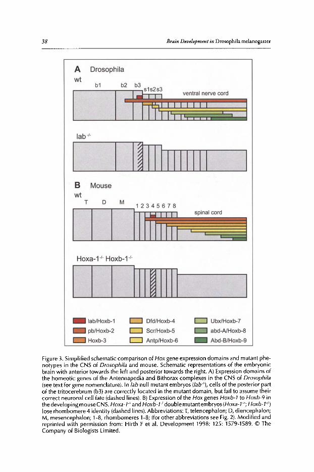
38 Brain Developmentin Drosophila melanogasrer
A Drosophila
wtb1
lab"
I,,--omtrrrrm_B Mousewt
T D M1 2 3 4 5 6 7 8.. I I I I spinal cord
IIII
Hoxa-t" Hoxb- t"
CD U'-----_lab/Hoxb-1
pb/Hoxb-2
Hoxb-3
c:J Dfd/Hoxb-4
c:J Scr/Hoxb-5
c:J Antp/Hoxb-6
c:J UbxlHoxb-7
abd-A/Hoxb-8
Abd-B/Hoxb-9
Figure 3. Simplified schematic comparison of Hox gene expression domains and mutant phenotypes in the CNS of Drosophila and mouse. Schematic representations of the embryonicbrain with anterior towards the left and poster ior towards the right. A) Expression domains ofthe homeotic genes of the Antennapedia and Bithorax complexes in the CNS of Drosophila(seetext for gene nomenclature). In lab null mutant embryos (lab·I .), cells of the posterior partof the tritocerebrum (b3) are correctly located in the mutant domain, but fail to assume theircorrect neuronal cell fate (dashed lines). B) Expression of the Hox genes Hoxb-l to Hoxb-9 inthe developing mouse CNS. Hoxa- F and Hoxb-l'l-double mutant embryos tHoxe-t"; Hoxb-tr;lose rhombomere 4 identity (dashed lines). Abbreviations: T, telencephalon; D, diencephalon;M, mesencephalon; 1-8, rhombomeres 1-8; (for other abbreviations see Fig. 2). Modified andreprinted with permission from : Hirth F et al. Development 1998; 125: 1579-1589. © TheCompany of Biologists Limited .
AnteroposteriorRegionalization oftheBrain: Genetic and Comparative Aspects 39
Gene expression studies indicate that a similar tripartite ground plan for anteroposteriorregionalization of the embryonic brain is also present in Drosophila. The Drosophila genomecontains two genes, Pox neuro (Poxn) and Pax2, which are together considered to be orthologsof the Pax2/5/8 genes." Remarkably, expression of both orthologs is present at the interface ofotd and the Drosophila Gbx2orthologunplugged (unpg),anterior to aHox-expressing region (Fig.4A,B).44Although Poxnand Pax2are expressedin a segmentally reiterated pattern along the entireembryonic eNS, their expression at the otdlunpg interface is exceptional in two ways. The twogenes are expressed in adjacent domains delineating together a transversal stripe ofthe brain andthis is the only position along the neuraxis where expression ofboth genes coincides with a brainneuromere boundary, the deutocerebral-trltocerebral boundary (DTB) (Fig. 4A,B).44 Analysesofeither otd or unpgmutants reveal a mutually repressive function of the two genes during earlybrain patterning. Thus, in otd mutant embryos a rostral extension ofthe unpgexpression domainis observed (in addition to the deletion of the anterior brain). On the other hand, mutationalinactivation of the unpg gene results in a caudal shift of the posterior limit of otd expression."Therefore, in both Drosophila and mouse, the early interaction of otdlOtx2 and unpglGbx2 isessential for the correct positioning of an intermediate brain domain characterized by a sharplydelimited otdlOtx2 and unpgl Gbx2 interface and the expression ofPax2/5/8 genes. In contrast tovertebrates, mutational inactivation of the Drosophila Pax2/5/8 orthologs Poxnor Pax2does notappear to result in brain patterningdefects. Moreover, to date, there is no evidence ofan organizeractivity at the fly DTB, suggesting that the organizer function at the otdlOtx2 and unpglGbx2
c
spinal cord
hindbrain
forebrain
midbrain
Mouse
IotdlOtx2
1Pax 21518
IHox
I
B
protocereb rum
ventral nervecord
deutocerebrum
tritocerebrum
subesophagea lgang lion
Drosophila
c::J otdlOtx2
l!ZZI unpglGb x2
_ Pax21518
_ Hox1 ortho logs
Figure4. Tripartite organization of the embryonic CNS in Drosophila and mouse. A) Expressionof Pax2and Poxnin the brain of stage13/14embryos.At the deutocerebral-tritocerebral bound ary (indicated by white arrows), Pax2 (white dots) and Poxn (white asterisks) are expressedin adjacent domains forming a transversal line in the CNS (immunolabelled with antiHRPand shown in grey). B,C) The expression of otd/Otx2, unpg/Cbx2, Pax2/5/B and Hoxl geneorthologs in the developing CNS of Drosophila (B)and mouse (C). (In this schematic, anterioris towards the top and posterior is towards the bottom.) In both cases,otd/Otx2 is expressedin the anterior nervous system rostral to a Hox-expressing region in the posterior nervoussystem. In addition , a Pax2/5/B-expressing domain posit ioned at the interface between theanterior otd/Otx2 domain and the posteriorly abutting unpg/Cbx2 expression domain iscommon to both nervous systems. Modified and reprinted with permission from : Hirth F etal. Development 2003; 130: 2365-2373. © The Company of Biologists Limited.
40 Brain Development in Drosophila melanogaster
interface might have emerged after the protostome/deuterostome divergence that separated insectsand vertebrates. In fact, an organizer activity ofthe MHB region has so far only been demonstratedfor vertebrate species within deuterostomes.
In summary, current comparative data indicates that similar genetic patterning mechanismsact in anteroposterior regionalization ofthe developing brain in Drosophila and vertebrate speciesand establish a common, evolutionarily conserved tripartite ground plan. This suggests that a corresponding tripartite organization ofthe developing brain was already present in the last commonbilateral ancestor of insects and vertebrates.
References1. Arendt D, Nubler-jung K. Comparison of early nerve cord development in insects and vertebrates.
Development 1999; 126(11):2309-2325.2. Reichert H, Simeone A. Conserved usage of gap and homeotic genes in patterning the CNS. Curr Opin
Neurobiol1999; 9(5):589-595.3. Adoutte A, Balavoine G, Lartillot N et al. The new animal phylogeny: reliability and implications. Proc
Nat! Acad Sci USA 2000; 97(9):4453-4456.4. Younossi-Hartenstein A, Nassif C, Green P et al. Early neurogenesis of the Drosophila brain. J Comp
Neurol 1996; 370(3):313-329.5. Lumsden A, KrumlaufR. Patterning the vertebrate neuraxis. Science 1996; 274(5290):1109-1115.6. Puelles L, Rubenstein JL. Forebrain gene expression domains and the evolvingprosomeric model. Trends
Neurosci 2003; 26(9):469-476.7. Dalton D, Chadwick R, McGinnis w: Expression and embryonic function of empty spiracles: a Dro
sophila homeo box gene with two patterning functions on the anterior-posterior axis of the embryo.Genes Dev 1989; 3(12A):1940-1956.
8. Finkelstein R, Perrimon N. The orthodenticle gene is regulated by bicoid and torso and specifies Drosophila head development. Nature 1990; 346(6283):485-488.
9. Walldorf U, Gehring WJ. Empty spiracles, a gap gene containing a homeobox involved in Drosophilahead development. EMBO J 1992; 11(6):2247-2259.
10. Cohen SM, Jurgens G. Mediation of Drosophila head development by gap-like segmentation genes.Nature 1990; 346(6283):482-485.
11. Schmidt-Ott U, Gonzalez-Gaitan M, JackIe H et al. Number, identity and sequence of the Drosophilahead segments as revealed by neural elements and their deletion patterns in mutants. Proc Nat! AcadSci USA 1994; 91(18):8363-8367.
12. Urbach R, Technau GM. Molecular markers for identified neuroblasts in the developing brain of Drosophila. Development 2003; 130(16):3621-3637.
13. Younossi-Hartenstein A, Green P, Liaw GJ et al. Control of early neurogenesis of the Drosophila brainby the head gap genes tll, otd, ems and brd, Dev Biol 1997; 182(2):270-283.
14. Hirth F, Therianos S, Loop T et al. Developmental defects in brain segmentation caused by mutations ofthe homeobox genes orthodenticle and empty spiracles in Drosophila. Neuron 1995; 15(4):769-778.
15. Hartmann B, Hirth F, Walldorf U et al. Expression, regulation and function of the homeoboxgene empty spiracles in brain and ventral nerve cord development of Drosophila. Mech Dev 2000;90(2):143-153.
16. Leuzinger S, Hirth F, Gerlich D et al. Equivalence of the fly orthodenricle gene and the human OTXgenes in embryonic brain development of Drosophila. Development 1998; 125(9):1703-1710.
17. Acampora D, Annino A, Tuorto F et al. Otx genes in the evolution of the vertebrate brain. Brain ResBull 2005; 66(4-6):410-420.
18. Cecchi C. Emx:2 a gene responsible for cortical development, regionalization and area specification.Gene 2002; 291(1-2):1-9.
19. Simeone A, Acampora D, Gulisano M et al. Nested expression domains of four homeobox genes indeveloping rostral brain. Nature 1992; 358(6388):687-690.
20. Acampora D, Mazan S, Avantaggiato V et al. Epilepsy and brain abnormalities in mice lacking the Otxlgene. Nat Genet 1996; 14(2):218-222.
21. Acampora D, Mazan S, Lallemand Y et al. Forebrain and midbrain regions are deleted in Otx2-/- mutants due to a defective anterior neuroectoderm specification during gastrulation. Development 1995;121(10):3279-3290.
22. Acampora D, Avantaggiato V, Tuorto F et al. Murine Otxl and Drosophila otd genes share conservedgenetic functions required in invertebrate and vertebrate brain development. Development 1998;125(9):1691-1702.
AnteroposteriorRegionalization ofthe Brain: Genetic and Comparative Aspects 41
23. Acampora D, Boyl PP, Signore M et al. OTD/OTX2 functional equivalence depends on 5' and 3'UTR-mediated control of Otx2 mRNA for nucleo-cytoplasmic export and epiblast-restricted translation. Development 2001; 128(23):4801-4813.
24. Simeone A, Gulisano M, Acampora D et al. Two vertebrate homeobox genes related to the Drosophila empty spiracles gene are expressed in the embryonic cerebral cortex. EMBO I 1992;11(7):2541-2550.
25. Gulisano M, Broccoli V, Pardini C et al. Emxl and Emx2 show different patterns of expression duringproliferation and differentiation of the developing cerebral cortex in the mouse. Eur ] Neurosci 1996;8(5):1037-1050.
26. Bishop KM, Rubenstein ]L, O'Leary DD. Distinct actions of Emxl, Emx2 and Pax6 in regulating thespecification of areas in the developing neocortex. ] Neurosci 1 2002; 22(17):7627-7638.
27. Muzio L, DiBenedetto B, StoykovaA et ale Conversion of cerebral cortex into basal ganglia in Emx2(-/-)Pax6(Sey/Sey) double-mutant mice. Nat Neurosci 2002; 5(8):737-745.
28. Qiu M, Anderson S, Chen S et ale Mutation of the Emx-l homeobox gene disrupts the corpus callosum.Dev Bioi 1996; 178(1):174-178.
29. Yoshida M, Suda Y, Matsuo I et ale Emxl and Emx2 functions in development of dorsal telencephalon.Development 1997; 124(1):101-111.
30. Ferrier DE, Holland PW. Ancient origin of the Hox gene cluster. Nat Rev Genet 2001; 2(1):33-38.31. Hughes CL, Kaufman TC. Hox genes and the evolution of the arthropod body plan. Evol Dev 2002;
4(6):459-499.32. Mann RS. Why are Hox genes clustered? Bioessays 1997; 19(8):661-664.33. Duboule D, Morata G. Colinearity and functional hierarchy among genes of the homeotic complexes.
Trends Genet 1994; 10(10):358-364.34. Hirth F,Hartmann B, Reichert H. Homeotic gene action in embryonic brain development of Drosophila.
Development 1998; 125(9):1579-1589.35. Kourakis M], Master VA, Lokhorst DK et al. Conserved anterior boundaries of Hox gene expression
in the central nervous system of the leech Helobdella. Dev Bioi 1997; 190(2):284-300.36. Lowe C], Wu M, Salic A et al. Anteroposterior patterning in hemichordates and the origins of the
chordate nervous system. Cell 2003; 113(7):853-865.37. Wilkinson DG, Bhatt S, Cook M et al. Segmental expression of Hox-2 homoeobox-containing genes
in the developing mouse hindbrain. Nature 1989; 341(6241):405-409.38. Hunt P, Krumlauf R. Deciphering the Hox code: clues to patterning branchial regions of the head. Cell
1991; 66(6):1075-1078.39. GavalasA, Studer M, Lumsden A et al. Hoxal and Hoxbl synergize in patterning the hindbrain, cranial
nerves and second pharyngeal arch. Development 1998; 125(6):1123-1136.40. Studer M, Lumsden A, Ariza-McNaughton Let al. Altered segmental identity and abnormal migration
of motor neurons in mice lacking Hoxb-l. Nature 1996; 384(6610):630-634.41. Bruce AE, Shankland M. Expression of the head gene Lox22-0tx in the leech Helobdella and the origin
of the bilaterian body plan. Dev Biol 1998; 201(1):101-112.42. Canestro C, Bassham S, Postlethwait J. Development of the central nervous system in the larvacean
Oikopleura dioica and the evolution of the chordate brain. Dev Biol 2005; 285(2):298-315.43. Castro LF, RasmussenSL, Holland PWet ale A Gbx homeobox gene in amphioxus: Insights into ancestry
of the ANTP class and evolution of the midbrain/hindbrain boundary. Dev Biol 2006.44. Hirth F, Kammermeier L, Frei E et al. An urbilaterian origin of the tripartite brain: developmental
genetic insights from Drosophila. Development 2003; 130(11):2365-2373.45. Liu A, Joyner AL. Early anterior/posterior patterning of the midbrain and cerebellum. Annu Rev
Neurosci 2001; 24:869-896.46. Wada H, Saiga H, Satoh N et al. Tripartite organization of the ancestral chordate brain and the
antiquity of placodes: insights from ascidian Pax-2/5/8, Hox and Otx genes. Development 1998;125(6):1113-1122.
47. Wurst ~ Bally-Cuif L. Neural plate patterning: upstream and downstream of the isthmic organizer.Nat Rev Neurosci 2001; 2(2):99-108.
48. Noll M. Evolution and role of Pax genes. Curr Opin Genet Dev 1993; 3(4):595-605.
CHAPTER 3
Dorsoventral Patterning ofthe Brain:AComparative ApproachRolfUrbach* and Gerhard M. Technau
Abstract
Development of the central nervous system (CNS) involves the transformation of atwo-dimensional epithelial sheet ofuniform ectodermal cells, the neuroectoderm, intoa highly complex three-dimensional structure consisting of a huge variety of different
neural cell types. Characteristic numbers ofeach cell type become arranged in reproducible spatialpatterns, which is a prerequisite for the establishment ofspecific functional contacts. Specificationofcell fate and regional patterning critical depends on positional information conferred to neuralstem cells early in the neuroectoderm. This chapter compares recent findings on mechanisms thatcontrol the specification ofcell fates along the dorsoventral axisduring embryonic development ofthe CNS in Drosophila andvertebrates. Despite the clear structural differences in the organizationofthe CNS in arthropods and vertebrates, correspondingdomains within the developingbrain andtruncal nervous system express a conserved set ofcolumnar genes (msh/Msx, ind/Gsh,vnd/Nkx)involved in dorsoventral regionalization. In both Drosophila and mouse the expression of thesegenes exhibits distinct differences between the cephalic and truncal part ofthe CNS. Remarkably,not only the expression ofcolumnar genes shows striking parallels between both species, but tosome extent also their genetic interactions, suggesting an evolutionary conservation ofkey regulators ofdorsoventral patterning in the brain in terms ofexpression and function.
IntroductionThe central nervous system (CNS) in Drosophila and in vertebrates can be subdivided into
two main portions, a truncal part (ventral nerve cord (VNC) in Drosophila and spinal cord invertebrates) composed of repetitive segmental units and an anterior part, the brain, exhibiting aless overt segmental composition (Fig. 1).
In Drosophila, the CNS develops from a bilaterally symmetrical sheet ofneuroectodermal cellson the ventral side ofthe embryo. It gives rise to a fixed number ofneural stem cells, called neuroblasts (NBs), which segregate to the interior ofthe embryo. NBs which form the VNC and braindescend from the truncal and procephalic neuroectoderm, respectively (Fig. 2).1,2 In vertebrates,the CNS forms from a bilaterally symmetrical neuroectoderm on the dorsal side of the embryo.The whole neuroectodermal sheet invaginates to form the neural tube, which develops into thespinal cord and brain. Accordingly, the differentiatingNBs do not delaminate but maintain contactwith the epithelial surfaces (for a review see ref 3). Insect and vertebrate NBs divide reiterativelyto give rise to specific types ofneurons (motoneurons, interneurons) and glial cells.
In Drosophila, the border between neurogenic and nonneurogenic ectoderm becomes definedby two antagonistically acting extracellular factors encoded byshortgastrulation (sog) and decapentaplegic (dpp). The homologous genes in Xenopus (vertebrates), Cbordin and Bonemorphogenetic
*Corresponding Author: Rolf Urbach-Institute of Genetics, University of Mainz, 0-55099Mainz, Germany. Email: urbachesuni-mainz.de
BrainDevelopment in Drosophila melanogaster, edited by Gerhard M. Technau.©2008 Landes Bioscience and Springer Science+ Business Media.
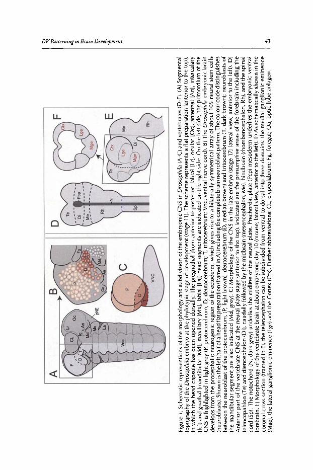
F
Figu
re1.
Sch
emat
icre
pres
enti
ons
of
the
mo
rph
olo
gyan
dsu
bd
ivis
ion
ofth
ee
mb
ryo
nic
CN
Sin
Dro
soph
ila
(A-C
)an
dve
rteb
rate
s(D
-F).
(A)
Seg
men
tal
topo
grap
hy
of
the
Dro
sop
hil
aem
bry
oat
the
phyl
oty
pic
stag
eo
fde
velo
pm
ent
(sta
ge11
).T
he
sche
me
repr
esen
tsa
flatp
rep
arat
ion
(an
teri
or
toth
eto
p),
inw
hic
hth
ehe
adca
psu
leh
asbe
enop
ened
do
rsal
ly.
The
preg
nat
hal
(fro
man
teri
orto
post
eri
or:
labr
al[L
r],
ocu
lar
[Oc]
,an
ten
nal
[An
],in
terc
alar
y[Ie
))an
dgn
ath
al(m
and
ibu
lar
[Md
],m
axi
llary
[Mx]
,la
bial
[La)
)he
adse
gme
nts
are
ind
ica
ted
onth
eri
ght
sid
e.O
nth
ele
ftsi
de,
the
prim
ord
ium
ofth
eC
NS
ish
igh
light
edin
light
gre
y(P
,p
roto
cere
bru
m;
D,
deu
toce
reb
rum
;T
,tri
toce
rebr
um;
Vn
c,ve
ntra
lne
rve
cord
).B
)T
heD
roso
ph
ila
embr
yon
icbr
ain
dev
elop
sfr
omth
epr
oce
phal
icne
uro
geni
cre
gio
nof
the
ecto
derm
,w
hic
hgi
ves
rise
toa
bila
tera
llysy
mm
etri
cal
arra
yo
fa
bo
ut
10S
neur
alst
emce
lls(n
euro
bla
sts)
.Sho
wn
isth
ele
fth
alfo
fahe
adfla
tpre
para
tion
(fra
med
inA
)in
clu
din
gth
eco
mp
lete
brai
nn
eu
rob
last
pa
tte
rn.T
heco
lour
cod
edi
stin
guis
hes
bet
wee
nth
en
eu
rob
last
of
the
pro
toce
reb
rum
,(P
,lig
htb
row
n),
de
uto
cere
bru
m(D
,m
edia
nb
row
n)
and
trito
cere
bru
m(T
,d
ark
bro
wn
);n
eu
rob
last
so
fth
em
an
dib
ula
rse
gme
ntar
ea
lso
indi
cat
ed(M
d,g
rey)
.C
)M
orp
ho
log
yof
the
CN
Sin
the
late
embr
yo(s
tage
17;
late
ral
view
,an
teri
orto
the
left
).D
)A
nter
ior
part
ofth
eve
rteb
rate
CN
Sat
the
neur
alpl
ate
stag
e(a
nte
rior
toth
eto
p).
Ind
icat
edar
eth
ep
resu
mp
tive
area
sof
the
fore
bra
inin
clu
din
gth
ete
len
cep
ah
lon
(Te)
and
dien
ceph
alon
(Di),
cau
da
llyfo
llow
ed
byth
em
idbr
ain
(mes
ence
pha
lon,
Me
),h
ind
bra
in(r
ho
mbe
nce
pha
lon,
Rh)
,and
the
spin
alco
rd(S
p),
Th
en
oto
chor
d(N
,d
ark
gre
y)un
der
lies
the
mid
line
ofth
ene
ural
plat
e.
Pre
chor
dal
plat
e(P
ep)
mes
oder
mun
de
rlie
sth
ee
mb
ryo
nic
ven
tral
fore
brai
n.E)
Mo
rph
olo
gyof
the
vert
eb
rate
brai
nat
abo
utem
bry
on
icda
y10
(mo
use;
late
ralv
iew
,an
teri
or
toth
ele
ft).
F)A
ssc
hem
ati
cally
show
nin
the
coro
nalc
ross
sect
ion
(fra
me
din
E),
the
tele
nce
phal
onca
nb
esu
bd
ivid
edfr
om
ven
tral
todo
rsal
into
thre
edo
mai
ns:
the
me
dia
lga
nglio
nic
emin
en
ce(M
ge)
,the
late
ral
gang
lion
ice
min
enc
e(L
ge)a
ndth
eC
orte
x(C
tx).
Fur
ther
ab
bre
viat
ion
s:C
L,c1
ypeo
lab
rum
;F
g,fo
reg
ut;
Oa,
opti
clo
bean
lage
n.
b ~ ;;p ~ ~. S· b:;
i:l S'~ ~ s- ] ~ ..... ~
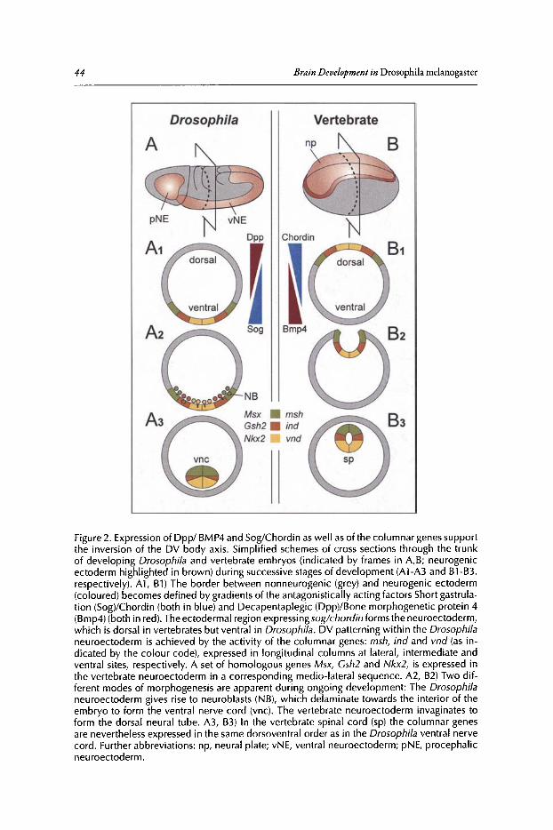
44
Drosophila
Brain Development in Drosophila melanogaster
Vertebrate
Figure 2. Expressionof Dpp/ BMP4 and Sog/Chordin aswell asof the columnar genessupportthe inversion of the OV body axis. Simplified schemes of cross sections through the trunkof developing Drosophila and vertebrate embryos (indicated by frames in A,B; neurogenicectoderm highlighted in brown) during successive stagesof development (A1-A3 and B1-B3,respectively). A1 , B1) The border between nonneurogenic (grey) and neurogenic ectoderm(coloured ) becomes defined by gradients of the antagonistically acting factors Short gastrulation (Sog)/Chordin (both in blue) and Oecapentaplegic (Opp)/Bone morphogenetic protein 4(Bmp4)(both in red).The ectodermal region expressing sog/ch ordin forms the neuroectoderm,which is dorsal in vertebrates but ventral in Drosophila. OV patterning within the Drosophilaneuroectoderm is achieved by the activity of the columnar genes: msh, ind and vnd (as indicated by the colour code), expressed in longitudinal columns at lateral, intermediate andventral sites, respectively. A set of homologous genes Msx, Gsh2 and Nkx2 , is expressed inthe vertebrate neuroectoderm in a corresponding medio-Iateral sequence. A2, B2) Two different modes of morphogenesis are apparent during ongoing development: The Drosophilaneuroectoderm gives rise to neuroblasts (NB), which delaminate towards the interior of theembryo to form the ventral nerve cord (vnc). The vertebrate neuroectoderm invaginates toform the dorsal neural tube. A3, B3) In the vertebrate spinal cord (sp) the columnar genesare nevertheless expressed in the same dorsoventral order as in the Drosophila ventral nervecord . Further abbrev iations: np, neural plate; vNE, ventral neuroectoderm; pNE, procephalicneuroectoderm.
DVPatterning in Brain Development 45
protein 4 (BMP4), respectively, basically serve the same function." In both species, the region inwhich sog/Chordin is expressed forms the neuroectoderm. Since the neuroectoderm is ventral inarthropods but dorsal in vertebrates, this has supported the hypothesis that the dorsoventral (DV)body axisbecame inverted during chordate evolution. This concept suggestsa monophyletic originand thus, homology ofthe CNS in protostomes and deuterostomes.t"
Remarkably, despite the clear structural differences in the mature CNS, corresponding DVsubdomains within arthropod and vertebrate neuroectoderm express homologous genes (knownas "columnar" genes; described below). This suggests that aspects ofDV patterning of the neuroectoderm have been evolutionarily conserved as well, which further supports homology ofthearthropod and vertebrate CNS.
The less complex truncal nervous systems in Drosophila and vertebrates (mouse, chick, frog)have provided useful models to study the mechanisms ofpatterning and the generation ofneuralcelldiversity. Many ofthe developmental processes that underlie NB formation, cell fate specification and pattern formation have been extensively studied in this more accessiblepart ofthe CNS(for a review see refs. 7-11). How cell diversity and patterning are achieved in the brain ofbothanimal phyla is less well understood.
Here, we compare recent findings on mechanisms that specify DV fates in the early (embryonic) brain of Drosophila and vertebrates and compare these mechanisms with those acting inthe truncal CNS.
DV Patterning ofthe Truncal Part ofthe eNSin Drosophila and Vertebrates
DVPatterningofthe VNCin DrosophilaIn Drosophila, the truncal neuroectoderm gives rise to the clearly metamerically organized
VNC, which comprises 8 abdominal, 3 thoracic and 3 gnathal segmental units (neuromeres).The primordium of the VNC (neuroectoderm and NBs) is subdivided along the DV axis intoadjacent longitudinal columns mainly by the activity ofthree homeobox genes (columnar genes).ventralnervous system defective (vnd) is expressed in the ventral, intermediateneuroblasts defective(ind) in the intermediate and muscle segmenthomeobox (msh; Drop [Dr]-FlyBase) in the dorsalneuroectodermal column (Fig. 3A).12-18 Onset oftheir expression is at the blastoderm stage.
The genetic mechanisms establishing and maintaining the sharp borders between the domainsofcolumnar gene expression in the VNC have been explored in detail. The columnar genes interact in a hierarchical cascade of transcriptional repression (also known as "ventral dorninance'T')according to which vnd represses ind (and msh) in the ventral column and ind represses msh inthe intermediate column. Thus, Vnd determines the ventral border of the Ind domain and Indthe ventral border of the Msh domain. The ventral border of the Vnd domain is defined by themesoderm-specific genes twistand snail(for a review see refs.20, 21). It is lessclearhow their dorsalborders are established. msh expression seems to be dorsally confined by the repressive activity ofgraded levelsofDpp and vnd expression by the Dorsal gradient, which activates vnd.22 The dorsalborder ofind expression may be formed by the limited activity ofEpidermalgrowthfactorreceptor(Egfr) and Dorsal,21but alsoby the activityofspatiallylocalized repressors,which areyet unknown."Egfr activity in the ventral and intermediate column regulates the fate ofNBs derived from thesecolumns and is further necessary for the maintenance ofvnd expression in the ventral column.24
-26
Furthermore, positional information provided by Dpp/BMP signalling contributes to patterningthe neuroectoderm by repressing columnar genes in a threshold-dependent fashion."
Columnar genes encode key regulators ofNB identity and each column thereby gives rise to apopulation ofdistinctly specified NBs. However, whereas vnd,ind and Egfr have also been shownto be crucial for the formation ofNBs in their respective column, this role appears dispensable formsh (for a review see ref. 20).

46
A
Brain Development in Dro sophila melanogaster
Dpp--_~----"""T---""
nuclear Dorsal
B
RA
CHRD ENOG GliFST RA
msom ~
Nkx2.2 <:
dorsal
intermediate
ventral
Figure 3. Genetic interactions controlling DV patterning of the truncal nervous system inDrosophila and vertebrates. A) Schematic cross section through a Drosophila embryo at theblastodermal stage. Intensity of red colour indicates gradient of Decapentaplegic (Dpp) whichin the dorsal ectoderm decreasesfrom dorsal to ventral. Dpp isventrally confined by repressiveactivity of Short gastrulation (Sog), Intensity of blue colour indicates the gradient of nuclearDorsal protein, wh ich increases from dorsal to ventral. Within the neuroectoderm, the properspatial doma ins of columnar gene expression are regulated by transcriptional repression: vndrepresses ind (and msh) in the ventral and ind represses msh in the intermediate neuroectoderm . Vnd and Epidermal growth factor receptor (Egfr)are ventrally delim ited by repressiveactivity of snail (from the mesoderm). Msh and SoxNeuro (SoxN) are dorsally repressed byDpp. Cleaved Spitz (cSpitz) emanating from mesectodermal cells (framed in pink) activatesEGFR signall ing. Nuclear Dorsal activates vnd, Egfr and msh in a concentration-dependentmanner. Figure legend continued on following page.
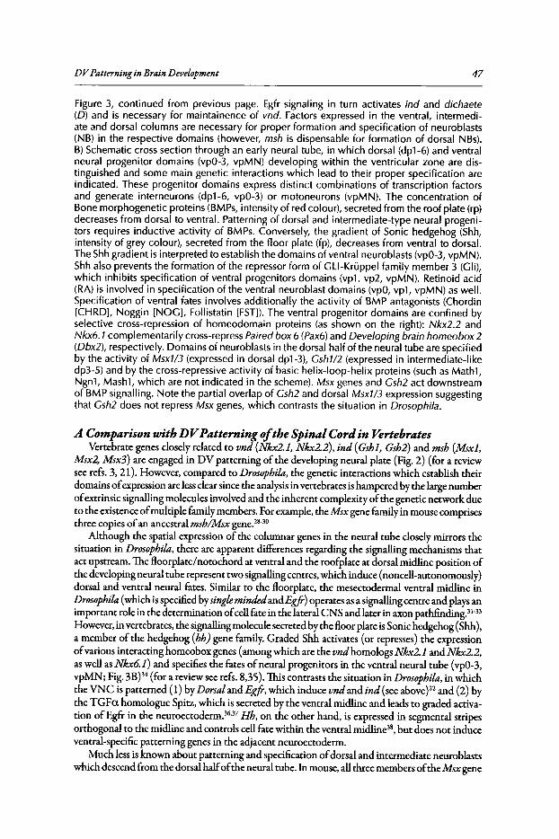
DVPatterning in Brain Development 47
Figure 3, continued from previous page. Egfr signaling in turn activates ind and dichaete(0) and is necessary for maintainence of vnd. Factors expressed in the ventral, intermediate and dorsal columns are necessary for proper formation and specification of neuroblasts(NB) in the respective domains (however, msh is dispensable for formation of dorsal NBs).B) Schematic cross section through an early neural tube, in which dorsal (dpl-6) and ventralneural progenitor domains (vpO-3, vpMN) developing within the ventricular zone are distinguished and some main genetic interactions which lead to their proper specification areindicated. These progenitor domains express distinct combinations of transcription factorsand generate interneurons (dpl-6, vpO-3) or motoneurons (vpMN). The concentration ofBone morphogenetic proteins (BMPs, intensity of red colour), secreted from the roof plate (rp)decreasesfrom dorsal to ventral. Patterning of dorsal and intermediate-type neural progenitors requires inductive activity of BMPs. Conversely, the gradient of Sonic hedgehog (Shh,intensity of grey colour), secreted from the floor plate (fp), decreases from ventral to dorsal.The Shhgradient is interpreted to establish the domains of ventral neuroblasts (vpO-3,vpMN).Shh also prevents the formation of the repressor form of Gl.l-Kruppel family member 3 (Gli),which inhibits specification of ventral progenitors domains (vpl, vp2, vpMN). Retinoid acid(RA) is involved in specification of the ventral neuroblast domains (vpO,vpl, vpMN) as well.Specification of ventral fates involves additionally the activity of BMP antagonists (Chordin[CHRD], Noggin [NOG], Follistatin [FST]). The ventral progenitor domains are confined byselective cross-repression of homeodomain proteins (as shown on the right): Nkx2.2 andNkx6.1 complementarily cross-repressPaired box 6 (Pax6)and Developing brain homeobox 2(Dbx2), respectively. Domains of neuroblasts in the dorsal half of the neural tube are specifiedby the activity of Msxl/3 (expressed in dorsal dpl-3), Cshl/2 (expressed in intermediate-likedp3-5) and by the cross-repressive activity of basic helix-loop-helix proteins (such as Mathl,Ngnl, Mashl, which are not indicated in the scheme). Msx genes and Csh2 act downstreamof BMP signalling. Note the partial overlap of Csh2 and dorsal Msxl/3 expression suggestingthat Csh2 does not repress Msx genes, which contrasts the situation in Drosophila.
A Comparison with D V Patterning ofthe Spinal Cord in VertebratesVertebrate genes closely related to vnd (Nkx2.1, Nkx2.2), ind (Gshl, Gsh2) and msh (Msxl,
Msx2, Msx3) are engaged in DV patterning of the developing neural plate (Fig. 2) (for a reviewsee refs. 3,21). However, compared to Drosophila,the genetic interactions which establish theirdomains ofexpression are lessclear since the analysisin vertebrates ishampered by the large numberofextrinsic signalling molecules involved and the inherent complexity ofthe genetic network dueto the existence ofmultiple family members. For example, theMsx gene family in mouse comprisesthree copies ofan ancestral msb/Msx gene.28
-30
Although the spatial expression of the columnar genes in the neural tube closely mirrors thesituation in Drosophila,there are apparent differences regarding the signalling mechanisms thatact upstream. The floorplatelnotochord at ventral and the roofplate at dorsal midline position ofthe developingneural tube represent two signalling centres, which induce (noncell-autonomously)dorsal and ventral neural fates. Similar to the floorplate, the mesectodermal ventral midline inDrosophila (which is specified bysinglemindedandEgfr) operates as a signalling centre and plays animportant role in the determination ofcell fate in the lateral CNS and later in axon parhfinding.v"However, in vertebrates, the signalling molecule secreted by the floor plate isSonic hedgehog (Shh) ,a member of the hedgehog (hh) gene family. Graded Shh activates (or represses) the expressionofvarious interacting homeobox genes (among which are the vndhomologs Nkx2.1 and Nkx2.2,as well as Nkx6.1) and specifies the fates ofneural progenitors in the ventral neural tube (vpO-3,vpMN; Fig. 3B)34 (for a review see refs. 8,35).This contrasts the situation in Drosophila,in whichthe VNC is patterned (1)by Dorsal and Egfr, which induce vnd and ind (see above)22 and (2)bythe TGFa homologue Spitz, which is secreted by the ventral midline and leads to graded activation ofEgfr in the neuroectoderm.w" Hh, on the other hand, is expressed in segmental stripesorthogonal to the midline and controls cell fate within the ventral midline", but does not induceventral-specific patterning genes in the adjacent neuroectoderm.
Much less is known about patterning and specification ofdorsal and intermediate neuroblastswhich descend from the dorsal halfofthe neural tube. In mouse, allthree members oftheMsx gene

48 Brain Development in Drosophila melanogascer
family are expressed in dorsalmost neuroblasts. While patterns ofMsxl and Msx2 expression arelargely overlapping in several nonneural tissues as well, Msx3 is exclusively restricted to the dorsalcolumn ofneural progenitors" (for a review see ref 30). Msx expression in the dorsal column isdetermined by molecules ofthe TGF-~ family secreted from the roofplate, among which are theDpp-related BMP2/4. BMP2/4 activate and seem to define both the dorsal and ventral border ofMsx expression/" This appeared to be in contrast to Drosophila, where Dpp represses mshand thusdefines only the dorsal border ofits expression.f However, it has recentlybeen reported that gradedDpp activity helps establish the mshlindand the indlvndbordersaswell by repressingmsh, ind andvnd in a threshold-dependent fashion and that BMPs act in a similar fashion in chick neural plateexplants." Gsh1 and Gsh2 are expressed in an intermediate column in the neural tube. Both genesare necessary for fate specification ofintermediate progenitors (in the progenitor domains dp3-S;Fig. 3B). Gsh2 is proposed to act downstream ofBMP/TGFB signalling." In Drosophila normallevel ofind expression critically depends on Eg{rsignalling. Although an Eg{rhomolog has beenidentified in zebrafish, it does not seem to have an instructive function in neural patterning ofthespinal cord" (for a review see ref 21). A further difference is that the expression domain ofGsh2partly overlaps with that ofthe Msx genes and that expression ofMsxl and Msx3 is unchanged inGsh2 single or Gshll2 double mutants. This indicates that Gsh2 cannot repress Msxll3, oppositeto the Drosophila VNC, where ind clearly represses msh.On the other hand, in both the vertebratespinal cord and the Drosophila VNC, Msx/msb does not repress Gshlind.18
,41
Interestingly, it has been shown that mouse Gsh1 (andNkx2.2) does not function in DrosophilaVNC development, suggesting that functional domains have become distinct over time. In contrast,function ofzebrafish Nkx6.1 and fly Nkx6 seems conserved since in both species overexpressionofthe respective ortholog leads to the induction ofsupernumerary motoneurons.t'
Further factors involved in the specification ofintermediate identities remain to be resolved,as for example signals involved in fate specification of"dp6" progenitors. Such signals may includeretinoid acid, which is also necessary for proper development of the adjacent ventral neural progenitors (for a review see ref 44). In the vertebrate neural tube, gaps have been observed betweenthe expression domains of the columnar genes, raising the possibility that other genes might fillin these gaps." It has been suggested that, in addition to the columns ofmsb/Msx,indlGsh andvnd/Nkx2, the early neural tube includes at least a fourth DV column which expresses the developing brainhomeobox2 (Dbx2) gene.41,4S,46 The Dbx2 expression domain is positioned betweenthe intermediate GshllGsh2and ventral Nkx6.1 column and includes the "dp6" progenitors. Itsventral border is determined by repressive activity ofNkx6.1, but the factor controlling its dorsalborder is unknown. However, the Drosophila Dbx homolog, H2.0, although expressed in subsetsofNBs and progeny cells, does not seem to be involved in DV specification ofNBs since it is notexpressed in the truncal neuroectoderm."
DV Patterning ofthe Brain in DrosophilaThe Drosophila larval brain develops from the procephalic neuroectoderm (pNE) which gives
rise to a bilaterally symmetrical array ofabout 100 embryonic NBs.47 Presumably all embryonic NBsbecome postembryonically reactivated to form the adult brain," whereas in the VNC postembryonic mitotic activity becomes restricted to segment-specific subpopulations ofNBs. The patternof embryonic brain NBs neither exhibits an ordered segmental assembly, nor morphologicallydistinct subdivisions into anteroposterior rows or dorsoventral columns, as is at least transiently thecase in the VNC. Accordingly, the segmental composition ofthe brain is not obvious. The brain issubdivided, from posterior to anterior, into the tritocerebrum, deutocerebrum and protocerebrum(Fig. lA-C). The embryonic trito- anddeutocerebrum correspond to one neuromere each, derivingform the intercalary and antennal segment, respectively. There is evidence that the protocerebrummay consist oftwo neuromeres, a large one deriving from the ocular segment and a small remnantof the labral segment.49,SO Likewise, DV regionalisation of the early embryonic brain is not overtand the underlying patterning mechanisms are only rudimentarilyunderstood. The columnar genesare expressed in distinct areas ofthe pNE and the developing brain. Although their expression is
DVPatterning in Brain Development 49
consistent with their role in DV patterning being principally conserved in the procephalon, thereare also significant differences in their patterns ofexpression as compared to the trunk.
Expression ofColumnar Genes in the Early Embryonic BrainAt the gastrula stage, vnd is expressed in the ventral pNE, covering the prospective ventral
parts of the trito-, deuto- and protocerebrum. While Vnd in the trunk, is maintained within acontinuous ventral neuroectodermal column during subsequent stages, vnd expression in the earlybrain ishighly dynamic. It becomes progressivelyconfined to three separate ventral domains at theposterior border ofthe trito-, deuto- and protocerebrum, encompassingdifferent numbers ofNBsand progeny cells (Fig. 4A).51.52 msh is expressed in the dorsal neuroectoderm of the intercalaryand antennal segments which give rise to trito- and deutocerebral NBs. It is not expressed in theprimordium ofthe protocerebrum. In the trunk, ind is expressed in a continuous column ofintermediate neuroectoderm, whereas in the procephalic neuroectoderm it is found in three separatespots (in the intercalary, antennal and ocular segment). The intercalary and antennal ind spot arelocated at intermediate position between the dorsal Msh and ventral Vnd domain. Opposed tothat, the ocular ind spot is spatially clearly separated from the ventral Vnd domain (and msh is notexpressed). Due to the insulated expression of ind, the intercalary and antennal domains ofmshand vnd expression share a common border at sites lacking an intervening ind domain."
Another conspicuous difference to the trunk (and to the TC and DC as well) is that a largeamount of the protocerebral NBs (more than 50%) does not express any of the three columnargenes (Fig. 4A).50 Similarly, in the vertebrate spinal cord gaps have been detected between thedomains ofcolumnar gene expression.18 Thus, DVpatterningofthe protocerebral primordia ofthebrain anlagen requires factors additional to those encoded by the columnar genes. Candidate genesmight include Egfr,24-26 the Sox genes SoxNeuro and Dicheate,53-55 the Nkx2.1 homologous genescarecrow." the Nkx6 family related gene Nk6,43.57 or perhaps the Dbx homologous gene, H2.0.58Most ofthese, except scarecrow and H2.0,are known to have a function in fate specification and/orformation of NBs in the trunk. Egfr, both Sox genes and Nk6 are expressed in the pNE beforeand during the phase ofNB formation43
•57(J.Seibert and R. Urbach, unpublished observations),
however, their role in the formation/specification ofbrain NBs is yet unknown.
Segment-Specific Regulation ofColumnar GenesRecent reports gave first insights into the interactions and function ofcolumnar genes during
DV patterning of the embryonic brain. 51.52Although principally the same DV patterning genesoperate in large parts ofthe pNE, their regulation revealssegment-specific differences both amongthe brain segments and compared to the trunk (Fig. 4B).
For example, contrary to the trunk, in vnd mutant background derepression ofind within theventral pNE does not occur in the antennal segment. Instead, ind expression is completely absent,indicating that, at least in this part of the pNE and brain, vnd is necessary for activation and/ormaintenance ofind rather than for its repression (as in the trunk). This is supported by the finding that ectopic expression of vnd does not repress ind in the antennal segment. The ocular indspot is often ventrally expanded in the absence ofVnd, which is reminiscent ofthe situation in thetrunk. However, in the wildtype, the ocular ind spot does not adjoin the ventral domain of vndexpression. Hence, a ventral expansion ofocular ind in vnd mutants cannot be due to the lack ofrepression by Vnd and may be regulated noncell-auronomously.P
Moreover, in the absence of Vnd, expression of msh reveals segment-specific differences. Itsexpression is ectopically expanded into the ventral pNE ofthe intercalary and antennal segment,due to lack ofrepression by Vnd and Ind. This is not the case in the neuroectoderm ofthe protocerebrum and trunk. In the latter ind is derepressed instead ofmsh.
Taken together, expression and interactions ofcolumnar genes (Le.,the cascadeoftranscriptionalrepression which establishes the ventral border ofthe mshand ind domain) appears to be conservedin the most posterior brain, the tritocerebrum. Although the expression ofcolumnar genes is tosome extent conserved in the deutocerebrum as well, their genetic interactions are more derivedin the deuto- and protocerebrum.V So far it is not settled how these segment-specific differences
50 Brain D evelopment in Drosophila melanogaster
D
c
p
v
telen cephalon
p
..~s: fJ?!••
VNCT.Dwt
A
B
intermediate
ventral
dorsal E.•• •• ••••• • • • dorsal !
intermediate
Figure 4. OV patterning of the embryonic brain in Drosophila and vertebrates. A) Expressionof the columnar genes msh , ind and vnd (see colour code) in neuroblasts of the tritocerebrum(T), deutocerebrum (0) and protocerebrum (P) at the embryonic stages9 and 11 . Each schemerepresents the left half of a head flat preparation (compare with Fig. 1B) including the full complement of neuroblasts at the respective stages (encircled). Red stippled lines indicate bordersbetween neuromeres . vnd is expressed in ventral (v), ind in intermediate and msh in dorsal (d)neuroblasts . Vnd becomes progressively expressed at the posterio-ventral border of the trito-,deuto - and protocerebrum. Msh expression is confined to dorsal neuroblasts of the trito- anddeutocerebrum, but is not found in the protocerebrum. Expression of ind is confined to threeseparate spots of neuroblasts in the trito-, deuto- and protocerebrum. Note that domains of Mshand Vnd share common borders at sites where expression of ind is lacking . Further abbreviations: An, antennal; CL, c1ypeolabral; Md; madibular appendage. B) Oiagramm summariz ing thesegment-specific differences in the regulatory interactions of columnar genes in the ventral nervecord (VNC), trito- (T), deuto- (0 ) and protocerebrum (P) in wildtype (wt), vnd loss-of-function(lof) and vnd gain-of-function (gof) embryos (for details see text). C) Schematic of a coronalsection of the mouse telencephalon at about embryonic day 10 (compare with Fig. 1F). The vndhomolog Nkx2 .1 is expressed in the most ventral area, the medial ganglion ic eminence (Mge),the ind homolog Gsh2 in the lateral ganglionic eminence (Lge) and the eyeless (ey) homologPax6 in the dorsal cortex (O x), where , similar to the situation in Drosophila, Msx genes arenot expressed. 0 ) Oiagramm comparing the regulatory interactions between Pax6, Gsh2 andNkx2 .1 in the vertebrate telencephalon with those of the homologous genes ey, ind and vnd inthe Drosophila protocerebrum (P). For details see text. In the vertebrate telencephalon, oppositeto the situation in the spinal cord , Pax6 and Gsh2 mutually repressone another (compare withFig. 3B). Gsh2 and Nkx 2.1 do not act in a cross-repressive manner (as indicated by red crossedrepression symbols)." Similarly, vnd and ind in the protocerebrum do not directly interfere(indicated by stippled red crossed repression symbols). Although the altered extent of the indexpression doma in in vnd lof and gaf backgrounds indicates regulator y interactions (see Fig.4B), they must be noncell-autonomous, since the normal vnd and ind expression doma ins donot abut each other. Expression of ey (and twin of eyeless, not indicated) is unaffected in indmutants , suggesting that, in contrast to the situation in the telencephalon, ind does not repressey (indicated by red crossed repression symbol ). It is not yet clear, if conversely, ind expressionis affected in the absence of ey and/or twin o f eyeless .
D V Patterningin Brain Development 51
are regulated. It also remains to be clarified in how far other factors (e.g., those described above)genetically interfere with the columnar genes in the pNE and may establish the gene-specific extentoftheir expression both in the DV and AP axis.
Role ofColumnar Genes in Formation ofBrain NBsIn vnd mutants, ventral brain NBs are largely absent indicating that, similar to the situation
in the trunk, vnd promotes formation ofNBs. In the absence ofVnd, cell death is increased andacts at the level ofboth, neuroectodermal progenitors cells and NBs.52It is not yet resolved ifthereduction in ventral NBs is solely due to an increase in cell death or involves other factors knownto be engaged in NB formation, such as proneural genes oftheAS-complex (acheate, scute,lethalofscute [l 'sc]; for a review see ref 59).In the trunk, there is evidence that vnd interacts with proneuralgenes, but may also have additional function in promoting NB formation. Accordingly, in vndmutant embryos, l 'scis still expressed in ventral proneural clusters, although the respective NBs willnot form (e.g., NB5-2).13.60 In the pNE, genes ofthe AS-complex are expressed in large proneuraldomains and the acheate and l 'sc domain seem to overlap with the domain ofvnd expression, compatible with a genetic interaction.v/" However, in vnd mutant embryos no substantial differenceto the wildtype expression pattern ofl 'sctranscript is observed (R.ll, unpublished observation),suggesting that, if vnd has proneural activity, it is rather independent of l 'sc. Nevertheless, theexpression ofanother proneural gene, atonal (in the pNE normally expressed in proneural clusters and developing sensory precursors ofthe hypopharyngeal-/latero-hypopharyngeal organ), isoften missing indicating its dependence on Vnd.52Thus far it is unclear ifmsh,ind and Eg{r exerta similar function in brain NB formation. Whereas Eg{r mutant embryos exhibit strong defectsin the number and pattern ofbrain NBs, they appear rather unaffected in mshmutants (J.Seibertand R.U., unpublished observation), indicating that at least mshdoes not playa role in brain NBformation. Eg{r signalling has also been shown to be necessary for the proper development ofmedial brain structures deriving from the head midline, which behaves like the mesectoderm in thetrunk.f Placode-like groups ofcells from the head midline invaginate and contribute subpopulations of cells to the brain." Loss ofEg{r signalling results in severe reduction or absence of therespective head midline derivatives.
Role ofvnd in Specification ofBrain NBsIn the trunk, evidence has been provided, that the set ofgenes expressed within a proneural
cluster specifies the individual identityofthe NB it gives rise to. Such a combinatorial code, whichis unique for each NB, is provided mainly by the superimposition ofthe acitivity ofDV patterninggenes and segment polarity genes (AP axis) and a number ofother factors (for a review see refs. 9,20, 63). Most ofthese genes are also expressed in specific procephalic neuroectodermal domainsbefore NBs delaminate, implying that these genes might be required for specification ofindividualbrain NBs aswell.64 Analysis ofan array ofsuch NB identitygenes in vnd loss- and gain-offunctionbackgrounds indicates that, similar to the situation in the trunk, vnd influences their expressionalready in the pNE, before the formation ofNBs. 52
In vnd loss-of-function background, dorsal-specific gene expression is derepressed in the ventral pNE and descending NBs and conversely, ventral-specific gene expression is lost, suggesting aventral-to-dorsal transformation ofthe mutantventralpNE and residual ventral NBs. This indicatesthat vndnormally activates genes specific for the ventralpNE and represses genes specific for dorsalpNE and is required for fate specification ofventral brain NBs. This is further supported by theproduction ofectopic glial cells derived from transfated ventral NBs in the trito- and deutocerebrum, which normally is a specific trait ofdorsal brain NBs. Later in embryogenesis a severe lossofneural tissue associated with increased apoptotic activity has been observed in the tritocerebrum,presumably as a consequence ofidentity changes imposed on vnd deficient NB lineages."
Upon vnd overexpression, there is a wide-ranging loss of dorsal-specific gene activity in thedorsal pNE and NBs, but a largely unaffected ventral-specific gene activity in ventral parts.Moreover, there is evidence for a partial dorsal-to-ventral transformation of dorsal parts of thepNE and corresponding NBs, which indicates that Vnd is not only necessary but to some extentalso sufficient to induce ventral traits.
52 Brain Development in Drosophila melanogaster
A Comparison with DV Regionalization ofthe VertebrateTelencephalon
The telencephalon derives from paired evaginations of the anterior forebrain that constitutethe most complex structures of the vertebrate CNS. Progress has been made in understandingthe early regional patterning ofthe telencephalon, although the present knowledge about its DVregionalization is still rudimentary. The telencephalon can be subdivided into a dorsal or pallialand aventral or subpallial territory and the subpallium further into the lateral ganglionic eminence(LGE) and aventralmost part, the medial ganglionic eminence (M GE) (Fig. 1).The pallium givesrise to the cortex, the subpallium to the basal ganglia. The future telencephalic territories can bedefined early in development by the expression ofNkx2.1 in the ventral MGE and Gsh1, Gsh2inthe intermediate LGE, resembling the expression ofvnd and ind in the anlagen ofthe Drosophilabrain. Pax6, the homolog of Drosophila eyeless (ey), is expressed in the dorsal telencephalon(Fig. 4C). Pax6 is involved in the specification of pallial identity (for a review see refs. 66, 67)instead ofMsx genes which are not expressed in the telencephalon. Interestingly, Drosophila eyis likewise preferentially expressed in dorsal/intermediate NBs ofthe protocerebrum, which lackmshexpression", suggesting that eymay to some extent play the role ofmsh in the anterior brain.In the telencephalon, Nkx2.1, Gsh2and Pax6are complementary expressed, provide some oftheearliest markers for the respective territories and are key regulators for their normal development(for a review see ref 67).
Genetic Interactions ofColumnar GenesAlthough a conserved set ofhomeobox genes is expressed at corresponding DV positions in
the brains ofarthropods and vertebrates, there are differences in their genetic interactions. In thetelencephalon Gsh2and Pax6cross-repress each other, which results in the formation ofa sharpborder between the dorsal and intermediate domains (Fig. 4C, D). Accordingly, in Pax6mutantmice there is evidence for a dorsal-to-ventral transformation ofdorsal (pallial) structures, which isopposite to the phenotype in Gsh2 mutants.68-70This behaviour is specific to the telencephalon andnot observed in the spinal cord. Similarly,in the Drosophila protocerebrum and deutocerebrum, eyand ind are largely expressed in complementary subsets ofNBs.64However, eyexpression does notseem to depend on Ind, since it does not expandventrally in ind mutants (R.U., unpublished observations), asopposed toPax6in the telencephalon ofGsh2 mutants." In the tritocerebrum, oppositeto the anterior brain, eyis coexpressed withind64
, resembling the situation in the vertebrate spinalcord, in which the domains ofPax6and Gsh2overlap.41.67 It is worth noting, that Drosophila has asecond Pax6gene, twin ofeyeless (toy), which is largely expressed in the protocerebrum. However,since ind is coexpressed with toyin the protocerebrum'", it is unlikely that ind and toy (instead ofey)genetically behave in a way similar to Pax6and Gsh2in the telencephalon.
Among the columnar genes,particularly the family ofNkx/vndgenes seemsto be well conservedin terms ofexpression and function. In mice carryinga deletion ofNkx2.1 ,a substantial lossofventral, especiallyofforebrain structures has been observed. The residual ventral (subpallial) structuresbecome transfated into dorsal striatal structures." Aninterestingcorrelation between the regulationofcolumnar genes in the vertebrate telencephalon and Drosophila deuto- and protocerebrum isthat Nkx2.1 and vnd do not repress the expression of Gsh and ind, respectively. Accordingly, inNkx2.1 knockout mouse, aswell as in Drosophila vnd mutants, the expression ofGsh2/indin thesebrain regions is not ventrally expanded 51.71, contrary to findings made in the truncal CNS (for areview see ref 21). Instead ofintermediate Gsb/ind, dorsal-specific marker genes are derepressedin ventralmost areas ofthe early brain; among these are Pax6,in the vertebrate telencephalon andey (to a minor extent) and especially msh in the Drosophila deuto-and tritocerebrum. Together,this suggests that in Nkx2.1/vnd mutant background, residual ventral brain territories undergo aventral-to-dorsal rather than a ventral-to-intermediate transformation, the latter being observedin the truncal CNS ofboth species.13·73.74
D V Patterningin Brain Development 53
Genetic Factors Upstream ofthe Columnar GenesSeveralextrinsic signalling molecules are involved in DVpatterningofthe telencephalon, among
which are BMPs, Wnts, Gli, FGFs, Nodal, retinoic acid and the central player Shh (for a reviewsee refs. 44, 67, 7S). The mechanisms by which Shh induces DV fate might differ between spinalcord and brain. Whereas in the spinal cord, the fates induced by Shh are concentration-dependent,Shh-induced fates in the telencephalon depend on timing rather than concentration (for a reviewsee ref 67). In the telencephalon, the source of secreted Shh is (among others) the prechordalplate, a mesodermal derivative. Remarkably, the Drosophila homolog, Hh, secreted from thehead mesoderm and foregut, acts on brain morphogenesis by regulating size and apoptosis. hh,expressed in the foregut, appears to mediate these effects via the Hh receptorpatched (expressedin brain cells surrounding the foregut). These similarities may indicate an ancient mechanism ofbrain patterning via induction." In how far other extrinsic signalling molecules are involved inDV patterning ofthe Drosophila brain remains to be shown.
ConclusionsA conserved set ofcolumnar genes (msh/Msx, ind/Gsh, vnd/Nkx) is involved in DV regional
ization ofthe brain and truncal CNS in vertebrates and arthropods (Drosophila). The expressionofcolumnar genes in the brain differs from the truncal CNS in both animal phyla. Remarkably,the brain-specific expression ofcolumnar genes exibits striking parallels between Drosophila andmouse in that the anterior borders oftheir domains are corresponding: Expression ofvnd/Nkx2extends most rostrally, followed by ind/Gshl and finally by msh/Msx3 (for a review see ref 77).Thus, the expression ofcolumnar genes in the brain is, to some extent, evolutionarily conserved,not only along the DV axis but also along the AP axis.
Moreover, brain-specific interactions among columnar genes bear some similarities betweenvertebrates and Drosophila. For example, Gsh/indare not repressed by Nkx2.1/vnd and expressionofdorsal factors, instead ofintermediate, is expanded into ventral domains in vnd/Nkx2 mutantbrains. This suggests that at least part of the genetic mechanisms governing DV fate in the brainhave been conserved as well. Differences may become more obvious at the level of upstreamregulating factors. However, in vertebrates, as well as in Drosophila, the genetic basis underlyingDV regionalization of the brain is far from being understood. The Drosophila brain, due to itscomparatively small size, allowing resolution at the level of individually identified cells and tothe powerful genetic and experimental tools available, provides a useful model system to studythese mechanisms in detail. This will facilitate the clarification of the processes underlying DVregionalization in the brain ofother organisms, including vertebrates.
AcknowledgementsWe are grateful to Ana Rogulja-Ortmann for comments on the manuscript. This work was
supported by the Deutsche Forschungsgemeinschaft and the EC.
References1. Hartenstein V, Campos-Ortega JA. Early neurogenesis in wild-type Drosophila melanogaster. Rouxs
Archives of Developmental Biology 1984; 193:308-325.2. Poulson DS. Histogenesis, organogenesis and differentiation in the embryo of Drosophila melanogaster
Meigen. In: Demerec M, ed. Biology of Drosophila. New York: Wiley, 1950:168-274.3. Arendt D, Niibler-Jung K. Comparison of early nerve cord development in insects and vertebrates.
Development 1999; 126:2309-2325.4. Holley SA, Jackson PD, Sasai Y et al. A conserved system for dorsal-ventral patterning in insects and
vertebrates involving sog and chordin, Nature 1995; 376:249-253.5. Arendt D, Niibler-Jung K. Dorsal or ventral: similarities in fate maps and gastrulation patterns in an
nelids, arthropods and chordates. Mech Dev 1997; 61:7-21.6. De Robertis EM, Sasai Y. A common plan for dorsoventral patterning in Bilateria. Nature. 1996;
380:37-40.7. Lumsden A, Krumlauf R. Patterning the vertebrate neuraxis. Science 1996; 274:1109-1115.8. Jessell TM. Neuronal specification in the spinal cord: inductive signals and transcriptional codes. Nat
Rev Genet 2000; 1:20-29.
54 Brain Development in Drosophila melanogaster
9. Skeath JB, Thor S. Genetic control of Drosophila nerve cord development. Curr Opin Neurobiol 2003;13:8-15.
10. Pearson BJ, Doe CQ Specification of temporal identity in the developing nervous system. Annu RevCell Dev BioI 2004; 20:619-647.
11. Price SR, Briscoe J. The generation and diversification of spinal motor neurons: signals and responses.Mech Dev 2004; 121:1103-1115.
12. Buescher M, Chi a W Mutations in lottchen cause cell fate transformations in both neuroblast and glioblastlineages in the Drosophila embryonic central nervous system. Development 1997; 124:673-681.
13. Chu H, Parras C, White K er al. Formation and specification of ventral neuroblasts is controlled byvnd in Drosophila neurogenesis. Genes Dev 1998; 12:3613-3624.
14. D'Alessio M, Frasch M. msh may playa conserved role in dorsoventral patterning of the neuroectodermand mesoderm. Mech Dev 1996; 58:217-231.
15. Isshiki T, Takeichi M, Nose A. The role of the msh homeobox gene during Drosophila neurogenesis: implication for the dorsoventral specification of the neuroectoderm. Development 1997; 124:3099-3109.
16. Jimenez F, Martin-Morris LE, VelascoL et al. vnd, a gene required for early neurogenesis of Drosophila,encodes a homeodomain protein. EMBO J 1995; 14:3487-3495.
17. Mellerick DM, Nirenberg M. Dorsal-ventral patterning genes restrict NK-2 homeobox gene expression tothe ventral half of the central nervous system of Drosophila embryos. Dev Biol 1995; 171:306-316.
18. Weiss JB, Von Ohlen T, Mellerick DM et al. Dorsoventral patterning in the Drosophila central nervoussystem: the intermediate neuroblasts defective homeobox gene specifies intermediate column identity.Genes Dev 1998; 12:3591-3602.
19. Cowden J, Levine M. Ventral dominance governs sequential patterns of gene expression across thedorsal-ventral axis of the neuroectoderm in the Drosophila embryo. Dev Biol 2003; 262:335-349.
20. Skeath JB. At the nexus between pattern formation and cell-type specification: the generation of individualneuroblast fates in the Drosophila embryonic central nervous system. Bioessays 1999; 21:922-931.
21. Cornell RA, Ohlen TV. Vnd/nkx, ind/gsh and msh/rnsx: conserved regulators of dorsoventral neuralpatterning? Curr Opin Neurobiol2000; 10:63-71.
22. von Ohlen T, Doe CQ Convergence of dorsal, dpp and egfr signaling pathways subdivides the Drosophila neuroectoderm into three dorsal-ventral columns. Dev Biol 2000; 224:362-372.
23. Stathopoulos A, Levine M. Localized repressors delineate the neurogenic ectoderm in the early Drosophila embryo. Dev BioI 2005; 280:482-493.
24. Skeath JB. The Drosophila EGF receptor controls the formation and specification of neuroblasts alongthe dorsal-ventral axis of the Drosophila embryo. Development 1998; 125:3301-3312.
25. Yagi Y, Suzuki T, Hayashi S. Interaction between Drosophila EGF receptor and vnd determines threedorsoventral domains of the neuroectoderm. Development 1998; 125:3625-3633.
26. Udolph G, Urban J, Riising G et al. Differential effects of EGF receptor signalling on neuroblast lineagesalong the dorsoventral axis of the Drosophila CNS. Development 1998; 125:3292-3299.
27. Mizutani CM, Meyer N, Roelink H et al. Threshold-dependent BMP-mediated repression: A modelfor a conserved mechanism that pattern the neuroectoderm. PLOS Biology 2006; 4:e313.
28. Catron KM, Wang H, Hu G et al. Comparison of MSX-1 and MSX-2 suggests a molecular basis forfunctional redundancy. Mech Dev 1996; 55:185-199.
29. Wang ~ Chen X, Xu H et al. Msx3: a novel murine homologue of the Drosophila msh homeoboxgene restricted to the dorsal embryonic central nervous system. Mech Dev 1996; 58:203-215.
30. Ramos C, Robert B. msh/Msx gene family in neural development. Trends Genet 2005; 21:624-632.31. Crews ST. Control ofcell lineage-specific development and transcription by bHLH-PAS proteins. Genes
Dev 1998; 12:607-620.32. Kim 10, Kim IC, Kim Set al. CNS midline cells contribute to maintenance of the initial dorsoventral
patterning of the Drosophila ventral neuroectoderm. J Neurobiol 2005; 62:397-405.33. Jacobs JR. The midline glia of Drosophila: a molecular genetic model for the developmental functions
of glia. Prog Neurobiol2000; 62:475-508.34. Ericson J, Muhr J, Placzek M et al. Sonic hedgehog induces the differentiation of ventral forebrain
neurons: a common signal for ventral patterning within the neural tube. Cell 1995; 81:747-756.35. Wilson L, Maden M. The mechanisms of dorsoventral patterning in the vertebrate neural tube. Dev
BioI 2005; 282:1-13.36. Golembo M, Raz E, Shilo BZ. The Drosophila embryonic midline is the site of Spitz processing and
induces activation of the EGF receptor in the ventral ectoderm. Development 1996; 122:3363-3370.37. Schweitzer R, Shilo BZ. A thousand and one roles for the Drosophila EGF receptor. Trends Genet
1997; 13:191-196.38. Bossing T, Brand AH. Determination of cell fate along the anteroposterior axis of the Drosophila ventral
midline. Development 2006; 133:1001-1012.

D V Patterning in Brain Development 55
39. Liu Y, Helms AW,Johnson ]E. Distinct activities ofMsxl and Msx3 in dorsal neural tube development.Development 2004; 131:1017-1028.
40. Suzuki A, Veno N, Hemmari-Brivanlou A. Xenopus msxl mediates epidermal induction and neuralinhibition by BMP4. Development 1997; 124:3037-3044.
41. Kriks S, Lanuza GM, Mizuguchi Ret al. Gsh2 is required for the repression of Ngnl and specificationof dorsal interneuron fate in the spinal cord. Development 2005; 132:2991-3002.
42. Strahle V, ]esuthasan S, Blader P et al. one-eyed pinhead is required for development of the ventralmidline of the zebrafish (Danio rerio) neural tube. Genes Funct 1997; 1:131-148.
43. Cheesman SE, Layden M], Von Ohlen T et al. Zebrafish and fly Nkx6 proteins have similar CNSexpression patterns and regulate motoneuron formation. Development 2004; 131:5221-5232.
44. Zhuang B, Sockanathan S. Dorsal-ventral patterning: a view from the top. Curr Opin Neurobiol2006;16:20-24.
45. Fjose A, Izpisua-Belmonte ]C, Fromental-Ramain C et ale Expression of the zebrafish gene hlx-l in theprechordal plate and during CNS development. Development 1994; 120:71-81.
46. Pierani A, Moran-Rivard L, Sunshine M] et al. Control of interneuron fate in the developing spinalcord by the progenitor homeodomain protein Dbx1. Neuron 2001; 29:367-384.
47. Vrbach R, Schnabel R, Technau GM. The pattern of neuroblast formation, mitotic domains and proneuralgene expression during early brain development in Drosophila. Development 2003; 130:3589-3606.
48. Lee C~ Robinson K], Doe CQ LgI, Pins and aPKC regulate neuroblast self-renewalversus differentiation. Nature 2006; 439:594-598.
49. Schmidt-Ott V, Technau GM. Expression of en and wg in the embryonic head and brain of Drosophilaindicates a refolded band of seven segment remnants. Development 1992; 116:111-125.
50. Vrbach R, Technau GM. Segment polarity and DN patterning gene expression reveals segmentalorganization of the Drosophila brain. Development 2003; 130:3607-3620.
51. Sprecher S, Vrbach R, Technau GM et al. The columnar gene vnd is rquired for tritocerebral neuromereformation during embryonic brain development of Drosophila. Development 2006; 133:4331-4339.
52. Vrbach R, Volland D, Seibert] et al. Segment-specific requirements for dorsoventral patterning genesduring early brain development in Drosophila. Development 2006; 133:4315-4330.
53. Buescher M, Hing FS, Chia w: Formation of neuroblasts in the embryonic central nervous system ofDrosophila melanogaster is controlled by SoxNeuro. Development 2002; 129:4193-4203.
54. Sanchez-Soriano N, Russell S. Regulatory mutations of the Drosophila Sox gene Dichaete reveal newfunctions in embryonic brain and hindgut development. Dev BioI 2000; 220:307-321.
55. Zhao G, Skeath ]B. The Sox-domain containing gene Dichaete/Iish-hook acts in concert with vnd andind to regulate cell fate in the Drosophila neuroectoderm. Development 2002; 129:1165-1174.
56. Zaffran S, Das G, Frasch M. The NK-2 homeobox gene scarecrow (scro) is expressed in pharynx, ventralnerve cord and brain of Drosophila embryos. Mech Dev 2000; 94:237-241.
57. Uhler], Garbern], YangL et al. Nk6, a novel Drosophila homeobox gene regulated by vnd. Mech Dev2002; 116:105-116.
58. Barad M, Jack T, Chadwick R et ale A novel, tissue-specific, Drosophila homeobox gene. EMBO I 1988;7:2151-2161.
59. Campos-Ortega ]A. Genetic mechanisms of early neurogenesis in Drosophila melanogaster. Mol Neurobiol 1995; 10:75-89.
60. Skeath ]B, Panganiban GF, Carroll SB. The ventral nervous system defective gene controls proneuralgene expression at two distinct steps during neuroblast formation in Drosophila. Development 1994;120:1517-1524.
61. Younossi-Hartenstein A, Nassif C, Green P et al. Early neurogenesis of the Drosophila brain.] CompNeurol 1996; 370:313-329.
62. Dumstrei K, Nassif C, Abboud G et al. EGFR signaling is required for the differentiation and maintenance of neural progenitors along the dorsal midline of the Drosophila embryonic head. Development1998; 125:3417-3426.
63. Bhat KM. Segment polarity genes in neuroblast formation and identity specification during Drosophilaneurogenesis. Bioessays 1999; 21:472-485.
64. Urbach R, Technau GM. Molecular markers for identified neuroblasts in the developing brain of Drosophila. Development 2003; 130:3621-3637.
65. Wilson SW, Rubenstein ]L. Induction and dorsoventral patterning of the telencephalon. Neuron 2000;28:641-651.
66. Manuel M, Price DJ. Role of Pax6 in forebrain regionalization. Brain Res Bull 2005; 66:387-393.67. Rallu M, Corbin ]G, Fishell G. Parsing the prosencephalon. Nat Rev Neurosci 2002; 3:943-951.68. StoykovaA, Treichel D, Hallonet M et ale Pax6 modulates the dorsoventral patterning of the mammalian
telencephalon. J Neurosci 2000; 20:8042-8050.

56 Brain Development in Drosophila melanogaster
69. Toresson H, Potter SS, Campbell K. Genetic control of dorsal-ventral identity in the telencephalon:opposing roles for Pax6 and Gsh2. Development 2000; 127:4361-4371.
70. Yun K, Potter S, Rubenstein ]L. Gsh2 and Pax6 play complementary roles in dorsoventral patterningof the mammalian telencephalon. Development 2001; 128:193-205.
71. SusselL, Marin 0, Kimura S et al. Loss of Nkx2.1 homeobox gene function results in a ventral to dorsalmolecular respecification within the basal telencephalon: evidence for a transformation of the palliduminto the striatum. Development 1999; 126:3359-3370.
72. Corbin ]G, Rudin M, Gaiano N et al. Combinatorial function of the homeodomain proteins Nkx2.1and Gsh2 in ventral telencephalic patterning. Development 2003; 130:4895-4906.
73. Briscoe ], Sussel L, Serup P et ale Homeobox gene Nkx2.2 and specification of neuronal identity bygraded Sonic hedgehog signalling. Nature 1999; 398:622-627.
74. McDonald ]A, Holbrook S, Isshiki T et ale Dorsoventral patterning in the Drosophila central nervoussystem: the vnd homeobox gene specifiesventral column identity. Genes Dev 1998; 12:3603-3612.
75. Lupo G, Harris WA, Lewis KE. Mechanisms of ventral patterning in the vertebrate nervous system.Nat Rev Neurosci 2006; 7:103-114.
76. Page DT. Inductive patterning of the embryonic brain in Drosophila. Development 2002;129:2121-2128.
77. Urbach R, Technau GM. Neuroblast formation and patterning during early brain development inDrosophila. BioEssays 2004; 26:739-751.
CHAPTER 4
Dissection ofthe Embryonic BrainUsing Photoactivated Gene ExpressionJonathan Minden"
Abstract
T he Drosophila brain is generated by a complex series of morphogenetic movements. Tobetter understand brain development and to provide a guide for experimental manipulation ofbrain progenitors, we created a fate map using photoactivated gene expression to
mark cells originatingwithin specific mitotic domains and time-lapse microscopy to dynamicallymonitor their progeny. We show that mitotic domains 1, 5, 9, 20 and B give rise to discrete cellpopulations within specific regions ofthe brain. Mitotic domains 1, 5 , 9 and 20 give rise to brainneurons; mitotic domain B produced glial cells. Mitotic domains 5 and 9 produce the antennaland visual sensory systems, respectively,where each sensory system is composed ofseveraldisparatecell clusters. Time-lapse analysis ofmarked cells showed complex mitotic and migratory patternsfor cells derived from these mitotic domains.
IntroductionFate maps serve as critical tools for developmental biologists to chart tissue morphogenesis and
as guides for experimental manipulation. The ideal fate map should contain information aboutcell movements, mitotic patterns, morphology, cell-cell contacts and cell death as well as specificpatterns ofgene expression and the consequence ofaltered gene expression and cellular interactions. Drosophila fate maps start at the cellular blastoderm stage, which is composed of about5,000 cells.' Prior to this stage there are no lineage-restricted fates, aside from the pole cells.i Theonly physical landmarks at cellular blastoderm are the anterior-posterior and dorsoventral axes.Tofate map the embryo, a Cartesian coordinate system relative to percent position along these axeswas used to mark the initial position ofcells in the blastoderm.v' Mapping was originally done byablation'< and more recently by dye marking ofcells.6,7 Ablation studies required the removal ofrather large numbers ofcells since embryos were able to compensate for small losses ofcells.5 Thedye marking approaches have been very successful, but are limited in that they do not provide ameans to alter the behavior ofthe marked cells.
Alternative fate mapping methods are: gynandomorph analysis"and the generation ofmitoticclones.t The latter method is useful for producing marked clones ofcells.These methods producegenetically perturbed clones ofcells, but there is little control over their location.
To develop a more reliable and precise coordinate system than the Cartesian coordinate system,we took advantage ofthe mitotic domain map. Mitotic domains are bilaterally symmetric groupsofcells that divide in a stereotypic sequence that are indicators ofcell fate.IO,II Cells within a mitotic domain are restricted to a limited set of fates that are distinct from the sets of cellular fatesobserved in neighboring mitotic domains.I2,13
*Jonathan Minden-Department of Biological Sciences and Science, Carnegie Mellon University,4400 Fifth Avenue, Pittsburgh, Pennsylvania 15213, USA. Email: minden@cmuedu
BrainDevelopment in Drosophila melanogaster, edited by Gerhard M. Technau.©2008 Landes Bioscience and Springer Science+Business Media.
58 Brain Development in Drosophila melanogaster
To enable the marking ofcells in a spatially and temporally restricted manner, we developed amethod for activatinggene expression using a micro-beam oflight. 12 This method, which is referredto as photoactivated gene expression, is based on the GAL4-expression method." Instead ofsupplying GAL4 genetically, chemically "caged" GAL4VP16 is injected into syncytial stage embryosthat carry a UAS-transgene. Expression ofthe UAS-transgene is activated by briefly irradiating thecell, or cells, of choice with a long-wavelength UV microbeam, thus un-caging the GAL4VP16protein. This method has been used to activate the expression ofbenign markers, such as LacZ andGFP, and to alter cell behavior. Time-lapse microscopy and whole-mount embryo preparationsare used to track the behavior ofmarked cells.
This chapter focuses on the origin of the embryonic brain. We show that the brain is derivedfrom five separate mitotic domains, each ofwhich undergo distinct morphogenetic behaviors togenerate discrete, non-overlapping regions ofthe brain. Several different mechanisms are used tointernalize blastoderm cells.
Procephalic Blastoderm Fate MapThe procephalic region ofthe embryo is made up ofthirteen mitotic domains (individual mi
totic domains willbe abbreviated as f}N).We have fate mapped eleven procephalic mitotic domains(for sz, f}8, f}10, f}15 see ref 12; for f}3, f}18, f}20see ref 13; for f}1, f}5, f}9, f}B see ref 15). All ofthese mitotic domains produced non-overlapping sets of distinctly fated cells. Of these mitoticdomains, f}1, f}5, f}9, f}20 and f}B form the embryonic brain. We were interested in determiningthe morphogenetic movements ofbrain-forming cells. How are these cells internalized? Do theyform discrete brain regions? Do they differentiate into neurons and glia? What other cell-types aregenerated by these mitotic domains? To map the fates ofcellswithin selected mitotic domains, weused photoactivated gene expression to initially mark cellsand monitored their development eitherby three-dimensional, time-lapse microscopy or post-fixation immunohistochemical staining.
Brain-Forming Mitotic Domains Populate Distinct Brain RegionsMitotic domains 1, 5, 9 and B occupy a large area that roughly corresponds to the proce
phalic neuroectoderm (Fig.lA). The strategy for mapping how these mitotic domains contributeto the brain, was to photoactivate patches ofcells within a chosen mitotic domain in UAS-lacZor UAS-nGFP embryos during stage 8. Photoactivated embryos were aged to stages 14-16 andimmuno-stained or live-imaged to detect the expression of the' UAS-transgene product. Mitoticdomains 1, 5 and 9 generated cells that occupied discrete regions ofthe brain, suggesting that theymay be neurons rather than glial cells,which are scattered," Mitotic domain B produced a dispersedpopulation ofcells that will be discussed later. A compendium ofmany mapping experiments wasprepared (Figs. IB,C and 2B). Each colored line in Figure IB,C outlines the region ofmarked cellsobserved in a single embryo mapped onto a dorsal or lateral view ofthe embryonic brain. Thesedata show that all three mitotic domains give rise to three distinct, non-overlapping regions oftheembryonic brain, demonstrating their early regional specification. The axons emanating from thesemitotic domains follow very different paths, indicating their distinct character.
Time-lapse recordings ofphotoactivated UAS-nGFP embryos revealed the complex morphogenetic movements made by each ofthese mitotic domains to form part ofthe brain (Fig. 2). Theschematic shown in Figure 2B starts at stage 9 when GFP fluorescence is clearly visible, 60-90minutes following photoactivation. A significant amount ofcell movement takes place in the headbetween stage 7, when cells were photoactivated, and stage 9, placing the cells from each mitoticdomain some distance from the site ofphotoactivation (compare Fig. lA and Fig 2B, stage 9). Themigration pattern is also distinct for each mitotic domain. The following sections will highlightunique features of these mitotic domains.
Mitotic Domain 1 Generates AnteriorProtocerebrum NeuronsMitotic domain 1 is a large, two-lobed region. Photoactivation ofdifferent regions off}1 gener
ated clones ofdifferent cell-types. Photoactivation ofthe anterior-ventral region off}1 revealed that
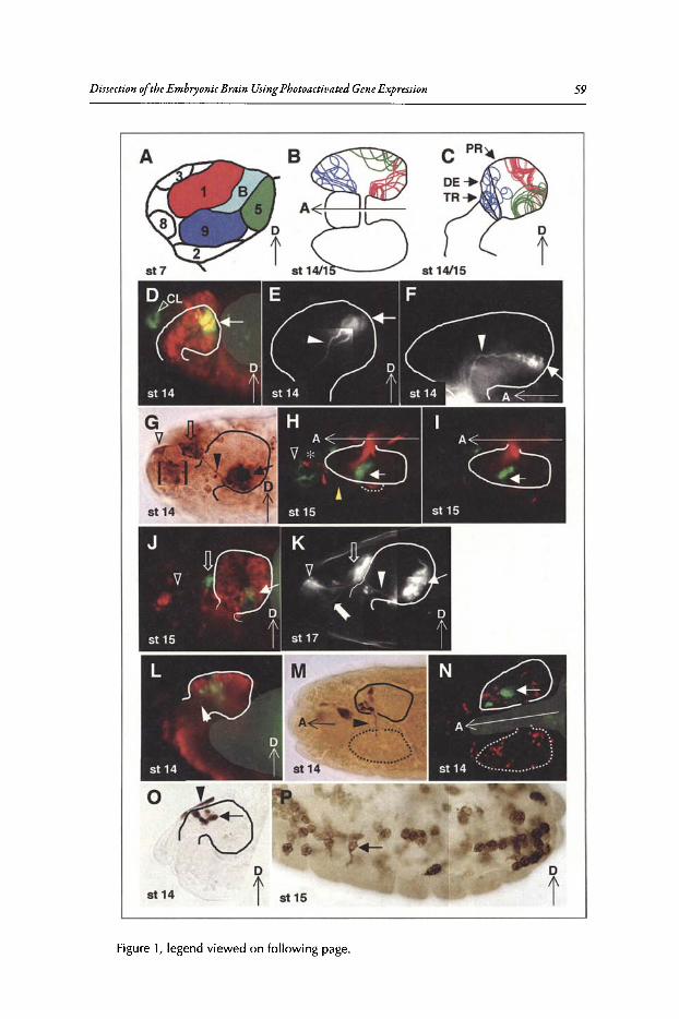
Dissection ofthe Embryonic Brain UsingPhotoactivatedGeneExpression
©D
st 14 t st 15
Figure 1, legend viewed on following page.
59
60 Brain Development in Drosophila melanogascer
Figure 1, viewed on previous page. Fate mapping of 01, 05, and 09 cells. For all figures, theembryo anterior is to the left. Dorsally mounted embryos are indicated by a horizontal arrowpointing to the anterior, marked A. Laterally mounted embryos are indicated by a verticalarrow pointing dorsally, marked D. The stage of the embryo is indicated in the bottom leftcorner of each panel. A) Schematic representation of the head mitotic domains at stage 7,which was used to guide photoactivation experiments. B,C) Cartoon of the regions within theembryonic brain that are populated by each mitotic domain. Each line represents results froman individual embryo (ol-red n =31, OS-green n =21, 09-blue n =31). The remaining panelsshow micrographs of photoactivated embryos. The affected brain hemisphere is bounded bya solid line. D-F) Photoactivation of 01. D) 2-4 cell photoactivation of a UAS-nGFPembryostained with anti-GFP antibody (green) and anti-ELAV (red). The 01 derived cells are visiblein both the brain (solid arrow) and the clypeolabrum (CL). E,F) 2-4 cell photoactivation ofUAS-tauGFPembryos. The arrowheads indicate the pioneer axons of the embryonic peduncle.G-K) Photoactivation of 05. All images are of 5-8 cell photoactivations. G) A photoactivatedUAS-lacZ embryo showing the four different 05 structures: the posterior group within thebrain (solid arrow), the middle group just anterior to the brain (open arrow), the anteriorgroup (open arrowhead) and the epithelial group (bracketed). The axon connecting theposterior and middle groups is indicated by the closed arrowhead. H,I) A UAS-lacZ embryostained with antibodies against f3-galactosidase (green) and Fasll (red). H) Composite imageof 3 adjacent optical sections showing Fasll-positive: optic lobe (outlined with dashed line),Bolwig's organ (asterisk) and Bolwig's nerve (yellow arrowhead). GFP-expressing, 05 braincells are in a different focal plane (solid arrow) that is adjacent to optic lobe. The 05 anteriorgroup (open arrowhead) is adjacent to Bolwig's organ. I) An in-focus optical section of theGFP-positive 05 cells within the brain (solid arrow). J) A 05 photoactivated UAS-nGFPembryostained with anti-GFP (green) and anti-ELAV (red; using the same arrow scheme as panel G).K) Composite of projected images of a os-photoactivated, UAS-tauGFP embryo (using thesame arrow scheme as panel G). The bifurcated axon tract projecting to the maxillary complex is indicated by a notched arrow. L-P) Photoactivation of 09. L) 2-4 cell photoactivationof a UAS-lacZ embryo stained with anti-f3-galactosidase (green) and anti-ELAV (red). Theclosed arrowhead indicates an axon extending toward the ventral nerve cord. M) Single cellphotoactivation of a UAS-lacZ embryo histochemically stained with anti-f3-galactosidase.The closed arrowhead indicates an axon extending to contralateral brain hemisphere. N) 2-4cell photoactivation of a UAS-lacZ embryo stained with anti-f3-galactosidase (green) andanti-Repo (red). The arrow indicates the patch of f3-galactosidase-positive cells that were notexpressing Repo. 0) Single cell photoactivation of 09 in a UAS-lacZ embryo histochemicallystained with anti-f3-galactosidase showing marked epidermal (closed arrowhead) and braincells (closed arrow). P) 5-8 cell photoactivation of 09 in a UAS-lacZ embryo histochemicallystained with anti-f3-galactosidase showing marked migratory cells (arrow) throughout theentire embryo. Yolk auto-fluorescence which appears in the green fluorescence channel ismasked gray in D, J, Land N. Reprinted from: Robertson K et al. Dev Bioi 2003; 260:124-137;©2003 with permission from Elsevier."
this region contributed mostly to the clypeolabrum (Fig. ID, see the green fluorescent cells outsideof the brain as indicated by the letters CL). The posterior-dorsal region gave rise to cells locatedpredominantly in the anterior-medial part ofthe protocerebrum (when referring to brain location,we use the neuroaxis as the frame ofreference) (Fig. 1D- F and cartooned in Fig. 1B,C, see the areasbounded by the red lines). These results indicate that 01 is divided into two sub-regions.
In the protocerebrum, marked 01 cells populated two adjacent clusters of cells. These cellsco-labeled with the pan-neuronal marker, ELAV (Fig.ID, arrow).'? In contrast, very few oI-derivedcells expressed the glial cell marker, Repo." Less than 2%ofthe marked 01 cells were glia, indicating that 01 cells gave rise to neurons rather than bipotential progenitors. Time-lapse analysis ofphotoactivated 01 cells showed that these cells were internalized en mass. The mass then movedposteriorly along the midline to their final position in the protocerebrum (Fig. 2B).
The location and double cluster appearance of 01 neurons suggested that they may form theembryonic mushroom bodies. To further test this possibility, 01 axons were marked by photoactivation using UAS-tauGFP embryos. These TauGFP marked axons had the typical morphologyof the embryonic mushroom bodies {Fig. IE,F, arrowhead).19.2o
Dissection ofthe Embryonic Brain Using Photoactivated Gene Expression 61
Mitotic Domain 5 Produces the EmbryonicAntennalSystemMitotic domain 5,which is initially located just anterior to the cephalic furrow near the dorsal
midline (Fig. lA), produces four distinct cellpopulations; one epidermal and three neuronal (Fig.IG, 2A,B). Time-lapse analysis of photoactivated UAS-nGFP embryos revealed the complexmigration pattern of this mitotic domain (Fig. 2A). The photoactivated patch ofcells first elongated along the edge ofthe cephalic furrow adjacent to the maxillary segment (Fig. 2A, frame 1,stage 9). The most anterior-ventral cells remained in the epidermis and moved to the anterior tipof the embryo. As head involution began, the non-epidermal ~5 progeny became internalized atthe boundary between the mandibulary and maxillary segments and separated into two populations (Fig. 2A, frames 5-6, stages 13-14). The inward movement ofthese cells appeared to be viainvagination. One population, the posterior group, which ultimately forms the antennallobeof the brain, remained stationary at the mid-anterior region of the embryonic brain, while thesecond group migrated over the ventral surface ofthe developing brain (Fig. 2A, frames 6-7, stage14-15). This was followed by another splitting of cells from the second group, which migratedinto the position ofthe antennal sensory organ (Fig. 2A, frames 7, stage 15). This culminated inpopulations of "oJ20 posterior group, "oJ5-6 middle group and 2-4 anterior group cells; the numberofepidermal cellswas not determined.
Immuno-chemical staining of~5 photoactivated UAS-lacZ embryos showed that the threeinternalized populations were connected by axonal fibers (Fig. IG, solid arrowhead). All fourgroups of cells arising from ~5 are shown in Fig. 1G. The neuronal character of cells withinthese groups was revealed by counter-staining with anti-ELAV antibody; about half of thephotoactivated cells within the anterior and posterior groups expressed ELAV (Fig. IJ, arrow,open arrowhead). We further confirmed the neuronal nature ofthe ~5 derived brain cells,aswellas those ofthe anterior group, by photoactivating ~5 cells in UAS-tauGFP embryos. Tau-GFPhighlighted the axons ofthe posterior group within the brain, the axon tracts between the groupsand the structure of the most anterior group (Fig. lK). The axons of the ~5-derived brain cellscan also be seen extending into other parts ofthe brain (Fig. 1K). Many ofthese processes appearto terminate in the region of the brain populated by ~1 mushroom body precursors (compareFig. lK and E, which correspond to ~5 and ~1, respectively).
The pattern of~5 cell types was reminiscent of the cell types produced by ~20, which will bedescribed later.13 The morphogenetic movements of~5 and ~20 were also similar; but not identical, ~20 cells form a more elongated pattern prior to internalization. We confirmed that mitoticdomains 5 and 20 yielded different structures by photoactivating ~5 cells in UAS-lacZ embryosand immuno-stained for FasII and (3-galactosidase expression. FasII is expressed in the optic lobe,Bolwig's nerve and Bolwig'sorgan, but not antennal cells." FasII was not expressed in any ofthephotoactivated ~5 cells (Fig.lH,I). ~5-derivedbrain cells (Fig. IH,I, closed arrow) were adjacentto the optic lobe (Fig. IH and I, broken line); there was no overlap. Likewise,~5 cells in the anterior group (Fig. IH, open arrowhead) were adjacent to Bolwig'sorgan, not overlapping (Fig. IH,asterisk). Thus, ~5-derived cells do not contribute to any part ofthe visual system.
The morphology and position of the ~5-derived cells indicate that this mitotic domain givesrise to the antennal sensory system,where the anterior group corresponds to the antennal sensoryorgan and the posterior group, which is in the brain, corresponds to the antennallobe.
Mitotic Domain 9Produces ThreeApparently Unrelated Cell TypesA unique feature of~9 is that the entire cell population divides perpendicularly to the embry
onic surface during the 14th mitosis, creating two populations ofcells,predicted to be epidermaland brain.'? To ensure that both layers ofprogeny were marked, ~9 cellswere irradiated prior to,or during, the 14th mitosis. Three distinct cell types were derived from ~9: posterior brain (Fig.lL-N), dorsal midline epidermis (Fig. 10, closed arrowhead) and an unidentified population ofmigratory cells (Fig. IP).
To determine the lineage relationship ofthese three cellpopulations, different-sized patches ofcellswithin ~9 were photoactivated in UAS-lacZ and UAS-GFP embryos (Table 1). Time-lapse
62 Brain Development in Drosophila melanogaster
Table 1. Distribution ofdifferent cell types arising from {)9
Cell Type(s)*
EBME+BE+MB+ME+ B +M
Single CellPhotoactivation(Percent, n =29)
10317
45oo7
2-4 CellPhotoactivation(Percent, n =72)
4152113o2819
5-8 CellPhotoactivation(Percent, n =24)
oo1213o2946
*E indicates epithelial cells; B indicates brain cells; M indicates migratory cells.Different sized patches of 69 cells were photoactivated in UAS-lacZ embryos just prior to, or during,the 14th mitosis. The embryos were aged to stage 14 through 16 and stained with anti-B-galactosidaseantibody. Only embryos with multiple marked cells were scored. Reprinted from: Robertson K etal. Dev Bioi 2003; 260:124-137; ©2003 with permission from Elsevier,"
recordings showed that all three cell types experienced significant amounts ofcell death, makingit extremely difficult to draw firm conclusions about lineage relationships. The origin ofthe migratory cells is not clear. None of the clones were composed ofboth epithelial and migratory cells,indicating that epithelial cells do not give rise to migratory cellsdirectly. Thus, the migratory cellsare either derived from brain progenitors or delaminated directly from the blastoderm. A significantfraction ofembryos had marked migratory-only clones, particularlywith 2-4 cellphotoactivation,supporting the delamination hypothesis. The brain- and migratory-cell progenitors appear to beevenly distributed across ~9.
Time-lapse analysisrevealed that initially the brain and epidermal progenitors moved in unisonanterior and dorsally, before separating, leaving the epidermal cells at the dorsal midline (Fig. 2B,stage 12, blue hatching), while the brain progenitors continue to move posteriorly to their finallocation in the brain (Fig. 2B).All photoactivated ~9 brain cells expressed ELAV (Fig. lL); noneexpressed Repo (Fig. IN), indicating69-derived brain cellsare neurons, not glia. These neurons occupied the deutero-, proto- and tritocerebrum (Fig. 1C,L), thus, the formation ofthe three cerebralneuromeres does not appear to be specified by separate mitotic domains. In many embryos an axoncould be seen to project either through the tritocerebrum toward the ventral nerve cord (Fig. lL,closed arrowhead) or toward the contralateral hemisphere through the tritocerebral commissure(Fig. 1M, closed arrowhead). These structures are similar to those described by Therianos et al.22
Mitotic Domain 20 Generates the Entire Visual SystemMitotic domain 20 is the most posterior of the three dorsal head mitotic domains (Fig. 3A).
A small number of cells within ~20 was marked by photoactivating UAS-lacZ expression. Sincethe cells in ~20 divide much later than surrounding mitotic domains and most ofthe cells divideinside the cephalic furrow," it was difficult to distinguish ~20 cells as they divide. Therefore, ~20cells were identified as those cells surrounded by the amnioserosa and mitotic domains 5, 18 andB (Fig. 3A, green circle).
Photoactivating cells in the center of~20 gave rise to a set ofbilaterally symmetrical structuresspanning from the anterior tip to the brain (Fig. 3B), including many head sensory organs andnerves of the peripheral nervous system (PNS), the posterior part of the brain, and the dorsalpouch epithelium above the clypeolabrum. Activation of~20 cells also gave rise to a significantamount ofcellular debris, indicating that some cells were dying. Photoactivation procedure doesnot affect cell death patterns."
Dissection of the EmbryonicBrain Using Pbotoactiuated GeneExpression 63
Figu re 2. Time-lapse images and cartoon of UAS-nCFP embryo followingphotoactivation. Column A) A seriesof images from a time-lapse recording of a 05 photoact ivated embryo.The GFP fluorescence is shown innegative so that marked cells appearblack overlaying transmitted light images. Lateral view of stages 9-16 asa projection of seven 5 lim opticalsections. Column B) Diagrammaticrepresentation of the position of theprogenyfrommitoticdomains1, 5, and9 from stage 9 to 16 shown as a lateral view. This series was constructedfrom multipletime-lapse experiments(ol- red, oS-green, 09-blue, the brainis outlined in black). Reprinted from:Robertson K et al. Dev Bioi 2003;260:124-137; ©2003 with permissionfrom Elsevier."

64 Brain Development in Dro sophila melanogasrer
B '\
Figure 3. Fate mapping of lI20. A) Individual cells of the dorsal head mitotic doma ins werevisualized by the expression of nuclear GFP (Ubi-CFPnls) by confocal microscopy. lI20 ishighlighted with a gray border. The numbers indicate mitotic domains. Interphase nuclei appear bright and have sharp edges while mitotic cells are large and appear diffuse. The whitearrows point to the cephalic furrow. The gray circle indicates a typical size and location of theUVphotoactivation beam. All embryos are shown with anterior to the left. B) Fatesof mitoticdomains were visualized by GAL4 dependent activation of lacZ usingthe photoactivated geneexpression system. Shown here is a dorsal view of a stage 17 embryo with photoactivatedlI20cells. Cells in the posterior part of the brain (white arrow) and head PNS (including axonsprojecting to the brain; arrows) were marked as well as cellular debris (arrow heads). CoG)Developmental time-course oflI20. Dorsal(C)and lateral(Dto G)imagesoflI20-photoactivatedembryos. The marked cells were visualized with an anti-f-galactosldase antibody. C)The cellsin lI20 moved away from the dorsal midline during germband extension. D) At late stage 11 ,the cells reached the dorsal border of the gnathal segments. The first sign of cell death wasapparent as a small spot moving away from the group of marked cells (arrow). E) At stage 13,the marked lI20cells extended along the lateral surface. F) During stage 14, the ventral cellscont inued to move anteriorly into the stomodeal invagination. G)Atthe end ofembryogenesis,cells were distributed into three clusters (arrowheads); the anterior tip, dorsal pouch and thebrain, connected via nerve-like projections (arrow). Reprinted from: Namba R, Minden JS .Dev Bioi 1999; 212:465-476; '01999 with permission from Elsevier."
Dissection ofthe EmbryonicBrain UsingPhotoactiuated GeneExpression 65
(\20 starts as a single domain on the dorsal midline. Duringgerm band extension, the cellsof(\20moved bilaterally away from the dorsal midline (Fig . 3C). By two hours after photoactivation atstage 7, most (\20 cells had migrated laterally away from the dorsal midline where they formed thedorsal border ofthe gnathal segments (Fig. 3D). By the end ofstage 13, the cells formed a narrowstrip spanning from the ventral to the dorsal surface (Fig. 3E) . At the end ofembryogenesis, cellsin this narrow strip were distributed into three clusters spanning the entire length ofthe head (Fig.3F). The ventral (\20 cells moved anteriorly with the gnathal segments during stomodeal invagination (Fig. 3G) and eventually reached the anterior tip . The more dorsal (\20 cells formed the dorsalridge and became a part ofthe dorsal pouch,while some cells delaminated and occupied the ventralposterior part ofthe brain lobe. The cells in the brain lobe were usually connected to a cell clusterin the anterior tip of the embryo by long nerve-like projections (Fig. 3G arrow).
Photoactivation of(\20 marked a pair oflateral clusters ofcells in the dorsal pouch adjacent to thepharynx in the stage 16 embryo (Fig. 4A). This cluster projected a nerve to the posterior part ofthebrain and the entire projection path was marked by the lacZexpression. The location and morphology ofthis structure suggested that it was the larval photoreceptor organ, Bolwigs organ,which wasconfirmed by stainingwith a PNS-specific antibody, mAb 22C10.23
•24 Double staining (\20 activated
embryos for ~-galactosidase expression and with mAb22C 10 showed that the photoactivared (\20cells coincided with the Bolwig's organ, fasciculated axons ofthe Bolwig 's organ (Bolwig's nerve),and cells at the termini ofBolwig's nerve presumably in the optic lobe (Fig. 4B).
In addition to producing the larval visual system, some (\20 cells were observed to form anepithelium on top ofthe Bolwig'snerve projection path (Fig. 4, white arrows). The Bolwig's nerve
c
OL
D
BO
,.•
. OL
Figure 4.020 generates the larval visual system. A,B) Dorsal view of 020 marked embryos . A)~-galactosidase was expressed in the Bolwig's organ (BO), Bolwig's nerve (BN) and the opticlobe (Ol.). White arrows point to cells in the dorsal pouch on top of the Bolwig's nerve path. B)~-galactosidase expression (blue) in the larval visual system overlaps with PNSmarker expression as visualized with mAb22C10 (brown). C-E) Single-cell photoactivation of 020; markedcells were found in the developing larval visual system at stage 14 (C) and at stage 17 (D),both lateral views. They were confined to the Bolwig's organ (BO), the optic lobe (Ol.) and thedorsal pouch (arrows). E) A dorsal view of an embryo with marked cells in both optic lobes.Reprinted from : Namba R, Minden JS. Dev Bioi 1999; 212:465-476; ©1999 with permissionfrom Elsevier." A color version of this figure is available onl ine at www.eurekah.com .
66 Brain Development in Drosophila rnelanogascer
extends posteriorly from the Bolwig's organ located inside the dorsal pouch epithelium and makesa sharp, ventral turn near the posterior edge ofthe dorsal pouch to follow along the basal surfaceofthe brain into the optic lobe. The 620 cells that formed the epithelial structure were often foundat or near where the Bolwigs nerve made the ventral turn, which corresponds to the location ofthe eye-antennal disc placode.
Photoactivation ofsingle cells in the center of620 gave rise to marked cells in the optic lobe,Bolwig's organ, and a small area of the dorsal pouch, presumably the eye-antennal disc placode,exclusively (Fig. 4C,D). This photoactivation typically marked the larval visual system either onthe left- or right-hand side of the embryo, while a small fraction of these embryos had marked,visual system cells on both sides ofthe embryo midline (Fig. 4E) .These results show that all ofthecell-types that make up the larval visual system can be derived from a single 620 cell.
Mitotic Domain B Generates Brain GliaProgeny of mitotic domains 1, S and 9 populated almost all of the brain volume (Fig. IB,C).
None ofthese mitotic domains generated significant numbers ofglial cells. Photoactivation oftheremaining mitotic domain,6B, revealed a major source ofbrain glia. Photoactivation ofcellsin threelocations along the length ofth iselongated mitotic domain (Fig. IA) in UAS-nGFP embr yos revealedthat their progeny formed small clusters ofcells in the presumptive protocerebrum at stage 14 (Fig.SA, solid arrow). The distribution ofthese clusters in the stage 14 embryonic brain is diagrammedin Figure SE,F.These clusters were located deep within the brain and were variable in size. Each ofthe clusters ofmarked 6B cellswas surrounded by dispersed cells (Fig. SA and cartooned as dots in
Figure 5. Brain glia originate from oB. A-C) 2-4 cell photoactivation of oB in a UAS-nGFPembryo stained with anti-GFP (green) and anti-Repo (red). A) Shows the green fluorescentchannel. A cluster of GFP-positive cells below the focal plane (indicated by the solid arrow)that is surrounded by individual cells (solid arrowhead) . B)Shows the anti-Repo signal revealing glial cells. C) Shows the superposition of A and B. Notice the double labeled cells (solidarrowhead) and Repo-only glial cells (open arrowhead) . D) 2-4 cell photoactivation of oB in aUAS-nGFP embryo stained with anti-GFP (green) and anti-ELAV (red). Not ice that none of theGFP-positive cells also express the neuronal marker, ELAV. E,F) Schematic representations ofmarked oBcells within the brain , lateral and dorsal views, respectively. The blue outlined areasrepresent marked clusters; the blue dots represent isolated cells. Reprinted from: RobertsonK et al. Dev Bioi 2003; 260:124-137; ©2003 with permission from Elsevier."
Dissection oftheEmbryonicBrain UsingPh0toactioated GeneExpression 67
Fig. SE,F). Three-quarters of~Bphotoactivated embryos had marked, dispersed brain cells that alsoexpressed Repo, indicating that they were glial cells (Fig. SA-C). None of the marked cells in bBphotoactivated embryos expressed ELAV (Fig. SD), indicating that they are unlikely to be neurons.There are two classesofembryonic brain glia: the subperineural glia that are mostly located in thebrain periphery and the neuropil glia.16 Glial cells arising from bB were identified as subperineuralglia by their position. Neuropil glia were never observed, suggesting that this subtype ofglial cellsmay arise from a different source.
ConclusionThe most difficult aspect offate mapping the head region ofthe Drosophila embryo is its com
plex morphogenesis. We have fate mapped the majority ofmitotic domains within the Drosophilaprocephalic blastoderm using the photoactivated gene expression system and determined that theembryonic brain develops from five mitotic domains: bl (posterior-dorsal part), bS, ~9, ~20 andbB. The final position ofthe mitotic domain progenywithin the brain does not reflect their relativeblastoderm positions. Thus, the mitotic domains follow specific morphogenetic trajectories. Severaldifferent mechanisms are employed to internalize brain progenitors: the posterior-dorsal part ofbland bB invaginate en mass, ~S and b20 also invaginate together, and ~9 uses oriented mitosisand possibly delamination. Together, these mitotic domains constitute non-overlapping regionsofthe brain. This fate map will provide an avenue for performing region-specific experiments. Thediscrete behavior ofthe brain-forming mitotic domains raises several interesting questions aboutthe ancestral origin ofthe brain. One such question is, did the various brain compartments evolvefrom a common group ofcells and later specialize or did the compartments evolve independentlyand later coalesce to form the brain?
References1. Foe VE, Odell GM, Edgar BA. Mitosis and morphogenesis in the Drosophila embryo. In: Bate M,
Martinez AA, eds. The Development of Drosophila melanogaster. New York: Cold Spring HarborLaboratory Press, 1993:149-300.
2. St. Johnston D. Pole plasm and the posterior group genes. In: Bate M, Martinez AA, eds. The Development of Drosophila melanogaster. New York: Cold Spring Harbor Laboratory Press, 1993:325-364.
3. Hartenstein V, Technau GM, Campos-Ortega JA. Fate-mapping in wild-type Drosophila melanogaster.III. A fate map of the blastoderm. Rouxs Arch Dev Biol 1985; 194:213-216.
4. Jurgens GR, Lehman M, Schardin M et al. Segmental organization of the head in the embryo of Drosophila melanogaster. A blastoderm fate map of the cuticle structures of the larval head. Rouxs ArchDev BioI 1986; 195:359-377.
5. Underwood EM, Turner FR, Mahowald AP. Analysis of cell movements and fate mapping during earlyembryogenesis in Drosophila melanogaster. Dev Biol 1980; 74(2):286-301.
6. Bossing T, Technau GM. The fate of the CNS midline progenitors in Drosophila as revealed by a newmethod for single cell labelling. Development 1994; 120(7):1895-1906.
7. Technau GM, Campos-Ortega JA. Fate-mapping in wild-type Drosophila. II. Injections of horseradishperoxidase in cells of the early gastrula stage. Roux's Arch Dev Biol 1985; 194:196-212.
8. Gehring WJ, Wieschaus E, Holliger M. The use of 'normal' and 'transformed' gynandromorphs inmapping the primordial germ cells and the gonadal mesoderm in Drosophila. J Embryol Exp Morphol1976; 35(3):607-616.
9. Janning W Aldehyde oxidase as a cell marker for internal organs in Drosophila melanogaster. Naturwissenschaften 1972; 59(11):516-517.
10. Foe VE. Mitotic domains reveal early commitment of cells in Drosophila embryos. Development 1989;107(1):1-22.
11. Minden JS, Agard DA, Sedat J~ et al. Direct cell lineage analysis in Drosophila melanogaster bytime-lapse, three-dimensional optical microscopy of living embryos. J Cell Biol 1989; 109(2):505-516.
12. Cambridge SB, Davis RL, Minden JS. Drosophila mitotic domain boundaries as cell fate boundaries.Science 1997; 277(5327):825-828.
13. Namba R, Minden JS. Fate mapping of Drosophila embryonic mitotic domain 20 reveals that the larvalvisual system is derived from a subdomain of a few cells. Dev Biol 1999; 212(2):465-476.
14. Brand AH, Perrimon N. Targeted gene expression as a means of altering cell fates and generatingdominant phenotypes. Development 1993; 118(2):401-415.
68 Brain Development in Drosophila melanogaster
15. Robertson K, Mergliano ], ·Minden ]S. Dissecting Drosophila embryonic brain development usingphotoactivated gene expression. Dev Bioi 2003; 260(1):124-137.
16. Hartenstein Y, Nassif C, Lekven A. Embryonic development of the Drosophila brain. II. Pattern ofglial cells.] Comp Neuro11998; 402(1):32-47.
17. Koushika SP,Lisbin M], White K. ELAY, a Drosophila neuron-specific protein, mediates the generationof an alternatively spliced neural protein isoform. Curr Bioi 1996; 6(12):1634-1641.
18. Halter DA, Urban], Rickert C et al. The homeobox gene repo is required for the differentiation andmaintenance of glia function in the embryonic nervous system of Drosophila melanogaster. Development 1995; 121(2):317-332.
19. Tettamanti M, Armstrong, D], Endo K et ale Early development of the Drosophila mushroom bodies,brain centers for associative learning and memory. Dev Genes Evo11997; 207:242-252.
20. Kurusu M, Awasaki T, Masuda-Nakagawa LM et al. Embryonic and larval development of the Drosophila mushroom bodies: concentric layer subdivisions and the role of fasciclin II. Development 2002;129(2):409-419.
21. Holmes AL, Heilig ]S. FasciclinII and Beaten path modulate intercellular adhesion in Drosophila larvalvisual organ development. Development 1999; 126(2):261-272.
22. Therianos S, Leuzinger S, Hirth F et al. Embryonic development of the Drosophila brain: formationof commissural and descending pathways. Development 1995; 121(11):3849-3860.
23. Fujita SC, Zipursky SL, Benzer S et al. Monoclonal antibodies against the Drosophila nervous system.Proc Nat! Acad Sci USA 1982; 79(24):7929-7933.
24. Goodman CS, Bastiani M], Doe CQ et al. Cell recognition during neuronal development. Science 1984;225(4668):1271-1279.
CHAPTERS
Design ofthe Larval Chemosensory SystemReinhard F. Stocker"
Abstract
Given that smell and taste are vital senses for most animal species, it is not surprising thatchemosensation has become a strong focus in neurobiological research. Much of whatwe know today about how the brain "mirrors" the chemical environment has derived
from simple organisms like Drosophila. This is because their chemosensory system includes onlya fraction of the cell number of the mammalian system, yet often exhibits the same basic design.Recent studies aimed at establishing fruitfly larvae as a particularly simple model for smell andtaste have analyzed the expression patterns ofolfactory and gustatory receptors, the circuitry ofthe chemosensory system and its behavioral output. Surprisingly, the larval olfactory system sharesthe organization ofits adult counterpart, though comprising much reduced cell numbers. It thusindeed provides a "minimal" model system of general importance. Comparing adult and larvalchemosensory systems raises interesting questions about their functional capabilities and aboutthe processes underlying its transformation through metamorphosis.
IntroductionThe sensesofsmell and taste create representations of the chemical environment in the brain.
Understandinghow the nervous system fulfills this amazing task-given the diversity ofmolecules,concentrations and blends-is a major challenge in neurobiology. A breakthrough in chemosensoryresearch was prompted by the identification ofodorant receptor genes in rodents,' in C. elegans,2and in Drosopbila.r' The expression patterns of these genes turned out to be an ideal tool fordissecting the olfactory circuits.l" These studies allowed to confirm earlier assumptions that theolfactory systems ofmammals and insects are organized according to common principles,"!' eventhough the insect brain comprises only a fraction ofthe cell numbers ofthe mammalian brain. It istherefore not surprising that insect species like Manduca sexta, Apismellifera and D. melanogasterhave become attractive models for investigating the chemical senses.
Does the larval chemosensory system of flies or other holometabolous insects offer an evensimpler alternative? Adults and larvae are anatomically and behaviorally much different, reflectingtheir different life-styles.Adult flies,for example, search for food, mates, and egg-layingsubstrates,all ofwhich requires sophisticated odor analysis. Fly larvae, in contrast, live directly on their food,and hence may not need long-range odor detection. Compatible with this notion, their olfactorysystem in terms of cell numbers is massively reduced. Nevertheless, its basic organization is surprisingly similar to the adult design, turning the Drosophila larva into a new, "elementary" modelsystem for srnell.!':"
*Dr. Reinhard F. Stocker-Department of Biology, University of Fribourg,l 0, Chemin du Musee,CH-1700 Fribourg, Switzerland. Email: [email protected]
Brain Developmentin Drosophila melanogaster, edited by Gerhard M. Technau.©2008 Landes Bioscience and Springer Science+ Business Media.
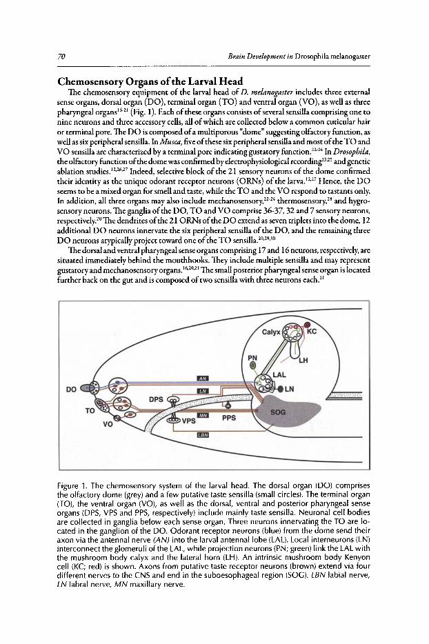
70 Brain Development in Drosophila melanoga srer
Chemosensory Organs ofthe Larval HeadThe chemosensory equipment of the larval head of D. melanogaster includes three external
sense organs, dorsal organ (DO), terminal organ (TO) and ventral organ (VO), as well as threepharyngeal organs 16.21(Fig. 1).Each of these organs consists ofseveral sensilla comprising one tonine neurons and three accessory cells, allofwhich are collected below a common cuticular hairor terminal pore . The DO is composed ofa multiporous "dome" suggesting olfactory function, aswell as six peripheral sensilla.InMusca, fiveofthese six peripheral sensilla and most ofthe TO andVO sensilla are characterized by a terminal pore indicating gustatory function.22.24In Drosophila,the olfactory function ofthe dome was confirmed by electrophysiological recording'P?and geneticablation studiesP·26.27 Indeed, selective block of the 21 sensory neurons of the dome confirmedtheir identity as the unique odorant receptor neurons (ORNs) of the larvap·27 Hence, the DOseems to be a mixed organ for smell and taste, while the TO and the VO respond to tastants only.In addition, all three organs may also include mechanosensory.P'" therrnosensory," and hygrosensory neurons. The ganglia ofthe DO, TO and VO comprise 36-37,32 and 7 sensory neurons,respectively," The dendrites ofthe 21 ORNs ofthe DO extend as seven triplets into the dome, 12additional DO neurons innervate the six peripheral sensilla of the DO, and the remaining threeDO neurons atypically project toward one ofthe TO sensilla.20,29,30
The dorsal and ventral pharyngeal sense organs comprising 17and 16neurons, respectively, aresituated immediately behind the mouthhooks. They include multiple sensilla and may representgustatory and mechanosensory organs .16,20,21 The small posterior pharyngeal sense organ is locatedfurther back on the gut and is composed oftwo sensilla with three neurons each."
Figure 1. The chemosensory system of the larval head. The dorsal organ (DO) comprisesthe olfactory dome (grey) and a few putative taste sensilla (small circles). The terminal organ(TO), the ventral organ (VO), as well as the dorsal, ventral and posterior pharyngeal senseorgans (DPS, VPSand PPS, respectively) include mainly taste sensilla. Neuronal cell bodiesare collected in ganglia below each sense organ. Three neurons innervating the TO are located in the ganglion of the DO . Odorant receptor neurons (blue) from the dome send theiraxon via the antennal nerve (AN) into the larval antennallobe (LAL). Local interneurons (LN)interconnect the glomeruli of the LAL, while projection neurons (pN; green) link the LAL withthe mushroom body calyx and the lateral horn (LH). An intrinsic mushroom body Kenyoncell (KC; red) is shown. Axons from putative taste receptor neurons (brown) extend via fourdifferent nerves to the CNS and end in the suboesophageal region (SOG). LBN labial nerve,LN labral nerve, MN maxillary nerve.
Larval Chemosensory System 71
Olfactoryprojections in the eNS are supra-oesophageal, whereas taste information issent to thesub-oesophageal ganglion (Fig. 1). Different from adults, all olfactory projections remain ipsilateral. Neurons from the DO ganglion, regardless oftheir modality and irrespective ofwhether theyextend to the DO or TO, connect to the brain via the antennal nerve. 20,31 The supra-oesophageallabral nerve carries the afferents from the dorsal and posterior pharyngeal sense organs, whereasthe sub-oesophageal maxillary and labial nerves comprise those from the TO and VO ganglia,and from the ventral pharyngeal sense organ, respectively.17,18,20,21
OlfactorySystem
Odorant Receptors and Their Expression PatternsOdorant receptors (0Rs) define the spectrum ofdetectable odors. Their expression pattern
across the population of0 RNs provides the basis for a combinatorial code in their target areas inthe brain which allows to interpret a practically unlimited number ofodors and odor mixtures,This seems to be true both for mammals'V' and for Drosophila. 3,4,7,8
In adult Drosophila, two related sub-families ofchemosensory receptors have been identified,an OR family comprising 62 members3,4,32,33 and a family of gustatory receptors (GRs) with 60members (see Gustatory Receptorsand TheirExpression Pattern).32-36 Similar to mammals, fly0 RNsexpress in general a single OR.3,4,37,38 For many ORs, odorant response spectra and expression patterns have been studied.F'" Afferents ofORNs expressing a given OR converge onto one or twoglomeruli in the antennal Iobe.Y'" analogous to the mammalian olfactory system. Hence, odorinformation carried by ORNs is translated into a pattern ofglomerular activation.v'"
The logic ofOrgene expression in the larval olfactorysystem, despite its simplicity, is surprisinglysimilar to the adult logic.12,13,27 For 2S Orgenes, expression was shown by in situ hybridization andvia Or-Gal4 driver lines (Table 1).12 However, there is evidence ofa few additional candidate larvalOrgenes.12,13,45 Each ofthe 21 larval 0 RNs expressesthe atypical receptor 0 R83b, known also fromadult flies,which is involved in proper localization and function of"conventional" 0 Rs.27,46,47 Thelarge majority of the ORNs express one conventional OR along with OR83b, while two ORNswere shown to coexpress two additional ORs apart from OR83b.12 Given that the number ofidentified ORs exceeds the total number ofORNs, a few more cases of triple OR expression areto be expected. Taken together, the number ofprimary olfactory "qualities" in the larva, reflectedby the number of0 Rs expressed, is considerably smaller compared to the approximately 60 qualities in adults. Interestingly, of the 2S well characterized larval Orgenes, 13 are larval-specific.P'"whereas the remaining 12 Orgenes are expressed in adults as well (Table 1).3,4,8,32,48
Using the "emtpy neuron approach', i.e., expressing single Or genes in adult anosmic mutantORNS,37-39 the electrophysiological responses of 11 larval ORs to a panel of 29 known adult orlarval stimulants26,38,39,49-51 were recorded.P'Ihe reaction spectra observed were very diverse, rangingfrom an 0 R that responded only to a single tested odorant, to 0 Rs which responded up to nineodorants." Odorants that elicited strong responses usually did so from multiple receptors. SomeoRs responded most strongly to aliphatic compounds, while others were preferentially tunedto aromatic compounds. Most of the responses were excitatory, but some ORs were stronglyinhibited by one compound and excited by another. Response dynamics and odor sensitivitiesvaried largely among different receptors. .
GlomerularArchitecture ofthe LarvalAntennalLobeThe larval olfactory circuitry is surprisingly similar to the adult circuitry, though much reduced
in terms ofcell numbers. Olfactoryafferents terminate in the larval antennallobe (LAL). Theirtargets are local interneurons, which provide lateral connections in the LAL, and projectionneurons (PNs), which link the LAL via the inner antennocerebral tract with higher order olfactory centers, the mushroom body (MB) calyx and the lateral horn (Fig. 1).15,20,52 Analogous tothe adult fly, larval ORNs and PNs seem to be cholinergic, whereas most or even all of the localinterneurons may be GABAergic.53
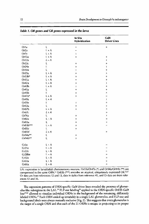
72 Brain Development in Drosophila melanogaster
Table 1. ORgenes and GRgenes expressedin the larva
In Situ Gal4Hybridization Driver Lines
Or1a L + +Or2a L+A +Or7a L +A +Or13a L+A + +Or22a L+A +Or22c L + +Or24a L + +Or30a L + +Or33a L +A + +Or33b* L + A + +Or35a L+A + +Or42a L+A + +Or42b L+A + +Or45a L + +Or45b L + +Or47a* L+A + +Or49a L+A +Or59a L + +Or63a L + +Or67b L +A + +Or67c L+A +Or74a L + +Or82a L+A + +Or83a L + +Or83b*** L +A + +Or85c L +Or85d L+A +Or94a** L +Or94b** L +
Gr2a L +A +Gr21a L+A +Gr22e L +A +Gr28be L+A +Gr32a L+A +Gr63a L+A +Gr66a L +A +
L/A: expression in larval/adult chemosensory neurons. Or33b/Or47a (*) and Or94a/Or94b (**) arecoexpressed in the same ORN.12 Or83b (***) encodes an atypical, ubiquitously expressed OR.12,27Or data are from references 12 and 13, data in italics from reference 45, and Gr data are from references 12 and 36.
The expression patterns of ORN-specific Gal4driver lines revealed the presence of glomerulus-like subregions in the LAL.20 FLP-out labeling'" applied to the ORN-specific Or83b-Ga14line27,42 allowed to visualize individual ORNs in the background of me remaining, differentlylabeled ORNs.1s Each ORN ended up invariably in a single LAL glomerulus, and FLP-out andbackground labels were alwaysmutually exclusive (Fig. 2).This suggests that every glomerulus isthe target of a single ORN and that each of me 21 ORNs is unique in projecting to its proper
Fig
ure
2.D
isse
ctio
nof
the
larv
alo
lfact
ory
path
way
by
sing
lece
llan
alys
is.(
A,A
')T
erm
inal
sof
two
od
ora
ntr
ece
pto
rne
uron
s(O
RN
s)in
ind
ivid
ual,
an
nota
ted
glo
me
ruli
ofth
eLA
Lat
the
3rd
larv
alin
star
,vi
sual
ized
by
FLP
-out
inth
eO
RN
-sp
eci
fic
Or8
3b-G
a/4
line.
OR
Ns
that
un
de
rwe
nt
FLP
-me
dia
ted
reco
mb
ina
tion
are
lab
eled
ingr
een
;th
ere
mai
nin
gO
RN
sar
ela
be
led
inm
agen
ta.
Mu
tua
llyex
clus
ive
gree
n/m
agen
tala
be
ling
dem
onst
rate
sth
atea
chgl
om
eru
lus
isth
eta
rget
ofa
sing
leO
RN
.(B
,B',B
")In
agre
em
ent
with
the
FLP
-ou
tda
ta,
GF
Pre
po
rte
rex
pres
sion
(gre
en)
dri
ven
byd
iffe
ren
tO
r-G
a/4
line
ssh
ows
that
each
OR
Nta
rget
sa
sing
le,
ind
ivid
ua
lLA
Lgl
om
eru
lus
(3ex
amp
les
show
n).
(C)
Glo
me
rula
rm
apof
the
LAL
base
don
the
term
inal
sof
dif
fere
ntO
r-G
a/4
lines
.(0
,0')
Sin
gle
pro
ject
ion
neur
ons
(PN
s)la
bele
dvi
aFL
P-o
ut
(in
gre
en)
onto
pof
othe
rP
Ns
(mag
enta
).Es
sen
tially
,ea
chP
Nlin
ksa
sin
gle
glo
me
rulu
sof
the
LAL
wit
ha
sing
legl
omer
ulu
sof
the
mu
shro
om
bo
dy
caly
x.T
hetw
opa
nels
show
that
for
man
yP
Ns,
ag
iven
inp
ut
glo
mer
ulu
sin
the
LAL
(e.g
.,P
3,M
10)
isco
rrel
ated
with
ap
arti
cula
rou
tput
glo
me
rulu
sin
the
caly
x(m
5,m
9).
(A,A
',D, D
':re
pri
nte
dfr
om
:R
amae
kers
A,
Mag
nen
atE,
Mar
inE
Cet
al.
Glo
me
rula
rm
aps
wit
ho
ut
cellu
lar
redu
ndan
cyat
succ
ess
ive
leve
lsof
the
Dro
sop
hila
larv
alo
lfact
ory
circ
uit.
Cur
rB
ioi
20
05;
15:9
82-9
92.
<020
06
wit
hpe
rmis
sion
from
Els
evie
r;B,
B',B
",C
:rep
rin
ted
fro
m:F
ishi
levi
chE,
Do
min
gos
AI,
Asa
hina
Ket
al.
Ch
em
ota
xis
beh
avio
rm
edia
ted
by
sin
gle
larv
alo
lfa
cto
ryne
uron
sin
Dro
soph
ila.C
urr
Bio
i20
05;
15:2
086
-209
6.<
020
06
with
perm
issi
onfr
omEl
sevi
er)
.
t' ., lii - 9 ~ § :s ..::! ~ ~ ~
74 Brain Development in Drosophila melanogaster
glomerulus among 21 spatially identifiable LAL glomeruli." Compatible with the FLP-out data,the axons of 0 RNs expressing Gal4under the control of 22 different Or gene promoters endedup in a different glomerulus each (Fig. 2).12.13 Moreover, combinations of two Or-Ga14 driverconstructs normally labeled two ORNs each of which projected to a different glomerulus. Anobvious exception were Orgenes coexpressed in the same 0 RN; these had a common glomerulusas a target. Having identified ligands for some of the ORs (see above), a map ofodor representation in the LAL was established," Accordingly, target glomeruli of receptors tuned to aliphaticcompounds and target glomeruli of receptors tuned to aromatic compounds appeared to clusterat distinct sites ofthe LAL.
Using the same FLP-out strategy as for ORNs, but in the PN-specific GH146-Ga14 driver,55the dendrites of larval PNs were found to be restricted to single LAL glomeruli, comparable tothe adult antennallobe (Fig. 2).15 UsingMARCM labeling," aminorityofPNs were found to bebi-glomerular.F Mutually exclusive FLP-out and background labels suggested that each glomerulus is innervated by a single GHl46-positive PN. Hence, the total number ofPNs may roughlymatch the total number of LAL glomeruli." The glomeruli recognized by PNs correspond tothose identified via the 0 RN terminals, indicating that LAL glomeruli meet the wiring criteriaoftypical insect glomeruli.
Glomerular Organization ofthe Mushroom Body CalyxThe adult MB calyx comprises hundreds of glomeruli.57 Adult PNs establish I-II terminal
boutons in variable calyxregions,54 each bouton probably correspondingto a single glomerulus. 57 Incontrast, the larval MB calyxconsists ofa small number ofwell-defined, relativelylarge glomeruli,"which has allowed to establish annotated glomerular maps. By expressing GFP-actin under thecontrol ofPN-specific and MB-specific Gal4lines or based on immunoreactivity patterns againstcholine acetyl transferase in the terminals of PNs, up to 34 calyx glomeruli were identified.":"Fine structural data suggest that each calyx glomerulus is filled by a large, bouton-like terminal ofa single PN.52Most of the PNs terminate in a single calyx glomerulus, except a minority ofPNswhich target two different glomeruli.14.15.52 Again, calyxglomeruli seem to be innervated by singleGHl46-positive PNS.15
A comparison of the input and output sites of PNs revealed at least seven types of PNs thatstereotypically link a specific LAL glomerulus with a specific calyx glomerulus (Fig. 2).15 Thus,the activity pattern set up in LAL glomeruli, as a result of ORN input and modulation by localinterneurons, seems to be rather faithfully transmitted to the calyx.This straightforward circuitryseems well suited for analyzing calyx function, although it remains to be shown whether strictinput-output correlations apply to all larval PNs.
FLP-out and MARCM labeling in MB-specific Gal4lines allowed to classify MB y neurons(the only type ofmature MB neurons present in the larva'"] according to their dendritic patternsin the calyx. While a minority of these neurons establish dendritic projections in a single calyxglomerulus," most of them have multiple arbors in up to seven glomeruli.lv" When studyingthe MB y neuron progenies deriving from the four MB neuroblasts, specific subsets of calyxglomeruli appeared to be preferentially targeted to some extent." In terms of cell numbers,roughly 21 PNs (or perhaps a few more) may be confronted with an estimated 600 functionalMB y neurons (L. Luo, personal communication). Hence, the larval calyx, similar to its adulthomologue, is a site of divergence; 14,15 it is in fact the only such site along the larval olfactorypathway (see Fig. 3).
The Larval Olfactory Pathway: Possible Rules ofOdor CodingAs shown above, larval ORNs express only one or two Orgenes along with the ubiquitously
expressed Or83bgene.12.13.27 This is similar to adult flies and mammals but differs from C. elegans,in which ORNs express multiple ORS.59 By using "subtractive" and "additive" ORN strategies,possible rules of olfactory coding were investigated in larval chemotaxis assays." In the firststrategy, in which selected ORNs were genetically ablated via toxin expression, two types of
Larval Chemosensory System 75
results were obtained. Animals lacking the ORIa-expressing neuron or the OR49a-expressingneuron showed reduced chemotaxis to only one of 20 odors tested. This mild effect is consistent with the broad and overlapping ligand tuning of many ORNs in adults.'? and larvae.'? Incontrast, loss of the neuron expressing OR42a resulted in behavioral anosmy to four of the 20odors. In the additive approach, larvae with one or two functional ORNs were generated usingOrla, Or42aor Or49adriver lines.'? Consistent with the stronger OR42a-ablated phenotype,OR42a-functionallarvae responded behaviorally to 22 of 53 odors tested (compared to 36 inthe wildtype), including three offour odors to which OR42a-ablated animals are anosmic. Thebroad response profile for OR42a-functionallarvae is in agreement with the broad ligand tuningofthis receptor.P'" In contrast, ORIa- and OR49a-functionallarvae did not exhibit significantchemotaxis to any of the 53 odors, consistent with the weak phenotype of the correspondingablated larvae and with electrophysiological responses." Animals with two functional ORNs(ORIa/OR42a) responded to a somewhat different subset of odors than larvae having eithersingle functional neuron alone.F
The minimal effects on chemotaxis observed after ablating the ORIa or OR49a neuronssuggest a certain degree of functional redundancy. This sounds surprising, given the smallnumber of ORNs in the larval system. Yet, subtle effects exerted by seemingly "unimportant"neurons could be crucial for cooperative processes. On the other hand, the OR42a neuron playsa particularly important role; it is sufficient to initiate chemotaxis to many odors, and its lossleads to severe behavioral defects. Finally, cooperativity is suggested by the modified responsesof ORIa/OR42a-functional animals compared to the single functional animals. Olfactorycoding thus does not simply rely on additive activation of 21 parallel pathways, but involveslateral interactions as well. Cross-talk may occur in particular via the local interneurons in theLAL. 1s Transformation of olfactory signals is known from the antennallobe of a number ofinsects including Drosophila.6
0-63 Integration of olfactory information may sharpen quantitative and qualitative parameters, such as detection threshold and odor discrimination. Whilechemotaxis assaysdo not answer how odors are distinguished from each other, it is reasonableto assume that integrative processes may be particularly crucial ifvery few channels have to dealwith many odors.
Further processing occurs in higher brain centers, such as the MBs. The different classes oflarval MB y neurons, innervating various numbers of calyx glomeruli, obviously allow differentmodes ofsignal transfer. Uniglomerular MB y neurons may be involved in elementary coding ofodor features, whereas multiglomerular MB y neurons receiving input from several PNs may actas coincidence detectors.14
,IS.64.6S Hence, although both LAL and larval calyx are glomerular, thelogic ofconnectivity is different. LAL glomeruli exhibit stereotypic connectivity between definedoRNs and PNs, whereas calyxglomeruli show stereotypic input but mostly nonstereotypic, highlycombinatorial MB y neuron output. 14
Distinctive Features ofLarvalandAdult Olfactory CircuitsWhereas the general design of the larval olfactory pathway is similar to its adult counter
part, larval 0 RNs and most (perhaps all) larval PNs appear to be unique, leading to an almostcomplete lack ofcellular redundancy (Fig. 3). Consequently, any cell loss should affect olfactoryfunction more severely than in the adult system. Moreover, the presence of no more than 21ORNs and 21 LAL glomeruli suggests that the number of primary olfactory qualities in thelarva is largely reduced compared to adults comprising about SO glomeruli.r"Also, given theuniglomerular projections of ORNs and PNs and the almost equal number of ORs, ORNs,LAL glomeruli, PNs and calyx glomeruli, the larval olfactory pathway lacks convergent anddivergent connectivity up to the calyx and is organized in a 1:1:1:1:1 manner. This contrasts withthe adult olfactory pathway, in which 1,300 ORNs converge onto about SO glomeruli, whichdiverge again to approximately 1SO PNs and hundreds ofcalyx glomeruli.67,68 Convergence andcellular redundancy in sensory systems are known to increase the signal-to-noise ratio, whereasdivergent connectivity very likely improves signal discrimination. In the larval olfactory system,
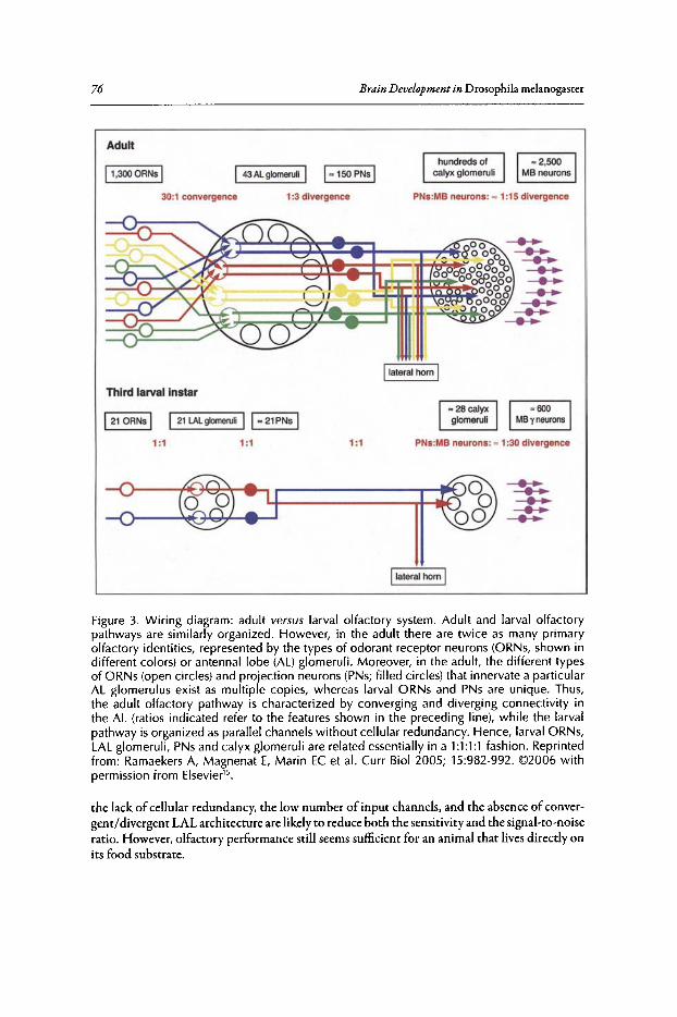
76
Adult
Brain Development in Drosophila melanogascec
11,300 CRNs I I43 AL glomeruli I 1 ~1 50 PNSIhundr eds of
ca lyx glomeru li
30:1 convergence 1:3 divergence PNs:MB neurons: = 1:15 divergence
Ilateral hom IThird larval Instar
I21 CRNs I I 21 LALglomeruli I I~ 21PNs I1 :1 1 :1 1 :1 PNs:MB neurons: = 1:30 divergence
Ilateral horn I
Figure 3. Wiring diagram: adult versus larval olfactory system. Adult and larval olfactorypathways are similarly organized . However, in the adult there are twice as many primaryolfactory identities, represented by the types of odorant receptor neurons (ORNs, shown indifferent colors) or antennal lobe (AL) glomeru li. Moreover, in the adult, the different typesof ORNs (open circles) and projection neurons (pNs; filled circles) that innervate a particularAL glomerulus exist as multiple copies, whereas larval ORNs and PNs are unique . Thus,the adult olfactory pathway is character ized by converging and diverging connectivity inthe AL (ratios indicated refer to the features shown in the preceding line), while the larvalpathway is organized asparallel channels without cellular redundancy . Hence, larval ORNs,LAL glomeruli, PNs and calyx glomeruli are related essentially in a 1:1 :1 :1 fashion. Reprintedfrom : Ramaekers A, Magnenat E, Marin EC et al. Curr Bioi 2005; 15:982-992. ©2006 withpermission from Elsevier" .
the lack of cellular redundancy, the low number of input channels, and the absence ofconvergent/divergent LAL architecture are likely co reduce both the sensitivity and the signal-co-noiseratio. However, olfactory performance still seems sufficient for an animal that lives directly onits food substrate.
Larval Chemosensory System 77
Gustatory System
Gustatory Receptors and Their Expression PatternIn contrast to smell, taste deals with a very limited number ofqualities, like "sweet" or "bitter"
This characteristic isnot due to a smaller diversity ofexisting tastants, but to the specific rationale ofthe gustatory system, which isdesigned for classifyingsubstances rather than identifyingparticularmolecules. This is particularly true for potentially harmful (bitter) compounds: they are chemically very diverse but should trigger the same behavioral response, Le.,aversion. It is therefore notsurprising that in mammals receptor neurons tuned to such compounds express multiple typesofgustatory receptors (GRS),69 suggesting that the capacity of these cells to distinguish betweendifferent bitter substances is limited.
In adult Drosophila, evidence suggests that the GR family (see OdorantReceptors and TheirExpression Patterns) mediates both sweet and bitter responses. Yet,because oflow expression levels,Grexpression patterns were studied exclusivelyby Grgene promoter-Gal4analysis.35.36 Similar tomammals, neurons responding to sugars appear to express onlyone or a few GRs, whereas neuronsthat bind bitter compounds express multiple GRS.70.71 This design allows to establish distinct attractive and aversive gustatory pathways. Surprisingly, three GRs are expressed on the antenna,suggesting that GRs are not strictly associated with taste function." Indeed, the Gr21agene isexpressed in CO2-sensitivecells ofthe antenna."
From the few Grgenesthat have been studied in the larva by promoter-Gal4 analysis,I2,36 Gr2a,Gr21a, Gr22e, Gr28be, Gr32aand Gr66a-all of which are expressed also in the adult-showexpression in one or two neurons ofthe TO (Table 1). Gr2alabels in addition two non-olfactoryneurons of the DO. GR22e, GR28be, GR32a and GR66a were suspected to represent "bitter"receptors in the adult, as they are coexpressed in many neurons.Y" However, reporter expressiondriven by the gene promoter pairs Gr66a/Gr21a or Gr66a/Gr32a labeled two larval neurons each.36Yet,the small number ofavailabledata does not allow to draw any conclusion about the numbers ofGRs expressed by individual neurons. Interestingly, the putative COrreceptor GR21a (see above),is expressed in the TO. Furthermore, Gr2aand several Orgene members (Or30a, Or42a, Or49a,Or63a), are expressed in both DO and TO.12.13.36 Thus, as in adults, gene family membership andsite ofexpression are not strictly linked. Finally, salt detection is mediated by degenerin/epithelialNa + channels, which are expressed in the TO as well as in adult taste bristles."
Primary Gustatory CentersLittle is known about the organization of primary taste centers, mainly because they lack
discrete glomerular architecture. In the adult, gustatory afferents from the pharynx, labellumand legs terminate in distinct regions of the suboesophageal ganglion (SOG).70.71 Neurons fromsensilla on different body regions projecting to different SOG regions may express the same GR,suggesting that the same tastant may trigger different behaviors, depending on the stimulationsite. Labellar neurons expressing putative bitter receptors and labellar neurons expressing sugarreceptors establish distinct but overlapping projections in the SOG?O.71.74
In the larva, the few existing data do not allow any meaningful generalizations. Yet, one TOneuron expressing Gr32aand three other neurons expressing Gr66a-one from the TO and twofrom pharyngeal sense organs-were shown to project to the ipsilateral SOG.36Gr66aand Gr32aprojections are adjacent to each other but do not overlap. A third receptor, GR2a that is expressedin two DO neurons and one TO neuron, has two targets in the SOG.36This suggests again thata given tastant can elicit different behaviors depending on the stimulation site. It is also worthnotifying that the Gal41ines Or30a, Or42a, and Or49a (Table 1) are not only expressed in theDO and TO, but also label sensory terminals in the SOG.l3
Recently, a genetically defined subset ofapproximately 20 putative first-order gustatory targetneurons was identified in the larval SOG?S These neurons provide output to the protocerebrum,the ventral nerve cord, the ring gland and pharyngeal muscles. They express the hugin gene,which generates two neuropeptides, and which appears to be upregulated in the absence
78 Brain Development in Drosophila rnelanogaster
of the feeding-regulatory transcription factor klumpfuss (P [9036])and downregulated by aminoacid-deficient conditions. Also, blocking the output from hugin-expressingneurons increases feeding. Apparently these neurons integrate taste processing, the endocrine system, higher-order braincenters and motor output. The dopaminergic nature ofsome ofthe hugin-expressing neurons" renders them interesting candidates as regulators offeeding, the most striking behavior oflarvae.
The Drosophila Larva as a Model for Smell and TasteThe usefulness ofDrosophila fliesas an olfactory model system isobvious, given the genetic and
molecular tools available, the simplicity oftheir olfactory system in terms ofcell numbers and thestriking similarities with the mammalian olfactory system. Surprisingly, even the larval olfactorysystem shares the design ofthe mammalian system, in the simplest conceivable form (Fig. 3). Thelarva may thus turn into a higWy attractive "minimal" model for olfactory studies, in particularbecause it permits the generation ofanimals with a single functional 0 RN. In such larvae, odors,ORs, and ORNs can be directly correlated with behavioral output, allowing to track down theolfactory code to the level ofidentified receptor neurons.
The model character of Drosophila for the gustatory system is less obvious, both in adultsand larvae. Anatomically, the taste systems of mammals and insects are different. Yet, there area number of interesting parallels: (1) both insect and mammalian taste receptor neurons seemto be tuned to either attractive or aversive stimuli; (2) many more of the taste receptors may bededicated to repulsive ligands than to attractive ones and, (3) cells responding to attractive cuesseem to express only one or a few receptors, whereas those responding to bitter substances expressmultiple receptors.
The parallels in the chemosensorysystemsofvertebrates and insects are not necessarilyevidencefor a common ancestry. Rather, the similarities may reflect functional constraints for efficient smelland taste systems. Understanding these properties will surely aid understanding chemosensoryfunction. In this context, Drosophila with its simple nervous system combined with a wealth ofmolecular tools, will contribute to our comprehension ofsmell and taste in general.
AcknowledgementsI am very grateful to N. Gendre (Fribourg) for providing Figure 1,and to B. Gerber (Wtirzburg)
for comments on the manuscript. This work was supported by the SwissNational Fund (grants31-63447.00, 3100AO-IOSSI7 and 3234-069273.02) and the Roche Research Foundation.
References1. Buck L, Axel R. A novel multigene family may encode odorant receptors: a molecular basis for odor
recognition. Cell 1991; 65:175-187.2. Sengupta P, Chou ]H, Bargmann CI. odr-l0 encodes a seven transmembrane domain olfactory receptor
required for responses to the odorant diacetyl. Cell 1996; 84:899-909.3. Clyne P], Warr CG, Freeman MR et al. A novel family of divergent seven-transmembrane proteins:
candidate odorant receptors in Drosophila. Neuron 1999; 22:327-338.4. Vosshall LB, Amrein H, Morozov PS et al. A spatial map of olfactory receptor expression in the
Drosophila antenna. Cell 1999; 96:725-736.5. ResslerK], SullivanSL, Buck LB. Information coding in the olfactory system: Evidencefor a stereotyped
and highly organized epitope map in the olfactory bulb. Cell 1994; 79:1245-1255.6. VassarR, Chao SK, Sitcheran R et al. Topographic organization of sensory projections to the olfactory
bulb. Cell 1994; 79:981-991.7. Gao Q Yuan B, Chess A. Convergent projections of Drosophila olfactory neurons to specificglomeruli
in the antennallobe. Nat Neurosci 2000; 3:780-785.8. Vosshall LB, Wong AM, Axel R. An olfactory sensory map in the fly brain. Cell 2000; 102:147-159.9. Hildebrand]G, Shepherd G. Mechanisms of olfactory discrimination: converging evidence for common
principles across phyla. Annu Rev Neurosci 1997; 20:595-631.10. Strausfeld N], Hildebrand ]G. Olfactory systems: common design, uncommon origins? Curr Opin
Neurobiol1999; 9:634-639.11. Ache B~ Young]M. Olfaction: diverse species, conserved principles. Neuron 2005; 48:417-430.12. Fishilevich E, Domingos AI, Asahina K et al. Chemotaxis behavior mediated by single larval olfactory
neurons in Drosophila. Curr Bioi 2005; 15:2086-2096.

Larval Chemosensory System 79
13. Kreher SA, Kwon AY, Carlson JR. The molecular basis of odor coding in the Drosophila larva. Neuron2005; 46:445-456.
14. Masuda-Nakagawa LM, Tanaka NK, O'Kane CJ. Stereotypic and random patterns of connectivity inthe larval mushroom body calyxof Drosophila. Proc Nat! Acad Sci USA 2005; 102:19027-19032.
IS. RamaekersA, Magnenat E, Marin EC et al. Glomerular maps without cellular redundancy at successivelevels of the Drosophila larval olfactory circuit. Curr Biol 2005; 15:982-992.
16. Singh RN, Singh K. Fine structure of the sensory organs of Drosophila melanogaster Meigen Larva(Diptera: Drosophilidae), Int J Insect Morphol Embryol 1984; 13:255-273.
17. Schmidt-Ott U, Gonzalez-Gaitan M, jackie H et al. Number, identity, and sequence of the Drosophilahead segments as revealed by neural elements and their deletion patterns in mutants. Proc Nat! AcadSci USA 1994; 91:8363-8367.
18. Campos-Ortega JA, Hartenstein V. The Embryonic Development of Drosophila melanogaster. Berlin,Heidelberg, New York: Springer, 1997.
19. Singh RN. Neurobiology of the gustatory systemsof Drosophila and some terrestrial insects. Micr ResTechn 1997; 39:547-563.
20. Python F, Stocker RF. Adult-like complexity of the larval antennal lobe of D. melanogaster despitemarkedly low numbers of odorant receptor neurons. J Comp Neuro12002; 445:374-387.
21. Gendre N, Liier K, Friche S et al. Integration of complex larval chemosensory organs into the adultnervous system of Drosophila. Development 2004; 131:83-92.
22. Chu I~ Axtell RC. Fine structure of the dorsal organ of the house By larva, Musca domestica L. ZZellforsch Mikrosk Anat 1971; 117:17-34.
23. Chu-WangI~ Axtell RC. Fine structure of the terminal organ of the house Bylarva, Musca domesticaL. Z. Zellforsch Mikrosk Anat 1972; 127:287-305.
24. Chu-Wang, I~ Axtell RC. Fine structure of the ventral organ of the house Bylarva, Musca domesticaL. Z Zellforsch Mikrosk Anat 1972; 130:489-495.
25. Oppliger FY, Guerin PM, Vlimant M. Neurophysiological and behavioural evidence for an olfactoryfunction for the dorsal organ and a gustatory one for the terminal organ in Drosophila melanogasterlarvae. J Insect Physiol 2000; 46:135-144.
26. Heimbeck G, Bugnon V, Gendre N et al. Smell and taste perception in D. melanogaster larva: toxinexpression studies in chemosensory neurons. J Neurosci 1999; 19:6599-6609.
27. Larsson MC, Domingos AI, Jones WD et al. Or83b encodes a broadly expressed odorant receptoressential for Drosophila olfaction. Neuron 2004; 43:703-714.
28. Liu L, Yermolaieva 0, Johnson WA et al. Identification and function of thermosensory neurons inDrosophila larvae. Nat Neurosci 2003; 6:267-273.
29. Kankel DR, Ferrus A, Garen SH et al. The structure and development of the nervous system. In:Ashburner M, Wright TRF, eds. The Genetics and Biology of Drosophila. London, New York,San Francisco: Academic Press, 1980:295-368.
30. Frederik RD, Denell RE. Embryological origin of the antenno-maxillary complex of the larva ofDrosophila melanogaster Meigen. Int J Insect Morphol Embryol1982; 11:227-233.
31. Tissot M, Gendre N, Hawken A et al. Larval chemosensory projections and invasion of adult afferentsin the antennal lobe of Drosophila melanogaster.J Neurobiol1997; 32:281-297.
32. Robertson HM, Warr CG, CarlsonJR. Molecularevolution of the insect chemoreceptor gene superfamilyin Drosophila melanogaster. Proc Nat! Acad Sci USA 2003; 100:14537-14542.
33. Hallem EA, Dahanukar A, Carlson JR. Insect odor and taste receptors. Annu Rev Entomol 2006;51:113-135.
34. Clyne P], Warr CG, Carlson JR. Candidate taste receptors in Drosophila. Science 2000;287:1830-1834.
35. Dunipace L, Meister S, McNealy C et al. Spatially restricted expression of candidate taste receptors inthe Drosophila gustatory system. Curr Blol 2001; 11:822-835.
36. Scott K, Brady R Jr, Cravchik A et al. A chemosensory gene family encoding candidate gustatory andolfactory receptors in Drosophila. Cell 2001; 104:661-673.
37. Dobritsa AA, van der Goes van Naters~ Warr CG et al. Integrating the molecular and cellular basisof odor coding in the Drosophila antenna. Neuron 2003; 37:827-841.
38. Goldman AL, van der Goes van Naters ~ Lessing, D et al. Coexpression of two functional odorreceptors in one neuron. Neuron 2005; 45:661-666.
39. Hallem EA, Ho MG, Carlson JR. The molecular basis of odor coding in the Drosophila antenna. Cell2004; 117:965-979.
40. Bhalerao S, Sen A, Stocker RF et al. Olfactory neurons expressing identified receptor genes projectto subsets of glomeruli within the antennal lobe of Drosophila melanogaster. J Neurobiol 2003;54:577-592.
80 Brain Development in Drosophila melanogaster
41. Fiala A, Spall T, Diegelmann S et al. Genetically expressed cameleon in Drosophila melanogaster is usedto visualize olfactory information in projection neurons. Curr Biol 2002; 12:1877-1884.
42. Ng M, Roorda RD, Lima SQ et al. Transmission of olfactory information between three populationsof neurons in the antennallobe of the fly. Neuron 2002; 36:463-474.
43. Wang~ Wong AM, Flores] et al. Two-photon calcium imaging revealsan odor-evoked map of activityin the fly brain. Cell 2003; 112:271-282.
44. Yu D, Ponomarev A, Davis RL. Altered representation of the spatial code for odors after olfactoryclassical conditioning: memory trace formation by synaptic recruitment. Neuron 2004; 42:437-449.
45. Couto A, Alenius M, Dickson B]. Molecular, anatomical, and functional organization of the Drosophilaolfactory system. Curr Bioi 2005; 15:1535-1547.
46. Neuhaus E, Gisselmann G, Zhang W et al. Odorant receptor heterodimerization in the olfactory systemof Drosophila melanogaster. Nat Neurosci 2004; 8:15-17.
47. Benton R, Sachse S, Michnick SW et al. Atypical membrane topology and heteromeric function ofDrosophila odorant receptors in vivo. PLoS Biology 2005; 4(2):e20, DOl: 10.1371/journal pbio004002046.
48. Komiyama T, Carlson ]R, Luo L. Olfactory receptor neuron axon targeting: intrinsic transcriptionalcontrol and hierarchical interactions. Nat Neurosci 2004; 7:819-825.
49. Rodrigues V. Olfactory behavior of Drosophila melanogaster. In: Siddiqi 0, Babu P, Hall LM, Hall ]C,eds. Development and Neurobiology of Drosophila. New York, London: Plenum, 1980:361-371.
50. Monte P, Woodard C, Ayer R et al. Characterization of the larval olfactory response in Drosophila andits genetic basis. Behav Genet 1989; 19:267-283.
51. Cobb M. What and how do maggots smell? BioI Rev 1999; 74:425-459.52. Marin EC, Watts R], Tanaka NK et aI. Developmentally programmed remodeling of the Drosophila
olfactory circuit. Development 2005; 132:725-737.53. Python F, Stocker RF. Immunoreactivity against choline acetyltransferase, gamma-aminobutyric acid,
histamine, octopamine, and serotonin in the larval chemosensory system of Drosophila melanogaster.J Comp Neurol 2002; 453:157-167.
54. Wong AM, Wang~ Axel R. Spatial representation of the glomerular map in the Drosophila protocerebrum. Cell 2002; 109:229-241.
55. Stocker RF, Heimbeck G, Gendre N et al. Neuroblast ablation in Drosophila P[GAL4] lines revealsorigins of olfactory interneurons.] Neurobiol1997; 32:443-456.
56. Lee T, Luo L. Mosaic analysis with a repressible cell marker for studies of gene function in neuronalmorphogenesis. Neuron 1999; 22:451-461.
57. Yasuyama K, Meinertzhagen lA, Schiirmann F~ Synaptic organization of the mushroom body calyxin Drosophila melanogaster.] Comp Neurol 2002; 445:211-226.
58. Lee T, Lee A, Luo L. Development of the Drosophila mushroom bodies: sequential generation of threedistinct types of neurons from a neuroblast. Development 1999; 126:4065-4076.
59. Troemel ER, Chou ]H, Dwyer ND et al. Divergent seven transmembrane receptors are candidatechemosensory receptors in C. elegans. Cell 1995; 83:207-218.
60. SachseS, Galizia CG. Role of inhibition for temporal and spatial odor representation in olfactory outputneurons: a calcium imaging study, ] Neurophysiol2002; 87:1106-1117.
61. Lei H, Christensen TA, Hildebrand ]G. Spatial and temporal organization of ensemble representationsfor different odor classes in the moth antennallobe. ] Neurosci 2004; 24:11108-11119.
62. Wilson RI, Turner GC, Laurent G. Transformation of olfactory representations in the Drosophilaantennallobe. Science 2004; 30:366-370.
63. Wilson RI, Laurent G. Role of GABAergic inhibition in shaping odor-evoked spatiotemporal patternsin the Drosophila antennallobe. I Neurosci 2005; 25:9069-9079.
64. Perez-Orive ], Mazor 0, Turner GC et al. Oscillations and sparsening of odor representations in themushroom body. Science 2002; 297:359-365.
65. Heisenberg M. Mushroom body memoir: from maps to models. Nat Revs Neurosci 2003; 4:266-275.66. Laissue PP, Reiter C, Hiesinger PR et al. Three-dimensional reconstruction of the antennallobe in
Drosophila melanogaster.] Cornp Neuro11999; 405:543-552.67. Stocker RF. The organization of the chemosensory system in Drosophila melanogaster: a review. Cell
Tiss Res 1994; 275:3-26.68. Stocker RF. Drosophila as a focus in olfactory research: mapping of olfactory sensilla by fine struc
ture, odor specificity, odorant receptor expression and central connectivity. Micr Res Techn 2001;55:284-296.
69. Montmayeur ]P, Matsunami H. Receptors for bitter and sweet taste. Curr Opin Neurobiol 2002;12:366-371.
70. Thorne N, Chromey C, Bray S et al. Taste perception and coding in Drosophila. Curr Biol 2004;14:1065-1079.

Larval Chemosensory System 81
71. Wang Z, Singhvi A, Kong P et al. Taste representations in the Drosophila brain. Cell 2004;117:981-991.
72. Suh GSB, Wong AM, Hergarden AC et al. A single population of olfactory sensory neurons mediatesan innate avoidance behavior in Drosophila. Nature 2004; 431:854-859.
73. Liu L, Leonard AS, Motto DG et al. Contribution of Drosophila DEG/ENaC genes to salt taste.Neuron 2003; 39:133-146.
74. Marella S, Fischler~ Kong P et al. Imaging taste responses in the Bybrain reveals a functional mapof taste category and behavior. Neuron 2006; 49:285-295.
75. Melcher C, Pankratz MJ. Candidate gustatory interneurons modulating feeding behavior in theDrosophila brain. PLoS BioI 2005; 3(9):e305, DOl: 10.1371/journal pbio 0030305.
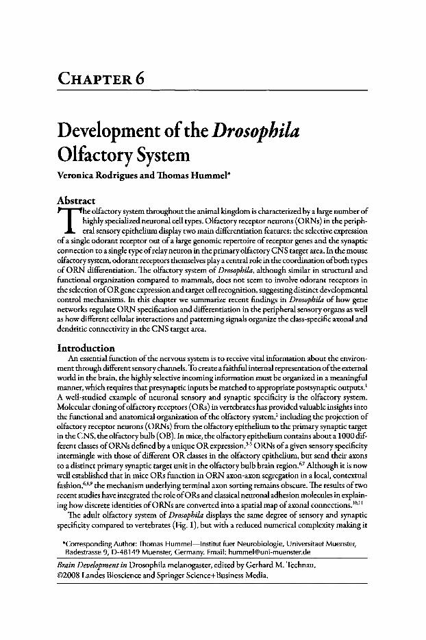
CHAPTER 6
Development oftheDrosophilaOlfactory SystemVeronica Rodrigues and Thomas Hummel"
Abstract
T he olfactory system throughout the animal kingdom is characterized by a large number ofhighly specialized neuronal cell types. Olfactory receptor neurons (0RNs) in the peripheral sensory epithelium display two main differentiation features: the selective expression
ofa single odorant receptor out ofa large genomic repertoire ofreceptor genes and the synapticconnection to a single type ofrelay neuron in the primary olfactory CNS target area. In the mouseolfactory system, odorant receptors themselves playa central role in the coordination ofboth typesof 0 RN differentiation. The olfactory system ofDrosophila, although similar in structural andfunctional organization compared to mammals, does not seem to involve odorant receptors inthe selection of0 R gene expression and target cell recognition, suggesting distinct developmentalcontrol mechanisms. In this chapter we summarize recent findings in Drosophila of how genenetworks regulate ORN specification and differentiation in the peripheral sensory organs as wellas how different cellular interactions and patterning signals organize the class-specific axonal anddendritic connectivity in the CNS target area.
IntroductionAn essential function ofthe nervous system is to receive vital information about the environ
ment through different sensorychannels. To create a faithful internal representation ofthe externalworld in the brain, the highly selective incoming information must be organized in a meaningfulmanner, which requires that presynaptic inputs be matched to appropriate postsynaptic outputs.'A well-studied example of neuronal sensory and synaptic specificity is the olfactory system.Molecular cloning ofolfactory receptors (0Rs) in vertebrates has provided valuable insights intothe functional and anatomical organization ofthe olfactory system.' including the projection ofolfactory receptor neurons (0RNs) from the olfactory epithelium to the primary synaptic targetin the CNS, the olfactory bulb (0B). In mice, the olfactory epithelium contains about a 1000 different classesofORNs defined by a unique OR expression.l" ORNs ofa given sensory specificityintermingle with those ofdifferent OR classes in the olfactory epithelium, but send their axonsto a distinct primary synaptic target unit in the olfactory bulb brain region. 6
,7 Although it is nowwell established that in mice ORs function in ORN axon-axon segregation in a local, contextualfashion,6,g,9 the mechanism underlying terminal axon sorting remains obscure. The results oftworecent studies have integrated the role of0 Rs and classicalneuronal adhesion molecules in explaining how discrete identities of0 RNs are converted into a spatial map ofaxonal connections.l"!'
The adult olfactory system of Drosophila displays the same degree of sensory and synapticspecificity compared to vertebrates (Fig. 1), but with a reduced numerical complexity making it
*Corresponding Author: Thomas Hummel-Institut fuer Neurobiologie, Universitaet Muenster,Badestrasse 9, D-48149 Muenster, Germany. Email: hurnmelwuni-muenster.de
BrainDevelopment in Drosophila melanogaster, edited by Gerhard M. Technau.©2008 Landes Bioscience and Springer Science+Business Media.
DevelopmentoftheDrosophila 0lftctorySystem 83
an excellent experimental model to determine developmental control mechanisms.P'P The recentflurr yofresearch on Drosophila olfactory system development and function has been catalyzed bythe discovery and analysis ofodorant receptor genes by th e laboratories of Leslie Vosshall, JohnCarlson and Andrew Chess (reviewed in Laissue and Vosshall, this issue and references there-in).
Figure 1. Organization of the Drosophila adult olfactory system. A) Whole mount preparation of the developing adult Drosophila brain, showing the projections of olfactory receptorneurons (O RNs) from the antenna (ANT) through the antennal nerve (AN) into the antennallobe (AL), which is localized just ventrally to the mushroom body (M B) neuropil. The position of the lateral horn (LH) and the opti c lobe (OL) is indicated. The inset indicates theposit ion of the two olfactory appendages, the antenna (ANT) and the maxillary palp (MP).(B, B') Schematic drawing of the neuronal circuitry in the olfactor y system: 1) Antennal ORNproject their axons, associated with different types of glial cells (GCs), into the ipsi-IateralAL and axons converge into glomeruli accord ing to the OR expression (red and green ORNclass). B') Inside each glomerulus , ORN axon terminal branches interact with dendr ites ofProjection Neurons (PNs, mostly uni-glomerular projections) and Local Interneurons (LNs,multi-glomerular proje ctions). 2) Most ORN classes send a projection across the comm issure to innervate the corresponding glomerulus in the contra-lateral AL. 3) PNs transmit theolfactory information along their axons onto the MBs and neurons of the LH. Glial cells (GCs)cover the surface of the AL and send processes into the synapt ic AL. C) Organization of ORNprojections into three main fascicles in the third antennal segment. D) Subdivision of the ALneuropil into glom erular synaptic units (Glo), which can be individually recognized basedon their position, size and shape. E) Distribution of two ORN classes (47b and 88a) acrossthe antennal surface. ORN4 7b and ORN88a are localized together in the same sensillum (F)and project to neighboring glomeruli in the AL (G). H, I) Multi-glomerular innervat ion of LNdendrites (red) in the AL (dotted line). ORN axon termin als (green) occup y a region insidethe glomerulus different from LN dendr itic branches (red). J) A group of three glomeruli isshow n, occup ied by two classes of PNs (red, dotted lines) and a non-overlapp ing innervation by an ORN class (green). Red labeling: N-Cadherin (A,D), C02 (E-J), 22C10 (C); Greenlabelin g: GFP(A-J).
84 Brain Development in Drosophila melanogaster
The G-coupled ORs are encoded by a family of-60 genes each ofwhich is expressed in a subsetof ORNS.20,21 Unlike the mammalian ORNs, where OR choice is believed to be stochastic.F expression in Drosophila neurons is defined by a combinatorial code of transcription factors someofwhich also playa role in determination of sense organ-type." It therefore becomes necessaryto understand how gene networks act to specify sense organs and determine cell fate in the olfactory system; this will be discussed in the first part of this review. Further, we now know thattargeting of the 0 RNs is 'receptor-topic' where neurons expressing a given receptor wire to thesame glomerulus(i) in the antennallobe thus forming the basis ofodor encoding (reviewed in ref.17). The mechanisms that control this projection pattern and the development of the olfactorycircuits are the subject ofthe second part ofthis review. For a more comprehensive view on olfactory system morphological and functional organization in the adult Drosophila see accompaniedchapter by Laissue and Vosshall. The characteristics of the Drosophila larval olfactory system arediscussed in the chapter by R. Stocker.
Organization ofthe Drosophila Adult Olfactory SystemIn adult Drosophila melanogaster, two types of bilaterally symmetric, peripheral sensory ap
pendages, the antenna and maxillary palpus, carry about 1200 and 120 ORNs respectively (Figs.lA,2A).13.14 The ORNs send their axons, associated with different types ofperipheral glial cells(GCs), via the antennal and labial nerve towards the antennallobe (AL; Fig. lA-C). Here, allaxons ofa single 0 RN class that express the same 0 R send terminal synaptic branches into oneout ofabout 50 glomeruli,16.24-26 individually recognizable by a characteristic size, shape and position inside the AL (Fig. ID,G).27Axons ofmost ORN classesshow, in addition to the ipsi-Iateralinnervation, an extension across the dorsal commissure to contact the identical glomerulus in thecontra-lateralAL (Fig.1B).28 Beside the terminal branches ofORN axons, each ofthe 50 glomerularunits in the adult antennallobe contains the processes ofthree additional cell types: the dendriticarborizations of Projections Neurons (PNs) and Local Interneurons (LNs) as well as differentglial cells (G Cs), which insulate individual glomeruli and also send processes into the glomeruluscompartmenr'f" (Fig. IB,B ').The cholinergic PNs are the main relay neurons in the AL, whichtransduce olfactory information to the Mushroom Bodies (MBs) and the Lateral Horn (LH).While most ofthe roughly 200 PNs display uni-glomerular dendritic projections (Fig. 11),a groupofmulti-glomerular PNs in the ventral cluster has been described.P'" In contrast, multi-glomerulardendritic innervation is the main feature of the LNs to modulate the transmission of olfactoryinformation between ORNs and PNs (Fig. IB ',H,I). Although the organization ofLNs in theadult AL is lesswell characterized compared to PNs, distinct morphological and functional (e.g.,excitatory and inhibitory) classes have been identified.34-37 Studies in other insects have shownthat these different axonal and dendritic elements establish a complex synaptic network in whichalmost any connectivity permutation is possible (Fig. IB ').38-40
Specification ofthe Olfactory Sense OrgansOlfactory sense organs are sensilla bearing 4-20 urn cuticular protuberances with microscopic
pores or grooves presumably allowing entry ofodorants into the sensillar lymph." There are -450sensilla (-419 in males and -457 in females), located on the third segment of the antenna with-60 on the maxillary palp.13,42 These are of three main morphological types-the trichoidea,basiconica, coleoconica-and a less well-defined intermediate type (Fig. 2A-D). Sensilla basiconica are innervated by either two or four neurons, coeloconica by two or three and trichoideaby between one and three neurons. Electrophysiological responses from single sense organs haveclassified ORNs into functional types based on their response to chemical components of foodand pheromones as well as CO2and humidity.43-48The neuronal composition and properties ofasense organ at a particular position on the antennal surface is conserved between different animalssuggesting a link between mechanisms that determine sense organ specification and positionalcues that form the antenna.
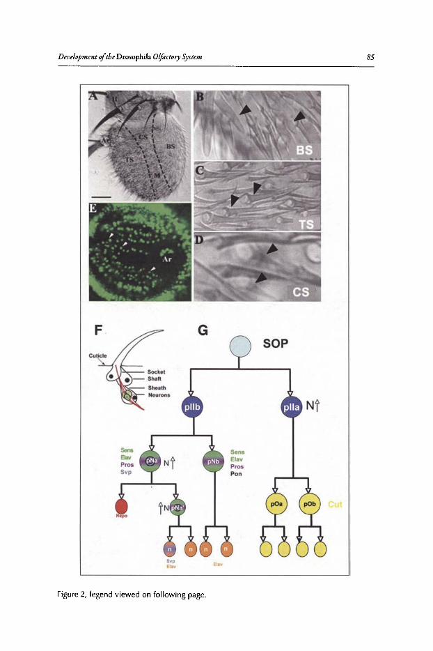
Development ofthe Drosophila OlfactorySystem 85
G
SensElavProsPon
SOP
SYPelay
Figure 2, legend viewed on followin g page.
elay
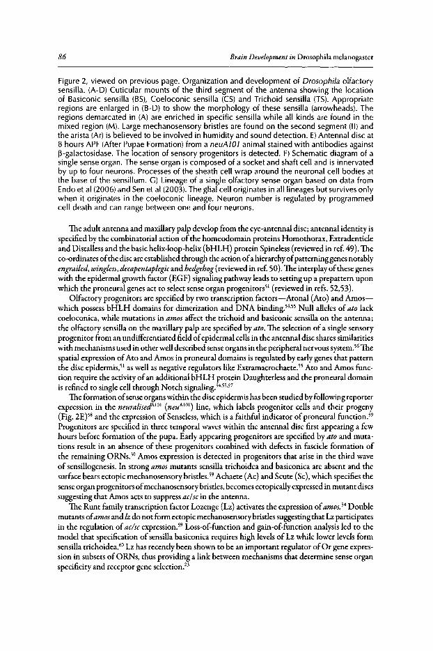
86 Brain Development in Drosophila melanogaster
Figure 2, viewed on previous page. Organization and development of Drosophila olfactorysensilla. (A-D) Cuticular mounts of the third segment of the antenna showing the locationof Basiconic sensilla (BS), Coeloconic sensilla (CS) and Trichoid sensilla (TS). Appropriateregions are enlarged in (B-O) to show the morphology of these sensilla (arrowheads). Theregions demarcated in (A) are enriched in specific sensilla while all kinds are found in themixed region (M). Large mechanosensory bristles are found on the second segment (II) andthe arista (Ar) is believed to be involved in humidity and sound detection. E)Antennal disc at8 hours APF (After Pupae Formation) from a neuA 101 animal stained with antibodies against~-galactosidase. The location of sensory progenitors is detected. F) Schematic diagram of asingle sense organ. The sense organ is composed of a socket and shaft cell and is innervatedby up to four neurons. Processes of the sheath cell wrap around the neuronal cell bodies atthe base of the sensillum. G) Lineage of a single olfactory sense organ based on data fromEndo et al (2006) and Sen et al (2003). The glial cell originates in all lineages but survives onlywhen it originates in the coeloconic lineage. Neuron number is regulated by programmedcell death and can range between one and four neurons.
The adult antenna and maxillary palp develop from the eye-antennal disc; antennal identity isspecified by the combinatorial action of the homeodomain proteins Homothorax, Extradenticleand Distalless and the basic helix-loop-helix (bHLH) protein Spineless (reviewed in ref 49). Theco-ordinates ofthe disc are established through the action ofa hierarchy ofpatterninggenes notablyengrailed, wingless, decapentaplegic and hedgehog (reviewed in ref. 50). The interplay ofthese geneswith the epidermal growth factor (EG F) signaling pathway leads to setting up a prepattern uponwhich the proneural genes act to select sense organ progenitors" (reviewed in refs. 52,53).
Olfactory progenitors are specified by two transcription factors-Atonal (At0 ) and Amoswhich possess bHLH domains for dimerization and DNA binding.54's5 Null alleles of ato lackcoeloconica, while mutations in amos affect the trichoid and basiconic sensilla on the antenna;the olfactory sensilla on the maxillary palp are specified by ato. The selection of a single sensoryprogenitor from an undifferentiated field ofepidermal cells in the antennal disc shares similaritieswith mechanisms used in other well described sense organs in the peripheral nervous system.56 Thespatial expression ofAto and Amos in proneural domains is regulated by early genes that patternthe disc epidermis." as well as negative regulators like Extramacrochaete." Ato and Amos function require the activity ofan additional bHLH protein Daughterless and the proneural domainis refined to single cell through Notch signaling.54.55.5?
The formation ofsense organs within the disc epidermis has been studied by following reporterexpression in the neuralised/r" (neuA l Ol
) line, which labels progenitor cells and their progeny(Fig. 2£)58and the expression of Senseless, which is a faithful indicator ofproneural functlon."Progenitors are specified in three temporal waves within the antennal disc first appearing a fewhours before formation of the pupa. Early appearing progenitors are specified byato and mutations result in an absence of these progenitors combined with defects in fascicle formation ofthe remaining ORNS.30 Amos expression is detected in progenitors that arise in the third waveof sensillogenesis. In strong amos mutants sensilla trichoidea and basiconica are absent and thesurface bears ectopic mechanosensory bristles." Achaete (Ac) and Scute (Sc), which specifies thesense organ progenitors ofmechanosensory bristles, becomes ectopically expressed in mutant discssuggesting that Amos acts to suppress aclscin the antenna.
The Runt family transcription factor Lozenge (Lz) activates the expression ofamos. 54 Doublemutants ofamosand lz do not form ectopic mechanosensory bristles suggesting that Lz participatesin the regulation ofac/sc expression." Loss-of-function and gain-of-function analysis led to themodel that specification of sensilla basiconica requires high levels of Lz while lower levels formsensilla trichoidea." Lz has recently been shown to be an important regulator ofOr gene expression in subsets ofORNs, thus providing a link between mechanisms that determine sense organspecificity and receptor gene selection.f
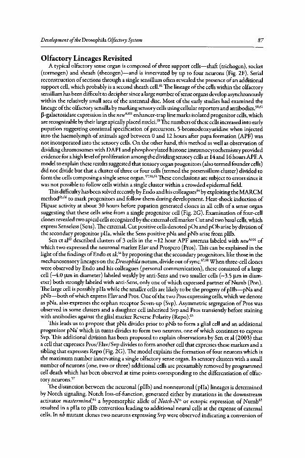
Development ofthe Drosophila Olfactory System 87
Olfactory Lineages RevisitedA typical olfactory sense organ is composed of three support cells-shaft (trichogen), socket
(tormogen) and sheath (thecogen)-and is innervated by up to four neurons (Fig. 2F). Serialreconstruction ofsections through a single sensillum often revealed the presence ofan additionalsupport cell, which probably is a second sheath cell." The lineage ofthe cells within the olfactorysensillum has been difficult to decipher since a large number ofsenseorgans develop asynchronouslywithin the relatively small area of the antennal disc. Most of the early studies had examined thelineage ofthe olfactory sensilla by marking sensory cellsusing cellular reporters and antibodies.58.62
~-galactosidase expression in the neuA101 enhancer-trap line marks isolated progenitor cells,whichare recognizable by their large apicallyplaced nuclei.58 The numbers ofthese cellsincreased into earlypupation suggesting continual specification of precursors. 5-bromodeoxyuridine when injectedinto the haemolymph of animals aged between 0 and 12 hours after pupa formation (APF) wasnot incorporated into the sensory cells. On the other hand, this method as well as observation ofdividing chromosomes with DAPI and phosphorylated histone immunocytochemistry providedevidence for a high levelofproliferation among the dividing sensory cellsat 14 and 16 hours APE Amodel to explain these results suggested that sensory organ progenitors (also termed founder cells)did not divide but that a cluster ofthree or four cells (termed the presensillum cluster) divided toform the cells composing a single sense organ. 57,58,63 These conclusions are subject to errors since itwas not possible to follow cells within a single cluster within a crowded epidermal field.
This difficulty has been solved recently by Endo and his colleagues'? by exploiting the MARCMmethod65,66 to mark progenitors and follow them during development. Heat-shock induction ofFlipase activity at about 30 hours before pupation generated clones in all cells of a sense organsuggesting that these cells arise from a single progenitor cell (Fig. 2G). Examination of four-cellclones revealed two apical cellsrecognized by the external cellmarker Cut and two basal cells,whichexpress Senseless (Sens).The external, Cut positive cellsdenoted pOa and pOb arise by division ofthe secondary progenitor pIIa, while the Sens positive pNa and pNb arise from pIIb.
Sen et al63described clusters of 3 cells in the -12 hour APF antenna labeled with neuA101 ofwhich two expressed the neuronal marker Elav and Prospero (Pros). This can be explained in thelight ofthe findings ofEndo et al,64 by proposing that the secondary progenitors, like those in themechanosensory lineages on the Drosophila notum, divide out ofsync.67.68 When three-cell cloneswere observed by Endo and his colleagues (personal communication), these consisted of a largecell (-4.0 urn in diameter) labeled weakly by anti-Sens and two smaller cells (-3.5 urn in diameter) both strongly labeled with anti-Sens, only one ofwhich expressed partner ofNumb (Pon).The large cell is possibly pIIa while the smaller cells are likely to be the progeny ofpIIb-pNa andpNb-both ofwhich express Elav and Pros. One ofthe two Pros expressing cells,which we denoteas pNa, also expresses the orphan receptor Seven-up (Svp). Asymmetric segregation of Pros wasobserved in some clusters and a daughter cell inherited Svp and Pros transiently before stainingwith antibodies against the glial marker Reverse Polarity (Repo)."
This leads us to propose that pNa divides prior to pNb to form a glial cell and an additionalprogenitor pNa' which in turns divides to form two neurons, one ofwhich continues to expressSvp. This additional division has been proposed to explain observations by Sen et al (2003) thata cell that expresses Pros/Elav/Svp divides to form another cell that expresses these markers and asibling that expresses Repo (Fig. 2G).The model explains the formation offour neurons which isthe maximum number innervating a single olfactory sense organ. In sensory clusters with a smallnumber ofneurons (one, two or three) additional cells are presumably removed by programmedcell death which has been observed at time points corresponding to the differentiation of olfactory neurons."
The distinction between the neuronal (pIIb) and nonneuronal (pIIa) lineages is determinedby Notch signaling. Notch loss-of-function, generated either by mutations in the downstreamactivator mastermind,64 a hypomorphic allele of Notcb-N" or ectopic expression of Numb'"resulted in a pIIa to pIIb conversion leading to additional neural cells at the expense ofexternalcells. In nb mutant clones two neurons expressing Svp were observed indicating a conversion of
88 Brain Development in Drosophila melanogaster
Figure 3. Glial cells in the antenna. At 36 hours APF, the sense organs in the antenna appear fully differentiated and ORNs can be identified by mAb22C10 staining (A,C,D). Axonbundles transit the third segment in distinct fascicles (long arrows). Prospero is expressed intwo nonneuronal cells in each sensory cluster (blue in B). Glial cells stained with antibodiesagainst Repo line the axonal bundles in the third segment (A; small arrows in B). The glialcells are of two subtypes: those labeled with Mz317-Ga/4 (C) and the GHI46-Ga/4 subset(D). Ar-Arista.
pNb to pNa. 64 This implies that neuronal cells generated from pNa experience high N signalingwhile those from pNb are low in N levels. This binary switch in N signaling levels could act indifferentiation of two populations ofneurons within a single sensillum. This has importance inregulation of the wiring of ORNs to their glomerular targets and will be discussed later.64The
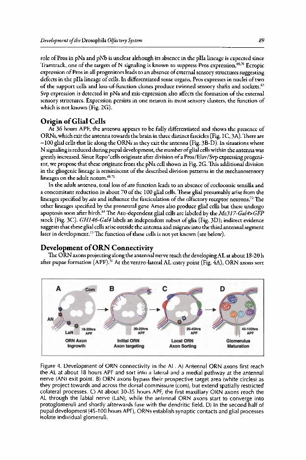
Development oftheDrosophila Olfactory System 89
role of Pros in pNa and pNb is unclear although its absence in the pIla lineage is expected sinceTramtrack, one of the targets of N signaling is known to suppress Pros expression. P'?' Ectopicexpression ofPros in all progenitors leads to an absence ofexternal sensory structures suggestingdefects in the pIla lineage ofcells. In differentiated sense organ s, Pros expresses in nuclei of twoof th e support cells and loss-of-function clones produce twinned sensory shafts and sockets."Svp expression is detected in pNa and rnis-expression also affects the formation of the externalsensory structures. Expression per sists in one neuron in most sensory clusters. the fun ct ion ofwhich is not known (Fig. 2G ).
Origin ofGlial CellsAt 36 hours APF, the antenna appears to be fully differentiated and shows the presence of
ORNs. which exit the antenna towards the brain in three distinct fascicles (Fig. l C , 3A). There are-100 glial cells that lie along the ORNs as they exit the antenna (Fig. 3B-D) . In situations whereN signaling is reduced during pupal development, the number ofglial cellswithin the antenna wasgreatly increased. Since Repo tcells originate after division ofa Pros/Elav/ Svp expressing progenitor, we propose that th ese originate from the pNa cell shown in Fig. 2G. This additional divisionin the gliogenic lineage is reminiscent of the described division patterns in the mechanosensorylineages on the adult notum.68•71
In the adult antenna, total loss ofato function leads to an absence of coeloconic sensilla anda concomitant reduction in about 70 of the 100 glial cells. These glial presumably arise from thelineages specified by ato and influence the fasciculation of the olfactory receptor neurons." Theother lineages specified by th e proneural gene Amos also produce glial cells but these undergoapoptosis soon after birth. " The Aro -dependent glial cells are labeled by the M z317-GaI4>GFPstock (Fig. 3C). GH146-GaI4Iabels an independent subset of glia (Fig. 3D); indirect evidencesuggests that these glial cells arise outside the antenna and migrate into the third antennal segmentlater in development ." The function of these cells is not yet known (see below).
Development ofORN ConnectivityThe 0 RN axons projectingalong the antennal nerve reach the developing AL at about 18-20 h
after pupae formation (APF)Y At the ventro -lateral AL entry point (Fig. 4A), ORN axons sort
ORN AxonIngrowth
InltlalORNAxon targeting
LocalORNAxon Sortlng
D
45-100hrsAPF
GlomerulusMaturation
Figure 4. Development of ORN connectivit y in the AL. A) Antennal ORN axons first reachthe AL at about 18 hours APF and sort into a lateral and a medial pathway at the antennalnerve (AN) exit point. B) ORN axons bypass their prospect ive target area (white circles) asthey project towards and across the dorsal commissure (com), but extend spatially restrictedcolateral processes. C) At about 30-35 hours APF, the first maxillar y ORN axons reach theAL through the labial nerve (LaN), wh ile the antennal ORN axons start to converge intoprotoglomerul i and shortl y afterwards fuse with the dendritic f ield. D) In the second half ofpupal development (45-100 hours APF), ORNs establish synapt ic contacts and glial processesisolate individual glomeruli.
90 Brain Development in Drosophila melanogaster
out, turn either into a medial or a lateral direction and continue to project in two broad pathwaysacross the surface ofthe AL towards the dorso-medial corner," (TH, unpublished observation).Inside these axon tracks, 0 RN axons initially bypass their prospective glomerular target area toproject across the dorsal commissure, but individual axons extend small collateral processes in theregion oftheir class-specific convergence (Fig. 4B)?4 The first axons cross the midline and extendinto the contra-lateral AL by about 20 hours APF. ORN axons stay within the peripheral nervefiber layer over the next 15 hours (Fig. 4C). During this period, the collateral axonal extensionelongate followed by a sequential, spatially restricted process ofaxon condensation into increasinglydiscrete protoglomeruli that spreads across the developingAL?4 These axonal protoglomeruli thenbegin to merge with the dendritic field ofprojection neurons so that by around 35 hours the firstglomeruli can be distinguished and by 50 hours APF most glomeruli have formed (Fig. 4D).31.74Compared to the antenna, ORNs from the maxillary palps develop later and maxillary axonsreaching the AL around 30 h APF at a ventral position to integrate into the antennal glomerularfield (Fig. 4C)?5 Following the assembly ofORN axons and PN dendrites into glomeruli duringthe first halfof pupal development, glomerulus maturation occurs in the remaining two days ofthe pupal phase, in which OR genes are turned on in the antenna and 0 RN axons form synapsesin the antennallobe (Fig. 4D).76,77
Olfactory Map OrganizationThe highly specific Drosophila olfactory circuit, composed of 50 individual channels, each
ofwhich is organized by the convergence of about 30 0 RN axons, presents a fascinating wiringproblem. Although the 0 RN projections from the periphery onto the glomerular array followthe principle ofa discrete sensory map," in which axons from spatially dispersed neurons with thesame sensory identity project onto one location in the target field, some more global organizationdomains have been noticed.
As the different sensilla classesoccupy distinct areas on the antenna, with the basiconic sensillabroadly located along the medial antennal surface and the trichoids more restricted to the lateralantennal areas (Fig. 2A), the topography ofORNs in the peripheral epithelium is approximatelymaintained in their projection into the brain (Fig. 5).25 Although ORN axons do not fasciculate inthe antenna strictly according to sensillum type, the 3 main fasciclesdescribed above contain 0 RNaxons from the sensillum type enriched regions on the antenna! surface (Fig. 5A,TH unpublished).When 0 RN axons reach the brain they defasciculate and become reorganized into several axonpathways with different medio-Iateral positions across the AL surface: basiconic ORNs accumulatein the medial tracts and trichoid ORNs in the lateral tracts (Fig. 5B).25 In Manduca, a special classofglial cells, important for 0 RN axon projection, has been identified that are located where theantennalnerve enters theAL?9,80 It is tempting to speculate that, in Drosophila, the GH146-positiveglial cells described above, are involved in this reorganization of0 RN axon projection."
Inside these sensilla-specific AL domains, the spatial organization of 0 RN classes in theperiphery is not maintained and ORN classes which are housed in the same sensillum oftenproject to glomeruli which are localized quite some distance apart. 25,26 Nevertheless, the 0 RNclass specific axon convergence in the AL seem to be linked to cell fate specification in the courseofdifferential divisions by the SOP progeny. The diversification of0 RN precursors in the SOPlineage through the differential activation of the Notch signalling pathway (see above) does notonly lead to a differential 0 R expression but also correlate with the global organization of0 RNaxon projection/" Endo and collaborators showed that axonal projections of "high-Notch" and"low-Notch" ORN classessegregate into a few multi-glomerular domains along the dorso-ventralAL axis (Fig. 5C). The subdivision of0 RN classconnectivity into different global domains alongthe dorso-ventral and medial-lateral axis suggest that a seriesofhierarchical decisions are made byoRN axons on their way from the peripheral sensory epithelium to the synaptic target region (seebelow). These sequentially acting signalling mechanisms would reduce the complexity to generateoRN synaptic specificity, leading to the positioning of0 RN axons into a selected AL area, wherelocal cues organizes the 0 RN class specific convergence.
Development oftheDrosophila Olfactory System
B
BasiconlcArea
c
TrichoidArea
91
Figure 5. Global organization of ORN projections into the AL. A) ORNs housed in basiconicsensilla are enriched on the medial antennal surface and project into two main fascicles (blueand green) towards the AL. Trichoid sensilla neuronal projections from the lateral antennalsurface are enriched in the third main fascicle (yellow) . The ORNs of each sensillum can besubdivided into one "Notch OFF" neuron and one to three "Notch ON " neurons based onthe position within the SOP lineage (see text). The axonal projections of "Notch OFF"- and"Notch ON"-type ORNs are intermingled within each of the three fascicles. B) The overalltopographic organization of basiconic and trichoid ORNs is maintained in the ORN axonprojections in the AL. C) "Notch OFF"- and "Notch ON"-type ORNs segregate into broadglomerular domains along the dorso-ventral axis. Red staining in A: 22ClO-positive ORNs.
Cellular and Molecular Mechanisms ofORN Wiring SpecificityRecent progress in understanding the molecular basis of ORN wiring specificity has been
achieved through the systematic use of an inducible genetic mosaic system (MARCM),65.66 inwhich gene functions can be specifically removed from projecting ORNs (Fig. SA-C). The induction ofMARCM clones under the control ofthe eyeless promotor generates large regions ofhomozygous 0 RNs in the antennal epithelium but does not affect the precursors ofthe PNs andLNs in the developing AL.81In addition, the expression ofFlipase under heat -shock (hs) promotor control enables the generation of single homozygous ORNs as well the induction ofgeneticmosaics in different populations of AL neurons." This mosaic system has been used to addressmany aspects ofolfactory system development, e.g., the origin ofthe different cell types and theirclonal relationship in the antenna and AL,64.83 the functional interplay ofaxons and dendrites during axonal and dendritic wiring82.84.85 and the characterization ofcandidate genes in this cell-cellcommunication process," Axonal connectivity phenotypes of candidate molecules (see below)and a large-scale histological screen (TH unpublished) further support the idea that hierarchicalmechanisms control ORN axon targeting in the AL. Most mutant phenotypes can be classifiedasglobal or local misprojection (Fig. 6D-I), suggesting that there are distinct and sequentially actingsignalling mechanisms in which initial axon guidance to a coarse region ofthe AL is followed bylocal interactions that control the final refinement and precise matching ofpre-and postsynapticneuronal processes.
The first neuronal guidance molecules that have been shown to be required within ORNs areDscam, a member ofthe Ig-domain superfamily and two Dscam downstream effectors, the SHUSH3adapter Dock and the serine/threonine kinase Pak.81.87All three genes are broadly expressedin the developing peripheral and central olfactory system. In eyFlpmosaics, Dscam mutant ORNaxons still follow an approximately normal path across the antennal lobe, but converge prematurely
92 Brain Development in Drosophila melanogasrer
at ectopic locations. Interestingly, they do not integrate into existingglomeruli at these ectopic sites;rather multiple Dscam mutant ORN axons converge to form novel glomerulus-like structures."Labeling of multiple 0 RN classes revealed that Dscam mutant 0 RN axons segregate at ectopicsites, even outside the AL, in an ORN classspecific manner (Fig. 6F-G') indicating the existenceof a unique axonal identity independent of the target area, which is counteracted by the Dscamactivity (Fig. 6J; T.H. unpublished observations). dockandPak mutant ORN axons show a moresevere phenotype in which they grow along inappropriate pathways and therefore form ectopicterminations all over the AL. The Robo receptors are a second classofguidance molecules involvedin the initial coarse targeting of 0 RN axons. Robo, Rob02 and Rob03 are expressed in discretesubsets of 0 RN axons that segregate from one another and take different medial versus lateralpathways across the developing AL.29 Widespread mistargeting defects when Robo receptors areremoved from ORNs or ectopically expressed suggest a crucial role for Robo signalling in ORNaxon positioning.
Following the crude and overlapping positioning of 0 RN axon terminals ofdifferent classeswithin a restricted AL domain, local short-range inter and intra-class interactions lead to theclass-specific axon sorting into protoglomeruli (Fig.4C). Compared to the mutations describedabove, removal ofthe transmembrane molecule Semaphorin-la and the Ca-dependent cell adhesion molecule N-Cadherin leads to more local axon targeting defects (Fig. 6H-I '). Ncad mutantORN axons reach the vicinity of their AL target area, but the initial axonal convergence intoprotoglomeruli is disrupted; this in turn affects all subsequent steps of glomerulus maturationand axon-dendrite interaction finally results in a severedisorganisation ofthe adult AL neuropil."However, N-Cadherin does not appear to mediate class-specific interactions between differentORN axons; rather it seems to be a permissive factor for axonal interactions among all ORNS.74In contrast to N-Cadherin, Sema-la mutant ORN axons are able to induce local convergence,but axons ofthe same classsplit into multiple adjacent glomeruli or coconverge with ORN axonsof neighboring glomeruli (Fig. 6H-I ').15•88Whereas N-Cadherin is ubiquitously expressed onprojecting 0 RN axons, 0 RN axons converging into neighbouring glomeruli display differentlevelsofSemaphorin-Ia." Clonal analysis indicates a non-autonomous Sema-la function, mediated through the Plexin A receptor, onto neighbouring classes,most likely in a repulsive fashion.In summary, different types ofinter-axonal attractive (via N-Cadherin) and repulsive (via Dscamand Sema-la/Plexin A) interactions, lead to a final coalescence ofORN axons in OR type specificprotoglomeruli (Fig. 6J).
Specification ofProjection NeuronsIn contrast to 0 RN formation during pupal development, the generation of the olfactory
CNS neurons starts already during embryonic and larval stages, ensuring that a prepatterneddendritic target field is established by the time ORN axons project into the antennal lobe."Projection neurons (PNs) and local interneurons (LNs) originate from asymmetrically dividingneural precursor cells called neuroblasts (Fig. 7).89 At each division neuroblasts produce serially anew neuroblast and a ganglion mother cell, which divides once more to generate two terminallydifferentiated neurons (Fig. 7). PNs are derived from three neuroblasts: an antero-dorsal (ad), alateral (la) and a ventral (ve) neuroblast, corresponding to the three groups ofcell bodies." It hasbeen shown that the ad and la PN lineages send dendrites to stereotyped and mutually exclusivesets ofglomeruli. 12 Using the MARCM method to perform a systematic fate mapping analysis ofPNs, Jefferis et al could demonstrate a direct correlation between the larval PN lineage and birthtime with their dendritic targeting onto distinct glomeruli." In addition, embryonic-born PNsparticipate in both the larval and adult olfactory circuits." In the larva, these neurons generallyinnervate a single glomerulus in the antennallobe and persist in the adult olfactory circuit, alsoprespecified by birth order to innervate a subset of glomeruli distinct from larval-born PNs.Developmental studies indicate that these neurons undergo stereotyped pruningoftheir dendritesand axon terminal branches locally during early metamorphosis, which requires cell-autonomousreception of the nuclear hormone ecdysone.?"
lcfll: I
I(J • :::J
• ~.. •.I !e 'if )
Development of theDrosophila Olfactory System
HomozygousMutant OAN
Heterozygous OAN
HomozygousWlldtypeOAN
Heterozygous OAN
93
J Intra-ClassRepulsion Intra-Class
Attraction Inter-ClassRespulsi on
, Dscam
N-Cadherin
nSema-1a1I Plexin A
Figure 6. Analysis of gene functions involved in ORN connectivity development. A-C) Mosaicanalysis in developing ORNs using the MARCM system. Induction of MARCM clones undereyFlp control leads to homoz ygous (GFP-positive) ORNs in the maxillary palps A) and antenna (not shown) and allows to follow their axonal projections into the AL (B), whereas theAL target neurons (LNs and PNs) next to the AL remain unaffected (red, GFP-negative). C)Schematic illustrating the segregation of the different chromosomes (asterisk indicates themutation) from the heterozygous ORN precursor cell into the different daughter cells. Dueto the loss of the GalBO insertion after mitotic recomb ination at the FRT sites (black box), thehomoz ygous mutant ORNs start to expressGFP in a GaI4/UAS-dependent manner, whereasin homozygous wildtype or heterozygous ORNs the expression of GFP remains represseddue to the presence of GalBO. D, E, E/) The project ion of two ORN classes (green and red)onto neighbor ing glomeruli is shown. F, G, G') Loss of Dscam in ORN-specific MARCMclon es lead to a prematu re convergence inside and outsid e the AL, but mutant axons sortout according to their OR class identity. H, I, I') Following the removal of Semaphorin - Ia(Seme- le), mutant ORN axons project to the target region but fail to sort into class-speci ficglomeruli. J)Model of the inter-axonal interactions mediated by different neuronal cell surfacemolecules (see text).
94 Brain Development in Drosophilamelanogasrer
• Level
: Timei ~
rNBr8r8~
•A A A·-,8888 PNS8.....\'\\\ @;/ 1/
@@Iac33ve
Figure 7. Generation of Projection Neurons dur ing AL development. Projection Neurons (PNs)in each of the three PN clusters, the anterior-dorsal (ad), lateral (la) and ventral (ve) cluster,derive from a distinct neuroblast (NB). The different PN classesinside each cluster (e.g., PN1 ,PN2, etc.) are generated at defined time points from the dividing NB though an intermediateganglion mother cell (GMC). Recent data indicate that the level of the POU-domain typetranscription factor Chinmo in the dividing NB defines a critical determinant in the control ofPN identity, with high levels giving rises to early-born PNs and low levels to late-born PNs.
How is PN diversity generated through the series of NB divisions? First insights into themolecular mechanism that lead to PN diversification came through the identification of theputative transcription factor Chinmo that confers temporal identity on the neural progeny ofmushroom body and projection neurons neuroblasts (Fig. 7).91 In the PN lineage, loss ofChinmoautonomously causes early-born neurons to adopt the fates oflate-born neurons from the samelineages. Although the molecular mechanisms that control PN fate identity is not clear yet, studiesin mushroom bodies indicate the generation ofa temporal gradient ofChinmo (Fig. 7) throughprimarily posttranscriptional regulation which helps to specify distinct birth order-dependent cellfates in an extended neuronal lineage,"
Development oftheDrosophila Olfactory System 95
PN AxonOutgrowth
PN DendriteOutgrowth
PN DendriteSorting
D
Sema-1aLevel
+
I I L n Position.1 LL_~Dorso- Central Ventrolateral medial """' Intra-class Interaction
Figure 8. Development of PN connectivity. (A-C) Schematic drawings of the three main stepsin PN axon and dendri te development. A) Adult PNs first send out axonal processes toward sthe mushroom bod ies and lateral horn . B) PNs develop dendr it ic branches wh ich project todifferent regions, according to their later glomerular posit ion in the AL. C) Different typesof inter-dendritic interactions further restrict PN dend rites to their final AL posit ion . D) Thelevel of Semaphori n-l a in developing PNs determines their dendr ite posit ion ing along thedorso-ventral AL axis; high levels lead to a dorso-Iateral location whe reas PNs expressinglow Sema-la levels proj ect to ventro-medial posit ions. E) PN dendrites having reached theircrude position in the AL perform different types of interactions (mediated by N-Cadherin andDscam) to elaborate and restrict their dendritic branches.
Development ofPN DendritesShortly after birth during larval development, PNs extend axons along the main tracts connect
ing the antennallobe to h igher olfacto ry centres. the MB and LH. however no axonal arborizations are formed with in those CNS regions (Fig. 8A) .JlAt the time oflarval/pupal transition, PNdendrite development startswith the extension ofdendritic proc esses int o different regions oftheAL (Fig. 8B), so that by about 20 h APF spatially restr icted but still overlapping dendritic arboursoccupy the early AL.JlAt the same tim e, ORN axons have just reached the developing ant ennallobe. The directional outgrowth is followed by a period of int er-dendritic interaction (Fig. 8C).resulting in a further refinement ofarborization according to their PN class identi ry," Class-specificbranching ofPN axons is evident in the lateral horn by 24-30 h APF.JlThus the PN axonal development relevant to wiring occurs relatively synchronously with dendritic development.
Recently, several molecular determinates, e.g.•transcription factors and cell surface molecules.have been identified that are involved in the two main steps of PN dendrite development. directio nal outgrowth and class-specific sorting.84-86.92.93 Interestingly. the same molecule s described
96 Brain Development in Drosophila melanogaster
above to control ORN axon-axon interaction, Semaphorin-la, N-Cadherin and Dscam, also havean independent function during PN dendrite patterning. Somewhat surprising came the observation that Sema-la levels cell-autonomously direct initial PN targeting in the antennallobe alongthe dorsal-Iateral/ventral-medial axis (Fig. 80).86 This function requires the cytoplasmic domain,which could mediate signalling upon binding to a yet unknown ligand, distributed in a gradientalong in the early AL to specify dendrite positioning."
Mosaic analyses suggest that N-Cadherin mediates dendro-dendritic interactions betweenPNs and thus contributes to spatial restriction ofPN dendrites thereby sharpening the boundariesbetween glomeruli (Fig. 8E).84 Developmental studies reveal that the dendrites of N-Cadherinmutant PNs occupy the same global positions as their wild-type counterparts during early pupaldevelopment but fail to sort in a PN classspecific pattern. A possible explanation for the dendriticdefect is that N-Cadherin expressed on the surface ofPN dendrites confers proper adhesivenessto the dendrite during and after the initial targeting event. Loss ofN-Cadherin results in reducedcell adhesion, allowing dendrites to more easily invade the neighboring glomeruli. In contrast tothe inhibition ofdendritic extensions, Dscam acts in projection neurons and local interneurons tocontrol the elaboration ofdendritic fields (Fig. 8E).8S The removal ofDscam selectivelyfrom PNsorLNs leads to a marked reduction in their dendritic field size whereas Dscam overexpression causesdendrites to shift their relative local position. Thus, similar to the ORN axon pattering, sequentialattractive and repulsive interactions seem to mediate the final positioning ofPN dendrites.
Beside the cell surface molecules directly involved in inter-dendritic communication, thefunctional analysis ofPN-intrinsic mechanisms has led to the identification of several transcription factors (TFs) that control dendritic targetingS6
,92 Members of different TF families, e.g.,LIM-homeodomain TFs (Islet and Lim l ), homeodomain TFs (Cut), zinc-finger TFs (Squeeze)and POU-domain TFs (Acj6 and Drifter) lead in PN-specific loss-of-function mosaic clones toeither coarse or local dendrite targeting defects, suggesting that they have qualitatively differentinstructive information. Most ofthese TFs show a spatially restricted expression pattern, e.g.,Acj6and Drifter, are expressed in adPNs and laPNs respectively (Fig. 8E).92 Misexpression experimentsinduces specific changes oftargeting specificity suggest that PN classesare at least partially definedby combinatorial expression ofTFs that regulate different steps ofdendritic targeting, some specifying the coarse area (e.g., Cut), followed by others controlling local glomerular choice within thearea (e.g., Drifter and Acj6).
Concluding RemarksInitial studies in other insects and mammals have emphasized the organizing role of ORNs
in AL development. For instance in Manduca surgical removal of the antenna prevents normalglomerular formation94,9s and antennal disc transplantation between different sexesresults in theformation ofglomeruli with a morphology that is most typical of the donor sex." The results inDrosophila presented here suggested that both ORNs and PNs have substantial autonomous patterning ability in which both neuronal types target coarselywithout interactingwith their partners.Experiments in which 0 RN axon and PN dendrite targeting are differentially affected providefirst insights into the relative contribution ofboth synaptic partners in determining connectionspecificity. Cell-type specific removal ofN-Cadherin from PN dendrites does not affect the sitespecific convergence ofthe partner 0 RN s.84 In addition, the local shift in 0 RN axon convergenceor PN dendrite positioning leads to the corresponding displacement ofthe synaptic partner, whichso far is the strongest indication for a cell-type specific recognition code."
Based on the cellular interactions during pupal AL development and the resulting connectivitydefect following the cell-type specific mutant analysis described above we suggest the followingmodel for ORN-PN matching (Fig. 9). The first cellular event seems to be the generation of acoarse map ofPN dendrites in the developingAL (Step 1). Initial ORN axon targeting is equallyimprecise. The axons ofa particular 0 RN class reach their target area and intermingle with axonsof spatially related targets. Intra-class axonal attraction combined with inter-class axonal retraction forces this local blend ofdifferent axon classesto segregate from one another (Step 2).When
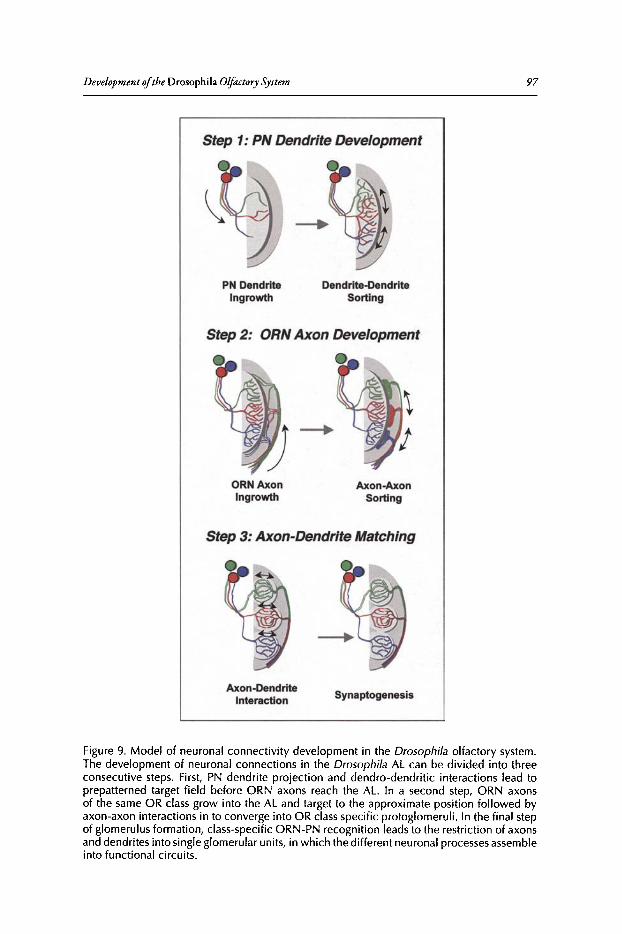
Development ofthe Drosophila Olftctory System
Step 1: PN Dendrite Development
97
PN DendriteIngrowth
Dendrite-DendriteSorting
Step 2: ORN Axon Development
ORNAxonIngrowth
Axon-AxonSorting
Step 3: Axon-Dendrite Matching
Axon-DendriteInteraction Synaptogenesis
Figure 9. Model of neuronal connectivity development in the Drosophila olfactory system.The development of neuronal connections in the Drosophila AL can be divided into threeconsecutive steps. First, PN dendrite projection and dendro-dendritic interact ions lead toprepatterned target field before ORN axons reach the AL. In a second step, ORN axonsof the same OR class grow into the AL and target to the approximate position followed byaxon-axon interactions in to converge into OR class specific protoglomeruli. In the final stepof glomerulus formation, class-specific ORN-PN recognition leads to the restriction ofaxonsand dendrites into single glomerular units, in which the different neuronal processes assembleinto functional circuits .
98 Brain Development in Drosophila melanogasrer
a critical concentration of similar axons is reached, axonal protoglomeruli begin to form at theperiphery of the antennallobe (Step 2). In the final step, class-specific ORN-PN recognitioninitiates the glomerulus assembly followed by synaptogenesis and glial mediated isolation ofnewemerging synaptic units. Once established these synaptic units are rather stable, manipulationsin the adult fly, e.g., the selective cell ablation or differential olfactory experience leads to onlyminor intra-glomerular changes?6.97 Based on this model 0 RN axon and PN dendrite targetingare initially two separate patterning events and subsequent axon-dendrite matching is the finalstep by which two prepatterned fields are merged.
How the differentiation ofabout SO distinct synaptic partners is coordinated remains one ofthe challenging questions in olfactory system research. In the peripheral olfactory system, distincttypes of0 RNs have to become specified and the subsequent differentiation has to be coupled tothe neurogenesis within developing sensilla, the development ofaxon projection patterns and theeventual expression ofindividual odorant receptors. In the central olfactory system, the development ofthe dendritic projection has to be coordinated with the distinct axonal branching patternin the higher brain cenrers.P:" The Drosophila olfactory system provides a powerful model toaddress these fundamental issuesofneuroscience research.
References1. Chklovskii DB, Koulakov AA. Maps in the brain: what can we learn from them? Annu Rev Neurosci
2004; 27:369-392.2. Miller G. 2004 Nobel Prizes. Axel, Buck share award for deciphering how the nose knows. Science
2004; 306(5694):207.3. Buck L, Axel R. A novel multigene family may encode odorant receptors: a molecular basis for odor
recognition. Cell 1991; 65(1):175-187.4. Chess A, Simon I, Cedar H et al. Allelic inactivation regulates olfactory receptor gene expression. Cell
1994; 78(5):823-834.5. VassarR, Ngai J, Axel R. Spatial segregation of odorant receptor expression in the mammalian olfactory
epithelium. Cell 1993; 74(2):309-318.6. Mombaerts P, Wang F, Dulac C et al. Visualizing an olfactory sensory map. Cell 1996;
87(4):675-686.7. Vassar R, Chao SK, Sitcheran Ret al. Topographic organization of sensory projections to the olfactory
bulb. Cell 1994; 79(6) :981-991.8. Feinstein P, Bozza T, Rodriguez I et al. Axon guidance of mouse olfactory sensory neurons by odorant
receptors and the beta2 adrenergic receptor. Cell 2004; 117(6):833-846.9. Feinstein P, Mombaerts P. A contextual model for axonal sorting into glomeruli in the mouse olfactory
system. Cell 2004; 117(6):817-831.10. Imai T, Suzuki M, Sakano H. Odorant receptor-derived cAMP signals direct axonal targeting. Science
2006; 314(5799):657-661.11. Serizawa S, Miyamichi K, Takeuchi H et al. A neuronal identity code for the odorant receptor-specific
and activity-dependent axon sorting. Cell 2006; 127(5):1057-1069.12. Stocker RF, Lienhard MC, Borst A et al. Neuronal architecture of the antennallobe in Drosophila
melanogaster. Cell Tissue Res 1990; 262(1):9-34.13. Stocker RF. The organization of the chemosensory system in Drosophila melanogaster: a review. Cell
Tissue Res 1994; 275(1):3-26.14. Stocker RF. Drosophila as a focus in olfactory research: mapping of olfactory sensilla by fine struc
ture, odor specificity, odorant receptor expression and central connectivity. Microsc Res Tech 2001;55(5 ):284-296.
IS. Stowers L. Neuronal development: specifying a hard-wired circuit. Curr BioI 2004; 14(2):R62-64.16. Vosshall LB, Wong AM, Axel R. An olfactory sensory map in the fly brain. Cell 2000;
102(2): 147-159.17. Komiyama T, Luo L. Development of wiring specificity in the olfactory system. Curr Opin Neurobiol
2006; 16(1):67-73.18. Jefferis GS, Marin EC, Watts RJ et al. Development of neuronal connectivity in Drosophila antennal
lobes and mushroom bodies. Curr Opin Neurobiol 2002; 12(1):80-86.19. Jefferis GS, Hummel T. Wiring specificity in the olfactory system. Semin Cell Dev BioI 2006;
17(1):50-65.20. Clyne PJ, Warr CG, Freeman MR et al. A novel family of divergent seven-transmembrane proteins:
candidate odorant receptors in Drosophila. Neuron 1999; 22(2):327-338.
Developmentofthe Drosophila Olfactory System 99
21. VosshallLB, Amrein H, Morozov PS, Rzhetsky A, Axel R. A spatial map of olfactory receptor expression in the Drosophila antenna. Cell 1999; 96(5):725-736.
22. Lomvardas S, Barnea G, Pisapia DJ et al. Interchromosomal interactions and olfactory receptor choice.Cell 2006; 126(2):403-413.
23. Ray A, van Naters WG, Shiraiwa T et al. Mechanisms of odor receptor gene choice in Drosophila.Neuron 2007; 53(3):353-369.
24. Gao Q Yuan B, Chess A. Convergent projections of Drosophila olfactory neurons to specificglomeruliin the antennallobe. Nat Neurosci 2000; 3(8):780-785.
25. Couto A, Alenius M, Dickson BJ. Molecular, anatomical and functional organization of the Drosophilaolfactory system. Curr Bioi 2005; 15(17):1535-1547.
26. Fishilevich E, Vosshall LB. Genetic and functional subdivision of the Drosophila antennallobe. CurrBioi 2005; 15(17):1548-1553.
27. Laissue PP, Reiter C, Hiesinger PR et ale Three-dimensional reconstruction of the antennallobe inDrosophila melanogaster. J Comp Neuro11999; 405(4):543-552.
28. Stocker RF, Singh RN, Schorderet M et al. Projection patterns of different types of antennal sensilla inthe antennal glomeruli of Drosophila melanogaster. Cell Tissue Res 1983; 232(2):237-248.
29. Jhaveri D, Saharan S, Sen A et al. Positioning sensory terminals in the olfactory lobe of Drosophila byRobo signaling. Development 2004; 131(9):1903-1912.
30. Jhaveri D, Sen A, Rodrigues V.Mechanisms underlying olfactory neuronal connectivity in Drosophila-theatonal lineage organizes the periphery while sensory neurons and glia pattern the olfactory lobe. DevBioi 2000; 226(1):73-87.
31. Jefferis GS, VyasRM, Berdnik D et al. Developmental origin of wiring specificity in the olfactory systemof Drosophila. Development 2004; 131(1):117-130.
32. Wong AM, WangJ~ Axel R. Spatial representation of the glomerular map in the Drosophila protocerebrum. Cell 2002; 109(2):229-241.
33. Marin EC, JefferisGS, Komiyama et al.Representation of the glomerular olfactory map in the Drosophilabrain. Cell 2002; 109(2):243-255.
34. Wilson RI, Turner GC, Laurent G. Transformation of olfactory representations in the Drosophilaantennallobe. Science 2004; 303(5656):366-370.
35. Wilson RI, Laurent G. Role of GABAergic inhibition in shaping odor-evoked spatiotemporal patternsin the Drosophila antennallobe. J Neurosci 2005; 25(40):9069-9079.
36. Olsen SR, Bhandawat V, Wilson RI. Excitatory interactions between olfactory processing channels inthe Drosophila antennal lobe. Neuron 2007; 54(1):89-103.
37. Shang Y, Claridge-Chang A, Sjulson L er al. Excitatory local circuits and their implications for olfactoryprocessing in the fly antennallobe. Cell 2007; 128(3):601-612.
38. Distler PG, Boeckh J. An improved model of the synaptic organization of insect olfactory glomeruli.Ann NY Acad Sci 1998; 855:508-510.
39. Distler PG, Gruber C, Boeckh J. Synaptic connections between GABA-immunoreactive neurons anduniglomerular projection neurons within the antennal lobe of the cockroach, Periplaneta americana.Synapse 1998; 29(1):1-13.
40. Distler PG, BoeckhJ. Synaptic connection between olfactory receptor cells and uniglomerular projectionneurons in the antennallobe of the American cockroach, Periplaneta americana. J Comp Neurol 1996;370(1):35-46.
41. Riesgo-EscovarJR, Piekos WB, Carlson JR. The Drosophila antenna: ultrastructural and physiologicalstudies in wild-type and lozenge mutants. J Comp Physiol [A] 1997; 180(2):151-160.
42. Shanbhag S, Muller, B, Steinbrecht A. Atlas of olffactory organs of Drosophila melanogaster: I Types,external organisation, innervation and distribution of olfactory sensilla. Int J Insect Morphol and Embryol 1999; 28:377-397.
43. Clyne PJ, Certel SJ, de Bruyne et al.The odor specificities of a subset of olfactory receptor neurons aregoverned by Acj6, a POD-domain transcription factor. Neuron 1999; 22(2):339-347.
44. de Bruyne M, Clyne PJ, Carlson JR. Odor coding in a model olfactory organ: the Drosophila maxillarypalp,J Neurosci 1999; 19(11):4520-4532.
45. de Bruyne M, Foster K, Carlson JR. Odor coding in the Drosophila antenna. Neuron 2001;30(2):537-552.
46. Elmore T, Ignell R, Carlson JR et al. Targeted mutation of a Drosophila odor receptor defines receptorrequirement in a novel class of sensillum. J Neurosci 2003; 23(30):9906-9912.
47. Yao CA, Ignell R, Carlson JR. Chemosensory coding by neurons in the coeloconic sensilla of theDrosophila antenna. J Neurosci 2005; 25(37):8359-8367.
48. van der Goes van Naters~ Carlson JR. Receptors and neurons for fly odors in Drosophila. Curr Biol2007; 17(7):606-612.
100 Brain Development in Drosophila melanogaster
49. Haynie JL, Bryant PJ. Development of the eye-antenna imaginal disc and morphogenesis of the adulthead in Drosophila melanogaster. J Exp Zoo11986; 237(3):293-308.
50. Cohen SM, Di Nardo S. Wingless: from embryo to adult. Trends Genet 1993; 9(6):189-192.51. Jhaveri D, Sen A, Reddy GV et al. Sense organ identity in the Drosophila antenna is specified by the
expression of the proneural gene atonal. Mech Dev 2000; 99(1-2):101-111.52. Gomez-Skarmeta JL, Campuzano S, Modolell J. Half a century of neural prepatterning: the story of a
few bristles and many genes. Nat Rev Neurosci 2003; 4(7) :587-598.53. Kiefer JC, Jarman A, Johnson J. Proneural factors and neurogenesis. Dev Dyn 2005; 234(3):808-813.54. Goulding SE, zur Lage P,Jarman AP. amos, a proneural gene for Drosophila olfactory sense organs that
is regulated by lozenge. Neuron 2000; 25(1):69-78.55. Gupta BP, Rodrigues V. Atonal is a proneural gene for a subset of olfactory sense organs in Drosophila.
Genes Cells 1997; 2(3):225-233.56. Jan YN, Jan LY. Neuronal cell fate specification in Drosophila. Curr Opin Neurobiol 1994;
4(1):8-13.57. Reddy G~ Gupta B, Ray K et al. Development of the Drosophila olfactory sense organs utilizes cell-cell
interactions as well as lineage. Development 1997; 124(3):703-712.58. Ray K, Rodrigues V. Cellular events during development of the olfactory sense organs in Drosophila
melanogaster. Dev Bioi 1995; 167(2):426-438.59. zur Lage PI, Prentice DR, Holohan EE et al. The Drosophila proneural gene amos promotes olfactory
sensillum formation and suppresses bristle formation. Development 2003; 130(19):4683-4693.60. Gupta BP, Flores GV, Banerjee U et al. Patterning an epidermal field: Drosophila lozenge, a member of
the AML-l/Runt familyof transcription factors, specifies olfactory sense organ type in a dose-dependentmanner. Dev Bioi 1998; 203(2):400-411.
61. Venkatesh SS, Sensilla RN on the third antennal segment of Drosophila melanogaster Meigen (Diptera:Drosophilidae). Int J Insect Morphol Embryol1984; 13:51-63.
62. Lienhard MC, Stocker RF. The development of the sensory neuron pattern in the antennal disc ofwild-type and mutant (lz3, ssa) Drosophila melanogaster. Development 1991; 112(4):1063-1075.
63. Sen A, Reddy G~ Rodrigues V. Combinatorial expression of Prospero, Seven-up and Elav identifiesprogenitor cell types during sense-organ differentiation in the Drosophila antenna. Dev BioI 2003;254(1):79-92.
64. Endo K, Aoki T, Yoda Y, Kimura K et al. Notch signal organizes the Drosophila olfactory circuitry bydiversifying the sensory neuronal lineages. Nat Neurosci 2007; 10(2):153-160.
65. Lee T, Luo L. Mosaic analysiswith a repressible cell marker (MARCM) for Drosophila neural development. Trends Neurosci 2001; 24(5):251-254.
66. Lee T, Luo L. Mosaic analysis with a repressible cell marker for studies of gene function in neuronalmorphogenesis. Neuron 1999; 22(3):451-461.
67. Manning L, Doe CQ Prospero distinguishes sibling cell fate without asymmetric localization in theDrosophila adult external sense organ lineage. Development 1999; 126(10):2063-2071.
68. Reddy GV, Rodrigues V. Sibling cell fate in the Drosophila adult external sense organ lineageis specified by prospero function, which is regulated by Numb and Notch. Development 1999;126(10):2083-2092.
69. Guo M, Bier E, Jan L~ et al. tramtrack acts downstream of numb to specify distinct daughter cell fatesduring asymmetric cell divisions in the Drosophila PNS. Neuron 1995; 14(5):913-925.
70. Jan YN, Jan LY. Asymmetric cell division. Nature 1998; 392(6678):775-778.71. Gho M, Bellaiche Y, Schweisguth F.Revisiting the Drosophila microchaete lineage: a novel intrinsically
asymmetric cell division generates a glial cell. Development 1999; 126(16):3573-3584.72. Jhaveri D, Rodrigues V. Sensory neurons of the Atonal lineage pioneer the formation of glomeruli within
the adult Drosophila olfactory lobe. Development 2002; 129(5):1251-1260.73. Sen A, Shetty C, jhaveri D et al. Distinct types of glial cells populate the Drosophila antenna. BMC
Dev BioI 2005; 5:25.74. Hummel T, Zipursky SL. Afferent induction of olfactory glomeruli requires N-cadherin. Neuron 2004;
42(1):77-88.75. Sweeney LB, Couto A, Chou YH et al. Temporal target restriction of olfactory receptor neurons by
Semaphorin-1a/PlexinA-mediated axon-axon interactions. Neuron 2007; 53(2):185-200.76. DevaudJM, Acebes A, Ramaswami M et al. Structural and functional changes in the olfactory pathway
of adult Drosophila take place at a critical age. J Neurobiol 2003; 56(1):13-23.77. Bhalerao S, Sen A, Stocker R et al. Olfactory neurons expressing identified receptor genes project
to subsets of glomeruli within the antenna] lobe of Drosophila melanogaster. J Neurobiol 2003;54(4):577-592.
78. Mombaerts P. Axonal wiring in the mouse olfactory system. Annu Rev Cell Dev Biol 2006;22:713-737.
Development ofthe Drosophila Olfactory System 101
79. Tolbert LP, Oland LA. A role for glia in the development of organized neuropilar structures. TrendsNeurosci 1989; 12(2):70-75.
80. Rossler~ Oland LA, Higgins MR et al. Development of a glia-rich axon-sorting zone in the olfactorypathway of the moth Manduca sexta.J Neurosci 1999; 19(22):9865-9877.
81. Hummel T, Vasconcelos ML, Clemens JC et al. Axonal targeting of olfactory receptor neurons inDrosophila is controlled by Dscam. Neuron 2003; 37(2):221-231.
82. Komiyama T, Carlson JR, Luo L. Olfactory receptor neuron axon targeting: intrinsic transcriptionalcontrol and hierarchical interactions. Nat Neurosci 2004; 7(8):819-825.
83. Jefferis GS, Marin EC, Stocker RF et al. Target neuron prespecificarion in the olfactory map ofDrosophila. Nature 2001; 414(6860) :204-208.
84. Zhu H, Luo L. Diverse functions ofN-cadherin in dendritic and axonal terminal arborization of olfactory projection neurons. Neuron 2004; 42( 1):63-75.
85. Zhu H, Hummel T, Clemens JC et al. Dendritic patterning by Dscam and synaptic partner matchingin the Drosophila antennallobe. Nat Neurosci 2006; 9(3):349-355.
86. Komiyama T, Luo L. Intrinsic control of precise dendritic targeting by an ensemble of transcriptionfactors. Curr Bioi 2007; 17(3):278-285.
87. Ang LH, Kim J, Stepensky V et alDock and Pak regulate olfactory axon pathfinding in Drosophila.Development 2003; 130(7):1307-1316.
88. Lattemann M, Zierau A, Schulte C et al. Semaphorin-1a controls receptor neuron-specific axonal convergence in the primary olfactory center of Drosophila. Neuron 2007; 53(2):169-184.
89. Stocker RF, Heimbeck G, Gendre N et al. Neuroblast ablation in Drosophila P[GAL4] lines revealsorigins of olfactory interneurons. J Neurobiol 1997; 32(5):443-456.
90. Marin EC, Watts RJ, Tanaka NK, et al. Developmentally programmed remodeling of the Drosophilaolfactory circuit. Development 2005; 132(4):725-737.
91. Zhu S, Lin S, Kao CF et al. Gradients of the Drosophila Chinmo BTB-zinc finger protein governneuronal temporal identity. Cell 2006; 127(2):409-422.
92. Komiyama T, Johnson WA, Luo L et al. From lineage to wiring specificity. POU domain transcription factors control precise connections of Drosophila olfactory projection neurons. Cell 2003;112(2):157-167.
93. Komiyama T, SweeneyLB, Schuldiner 0 et al. Graded expression of semaphorin-1a cell-autonomouslydirects dendritic targeting of olfactory projection neurons. Cell 2007; 128(2):399-410.
94. Tolbert LP, Sirianni PA. Requirement for olfactory axons in the induction and stabilization of olfactoryglomeruli in an insect. J Comp Neurol 1990; 298(1):69-82.
95. Oland LA, Orr G, Tolbert LP. Construction of a protoglomerular template by olfactory axons initiatesthe formation of olfactory glomeruli in the insect brain. J Neurosci 1990; 10(7):2096-2112.
96. Rossler~ Randolph P~ Tolbert LP et al. Axons of olfactory receptor cells of transsexually graftedantennae induce development of sexually dimorphic glomeruli in Manduca sexta, J Neurobiol 1999;38(4):521-541.
97. Berdnik D, Chihara T, Couto A et al. Wiring stability of the adult Drosophila olfactory circuit afterlesion. J Neurosci 2006; 26(13):3367-3376.
98. Jefferis GS, Potter CJ, Chan AM et al. Comprehensive maps of Drosophila higher olfactory centers:spatially segregated fruit and pheromone representation. Cell 2007; 128(6):1187-1203.

CHAPTER 7
The Olfactory Sensory Map inDrosophilaPhilippe P. Laissue and Leslie B. Vosshall*
Abstract
The fruit fly (Drosophila melanogaster) exhibits robust odor-evoked behaviors in response tocues from diversehost plants and pheromonal cuesfrom other flies.Understandinghow theadult olfactory system supports the perception ofthese odorous chemicals and translates
them into appropriate attraction or avoidance behaviors is an important goal in contemporarysensory neuroscience. Recent advances in genomics and molecular neurobiology have providedan unprecedented level of detail into how the adult Drosophila olfactory system is organized.Volatile odorants are sensed by two bilaterally symmetric olfactory sensory appendages, the thirdsegment of the antenna and the maxillary palps, which respectively contain approximately 1200and 120 olfactory sensory neurons (OSNs) each. These OSNs express a divergent family ofseventransmembrane domain odorant receptors (0Rs) with no homology to vertebrate 0 Rs, whichdetermine the odor specificity ofa given OSN. Drosophila was the first animal for which all ORgenes were cloned, their patterns ofgene expression determined and axonal projections ofmostOSNs elucidated. In vivo electrophysiology has been used to decode the ligand response profilesof most of the 0 Rs, providing insight into the initial logic of olfactory coding in the fly. Thischapter willreview the molecular biology, neuroanatomy and function ofthe peripheral olfactorysystem ofDrosophila.
IntroductionSensory systems-touch, hearing, vision, taste, smell-map features ofthe external world into
internal representations in the brain that ultimately allow all animals to navigate their environments. The physical sensesoftouch and vision use topographic mapping approaches to representdiscrete dimensions ofthe external world. For example, the visual system uses retinotopic mappingto organize the field ofview in the lateral geniculate nucleus, such that there is an orderly representation of the visual field in the brain.' The somatosensory system uses somatotopic mappingto project not the external world but the body plan onto the somatosensory cortex.P Thus it isnot the environment per se that is mapped, but the various parts ofthe body, allowing an animalto determine with precision where it is being touched by a physical stimulus. The auditory systemmaps sound frequencies along a tonotopic axis in the cochlea and the auditory cortex, allowingsound to be broken into its component parts and later synthesized into a coherent representation ofwhat was heard.v'An important feature of the auditory system is the precision by whichit permits animals to localize sound in space. This is accomplished by central brain comparisonsof input into the left and right ears. These mapping approaches allow visual, somatosensory andauditory cortex to represent important features ofvisual, mechanical and auditory stimuli andrelate them to physical space in the external world.
*Corresponding Author: Leslie B.Vosshall-Laboratory of Neurogenetics and Behavior,The Rockefeller University, 1230YorkAvenue, Box 63, New York, New York 10021, USA.Email: [email protected]
BrainDevelopment in Drosophila melanogaster, edited by Gerhard M. Technau.©2008 Landes Bioscience and Springer Science+ Business Media.

TheOlfactory Sensory Map in Drosophila 103
The chemical senses-taste and smell-are less well understood than the physical senses butappear to use a different strategy to represent gustatory and olfactory cues encountered in theenvironment. Instead ofmapping primarily the position ofthe external stimulus and its relationship to the individual, the gustatory and olfactory systems categorize the identity and quality ofthe stimulus. The tongue can detect at least five different taste qualities-bitter, sweet, sour, saltyand umami, the taste ofmonosodium glutamate. Insects appear to have all ofthese taste qualities,with the possible exception of umami and the addition of a "water" sense.Y Each of these tastequalities is perceived by structurally and functionally discrete gustatory neurons in the tongue ofvertebrates' and labial palps ofinsects.s" It is still unclear in the field whether these taste qualities remain segregated into stimulus-specific labeled lines from the periphery to higher brain centers,8.10.11or whether distributed coding across groups ofsensory and central brain neurons allows animalsto distinguish tastes ofdifferent modalities such as bitter and sweet. 12.13 There is clear evidence inDrosophila that pathways for bitter and sweet tastes are anatomically and functionally separatesenses that elicit innate aversive and appetitive responses, respectively."!'
The olfactory system is capable of detecting an extremely large number ofvolatile chemicalstimuli, possibly exceeding tens ofthousands, although the total olfactory coding capacity ofanyanimal has never been exhaustively catalogued." The ability to recognize such a vast numberof odorous ligands is thought to be due to the special properties of the 0 Rs, the large family ofmembrane proteins that is selectively expressed in OSNs in the olfactory epithelium ofvertebratesand antennae ofinsects. ORs have selective but broad ligand-binding properties, such that a givenOR is activated by multiple odors and a given odor activates multiple ORS. 1S-18This combinatorial coding strategy based on a large family of0 Rs with broad but selective ligand pharmacologyin part accounts for the ability of animals to detect and discriminate a number ofodors that farexceeds the number of0 Rs they possess.
In all arthropods and vertebrates studied to date, the early olfactory system is organized into alarge number ofspherical neuropil elements, called glomeruli.Pr" Olfactory glomeruli representpoints of convergence where OSNs expressing the same OR synapse with inhibitory local interneurons and secondary neurons that relay olfactory information to higher brain centers.21.2SThereis some evidence in mammals that the olfactory system maps odor stimuli along a chemotopic axisin the vertebrate olfactory bulb.26-28Thus neurons responsive to odors sharing an alcohol functionalgroup will tend to innervate adjacent regions in the bulb and these regions appear to be organizedby carbon chain length.26.27This type ofchemotopy is less apparent in insect systems.29-32
This chapter will review recent progress in our understandingofthe organization and function ofthe adult Drosophila olfactory system. The accompanying chapter by Veronica Rodrigues and ThomasHummelwilladdress the development and earlypatterningofthe olfactory system. The accompanyingchapter by Reinhard Stocker concerns the unique organization ofthe larval olfactory system.
Olfactory Organs and Olfactory Sensory Neurons ofDrosophilaFruit flies detect odors through two olfactory sensory organs on the head, the antenna and
maxillary palp (Fig. 1). These olfactory appendages are covered with a large number of sensoryhairs, called sensilla, which house and protect the underlying OSNs that are specialized to detectodors. Olfactory sensilla can be distinguished morphologically from thermo- and hygro-sensitivesensilla by the presence of a large number of small pores that perforate the shaft:of the sensillumand which are believed to allow access to odors (reviewed in ref 33).A total ofabout 410 olfactorysensilla cover the antenna, while the maxillary palp has about 60 olfactory sensilla. These hairs canbe divided into three distinct morphological and functional classes: Club-shaped basiconic sensilla,long and pointed trichoid sensilla and short, peg-shaped coeloconic sensilla (Fig. 1).
Further morphological and functional distinctions subdivide both basiconic and trichoidsensilla into additional subclasses, which differ by the size and density ofodor pores, the numberofneurons housed in each sensillum and their distribution on the antenna (Fig. 1)29.33-36 The different sensilla types are distributed in a highly stereotyped fashion over the surface ofthe antenna.Large basiconic sensilla are clustered at the medial face of the antenna, while the three types oftrichoid sensilla are arranged in diagonal bands across the lateral face of the antenna (Fig. 1).
104 Brain Development in Drosophila melanogaster
Figure1. Peripheralorganization of the Drosophila olfactorysystem. Scanningelectron micrograph of a Drosophila head indicating the two major sensory organs, the third segment of theantenna and the maxillarypalp. At right is a schematic of sensilla types and relative locationson both organs. Abbreviations: LB, large basiconic sensilla; TB, thin basiconic sensilla; SB,small basiconic sensilla; Tl-T3, three different types of trichoid sensilla. SEM image by j.Scottand R.Bhatnagar, AMF, Biological Sciences, Universityof Alberta. Reprinted with permissionfrom l. Scott ©2006 Biological Sciences. Cartoons adapted with permission from: Couto A,Alenius M, Dickson B. Curr Bioi 2005; 15:1535-1547. ©2005 Elsevier Press.
Coeloconic sensilla are interspersed with other sensilla types, but are concentrated at the centralface of th e antenna. The relative pos ition of these sensilla is well conserved as are the number ofneurons innervating a given sensillum. Trichoid sensilla are named TI, T2 and T3 and containone, two , or three OSNs, respectively. Most basiconic sensilla house two neurons, although thereare several cases of four neurons per basiconic sensillum. 29.33.36 Coeloconic sensilla typically havetwo or three neurons. Thus the third segment ofthe antenna is marked by a reproducibly orderedarray ofolfactory sensilla that house defined and stereotyped numbers ofOSNs. Thi s patterningarises through the interplay ofa cascade ofpatterning genes that act early in development and isdiscussed in the accompanying chapter by Hummel and Rodrigues.F'"
The maxillary palp is a simpler olfactory organ, containing fewer OSNs housed in a smallernumber ofbasiconic sensilla. Approximately sixty basiconic sensilla each housing two OSNs canbe found in this organ . Although these sensilla are externally similar, Shanbhag ecal used electronmicroscopic analysisofOSN terminal dendrite branching in the maxillary palp to further subdiv idepalp sensilla into three subtypes, PB·I, PB-II and PB-IIl,36 PB-I OSNs contain highly branchedterminal dendrites,while PB-II OSNs are characterized by ribbon-shaped dendrites. PB-III OSNsare rarer on the palp and have an unusual thick, hollow dendritic segment. The extent to whichthese ultrastructural differences in antennal and maxillary palp sensilla and OSNs have functionalimplications will be discussed below.
Odorant Receptor Gene ExpressionIn vertebrates, 0 Rs were first identified in 1991 as a very large family of related genes encoding
members of the G protein-coupled receptor (GPCR) superfamily, which couples ligand binding to production of cAMP second messenger signaling." During the 1990s, efforts by multipleinvestigators to find homologues ofvertebrate ORs in insect genomes failed. In 1999 three groupsused a combination ofdifference cloning" and miningofgenome databasesfor multi -transmembrane
TheOlfactory Sensory Map in Drosophila 105
domain proteins'<" to identify candidate Drosophila 0 Rs. There are a total of 62 0 Rs,encodedby a family of60 genes through alternative splicing." The fly OR genes encode a highly divergentfamily ofmembrane-associated proteins that are selectivelyexpressed in Drosophila OSNS.42-44Theseproteins are predicted to contain seven transmembrane domains, but contain no obvious homologyto vertebrate 0 Rs or the GPCR superfamily.42,46,47 Two recent reports that looked at the membrane
topology ofthe fly 0 R gene family suggested that these proteins adopt an orientation in the membrane that is inverted relative to GPCRs, such that the N-terminus faces the cytosol.46.47 Benton et al46
provided experimental evidence to support this atypical topology, calling into question the generalassumption that fly ORs are classic GPCRs. Furthermore, different members ofthe fly OR familyshow considerably less homology to each other than most vertebrate 0 Rs, leading to the hypothesisthat this is a rapidly evolving gene family,"
Detailed information about the expression ofeachDrosophila 0 R gene is now available. Initially,RNA in situ hybridization was used to examine in which tissue and in which OSNs a given ORisexpressed.v" In these early papers, it was already obvious that there is a segregation ofgene expression between the two major appendages: ORs expressed in the antenna are not expressed in themaxillary palp and vice versa. A later study that examined a group of 57 fly 0 Rs confirmed thisinitial impression ofsegregation in OR repertoire between antenna (Table 1)and palp (Table 2).These appendages express non-overlappingsubsets of32 and seven ORgenes, respectively (Fig. 2).25Two recent papers29,30 that monitored OR gene expression with transgenic reporter techniquesbring the total number ofantennal-specific genes to 40 and maxillary palp-specific genes to seven.The remaining 0 R genes are not detectably expressed in the adult and are now known to encodethe larval ORs, as discussed in the accompanying chapter by Reinhard Stocker.48,49
Each OR gene is expressed in a small subset of the OSNs in either olfactory organ, whichvaries from two to 50 OSNs per OR. The relative position and number ofOR-expressing OSNsis bilaterally symmetric in the two appendages and highly stereotyped between individual flies.Early reports discussed the existence of"zones" of0 R gene expression, reminiscent of the zonesof0 R gene expression on the olfactory turbinates of the rodent.50,51 Careful examination of therelationship between 0 R gene expression and sensilla type has revealed that there is a nearlyperfectcorrelation between the expression of0 R genes and subsets ofmorphologically distinct basiconic,trichoid and coeloconic sensilla (Table 1).29,52 Thus the same developmental pathways that specifythe morphology of the sensilla must also dictate the numbers and functional properties of theOSNs and the specific 0 Rs they express.
There are two unusual features of0 R gene expression in Drosophila that set this system apartfrom the vertebrate paradigm, in which each OSN expresses only a single 0 R gene.16,53 First, eachDrosophila OSN expresses a broadly expressed member of the OR gene family called Or83b,which associates with 0 Rs and is necessary for the proper ciliary targeting and function ofall0 Rgenes.46,54,55 Second, a given OSN can co-express up to three conventional 0 Rs mediating ligandselectivity along with the Or83bco-receptor.29.30,56 Thus mechanisms of0 R gene choice are likelyto be different in the fly compared to the mouse and the feedback system that limits vertebrateOSNs to express only a single OR allele does not operate in Drosophila.
LigandTuningProfilesWhat types ofodors activate fly 0 Rs and OSNs ?Extracellular electrophysiological recordings
that take advantage ofthe electrical isolation ofneurons housed in a given sensillum have been apowerful tool to answer this question. Such single sensillum recordings were used to define thecomplete olfactory profile ofthe maxillary palp? and the majority ofbasiconic" and coeloconic'?sensilla on the antenna. The tuning of trichoid sensilla is less well studied, but T 1 sensilla arethought to respond to the aggregation pheromone cis-vaccenyl acetate.34,60
From these initial electrophysiological experiments, it became clear that the morphologicaldifferences in the olfactory sensilla are reflected in functional differences of the OSNs that arehoused in the sensilla (Fig. 2). There is now excellent evidence that basiconic sensilla are specialized to detect food odors, both in the antenna and maxillary palp (Fig. 2, Tables 1and 2). Trichoid
106 Brain Development in Drosophila melanogaster
Table 1. Molecular and functionalorganization of the Drosophila antenna
Antenna
Odors Evoking Responses (of 110 Tested)"
OR Neuron Glomerulus +(-) Strongest Ligand
Or2a at3 DA4m 0(5) no strong ligandOr7a ab4A DL5 19 (30) E2-hexenalOr9a abB VM3 21 (0) 2-pentanolCrl0a ablD DL1Orl0a ablD DLl 9 (27) ethyl benzoateOr13a ail DC2Or19a at3 DCl 6 (26) t-octen-S-olOr19b at3 DClCr21a ablC V carbon dioxideOr22a ab3A DM2 29 (0) methyl hexanoateOr22b ab3A DM2Or23a at2 DA3 0(22) no strong ligandOr33a DA2Or33b ab5B+ab2B DM3+DM5 0(6) no strong ligandOr35a acl VC31 2B (14) l-hexanol
Or42b abl DMlOr43a at3 DA41 1 (34) l-hexanolOr43b abBA VM2 14 (0) ethyl butyrateOr47a ab5B DM3 11 (0) propyl acetateOr47b at4 VAlm+1 0(37) no strong ligandOr49a abl0 DL4Or49b ab6B VAS 3 (19) 2-methylphenolOr56a ab4B DA2Or59b ab2A DM4 6 (0) methyl acetateOr65a at4 DL3 0(3) no strong ligandOr65b at4 DL3Or65e at4 DL3Or67a abl0 DM6 31 (6) phenylethyl alcoholOr67b ab9 VA3Or67e ab7 VC3m B (9) ethyl lactateOr67d atl DAlOr69aA ab9 DOr69aB ab9 DOrB2a ab5A VA6 1 (5) geranyl acetateOrB3e at2 DC3OrB5a ab2B DM5 4 (31) ethyl 3-hydroxybutyrate
OrBSb ab3B VM5d* 22 (1) 6-methyl-5-hepten-2-one
OrBSf ab10 DL4 0(4) no strong ligand
OrB8a at4 VA1d a(11) no strong ligand
Or92a abl VA2Or9Ba ab7A VM5v 21 (B) ethyl benzoate
Or9Bb ab6B* VM5d*
*tentative; J\from Hallem and Carlson 2006; + =# odors eliciting activation of > 100 spikes/secondof 110 tested; - =# odors eliciting inhibition of > -10 spikes/second of 110 tested. Data from refer-ences29,30,32,5B.
The Olfactory Sensory Map in Drosophila 107
Table 2. Molecular and functional organization of the Drosophila maxillary palp
Maxillary Palp
OR Neuron Glomerulus Odors Evoking Strong Responses(of 10 odors)
Or33c
Or42a
Or46aA
Or59c
Or71a
Or85d
Or85e
pb2A
pblA
pb2B
pb3A
pblB
pb3Bpb2A
VCl
VM7
VA711
VC2
VA4VCl
ethyl acetate, cyclohexanone, (-) fenchone
ethyl acetate, isoamyl acetate, E2-hexenal,
cyclohexanone, 2-heptanone
4-methyl phenol
<none>
4-methyl phenolisoamyl acetate, 2-heptanone
ethyl acetate, cyclohexanone, (-) fenchone
Data from references29, 30, 56.
Antenna Palp
IBasiconic I
sensilla
Foododors
OrXlOR83b
~ltilrUOr83b
Or33cOr42aOr46aAOr59cOr71aOr85d0r85e
I
?
Unknownreceptors
? ?
water vaporammonia
putrescine
Coeloconicsensilla
Foododors
Or83b
0r35a
Or83b
Or2a0r19aOr19b0r23aOr43aOr47b0r65a0r65bOr6Se0r67d0r88a
Pheromones
Gr21a
Gr63a
Basiconicsensilla
Foododors
OrXlOR83bGr21a/Gr63a OrXlOR83b Or35a10r83b
Or83b
Or7a Or56aOr9a Or59b0r10a 0r67aGr10a 0r67bOr13a 0r67c0r22a 0r69aA0r22b 0r69aBOr33a Or82aOr33b Or83cOr42b 0r85aOr43b 0r85bOr47a Or85fOr49a 0r92aOr49b 0r98a
0r98b
II :r~~~~:= II'-------- --' '-----------'I
Figure 2. Molecular organization of the Drosophila olfactory system. Gene expression of chemosensory receptors responding to different classes of ligands is indicated. Co-receptors arelisted in gray. Cr21aand Cr63a comprise the CO 2 receptor; it is not clear if either or both servea co -receptor function. With the exception of Or35a/Or83b the coeloconic chemosensoryreceptors are still unknown. Data from references 30, 54, 56, 58-60 and 65-67.
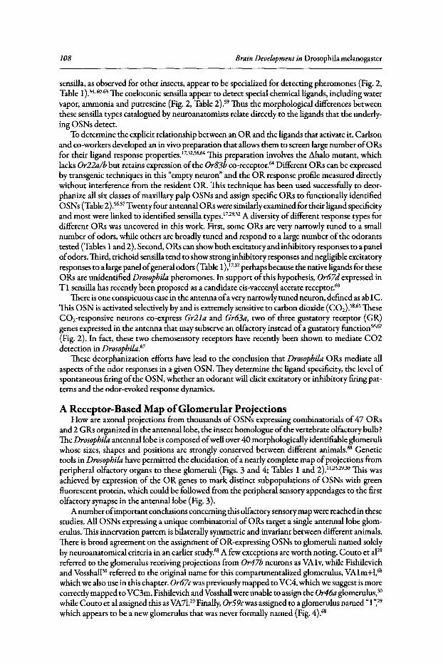
108 Brain Development in Drosophila melanogaster
sensilla, as observed for other insects, appear to be specialized for detecting pheromones (Fig. 2,Table 1).34.60-63 The coeloconic sensilla appear to detect special chemical ligands, including watervapor, ammonia and putrescine (Fig. 2, Table 2).59 Thus the morphological differences betweenthese sensilla types catalogued by neuroanatomists relate directly to the ligands that the underlying OSNs detect.
To determine the explicit relationship between an 0 R and the ligands that activate it, Carlsonand co-workers developed an in vivo preparation that allows them to screen large number of0 Rsfor their ligand response properties.17.32,56.64 This preparation involves the L\halo mutant, whichlacks Or22a/bbut retains expression ofthe Or83bco-receptor/" Different ORs can be expressedby transgenic techniques in this "empty neuron" and the OR response profile measured directlywithout interference from the resident OR. This technique has been used successfully to deorphanize allsix classes ofmaxillary palp OSNs and assign specific ORs to functionally identifiedOSNs (Table 2) .56.57 Twenty four antennal 0 Rs were similarly examined for their ligand specificityand most were linked to identified sensilla types.17.29.32 A diversity ofdifferent response types fordifferent 0 Rs was uncovered in this work. First, some 0 Rs are very narrowly tuned to a smallnumber ofodors, while others are broadly tuned and respond to a large number of the odorantstested (Tables 1 and 2). Second, 0 Rs can show both excitatory and inhibitory responses to a panelofodors. Third, trichoid sensilla tend to show stronginhibitory responses and negligible excitatoryresponses to a large panel ofgeneral odors (Table 1),17.32 perhaps because the native ligands for theseoRs are unidentified Drosophila pheromones. In support ofthis hypothesis, Or67d expressed inTI sensilla has recently been proposed as a candidate cis-vaccenylacetate receptor."
There is one conspicuous case in the antenna ofa very narrowly tuned neuron, defined as ab1C.This 0 SN is activated selectively by and is extremely sensitive to carbon dioxide (C02).58.65 TheseCO2-responsive neurons co-express Gr21a and Gr63a, two of three gustatory receptor (GR)genes expressed in the antenna that may subserve an olfactory instead ofa gustatory function66.67
(Fig. 2). In fact, these two chemosensory receptors have recently been shown to mediate C02detection in Drosophila."
These deorphanization efforts have lead to the conclusion that Drosophila 0 Rs mediate allaspects of the odor responses in a given OSN.They determine the ligand specificity, the level ofspontaneous firing ofthe OSN, whether an odorant will elicit excitatory or inhibitory firing patterns and the odor-evoked response dynamics.
A Receptor-Based Map ofGlomemlar ProjectionsHow are axonal projections from thousands of OSNs expressing combinatorials of47 0 Rs
and 2 GRs organized in the antennallobe, the insect homologue ofthe vertebrate olfactory bulb?The Drosophila antennallobe is composed ofwell over 40 morphologically identifiable glomeruliwhose sizes, shapes and positions are strongly conserved between different animals." Genetictools in Drosophila have permitted the elucidation ofa nearly complete map ofprojections fromperipheral olfactory organs to these glomeruli (Figs. 3 and 4; Tables 1 and 2).21,25.29.30 This wasachieved by expression of the OR genes to mark distinct subpopulations of OSNs with greenfluorescent protein, which could be followed from the peripheral sensory appendages to the firstolfactory synapse in the antennallobe (Fig. 3).
A number ofimportant conclusions concerningthis olfactory sensory map were reached in thesestudies. AllOSNs expressing a unique combinatorial ofORs target a single antennallobe glomerulus. This innervation pattern is bilaterally symmetric and invariant between different animals.There is broad agreement on the assignment of0 R-expressing OSNs to glomeruli named solelyby neuroanatomical criteria in an earlier study.68 A few exceptions are worth noting. Couto et aF9referred to the glomerulus receiving projections from Or47bneurons as VAlv, while Fishilevichand Vosshall'" referred to the original name for this compartmentalized glomerulus, VAlm+1,68which we also use in this chapter. Or67cwas previously mapped to VC4, which we suggest is morecorrectly mapped to VC3m. Fishilevich and Vosshallwere unable to assign the Or46a glomerulus, 30while Couto et al assigned this asVA7!.29 Finally, OrS9cwas assigned to a glomerulus named"1~29which appears to be a new glomerulus that was never formally named (Fig. 4).68

Fig
ure
3.M
ole
cula
rm
ap
pin
gof
the
Dro
soph
ila
ante
nnal
lob
e.
Ant
enna
llo
bes
oftr
ansg
enic
flies
carr
yin
gO
R-m
CD
8-G
FP
rep
ort
ers
stai
ned
wit
han
ti-G
FP
(gre
en)
and
the
gene
ral
ne
uro
pil
mar
ker
nc82
(mag
enta
)."
Fro
ntal
con
foca
lim
ages
are
alig
ne
dw
ith
dors
alup
and
late
ral
right
.B
oth
Or5
9can
dO
r67d
rep
ort
ers
labe
lan
ect
op
icg
lom
eru
lus
(inpa
rent
hese
s)w
hic
hre
sults
from
ect
op
icex
pres
sion
ofth
ep
rom
ote
rin
oth
er
OS
Ns
.T
helo
we
rrig
htpa
nel
repr
esen
tsco
elo
con
icg
lom
eru
lim
arke
dby
expr
essi
onof
the
aton
alG
al4
line
.D
ata
from
refe
renc
es.29
Rep
rinte
dw
ith
pe
rmis
sio
nfr
om:
Cou
toA
,A
len
ius
M,
Dic
kso
nB
.Cur
rB
ioi
2005
;15
:153
5-15
47.
©2
00
5E
lsev
ier
Pre
ss.
::;:l " o ~ ~ ~ ~ j ~ .... s o § .g e: o;- .... <:::
>'0
110 Brain Development in Drosophila melanogaster
0 antennalbold type: basiconicglomeru lus r-----.
1 1 antennal1 1 trichoid
italic type:1_____ .1
olfactory receptor · . antennal· .· . coeloconic· .-
o: :. .: .:
maxillary palpbasiconic
Unassigned
ttu targets * (tentalive)
Figure 4. Molecular and anatomical map of the Drosophila antennal lobe. Schematic of theantennal lobe presented as frontal sections from anterior to posterior, organized clockwisefrom top left. Glomeruli are depth-coded with black for deep, grayfor intermediate and whiteforsuperficialsections. Glomeruliare coded according to sensillumtype, chemosensory organand whether or not they are innervated by fruitless-positive neurons. Data from references29, 3D, 63, 66 and 68. Adapted with permission from: Fishilevich E, Vosshall LB. Curr Bioi2005; 15:1548-1553. ©2005 Elsevier Press.

TheO/ftctory Sensory Map in Drosophila 111
We synthesize the conclusions reached in these disparate studies and present a complete map ofthe antennallobe, indicating both the neuroanatomical and molecular name for each glomerulusin Figure 4. The antennallobe comprises a total of42 glomeruli, ofwhich seven are subdividedinto two compartments and one into three. While compartments were discovered on a purelymorphological basis," many have since been shown to express a single ORand due to these findings, compartments have been revealed in formerly undivided glomeruli. On a functional level,the antennallobe can thus be said to have a total of51 glomeruli. By including glomeruli VP 1-3,the total number ofglomeruli in the AL ofDrosophila amounts to 54 glomeruli. While VP 1-3 arevisible in staining using synaptotagmin antibody." they are not discernible with the monoclonalantibody nc82.25,29.30.68 This may be due at least partly to their posterior-most location deep intothe brain, where also the other glomeruli unassigned to 0 Rs lie. Glomeruli in the antennallobeare clustered into arrays, reflecting the bundlingofsensilla types on the antenna and the maxillarypalp. The glomeruli being connected with the maxillary palp are found predominantly in centralpositions, distinct from the glomeruli connected to the antenna. Antennal coeloconic OSNsproject mainly to the posterior face ofthe antennallobe, while antennal basiconic OSNs project tomedial anterior regions ofthe antennallobe. Trichoid OSNs project to the group oflarge glomerulithat lie at the extreme lateral regions of the antennallobe. While these arrays have a fairly fixeddesign, there is no evidence for a topographic point-to-point mapping from the antenna to theantennallobe. A direct correlation exists though between the sizeofa glomerulus and the numberofOSNs projecting to it. For instance, Or47b is expressed in approximately 50 OSNs and marksa large glomerulus, VAlm+l, while Or22a is expressed in approximately 25 OSNs and marks asmall glomerulus, DM2.
How accurate is this olfactory sensory map? Because these maps were generated with geneticreagents, it is important that the transgenic marker expression recapitulates the expression patternsofthe endogenous genes. In most cases, this has been verified. Expression ofthe transgene, closelymatching RNA in situ hybridization ofthe endogenous 0 R has been demonstrated for most published 0 R-Gal4 transgenes.21,25,29,30 Nevertheless, the transgenic approach has led to some variabilityin glomerular mapping that almost certainly reflects artefacts ofthe transgenic lines themselves. Forinstance, both early reports ofOr23a-expressing OSNs showed that these target two glomeruli inthe antennallobe.21.25 Subsequent analysis ofthese same transgenes showed that OSNs expressingOr23a innervate only one of the two original glomeruli.29.30 Ectopic expression of both Or59cand Or67d was observed, such that both transgenic reagents label one authentic and one ectopicglomerulus.21.25Sporadic cases of these transgenic reagents labeling multiple glomeruli have alsobeen reported and these are almost certainly due to ectopic expression ofthe transgenes inducedby position and other genetic background effects.52Another possible explanation for variation inthe olfactory map is that despite the highly conserved anatomy ofthe antennal lobe, additional ormissing glomeruli and compartments are observed between individual flies,suggesting a moderateplasticity of the olfactory system on the individual level.29.68 Despite this inherent limitation ofthe genetic reagents, they have proven to be powerful tools that allowed investigators to describethe molecular neuroanatomy ofthe antennallobe in unprecedented detail.
Sexual Dimorphism in the Drosophila Olfactory SystemOne further outcome ofthe molecular mapping ofthe antennallobe was that it allowed the
identification ofputative pheromone receptors. Previous reports that examined sexual dimorphism in the antennallobe ofHawaiian Drosophila species identified several prominent lateralglomeruli that are larger in male than female flies.61Both DL3 and DAI are considerably largerin male Hawaiian species than females. The same analysis in Drosophila melanogaster indicatesthat compared to the female, the male DAI and VAlm+1 are 62% and 33% larger, respectively,while DL3 and VAld are isomorphic in both sexes." These glomeruli receive input from OSNsexpressing Or67d (DA1) and Or47b (VAlm+I),29.30 both of which are housed in trichoidsensilla." The basis for this size increase in males is unknown, but earlier investigators noticedthat there is also a sexual dimorphism in sensilla number.Y" Males have more trichoid sensilla
112 Brain Development in Drosophila melanogaster
and fewer basiconic sensilla than females.33•36 Finally, neurons expressingfruitless, the master
transcriptional regulator ofsex-specificdevelopment and behavior project to these large lateral,sexually dimorphic glomeruli (Fig. 4, pink hatched glomeruli).62.69 Thus the hypothesis thatmale antennae are more sensitive to pheromones, as has been shown for a large number ofotherinsects and that this sensitivity is mediated by specialized pheromone-sensing OSNs housed intrichoid sensilla is well supported by the available data.
Concluding RemarksThe advanced state ofknowledge concerning gene expression and synaptic organization ofthe
early olfactory system ofthe flymakes this a compelling system to address questions in odor coding. For instance, it is not yet clear in any species how and where odor concentration is encoded;how the brain solves odor mixture problems, by far the most likely physiological stimulus ananimal will encounter; and how discrimination between perceptually similar odors is achieved."Functional calcium imaging71
-73 and electrophysiology'S" will be important tools in future re
search that seeks to answer these important questions at the cellular level.Finally,little is knownabout how the olfactory system processes odors to produce stereotyped behavioral outputs. Thesmall size, genetic manipulability and availability of robust olfactory behavior paradigms forDrosophila olfaction strengthen the role of this little insect as a powerful genetic model systemfor the foreseeable future.
References1. Roskies A, Friedman GC, O'Leary DD. Mechanisms and molecules controlling the development of
retinal maps. Perspect Dev Neurobiol1995; 3(1):63-75.2. Schieber MH. Constraints on somatotopic organization in the primary motor cortex. J Neurophysiol
2001; 86(5):2125-2143.3. Frostig RD. Functional organization and plasticity in the adult rat barrel cortex: moving out-of-the-box.
Curr Opin Neurobiol2006; 16(4):445-450.4. Shamma SA. Topographic organization is essential for pitch perception. Proc Nat! Acad Sci USA 2004;
101(5):1114-1115.5. Rubsamen R. Postnatal development of central auditory frequency maps. J Comp Physiol [A] 1992;
170(2):129-143.6. Dethier VG. The Hungry Fly: A Physiological Study of the Behavior Associated with Feeding. Cam
bridge: Harvard University Press, 1976.7. Arora K, Rodrigues V, Joshi S et ale A gene affecting the specificity of the chemosensory neurons of
Drosophila. Nature 1987; 330(6143):62-63.8. Zhang Y, Hoon MA, Chandrashekar Jet al. Coding of sweet, bitter and umami tastes: different receptor
cells sharing similar signaling pathways. Cell 2003; 112(3):293-301.9. Marella S, Fischler~ Kong P et al. Imaging taste responses in the fly brain reveals a functional map
of taste category and behavior. Neuron 2006; 49(2):285-295.10. Wang Z, Singhvi A, Kong P et al. Taste representations in the Drosophila brain. Cell 2004;
117(7):981-991.11. Thorne N, Chromey C, Bray S et al. Taste perception and coding in Drosophila. Curr Biol 2004;
14(12):1065-1079.12. Jones LM, Fontanini A, Katz DB. Gustatory processing: a dynamic systems approach. Curr Opin
Neurobiol2006; 16(4):420-428.13. Glendinning Jl, Davis A, Rai M. Temporal coding mediates discrimination of "bitter" taste stimuli by
an insect. J Neurosci 2006; 26(35):8900-8908.14. Firestein S. How the olfactory system makes sense of scents. Nature 2001; 413(6852):211-218.15. Araneda RC, Kini AD, Firestein S. The molecular receptive range of an odorant receptor. Nat Neurosci
2000; 3(12):1248-1255.16. Malnic B, Hirono J, Sato T et al. Combinatorial receptor codes for odors. Cell 1999; 96(5):713-723.17. Hallem EA, Ho MG, Carlson JR. The molecular basis of odor coding in the Drosophila antenna. Cell
2004; 117(7):965-979.18. Katada S, Hirokawa T, Oka Y et ale Structural basis for a broad but selectiveligand spectrum of a mouse
olfactory receptor: mapping the odorant-binding site. J Neurosci 2005; 25(7):1806-1815.19. Hildebrand JG, Shepherd GM. Mechanisms of olfactory discrimination: converging evidence for com
mon principles across phyla. Annu Rev Neurosci 1997; 20:595-631.
TheOlfactory Sensory Map in Drosophila 113
20. Strausfeld NJ, Hildebrand JG. Olfactory systems: common design, uncommon origins? Curr OpinNeurobiol 1999; 9(5):634-639.
21. Gao Q Yuan B, Chess A. Convergent projections of Drosophila olfactory neurons to specificglomeruliin the antennallobe. Nat Neurosci 2000; 3(8):780-785.
22. Mombaerts P, Wang F, Dulac C et al. Visualizing an olfactory sensory map. Cell 1996;87(4):675-686.
23. ResslerKJ, Sullivan SL, Buck LB. Information coding in the olfactory system: evidence for a stereotypedand highly organized epitope map in the olfactory bulb. Cell 1994; 79(7):1245-1255.
24. VassarR, Chao SK, Sitcheran R et al. Topographic organization of sensory projections to the olfactorybulb. Cell 1994; 79(6):981-991.
25. VosshallLB, Wong AM, Axel R. An olfactory sensory map in the Bybrain. Cell 2000; 102:147-159.26. Uchida N, Takahashi YK, Tanifuji M et al. Odor maps in the mammalian olfactory bulb: domain
organization and odorant structural features. Nat Neurosci 2000; 3(10):1035-1043.27. Mori K, Nagao H, Yoshihara Y. The olfactory bulb: coding and processing of odor molecule informa
tion. Science 1999; 286(5440):711-715.28. Friedrich R~ Korsching SI. Combinatorial and chemotopic odorant coding in the zebrafish olfactory
bulb visualized by optical imaging. Neuron 1997; 18(5):737-752.29. Couto A, Alenius M, Dickson B]. Molecular, anatomical and functional organization of the Drosophila
olfactory system. Curr Bioi 2005; 15(17):1535-1547.30. Fishilevich E, Vosshall LB. Genetic and functional subdivision of the Drosophila antennallobe. Curr
Bioi 2005; 15(17):1548-1553.31. Galizia CG, Sachse S, Rappen A et al. The glomerular code for odor representation is species specific
in the honeybee Apis mellifera. Nat Neurosci 1999; 2(5):473-478.32. Hallem EA, Carlson JR. Coding of odors by a receptor repertoire. Cell 2006; 125(1):143-160.33. Stocker RF. The organization of the chemosensory system in Drosophila melanogaster: a review. Cell
Tissue Res 1994; 275(1):3-26.34. Clyne P, Grant A, O'Connell R et al. Odorant response of individual sensillaon the Drosophila antenna.
Invert Neurosci 1997; 3:127-135.35. Shanbhag SR, Mueller B, Sreinbrecht RA. Atlas of olfactory organs of Drosophila melanogaster. 2. In
ternal organization and cellular architecture of olfactory sensilla. Arthr Struct Dev 2000; 29:211-229.36. Shanbhag SR, Mueller B, Steinbrecht RA. Atlas of olfactory organs of Drosophila melanogaster. 1.
Types, external organization, innervation and distribution of olfactory sensilla. Int J Insect MorpholEmbryol1999; 28(4):377-397.
37. Reddy G~ Gupta B, Ray K et al. Development of the Drosophila olfactory sense organs utilizes cell-cellinteractions as well as lineage. Development 1997; 124(3):703-712.
38. Gupta BP, Rodrigues V. Atonal is a proneural gene for a subset of olfactory sense organs in Drosophila.Genes Cells 1997; 2(3):225-233.
39. Sen A, Reddy GV, Rodrigues V. Combinatorial expression of Prospero, Seven-up and Elav identifiesprogenitor cell types during sense-organ differentiation in the Drosophila antenna. Dev Biol 2003;254( 1):79-92.
40. Goulding SE, zur Lage P,Jarman AP. amos, a proneural gene for Drosophila olfactory sense organs thatis regulated by lozenge. Neuron 2000; 25:69-78.
41. Buck L, Axel R. A novel multigene family may encode odorant receptors: a molecular basis for odorrecognition. Cell 1991; 65(1):175-187.
42. Vosshall LB, Amrein H, Morozov PS et al. A spatial map of olfactory receptor expression in the Drosophila antenna. Cell 1999; 96(5):725-736.
43. Clyne PJ, Warr CG, Freeman MR et al. A novel family of divergent seven-transmembrane proteins:candidate odorant receptors in Drosophila. Neuron 1999; 22(2):327-338.
44. Gao Q Chess A. Identification of candidate Drosophila olfactory receptors from genomic DNA sequence.Genomics 1999; 60(1):31-39.
45. Robertson HM, Warr CG, Carlson JR. Molecular evolution of the insect chemoreceptor gene superfamilyin Drosophila melanogaster. Proc Natl Acad Sci USA 2003; 100 SuppI2:14537-14542.
46. Benton R, Sachse S, Michnick SW et al. Atypical membrane topology and heteromeric function ofDrosophila odorant receptors in vivo. PLoS Bioi 2006; 4(2):e20.
47. Wistrand M, Kall L, Sonnhammer EL. A general model of G protein-coupled receptor sequences andits application to detect remote homologs. Protein Sci 2006; 15(3):509-521.
48. Fishilevich E, Domingos AI, Asahina K et al. Chemotaxis behavior mediated by single larval olfactoryneurons in Drosophila. Curr Biol 2005; 15(23):2086-2096.
49. Kreher SA, Kwon~ Carlson JR. The molecular basis of odor coding in the Drosophila larva. Neuron2005; 46:445-456.
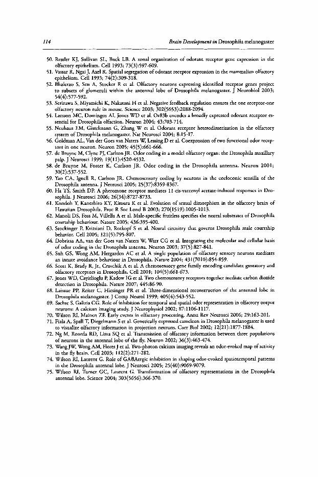
114 Brain Development in Drosophila melanogaster
50. Ressler KJ, Sullivan SL, Buck LB. A zonal organization of odorant receptor gene expression in theolfactory epithelium. Cell 1993; 73(3):597-609.
51. VassarR, Ngai J, Axel R. Spatial segregation of odorant receptor expression in the mammalian olfactoryepithelium. Cell 1993; 74(2):309-318.
52. Bhalerao S, Sen A, Stocker R et al. Olfactory neurons expressing identified receptor genes projectto subsets of glomeruli within the antennal lobe of Drosophila melanogaster. J Neurobiol 2003;54(4):577 -592.
53. Serizawa S, Miyamichi K, Nakatani H et al. Negative feedback regulation ensures the one receptor-oneolfactory neuron rule in mouse. Science 2003; 302(5653):2088-2094.
54. Larsson MC, Domingos AI, Jones WD et al. Or83b encodes a broadly expressed odorant receptor essential for Drosophila olfaction. Neuron 2004; 43:703-714.
55. Neuhaus EM, Gisselmann G, Zhang W et al. Odorant receptor heterodimerization in the olfactorysystem of Drosophila melanogaster. Nat Neurosci 2004; 8:15-17.
56. Goldman AL, Van der Goes van Naters~ LessingD et al. Coexpression of two functional odor receptors in one neuron. Neuron 2005; 45(5):661-666.
57. de Bruyne M, Clyne PJ, Carlson JR. Odor coding in a model olfactory organ: the Drosophila maxillarypalp, J Neurosci 1999; 19(11):4520-4532.
58. de Bruyne M, Foster K, Carlson JR. Odor coding in the Drosophila antenna. Neuron 2001;30(2):537-552.
59. Yao CA, Ignell R, Carlson JR. Chemosensory coding by neurons in the coeloconic sensilla of theDrosophila antenna. J Neurosci 2005; 25(37):8359-8367.
60. Ha TS, Smith DP. A pheromone receptor mediates l l-cis-vaccenyl acetate-induced responses in Drosophila. J Neurosci 2006; 26(34):8727-8733.
61. Kondoh Y, Kaneshiro~ Kimura K et al. Evolution of sexual dimorphism in the olfactory brain ofHawaiian Drosophila. Proc R Soc Lond B 2003; 270(1519):1005-1013.
62. Manoli DS, Foss M, Villella A et al. Male-specificfruitless specifies the neural substrates of Drosophilacourtship behaviour. Nature 2005; 436:395-400.
63. Stockinger P, Kvitsiani D, Rotkopf S et al. Neural circuitry that governs Drosophila male courtshipbehavior. Cell 2005; 121(5):795-807.
64. Dobritsa AA, van der Goes van Naters~ Warr CG et al. Integrating the molecular and cellular basisof odor coding in the Drosophila antenna. Neuron 2003; 37(5):827-841.
65. Suh GS, Wong AM, Hergarden AC et al. A single population of olfactory sensory neurons mediatesan innate avoidance behaviour in Drosophila. Nature 2004; 431(7010):854-859.
66. Scott K, Brady R, jr., Cravchik A et al. A chemosensory gene family encoding candidate gustatory andolfactory receptors in Drosophila. Cell 2001; 104(5):661-673.
67. Jones WD, Cayirlioglu P, Kadow IG et al. Two chemosensory receptors together mediate carbon dioxidedetection in Drosophila. Nature 2007; 445:86-90.
68. Laissue PP, Reiter C, Hiesinger PR et al. Three-dimensional reconstruction of the antennallobe inDrosophila melanogaster. J Comp Neuro11999; 405(4):543-552.
69. SachseS, Galizia CG. Role of inhibition for temporal and spatial odor representation in olfactory outputneurons: A calcium imaging study. J Neurophysiol2002; 87:1106-1117.
70. Wilson RI, Mainen ZE Early events in olfactory processing. Annu Rev Neurosci 2006; 29:163-201.71. Fiala A, Spall T, Diegelmann S et al. Genetically expressedcameleon in Drosophila melanogaster is used
to visualize olfactory information in projection neurons. Curr Bioi 2002; 12(21):1877-1884.72. Ng M, Roorda RD, Lima SQ et al. Transmission of olfactory information between three populations
of neurons in the antennallobe of the fly. Neuron 2002; 36(3):463-474.73. Wang~ Wong AM, FloresJ et al. Two-photon calcium imaging revealsan odor-evoked map of activity
in the fly brain. Cell 2003; 112(2):271-282.74. Wilson RI, Laurent G. Role of GABAergic inhibition in shaping odor-evoked spatiotemporal patterns
in the Drosophila antennallobe. J Neurosci 2005; 25(40):9069-9079.75. Wilson RI, Turner GC, Laurent G. Transformation of olfactory representations in the Drosophila
antennallobe. Science 2004; 303(5656):366-370.
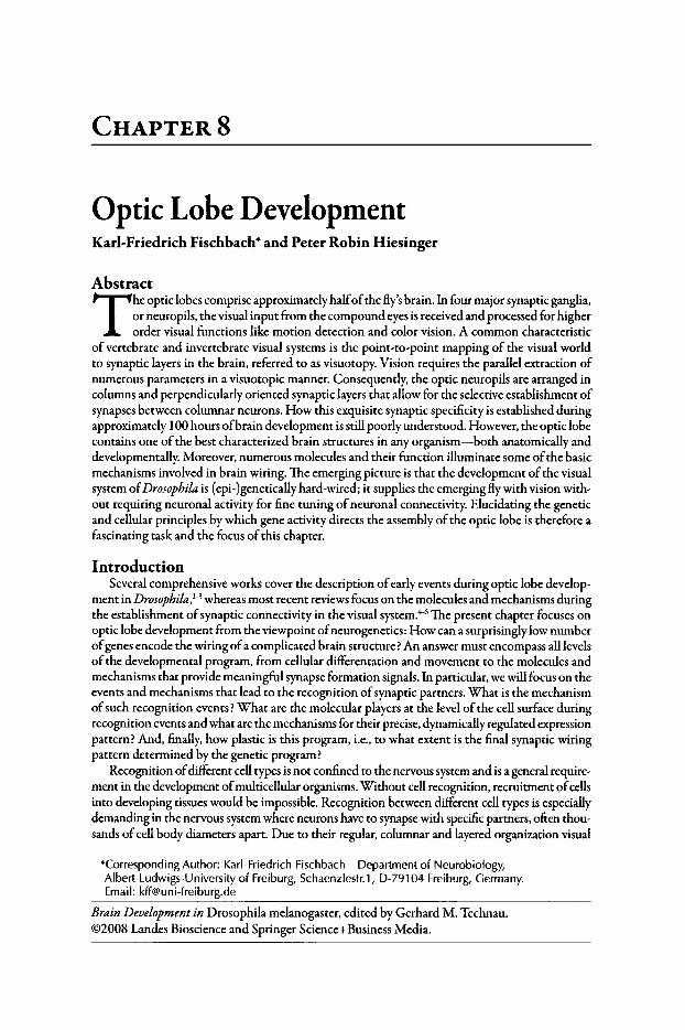
CHAPTER 8
Optic Lobe DevelopmentKarl-Friedrich Fischbach" and Peter Robin Hiesinger
Abstract
The optic lobes comprise approximately halfofthe fly'sbrain. In four major synaptic ganglia,or neuropils, the visual input from the compound eyes is received and processed for higherorder visual functions like motion detection and color vision. A common characteristic
of vertebrate and invertebrate visual systems is the point-to-point mapping of the visual worldto synaptic layers in the brain, referred to as visuotopy. Vision requires the parallel extraction ofnumerous parameters in a visuotopic manner. Consequently, the optic neuropils are arranged incolumns and perpendicularly oriented synaptic layers that allow for the selective establishment ofsynapses between columnar neurons. How this exquisite synaptic specificity is established duringapproximately 100 hours ofbrain development is still poorly understood. However, the optic lobecontains one of the best characterized brain structures in any organism-both anatomically anddevelopmentally. Moreover, numerous molecules and their function illuminate some ofthe basicmechanisms involved in brain wiring. The emerging picture is that the development ofthe visualsystem ofDrosophila is (epi-)genetically hard-wired; it supplies the emerging fly with vision without requiring neuronal activity for fine tuning of neuronal connectivity. Elucidating the geneticand cellular principles by which gene activity directs the assembly ofthe optic lobe is therefore afascinating task and the focus of this chapter.
IntroductionSeveral comprehensive works cover the description ofearly events during optic lobe develop
ment in Drosophila,1-3 whereas most recent reviews focus on the molecules and mechanisms duringthe establishment ofsynaptic connectivity in the visual systern.tf The present chapter focuses onoptic lobe development from the viewpoint ofneurogenetics: How can a surprisingly low numberofgenes encode the wiring ofa complicated brain structure? An answer must encompass all levelsof the developmental program, from cellular differentation and movement to the molecules andmechanisms that provide meaningful synapse formation signals. In particular, we will focus on theevents and mechanisms that lead to the recognition ofsynaptic partners. What is the mechanismofsuch recognition events? What are the molecular players at the level of the cell surface duringrecognition events and what are the mechanisms for their precise, dynamically regulated expressionpattern? And, finally, how plastic is this program, i.e., to what extent is the final synaptic wiringpattern determined by the genetic program?
Recognition ofdifferent cell types is not confined to the nervous system and is a general requirement in the development ofmulticellular organisms. Without cell recognition, recruitment ofcellsinto developing tissues would be impossible. Recognition between different cell types is especiallydemanding in the nervous system where neurons have to synapse with specific partners, often thousands of cell body diameters apart. Due to their regular, columnar and layered organization visual
*Corresponding Author: Karl-Friedrich Fischbach-Department of Neurobiology,Albert-Ludwigs-University of Freiburg, Schaenzlestr.1, 0-79104 Freiburg, Germany.Email: [email protected]
Brain Development in Drosophila melanogaster, edited by Gerhard M. Technau.©2008 Landes Bioscience and Springer Science+Business Media.
116 Brain Development in Drosophila melanogasrer
systemsare well suited to investigate the genetic determination and developmental rules that underliethe establishment ofneuronal connectivity. The repetitive organization ofabout 750 visual units orcolumns on each side ofthe fly's head allow the detection ofminor disturbances. The visual systemofDrosophila has the further advantage that an exceptionally powerful toolbox can be applied togenetically dissect the developmental programs.
The Adult Visual System Is Organized into Parallel VisuotopticFunctional Pathways
The adult optic lobes ofcoleoptera, lepidoptera and diptera7.8 are subdivided into four neuropils,the lamina, medulla, lobula and lobula plate (Figs. lA, 2A). Photoreceptor projections from theeye directly innervate the first two neuropils, lamina and medulla. In Drosophila, each single eye,or ommatidium, of the compound eye contains eight different photoreceptor cell types . Theirlight-sensing protrusion, the rhabdomeres, receive light along seven different optical axes underneath a single lens.The outer 6 rhabdomeres are formed by retinula cellsRI-6; the inner rhabdomerecomprises distally R7 and proximally R8.In all ommatidia, except those ofthe dorsal rim, the innerrhabdomeres are much thinner than the outer ones . Functionally, the outer photoreceptors areresponsible for spatial vision, whereas the inner photoreceptors convey color vision.
Three types ofommatidia can be distingulshed" according to the rhodopsin (Rh) content ofthe inner retinula cells R7 and R8: 30% ofommatidia are ofthe pale subtype, where R7 containsthe UV-sensitive Rh3 and R8 the blue-sensitive Rh5 , while the remaining 70% are of the yellowsubtype and contain UV-sensitive Rh4 in R7 and green-sensitive Rh6 in R8. Both types are ran domly distributed due to the stochastic expression pattern ofthe transcription factor and Dioxinreceptor homolog spineless in R7 cells. The expression ofSpineless in R7 cells specifies it as a Rh4cell. R7 then dictates the fate of the R8 cell to also assume the yellow subtype. In the absenceof spinelessor in spineless mutants, all R7 and most R8 cells adopt the pale (Rh5) fate, whereasoverexpression ofspineless is sufficient to induce the yellow R7 fate.1OThe molecular mechanismthat determines the stochastic expression ofSpineless as well as the functional significance oftherandom pale/yellow ommatidia distribution are currently unknown.
In addition to these two major ommatidial types there is a dorsal rim area of the compoundeyes"!' which is specialized for the detection of polarized light. Here R7 and R8 rhabdomereshave larger diameters and both express the UV-sensitive Rh3 . As the microvilli ofboth cell typesare perpendicularly oriented with respect to each other, this allows the evaluation ofthe vector of
Figure 1.The Drosophilaoptic lobe.A) Volumerendered optic lobe neuropilsbased on synapticstaining (n-Syb). Selected characterized cell types are depicted based on Golgi studies." la,lamina; dm, distal medulla; pm, proximal medulla; Ip, lobula plate; 10, lobula. B) The primaryvisual map. Lamina cross-sections of confocal images based on a photoreceptor-specificantibody staining. Scale bar 51Jm. C) EM micrograph of a single unit (cartridge) of the visualmap in the lamina. Color code as in A. Scale bar Ium.
OpticLobeDevelopment 117
A
B
o
Figure 2, legend viewed on fall .owmg page.
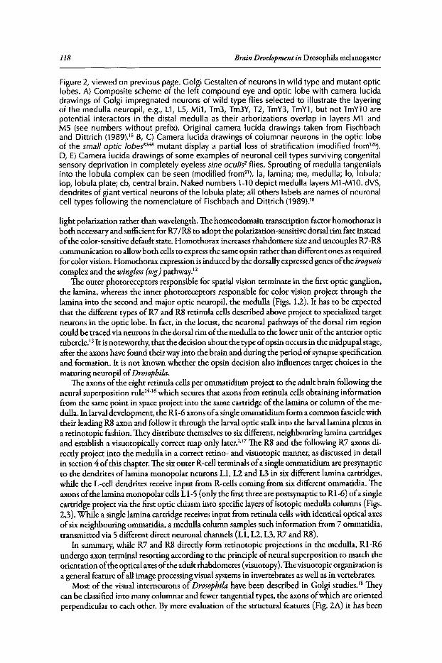
118 Brain Development in Drosophila melanogaster
Figure 2, viewed on previous page. Golgi Gestalten of neurons in wild type and mutant opticlobes. A) Composite scheme of the left compound eye and optic lobe with camera lucidadrawings of Golgi impregnated neurons of wild type flies selected to illustrate the layeringof the medulla neuropil, e.g., Ll, LS, Mil, Tm3, Tm3Y, T2, TmY3, TmYl, but not TmYl0 arepotential interactors in the distal medulla as their arborizations overlap in layers Ml andMS (see numbers without prefix). Original camera lucida drawings taken from Fischbachand Dittrich (1989).18 B, C) Camera lucida drawings of columnar neurons in the optic lobeof the small optic lobesKS58 mutant display a partial loss of stratification (modified from'"),D, E) Camera lucida drawings of some examples of neuronal cell types surviving congenitalsensory deprivation in completely eyelesssine oculis' flies. Sprouting of medulla tangentialsinto the lobula complex can be seen (modified from"), la, lamina; me, medulla; 10, lobula;lop, lobula plate; cb, central brain. Naked numbers 1-10 depict medulla layers Ml-Ml O.dVS,dendrites of giant vertical neurons of the lobula plate; all others labels are names of neuronalcell types following the nomenclature of Fischbach and Dittrich (1989).18
light polarization rather than wavelength. The homeodomain transcription factor homothorax isboth necessary and sufficient for R7/R8to adopt the polarization-sensitive dorsal rim fate insteadofthe color-sensitive default state. Homothorax increases rhabdomere size and uncouples R7-R8communication to allow both cellsto express the same opsin rather than different ones as requiredfor color vision. Homothorax expression is induced by the dorsally expressed genes ofthe iroquoiscomplex and the wingless (wg)pathway."
The outer photoreceptors responsible for spatial vision terminate in the first optic ganglion,the lamina, whereas the inner photoreceptors responsible for color vision project through thelamina into the second and major optic neuropil, the medulla (Figs. 1,2). It has to be expectedthat the different types of R7 and R8 retinula cells described above project to specialized targetneurons in the optic lobe. In fact, in the locust, the neuronal pathways of the dorsal rim regioncould be traced via neurons in the dorsal rim ofthe medulla to the lower unit ofthe anterior optictubercle.P It is noteworthy, that the decision about the type ofopsin occurs in the midpupal stage,after the axons have found their way into the brain and during the period ofsynapse specificationand formation. It is not known whether the opsin decision also influences target choices in thematuring neuropil ofDrosophila.
The axons ofthe eight retinula cells per ommatidium project to the adult brain following theneural superposition rule14-16which secures that axons from retinula cells obtaining informationfrom the same point in space project into the same cartridge ofthe lamina or column of the medulla. In larval development, the Rl-6 axons ofa single ommatidium form a common fasciclewiththeir leading R8 axon and follow it through the larval optic stalk into the larval lamina plexus ina retinotopic fashion. They distribute themselves to six different, neighbouring lamina cartridgesand establish a visuotopically correct map only later.2,17 The R8 and the following R7 axons directly project into the medulla in a correct retino- and visuotopic manner, as discussed in detailin section 4 ofthis chapter. The sixouter R-cell terminals ofa single ommatidium are presynapticto the dendrites of lamina monopolar neurons L1, L2 and L3 in six different lamina cartridges,while the L-cell dendrites receive input from R-cells coming from six different ommatidia. Theaxons ofthe lamina monopolar cellsL1-5 (only the first three are postsynaptic to Rl-6) ofa singlecartridge project via the first optic chiasm into specific layers of isotopic medulla columns (Figs.2,3). While a single lamina cartridge receives input from retinula cellswith identical optical axesofsix neighbouring ommatidia, a medulla column samples such information from 7 ommatidia,transmitted via 5 different direct neuronal channels (Ll, L2, L3, R7 and R8).
In summary, while R7 and R8 directly form retinotopic projections in the medulla, R1-R6undergo axon terminal resorting according to the principle ofneural superposition to match theorientation ofthe optical axesofthe adult rhabdomeres (visuotopy). The visuotopic organization isa general feature ofall image processing visual systems in invertebrates as well as in vertebrates.
Most of the visual interneurons of Drosophila have been described in Golgi studies. IS Theycan be classified into many columnar and fewer tangential types, the axons ofwhich are orientedperpendicular to each other. By mere evaluation of the structural features (Fig. 2A) it has been
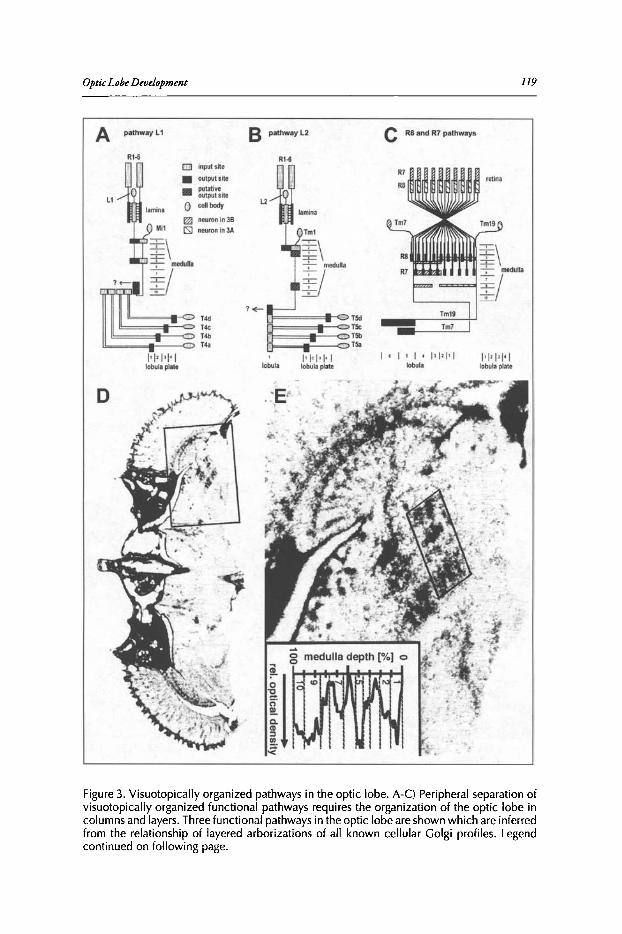
OpticLobeDevelopment 119
A pathwayL1 B pathwayL2 C RBand R7 pathways
Rt·6 R1-6
1'1' 1'1' 1lobula plate
I . 1 '1'1 ' 1'1'1tobula
III1' 1'1' 1'1lobula plate
11=== ==1[-<:"TSd
[;]I=~~~!::::::= T5c0= OT5bG OT~
lobula
G input site
• output site
II putative
L1output site
L20 cenbody
m neuronin38Mil lS1 neuronin3A
Figure 3. Visuotopically organized pathways in the optic lobe . A-C) Peripheral separation ofvisuotopically organized functional pathways requires the organization of the optic lobe incolumns and layers. Three functional pathways in the optic lobe are shown which are inferredfrom the relationship of layered arborizations of all known cellular Golgi profiles. Legendcontinued on following page.
120 Brain Development in Drosophila melanogaster
Figure 3, viewed on previous page. For simplicity, at the level of the medulla only typical neuronal types are shown. The L1 and L2 pathways are fed by Rl-6 and function in spatial vision,the R8 and R7 pathways in color vision. D) depicts an 3H-2-deoxyglucose autoradiogram of ahorizontal brain section after unilateral 120 min stimulation in two 15 x 15 degree sectors ofthe right visual field. The right optic lobe autoradiogram is enlarged in E. The anterior visualfield window (posterior medulla sector) was stimulated by upward motion, the posteriorvisual field window by horizontal progressive motion of the same spatial wavelength (usinga sinusoidally modulated gray scale). In both casesvisuotopically situated columnar neuronsof the L1 and L2 pathway layers (A,B) have taken up radioactive deoxyglucose. The densityprofile of the medulla sector stimulated by upward motion is shown in the inset. It is obviousthat the R7/R8 pathway layers M3 and M6 are silent under these conditions (modified fromFischbach et al 199222
) .
claimed that several, visuotopically organized, parallel visual pathways co-exist'? (Fig. 3A-C). Incombination with 2-deoxyglucose studies" a clear structural separation between the pathways formotion detection and colour vision could be demonstrated18-22 (see Fig. 3D,E).
This neuronal organization ofthe visual system ofDrosophila contrasts sharply with the olfactory system, where olfactory receptor cells with the same chemosensory specificity converge in socalled glomeruli of the antennallobe onto single large interneurons (relay neurons) that projectto the mushroom bodies and the lateral protocerebrum'V' (see Chapters by R. Stocker and byv. Rodrigues and T. Hummel). However, it has recently been pointed out that the output levelofthe visual system is also comparable to the olfactory system, as visuotopically organized lobulaoutput neurons of the same type converge in so-called optic glomeruli, where they synapse ontolarge projection neurons25.26 (Fig. 4). It is therefore tempting to suggest that the visuotopic, parallelpathway organization is an evolutionary added feature ofthe visual system.
What is known about the cellular and molecular mechanisms that enable the visuotopic andpathway-specific wiring in the optic lobe? We will first review data related to the dependenceofvisual neuropil development on retinal innervation and will consider some of the functionsofknown cellular and molecular factors involved in axonal pathfinding, target recognition andsynaptogenesis.
Lamina Development
RetinalInnervation: Axon Outgrowth andInterdependence with Optic LobeDevelopment
Axon outgrowth from the retina occurs in a developmental wave following the wave ofcellulardifferentiation in the eye disc. The first (pioneer) axons grow out from R8, followed by R2&RS,R3&R4, then Rl&R6 and R7 follow last.27The retinal axons project through the tubular optic stalkthat consists ofa monolayer ofsurface glia and forms before axon ingrowth under the control thefocal adhesion kinase FakS6D.28 The larval photoreceptor organ, the Bolwig's organ, is dispensablefor adult wild-type photoreceptor axons to project normally and is thus not an essential pioneer ofaxonal navigation to the lamina. Bolwig's organ later transforms into the four photoreceptors ofanextra-retinal posterior "eyelet', the so-called "Hofbauer-Buchner eyelet~29which is involved in thegeneration ofcircadian rhythm." The best characterized signal transduction pathway required forphotoreceptor growth cone guidance includes the Insulin receptor on the cell surface" and intracellularly dreadlocks (dock, a SH2/SH3 adaptor protein), pak (p21 activated protein kinase), trio (aRho family guanine exchange factor that activates Rae), misshapen (a Ste20-like serine/threoninekinase) and bifocal (a putative cytoskeletal regulator).32-37 These molecular components have beenproposed to constitute a signal transduction cascade from the cell surface to the actin cytoskeleton.Targeting choices of the different photoreceptor subtypes and the upstream guidance receptorsare described in more detail below.
While maintainance of the fly's retina requires that retinal axons connect to the optic lobe,"it is well established that retinae develop quite normally in ectopic positions without connectionsto the brain, either achieved by transplantation" or by ectopic expression of eyeless." Also the
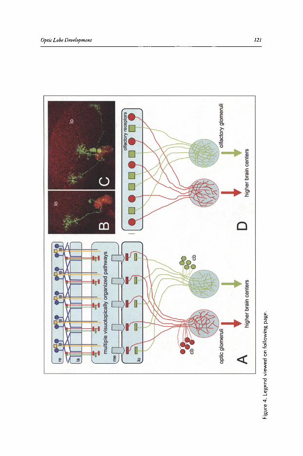
10
mul
tiple
visu
oto
pic
ally
orga
nize
dpa
thw
ays
... N ...~ ;:;.
t-. '" ~ )? 1t ..g ~ s ...
~~o
ltact
ory
glom
emll
high
er
brai
nce
nte
rsD
optic
glo
mem
ll~
~
Ahi
ghe
rbr
ain
cent
ers
Figu
re4
.Le
gend
view
edon
follo
win
gpa
ge.

122 Brain Development in Drosophila melanogasrer
Figure 4, viewed on previous page. Comparing wiring principles of the olfactory and thevisual system. A) Schematic view of the visual system. Visuotopy is maintained up to thelobula complex (Iobula plate has been omitted for simplicity). Different sets of lobula columnar neurons project to specific optic glomeruli, where they terminate in a nonvisuotopicmanner. B,C) GFP marked neurons resulting from MARCM using the irreC/rst-specific Gal4driver NP2044. Background staining with an IrreC/Rst-specific antibody. B)The terminals of asingle LC12 neuron branch throughout its glomerulus. C) A clone of three such LC12 neuronssubserving different parts of the visual field are shown. D) Schematic view of the organizationof the olfactory system. Here all olfactory receptor cells of the same kind directly project tothe same glomerulus. re, retina; la, lamina; me, medulla; 10, lobula; cb, cell bodies.
unconnected phenotype of the disconnected mutant, in which the retinula cell axons of the compound eye do not connect to the brain, demonstrates that retina development, which proceedsnormally, isautonomous." This does not hold for the optic lobe, the development ofwhich stronglydepends on retinal innervation (Fig. 2D,E). It was already demonstrated by Power in 1943 andconfirmed by Hinke in 1961 that optic lobe volume strongly correlates with the facet numberof the compound eye.41
,42 In his volumetric studies Power found that eyelessflies do not developa lamina at all and have a drastically reduced medulla and lobula complex (about 80% and 60%reduction respectively).
Optic lobe interneurons are the progeny of two groups of progenitor cells, arranged in theouter and inner optic anlagen. The lamina (together with the distal part of the medulla, see below) arise from the outer optic anlage." The strong correlation of lamina size with the numberof ommatidia is the direct consequence of an inductive influence of ingrowing retinula (R) cellaxons on neurogenesis of lamina neurons'v" and lamina glia.46 Photoreceptor innervation thustriggers the final cell-cycleoflamina precursor cells.Hedgehog, that is released from R-cell axons,induces the generation oflamina monopolar neurons from lamina precursor cellswhich-in theabsence ofHedgehog-are arrested in the Gl phase.47
-49 Hedgehog transport in photoreceptors
has recently been shown to depend on the competition between targeting signalsofthe HedgehogN- and C-termini. After Hedgehog cleavage, the N-terminal domain is targeted to the retina,while the C-terminal domain is responsible for Hedgehog transport along the axon.50 Togetherwith Hedgehog, the epidermal growth factor receptor (EGFR) ligand Spitz is transported downthe photoreceptor axons. The postsynaptic precursor cellsexpressEGFR and are thus initiated toassemble the postsynaptic cell complement for the lamina cartridge." By the concerted action ofHedgehog and Spitz, the number ofpresynaptic neurons determines the size ofthe postsynapticneuronal population. The fivelamina cell types L1-L5 are thereby specified.As young retinal ommatidia are added anteriorly, this also implies that the lamina grows from posterior to anterior.Lamina precursor cellsas well as glia cellsrequire the transcription factor Glia cellsmissing (gcm)and Glia cells missing 2 (gcm2), that were previously thought to be exclusively required for glialcell fate determination." Of further importance on the side of the lamina precursor cells is thegene product ofdally. In dally loss-of-function mutants the lamina precursors do not perform thesecond division that is triggered by ingrowing retinal fibres.52 Dally isa heparan sulfate proteoglycanattached to the membrane via a GPI-anchor and able to modulate Hedgehog signaling.53
The dependence of lamina differentiation upon the ingrowth of retinal fibres provides astraight-forward programming of retinotopic projections along the anterior-posterior axis.As a wave of differentiation (visible as the so-called morphogenetic furrow) sweeps along theeye-imaginal disc from posterior to anterior during the late larval and early pupal stage, the newommatidial axon bundles leave the eye imaginal disc anteriorly and accordingly induce laminadevelopment also at its anterior margin.27,54 Maturation of the eye imaginal disc and the laminatherefore occurs in parallel from posterior to anterior. Apoptosis ofexcesscellsconcludes the waveof development in the lamina. In vertebrates, Eph receptor tyrosine kinases have critical rolesin retinotopic map formation. Drosophila contains only one Eph gene, which has indeed beenimplicated in the targeting of retinotopic projections, although the precise cellular requirementand mechanism are less clear,"
OpticLobeDevelopment 123
Photoreceptor axon ingrowth
R1-R6 filopodial growth cone 'V onset of activity •overlapsPhase I Phase II
Phases of R7/R8 targeting ~ ~
L1-L2 filopodial invaginationsinto R1-R6 growth cones
~~ansmlsslon
Growth of L1/L2 neurites
Tetrad Synaptogenesis
Pupal development stage [%] 110 ~o ~o ~o ~o ~o jo I
~oI
0 80 100
Figure 5. Timeli ne of morp hogenetic events duri ng pupal optic lobe wiring. Depicted is thetemporal succession of different phases of photoreceptor and lamina monopolar cell (L1 /L2)growth, incorporating data from different f ly species. Innervation of the anterior lamina byphotoreceptors axons iscomplete by 20% of pupal development. Transient filopodial -growthcone invaginations and overlaps amongst Rl -R6 growth cones can be observed up to 75%of pupal development. The arrowheads show the approximate onset of Rl -R6 responsesrecorded using sharp electrodes in the blowfly Calliphora (open arrowhead)!" or whole-cellrecordings (filled arrowhead ) from dissociated ommatidia in Drosophila.128 In the first halfof pupal development Rl-R6 terminals are resorted accord ing to the neural superpositionrule.' -" Two phasesof R7/R8 target layer selection have been distinguished in the medulla."Growth of L1/L2 neurites and filopodial growth cone invaginations from at least one L1/L2axon into Rl-R6 growth cones can be observed through most of the second half of pupat ion.The grey arrowhead indicates the approx imate onset of synaptic transmission to L1/L2 basedon Calliphora data.!" Synaptogenesis takes place in the second half of pupal developmentand culminates in the formation of tetrads which unite elements of four different cell typesat a single synapse.' Modified from I. A. Meinertzhagen et al (2000).'29
No such helpful temporal gradient does exist when the establishment ofretinotopy along thedorso-ventral axes is considered. How is it secured that dorsal retinula axons project into the dorsallamina and ventral retinula axons project into the ventral lamina?By the use ofeye mutants withreduced facet number. it was demonstrated that navigation ofommatidial bundles is independentofeach other: Single bundles navigate more or less correctly in the absence ofneighbouring one.Genetically wild type axons are even able to innervate their correct brain region . when surrounding fibres are misprojecting due to the glassgenorype.f Which cues are these axons using for th eirnavigation?
DWnt4. a Drosophila member of the Wnt family ofsecreted glycoproteins, is specifically expressed in the ventral halfofthe developing lamina in the third instar larval stage.57 In the absenceof DWnt4, ventral retinal axons misproject to the dorsal lamina and can be redirected towardsan ectopic source of DWnt4. Wnt glycoproteins are known to activate via Frizzled (Fz) recep tors canonical (B-catenin dependent) as well as noncanonical (B-catenin independent) signalingpathways. Ventral retinula cells missing the Dfrizzled2 (Dfz2) receptor or the directly interactingDishevelled protein often misroute their axons dorsally and it could be shown that interferencewith noncanonical but not with canonical signaling affects axon targeting along the dorso-ventralaxis. These results suggest that secreted DWnt4 from the ventral lamina acts as an attractant forretinal axons that express Dfz2. In dorsal retinula cells the expression ofthe genes ofthe iroquoiscomplex seem to attenuate the competence ofDfz2 to respond to DWnt4.57
124 Brain Development in Drosophila melanogaster
Stop and Goat TheMarginal GliaIn the larva, the lamina neuropil (called lamina plexus at this stage) contains the RI-6 terminals
and is sandwiched between layers ofglial cells. Distally of the RI-6 terminals, the epithelial glialcells are situated and proximally the lamina marginal glial cells.They separate the RI-6 terminalsfrom the layer ofmedulla glia. Several lines ofevidence suggest that the lamina marginal glial cellsrepresent an intermediate target for RI-6 growth cones and cause them to stop at this point. Innonstop mutantsSS.59 glial cell development is disrupted and the axons ofRI-6 do not terminatein the lamina, but project down into the medulla. Nonstop is a ubiquitin-specific protease thatis required in glia cells. Similarly, the absence ofmarginal glia in clones mutant for Medea, whichcodes for a D PP signal transducer, results in RI-6 axon projection defects/"
Contacting glial cells as intermediate targets may be the price retinula cells have to pay forregulating the neurogenesis of their postsynaptic partners. These still have to differentiate and itis not before the second halfofpupal development that synapses are being formed2
,61,62 (Fig. 5).Neither the molecular nature ofthe stop signal emitted by marginal glial cells nor the receptor
in R-cells are currently known. However, it was shown that the absence of the receptor tyrosinephosphatase PTP69D in photoreceptors sometimes leads to their projection into the medulla.f' AsPTP69D is also required for the correct targeting ofR7 to layer M6 ofthe medulla (in its absenceR7 terminates in M3like R8) it has been suggested that PTP69D plays a permissive role in RI-6and R7 axonal targeting by helping to defasciculate from the leading R8 axon.64
After having stopped at the marginal glia, RI-6 growth cones are hanging around for quite awhile. Apparently the reception ofnitric oxide (NO), which isproduced by lamina cells,is requiredfor these growth cones not to project further down into the medulla/? Furthermore, Brakeless, anuclear protein is needed in retinula cells to stop their axons at the marginal glia.66
,67 Interestingly,Brakeless acts as a transcriptional repressor of the runt pair rule gene, which encodes the Runttranscription factor required for R7 and R8 axonal projections into the medulla. If repression ofrunt by Brakeless is abolished in R2 and R5 cells only, this is sufficient to induce the projectionofall six outer R-cells into the medulla/" This fact clearly indicates the existence of interactionsbetween the R-cell terminals in larval development. As R2 and R5 are determined directly after theR8-cell, their axons are the first to follow the R8 axon. When the first three axons ofan ommatidialbundle project into the medulla, the trailing axons might be forced to follow due to fasciculativeforces. During the pupal stage, afferent-afferent interactions also seem to play an important rolein the sprouting ofthe outer R-cell terminals to their correct visuotopic cartridges. 2
,17,69
Neural Superposition: Correcting the InitialRetinotopic Projectionsin the Lamina
Initially, in the larvae, all outer R-cell axons from a single ommatidium form a single fascicle.They terminate together, sandwiched between the epithelial and marginal glia in the lamina plexus,retaining their spatial relationship in the ommatidium. A column of5 lamina monopolar neuronsis induced by the incomingphotoreceptors distally. Lamina monopolar axons fasciculate with theR7/8 axons ofthe correspondingommatidium and project towards the medulla. Due to the axonalingrowth from new ommatidia and the corresponding recruitment oflamina neurons and gliaalongthe posterior-anterior axes, a precise retinotopic map is established. However, the retinotopicmap is oflittle use for RI-R6 in the lamina, as the RI-R6 from a single ommatidium look at different points in visual space. In order to obtain a visuotopic map from here, R-cell axons have tobe resorted so that axons coming from retinula cells looking at the same point in space in the adultare united in a single cartridge. This process takes place in the first halfofpupal developmenr-'?(see Figs. 5,6). It is interesting to note that the extensive resorting and hence rewiring ofphotoreceptor terminals in the lamina is a peculiarity solely made necessary by the fact that Drosophilaommatidia, like all Diptera, contain a split or open rhabdom system; the rhabdomeres receivelight from different points in space under the same lens. A single secreted protein, Spacemaker, isnecessary and sufficient for the formation ofan open system."
~ ....
1:;'
to-< <:> e- '" )? '" ~ ~ ;:l ~
Sy
na
pto
ge
ne
sis
(Tet
rad
Fo
rmat
ion
}-
-
Syn
apti
cP
artn
er
Sel
ecti
on
(Lo
cal
Den
dri
ticGrowt.
!!~
Tar
get
Ref
inem
ent
(Car
trid
ge
Fo
rmat
ion
}
0-20
%
Tar
get
Rec
og
nit
ion
Bt
""
Ii
,
L. €A \..~
#.
,,#
#,'#
Figu
re?
The
De
velo
pm
en
tof
the
Dro
sop
hila
Vis
ual
Map
.T
ime
serie
sof
de
velo
pm
en
tal
step
sn
orm
ally
lea
din
gto
the
form
atio
nof
visu
oto
pic
ally
corr
ect
syna
pses
.A
)R
l-R6
gro
wth
cone
sin
itia
llyst
opat
the
mar
gin
algl
ia.
So
rtin
gof
ph
oto
rece
pto
rte
rmin
als
into
cart
rid
ge
sth
atm
apn
eig
hb
ori
ng
po
ints
insp
ace
occ
urs
du
rin
gth
efir
stha
lfof
pupa
ld
eve
lop
me
nt.
The
seco
ndha
lfis
cha
ract
eri
zed
bysy
naps
efo
rma
tion
be
twe
en
syn
ap
ticp
art
ne
rsth
atw
ere
pre
spe
cifie
dd
uri
ng
cart
rid
ge
form
atio
n.
Ano
rmal
nu
mb
er
ofsy
naps
esfo
rms
inp
ho
tore
cep
tor
term
inal
sin
de
pe
nd
en
tof
syna
ptic
pa
rtn
er
acc
ura
cy.
Gre
en,
ph
oto
rece
pto
rte
rmin
als;
red,
post
syna
ptic
lam
ina
mo
no
po
lar
cells
.B
)T
ime
scal
e.
C)
Na
min
gof
deve
lop
me
nta
lst
eps.
Ad
ap
ted
from
Hie
sin
ge
ret
al2
00
671
•
.... ~
126 Brain Development in Drosophila melanogaster
By visualizing projections from single ommatidia labeled with DiI and by deleting subsets ofretinula cells, it was demonstrated that interactions among the R-cell population itself regulatecartridge selection." First it was shown that remaining R-cell terminals in mutants for phyllopod(RI, R6 and R7 are transformed into cone cells), lozenge'"!" (transforms R3 and R4 into R7 cells)and seven-up (transforms R3 and R4 and in addition RI and R6 into R7) are still able to defasciculate and to initiate their search for a lamina target. Therefore, this basic behavior seems to beindependent ofother R-terminals in the bundle. However, when RI and R6 were absent, the finalprojections of the remaining R3 and R4 terminals were invariably correct, while those ofR2 andR5 sometimes showed defects. RI and R6 are therefore not required for the correct projectionsofR3 and R4, but do influence R2 and R5 targeting. In lozenge sprite, absence ofR3 and R4leads tohighly aberrant targeting ofthe remaining R-cell axons (RI, R2, R5, R6). In the seven-up mutant,where in addition RI and R6 are missing, the remaining R2 and R5 are always making targetingerrors.'? In conclusion these results indicate a specific interaction between R-cell axons with regardto their final projections in the lamina.
Mutations in many genes have been identified in large screens using the eyFLP method'" thataffect this photoreceptor terminal resorting and thus lead to cartridges with too few or too manyR-cell terminals.TIhe list contains several guidance receptors and cell adhesion molecules, including DLar (a receptor tyrosine phosphatase), DN-Cadherin (a classical cadherin) and Flamingo (aprotocadherinj.P?" For N-Cadherin mediation ofattractive interaction between photoreceptoraxons during visual map formation has been demonstrated." All ofthese are also required for thetargeting ofR7/R8 in the medulla, as discussed in more detail below.
Guidance cues like the above-mentioned cell adhesion molecules must be accurately spatiotemporally regulated and localized in order to provide meaningful synapse formation signals. Vesicletrafficking has been implicated in the localization of cell adhesion molecules in photoreceptorsmutant for neuronal synaptobrevin, which encodes a vesicle protein critically required for vesiclefusion." More recently, loss ofavesicle-associated protein, the exocyst component Sec15, has beenshown to cause specific cartridge sorting and R7/R8 projection defects (Fig. 7). Importantly, photoreceptors mutant for sec15 display mislocalization phenotypes for a specific subset ofguidancemolecules, including DLar. 77 Which intracellular compartments are responsible for the dynamicand precise trafficking and localization ofguidance receptors is unknown.
Synapse Formation in the Lamina Is Activity-IndependentandSynapseNumber Is Presynaptically Determined
In vertebrates the refinement of retinotopic maps in the visual system is strongly affected byelectric neuronal activity and by competition between presynaptic rerrninals.V" Although visualdeprivation in early adulthood does reduce synapse number in the visual system ofDrosophila,80
neuronal activity is not required for synaptic partner selection, synapse formation or refinement ofsynapse numbers in pupal photoreceptors. The emerging fly is thus provided with a prespecified,functional visual system that has been built by activity-independent mechanisms." The argument is based on the evaluation of the brain structure ofmutants with defects in the generationof electrical potentials (norpAP24: phospholipase C, required for phototransduction" and trp343;trpp02: Ca 2+ channels required for evoked and spontaneous electrical potentials), or with defectsin the conduction ofelectrical potentials (paratsl
: sodium channel), or with defects in the releaseof neurotransmitter (hdcjk910, a histidine decarboxylase'Y'] and synaptotagmin (a Ca 2+-sensor
required for neurotransmitter release'"). Importantly, in spite of the absence of spontaneous orevoked electrical activity, cartridge sorting according to the principle of neural superposition aswell as the formation ofthe correct number ofsynapses in each cartridge are normal." Per R-cellterminal about SOevenly spaced synapses are formed.85.86
Synapse number is not only independent of electrical activity, but also independent fromhypo- or hyperinnervation of a single cartridge by R-cell terminals (Fig. 6). Synapse constancyper R-cell terminal was first suggested for house flies87 and recently shown for Drosophila?' Ina collection of cartridge missorting mutants, terminals in aberrant cartridges nevertheless form
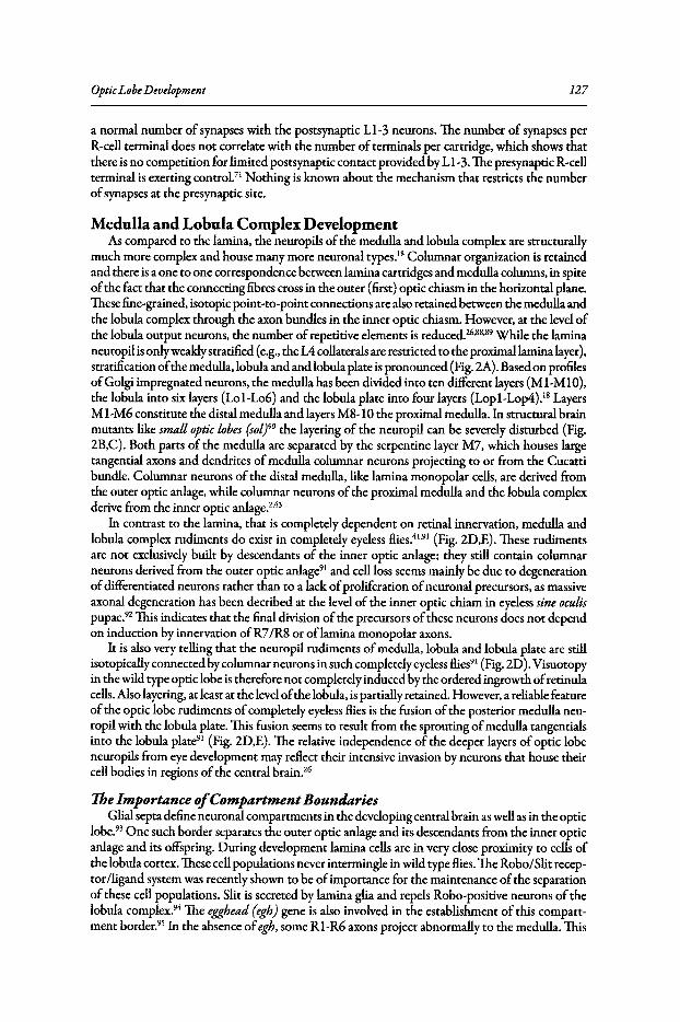
OpticLobeDevelopment 127
a normal number of synapses with the postsynaptic LI-3 neurons. The number of synapses perR-cell terminal does not correlate with the number ofterminals per cartridge, which shows thatthere is no competition for limited postsynaptic contact provided by LI-3.The presynaptic R-cellterminal is exerting control." Nothing is known about the mechanism that restricts the numberofsynapses at the presynaptic site.
Medulla and Lobula Complex DevelopmentAs compared to the lamina, the neuropils ofthe medulla and lobula complex are structurally
much more complex and house many more neuronal types." Columnar organization is retainedand there is a one to one correspondence between lamina cartridges and medulla columns, in spiteofthe fact that the connecting fibres cross in the outer (first) optic chiasm in the horizontal plane.These fine-grained, isotopic point-to-point connections are also retained between the medulla andthe lobula complex through the axon bundles in the inner optic chiasm. However, at the level ofthe lobula output neurons, the number ofrepetitive elements is reduced.26,88,89 While the laminaneuropil isonly weakly stratified (e.g.,the L4collaterals are restricted to the proximal lamina layer),stratification ofthe medulla, lobula and and lobula plate ispronounced (Fig. 2A). Based on profilesofGolgi impregnated neurons, the medulla has been divided into ten different layers (M I-M10),the lobula into six layers (Lol-Lo6) and the lobula plate into four layers (Lopl-Lop4).18 LayersM I-M6 constitute the distal medulla and layers M8-1 0 the proximal medulla. In structural brainmutants like smalloptic lobes (sOl)90 the layering of the neuropil can be severely disturbed (Fig.2B,C). Both parts of the medulla are separated by the serpentine layer M7, which houses largetangential axons and dendrites of medulla columnar neurons projecting to or from the Cucattibundle. Columnar neurons of the distal medulla, like lamina monopolar cells, are derived fromthe outer optic anlage, while columnar neurons ofthe proximal medulla and the lobula complexderive from the inner optic anlage.2,43
In contrast to the lamina, that is completely dependent on retinal innervation, medulla andlobula complex rudiments do exist in completely eyeless flies.4I,9I (Fig. 2D,E). These rudimentsare not exclusively built by descendants of the inner optic anlage; they still contain columnarneurons derived from the outer optic anlage?' and cell loss seems mainly be due to degenerationofdifferentiated neurons rather than to a lack ofproliferation ofneuronal precursors, as massiveaxonal degeneration has been decribed at the level of the inner optic chiam in eyeless sine oculispupae." This indicates that the final division ofthe precursors ofthese neurons does not dependon induction by innervation ofR7/R8 or oflamina monopolar axons.
It is also very telling that the neuropil rudiments ofmedulla, lobula and lobula plate are stillisotopically connected by columnar neurons in such completelyeyelessflies9I (Fig. 2D). Visuotopyin the wild type optic lobe is therefore not completely induced by the ordered ingrowth ofretinulacells.Also layering, at least at the levelofthe lobula, is partially retained. However, a reliable featureof the optic lobe rudiments ofcompletely eyeless flies is the fusion of the posterior medulla neuropil with the lobula plate. This fusion seems to result from the sprouting ofmedulla tangentialsinto the lobula plate" (Fig. 2D,E). The relative independence of the deeper layers of optic lobeneuropils from eye development may reflect their intensive invasion by neurons that house theircell bodies in regions ofthe central brain."
TheImportance ofCompartmentBoundariesGlial septa define neuronal compartments in the developing central brain aswell as in the optic
lobe." One such border separates the outer optic anlage and its descendants from the inner opticanlage and its offspring. During development lamina cells are in very close proximity to cells ofthe lobula cortex. These cell populations never intermingle in wild type flies.The Robe/Slit receptorlligand system was recently shown to be ofimportance for the maintenance ofthe separationof these cell populations. Slit is secreted by lamina glia and repels Robo-positive neurons of thelobula complex." The egghead (egh) gene is also involved in the establishment of this compartment border," In the absence ofegh, some RI-R6 axons project abnormally to the medulla. This
128 Brain Development in Drosophila melanogaster
Figure 7. Normal and aberrant photoreceptor projections in the optic lobe. A, C) 3D reconstruct ions from confocal stacks of photoreceptor projections in newly eclosed flies, viewedfrom inside the brain . B, D) Project ions views of respective brains at higher magnification. A,B) W ild type R1 -R6 projections form a dense synaptic layer in the lamina (la) , R7/R8 projectthrough the outer chiasm and terminate in separate layers in the medulla (drn, distal medulla ).C, D) Mutants defective for correct synaptic partner selection (shown here are photoreceptorsmutant for sectS) are characterized by a loss of the precise and regular projection pattern inboth neuropils (adapted from Mehta et al 2005 77) .
is not due to a loss of egh function in the eye or in the neurons and glia of the lamina. Instead,clonal analysisand cell-specific rescue experiments showed that egh is required in cellsofthe lobulacomplex primordium, which abuts the lamina and medulla in the developing larval brain. In theabsence ofegh. sheath-like glial processes at the boundary region delimiting lamina glia and lobulacortex are in disorder and inappropriate invasion oflobula cortex cells across this boundary regiondisrupts the pattern of lamina marginal glia which normally provides the stop signal for RI -6axons," egghead encodes a beta-i-rnannosyltransferase'" which is involved in Glycosphingolipidbiosynthesis. Glycosphingolipids have been implicated in EGFR signaling in Drosophila."
Selecting the CorrectMedulla Target LayerIn the medulla the visual information channels fed by RI-6 are relayed via lamina neuron pro
cessesto higher order interneurons. In addition, photoreceptors R7/8 terminate and form synapsesexclusivelyin the medulla (Fig. 7A), where therefore the color vision circuit ispredicted to reside9•19
(Fig. 3). Layering of the medulla reflects the requirement for the establishment ofvisuotopicallyorganized synapses between these different sets of columnar neurons. In the adult optic lobe thefivelamina monopolar neurons and R7 and R8 terminate in different layersofthe distal medulla.18
This enables them to relayon characteristic sets ofhigher order columnar neurons which project tothe lobula complex, most importantly onto transmedulla cells (Tm) projecting to the lobula and
Optic Lobe Development 129
transmedulla Y cells (TmY), the axons ofwhich branch in the inner optic chiasm and terminate inlobula and lobula plate (Fig. 2). By inspection ofR8 and R7 targeting it was shown that the adultsituation isestablished in an at least two-staged layer-selection process" (Fig. 5). Duringearlypupaldevelopment the newest leading R8 axon terminates superficially in the distal medulla neuropileand isovertaken by the following R7 axon that temporarily occupies the immediate adjacent deeperlayer.These temporary layersofR8 and R7 are more and more pushed apart by the growth cones ofthe fivelamina monopolar cells,which follow and elaborate their arborizations in the spacebetweenthe R-cell terminals during the first 40% ofpupal development. The lamina gradient ofmaturationfrom posterior (oldest) to anterior (youngest) is thus reflected as a spatial gradient ofthe thicknessof the medulla in the horizontal plane during early pupal development. At about 50% ofpupaldevelopment all R7 and R8 growth cones simultaneously become mobile again and target to theirfinal layers M6 and M3. 6,98 The nature ofthe global trigger ofthis event is still unknown.
In sevenless mutants lacking R7, the axons ofR8 and lamina monopolar neurons behave normally during targeting stage I (Fig. 5). The same is true for R8 and R7 terminals in the absenceoflamina monopolar neurons. Therefore R8, R7 and LI-L5 axons target independently to theirtemporary terminal layers at the first layer-selection stage." This layer selection therefore doesnot seem to depend on interactions between the afferents, but rather on interactions with cellsin the target area.
Some factors have been identified that are required for target layer selection. One interestingexample is the homophilic celladhesion protein Capricious (CAPS) with leucine rich repeats, whichis present only in R8 and in medulla cells, but not in other retinula cells and not in the lamina."In the medulla neuropil of the third larval instar CAPS is uniformly expressed, but is restrictedto specific layers during pupal development sparing the final R7 recipient layer. In flies mutant forcaps, R-cell terminals in the medulla do not form a regular array and many R8-cell terminals seemto invade neighbouring columns. IfCAPS is misexpressed in R7 cells, the first stage ofR7 targetlayer selection is only mildly affected, but the growth cones remain in the final R8 recipient layer.This is evidence that CAPS plays an instructive role in the targetingofR8 terminals."
Other factors required for target layer selection ofretinula cells are more widely expressed inthe target region and may playa permissive role, e.g., N-cadherin.6,72 Homophilic cell adhesionmediated by the extracellular domain rather than signaling is important, because the cytoplasmic domain is dispensable not only for N-cadherin mediated cell adhesion in S2 cells but alsofor targeting of R7 growth cones. However, the cytoplasmic domain is required for normal R7growth cone morphology.l'" In the lamina, N-cadherin seems to function in a very similar way inthe targeting ofRI-6 axons as it is expressed and required in the R-cells as well as in the laminamonopolar neurons."
As N-cadherin is not exclusivelyexpressed in specific subsets ofneurons in the respective targetareas, it isworth mentioning that N-cadherin exists in 12 splice isoforms. In fact, it could be shownthat the isoform specific N-cad (18Astop) allele selectively affects the second stage of R7 targetselection.'?' This allele eliminates the six isoforms containing alternative exon 18A. N-cadherinisoforms containing exon 18B are sufficient for the first stage of R7 targeting to its temporarylayer, while the 18A isoforms are preferentially expressed in R7 during the second halfofpupaldevelopment and are necessary for R7 to terminate in the appropriate synaptic layer M6 of themedulla.'?' However, it is very unlikely that the N-cadherin isoforms constitute something like acombinatorial code for the selective recognition ofsynaptic partners, as expression ofany isoformis able to rescue the function ofthe other and the various isoforms mediate promiscuous heterophilic interactions with each other.98,IOI The function ofthe structural variations in the isoforms isthus still unknown. It is conceivable that they affect interactions with other proteins rather thanhomophilic adhesiveness. Therefore N-cadherin can be considered as a homophilic cell adhesionprotein providing permissive stabilizing interactions in target selection.
Mutant alleles of the receptor tyrosine phosphatase LAR and its downstream interactor, thescaffolding protein Liprin-a, produce N-cadherin mutant-like targetingdefects ofR_axons.74,I02-104
Both proteins are expressed like N-cadherin in all R-cells and in neurons of the target areas and
130 Brain Development in Drosophila melanogaster
their involvement in the regulation ofN-cadherin has been shown.l'" However, the requirementofLiprin-a and LAR for R-cell targeting is exclusively on the presynaptic site.l03,104 This impliesthat N-cadherin regulation is different on the dendritic and on the axonal site. Two heparan sulfate proteoglycans have been identified as ligands for LAR: Dally-like and Syndecan. Both havebeen implicated in LAR-dependent axon guidance: Syndecan as a promotor and Dally-like as aninhibitor ofLAR signaling. 106-108
The G-protein coupled, 7-passtransmembrane receptor Flamingo isan atypical cadherin, whichhas recently been shown to regulate synaptogenesis at the neuromuscular junction. In addition,Flamingo is required to prevent axonal and synaptic degeneration in Drosophila.109 Its involvementin optic lobe development is alsowell established?3,110,111 Mutations in the flamingo (fmi) gene havebeen discovered in screens for abnormal R-cell connectivity'!' and for defects in visual behaviour,"While Flamingo is required for the sorting of RI-6 terminals to their correct lamina cartridges,it has at least two important functions during R8 axon targeting as well: it facilitates competitiveinteractions between adjacent R8 axons to ensure their correct spacing73,111 and it promotes theformation ofstable connections between R8 axons and their target cells in the medulla.l'?''!' Thetiling function of Flamingo is not restricted to axonal projections. In other systems, it has beenshown to function in the shaping ofdendritic fields as well112and it was recently shown that ingrowing R8 axons induce layer-specific expression ofFlamingo in the medulla viaJelly belly (Jeb)signaling. 110Its receptor, the anaplastic lymphoma kinase (Alk), is expressed and required in targetneurons in the optic lobe. Jeb is generated by photoreceptor axons and controls target selection ofRl-R6 axons in the lamina and R8 axons in the medulla. Loss ofjeb/Alk function affects medullalayer-specific expression not only ofFlamingo, but also oftwo cell-adhesion molecules ofthe immunoglobulin superfamily, Roughest/IrreC and Kirre/Dumbfounded.!" These closely relatedsingle pass transmembrane proteins are known from their function in muscle fusion,113,114 eyedevelopmenr'V" and optic chiasm formation. 116-118Loss of Roughest/IrreC leads to misroutingvia the inner optic chiasm ofposterior R8/R7 and lamina monopolar axons to their visuotopictarget area.116 The axonal bundles in the first optic chiasm which connect single lamina cartridgeswith isotopic medulla columns tend to fasciculate in loss offunction mutants,118 which copies theloss offlamingophenotype in the first chiasm," indicating that the Roughest/IrreC protein helpsto keep columnar fibre bundles apart from each other.
Columnar TilingWhile the stratification ofcolumnar neurons reflects their cell type specific connectivity, it is
the lateral extent of the arborizations that determines the visuotopic precision ofthe adult neurons and affects the size and position of their visual fields. It was shown in a classical paper thatcompetition between R7 terminals occurs to a limited degree in the target region.!" In the thirdinstar, R7 axons transiently display overlapping halos offilopodia, but in genetic mosaics vacantsites are only invaded by neighbouring R7 terminal extensions, ifextra R7 axons due to the moreinnerphotoreceptors mutation are available in the juxtaposed medulla columns.119
The appropriation of territory by neuronal arborizations has at least two aspects. First, theprocesses ofthe same neuron have to recognize and arrange themselves. Dendritic aswell as axonalarborizations should more or less evenly cover their appropriate target space. Second, neurons ofthe same type should respect each others territory. The second process is known as "tiling" but thefirst process is related. It has to be assumed that in both processes recognition of"self" or of"samekind" has to be followed by repulsion. Interestingly homophilic receptors ofthe conserved familyof the Down syndrome cell adhesion molecules (DSCAMs), members of the immunoglobulinsuperfamily, have been found to function in both aspects ofneuronal tiling. 12Q-122 There are fourDscam genes in the Drosophila genome, called Dscam and Dscam2-4. Dscam is special in that itdisplays an extraordinary molecular diversity. Due to four casettes ofalternative spliced exons itcan generate 38016 different proteins.123 Most interestingly, isoform-specific homophilic adhesionseems to induce repulsion in dendrites and thus helps to avoid selfcrossing and contributes to aneven coverage ofthe dendritic field in all four classesofdendrite arborization neurons, a group ofsensory neurons with a stereotyped dendritic branching pattern.120,122 For Dscam2 two isoforms
OpticLobeDevelopment 131
(Dscam2A and Dscam2B) have been described. They are able to mediate isoform-specific homophilic adhesion in 52-cells and do not bind to other Dscam family members. Dscam2 plays a rolein tiling amongLl terminals (Fig. 2A) within the distal medulla. Dscam2 homophilic interactionsmediate repulsion between Ll axonal terminals in neighbouring columns. Loss ofDscam2 function leads to an overlap ofthe L1 terminals.!"
The repulsive effect ofproteins that are able to mediate homophilic adhesion in cell cultureexperiments demonstrates the importance of signaling for the understanding of cellular responses in vivo. Due to the high number ofdifferent types of columnar neurons in the medullaof DrosophilaI8 it is likely that still other receptors will be described that function in tiling ofcolumnar cell types.
Connecting Optic Lobes with andacross the CentralBrainNeuronal connections between the optic lobe and central brain have recently been systemati
callymapped in considerable detail." Comparably little is known about the development oftheseprojections. Through the study of the transcription factor Atonal, which is originally known tobe required for the specification ofthe R8 ommatidial founder photoreceptor, a dorsal cluster ofoptic lobe neurons was discovered that connects both optic lobes across the central brain duringlarval developrnent.P' The dorsal cluster neurons project contralaterally towards the lobula complex where they fan out over the lobula complex and inner chiasm and additionally form a precisenumber ofprojections towards the medulla. This reproducibly accurate projection pattern has beenemployed to identify an integrative signaling network encompassing the Jun N-terminal kinase,the GTPase Rae, the secreted morphogen Wnt, its receptor Frizzled, the FGF Branchless and theFGF receptor. Importantly, this network regulates the extension and retraction ofaxonal branches,but not axon guidance, indicating that these processes are regulated independently.125 Finally, thedorsal cluster neuron projections have also been shown to form independent ofneuronal activity,further supporting the notion that wiring of the optic lobes, from cellular differentiation downto the specification ofsynapses, follow a genetic program.71,125
Concluding RemarksWhile many steps in optic lobe development are still not yet understood, it is clear that a
combination oftimingofneuronal and glial cell fate specification, axonal outgrowth, ofinductiveevents and ofspecific recognition processes between "self" and "not self" direct the wiring oftheneural machinery ofthe optic lobe. It is therefore a genetically encoded developmental programthat ensures all aspects ofvision required for the survival of the newly emerging fly. Adult opticlobe development is optimized for speed and precision. However, the adult optic lobe also displaysa certain degree ofplasticity. Deprivation ofvisual input after the optic lobe is formed can lead toa reduction in synapse numbers in the lamina during a critical time window in early adulthood/"However, such plasticity is apparently not required to wire a functional optic lobe. It is thereforean important realization that a brain structure like the Drosophila optic lobe is as much the product ofa genetically encoded developmental program as the eye or a wing. Given the rich genetictool box available and the wealth ofknowledge about Drosophila development, the optic lobe isa wonderful model system to decipher this developmental program and attain knowledge aboutthe extend to which a brain structure can be "genetically encoded"
AcknowlegementsWe thank Gerhard Technau for his encouragement throughout the process ofwriting. KFF
thanks the SFBSOS. PRH is a Eugene McDermott Scholar in Biomedical Research at UTSouthwestern Medical Center in Dallas. We thank Claude Desplan and Bassem Hassan for comments on parts of the manuscript. Till Andlauer provided MARCM data. We apologize to allcolleagues whose work was not discussed due to space constraints or our shortcomings.
132 Brain Development in Drosophila melanogaster
References1. Green P, Hartenstein AY, Hartenstein V. The embryonic development of the Drosophila visual system.
Cell Tissue Res 1993; 273(3):583-598.2. Meinertzhagen lA, Hanson TE. The development of the optic lobe. In: Bate M, Martinez-Arias A,
eds. The Development of Drosophila melanogaster. Cold Spring Harbor: Cold Spring Harbor Press,1993:1363-1491.
3. Nassif C, Noveen A, Hartenstein V. Early development of the Drosophila brain: III. The pattern ofneuropile founder tracts during the larval period. J Comp Neurol 2003; 455(4):417-434.
4. Clandinin TR, Zipursky SL. Making connections in the fly visual system. Neuron 2002;35(5):827-841.
5. Mast JD, Prakash S, Chen PL et al. The mechanisms and molecules that connect photoreceptor axonsto their targets in Drosophila. Semin Cell Dev Bioi 2006; 17(1):42-49.
6. Ting CY, Lee CH. Visual circuit development in Drosophila. Curr Opin Neurobiol 2007;17(1):65-72.
7. Cajal SR, Sanchez D. Contribucion al concocimiento delos centros nerviosos de los insectos. Trab LabInvest Bioi 1915; 13:1-167.
8. Strausfeld NJ. Golgi studies on insects. Part II: The optic lobes of Diptera. Philos Trans R Soc Lond B1970; 258:135-223.
9. Morante J, Desplan C. Building a projection map for photoreceptor neurons in the Drosophila opticlobes. Semin Cell Dev Bioi 2004; 15(1):137-143.
10. Wernet MF, Mazzoni EO, Celik A et al. Stochastic spineless expression creates the retinal mosaic forcolour vision. Nature 2006; 440(7081):174-180.
11. Tomlinson A. Patterning the peripheral retina of the fly: decoding a gradient. Dev Cell 2003;5(5):799-809.
12. Wernet MF, Labhart T, Baumann F et al. Homothorax switches function of Drosophila photoreceptorsfrom color to polarized light sensors. Cell 2003; 115(3):267-279.
13. Homberg U, Hofer S, PfeifferK et ale Organization and neural connections of the anterior optic tuberclein the brain of the locust, Schistocerca gregaria. J Comp Neuro12003; 462(4):415-430.
14. Braitenberg V. Pattern of projections in the visual system of the Ay. I. Retina lamina projections. ExpBrain Res 1967; 3:271-298.
15. Kirschfeld K. The projection of the optical environment on the screen of the rhabdomere in the compound eye of Musca. Exp Brain Res 1967; 3:248-270.
16. Trujillo-Cenoz 0, Melamed J. Electron microscope observations on the peripheral and intermediateretinas of dipterans. In: Bemhad CG, ed. The Functional Organization of the Compound Eye. NewYork: Pergamon Press, 1966:339-361.
17. Clandinin TR, Zipursky SL. Afferent growth cone interactions control synaptic specificity in theDrosophila visual system. Neuron 2000; 28(2):427-436.
18. Fischbach K, Dittrich AP. The optic lobe of Drosophila melanogaster. I. A Golgi analysis of wild typestructure. Cell Tissue Res 1989; 256:441-475.
19. Bausenwein B, Dittrich AP, Fischbach KF. The optic lobe of Drosophila melanogaster. II. Sorting ofretinotopic pathways in the medulla. Cell Tissue Res 1992; 267(1):17-28.
20. BuchnerE, BuchnerS.Mappingstimulus-inducednervousactivityin smallbrainsby [3H]2-deoxy-D-glucose.Cell Tissue Res 1980; 211(1):51-64.
21. Bausenwein B, Fischbach KF. Activity labeling patterns in the medulla of Drosophila melanogastercaused by motion stimuli. Cell Tissue Res 1992; 270(1):25-35.
22. Fischbach K, Ramos RG, Bausenwein B. Der optische Lobus von Drosophila melanogaster: Experimentelle Ansaetze zum Studium von Struktur, Funktion und Entwicklung. Verh Dtsch Zool Ges 1992;85.2:133-148.
23. Stocker RF. The organization of the chemosensory system in Drosophila melanogaster: a review. CellTissue Res 1994; 275(1):3-26.
24. Stocker RF, Lienhard MC, Borst A et al. Neuronal architecture of the antennal lobe in Drosophilamelanogaster. Cell Tissue Res 1990; 262(1):9-34.
25. Campos AR, Fischbach KF, Steller H. Survival of photoreceptor neurons in the compound eye ofDrosophila depends on connections with the optic ganglia. Development 1992; 114(2):355-366.
26. Otsuna H, Ito K. Systematic analysis of the visual projection neurons of Drosophila melanogaster. I.Lobula-specific pathways. J Comp Neuro12006; 497(6):928-958.
27. Wolff T, Ready DE Pattern formation in the Drosophila retina. In: Bate M, Martinez-Arias A, eds.The Development of Drosophila melanogaster. Cold Spring Harbor: Cold Spring Harbor Press,1993:1363-1491.
28. Murakami S, Umetsu D, Maeyama Y et al. Focal adhesion kinase controls morphogenesis of theDrosophila optic stalk. Development 2007; 134(8):1539-1548.
Optic Lobe Development 133
29. Hofbauer A, Buchner E. Does Drosophila have seven eyes? Naturwissenschaften 1989; 76:335-336.30. Helfrich-Forster C, Edwards T, Yasuyama K et al. The extraretinal eyelet of Drosophila: development,
ultrastructure and putative circadian function. ] Neurosci 2002; 22(21):9255-9266.31. Song], Wu L, Chen Z et al. Axons guided by insulin receptor in Drosophila visual system. Science
2003; 300(5618):502-505.32. Garrity PA, Rao ~ Salecker I et al. Drosophila photoreceptor axon guidance and targeting requires the
dreadlocks SH2/SH3 adapter protein. Cell 1996; 85(5):639-650.33. Hing H, Xiao ], Harden N et al. Pak functions downstream of Dock to regulate photoreceptor axon
guidance in Drosophila. Cell 25 1999; 97(7):853-863.34. Houalla T, Hien Vuong D, Ruan W et al. The Ste20-like kinase misshapen functions together with
Bicaudal-D and dynein in driving nuclear migration in the developing drosophila eye. Mech Dev 2005;122(1):97-108.
35. Newsome TP, Schmidt S, Dietzl G et al. Trio combines with dock to regulate Pak activity duringphotoreceptor axon pathfinding in Drosophila. Cell 2000; 101(3):283-294.
36. Ruan w: Long H, Vuong DH et al. Bifocal is a downstream target of the Ste20-like serine/threoninekinase misshapen in regulating photoreceptor growth cone targeting in Drosophila. Neuron 2002;36(5):831-842.
37. Ruan w: Pang P, Rao Y. The SH2/SH3 adaptor protein dock interacts with the Ste20-like kinase misshapen in controlling growth cone motility. Neuron 1999; 24(3):595-605.
38. Chevais S. Sur la structure des yeux implantes de Drosophila melanogaster. Archs Anat Microsc 1937;33:107-112.
39. Halder G, Callaerts P, Gehring W]. New perspectives on eye evolution. Curr Opin Genet Dev 1995;5(5) :602-609.
40. Steller H, Fischbach KF, Rubin GM. Disconnected: a locus required for neuronal pathway formationin the visual system of Drosophila. Cell 1987; 50(7):1139-1153.
41. Power ME. The effort of reduction in numbers of ommatidia upon the brain of Drosophila melanogaster.J Exp Zoo11943; 94:33-71.
42. Hinke W Das relative postembryonale Wachstum der Hirnteile von Culex pipiens, Drosophila melanogaster and Drosophila mutanten. Z. Morph Okol Tiere 1961; 50:81-118.
43. Campos-Ortega ]A, Hofbauer A. Proliferation pattern and early differentiation of the optic lobes inDrosophila melanogaster. Rouxs Arch Dev Biol 1990; 198:264-274.
44. Selleck SB, Gonzalez C, Glover DM et al. Regulation of the Gl-S transition in postembryonic neuronalprecursors by axon ingrowth. Nature 1992; 355(6357):253-255.
45. Selleck SB, Steller H. The influence of retinal innervation on neurogenesis in the first optic ganglionof Drosophila. Neuron 1991; 6(1):83-99.
46. Winberg ML, Perez SE, Steller H. Generation and early differentiation of glial cells in the first opticganglion of Drosophila melanogaster. Development 1992; 115 (4) :903-911.
47. Huang Z, Shilo BZ, Kunes S. A retinal axon fascicle uses spitz, an EGF receptor ligand, to construct asynaptic cartridge in the brain of Drosophila. Cell 1998; 95(5):693-703.
48. Huang Z, Kunes S. Hedgehog, transmitted along retinal axons, triggers neurogenesis in the developingvisual centers of the Drosophila brain. Cell 1996; 86(3):411-422.
49. Huang Z, Kunes S. Signals transmitted along retinal axons in Drosophila: Hedgehog signal receptionand the cell circuitry of lamina cartridge assembly. Development 1998; 125(19):3753-3764.
50. Chu T, Chiu M, Zhang E et al. A C-terminal motif targets Hedgehog to axons, coordinating assemblyof the Drosophila eye and brain. Dev Cell 2006; 10(5):635-646.
51. Chotard C, Leung w: Salecker I. glial cells missing and gcm2 cell autonomously regulate both glial andneuronal development in the visual system of Drosophila. Neuron 2005; 48(2):237-251.
52. Nakato H, Futch TA, Selleck SB. The division abnormally delayed (dally) gene: a putative integralmembrane proteoglycan required for cell division patterning during postembryonic development of thenervous system in Drosophila. Development 1995; 121(11):3687-3702.
53. Takeo S, Akiyama T, Firkus C et al. Expression of a secreted form of Dally, a Drosophila glypican, inducesovergrowth phenotype by affecting action range of Hedgehog. Dev Biol 2005; 284(1):204-218.
54. Dickson B, Hafen E. Genetic dissection of eye development in drosophila. In: Bate M, Martinez-AriasA, eds, The Development of Drosophila melanogaster. Cold Spring Harbor: Cold Spring Harbor Press,1993:1363-1491.
55. Dearborn R, He Q Kunes S et al. Eph receptor tyrosine kinase-mediated formation of a topographicmap in the Drosophila visual system.] Neurosci 2002; 22(4):1338-1349.
56. Kunes S, Steller H. Topography in the Drosophila visual system. Curr Opin Neurobiol 1993;3( 1):53-59.
57. Sato M, Umetsu D, Murakami Set al. DWnt4 regulates the dorsoventral specificity of retinal projectionsin the Drosophila melanogaster visual system. Nat Neurosci 2006; 9( 1):67-75.

134 Brain Development in Drosophila melanogaster
58. Martin KA, Poeck B, Roth H et ale Mutations disrupting neuronal connectivity in the Drosophila visualsystem. Neuron 1995; 14(2):229-240.
59. Poeck B, Fischer S, Gunning D et al. Glial cells mediate target layer selection of retinal axons in thedeveloping visual system of Drosophila. Neuron 2001; 29(1):99-113.
60. YoshidaS, SoustelleL, Giangrande A et ale DPP signalingcontrols development of the lamina glia requiredfor retinal axon targeting in the visual system of Drosophila. Development 2005; 132(20):4587-4598.
61. Frohlich A, Meinertzhagen lA. Synaptogenesis in the first optic neuropile of the fly's visual system. JNeurocytol1982; 11(1):159-180.
62. Frohlich A, Meinertzhagen IA. Quantitative features of synapse formation in the fly'svisual system. 1.The presynaptic photoreceptor terminal. J Neurosci 1983; 3(11):2336-2349.
63. Garrity PA, Lee CH, Salecker I et al. Retinal axon target selection in Drosophila is regulated by areceptor protein tyrosine phosphatase. Neuron 1999; 22(4):707-717.
64. Newsome TP, Asling B, Dickson BJ.Analysisof Drosophila photoreceptor axon guidance in eye-specificmosaics. Development 2000; 127:851-860.
65. Gibbs SM, Truman ]W. Nitric oxide and cyclic GMP regulate retinal patterning in the optic lobe ofDrosophila. Neuron 1998; 20(1):83-93.
66. Rao Y, Pang P, Ruan W et ale brakelessis required for photoreceptor growth-cone targeting in Drosophila.Proc Nat! Acad Sci USA 2000; 97(11):5966-5971.
67. Senti K, Keleman K, Eisenhaber F et al. brakeless is required for lamina targeting of Rl-R6 axons inthe Drosophila visual system. Development 2000; 127(11):2291-2301.
68. Kaminker JS, Canon J, Salecker I et al. Control of photoreceptor axon target choice by transcriptionalrepression of Runt. Nat Neurosci 2002; 5(8):746-750.
69. Tayler TD, Garrity PA. Axon targeting in the Drosophila visual system. Curr Opin Neurobiol 2003;13(1):90-95.
70. Zelhof AC, Hardy R~ Becker A et al. Transforming the architecture of compound eyes. Nature 2006;443(7112):696-699.
71. Hiesinger PR, Zhai RG, Zhou Y et al. Activity-independent prespecification of synaptic partners in thevisual map of Drosophila. Curr Bioi 2006; 16(18):1835-1843.
72. Lee CH, Herman T, Clandinin TR et al. N-cadherin regulates target specificity in the Drosophila visualsystem. Neuron 2001; 30(2):437-450.
73. Lee RC, Clandinin TR, Lee CH et al. The protocadherin Flamingo is required for axon target selectionin the Drosophila visual system. Nat Neurosci 2003; 6(6):557-563.
74. Maurel-Zaffran C, Suzuki T, Gahmon G et al. Cell-autonomous and -nonautonomous functions ofLARin R7 photoreceptor axon targeting. Neuron 2001; 32(2):225-235.
75. Prakash S, Caldwell]C, Eberl DF et ale Drosophila N-cadherin mediates an attractive interaction betweenphotoreceptor axons and their targets. Nat Neurosci 2005; 8(4):443-450.
76. Hiesinger PR, Reiter C, Schau H et al. Neuropil pattern formation and regulation of cell adhesion molecules in Drosophila optic lobe development depend on synaptobrevin. J Neurosci 1999;19(17):7548-7556.
77. Mehta SQ Hiesinger PR, Beronja S et ale Mutations in Drosophila sec15 reveal a function in neuronaltargeting for a subset of exocyst components. Neuron 2005; 46(2):219-232.
78. Katz LC, Shatz C]. Synaptic activity and the construction of cortical circuits. Science 1996;274(5290):1133-1138.
79. Shatz C]. Emergence of order in visual system development. Proc Nat! Acad Sci USA 1996;93(2):602-608.
80. Barth M, Hirsch HV, Meinertzhagen IA et al. Experience-dependent developmental plasticity in theoptic lobe of Drosophila melanogaster. J Neurosci 1997; 17(4):1493-1504.
81. Bloomquist BT, Shortridge RD, Schneuwly S et al. Isolation of a putative phospholipase C gene ofDrosophila, norpA and its role in photorransduction. Cell 1988; 54(5):723-733.
82. Burg MG, Sarthy pv, Koliantz G et ale Genetic and molecular identification of a Drosophila histidinedecarboxylase gene required in photoreceptor transmitter synthesis. EMBO J 1993; 12(3):911-919.
83. Hardie RC. Is histamine a neurotransmitter in insect photoreceptors? J Comp Physiol [A] 1987;161(2):201-213.
84. Koh T~ Bellen HJ. Synaptotagmin I, a Ca2+ sensor for neurotransmitter release. Trends Neurosci2003; 26(8):413-422.
85. Meinertzhagen lA, Hu X. Evidence for site selection during synaptogenesis: the surface distribution ofsynaptic sites in photoreceptor terminals of the files Musca and Drosophila. Cell Mol Neurobiol 1996;16(6):677-698.
86. Meinertzhagen lA, Sorra KE. Synaptic organization in the fly's optic lamina: few cells, many synapsesand divergent microcircuits. Prog Brain Res 2001; 131:53-69.
OpticLobeDevelopment 135
87. Frohlich A, Meinertzhagen IA. Regulation of synaptic frequency: comparison of the effectsof hypoinnervation with those of hyperinnervation in the fly's compound eye. J Neurobiol 1987; 18(4):343-357.
88. Strausfeld NJ. Atlas of an Insect Brain. Heidelberg: Springer Verlag; 1976.89. Strausfeld NJ, Okamura JY. Visual system of calliphorid flies: organization of optic glomeruli and their
lobula complex efferents. J Comp Neurol 2007; 500(1):166-188.90. Fischbach KF, Heisenberg M. Structural brain mutant of Drosophila melanogaster with reduced cell
number in the medulla cortex and with normal optomotor yaw response. Proc Natl Acad Sci USA 1981;78(2):1105-1109.
91. Fischbach KF. Neural cell types surviving congenital sensory deprivation in the optic lobes of Drosophilamelanogaster. Dev BioI 1983; 95(1):1-18.
92. Fischbach KF, Technau G. Cell degeneration in the developing optic lobes of the sine oculis andsmall-optic-Iobes mutants of Drosophila melanogaster. Dev BioI 1984; 104( 1):219-239.
93. Younossi-Hartenstein A, Salvaterra PM, Hartenstein V. Early development of the Drosophila brain: IV.Larval neuropile compartments defined by glial septa. J Comp Neuro12003; 455(4):435-450.
94. TaylerTD, Robichaux MB, Garrity PA. Compartmentalization of visual centers in the Drosophila brainrequires Slit and Robo proteins. Development 2004; 131(23):5935-5945.
95. Fan Y, Soller M, Flister S et al. The egghead gene is required for compartmentalization in Drosophilaoptic lobe development. Dev Bioi 2005; 287(1):61-73.
96. Wandall HH, Pizette S, Pedersen JW et al. Egghead and brainiac are essential for glycosphingolipidbiosynthesis in vivo. J BioI Chern 2005; 280(6):4858-4863.
97. Chen~ Pedersen~Wandall HH et al. Glycosphingolipids with extended sugar chain have specialized functions in development and behavior of Drosophila. Dev Bioi 2007; 306(2):736-749.
98. Ting C~ YonekuraS, Chung P et al. Drosophila N-cadherin functions in the first stage of the two-stagelayer-selection process of R7 photoreceptor afferents. Development 2005; 132(5):953-963.
99. Shinza-Kameda M, Takasu E, Sakurai K et al. Regulation of layer-specific targeting by reciprocal expression of a cell adhesion molecule, capricious. Neuron 2006; 49(2):205-213.
100. Yonekura S, Xu L, Ting CY et al. Adhesive but not signaling activity of Drosophila N-cadherin isessential for target selection of photoreceptor afferents. Dev Biol 2007; 304(2):759-770.
101. Nern A, Nguyen L~ Herman T et al. An isoform-specific allele of Drosophila N-cadherin disrupts alate step of R7 targeting. Proc Nat! Acad Sci USA 2005; 102(36):12944-12949.
102. Clandinin TR, Lee CH, Herman T et al. Drosophila LAR regulates R1-R6 and R7 target specificityin the visual system. Neuron 2001; 32(2):237-248.
103. Choe KM, Prakash S, Bright A et al. Liprin-alpha is required for photoreceptor target selection inDrosophila. Proc Natl Acad Sci USA 2006; 103(31):11601-11606.
104. Hofmeyer K, Maurel-Zaffran C, Sink H et al. Liprin-alpha has LAR-independent functions in R7photoreceptor axon targeting. Proc Natl Acad Sci USA 2006; 103(31):11595-11600.
105. Kypta RM, Su H, Reichardt LF. Association between a transmembrane protein tyrosine phosphataseand the cadherin-catenin complex. J Cell Bioi 1996; 134(6):1519-1529.
106. Fox AN, Zinn K. The heparan sulfate proteoglycan syndecan is an in vivo ligand for the DrosophilaLAR receptor tyrosine phosphatase. Curr BioI 2005; 15(19):1701-1711.
107. Rawson JM, Dimitroff B,Johnson KG et al. The heparan sulfate proteoglycans Dally-like and Syndecanhave distinct functions in axon guidance and visual-system assembly in Drosophila. Curr Biol 2005;15(9):833-838.
108. Johnson KG, Tenney AP, Ghose A et al. The HSPGs Syndecan and Dallylike bind the receptor phosphatase LAR and exert distinct effects on synaptic development. Neuron 2006; 49(4):517-531.
109. Bao H, Berlanga ML, Xue M et al. The atypical cadherin flamingo regulates synaptogenesis and helpsprevent axonal and synaptic degeneration in Drosophila. Mol Cell Neurosci 2007; 34(4):662-678.
110. Bazigou E, Apitz H, Johansson J et al. Anterograde Jelly belly and Alk receptor tyrosine kinase signalingmediates retinal axon targeting in Drosophila. Cell 2007; 128(5):961-975.
111. Senti KA, Usui T, Boucke K et al. Flamingo regulates R8 axon-axon and axon-target interactions in theDrosophila visual system. Curr BioI 2003; 13(10):828-832.
112. Kimura H, Usui T, Tsubouchi A et al. Potential dual molecular interaction of the Drosophila 7-pass transmembrane cadherin Flamingo in dendritic morphogenesis. J Cell Sci 2006; 119(Pt 6):1118-1129.
113. Ruiz-Gomez M, Coutts N, Price A et al. Drosophila dumbfounded: a myoblast attractant essential forfusion. Cell 2000; 102(2):189-198.
114. Strunkelnberg M, Bonengel B, Moda LM et al rst and its paralogue kirre act redundantly duringembryonic muscle development in Drosophila. Development 2001; 128(21):4229-4239.
115. Reiter C, Schimansky T, Nie Z et al. Reorganization of membrane contacts prior to apoptosis in theDrosophila retina: the role of the IrreC-rst protein. Development 1996; 122(6):1931-1940.
116. Boschert U, Ramos RG, Tix S et al. Genetic and developmental analysis of irreC, a genetic functionrequired for optic chiasm formation in Drosophila. J Neurogenet 1990; 6(3):153-171.
136 Brain Development in Drosophila melanogaster
117. Ramos RG, Igloi GL, Lichte B et ale The irregular chiasm C-roughest locus of Drosophila, which affectsaxonal projections and programmed cell death, encodes a novel immunoglobulin-like protein. GenesDev 1993; 7(12B):2533-2547.
118. Schneider T, Reiter C, Eule E et ale Restricted expression of the irreC-rst protein is required for normalaxonal projections of columnar visual neurons. Neuron 1995; 15(2):259-271.
119. Ashley JA,Katz FN. Competition and position-dependent targeting in the development of the DrosophilaR7 visual projections. Development 1994; 120(6):1537-1547.
120. Hughes ME, Bortnick R, Tsubouchi A et al. Homophilic Dscam interactions control complex dendritemorphogenesis. Neuron 2007; 54(3):417-427.
121. Millard SS, Flanagan JJ, Pappu KS et ale Dscam2 mediates axonal tiling in the Drosophila visual system.Nature 2007; 447(7145):720-724.
122. Soba P, Zhu S, Emoto K et al. Drosophila sensory neurons require Dscam for dendritic self-avoidanceand proper dendritic field organization. Neuron 2007; 54(3):403-416.
123. Schmucker D, Clemens JC, Shu H et al. Drosophila Dscam is an axon guidance receptor exhibitingextraordinary molecular diversity. Cell 2000; 101(6) :671-684.
124. Hassan BA, Bermingham NA, He Y et al. Atonal regulates neurite arborization but does not act as aproneural gene in the Drosophila brain. Neuron 2000; 25(3):549-561.
125. Srahna M, Leyssen M, Choi CM et al. A signaling network for patterning of neuronal connectivity inthe Drosophila brain. PLoS Bioi 2006; 4(11):e348.
126. Fischbach KF. Neurogenetik am Beispiel des visuellen Systems von Drosophila melanogaster.: Habilitationsschrift. Bayerische julius-Maximilians-Universitat Wurzburg; 1983.
127. jarvilehto M, Finell N. Development of the function of visual receptor cells during the pupal life of thefly Calliphora. J Comp Physiol [A] 1983; 150:529-536.
128. Hardie RC, Peretz A, PollockJA et al. Ca2+ limits the development of the light response in Drosophilaphotoreceptors. Proc BioI Sci 1993; 252(1335):223-229.
129. Meinertzhagen lA, Piper ST, Sun XJ et al. Neurite morphogenesis of identified visual interneurons andits relationship to photoreceptor synaptogenesis in the flies, Musca domestica and Drosophila melanogaster. Eur J Neurosci 2000; 12(4):1342-1356.

CHAPTER 9
Clonal UnitArchitecture ofthe Adult FlyBrainKei Ito" and Takeshi Awasaki
Abstract
During larval neurogenesis, neuroblasts repeat asymmetric cell divisions to generate clonallyrelated progeny. When the progeny ofa single neuroblast is visualized in the larval brain,their cell bodies form a cluster and their neurites form a tight bundle. This structure persists
in the adult brain. Neurites deriving from the cells in this cluster form bundles to innervate distinctareas of the brain. Such clonal unit structure was first identified in the mushroom body, whichis formed by four nearly identical clonal units each ofwhich consists ofdiverse types ofneurons.Organised structures in other areas ofthe brain, such as the central complex and the antennallobeprojection neurons, also consist ofdistinct clonal units. Many clonally related neural circuits areobserved also in the rest ofthe brain, which is often called diffused neuropiles because ofthe apparent lack ofclearly demarcated structures. Thus, it is likely that the clonal units are the buildingblocks ofa significant portion ofthe adult brain circuits. Arborisations ofthe clonal units are notmutuallyexclusive,however. Rather, several clonal units contribute together to form distinct neuralcircuit units, to which other clones contribute relatively marginally. Construction ofthe brain bycombiningsuch groups ofclonally related units would have been a simple and efficient strategy forbuilding the complicated neural circuits during development as well as during evolution.
IntroductionThe fly brain consists of a complicated meshwork of neural circuirs.l-' Each neuron projects
to and arborises in its distinct subareas. Visualisation of specific subtypes of neurons, either byantibody staining or by expression of reporter genes, suggests that, although certain variabilityis observed in the number of the labelled cells, the projection patterns of the labelled neuronsare rather stereotyped in the adult brain."? Molecular mechanisms underlying the formation ofsuch complicated but stereotyped neural architecture have been studied extensively during thepast few decades. Neurons are generated by asymmetric division of the stem cells called neuroblasts.6
•7 Each neuroblast gives birth to a series ofclonal progeny during neurogenesis. The brainis therefore composed of"families" ofclonally related cells. In this chapter, we examine how suchlineage-dependent groups ofneurons contribute to the formation ofthe elaborated neural circuitsofthe adult fly brain.
Structure ofthe Adult BrainBefore discussing the relationship between clones and neural network, we will briefly overview
the general structure ofthe adult fly brain (for structure and development ofthe larval brain, seechapter by V Hartenstein et al). The adult brain is a mass ofneurons that is about 500 urn wide,200 Jlm thick and 2SO urntall. It consists of three parts, the central brain and an optic lobe oneither side. The latter is the lower-order sensory centre specialised for visual information processing,8.9 whereas the former contains lower-order centres of other sensory modalities (olfactory,
*Corresponding Author: Kei Ito-Institute of Molecular and Cellular Biosciences, the Universityof Tokyo,Yayoi, Bunkyo-ku,Tokyo 113-0032, Japan. Email: [email protected]
BrainDevelopment in Drosophila melanogaster, edited by Gerhard M. Technau.©2008 Landes Bioscience and Springer Science+Business Media.
138 Brain Development in Drosophila melanogaster
etc.) as well as integrative and associative centres and higher-order motor control centres. FiguresIA,B show sections ofa silver-stained adult fly brain. The area near the brain surface is occupiedby the rind, or cortex, where cell bodies of all the neurons are confined (yellow areas). Unlikevertebrates, insect neurons have no synapses around their cell bodies. Thus, there are no synapsesin the rind. All the brain neurons are monopolar, sending single neurites (cell body fibres) deeperinto the brain and form synaptic connections' (Fig. IC). The area occupied by these fibres andsynapses is called the neuropile.
The thickness ofthe rind isdifferent dependingon the area ofthe brain. It is thickest in the areacalled the lateral cell body region (LCBR), which is between the central brain and the optic lobe(Fig. IA,B). The rind is thin in the areas where the underlying neuropiles are protruded. Especially,there are essentially no cell bodies in the anteriormost surface area ofthe suboesophageal ganglion(SaG), antennallobe (AL), ventrolateral protocerebrum (vlpr) and the anterior inferiorlateralprotocerebrum (aimpr) (Fig. ID). The ventral area ofthe posterior brain has no cell bodies, either,because this area is occupied by the cervical connective that houses the descending and ascendingneural fibres to and from the thoracic ganglion (Fig. 1E). The diameter of the neural cell bodiestend to be smaller in the optic lobe and in the area above the calyx (ca) of the mushroom body(MB) than in other areas ofthe central brain (Fig. IE).
Neurites generally form arborisations in several areas along their trajectories (Fig. IC). Thearborisations that are closest from the cell bodies are called the primary arborisations and thosethat are farthest are the terminal arborisations. In a simplistic view, the primary arborisation isoften regarded as "postsynaptic dendrites" or "input areas," whereas the terminal arborisation isoften called "presynaptic axon terminals" or "output areas." Though this is true in some cases, thesituation is often more complicated. For example, many projection neurons that convey olfactoryinformation from the AL to the second-order olfactory centres (the MB and the lateral horn, LH)have presynaptic sites not only in their terminals in the MB and LH but also in their dendrites inthe AL (R Okada and KI, unpublished observation). Kenyon cells of the MB have postsynapticsites not only in the calyx,which is supposed to be the input area ofthe MB, but also in the lobes,which is regarded as its output area.'? Thus, pre and postsynaptic sites may in various cases co-existin the same branches ofneurites. Presynaptic sites in the primary arborisations may function foremitting local feedback signals and postsynaptic sites in the terminal arborisations might receivelocal modification signals for their output. On the other hand, there are indeed some neuronsin which pre and postsynaptic sites are preferentially distributed in the proximal and distal areasof the neurites, respectively." The direction of information therefore is not self evident from theprojection pattern alone. Because the term "dendrite" often infers its role as input sites, care shouldbe taken when using this word for referring to certain primary arborisations.
The brain consists ofneurons and glial cells.Figure 1F,Gshow cross sections ofthe brain labelledfor synaptic areas (with monoclonal antibody nc8211
) and glial processes (with GFP driven by theglial specific repo-GAL4 driver.) The rind is contributed extensively by the processes ofcell bodyglia (or cortex glia)," which ensheath each neural cell body. As explained before, synapses existonly in the neuropile. By comparing Figure IA and 1G, which show the sections ofthe same levelof the brain, it is clear that the neuropile areas that are occupied by large tracts of neural fibres(bundles ofthick lines in Fig. IA) are devoid ofsynapses (black areas in Fig. 1G). These tracts arecovered by the processes of the neuropile glial cells.
The neuropile glia also separate the borders between major brain areas. For example, the borders around the AL, MB and the central complex, as well as the border between the suboesophageal ganglion (SaG) and the supraoesophageal ganglion, are covered by the glial sheath. Glialprocesses, however, do not always demarcate borders between functional areas of the neuropile.For example, although the MB is covered extensively by glial processes, there is no glial sheathstructure between the LH-the other second-order olfactory centre-and the surrounding neuropiles. Similarly, although the anterior half of the ventrolateral protocerebrum (vlpr) is clearlydemarcated by glial processes, the border between its posterior halfand neighbouring neuropilesis more ambiguous.

Clonal Unit Architecture ofthe Adult Fly Brain 139
Whereas three particular regions of the central brain, the AL, MB and the central complex,have clear glial sheaths that demarcate their borders and simple and organised circuit structureswithin them, neural fibres in the rest ofthe central brain do not form clearly distinguishable unitstructures. These areas are often collectively called "diffused neuropiles." Short ofa comprehensiveknowledge about the circuit structures in the diffused neuropiles, it is not possible to determine thefunctional areasunambiguously in these brain areas.Therefore we here relyon a simple block-basedterminology system to describe the subregions in these neuropiles (Fig. 1H-N).2.S
The central brain is divided into two parts: the supraoesophageal and suboesophageal ganglia.They are separated clearly in insect species that appeared earlier during evolution, but in flies theyare fused with no clear external border (Fig. 1A,D). The supraoesophageal ganglion is dividedinto three neuromeres, the proto-, deuto- and tritocerebrum. The protocerebrum occupies mostarea of the supraoesophageal ganglion. The deutocerebrum is a small, flat area that lies beneaththe protocerebrum and spans on both sides of the SOG. The neuropiles that receive sensoryprojections from the antennae, i.e., the AL and the antennal mechanosensory and motor centre(AMMC), are parts of the deutocerebrum (Fig. 1M,N).13-1S Evolutionary studies and analysesofearly embryogenesis suggest that the animal body anterior to the oesophagus is likely to consistof three segments (Chapter 2). Thus, the third supraoesophageal neuromere, the tritocerebrum,should exist somewhere between the deutocerebrum and the SOG. Such neuromere is not clearlydiscernible in the adult fly brain, however (Fig. 1A,G).
The SOG can also be divided into three neuromeres: the mandibular, maxillary and labialneuromeres. They derive from the three head segments posterior to the oesophagus and eachneuromere receivesperipheral nerves from the correspondinghead segment. The internal bordersbetween these neuromeres within the brain, however, are difficult to identify. The SOG consistsmainly of the terminals of sensory neurons from the mouth and the surface of the head capsuleand dendrites of the motor neurons for the head muscles. Judging from its primary role that isclosely associated with the peripheral nervous systems, the SOG is functionally more similar tothe thoracic ganglion than to the supraoesophageal ganglion. For this reason, the term "brain"sometimes refers specifically to the supraoesophageal ganglion.
As this example shows, the definition ofthe word "brain" is somewhat ambiguous in the insectnervous system (Table 1). Depending on the context, it refers to either all the central nervoussystem that resides in the head capsule, the supraoesophageal ganglion including the optic lobes,the combination of the SOG and the central part of the supraoesophageal ganglion, or only thecentral part ofthe supraoesophageal ganglion. To avoid confusion, in this chapter we use the word"brain" to refer to all the central nervous system in the head and use the words shown in parenthesesofTable 1 to refer to each specific part ofit.
Techniques for Visualising CIonally Related ProgenyNeuroblasts divide asymmetrically to generate their progeny (Fig.2A).The proliferation pattern
is rather different between the optic lobe and the central brain (inset photograph in Fig.2A). In theoptic lobe, precursor cells arranged in the two optic anlagen first divide symmetrically to increasetheir number and than asymmetrically, to produce large numbers of progeny" (see Chapter byKF Fischbach and PR Hiesinger). In the central brain, the proliferation pattern is essentially thesame as in the thoracic ganglion (the ventral nerve cord), where a limited number ofneuroblasts
Table 1. Classification of the brain areas
Suboesophageal ganglion
SOG
SOG
Supraoesophageal ganglion without optic lobe
BrainBrain (-+ su
Optic lobe
140 Brain D evelopment in Drosophila melanogaster
Figure 1, legend viewed on following pages.
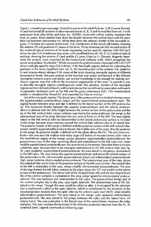
Clonal Unit Architecture ofthe AdultFly Brain 141
Figure 1, viewed on prvious page. Overall structure of the adult fly brain. A,B) Coronal (frontal,A) and horizontal (B) sections of silver-stained brains (A, B, D and N modified from ref. 5 withpermission from John Wiley and Sons, Inc. (2006). Areas with yellow overlay represent therind, or cortex. Black dashed lines show the border between the central brain and the opticlobe and between neuromeres (A). White lines show the arbitrary border of the neuropile regions. C) Scheme of a neuron in the brain. D,E) Distribution of the neural cell bodies, showingthe anterior (D) and posterior (E) views of the brain. Three-dimensional (3D) reconstruction ofthe confocal optical sections of the brain expressing nuclear-specific reporter UAS-NLS-/acZdriven by elav-GAL4 enhancer trap strain c155 (modified from ref. 10). F, G) Confocal opticalsections, showing the anterior (F) and middle (G) areas (Data by H. Otsuna). Magenta represents the synaptic areas visualised by the monoclonal antibody nc82, which recognises theactive zone protein, Bruchpilot." White represents the glial processesvisualised with UAS-CFPdriven with glia-specific repo-GAL4 driver. H-N) Neuropile regions defined for indicating thepositions in the brain (modified from ref. 5). 3D reconstruction of the anterior (H), posterior(I) and anterior-dorsal oblique (J-N) views of the brain showing neuropile regions at differentdorsoventral levels. Because analysis of the function and neural architecture of the diffusedneuropiles remains scarce and spotty, our current knowledge is not enough for making conclusive regional map that reflects the functional organisation of this area. To provide a wayto describe neuropile regions unambiguously under this situation, borders of the neuropileregions are here defined arbitrarily with simple planes that are defined in association with easilyrecognisable landmarks such as the MB and the great commissure (GC). This nomenclaturesystem is introduced by Strausfeld? and expanded by Otsuna and Ito.'
Listof the neuropile regions: The dorsal area of the protocerebrum is divided into two areas:the superiormedial protocerebrum (smpr) and the superiorlateral protocerebrum (slpr). Thesagittal border between smpr and slpr is defined by the lateral surface of the MB pedunculus(p), The horizontal border between the superior protocerebrum and the inferior protocerebrum is defined with the 50% height between the ventral surface of the pedunculus and thetip of the MB vertical lobe. asmpr (anterior superiormedial protocerebrum): the asmpr is theanteriormost area of the smpr, between the two vertical lobes (v) of the MB. The area slightlylateral to the MB vertical lobe but dorsomedial to the lateral pedunculus surface is includedin the aimpr, because many neurons around the vertical lobe arborise also in its lateral side.The posterior border of the asmpr is defined with the posterior surface of the MB vertical lobe.msmpr (middle superiormedial protocerebrum): the middle area of the smpr, directly posteriorto the asmpr. Its posterior border is defined with the plane above the GC. The pars intercerebralis-the area near the midline with many large cell bodies of neurosecretory cells-lies inthe medilalmost region of the msmpr. psmpr (posterior superiormedial protocerebrum): theposteriormost area of the smpr, spanning above and anterodorsal to the MB calyx (ca). mslpr(middle superiorlateral protocerebrum): the area lateral to the msmpr. Note that there is no areacalled the aslpr, because there is no neuropile anterolateral to the MB vertical lobe (see Fig.1J). pslpr (posterior superiorlateral protocerebrum): the area lateral to the psmpr, dorsolateralto the MB calyx. The area below the superior protocerebrum and above the ventral surface ofthe pedunculus is the inferiormedial protocerebrum (impr) and inferiorlateral protocerebrum(i1pr). aimpr(anterior inferiormedial protocerebrum): The anteriormost area of the impr, abovethe antennallobe and in front of the posterior surface of the MB vertical lobe. The medial lobeof the MB is embedded in this area. mimpr (middle inferiormedial protocerebrum): The areaof the impr behind the MB lobes, anterior to the plane above the GC and medial to the lateralsurface of the pedunculus. The dorsal half of the ellipsoid body (eb) and the fan-shaped body(fb) of the central complex is contained in this area. pimpr (posterior inferiormedial protocerebrum): The area between and anteromedial to the calyx. The protocerebral bridge (pb) ofthe central complex lies in this area. optu (optic tubercle): The anteriormost area of the i1pr,lateral to the aimpr. Though this area could be called as ailpr, it is occupied by the structurethat is traditionally called as the optic tubercle, which is contributed by the terminals of thevisual projection neurons from the optic lobe via the anterior optic tract (AOT). milpr (middleinferiorlateral protocerebrum): The area lateral to the mimpr. pilpr (posterior inferiorlateralprotocerebrum): The area lateral to the pimpr, between the calyx and the lateral horn. LH(lateral horn): The area protruded in the lateral area of the central brain, between the milprand pilpr. This area contains the terminals of the olfactory projection neurons from the AL. AL(antennal lobe). Legend continued on following page.
142 Brain Development in Drosophila melanogaster
Figure 1, viewed on page 138. The anterior protrusion of the medial cerebrum, receivingprojections of the sensory neurons of the antennae via the antennal nerve (AN). It is a partof the deutocerebrum. vmpr (ventromedial protocerebrum): The area just posterior to theAL, in front of the GC and ventromedial to the MB pedunculus. Unlike the AL, it is a part ofthe protocerebrum. It houses the ventral half of the ellipsoid body and the fan-shaped bodyas well as the lateral accessory lobe (also called the ventral body), an annex of the centralcomplex that is important for motor control. spsl (superior posterior slope): Dorsal part of thearea in the posterior brain surrounding the oesophagus foramen. It receives projections fromthe ocellar nerve and is also contributed by the dendrites of descending neurons. ipsl (inferiorposterior slope): The area of the posterior slope ventral to the oesophagus foramen, whichalso houses dendrites of descending neurons. vlpr(ventrolateral protocerebrum): A large areain the lateral cerebrum in front of the GC. It is also called the anterior optic foci, because itreceives many visual projections from the optic lobe. Their terminals in this area form severalglomerular structures called the optic glomeruli. pip' (posteriorlateral protocerebrum): The areabehind the vlpr, which is also called as the posterior optic foci. Like vlpr, many visual projection neurons terminate in the plpr. de (deutocerebrum): The area posterior ventral to the AL. Ithouses the antennal mechanosensory and motor centre (AMMC), which receives projectionsof auditory and mechanosensory neurons from the antennae. The AL is actually also a partof the de. SOC (suboesophageal ganglion): The neuromere ventral to the oesophagus. Otherlabelled structures: la: lamina, me: medulla, lo:lobula, lop: lobula plate, AOT: anterior optictract, POT: posterior optic tract, LCBR: lateral cell body region.
distributed around the surface ofthe nervous system each generates a large number ofneurons.6.7
Each cell division yields a neuroblast and a ganglion mother cell (GMC). It is generally believedthat a GMC divides once more to generate two neural progeny. Most neuroblasts proliferate at twoseparate periods during neurogenesis," The first proliferation occurs during mid to late embryonicstage, whereas the second proliferation starts from between the late first and late second larvalinstar and ends during the first day ofthe pupal stage. Thus, the clonal progenyofmost neuroblastsconsists ofembryonic and postembryonic neurons (Fig. 2A).17
In the larval brain, there are about 100 neuroblasts per hemisphere in the cerebrum':":'? andabout 80 per hemisphere in the SOG (R Urbach and GM Technau, personal communication).There are therefore in total about 180 neuroblasts in a central brain hemisphere. Counting ofcell bodies in the nuclear-labelled brain samples suggests that there are about 18,000 cells perhemisphere in the adult central brain including the SOG (T Shimada and KI, unpublished observation). Considering that some neuroblasts, such as those that generate the MB Kenyon cells,give birth to several hundred progeny," the number ofprogeny ofmost other neuroblasts shouldbe less than a hundred.
How, then, does each family ofclonally related neurons contribute to the formation ofthe adultneural circuits? One possibility is that each neuron differentiates and sends its neurites independently from cell lineage (left panel ofFig. 2B). The other possibility is that neurons ofa particularclone form distinct subcomponents ofthe neural circuits (right panel ofFig. 2B).
To determine which is more likely,a technique is required to visualise the projection pattern ofall the progeny ofone neuroblast in the adult nervous system. This has not been an easy task. Celllineage can in principle be traced by injecting dyes to a cell early during development.2o-24Thoughthis worked well for analysing celllineage in embryos, postembryonic progeny could not be labelledwith this technique, because injected dye is diluted below detection level as neuroblasts repeatcell division. To circumvent this problem, transplantation ofgenetically labelled neuroblasts wasdeveloped.F'" In this technique, a neuroblast is picked out from an embryo expressing a reportergene (e.g., lacZ) under control of a ubiquitous promoter. The neuroblast is then transplanted toa host embryo that does not carry the reporter gene. Though this system is versatile,26-28 technicalexpertise is required for cell transplantation and differences in the cellpositions and developmentalstages between donor and host embryos might affect subsequent development ofthe transplantedneuroblast. Thanks to the powerful Drosophila genetics, however, several techniques that areeasier to label clonally related cellswere developed during the last decade. They use genetic mosaic
Clonal Unit Architecture ofthe Adult Fly Brain 143
cell- lineage dependent
optic lobe
central bra in
in the adult
cell-lineage independent
:. c~~ <\'1:'A:f~:se ~~~~f; ~ \\
I ' ,., ,, ,neural circuit made
during embryogenesis
A neuroblast during development
0 ....../neuroblast /
GMCpostembryonicneurons
embryonic - neurons
axons ofpostembryonicneurons
cis recombinationC FLP-out GAL4
trans recombinationEMARCM
tUb~
tub ~FAT
reporter expressionhomozygous mutant phenotype
Sister cell 2
reporter---+
FRT FRT
UAS reporteri11!i43!1l4'l'llillii
GAL<.nhancer trap ~W"ijiiGAL4 mut'!J!!!n
---JFRT ~re:U~~:;sion
hs promoter 'd·"y·I.1dY'4 i']'4ft"',1-6-c::::>- chromosomal recombin-
flippase ation at low frequency
Sister cell 1
~ mutationUAS reporter ~
GAL4 _•• _~ . GAL4., FAT L,;ltUAS reporterl&illii1&Lliit1!1l4'l'llillii GAL4-t> ~ ~mu~on
FAT UAS repo;erFATcassette
i
GAL4
hs promoter
1+-- - --C:>-<=>-flippase
FLP-out of FRT cassete
r~FAT
act
G~ GAL~UAS ~ reporteri!i!&tiiii4iit!
hs promoter
~ FLP-out of FRT cassete
GAL4 enhancer trap FRTr~
ubiquitus actin promoter
~FRT cassette ,
FRT FRT-e:J-1_ r>-
act yellow GAL4---+
D UAS-FLP-out
Figure 2. Neuroblast proliferation and techniques for labelling clonally related cells . A) Schemeof the neuroblast and its progeny in the larval brain . GMC: ganglion mother cell. Photograph icinset: Larval brain shortly before pupa rium formation. Prolife rating cells are labelled withbromodeoxyuridine (BrdU) incorporation by larval feeding and visualised with ant i-BrdUantibody. Figure legend continued on next page.

144 Brain Development in Drosophila melanogaster
Figure 2, viewed on previous page. B)Two possible strategies for constructing the neural cicuitsof the adult brain. C) FLP-out-GAL4 (FRT-GAL4)system. In the first component, a marker geneyellow flanked by a pair of FRTs (FRTcassette) is inserted between the ubiquitous actin promoterand the transcription activator CAL4. Because there is no promoter directly in front of CAL4,only yellow is expressed. By applying mild heat shock to the animal during development, theheat-shock (hs) promoter activates the expression of the second component, hs-flippase, insome cells. Flippase protein induces recombination between the two FRTs, excising the yellowgene between them (FLP-out). In the cells in which this recombination occurred and also inits progeny, the actin promoter starts driving the expression of CAL4. The expression of thethird component, a reporter gene such as CFP under control of the GAL4-target sequenceUAS, is activated only in these cells. D) UAS-FLP-out system: The GAL4 gene is expressedin a cell-type specific manner using certain promoter or GAL4 enhancer-trap strains. Thesecond component features UAS and a reporter gene separated by an FRT cassette containing the C02 gene. GAL4 activates the expression of only C02. A mild heat shock activatesflippase, which excises the FRTcassette. This enables the expression of the reporter gene inthe cell and its progeny. E) MARCM system: A ubiquitous tublin promoter drives constitutiveexpression of yeast-derived GALBO, which suppresses expression of the UAS-linked reportergene even in the presence of CAL4. The tublin-CALBO and the reporter gene are put in thehomologous chromosome in trans and the FRT sequence is put in the locus close to thecentromere of each chromosome. Upon mild heat shock, trans recombination between twochromosomes occurs in some of the cells during mitosis. One of the daughter cell becomeshomozygous for the UAS-reporter. Because GALBO no longer exists in the genome of thiscell and its progeny, the cells are visualised by the reporter.
analysis combined with yeast-derived GAL4-UAS29-31 and flippase-FRT systerns'<" and can becategorised into two groups.
cis-Recombination SystemsFlippase is the enzyme that induces recombination between two sequences called the Hippase
recognition targets (FRTs). The first group oftechniques label cellsby inducingcis-recombinationbetween two FRT sequences on the same chromosome. First, a gene or a stop-codon sequenceis placed between the two FRTs. This "FRT cassette" is then put between a reporter gene and apromoter of a ubiquitous house-keeping gene, e.g., actin or tublin. Because of the inserted FRTcassette, the ubiquitous promoter cannot drive the expression of the reporter gene. By inducingthe expression offlippase transiently during development, e.g., by putting the flippase gene underthe heat-shock promoter and giving temporal heat shock to the transgenic animals, recombination between the two FRTs would occur in some cells. This removes the FRT cassette (flip-outor Fl.Peout) and connect the ubiquitous promoter and the reporter gene directly. The reportergene would be expressed specifically in these cells as well as in their progeny. If the recombinationoccurs in the GMC or in the postmitotic cells, single or a few scattered cells would be labelled. Ifthe recombination occurs in the neuroblast, on the other hand, a group of clonally related cellscan be visualised.
Such system was first developed by putting the lacZ gene after the FRT cassette." An improvedversion featured GAL4 instead oflacZ,which can activate the expression ofdiverse types ofreportergenes to visualise different aspects ofthe labelled cells (FRT-GAL4, or FLP-out GAL4 system, Fig.2C).36 Because GAL4 can activate multiple UAS targets, genes that affect the function or development of the cells-so called effector genes-can be expressed simultaneously with the reportergenes, enabling the functional analyses of the expressed genes using this system.
Another approach is to put the FRT cassette between the UAS and the reporter gene (theUAS-FLP-out system, Fig. 20).37 This system can be combined with a wide variety of promoter-GAL4lines and GAL4 enhancer-trap strains currently available, in which GAL4 is expressedspecificallyin particular cells.Dependingon whether the recombination occurred in the neuroblastor in the postrnitotic cells, the UAS-FLP-out system visualizes a clonally related subset or themorphology of the single cells out ofthe GAL4-expressing cell population.1
5,38
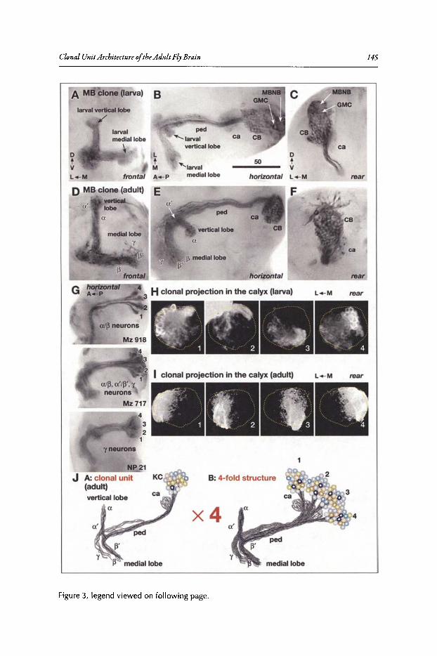
Clonal Unit Architecture ofthe Adult Fly Brain 145
A MB clone (larva)
rear
c
o50 t
Vhorizontal L_M
'larvalmedial lobe
LtMA_Pfrontal
larvalmedial lobe
~
larval vertical lobe
/
otVL-M
4
F
B: 4-fold structure
x4
NP21
f3 medial lobe
yneurons
3 Hclonal projection in the calyx (larva)
...s::~=
J A: clonal unit(adult)vertical lobe
(J.
D MB clone (adult) Everticallobe
f3_____-!.t!o.ro~ ntal
G horizontalA_P
Figure 3, legend viewed on following page.
146 Brain Development in Drosophila melanogaster
Figure 3, viewed on previous page. Clonal units in the mushroom body. A-F) A clone labelledin larvae just after hatching and visualised at the end of the larval stage (A-C) and anotherclone visualised in the adult (D-F), respectively (modified from ref. 36). (FLP-out-GAL4 clonesvisualised with UAS-tau reporter. UAS-tau and UAS-GFP reporters label essentially similarstructures, except that Tau labels dendritic arborisations more weakly and occasionally causesmild disturbance of the neural function.) Optical sections at different levels were taken withNomarski optics and montaged. Frontal, horizontal and rear views of the same clone wasvisualized by rotating the specimen. MBNB: mushroom body neuroblasts, CB: cell bodies,ca: calyx, ped: pedunculus. G) Four-fold labelling pattern of enhancer-trap strains labellingsubsets of the MB Kenyon cells. (Modified from ref. 36, UAS-tau reporter). H) Cross sectionof the calyx in the larval brain, showing areas of arborisations of each of the four clonal units.(Modified from ref. 42, ©200S National Academy of Sciences, U.S.A., MARCM clone withUAS-GFP reporter.) I) Cross section of the calyx in the adult brain, showing areas of arborisations of each of the four clonal units. (Data by Nobuaki Karl Tanaka. MARCM clone withUAS-GFP reporter.) J: Scheme of the four-fold clonal units in the MB.
Note on the name of the lobes: The names of the lobes have been changed drasticallyduring the last few years of the last century. The terms of the a and ~ lobes originated fromthe study of the bee brain'" to refer to the vertical and medial lobes. The y lobe derived fromthe study of sphinx moth." These terms are adopted to describe the fly MB.2,62 Because ofthe apparent structural similarity, vertical and medial lobes in the larval MB had also beencalled as larval a and ~ lobes."
Analysis of clones and GAL4 enhancer-trap strains revealed a characteristic subdivision ofthe a lobe and defined it as the a' lobe, but failed to recognisethe corresponding subdivision inthe ~ lobe." The latter subdivision was identified by the comparison of labelling pattern of various antibodies and named as the Wlobe.' Until this period, it was not known that the neuronsinnervating the y lobe have no vertical branches. Though such unbranched neurons had beenobserved in Golgi impregnatedsamples, the non-existenceof the branch could not be determinedconclusively because Golgi labelling may not always label all the branches of a neuron.
Finally, systematic flippase-mediated single-cell analysesrevealed that the neurons contributing to the y lobe, a'/~' lobes and a/~ lobes are generated in this order and that the verticaland medial lobes of the larval MB is contributed exclusively by the neurons that composethe adult y lobe as a result of reorganization." The larval vertical and medial lobes, therefore,have nothing to do with the neurons of the adult alp lobes.
To avoid confusion, it is better not to use the term a/~ lobes for the larval MB but to usethe generic term vertical/medial lobes instead. Also, the adult vertical lobe should not generally be called the a lobe, as it actually consists of a and a' lobes each of which is likely tohave rather different functions.
The vertical and medial lobes are sometimes called dorsal and horizontal lobes, respectively. In various insects, however, the vertical lobe does not project dorsally but anteriorly oranterodorsally. The medial lobe projects medially (towards the midline) in all insect species,but the inclination of the lobe may not always be horizontal. Thus, the combination of "vertical" and "medial" seems more appropriate when considering cross-species compatibility.A-G, J reproduced with permission of the Company of Biologists.
trans-Recombination SystemsOne ofthe classicmethods for analysing lineage-associated cells is to induce somatic recombina
tion by irradiating the animals with X ray or y ray. Recombined cells can be identified by puttinga marker gene in one ofthe chromosomes. As a more controllable and easy-to-use approach, FRTwas put into the chromosome to induce flippase-dependent trans-recombination.34 The lack ofconvenient reporter systems for detecting the neurons that experienced recombination has madeit difficult to apply this technique for brain research. The mosaic analysis with a repressible cellmarker (MARCM) system solved this problem." The MARCM system features GAL80, whichworks antagonistically to GAL4 (Fig. 2£). GAL80 suppresses expression of the VAS-linked reporter gene even in the presence ofGAL4. Flippase-induced somatic recombination between theFRT sequences removes GAL80 gene in one of the daughter cells. VAS-linked reporter/effectergenes will be expressed specifically in this cell and its progeny.
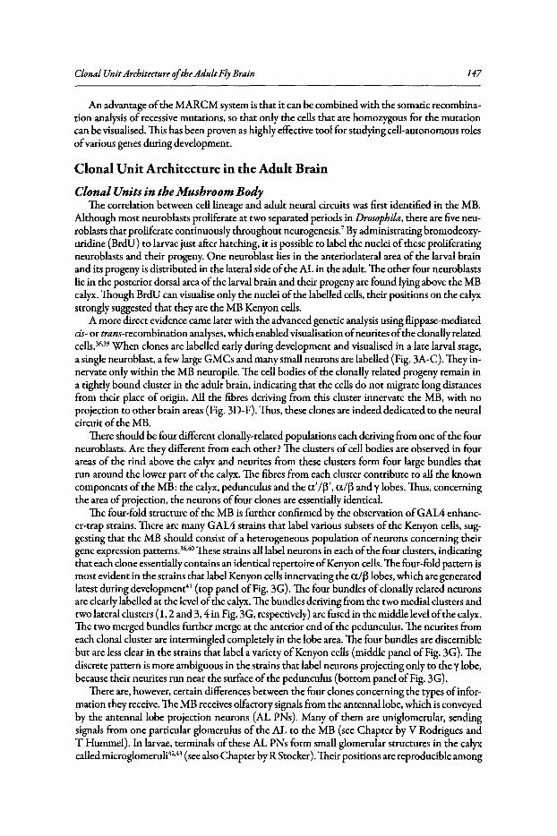
Clonal Unit Architecture ofthe AdultFly Brain 147
An advantage ofthe MARCM system is that it can be combined with the somatic recombination analysis of recessive mutations, so that only the cells that are homozygous for the mutationcan be visualised. This has been proven as highly effective tool for studying cell-autonomous rolesofvarious genes during development.
Clonal Unit Architecture in the Adult Brain
Clonal Units in the Mushroom BodyThe correlation between cell lineage and adult neural circuits was first identified in the MB.
Although most neuroblasts proliferate at two separated periods in Drosophila, there are five neuroblasts that proliferate continuously throughout neurogenesis," By administrating bromodeoxyuridine (BrdU) to larvae just after hatching, it is possible to label the nuclei ofthese proliferatingneuroblasts and their progeny. One neuroblast lies in the anteriorlateral area of the larval brainand its progeny is distributed in the lateral side ofthe AL in the adult. The other four neuroblastslie in the posterior dorsal area ofthe larval brain and their progeny are found lying above the MBcalyx. Though BrdU can visualise only the nuclei ofthe labelled cells, their positions on the calyxstrongly suggested that they are the MB Kenyon cells.
A more direct evidence came later with the advanced genetic analysis using £lippase-mediatedcis- or trans-recombination analyses, which enabled visualisation ofneurites ofthe clonally relatedcells.36•39 When clones are labelled early during development and visualised in a late larval stage,a single neuroblast, a few large GMCs and many small neurons are labelled (Fig. 3A-C). They innervate only within the MB neuropile. The cell bodies of the clonally related progeny remain ina tightly bound cluster in the adult brain, indicating that the cells do not migrate long distancesfrom their place of origin. All the fibres deriving from this cluster innervate the MB, with noprojection to other brain areas (Fig. 3D-F). Thus, these clones are indeed dedicated to the neuralcircuit ofthe MB.
There should be four different cionally-related populations each deriving from one ofthe fourneuroblasts. Are they different from each other? The clusters ofcell bodies are observed in fourareas of the rind above the calyx and neurites from these clusters form four large bundles thatrun around the lower part of the calyx. The fibres from each cluster contribute to all the knowncomponents ofthe MB: the calyx, pedunculus and the a'/~', a/~ and y lobes. Thus, concerningthe area ofprojection, the neurons offour clones are essentially identical.
The four-fold structure ofthe MB is further confirmed by the observation ofGAL4 enhancer-trap strains. There are many GAL4 strains that label various subsets of the Kenyon cells, suggesting that the MB should consist of a heterogeneous population ofneurons concerning theirgene expression patterns.36•4OThese strains all label neurons in each ofthe four clusters, indicatingthat each clone essentially contains an identical repertoire ofKenyon cells.The four-fold pattern ismost evident in the strains that label Kenyon cells innervating the a/f3lobes, which are generatedlatest during development" (top panel ofFig. 3G). The four bundles ofclonally related neuronsare clearly labelled at the level ofthe calyx. The bundles deriving from the two medial clusters andtwo lateral clusters (1,2 and 3, 4 in Fig. 3G, respectively) are fused in the middle level ofthe calyx.The two merged bundles further merge at the anterior end ofthe pedunculus. The neurites fromeach clonal cluster are intermingled completely in the lobe area. The four bundles are discerniblebut are less clear in the strains that label a variety ofKenyon cells (middle panel ofFig. 3G). Thediscrete pattern is more ambiguous in the strains that label neurons projecting only to the y lobe,because their neurites run near the surface ofthe pedunculus (bottom panel ofFig. 3G).
There are, however, certain differences between the four clones concerning the types ofinformation they receive. The MB receives olfactory signals from the antennallobe, which is conveyedby the antennallobe projection neurons (AL PNs). Many of them are uniglomerular, sendingsignals from one particular glomerulus of the AL to the MB (see Chapter by V Rodrigues andT Hummel). In larvae, terminals of these AL PNs form small glomerular structures in the calyxcalled microglomeruliv" (see also Chapter by R Stocker). Their positions are reproducible among
148 Brain Development in Drosophila melanogaster
individuals, showing that olfactory information from particular glomeruli in the larval AL istransmitted to distinct subregions ofthe calyx.The arborisations ofthe Kenyon cellsofeach cloneoccupy different, but partially overlapping, areas ofthe calyx (Fig. 3H).42 Thus, each clone shouldreceive a different repertoire ofolfactory information.
Because ofthe much larger number ofAL PNs and the number ofglomeruli in the adult AL,there are numerous very small microglomeruli in the calyxofthe adult MB, making their mappingmore complex (see Chapter by P Laissue and L Vosshall). Nevertheless, AL PNs from particularAL glomerulus terminate in specific concentric zones in the calyx." The Kenyon cellsofeach cloneagain arborise in distinct areas ofthe calyx (Fig. 31),4.44 suggesting that there may also be differencesin the repertoire ofolfactory information each clone would receive. For example, the two"outer"clones (1 and 4 in Fig. 31) may have fewer interaction with the projection neurons that terminatein the central area ofthe calyx than do the two "inner" clones (2 and 3 ofFig. 31).
Observations in the MB suggest that there are clonally-related unit structures in the adult brain.Progeny ofa single neuroblast may contain a functionally heterogeneous population ofneurons.Yet, they all innervate only a limited area ofthe brain and form a distinct neural circuit structure.There are four such clonal units in the Drosophila MB, which are essentially identical regardingtheir morphology and biochemical diversity but slightly different in the projection pattern intheir input areas (Fig. 3J).
Clonal UnitArchitecture in the Central ComplexClonal unit is not a unique feature ofthe MB. They are also observed in the central complex,
the neuropile that lies at the centre of the cerebrum-Y and is supposed to play important rolesin motor coordination control, visual memory, etc.464 8 The structure of the central complex ismuch more complex than the MB (Fig. 4A). It consists offour major components, the ellipsoidbody (eb), fan-shaped body (fb),protocerebral bridge (pb) and noduli (no).4S Whereas the cellbodies of the MB Kenyon cells are all confined in a small area just around the MB calyx, thosethat contribute to the central complex are distributed in various parts of the brain. Nevertheless,lineage-dependent cell labelling experiments revealed that several clones contribute specifically tothe central complex, each forming distinct building units ofits neural circuits.
The ellipsoid body is a round structure that forms the anteriormost part ofthe central complex.There is a pair ofclonal units with their cell bodies in the anterior brain above the aimpr area ofthe cerebrum, dorsolateral to the AL (EB-A1, Fig. 4A,B). A bundle ofneurites projects beneaththe medial lobe ofthe MB and forms the primary arborisation in the vmpr part ofthe cerebrum,forming the structure called the lateral triangle (ltr). From the ltr, some fibres project dorsally toreach the asmpr and aimpr and others project to the ellipsoid body from its central hole to formthe ring neurons ofthis neuropile.
The fan-shaped body consists ofan array ofradial projections and tangential neurons that arborise at its various dorsoventrallevels. One ofthe clonal units that form these tangential componentshave the cell body cluster in the dorsolateral area ofthe cerebrum, posterior to the LH (FB-DL1,Fig. 4A,C). The neurons form primary arborisations near the dorsal surface ofthe cerebrum abovethe LH and secondary arborisations in the msmpr and mslpr. The fibre bundle bifurcates, entersthe fan-shaped bodyfrom its anterior side at two levels (Fig.4C) and forms extensive branches thatspan tangentially. There are also other clonal units that form tangential arborisations in differentlevels of the fan-shaped body (not shown here).
The radial component of the fan-shaped body is formed by four clonal units per hemisphere(FB-P1-4, Fig. 4A,D). A row ofeight cell body clusters lies in the posterior brain right behind thefan-shaped body, flanked by the calycesofthe MB. The neurites form primary arborisation in theprotocerebral bridge and enter the inferior part of the fan-shaped body from its posterior side.They form two bundles that run radially in the fan-shaped body and terminate in the nodulus ofthe contralateral hemisphere.
The protocerebral bridge is divided into eight sections per hemisphere. Similarly, the radialcomponent of the fan-shaped body is organized in eight radial structures called the staves.2.4S
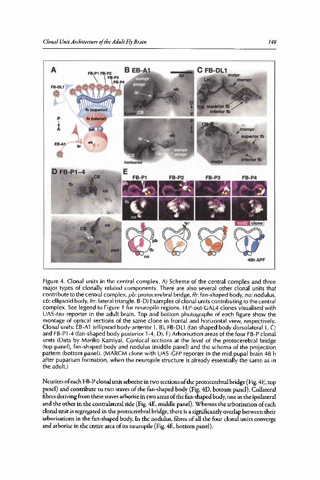
Clonal Unit Architecture ofthe Adult Fly Brain 149
A
p
iEIl-A1
FB-P1 FB-P2 B EB-A1
\FB-P3
\ FB-P4I
48hAPF
Figure 4. Clonal units in the central complex. A) Scheme of the central complex and threemajor types of clonally related components. There are also several other clonal units thatcontribute to the central complex. pb: protocerebral bridge, fb : fan-shaped body, no: nodulus ,eb: ellipsoid body, Itr : lateral triangle. B-D) Examplesof clonal units contributing to the centralcomplex. See legend to Figure 1 for neuropile regions. FLP-out-GAL4 clones visualised withUAS-tau reporter in the adult brain. Top and bottom photo graphs of each figure show themontage of optical sections of the same clone in frontal and horizontal view, respectively.Clonal units: EB-A1 (ellipsoid body -anterior 1, B), FB-DLl (fan-shaped body dorsolateral 1, C)and FB-Pl-4 (fan-shaped body posterior 1-4, D). E)Arborisation areas of the four FB-Pclonalunits (Data by Mariko Kamiya). Confocal sections at the level of the protocerebral bridge(top panel), fan-shaped body and nodulus (middle panel) and the schema of the projectionpattern (bottom panel). (M ARCM clone with UAS-GFP reporter in the mid pupal brain 48 hafter puparium formation, when the neuropile structure is already essentially the same as inthe adult. )
Neurite sofeach FB-P clonal unit arborise in two section softhe protocerebral bridge (Fig.4E. toppanel ) and contribute to two staves of the fan-shaped body (Fig. 4D. bottom panel). Collateralfibres deriving from these staves arbori se in two areas ofthe fan-shaped body,one in the ipsilateraland the other in the contralateral side (Fig.4E. middle panel). Whereas the arbori sation ofeachclonal unit is segregated in the protocerebral bridge , there is a significantly overlap between theirarbo risations in the fan-shaped body. In the nodulus, fibres ofall the four clonal units convergeand arbori se in the entire area ofits neuropile (Fig. 4E. bottom panel ).
150 Brain Development in Drosophila melanogaster
Clonal UnitArchitecture in Other Brain AreasCompared to the MB and the central complex, borders between neural circuits in the rest of
the central brain are much more obscure. Nevertheless, clonally related neurons innervate onlylimited areas ofthese neuropiles and form distinct unit structures.
Projection neurons from the antennallobe innervate the MB calyx, the LH and several otherareas of the brain.P:" GAL4 enhancer-trap strains such as GH146, NP22S and NPS288 labelmany ofthese neurons.4,49.50 The cell bodies ofthese neurons form at least four clusters around theAL.The anterior dorsal cluster (AL-DAI, Fig. SA) and a lateral cluster (AL-Ll, not shown here)consists ofthe neurons that innervate via the inner antennocerebral tract (iACT). The cell clusterthat lies ventral to the AL (AL-VI, not shown here) consists of the neurons of the middle ACT(mACT) pathway. There is yet another clone in the lateral area of the AL, which consists of theneurons that do not seem to be labelled in these GAL4 strains (AL-L2, Fig. SB). Neurons ofthisclonal unit project not only to the MB and calyx but also to the SOG and the plpr.
In the MB, neurons other than the Kenyon cells also innervate its neuropile. An example ofsuch clonal unit, MB-Al (Fig. SC), has the cell bodies in the anterior brain just in front of theMB vertical lobe.l"Neurons of this clone mainly innervate the distal area of the medial lobe andproject also to the neuropiles other than the MB in the aimpr and vmpr areas.
Neurons in the LH, which receives olfactory information from the AL like the MB Kenyoncells, are also organized in a clonally related manner. Several clonal units contribute to the neuropile ofthe LH.Their cell bodies form clusters in the LCBR. Some clones (e.g., LH-l, Fig. SO)consist oflocal neurons that arborise only in the LH. The neurites ofother clones (e.g., LH-2 and3, Fig. SE,F) arborise in the LH and project further to other areas ofthe protocerebral neuropiles.Depending on the clonal units, the neurites project to the LH either from inside (LH-2,3) orfrom outside (LH-l).
The superior lateral and superior medial protocerebrum occupies the dorsalmost area of thecerebrum. Because neural connections between these neuropiles and the neuropiles ofthe sensoryand motor pathways are still essentially unknown, the function ofthe neural circuits in these areasare yet to be determined. These neuropiles are also contributed by many clonal units. Short oftheknowledge ofdetermining neural structure in these areas, these clonal units are tentatively namedaccording to the neuropile region (Fig. 1H-N) in which they arborise most extensively. Some clonalunits, e.g., PSLPR-l and MSLPR-l (Fig. SG,H), arborise only in a small region of the neuropile.They tend to have simple structures, with a single bundle of neurites and arborisation in one oronly a few areas. Other clones, like MSLPR-2 and MSMPR-l (Fig. SI,J), arborise in multiple areas.The structure of these clonal units are more complex, with bifurcation or trifurcation of neuritebundles and extensive projections that span a long distance in the brain.
The ventrolateral part ofthe cerebrum (vlpr and plpr) is occupied by the neuropiles that extensively receive axons ofthe visual projection neurons, which connect the optic lobe and the centralbrain.P These areas are also formed by various clonal units, whose cell bodies lie in the LCBRor in the anterior lateral area of the cerebrum. Some clonal units form circuits that connect thecorrespondingneuropiles ofboth hemispheres (e.g., VLPR-l, Fig. 5K), whereas others connect avariety ofneuropile areas of the cerebrum (e.g., VLPR-2, Fig. 5L).
Formation ofthe Clonal Units During DevelopmentThe observations presented above suggest that a significant portion of the adult brain is com
posed in a cell lineage-dependent manner (Fig. 2B). Though the progeny of a single neuroblastare not as tightly packed as in the larval brain, they still form a cluster. Neurites deriving from thiscluster form tight bundles and innervate distinct areas of the brain.
How, then, is such clonal unit architecture in the adult brain composed during neurogenesis ?When the clones are visualised in late larval or early pupal brains, the progeny of a neuroblastform a tightly packed cluster, which sends a bundle ofneurites towards the neuropile (Fig. 6A,B).The bundle either projects to a single target or bifurcates when it enters the neuropile to innervate different areas of the brain.V The formation of the adult clonal units should depend on this

Clonal Unit Architecture ofthe Adult Fly Brain
AAL-DA1 ca- 50otV
151
Figure S. Clonal units in other brain areas. FLP-out -GAL4 clones visualised w ith UAS-taureporter. Top and bottom photographs of each figure (A-L) show the montage of frontaland hor izontal optical sections of the same sample, respectively. See legend to Figure 1 forneuropile regions.
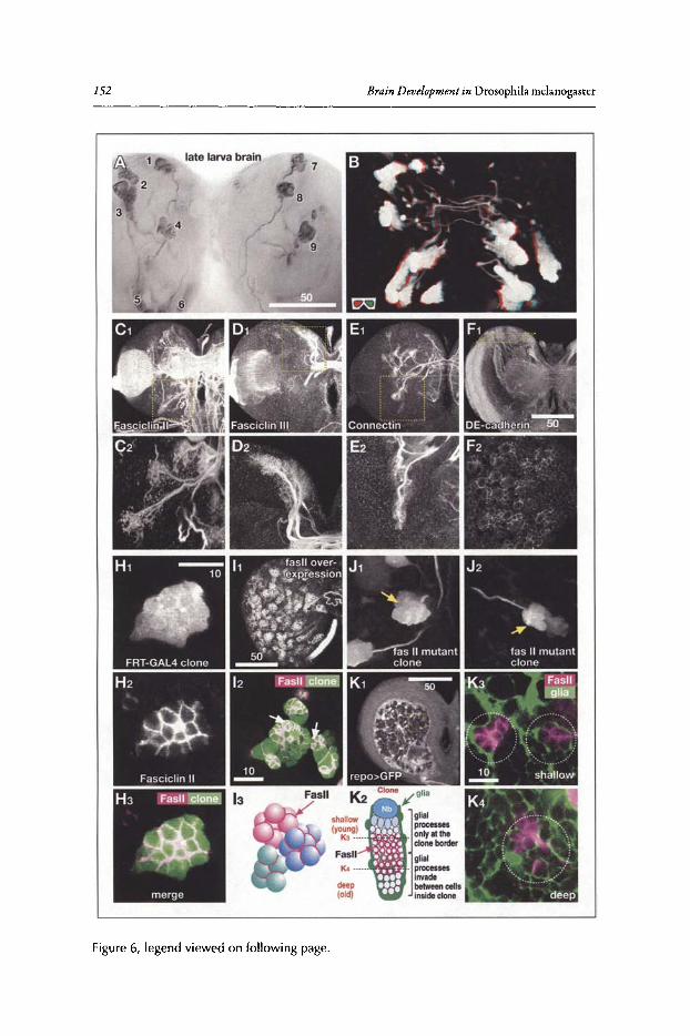
152
late larva brain
2
4
Figure 6, legend viewed on following page.
Brain Development in Drosophila rnelanogascer
Clonal Unit Architecture ofthe AdultFly Brain 153
Figure 6, viewed on previous page. Formation of clonal units in the larval brain. A, B)Clonal units in late larvae. FLP-out-GAL4 clones visualised with UAS-tau (A, montage ofoptical sections) and UAS-CFP (B, 3D-stereograph of confocal sections). C-F) Distributionof cell-adhesion molecules in the larval brain visualised with antibodies. Overall brain (toppanel) and blow-up view (bottom panel) showing the area indicated with dashed squaresin the top panel. Clonal-unit dependent distribution of Fasciclin II (Fasll, C), Fasciclin III (D)and Connectin (E) and pan-clonal distribution of DE-cadherin (F). H) Distribution of Fasllvisualised with anti-Fasll antibody in the cluster of clonal cell bodies. I) Over-expression ofFasll in all the neurons (using elav-CAL4C155 driver, 11) and in the MARCM clones (12). J)Effect of the homozygous mutation of Fasll in the MARCM clones. Two examples are shown.K) Distribution of Fasll (visualised with anti-Fasll antibody) and glial processes (visualisedwith UAS-CFP driven with glia-specific repo-GAL4 driver).
clonal cluster formation in larvae. Because formation of the lineage-dependent structure in thelarval brain is comprehensively described in the Chapter by V Hartenstein et al, here we discussthis issue only briefly.
One ofthe candidate mechanisms that promote bindingofthe clonally related cell bodies andneurites depends on homophilic cell adhesion molecules (CAMs). Ifsuch CAMs are expressed inthe clonally related neurons, they would facilitate adhesion ofthe cells and fibre bundles.51-53
According to their expression patterns, the homophilic CAMs can be classified into two types.The first type is expressed only in a small subset of the clones. This includes Fasciclin II (Fas II),Fasciclin III (Fas III) and Connectin (Fig. 6C-E). Interestingly, whereas the expression patterns ofthese CAMs are associated with the clonal units in the developing brain, they are not related withthe clonal units in the adult. This suggests that intra-clonal cell-cell adhesion would be mediatedby these CAMs during the formation ofcertain clones.
The other group is expressed in most of the developing clonal units: this category includesCAMs like DE-cadherin (DE-cad) and Neurotactin (Fig. 6F). The role ofsuch pan-clonal CAMsduring development has been studied using the ectopic expression ofthe dominant negative formof DE-cad, which affected the organisation of the developing clonal clusters. 54 Although theobserved abnormality was not severe, the function ofDE-cad at least seems to be involved in thecorrect formation ofthe clonal architecture.
The role ofthe clone-specific CAMs, on the other hand, is not yet clear.When the distributionofone such CAM, Fas II, isvisualised together with the clonal cluster, the protein isobserved onlyon the cell surface that is flanked by other siblings in the same clone but not on the outer surface ofthe cell body cluster (Fig. 6H). To determine whether Fas II is concentrated because ofthe homophilic interaction with the same molecule ofthe neighbouring cells,we over-expressed Fasll so thatcells in the neighbouring clones express the same protein. Even in this case, Fasll is concentratedonly along the cell border within each clone but not along the cell border between clones (Fig.612, 13). Ectopic expression of Fasll in all the neurons, which should negate the clone-specificrole of this molecule, affect neither the organised distribution of the clonal cell clusters nor theprojection patterns ofneurites (Fig. 611). Moreover, the formation of the clonal cell cluster andneurite bundles is not disturbed even when the function of Fasll is removed by inducingfizsIImutant clones using the MARCM system (Fig. 6J). Thus, removal ofjust one clone-specific CAMdoes not affect the formation and maintenance ofthe clonal architecture in the larval brain. It ispossible that pan-clonal and clone-specific CAMs might function cooperatively to facilitate theclone-specific cell-cell adhesion.
Another factor that would be important for the organisation of the clonal unit is the cellbody glial cells, which send processes between neural cell bodies.F The region of the rind nearthe surface ofthe larval brain is characterised by the glial processes that form large nest-like holes(Fig. 6Kl).55 Because each glial nest houses a neuroblast and its progeny, the surface ofthe clonalcluster is flanked by the glial sheath. This organisation explains why Fasll is accumulated only inthe intraclonal border of the cell bodies (Fig. 6K3). Because glial cells do not express Fasll, theglial sheath physically separates the cellsofthe Fasll-expressing clones even when they are flankedwith each other.
154 Brain Development in Drosophila melanogaster
In the deeper level ofthe rind, glial processes invade borders between neural cell bodies oftheclones. Though FasII is still distributed along the intraclonal cell border, neighbouring cell bodiesare separated by the invaded glial processes (Fig. 6K4).In the adult, all the neural cell body in therind are each surrounded by extensive glial processes (Fig. 1F, G). Thus, the glial nest seems to bea transiently structure formed due to the time required for the extension ofglial processes duringlarval neurogenesis. Although glial cellscontinue invadingall the space between neural cellbodies,a temporal delay is inevitable between the period when neurons are newly formed by the GMC andthe time when glial cellsoutside ofthe clonal cluster send processes between them (Fig. 61(2). Thisdelay results in the glial nest architecture in the larval brain. Clone-specific CAMs may stabilise theclusteringofsibling neurons during this time lag. Since the cell clusters are buttressed by the sheathofthe glial nest and because pan-clonal CAMs may function redundantly, over-expression or lackofa particular clone-specific CAM would not lead to significantly abnormal phenotypes.
Functional Importance ofthe Clonal UnitsBecause many areas of the brain neuropile are formed by the combination of clonal units,
they seem to be the fundamental building blocks of the adult fly neural circuits. There would beseveral advantages by organising the brain in such a clone dependent manner. Unlike in the simplenervous system ofearly embryos, neural fibres in the postembryonic brain must find their pathsthrough the three-dimensional space filled with tangled fibres ofother neurons. Ifeach neurondifferentiates and sends its neurite independently, a large variety ofattracting and repulsive signalswould be required for providing positional cues for these neurons (Fig. 7Al).s6 Because neuralfibres innervating different targets would criss-cross with each other, systems for avoiding unnecessary cross-talk between these signals would be inevitable. Ifneurons ofthe same cluster, on theother hand, form fascicles to project to only distinct areas of the brain, the guidance system forthe follower neurons should be much simpler (Fig. 7A2). Path finding ofindividual neurons willbe required only in the area near the target. Projection towards an additional target is a matter oflocating the branching point in the one-dimensional space along the neurite bundle. Even in suchclones, the first neuron (the so called pioneer neuron) has to extend its fibre without the help ofa pre-exiting fascicle. As this occurs in relatively early embryos, when the brain neuropile is stillsimpler and the distance between the cell body and the target is much shorter than in the adult,path finding would be relatively easy.
Although flippase-mediated labelling visualises clonal units so clearly,few molecular markerssuch as antibodies and enhancer-trap strains label neurons of a single clonal unit. Rather, theytend to label small subsets ofneurons scattered in many clonal units. This suggests that, althoughneurons ofeach clonal unit are relatively homogeneous regarding their overall projection patterns,they are rather heterogeneous concerning properties like gene expression patterns. They are alsoheterogeneous in the precise arborisations within the target areas. These suggest that a singleclonal unit would be a versatile functional unit in which a variety ofcomplicated computation ispossible. Organising the brain by the composition ofsuch units might have been an economicalway for developing complicated neural circuits during evolution. Just like duplication and subsequent modification ofgenes added new functions to the genome, addition ofnew clonal unitsby the formation ofadditional neuroblasts might be a convenient way of incremental evolutionofthe brain (Fig. 7B). The loss ofcertain clonal units might also have occurred during evolution.Consideringthat there are severalclonal units contributingoverlappingly to the same circuit moduleofthe brain (discussed later), such loss ofclonal units may not have jeopardised the architectureand function ofthe brain.
Whereas some clonal units consist of several hundreds of neurons, some have less than SOneurons. Such significant differences in cell number may affect the computational capacity ofthecircuits formed by that clonal unit. Because different insect species rely on very different sensorysignals depending on their habitats and life styles, computational requirements for the evolutionary comparable clonal units might vary. Not only duplication or removal ofclonal units but alsothe change in the cell numbers of clones might have been important during evolution. Though
Clonal UnitArchitecture oftheAdult FlyBrain 155
Clonal unit-based projection Vpathfinding onlyin a restriuced
target arearecognition of the .•......\"">,
branch point (1-0 space)! ~
I ~ :
.............../ t.~.....1neccesity of avoiding cross-talkbetween signals/neurons
Clone-independent projectionneccesity of a largevariety of attractive/repulsive signals("I
each neuron finds its targetindependetly in a 3-D space
/
Uclan""clan f1
lineage
01 clon al unit as segregatedfunctional block
in the adult
dupication /' ~....\ (,In
of a clone (
.1] 02 hierarchica l unit architectureVlineage
. ] birth orderG birth date
~ 0 ~ c ]c 0 C c
~hemilineage 1 hemilineage 2
during development
O~·neurobla~ gGMC ~
,1/2 daughter cells
c
Figure 7. Clonal unit architecture of the brain. A) Comparison of possible path-finding mechanisms between clone-independent (left) and clone-dependent (right) organisation of the brain.S) Hypothetical scheme of incremental complication of neural circuits. C) Possible factors thataffect the diversity of neurons within each clone. 0) Scheme of the arborisation area of eachclonal unit Each clone innervates segregated areas of the neuropile (01), or, Several clonesinnervate highly overlapping areas to form functional modules of the brain (02).
visualisation ofthe clonal units in the adult brain iscurrently possible only in Drosophila, comparative study ofclonal units across insect taxa in the future would provide important insights on thefunctional composition ofthe brain.
As for the heterogeneity within each clonal unit, there would be two candidate control factors(Fig.7C).The first factor is the order and timingofcellgeneration. Duringembryonic development,neuroblasts change their gene expression pattern drastically and neurons that are made at each timepoint are characteristically affected by this.l~.57In the postembryonic stages,expression patterns ofthe neuroblasts do not seem to change so quickly. Nevertheless, specific projection patterns oftheadult neurons in the target area, such as the arborisation ofAL-PNs in the AL and the LH andthat ofthe MB Kenyon cells in the lobes, are dependent on the birth date ofeach neuron duringlarval stage.41
•49 A BTB zinc-finger protein gene has been identified that governs neuronal temporal
identity during postembryonic fly brain development." Expression levels ofthis molecule in theclonal neurons are reduced gradually depending on their birth timing. Temporal gradient in theactivity ofsuch genes may specify cell fate in an extended neuronal lineage.
Other factors would control the differences between the two sibling neurons made by eachof the GMCs. Proteins such as Numb are distributed unevenly between the two daughter cells,activating the Notch signalling pathway in only one ofthem . This difference between sibling cells
156 Brain Development in Drosophila melanogaster
ofthe olfactory sensory neurons made by the same precursor causesclusteringofprojection targetsin the AL.59 Similar differences between sister cells,each ofwhich comprises a "hemilineage", mayoccur within the clonal units of the brain (Fig. 7C).
The concept ofthe adult brain made by the building blocks ofclonal units may give the impression that each clone occupies specific and discrete areas ofthe brain neuropile (Fig. 7D1).Indeed,3D reconstruction ofclonal units yields images ofcionallyrelated neuronal fibres that appear to fillparticular areas of the brain. This, however, might be a too simplistic view. Because the diameterofneural fibres is much smaller than the resolution ofthe optical microscopes, dense arborisationis visualized as a solid structure even when only a fraction of the volume is occupied by the visualised fibres. Volume- and surface-rendering algorithms ofthe 3D reconstruction software furtherremove fine detail of the visualised fibres, oversimplifying the projection pattern in the area. Forthe neurons of each clonal unit to communicate with neurons ofother units, their arborisationshave to be spatially colocalised and therefore intermingled. Thus, clonal units should in principlecontribute to significantly overlapping areas of the brain (Fig. 7D2). Interestingly, the degree ofoverlap appears to be larger in the arborisation areas that are distal from the cell body clusters.Both in the MB and FB-P clones, arborisations ofeach clone occupy distinct areas in the calyxandprotocerebral bridge but overlap completely in the lobes and nodulli (Figs. 3J,4E).
The degree ofoverlapbetween specificsetsofclonal units ismuch larger than the overlapwith therest ofthe clones. In another word, several clonal units contribute together to form distinct neuralcircuit units, to which other clones contribute only marginally. In these cases, the neural circuitformed by each clonal unit may be too small and simple to represent an independent functional unit.The neural circuits in the brain are therefore organised in a hierarchical manner. Neurons derivingfrom several cell lineages form a "clan~ which together contribute to the formation ofa functionalmodule of the brain circuit. The four clonal units of the MB, several clonal units around the ALthat all arborise in the AL and form the complete set ofACT pathways, clones in the anterior andposterior brain that together compose the central complex neuropile, are examples ofsuch clans.The clan might therefore be as important as lineage for understanding the functional dynamics ofthe brain, just like a clan ofpeople, who belong to a number oftightly-associated lineages, behavedas a functional group in the dynamics of the ancient human society (Fig. 7D2).
ConclusionComplicated neural circuits in the brain are composed by the combination ofrelatively simple
clonal units. A group ofclonal units together form a functional module ofthe brain. Developmentalmechanisms that form such lineage- and clan-dependent structures are not yet fully understood.Guidance molecules and interactions between neurites of the same clone and between those ofthe neighbouring clones would play important roles in this process. More detailed analysis ofthearborisation patterns and gene expression patterns of the neurons of each clonal unit would berequired. Analysis oftemporal aspects, not only about the order ofneuron formation within eachclone but also about the timing ofproliferation and neurite extension among clones of the sameclan, would also further our understanding about the process of the neural circuit formation.
AcknowledgementsWe are grateful to Kazumi Suzuki and Mariko Kamiya for the preparation of images of the
clonal units, Nobuaki Karl Tanaka for the unpublished data ofthe arborisation pattern ofthe clonalunits in the adult calyx and Liria Masuda-Nakagawa for the reproduction of images in the larvalcalyx.This work is supported by the grants ofthe Precursory Research for Embryonic Science andTechnology and the Institute for Bioinformatics Research and Development from Japan Scienceand Technology Agency to KI and TA and the Grant-in-Aid for Scientific Research from theMinistry ofEducation, Culture, Sports, Science and Technology ofJapan to KI.
References1. Power ME. The brain of Drosophila. J Morph 1943; 72:517-559.2. Strausfeld NJ. Atlas of an Insect Brain. Berlin, Heidelberg, New York, Tokyo: Springer-Verlag, 1976.
Clonal Unit Architecture ofthe AdultFly Brain 157
3. Crittenden JR, Skoulakis EM, Han KA et al. Tripartite mushroom body architecture revealed by antigenicmarkers. Learn Mem 1998; 5:38-51.
4. Tanaka NK, Awasaki T, Shimada T et al. Integration of chemosensory pathways in the Drosophilasecond-order olfactory centers. Curr BioI 2004; 14:449-457.
5. Otsuna H, Ito K. Systematic analysis of the visual projection neurons of Drosophila melanogaster I.Lobula-specific pathways. J Comp Neuro12006; 497:928-958.
6. Truman~ Bate M. Spatial and temporal patterns of neurogenesis in the central nervous system ofDrosophila melanogaster. Dev Biol 1988; 125:145-157.
7. Ito K, Horta Y. Proliferation pattern of postembryonic neuroblasts in the brain of Drosophila melanogaster. Dev BioI 1992; 149:134-148.
8. Fischbach KF, Dittrich APM. The optic lobe of Drosophila melanogaster I. A golgi analysis ofwild-typestructure. Cell Tissue Res 1989; 258:441-475.
9. Bausenwein B, Dittrich APM, Fischbach KF. The optic lobe of Drosophila melanogaster II. Sorting ofretinotopic pathways in the medulla. Cell Tissue Res 1992; 267: 17-28.
10. Ito K, Suzuki K, Estes P et al. The organization of extrinsic neurons and their implications in the functional roles of the mushroom bodies in Drosophila melanogaster Meigen. Learn Mem 1998; 5:52-77.
11. Wagh DA, Rasse TM, Asan E et al. Bruchpilot, a protein with homology to ELKS/CAST, is required forstructural integrity and function of synaptic active zones in Drosophila. Neuron 2006; 49:833-844.
12. Ito K, Urban J, Technau GM. Distribution, classification and development of Drosophila glial cells inthe late embryonic and early larval ventral nerve cord. Roux's Arch Dev Biol 1995; 204:284-307.
13. Stocker RF, Lienhard MC, Borst A et al. Neuronal architecture of the antennallobe in Drosophilamelanogaster. Cell Tissue Res 1990; 262:9-34.
14. Stocker RF. The organization of the chemosensory system in Drosophila melanogaster: a review. CellTissue Res 1994; 275:3-26.
15. Kamikouchi A, Shimada T, Ito K. Comprehensive classification of the auditory sensory projections inthe brain of the fruit fly Drosophila melanogaster. J Comp Neurol 2006; 499:317-356.
16. Hofbauer A, Campos-Ortega JA. Proliferation pattern and early differentiation of the optic lobes inDrosophila melanogaster. Roux's Arch Dev Biol 1990; 198:264-274.
17. Prokop A, Technau GM. The origin of postembryonic neuroblasts in the ventral nerve cord ofDrosophilamelanogaster. Development 1991; 111 :79-88.
18. Urbach R, Schnabel R, Technau GM. The pattern of neuroblast formation, mitotic domains and proneuralgene expression during early brain development in Drosophila. Development 2003; 130:3589-3606.
19. Urbach R, Technau GM. Molecular markers for identified neuroblasts in the developing brain ofDrosophila. Development 2003; 130:3621-3637.
20. Technau GM, Campos-Ortega JA. Fate-mapping in wild-type Drosophila melanogaster. II. Injectionsof horseradish peroxidase in cells of the early gatrula stage. Rouxs Arch Dev Biol 1985; 194:196-212.
21. Bossing T, Technau GM. The fate of the CNS midline progenitors in Drosophila as revealed by a newmethod for single cell labelling. Development 1994; 120:1895-1906.
22. Bossing T, Udolph G, Doe CQ et al. The embryonic central nervous system lineages of Drosophilamelanogaster. I. Neuroblast lineages derived from the ventral half of the neuroectoderm. Dev BioI 1996;179:41-64.
23. Schmidt H, Rickert C, Bossing T et al. The embryonic central nervous system lineages of Drosophilamelanogaster II. Neuroblast lineages derived from the dorsal part of the neuroectoderm. Dev Biol 1997;189:186-204.
24. Schmid A, Chiba A, Doe CQ Clonal analysis of Drosophila embryonic neuroblasts: neural cell types,axon projections and muscle targets. Development 1999; 126:4653-4689.
25. Technau GM. Lineage analysis of transplanted individual cells in embryos of Drosophila melanogaster I.The method. Roux'sArch Dev BioI 1986; 195:389-398.
26. Technau GM, Campos-Ortega JA. Lineage analysis of transplanted individual cells in embryos ofDrosophila melanogaster II. Commitment and proliferative capabilities of neural and epidermal cellprogenitors. Roux's Arch Dev Biol 1986; 195:445-454.
27. Technau GM, Campos-Ortega JA. Lineage analysis of transplanted individual cells in embryos of Drosophila melanogaster III. Commitment and proliferative capabilities of pole cells and midgut progenitors.Roux's Arch Dev BioI 1986; 195:489-498.
28. Prokop A, Technau GM. Early tagma-specific commitment of Drosophila CNS progenitor NB 1-1.Development 1994; 120:2567-2578.
29. Fischer JA, Ginigar E, Maniatis T et al. GAL4 activates transcription in Drosophila. Nature 1988;332:853-856.
30. Brand AH, Perrimon N. Targeted gene expression as a means of altering cell fates and generatingdominant phenotypes. Development 1993; 118:401-415.
31. Brand AH, Dormand EL. The GAL4 system as a tool for unravelling the mysteries of the Drosophilanervous system. Curr Opin Neurobiol1995; 5:572-578.
158 Brain Development in Drosophila melanogaster
32. Golic KG, Lindquist S. The FLP recombinase of yeast catalyzes site-specific recombination in theDrosophila genome. Cell 1989; 59:499-509.
33. Dang DT, Perrimon N. Use of a yeast site-specific recombinase to generate embryonic mosaics inDrosophila. Dev Genet 1992; 13:367-375.
34. Xu T, Rubin GM. Analysis ofgenetic mosaics in developing and adult Drosophila tissues. Development1993; 117:1223-1237.
35. Struhl G, Basler K. Organizing activity of wingless protein in Drosophila. Cell 1993; 72:527-540.36. Ito K, Awano ~ Suzuki K et al. The Drosophila mushroom body is a quadruple structure of clonal
units each of which contains a virtually identical set of neurones and glial cells. Development 1997;124:761-771.
37. Basler K, Struhl G. Compartment boundaries and the control of Drosophila limb pattern by hedgehogprotein. Nature 1994; 368:208-214.
38. Wong AM, Wang JW, Axel R. Spatial representation of the glomerular map in the Drosophila protocerebrum. Cell 2002; 109:229-241.
39. Lee T, Luo L. Mosaic analysis with a repressible cell marker for studies of gene function in neuronalmorphogenesis. Neuron 1999; 22:451-461.
40. YangMY: ArmstrongJD, VilinskyI et al. Subdivisionof the Drosophila mushroom bodies by enhancer-trapexpression patterns. Neuron 1995; 15:45-54.
41. Lee T, Lee A, Luo L. Development of the Drosophila mushroom bodies: sequential generation of threedistinct types of neurons from a neuroblast. Development 1999; 126:4065-4076.
42. Masuda-Nakagawa LM, Tanaka NK, O'Kane C]. Stereotypic and random patterns of connectivity inthe larval mushroom body calyx of Drosophila. Proc Nat! Acad Sci USA 2005; 102:19027-19032.
43. Ramaekers A, Magnenat E, Marin EC et al. Glomerular maps without cellular redundancy at successivelevels of the Drosophila larval olfactory circuit. Curr BioI 2005; 15:982-992.
44. Zhu S, Chiang AS, Lee T. Development of the Drosophila mushroom bodies: elaboration, remodelingand spatial organization of dendrites in the calyx. Development 2003; 130:2603-2610.
45. Hanesch U, Fischbach KF, Heisenberg M. Neuronal architecture of the central complex in Drosophilamelanogaster. Cell Tissue Res 1989; 257:343-366.
46. Strauss R, Heisenberg M. A higher control center of locomotor behavior in the Drosophila brain. JNeurosci 1993; 13:1852-1861.
47. Strauss R. The central complex and the genetic dissection of locomotor behaviour. Curr Opin Neurobiol2002; 12:633-638.
48. Liu G, Seiler H, Wen A et al. Distinct memory traces for two visual features in the Drosophila brain.Nature 2006; 439:551-556.
49. Jefferis GS, Marin EC, Stocker RF et al. Target neuron prespecification in the olfactory map ofDrosophila. Nature 2001; 414:204-208.
50. Marin EC, Jefferis GS, Komiyama T et al. Representation of the glomerular olfactory map in theDrosophila brain. Cell 2002; 109:243-255.
51. Pereanu~ Hartenstein V. Neural lineages of the Drosophila brain: a three-dimensional digital atlas ofthe pattern of lineage location and projection at the late larval stage. J Neurosci 2006; 26:5534-5553.
52. Akong K, McCartney BM, Peifer M. Drosophila APC2 and APC1 have overlapping roles in the larvalbrain despite their distinct intracellular localizations. Dev Biol 2002; 250:71-90.
53. Nassif C, Noveen A, Hartenstein V. Early development of the Drosophila brain: III. The pattern ofneuropile founder tracts during the larval period. J Comp Neurol 2003; 455:417-434.
54. Dumstrei K, Wang F, Hartenstein V. Role ofDE-cadherin in neuroblast proliferation, neural morphogenesis and axon tract formation in Drosophila larval brain development. J Neurosci 2003; 23:3325-3335.
55. Pereanu~ Shy D, Hartenstein V. Morphogenesis and proliferation of the larval brain glia in Drosophila.Dev BioI 2005; 283:191-203.
56. Tessier-Lavigne M, Goodman CS. The molecular biology of axon guidance. Science 1996;274:1123-1133.
57. Isshiki T, Pearson B, Holbrook S et al. Drosophila neuroblasts sequentially express transcription factorswhich specify the temporal identity of their neuronal progeny. Cell 2001; 106:511-21.
58. Zhu S, Lin S, Kao CF et al. Gradients of the Drosophila Chinmo BTB-zinc finger protein governneuronal temporal identity. Cell 2006; 127:409-422.
59. Endo K, Aoki T, Yoda Y et al. Notch signal organizes the Drosophila olfactory circuitry by diversifyingthe sensory neuronal lineages. Nat Neurosci 2007; 10:153-160.
60. VowlesD. The structure and connections of the corpora pedunculata in bees and ants. QJ Microsc Sci1955; 96:239-255.
61. Pearson L. The corpora pedunculata of Sphinx ligustri L. and other Lepidoptera: an anatomical study.Philos Trans R Soc Lond B 1972; 259:477-516.
62. Heisenberg M. Mutants of brain structure and function: what is the significance of the mushroombodies for behavior? Basic Life Sci 1980; 16:373-390.
INDEX
A
Adult brain 1,2,4,21,24,25,27-29,48,118,137,144,146-150,155,156
Antenna 77,83,84,86-91,93,96,102-106,108,111,112,139,142
Antennallobe 9,10,13,23,27,61,70,71,74-76,83,84,90-92,95,96,98,108-111,120,137,138,141,147,150
Axon 1,6,8-11,13,15,17,19-21,23-25,27-31,35,47,58,60-62,64,65,70,74,82-84,88-93,95-98, 118, 120, 122-124,126-131,138,150
Axonalpathfinding 120
B
Brain 1-13,15-21,23-25,27-40,42,43,45,47-53,57,58,60-67,69,71,75,78,82,83,89,90,98,102,103,111,112,115,118,120,122,123,126-128,131,137-139,141-144,146-151,153-156
Braindevelopment 2,30,32,34-37,57,115,155
cCell adhesionmolecule (CAM) 92, 126,
130,153,154Cell fate specification 45, 90, 131Cell recognition 82, 115Central complex(CC) 25,27-29,137-139,
141,142,148-150,156Central nervoussystem (CNS) 4, 6, 8, 10,
29-34,36-39,42,43,45,47,52, 53,70,71,82,92,95,139
Clan 156Clonal unit 30,137, 146-151,153-156Columnar gene 42,44-53Compartment 1,3, 8-11, 13, 17, 19,21,
23-25,27-29,36,67,111,126,127
D
Dendrite 1,9, 10,31,70,74,83,90-92,95-97,104,118,127,130,138,139,142
Dorsoventralpatterning 42Drosophila melanogaster 1-3,6,8-10,12,
17,25,29-40,42-48,50,52,53,57,67,69-71,73,75,77,78,82-84,86,87,90,96-98,102-112,115,116,118,120,122-126,128,130,131,142,147,148,155
E
Embryo 1-4,6,10,11,13,15-19,21,23,25,27-30,34-36,38,39,42-44,46,50,51,57,58,60-67,142,154
F
Fatemapping 57,60, 64, 67, 92
G
Ganglionmother cell(GMC) 1,3-7,10,16,17,92,94,142-144,147,154,155
Glia 1,2,6, 7,9, 10, 12, 13, 15, 16, 19,30,31,35,58,60,62,66,67,89,120,122,124,125,127,128,138,141,153
Glialcell 1,3,8, 10, 12, 13, 15, 19,20,30,31,42,51,57,58,60,66,67,83,84,86-90,122,124,131,138,153,154
Glomeruli 13,70-76,83,84,89,90,92,93,96-98,103,106-112,120,122,142,147,148
Gsh 42,48,52,53Gsh/ind 48, 52, 53Gustatoryreceptor (GR) 69,71, 72, 77, 108
H
Hox gene 8, 32, 36, 37, 38
I
Ind 42,44-53
L
Lamina 116,118,120,122-131,142
160
Larva 1,2,5,6,8,10,11,13,15,19,23,25,27-29,69-72,75,77,78,92,124,146,147,153
Larvalbrain 1-3,5-10,12,13,15-18,21,23-25,28,30,31,48,128,137,142,143,146,147,150,153,154
Lineage 1-3,5,6,8-11, 13, 15-17,19-21,23-25,27-31,51,57,61,62,86,87,89-92,94, 137, 142, 146-148,150, 153,155,156
Lobula 11,29,116,118,120,122,127-129,131,142
M
Maxillarypalp (MP) 83, 84, 86, 90, 93,102-105,107,108,111
Medulla 116,118,120,122-124,126-131,142
Midbrain-hindbrain boundaries (MHB) 37,40
Mitotic domain 57, 58,60-64, 66,67Mouse 32,33,35-39,42,43,45,47,48,50,
52,53,82,105Msx/msb 42,44-53Mushroom body (MB) 3,5,8-10, 16, 17, 19,
21,23,25,27-31, 60, 61, 70, 71, 73-75,83,84,94,95,120,137-139,141,142,146-148,150,155,156
N
Neural circuit 137, 142, 147, 148, 150,154-156
Neural superposition 118, 123, 124, 126Neural tube 42,44,47,48Neuroanatomy 3,10,17,102,111Neuroblast 1-8, 10-13,15-17,20,21,29,30,
34,35,42-45,47,48,50,74,92,94,137,139,142-144,146-148,150,153-155
Neuroectoderm 30, 34, 35,42,44-49, 58Neurogenetics 115Neuronal connectivity 1,3, 97, 115, 116NeuropUe 1,3,4,6,8-13,15-21,23,24,27,
29,31,129,137-139,141,147-151,154-156
Neuropilecompartment 1,3,8,10,13,19,21, 24, 29
Nkx 42, 52, 53Nkxlvnd 52
Brain Development in Drosophila melanogaster
oOdorant 69-71,73, 76, 77,82-84,98, 102,
104,108Odorant receptor (OR) 70-72,74,75,77,
78,82-84,90,92,93,97,98,102-109,111
Odor coding 74, 112Olfaction 112Olfactory 10,13,69-71, 73-78,82-84,86-92,
95,97,98,102-105,107,108,111,112,120,122,137,138,141,147,148,150,156
Otx gene 35, 36
p
Path finding 154Pattern formations 45Pheromone 84,105,108,111,112Photoactivation 58,60,62-66Protocerebrum 13, 17,28,29, 34,43,48-50,
52,58,60,66,77,138,139,141,142,150
R
Regionalization 32, 33, 39,40,42, 52, 53Retinotopy 123
sSensillum 83,86-91,103-105,110Sprouting 118, 124, 127Synapse formation 115, 125, 126
T
Target recognition 120TUing 130, 131Trachealcell 1,3Tripartite organization 37,39,40
vVertebrate 3,31-33,35-37,39,40,42-50,52,
53,78,82,102-105,108,115,118,122,126,138
Visuotopy 115,118,122,127Vnd 42,44-53