Embed Size (px)
Citation preview
BRAIN METASTASISIMAGING ,CLINICAL FEATURES
AND MANAGEMENT
INCIDENCE
98,000 to 170,000 new cases of metastatic brain tumor are diagnosed in US every year
CNS metastases occur in approx. 24% of patients with systemic cancer.
Other estimates suggest an incidence as high
as 45% 20-30% of all brain tumours in adults.

DISSEMINATION OF BRAIN METASTASIS
Commonly located beneath the gray-white matter junction and superficial distal arterial fields.
CNS metastases most often involved the brain parenchyma, leptomeninges, and spinal epidural space.
Distribution of lesions reflects the blood flow with approximately 80% in cerebrum, 15% in cerebellum, and 5% in brainstem
Clinical Features
Depends on the area of metastasis.
NEUROIMAGING OF METASTATIC BRAIN DISEASE
o Contrast CT scans are often the initial imaging modality for symptomatic patients with unknown brain
metastases.
o On MRI , most show mild T1 hypointensity with T2 hyperintensity and FLAIR hyperintensity.
o Metastases tend to be most commonly located in the supratentorial compartement with the exception of those from renal cell carcinoma that tend to be infratentorial.
MRI AND CT On MRI, mild T1 hypointensity with T2 hyperintensity and
FLAIR hyperintensity.
Hemorrhagic metastases demonstrate hyperdensity on NCCT and T1 hyperintensity on noncontrast MRI.
Metastases from malignant melanoma may demonstrate T1 hyperintensity because of hemorrhage or melanin components.
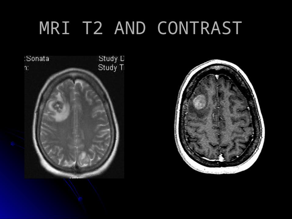
MRI T2 AND CONTRAST
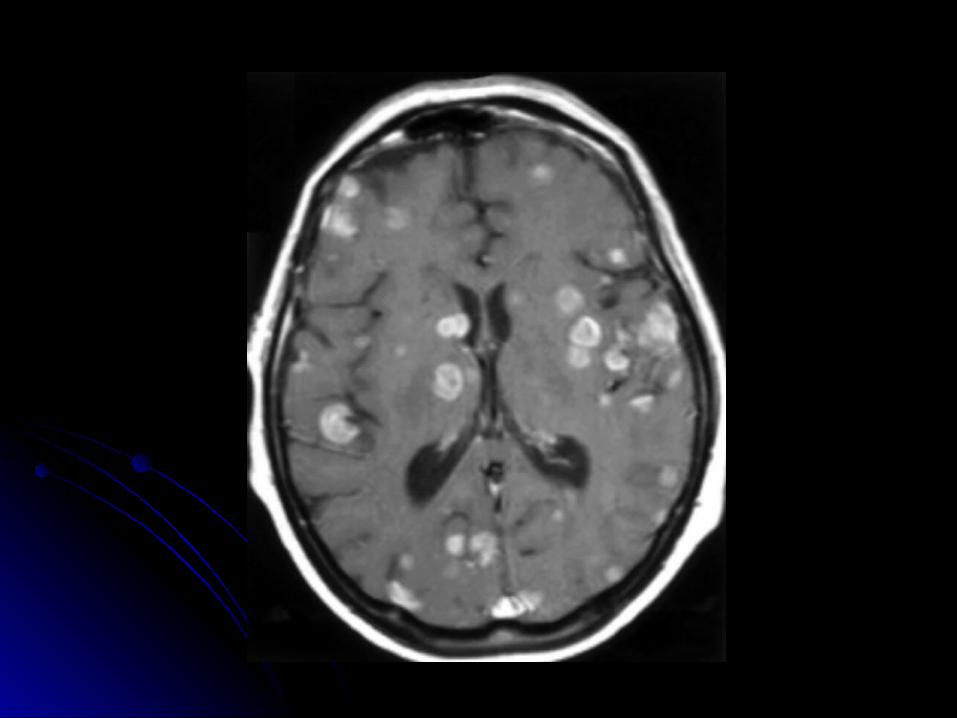
LEPTOMENINGEAL METASTASIS
The leptomeninges may be the site of metastases from
primary CNS malignancies (34%) extra-cranial hematological (11%) solid (46%) malignancies.
Solid tumors include adenocarcinomas from breast, lung(small cell type), stomach, and malignant melanoma.
.
LEPTOMENINGEAL METASTASES
Observed as curvilinear or nodular pial
enhancement along the basal cisterns or
sulci in 35% .
Hydrocephalus In 13%.
Cranial nerve deposits in 11%.
DURAL METASTASES
Commonly caused by systemic breast carcinoma, pulmonary adenocarcinoma, squamous cell carcinoma, and renal cell carcinomas .
More prone to recurrent disease(>41% ).
Overall survival is similar to that of patients with parenchymal disease.
Chronic subdural hematomas in cancer patients
may be masking dural metastases.
SPECT
Provides physiological imaging with a variety of radiopharmaceuticals.
99mTc-HMPAO , 123I-IMP, and thallium-201 (201TI).
Metastases often show decreased uptake with the exception of hypervascular metastases, such as malignant melanoma.
99mTc-MIBI demonstrates only a 50% sensitivity for brain metastases.
PET
Measures metabolic activity charged positrons.
Preferred over SPECT because of higher intrinsic resolution.
18F-fluorodeoxyglucose positron-emission tomography (FDG-PET). commonly done.
Methionine -, tyrosine-, and choline-labeled PET scanning are
newer technologies that are based on amino acid membrane transport, protein synthesis, and phospholipid uptake.

PET Absolute quantification of cerebral blood flow and uses
functional imaging to define eloquent cortex.
Evaluation of suspected metastatic lesions.
Whole-body staging in a convenient single examination .
Possible detection of the primary systemic tumor.

Perfusion MRI Provides visual maps of the regional variations in cerebral
microvasculature caused by intrinsic differences between tumoral and nontumoral capillary systems.
Exploits the T2* signal changes occurring during passage of contrast through the cerebrovascular system to attain cerebral blood flow and cerebral blood volume (CBV) information.
Lack of angiogenesis suggests tumor-mimicking lesions i.e., radiation necrosis, cerebral abscess, and tumefactive demyelination.
pMRI evaluation of the peritumoral T2 hyperintensity reliably differentiate the metastatic from high-grade gliomas.
Perfusion MRI Isolated dural-based metastasis, differentiation from meningioma
can be done.
Intratumoral rCBV measures are elevated in meningiomas because of increased vascularity and lack of BBB but are only mildly elevated in a number of metastatic tumors .
Hypervascular metastases from malignant melanoma, renal cell carcinoma, or Merkel carcinoma, however, may present with an elevated rCBV.

Spectroscopic MRI
• Provides semiquantitative evaluation of metabolite levels to characterize brain tumors.
• Commonly measured metabolites include choline (Cho), lactate(Lac), lipids (Lip), N- acetylaspartate (NAA), and creatineand creatine phosphate (Cr).
• Metastases show elevated Cho levels, Lac and Lip resonances,
and decreased NAA and Cr levels, with subsequently increased Cho /Cr and Cho/Cho (normal) and decreased NAA/Cr ratios.
• Peritumoral sMRI can be used to differentiate metastasis from high grade glioma
Functional Imaging
Relationship of tumor margins with
eloquent cortex and may change the operative approach.
Performed with PET,
magnetoencephalography (MEG), and functional MRI (fMRI).
FUNCTIONAL IMAGING
• MEG has the best temporal resolution, because it directly measures neuronal activity.
• fMRI has the advantage of concurrent acquisition of anatomic and functional images.
• PET and MEG require separate acquisition and fusion of the anatomic images.
• fMRI also has better spatial resolution than PET and MEG,but decreased precision. (indirectly measures venous/ venuolar deoxyhemoglobin changes ).
Intraoperative Imaging Neuronavigation systems improve lesion targeting and resection of
tumor tissue .
Frame-based and frame-less stereotactic systems suffer from diminished accuracy as the duration of surgery increases.
Intraoperative ultrasound has unprecedented real time scanning, but suffers from suboptimal definition of small lesions and tumor margins.
Intraoperative MRI has emerged as an important imaging guidance tool because of its superior spatial resolution and sensitivity.
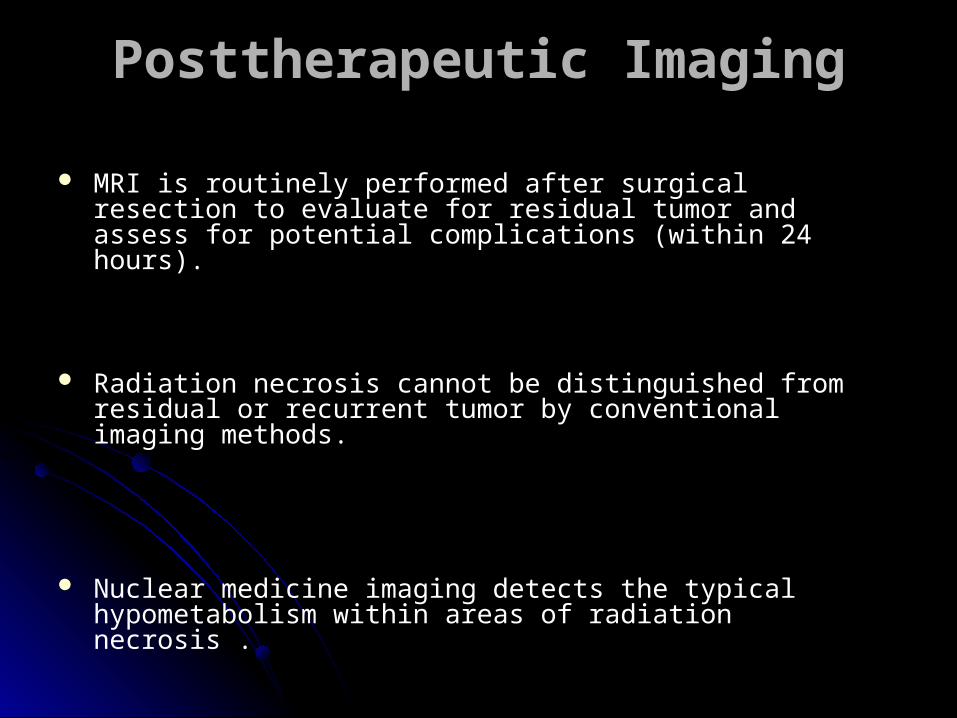
Posttherapeutic Imaging
MRI is routinely performed after surgical resection to evaluate for residual tumor and assess for potential complications (within 24 hours).
Radiation necrosis cannot be distinguished from residual or recurrent tumor by conventional imaging methods.
Nuclear medicine imaging detects the typical hypometabolism within areas of radiation necrosis .

IMAGING STRATEGY CT retains a limited but important role as an initial diagnostic tool to
exclude neurosurgical emergencies .
A contrast-enhanced MRI study with volumetric 3D sequences and diffusion data should be performed.
Spectroscopic and pMRI should be performed to help differentiate metastases from other tumor and tumor-mimicking lesions.
Both sMRI and pMRI can also be used to guide stereotactic biopsy to the most proliferative area of the tumor,
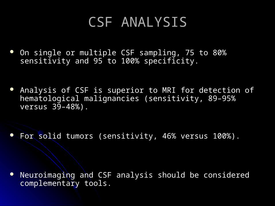
CSF ANALYSIS
On single or multiple CSF sampling, 75 to 80% sensitivity and 95 to 100% specificity.
Analysis of CSF is superior to MRI for detection of hematological malignancies (sensitivity, 89–95% versus 39–48%).
For solid tumors (sensitivity, 46% versus 100%).
Neuroimaging and CSF analysis should be considered complementary tools.
MANAGEMENT OPTIONS
Surgery RadiotheraphyRadiosurgeryPalliative treatment

National Comprehensive Cancer Network recommendations
TREATMENT GUIDELINES
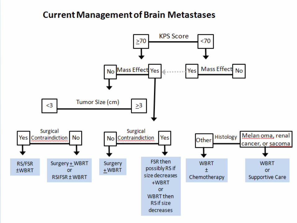
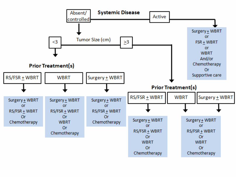
For patients with a resectable new solitary metastasis or a symptomatic metastasis with mass effect, surgery followed by WBRT is recommended,
WBRT for patients with active systemic disease and poor prognosis.
RPA Class 3 ; palliative care.
RS can be used for palliative care also.
RS should be considered when surgery is contraindicated.
Generally, metastases 3 cm or less in diameter, without abundant surrounding edema can be treated with RS. For tumors less than 3 cm in diameter, surgical resection and WBRT should be considered.
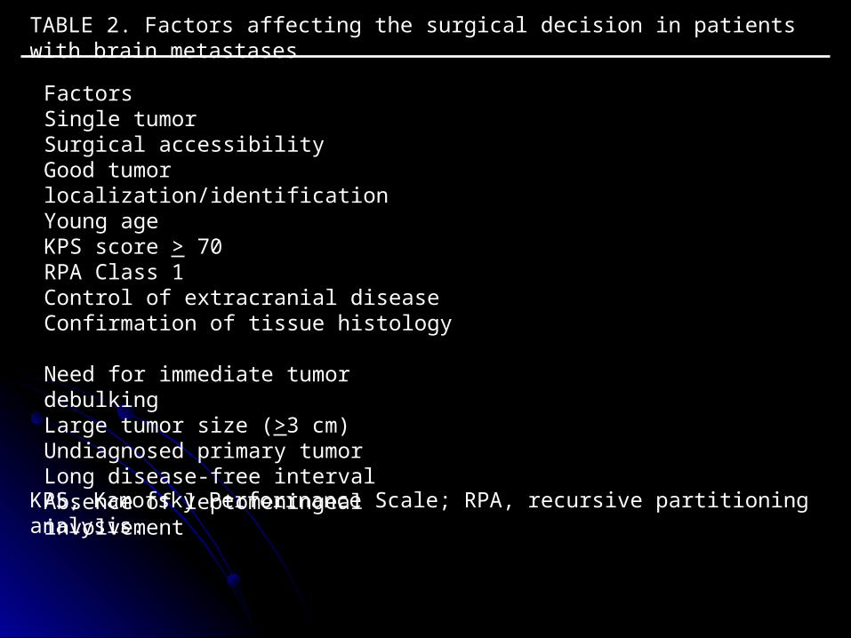
TABLE 2. Factors affecting the surgical decision in patients with brain metastases
Factors Single tumor Surgical accessibility Good tumor localization/identification Young age KPS score > 70 RPA Class 1 Control of extracranial disease Confirmation of tissue histology Need for immediate tumor debulking Large tumor size (>3 cm) Undiagnosed primary tumor Long disease-free interval Absence of leptomeningeal involvement
KPS, Kamofsky Perforrnance Scale; RPA, recursive partitioning analysis.
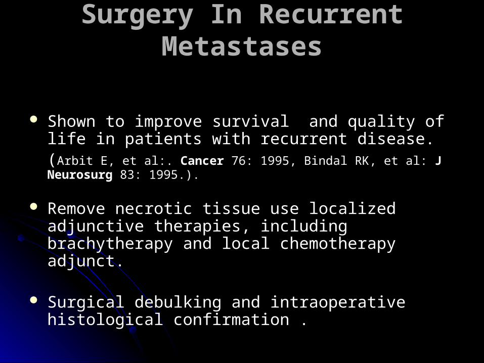
Surgery In Recurrent Metastases
Shown to improve survival and quality of life in patients with recurrent disease.(Arbit E, et al:. Cancer 76: 1995, Bindal RK, et al: J Neurosurg 83: 1995.).
Remove necrotic tissue use localized adjunctive therapies, including brachytherapy and local chemotherapy adjunct.
Surgical debulking and intraoperative histological confirmation .

WHOLE-BRAIN RADIATION THERAPY(WBRT)
Treatment of choice for metastases that impinge on eloquent areas, or are too large, numerous, or disseminated for surgery or RS.
Response rates after WBRT vary,( complete or partial responses 60% ) .
Acute toxicities, after treatment, may include nausea or vomiting, alopecia, hearing loss, acute or subacute skin reactions, and somnolence.
Long term complications ;necrosis, personality and memory changes (both short- and longterm memory), and neurocognitive deficits.
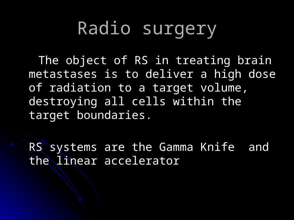
Radio surgery
The object of RS in treating brain metastases is to deliver a high dose of radiation to a target volume, destroying all cells within the target boundaries.
RS systems are the Gamma Knife and the linear accelerator
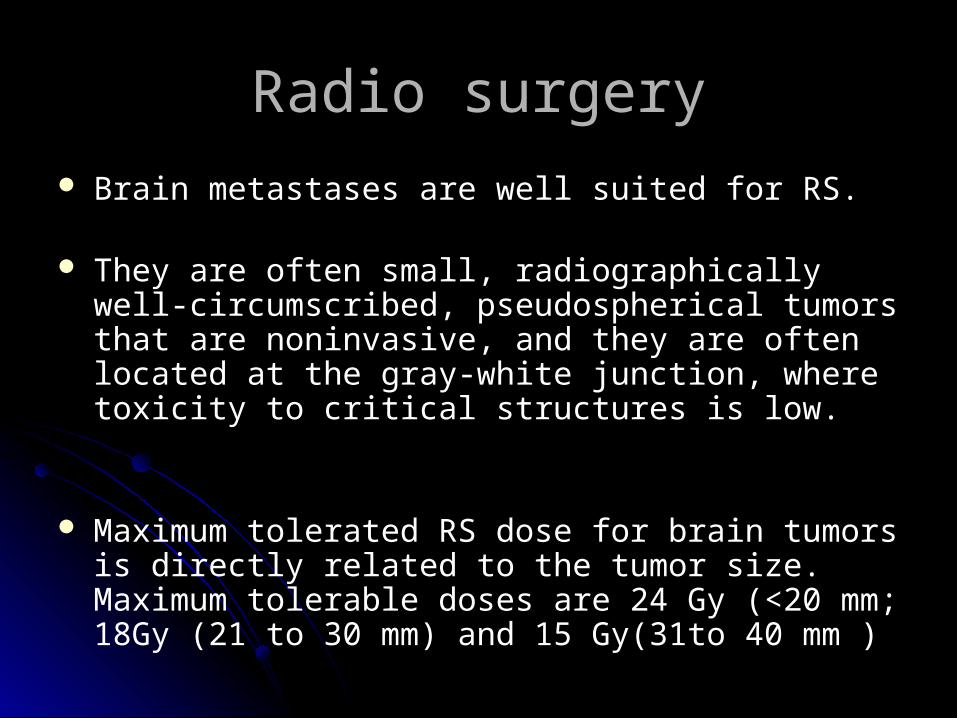
Radio surgery Brain metastases are well suited for RS.
They are often small, radiographically well-circumscribed, pseudospherical tumors that are noninvasive, and they are often located at the gray-white junction, where toxicity to critical structures is low.
Maximum tolerated RS dose for brain tumors is directly related to the tumor size. Maximum tolerable doses are 24 Gy (<20 mm; 18Gy (21 to 30 mm) and 15 Gy(31to 40 mm )
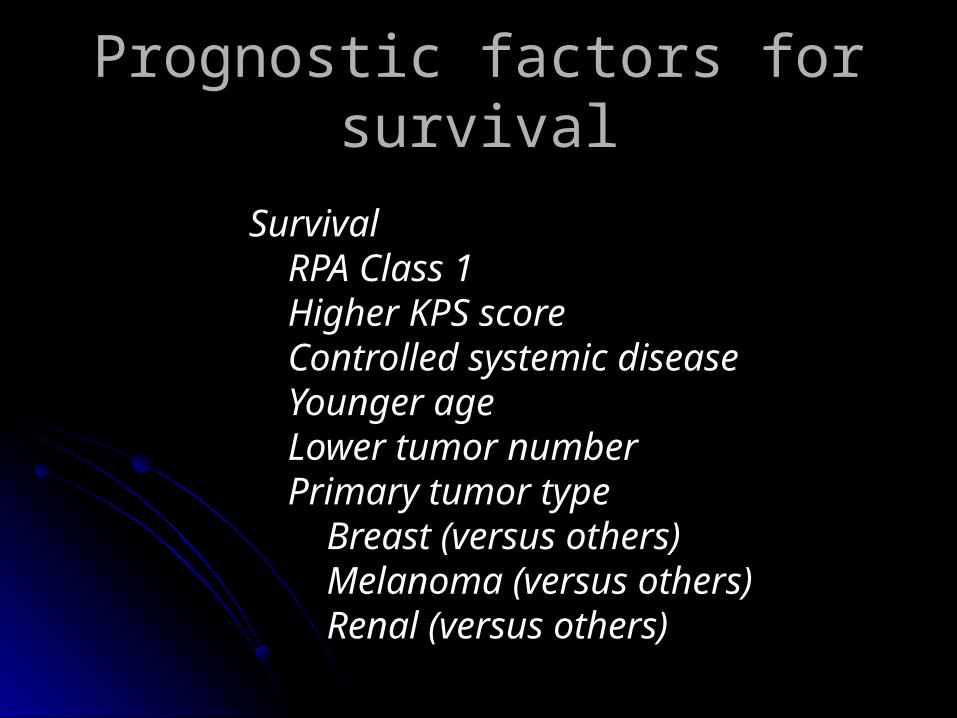
Prognostic factors for survival
Survival RPA Class 1 Higher KPS score Controlled systemic disease Younger age Lower tumor number Primary tumor type Breast (versus others) Melanoma (versus others) Renal (versus others)
Prognostic factors for tumor control
Smaller tumor size Lower tumor number L onger time to bra in metastases Adjunct WBRT Supratentorial location (versus infratentorial) New lesion (versus recurrent) Type of primary tumor Breast Melanoma (versus others) Renal cell (versus others) Homogeneous pattern of enhancement Higher radiosurgical dose
CHEMOTHERAPHY
For patients with metastases from chemosensitive tumors, such as germ cell tumors or lymphoma, chemotherapy should be administered first, with radiotherapy reserved for relapse.
Surgery or Radio surgery The tumor location and size and the presence of edema are
important considerations .
Tumors that are large, in a favorable location for resection, and are associated with mass effect should be surgically resected.
Surgery should also be considered for patients with an unknown primary lesion or at the time of a possible first metastasis from a known primary lesion because of the need for tissue diagnosis.
Small tumors (3 cm) should be treated with RS if they are unresectable.
Small tumors that are resectable and are associated with minimal edema can be treated with either surgery or RS.
Seizures Seizures are a presenting symptom in approximately
20% of patients with brain metastases .
Phenytoin, carbamazepine, and valproic acid are frequently used as first-line agents, along with newer agents in selected circumstances.
American Academy of Neurology now recommends that anticonvulsants should be administered only to those patients at risk for seizure, and their use should be minimized to single therapy at the lowest effective dose.

ROLE OF ANTICONVULSANTS
Meta-analysis of the available data showed that prophylactic anticonvulsants do not seems to significantly reduce the risk of a first seizure.
Anticonvulsant-related side effects were especially common in brain tumor patients (20–40%)
A combination of multiple anticonvulsants is needed to control seizures adequately .

THANK YOU