Embed Size (px)
Citation preview

Bent 0. Kjos1- 3
Richard Umansky4
A. James Barkovich3
Received January 3, 1990; revision requested March 9, 1990; revision received May 4, 1990; accepted May 7, 1990.
1 Magnetic Imaging Affiliates, 5730 Telegraph Ave., Oakland , CA 94609.
2 Present address: Department of Radiology, Swedish Hospital Medical Center, 747 Summit Ave. , Seattle, WA 98104. Address reprint requests to B. 0 . Kjos.
3 Department of Radiology, Neuroradiology Section , University of California School of Medicine, San Francisco, CA 94143.
'Child Development Center, Oakland Children's Hospital, Oakland, CA 94609 .
0195-6108/90/1105-1035 © American Society of Neuroradiology
1035
Brain MR Imaging in Children with Developmental Retardation of Unknown Cause: Results in 76 Cases
Seventy-six children with developmental retardation of unknown cause underwent MR imaging of the brain. Twenty-one (28%) had positive MR findings, including nine with atrophy, six with delayed myelination, four with multiple focal white matter lesions, three with hypoplastic white matter, and three with migration abnormalities. The frequency of abnormality was highest in nonautistic children with associated neurologic physical findings (61%) but was also significant in nonautistic children without neurologic findings (23%). We did not detect abnormalities on MR images of autistic retarded children. Delayed myelination and migration abnormalities were the predominant abnormalities in children with associated neurologic findings, whereas focal white matter lesions were more common in children without neurologic findings. Abnormalities were significantly more common in children with a small head circumference. Although MR did not have any effect on treatment or prognosis, it did aid the clinician in family counseling.
MR will reveal brain abnormalities in about one third of nonautistic children with developmental retardation of unknown cause, and more often in those with neurologic deficits, seizures, or a small head size.
AJNR 11: 1035-1040, September/October 1990
The cause of developmental retardation in children often remains unclear even after extensive medical workup, including history, physical and developmental examination, and laboratory and radiologic studies [1). Even with refinements in karyotyping , more sophisticated metabolic testing , and brain CT scanning, too often both physician and family are left with no explanation for the cause of this serious lifelong problem. The goal of this study was to determine the efficacy of the use of MR in evaluating children with developmental retardation of unknown origin.
Subjects and Methods
Brain MR examinations were performed between June 1986 and August 1988 in 76 children with developmental retardation in whom diagnostic evaluation at the Child Development Center at Children 's Hospital , Oakland , was unsuccessful in revealing the cause. Only patients whose developmental function was less than 80% of chronologie age were included. Besides brain MR, evaluations consisted of (1) medical , developmental , family , and pregnancy history; (2) pediatric physical examination and developmental assessment; and (3) laboratory investigations. Laboratory tests varied but usually included CBC, urinalysis (with reducing substances), SMAC 20, T4 and TSH , blood ammonia, blood and urine amino acid chromatography, urine organic acids, and karyotyping. Other tests were performed as indicated. Brain CT scanning was not done as part of this workup.
Excluded from this study were children with progressive or degenerative disorders, congential CNS infections, meningitis and encephalitis, known perinatal asphyxia, and recognized syndromes, including chromosomal disorders, known to cause developmental retardation. Retarded children with autism and so-called chronic pervasive developmental disorder
1036 KJOS ET AL. AJNR:11 , September/October 1990
were included only if their overall developmental function was less than 80% of their chronologie age. Chi ldren with findings of cerebral palsy (e.g., tone and movement abnormalities) functioning overall at less than 80% of their chronologie age were included if their most prominent feature was a global mental retardation . Thus, patients whose motor disabi lity was clinically more prominent than their cognitive deficit were excluded.
Of the 76 chi ldren, 60 were boys and 16 were girls. Their ages ranged from 4 months to 22 years (only two patients were more than 12 years old) (Fig . 1 ).
20
18 I 16 ' 14
12
No. ol Patients 10
8
6 I 4 I
2
0
< 1 10 11 12 > 12
Age
Fig. 1.-Age distribution of developmentally retarded children in this series.
For purposes of analysis, patients were divided into three main clinical categories: (1) retarded-neurologic-retardation associated with neurologic features such as seizures, cranial nerve abnormalities, hypotonia, and spasticity (18 patients , or 24%); (2) retarded-autisticretardation associated with abnormalities characteristic of autism or the allied state of chronic pervasive developmental disorder (15 patients, or 20%); and (3) retarded-general-retarded patients with neither neurologic nor autistic symptomatology (43 patients, or 57%).
Severity of retardation was expressed as a percentage of chronologie age (functional developmental age/chronologie age). These estimates were derived from the pediatric developmental examination andfor psychological testing. Patients were grouped into four categories by level of severity: those functioning at 0-20% of chronologie age, 21-40%, 41-60%, and 61-80%.
Head circumference was measured and expressed as number of standard deviations (SO) (to the nearest one half SO) from the mean for age and sex.
Brain MR imaging was performed on a 1.5-T imager (GE Signa, Milwaukee, WI). Spin-echo (SE) T2-weighted images, 2500-2800/ 35, 70-80/1 (TRfTEfexcitation), were obtained in each patient. Axial T2-weighted images were obtained in all cases except in one instance in which coronal T2-weighted images were obtained. In 63 cases sagittal (400-600/20-25/2) T1-weighted SE images were also acquired. Axial (600/20-25/2) T1-weighted SE images were also obtained in 40 patients, including children less than 1 year old , to evaluate the extent of myelination. In most cases 5-mm-thick slices were used; however, 3- and 4-mm-thick scans were obtained in some
TABLE 1: Abnormalities Seen on MR in the Different Clinical Subgroups
No. of Percent Abnormalities on MR No. of Clinical Subgroup
Patients Abnormal Abnormal Morpho- Delayed Foci of MRs MRs Atrophy genetic Myelination Long T2
All patients 76 21 28 9 7 6 4 Retarded-neurologic 18 11 61 4 5 5 0 Retarded-general 43 10 23 5 2 1 4 Retarded-autistic 15 0 0 0 0 0 0
Note.- Because some of the patients had multiple abnormalities on MR, the total number of abnormalities seen exceeds the number of abnormal MR examinations.
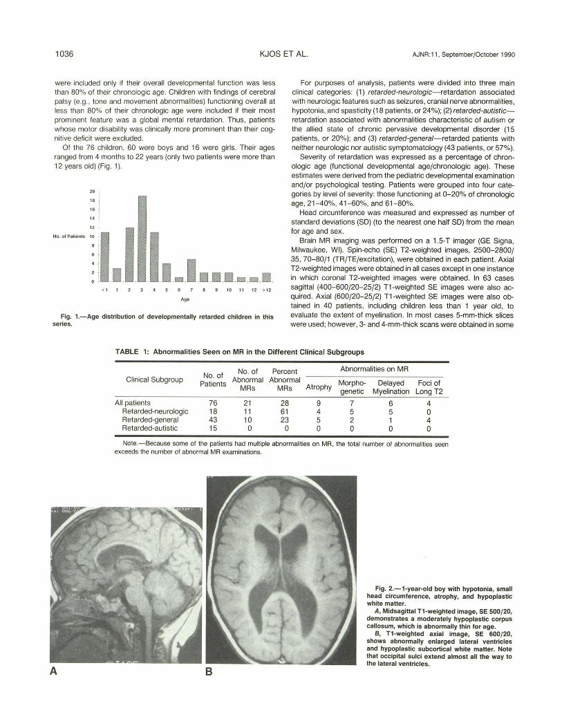
Fig. 2.-1-year-old boy with hypotonia, small head circumference, atrophy, and hypoplastic white matter.
A, Midsagittal T1-weighted image, SE 500/20, demonstrates a moderately hypoplastic corpus callosum, which is abnormally thin for age.
B, T1-weighted axial image, SE 600/20, shows abnormally enlarged lateral ventricles and hypoplastic subcortical white matter. Note that occipital sulci extend almost all the way to the lateral ventricles.

AJNR :11 , September/October 1990 BRAIN MR IN DEVELOPMENTAL RETARDATION 1037
of the smaller infants. IV contrast material was not used. MR studies were reviewed by the authors both separately and as a group. Degree of myelination was determined by using standards from the literature [2-4] . In evaluating for delayed myelination , chronologie age was corrected for prematurity.
Results
Twenty-one (28%) of the 76 developmentally retarded children had significant abnormalities on brain MR imaging. The frequency and type of abnormalities on MR differed significantly in the various clinical subgroups: 11 (61 %) of the 18 children in the retarded-neurologic subgroup had abnormal MR studies, 10 (23%) of the 43 children in the retardedgeneral subgroup had abnormalities on MR, while none of the 15 patients in the retarded-autistic subgroup had an abnormal brain MR examination (p < .01 ).
The frequency of abnormalities on MR was also significantly higher in patients with a small head circumference for age. Fifty-two percent of the 23 children whose head circumference was less than 1 SD below the mean for age had positive MR studies versus only 17% of the 53 remaining children (p < .01 ). The brain MR was normal in the three patients with a head circumference greater than 1 SD above normal for age. No significant correlation was found between the severity of retardation and the frequency of MR abnormalities.
The abnormalities seen with MR fell into four main categories (Table 1 ).
1. Atrophy, characterized by enlarged ventricles and for sulci, was the most common finding on MR and was seen in nine patients (12%) (Fig. 2). It was associated with a small head circumference (Table 2). In six of nine patients the head size was below normal for age by more than 1 SD (p < .01 ). Two of the patients had white matter hypoplasia while none had hydrocephalus (Table 3). Atrophy was seen in both the retarded-general subgroup (five patients) and in the retardedneurologic subgroup (four patients).
2. Congenital morphogenetic abnormalities were seen in seven patients (9%). Three patients had pachygyria (Fig. 3) , two patients had dysgenesis of the corpus callosum, and one
TABLE 2: Atrophy vs Head Circumference
No. of Patients Head Circumference (HC)*
Atrophy No Atrophy Total
HC < -2 SO 2 7 9 -2 SO :::; HC < - 1 SD 4 10 14 - 1 SO :::; HC :::; + 1 SD 3 41 44 +1 SO < HC :::; + 2 SO 0 5 5 HC > +2 SO 0 3 3
• Expressed as no. of standard deviations (SO) from mean for age.
patient had multiple periventricular nodules and lesions in the right occipital and right frontal subcortical white matter suggesting tuberous sclerosis but clinically lacked associated skin lesions or seizures (Fig. 4). Finally, one patient had unusual hypoplastic anterior limbs of the internal capsule. Congenital morphogenetic lesions were associated with small head circumference(< -1 SD) (five patients , p < .01), and were more common in the retarded-neurologic subgroup (five patients, p < .01 ). Patients with congenital lesions were younger than other patients in our series (p < .01 ), probably because the seizures and neurologic problems in these children caused the parents to seek medical attention sooner. All of these children were less than 4 years old ; four were less than 2 years old . Of these seven patients with congenital morphogenetic lesions, two had other abnormalit ies on cranial MR (Table 3).
3. A delayed pattern of myelination for age was seen in six patients, none of whom were premature, and was confined to patients less than 2 years old (Fig. 5). This was an isolated finding in three patients and in the other three pat ients was associated with other MR abnormalities , including atrophy (two cases) , dysgenetic corpus callosum (two cases), and white matter hypoplasia (two cases) (Table 3). Patients with delayed myelinat ion more often had a small head circumference (four patients, p < .05), and were more often in the retarded-neurologic subgroup (five patients, p < .01 ).
4. Multiple small high-intensity foci within the brain on T2-weighted images were seen in four patients. These areas of
TABLE 3: MR Findings of Patients' Multiple Abnormalities on MR
Patient No. Age
Sex MR Findings (months)
4 M Delayed myelination: myel ination similar to newborn brain. Atrophy: cerebellar greater than cerebral. Agenesis of corpus callosum.
2 21 F Delayed myelination: myelination simi lar to 15-month-old brain . Dysgenic corpus callosum with small spenium and no rostrum.
3 9 M Delayed myelination: myelination similar to 4-month-old brain. Atrophy. Cerebral white matter hypoplasia.
4 27 M Atrophy. Foci with long T2 in the occipital and pari-etal white matter bilaterally.
5 11 M Delayed myelination: myelination similar to 8-month-old brain. Cerebral white matter hypopla-sia.
6 12 M Atrophy. Cerebral white matter hypoplasia.
1038
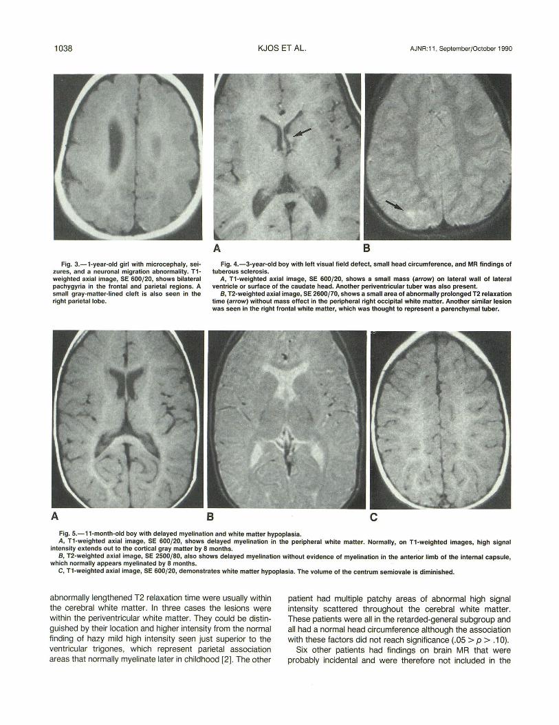
Fig. 3.-1-year·old girl with microcephaly, seizures, and a neuronal migration abnormality. T1-weighted axial image, SE 600/20, shows bilateral pachygyria in the frontal and parietal regions. A small gray-matter-lined cleft is also seen in the right parietal lobe.
KJOS ET AL. AJNR:11 , September/October 1990
A 8 Fig. 4.-3-year-old boy with left visual field defect, small head circumference, and MR findings of
tuberous sclerosis. A, T1-weighted axial image, SE 600/20, shows a small mass (arrow) on lateral wall of lateral
ventricle or surface of the caudate head. Another periventricular tuber was also present. 8, T2-weighted axial image, SE 2600/70, shows a small area of abnormally prolonged T2 relaxation
time (arrow) without mass effect in the peripheral right occipital white matter. Another similar lesion was seen in the right frontal white matter, which was thought to represent a parenchymal tuber.
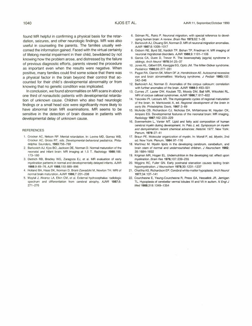
8 c Fig. 5.-11-month-old boy with delayed myelination and white matter hypoplasia. A, T1-weighted axial image, SE 600/20, shows delayed myelination in the peripheral white matter. Normally, on T1-weighted images, high signal
intensity extends out to the cortical gray matter by 8 months. 8 , T2-weighted axial image, SE 2500/80, also shows delayed myelination without evidence of myelination in the anterior limb of the internal capsule,
which normally appears myelinated by 8 months. C, T1-weighted axial image, SE 600/20, demonstrates white matter hypoplasia. The volume of the centrum semiovale is diminished.
abnormally lengthened T2 relaxation time were usually within the cerebral white matter. In three cases the lesions were within the periventricular white matter. They could be distinguished by their location and higher intensity from the normal finding of hazy mild high intensity seen just superior to the ventricular trigones, which represent parietal association areas that normally myelinate later in childhood [2) . The other
patient had multiple patchy areas of abnormal high signal intensity scattered throughout the cerebral white matter. These patients were all in the retarded-general subgroup and all had a normal head circumference although the association with these factors did not reach significance (.05 > p > .1 0).
Six other patients had findings on brain MR that were probably incidental and were therefore not included in the

AJNR:11 , September/October 1990 BRAIN MR IN DEVELOPMENTAL RETARDATION 1039
abnormal MR category. Four patients had one or a few questionable foci of prolonged T2 relaxation time in the cerebral white matter. Another had an enlarged empty sella. The last patient had borderline enlargement of the fourth ventricle.
Discussion
The results of this study suggest that, although the specific cause of developmental retardation often remains unknown, MR provides useful diagnostic information in a relatively high percentage of retarded children resistant to diagnosis by nonimaging methods. The sensitivity of MR in this population (austistics excluded), with a 34% rate of positivity, far exceeds that of all other laboratory tests. Of interest were clinical correlations with MR findings. When neurologic factors were associated with the retardation , not only was the likelihood of an MR abnormality increased, but the type of lesion appeared different. Such patients were more apt to have congenital malformations of brain structure and delayed myelination. Moreover, MR abnormalities were seen in almost a quarter of the retarded children who lacked distinguishing neurologic features, a large group that is especially baffling to clinicians seeking to establish cause. Though sometimes confined to atrophy, focal white matter lesions were also seen in this group.
Overall , the MR findings did not establish specific causations for the developmental retardation, but in many cases the MR findings focused attention on a specific period when the development was disrupted. Four general categories of MR findings were seen.
The most frequent MR finding was atrophy, enlargement of ventricles and sulci in a patient with normal or a small head size. Besides reflecting a brain that was small in relation to the surrounding skull (which is not necessarily abnormal in infants) [5] , it was indicative of reduced brain volume, since head circumference was small in most of these patients and normal in the remaining ones. Increased CSF spaces can also be seen in children with large heads. Without knowing the head size, atrophy and hydrocephalus may be difficult to distinguish from benign macrocephaly of infancy. Atrophy, by itself, is relatively nonspecific and provides few etiologic clues because it is the end result of many pathologic processes. Because small brain size is accompanied by enlargement of both ventricles and sulci, it suggests that the brain initially may have grown normally and then been subjected to a destructive process. Conversely, in patients with a small brain but no atrophy-for example, without ventricular and sulcal enlargement-the process may be primarily one of restriction of brain growth from an early period with little loss of tissue.
Congenital morphogenetic abnormalities represent disorders in development occurring early in gestation. In our series, three of the seven patients in this category had neuronal migration abnormalities. Neuroblasts migrate radially from the subependymal germinal matrix to the cerebral cortex in two major waves over a 2-month period extending from 8 to 16 weeks gestation. Additional neuronal migration continues until about 24-26 weeks [6-8]. Any insult to the brain during this period can disrupt this process; the end result is a thickened ,
disorganized cortex in the involved regions. Clinically, these disorders are associated with seizures as well as retardation . Genetic abnormalities also may be associated with disorders of neuronal migration [9-11]. Causative factors were unknown in our patients, but all were in the retarded-neurologic subgroup, all had a small head circumference for age, and all had seizures. Agenesis of the corpus callosum, seen in one patient, also represents a defect occurring early in gestation (8-15 weeks) [12, 13]. Because this is a critical period of brain development (the cerebrum and cerebellum are forming), abnormalities of the corpus callosum are often associated with a multitude of other malformations. Our patient with agenesis of the corpus callosum also had delayed myelination and cerebellar atrophy.
In humans and in experimental animals, delayed myelination has been associated with inborn errors of metabolism (amino and organic acidopathies), congenital rubella, and severe malnutrition [14-20] . In some of these processes the histologic and structural findings suggest a primary dysmyelination as well as a delay in myelination. Five of the six children with delayed myelination in this series exhibited abnormalities of muscle tone, predominantly spasticity. In two of these six cases of delayed myelination and in an additional child with atrophy, there was white matter hypoplasia on MR. This combination of pathologic findings has been reported as a severe, nonprogressive disorder characterized by intellectual impairment and spastic quadriparesis [21]. The striking neuropathologic finding is hypoplasia of the cerebral white matter, particularly centrum semiovale, with relatively normal cortex and deep nuclei . No gliosis or inflammatory changes are seen in the white matter, corresponding to the lack of a known intrauterine event and to a typical history of a normal pregnancy and delivery.
The multiple focal white matter lesions seen in four patients most likely represent areas of necrosis or demyelination from a previous, nonspecific insult. These children all had normal head sizes and did not have delayed myelination, morphogenetic congenital abnormalities, or abnormal neurologic findings. They appear to be a distinct group whose difficulties relate to pathology occurring late in development, after axonal and dendritic ramifications have occurred , perhaps not before the perinatal period. Although these relatively minor lesions may not cause significant developmental delay by themselves, they may be markers for more extensive brain disease not visible on MR.
In this series no qualitative MR abnormalities were found in the retarded-autistic patients. Although gross alterations have been reported in the brains of autistic patients, there is little agreement in the literature about the characteristic lesions in this condition. Recently, it has been reported that the volume of the posterior lobule of the cerebellar vermis is hypoplastic in autistic patients [22] . Lacking appropriate age-matched normal controls , we did not measure the relative volume of the lobules of the vermis. Therefore, we can neither exclude nor substantiate small , but significant, vermian hypoplasia.
The use of MR did not lead to a specific treatment in any patient in this series or alter the patient's developmental status. Nevertheless, the clinicians and the children 's parents
1040 KJOS ET AL. AJNR:11 , September/October 1990
found MR helpful in confirming a physical basis for the retardation , seizures, and other neurologic findings. MR was also useful in counseling the parents. The families usually welcomed the information gained. Faced with the virtual certainty of lifelong mental impairment in their child , bewildered by not knowing how the problem arose, and distressed by the failure of previous diagnostic efforts, parents viewed the procedure as important even when the results were negative. When positive, many families could find some solace that there was a physical factor in the brain beyond their control that accounted for their child's developmental abnormality or from knowing that no genetic condition was implicated.
In conclusion, we found abnormalities on MR scans in about one third of nonautistic patients with developmental retardation of unknown cause. Children who also had neurologic findings or a small head size were significantly more likely to have abnormal brain MR examinations. MR seems to be sensitive in the detection of brain disease in patients with developmental delay of unknown cause.
REFERENCES
1. Crocker AC, Nelson RP. Mental retardation . In: Levine MD, Gomez WB, Crocker AC, Gross RT, eds. Developmental-behavioral pediatrics. Philadelphia: Saunders, 1983:756- 769
2. Barkovich AJ, Kjos BO, Jackson DE, Norman D. Normal maturation of the neonatal and infant brain: MR imaging at 1.5 T. Radiology 1988;166: 173- 180
3. Dietrich RB, Bradley WG, Zaragoza EJ , et al. MR evaluation of early myelination patterns in normal and developmentally delayed infants. AJNR 1988;9 :69- 76, AJR 1988;150:889-896
4. Holland BA, Haas DK, Norman D, Brant-Zawadzki M, Newton TH . MRI of normal brain maturation. AJNR 1986;7:201-208
5. Maytal J, Alvarez LA, Elkin CM, et al. External hydrocephalus: radiologic spectrum and differentiation from cerebral atrophy. AJNR 1987;8: 271-276
6. Sidman RL, Rakic P. Neuronal migration, with special reference to developing human brain: A review. Brain Res 1973;62 :1-35
7. Barkovich AJ , Chuang SH, Norman D. MR of neuronal migration anomalies. AJNR 1987;8 :1009-1017
8. Osborn RE, Byrd SE, Naidich TP, Bohan TP, Friedman H. MR imaging of neuronal migrational disorders. AJNR 1988;9: 1101-1106
9. Garcia CA, Dunn D, Trevor R. The lissencephaly (agyria) syndrome in siblings. Arch Neuro/1978;54 :25-37
10. Jones KL, Gilbert ER , Kaviggia EG, Opitz JM. The Miller-Didker syndrome. Pediatrics 1980;66:277-281
11 . Pagon RA, Clarren SK, Milam DF Jr, Hendrickson AE. Autosomal recessive eye and brain abnormalities: Warburg syndrome. J Pediatr 1983;102: 542-546
12. Barkovich AJ, Norman D. Anomalies of the corpus callosum: correlation with further anomalies of the brain. AJNR 1988;9:493-501
13. Curnes JT, Laster DW, Koubek TD , Moody DM, Ball MR, Witcofski RL. MRI of corpus callosal syndromes. AJNR 1986;7:617-622
14. Yakovlev PI , Lecours AR. The myelogenetic cycles of regional maturation of the brain. In: Mankowski A, ed. Regional development of the brain in early life. Philadelphia: Davis, 1967:3-69
15. McArdle CB, Richardson CJ, Nicholas DA, Mirfakharee M, Hayden CK, Amparo EG. Developmental features of the neonatal brain: MR imaging. Radiology 1987;162:223-229
16. Svennerholm L, Vanier MT. Lipid and fatty acid composition of human cerebral myelin during development. In: Palo J, ed. Symposium on myelin and demyelination: recent chemical advances: Helsinki 1977. New York: Plenum, 1978:27-41
17. Braun PE. Molecular organization of myelin. In: Morell P, ed. Myelin , 2nd ed. New York: Plenum, 1984:97-116
18. Martinez M. Myelin lipids in the developing cerebrum , cerebellum, and brain stem of normal and undernourished children. J Neurochem 1982; 39:1684-1692
19. Krigman MR, Hogan EL. Undernutrition in the developing rat: effect upon myelination. Brain Res 1976;107:239-255
20. Wiggins RC, Fuller GN. Early postnatal starvation causes lasting brain hypomyelination. J Neurochem 1978;30 :1231-1237
21 . Chattha AS, Richardson EP. Cerebral white-matter hypoplasia. Arch Neurol 1977;34: 137-141
22 . Courchesne E, Yeung-Courchesne R, Press GA, Hesselink JR, Jernigan TL. Hypoplasia of cerebellar vermallobules VI and VII in autism. N Eng/ J Med 1988;318:1349-1354