Embed Size (px)
Citation preview
www.comascience.org
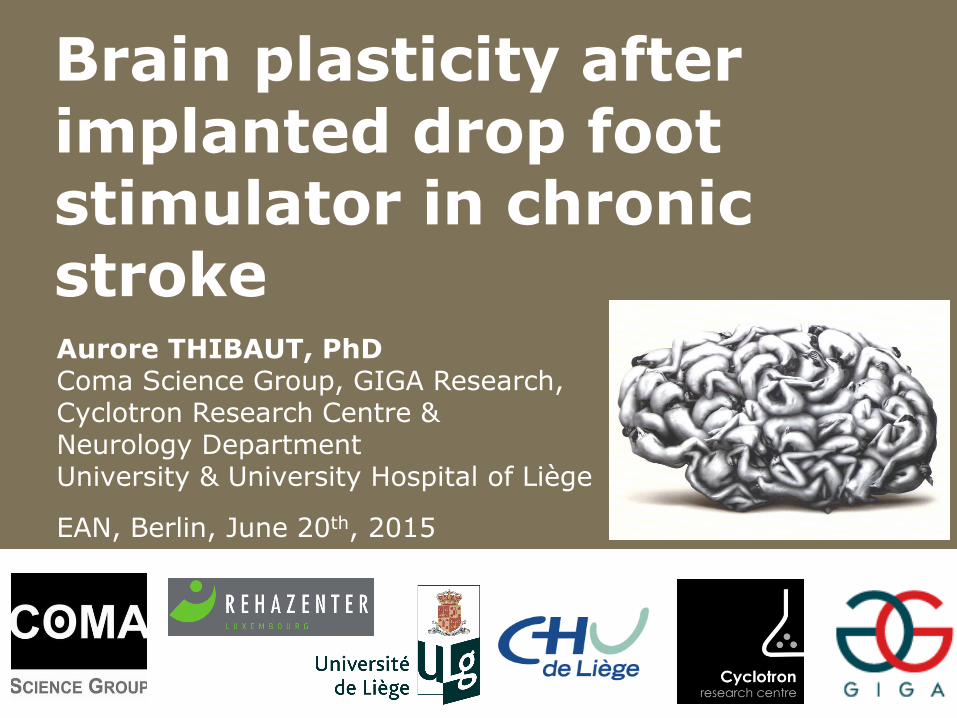
Aurore THIBAUT, PhD Coma Science Group, GIGA Research, Cyclotron Research Centre & Neurology Department University & University Hospital of Liège
EAN, Berlin, June 20th, 2015
Brain plasticity after implanted drop foot stimulator in chronic stroke
www.comascience.org
Workshop Ottobock Stimulation électrique fonctionnelle
implantée chez le patient hémiplégique
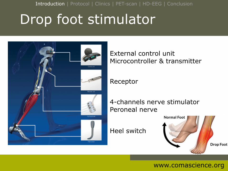
External control unit Microcontroller & transmitter
4-channels nerve stimulator Peroneal nerve
Drop foot stimulator
Receptor
Heel switch
Introduction | Protocol | Clinics | PET-scan | HD-EEG | Conclusion
www.comascience.org
disorders of consciousness | behavioural evaluation | electrophysiology | neuroimaging | methods, ethics & quality of life | perspectives
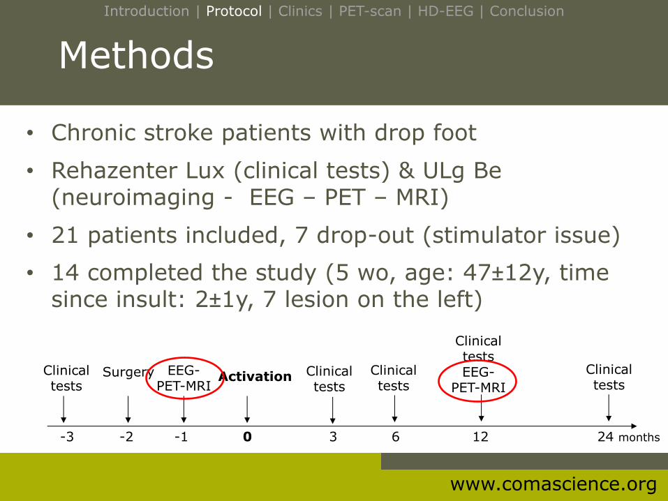
Methods
• Chronic stroke patients with drop foot
• Rehazenter Lux (clinical tests) & ULg Be (neuroimaging - EEG – PET – MRI)
• 21 patients included, 7 drop-out (stimulator issue)
• 14 completed the study (5 wo, age: 47±12y, time since insult: 2±1y, 7 lesion on the left)
Clinical
tests Surgery EEG-
PET-MRI Activation Clinical
tests
Clinical tests
Clinical tests EEG-
PET-MRI
Clinical tests
-3 -2 -1 0 3 6 12 24 months
Introduction | Protocol | Clinics | PET-scan | HD-EEG | Conclusion
www.comascience.org
Clinical improvement
M -1 M +12
Introduction | Protocol | Clinics | PET-scan | HD-EEG | Conclusion
www.comascience.org
PET-scan: Analyses
18FGD-PET-scan at rest
Pre-post : n=14 – right stroke: n=7; left stroke: n=7
7 patients with right lesion were flipped
all patients: lesion on the left hemisphere
Normalization with « flipped template »
Smoothing at 12 mm
Introduction | Protocol | Clinics | PET-scan | HD-EEG | Conclusion
www.comascience.org
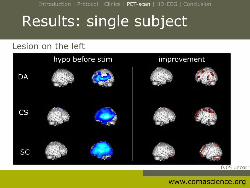
Results: single subject
Lesion on the left
hypo before stim improvement
DA
CS
SC
0.05 uncorr
Introduction | Protocol | Clinics | PET-scan | HD-EEG | Conclusion

www.comascience.org
Results: single subject
Lesion on the right
BD
JR
SV
0.05 uncorr
hypo before stim improvement
Introduction | Protocol | Clinics | PET-scan | HD-EEG | Conclusion
www.comascience.org
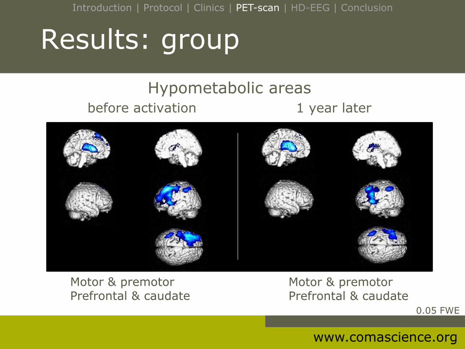
Results: group
Hypometabolic areas
before activation 1 year later
0.05 FWE
Motor & premotor Prefrontal & caudate
Motor & premotor Prefrontal & caudate
Introduction | Protocol | Clinics | PET-scan | HD-EEG | Conclusion
www.comascience.org
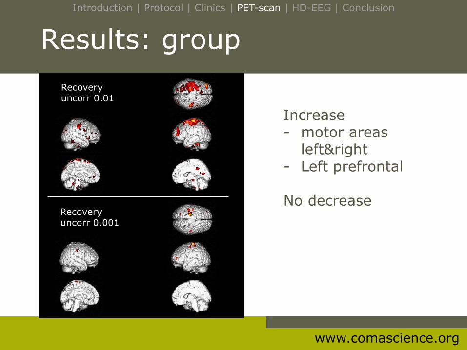
Results: group
Increase - motor areas
left&right - Left prefrontal No decrease
Recovery uncorr 0.01
Recovery uncorr 0.001
Introduction | Protocol | Clinics | PET-scan | HD-EEG | Conclusion
www.comascience.org
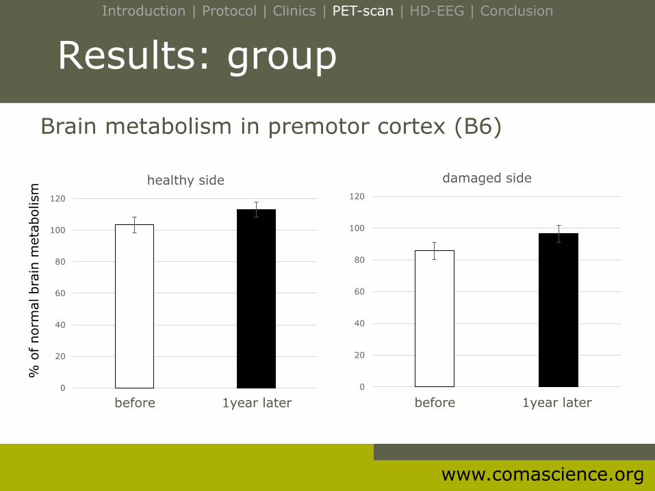
Results: group
0
20
40
60
80
100
120
healthy side
0
20
40
60
80
100
120
damaged side
before 1year later
Brain metabolism in premotor cortex (B6)
before 1year later
Introduction | Protocol | Clinics | PET-scan | HD-EEG | Conclusion
% o
f norm
al bra
in m
eta
bolism
www.comascience.org
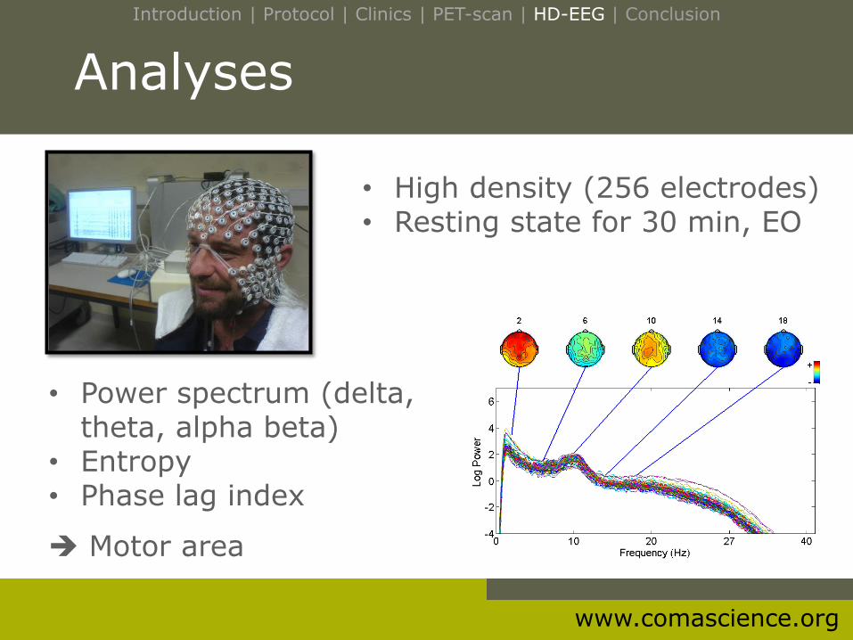
Analyses
• Power spectrum (delta, theta, alpha beta)
• Entropy • Phase lag index
Motor area
• High density (256 electrodes) • Resting state for 30 min, EO
Introduction | Protocol | Clinics | PET-scan | HD-EEG | Conclusion
www.comascience.org
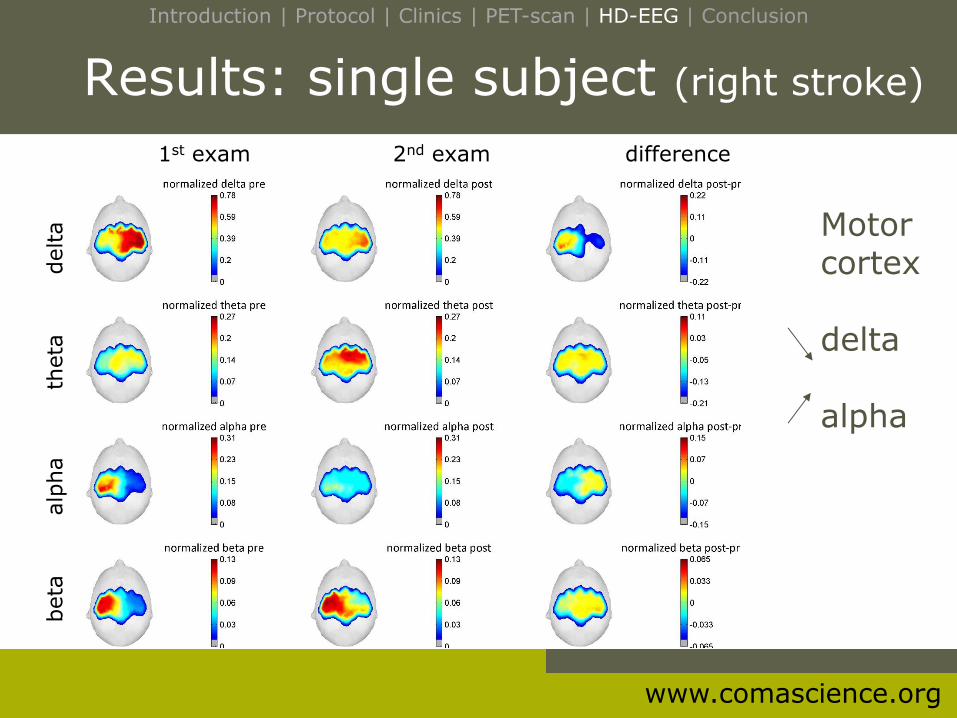
Results: single subject (right stroke)
Motor cortex delta alpha
1st exam 2nd exam difference
beta
alp
ha
t
heta
delta
Introduction | Protocol | Clinics | PET-scan | HD-EEG | Conclusion
www.comascience.org
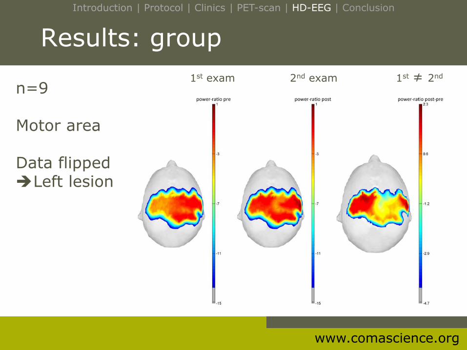
Results: group
n=9 Motor area Data flipped Left lesion
1st exam 2nd exam 1st ≠ 2nd
Introduction | Protocol | Clinics | PET-scan | HD-EEG | Conclusion
www.comascience.org
Conclusion
Clinical improvements correlates
• brain metabolism (PET-scan) in motor areas
(damaged & contralateral hemisphere)
• cortical activity (EEG) in motor area
(damaged hemisphere)
Plasticity of the damaged area in chronic stroke patients
Introduction | Protocol | Clinics | PET-scan | HD-EEG | Conclusion

www.comascience.org
Thank you!