Embed Size (px)
Citation preview

Brain tumor analysis
By:Ninad Mehendale

INTRODUCTION
Normally, the CT scanned and MRI scanned images gives the 2D images which sometimes are not sufficient for detecting the exact type of tumor and also its location, size and shape.

EXISTING SYSTEMS
1. CT SCANNED A 'computerized tomography' (CT) or 'computerized axial
tomography' (CAT) scan uses a computer that takes data from several X-ray images of structures inside a human's or animal's body and converts them into pictures on a monitor.

EXISTING SYSTEMS
2. MRI SCANNED Magnetic resonance imaging (MRI), nuclear magnetic resonance
imaging (NMRI), or magnetic resonance tomography (MRT) is a medical imaging technique used in radiology to investigate the anatomy and physiology of the body in both health and disease. MRI scanners use strong magnetic fields and radio waves to form images of the body.
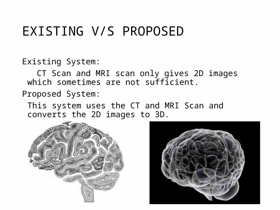
EXISTING V/S PROPOSED
Existing System: CT Scan and MRI scan only gives 2D images which sometimes are not
sufficient.Proposed System:
This system uses the CT and MRI Scan and converts the 2D images to 3D.

OBJECTIVES 2D CT or MRI scanned Images
can be used to form 3D model of brain
This is image processing based project
It will gives size & shape of tumor and also calculate its exact location in 3D space
With previous result and current result growth rate of tumor can be determined

Proposed system
The system will convert any Z-stack images to 3D modelThe programming will be done using MATLAB