Embed Size (px)
DESCRIPTION
simple to knowing tumor type by its location
Citation preview
Page 1Brain Tumor Diagnosis by Location
1/10/2013 4:31:04 PMhttp://rad.usuhs.mil/rad/home/locate/locate.html
Home » rad » home » locate » locate.html Turbo Search: Go!
Portions © 1999 - 2006 by James G. Smirniotopoulos, M.D. This page was last updated: 01/10/2013 16:30:14
Greetings! It's Thursday January 10, 2013
Please Try the Brain Lesion Locator™ !
REGIONAL NEUROANATOMYand DIFFERENTIAL DIAGNOSIS:PART 2 - Infratentorial LesionsPART 2 - Supratentorial Lesions
James G. Smirniotopoulos, M.D.Professor of Radiology, Neurology, and Biomedical InformaticsChairman, Department of Radiology and Radiological SciencesUniformed Services University of the Health Sciences
4301 Jones Bridge RoadBethesda, MD 20814Voice: 301-295-3145FAX: 301-295-3893
DISCLAIMER
The opinions expressed herein are those of the author(s), and are not necessarily representative of the UniformedServices University of the Health Sciences (USUHS), the Department of Defense (DOD); or, the World HealthOrganization (WHO).
Medicine is a constantly changing field, and medical information is subject to frequent correction and revision.Therefore the reader is entirely responsible for verifying the accuracy and relevance of the information containedherein.
Portions Copyright © 1997 - 2006 by James G. Smirniotopoulos, M.D.
Send EMail about this site.
Schematic of Locations
Page 2Brain Tumor Diagnosis by Location
1/10/2013 4:31:04 PMhttp://rad.usuhs.mil/rad/home/locate/locate.html
Schematic of Locations
PATTERN ANALYSIS
Basic Approach
Where is the lesion ?SupratentorialInfratentorial
Where is the lesion ?IntraaxialExtraaxial
How old is the patient ?ChildAdult
PATTERN ANALYSIS
Where is the Lesion?
INTRAAXIALinternal to PIA(brain parenchyma)
EXTRAAXIALexternal to PIA(meninges, nerve sheath)
INTRAVENTRICULARLateralThirdFourth
INTRAAXIAL:
Differential
CORTEXGRAY/WHITE JUNCTIONDEEP WHITE MATTERDEEP GRAY MATTER
INTRAAXIAL:
Differential
Page 3Brain Tumor Diagnosis by Location
1/10/2013 4:31:04 PMhttp://rad.usuhs.mil/rad/home/locate/locate.html
Differential
GliomaMedulloblastomaHemangioblastomaMetastasesInfarct/hematomaAVM/congenitalAbscess/inflammation
EXTRAAXIAL LESIONS
(Location)
SubarachnoidSubduralEpiduralCalvarium (skull base)SubgalealScalp (soft tissues)
EXTRAAXIAL LESIONS
(Differential):
MeningiomaPituitary adenomaCraniopharyngiomaSchwannomaChordomaDermoid/epidermoid, cyst, lipomaHematoma, metastasis, infection
CLASSIC LOCATIONS
Foramen magnumCerebellopontine angleFourth ventricle/CerebellumSella/parasellar/suprasellarBasal ganglia/Third ventricleLateral ventricle/Pineal regionDeep hemispheric/periventricularCortical/subcorticalConvexity
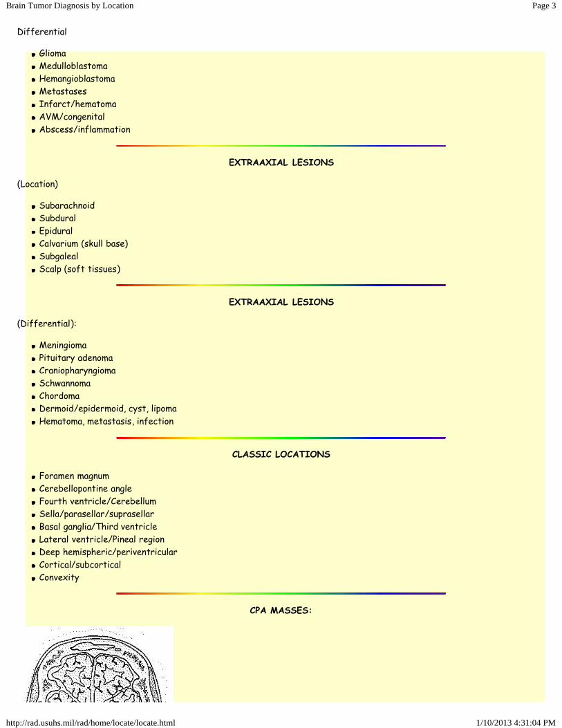
CPA MASSES:
Page 4Brain Tumor Diagnosis by Location
1/10/2013 4:31:04 PMhttp://rad.usuhs.mil/rad/home/locate/locate.html
Differential
S schwannoma (8th >> > 5th)A aneurysm, arachnoid cystM meningioma, metsE epidermoid, ependymoma, CPP
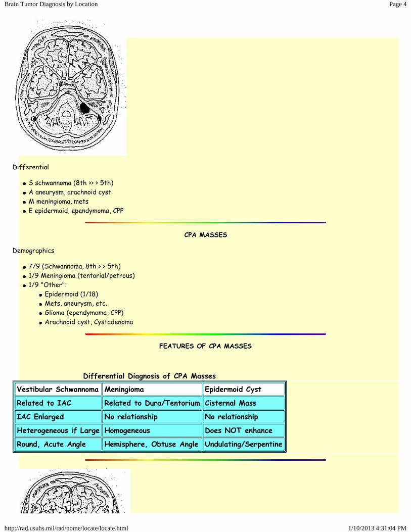
CPA MASSES
Demographics
7/9 (Schwannoma, 8th > > 5th)1/9 Meningioma (tentorial/petrous)1/9 "Other":
Epidermoid (1/18)Mets, aneurysm, etc.Glioma (ependymoma, CPP)Arachnoid cyst, Cystadenoma
FEATURES OF CPA MASSES
Differential Diagnosis of CPA Masses
Vestibular Schwannoma Meningioma Epidermoid Cyst
Related to IAC Related to Dura/Tentorium Cisternal Mass
IAC Enlarged No relationship No relationship
Heterogeneous if Large Homogeneous Does NOT enhance
Round, Acute Angle Hemisphere, Obtuse Angle Undulating/Serpentine
*
Page 5Brain Tumor Diagnosis by Location
1/10/2013 4:31:04 PMhttp://rad.usuhs.mil/rad/home/locate/locate.html
*
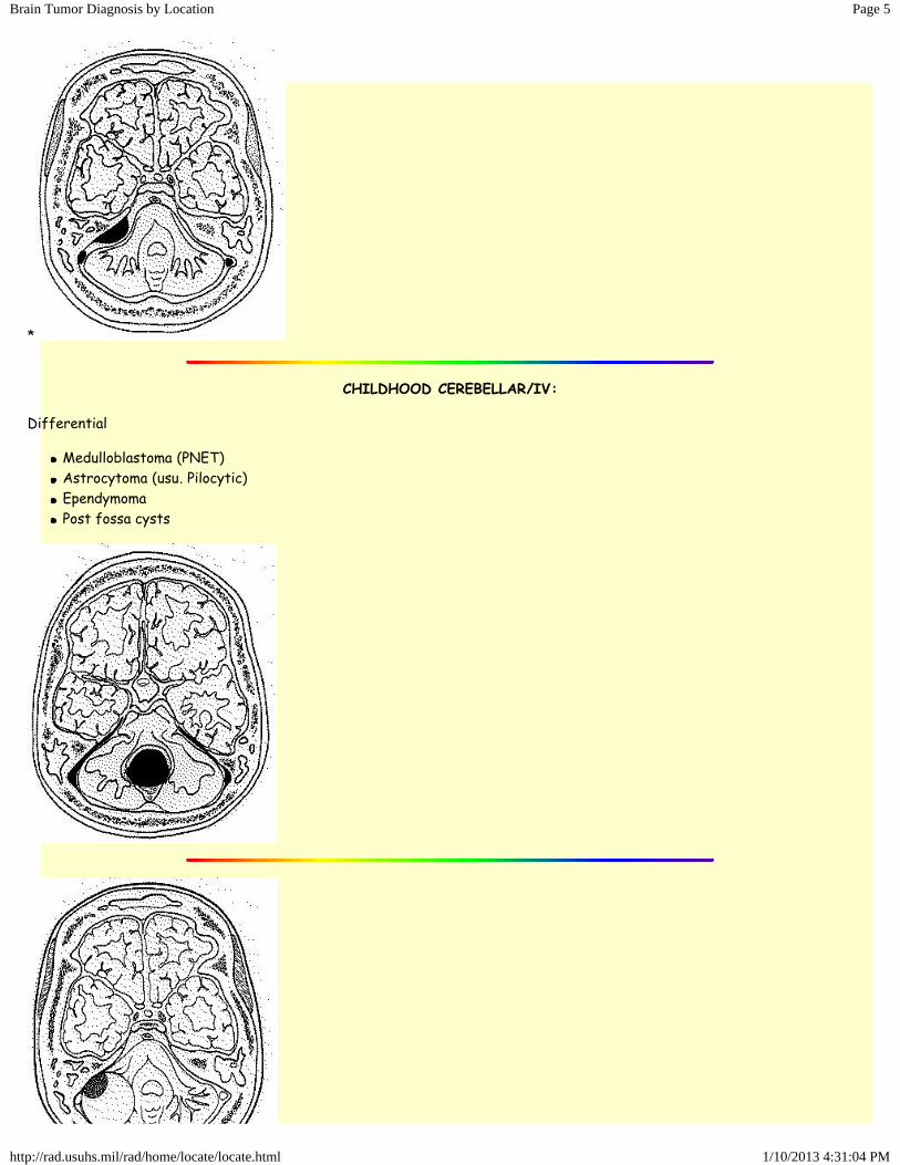
CHILDHOOD CEREBELLAR/IV:
Differential
Medulloblastoma (PNET)Astrocytoma (usu. Pilocytic)EpendymomaPost fossa cysts
Page 6Brain Tumor Diagnosis by Location
1/10/2013 4:31:04 PMhttp://rad.usuhs.mil/rad/home/locate/locate.html
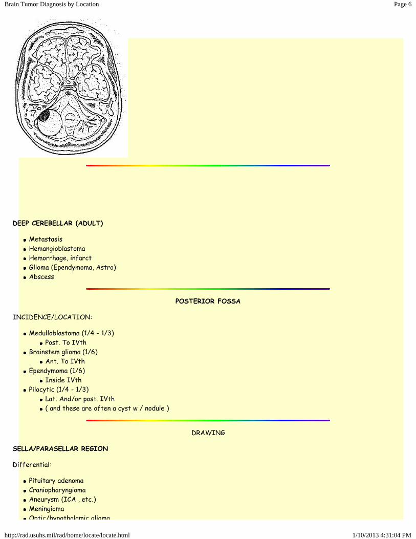
DEEP CEREBELLAR (ADULT)
MetastasisHemangioblastomaHemorrhage, infarctGlioma (Ependymoma, Astro)Abscess
POSTERIOR FOSSA
INCIDENCE/LOCATION:
Medulloblastoma (1/4 - 1/3)Post. To IVth
Brainstem glioma (1/6)Ant. To IVth
Ependymoma (1/6)Inside IVth
Pilocytic (1/4 - 1/3)Lat. And/or post. IVth( and these are often a cyst w / nodule )
DRAWING
SELLA/PARASELLAR REGION
Differential:
Pituitary adenomaCraniopharyngiomaAneurysm (ICA , etc.)MeningiomaOptic/hypothalamic glioma
Page 7Brain Tumor Diagnosis by Location
1/10/2013 4:31:04 PMhttp://rad.usuhs.mil/rad/home/locate/locate.html
Optic/hypothalamic gliomaChordomaGranuloma, e.g., hamartoma, cyst(arachnoid, dermoid/epi)Germ Cell (Germinoma)
PITUITARY ADENOMA
Sexual Dimorphism
paste table
SELLA/PARASELLAR
Differential Features:
CHILD - Craniopharyngioma / Glioma (hypothalamus or optic )ADULT - Pituitary adenomaSELLA NORMAL - NOT pituitaryCa++ - Craniopharyngioma, but...HYPEROSTOSIS - Meningioma ( exp. "blistering" )CLIVUS - Chordoma, mets, NP CaRemember - rule out vascular lesions (aneurysms)
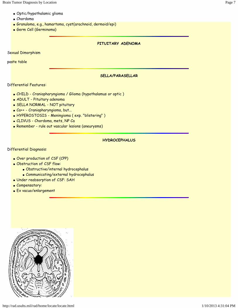
HYDROCEPHALUS
Differential Diagnosis:
Over production of CSF (CPP)Obstruction of CSF flow:
Obstructive/internal hydrocephalusCommunicating/external hydrocephalus
Under reabsorption of CSF: SAHCompensatory:Ex vacuo/enlargement
Page 8Brain Tumor Diagnosis by Location
1/10/2013 4:31:04 PMhttp://rad.usuhs.mil/rad/home/locate/locate.html
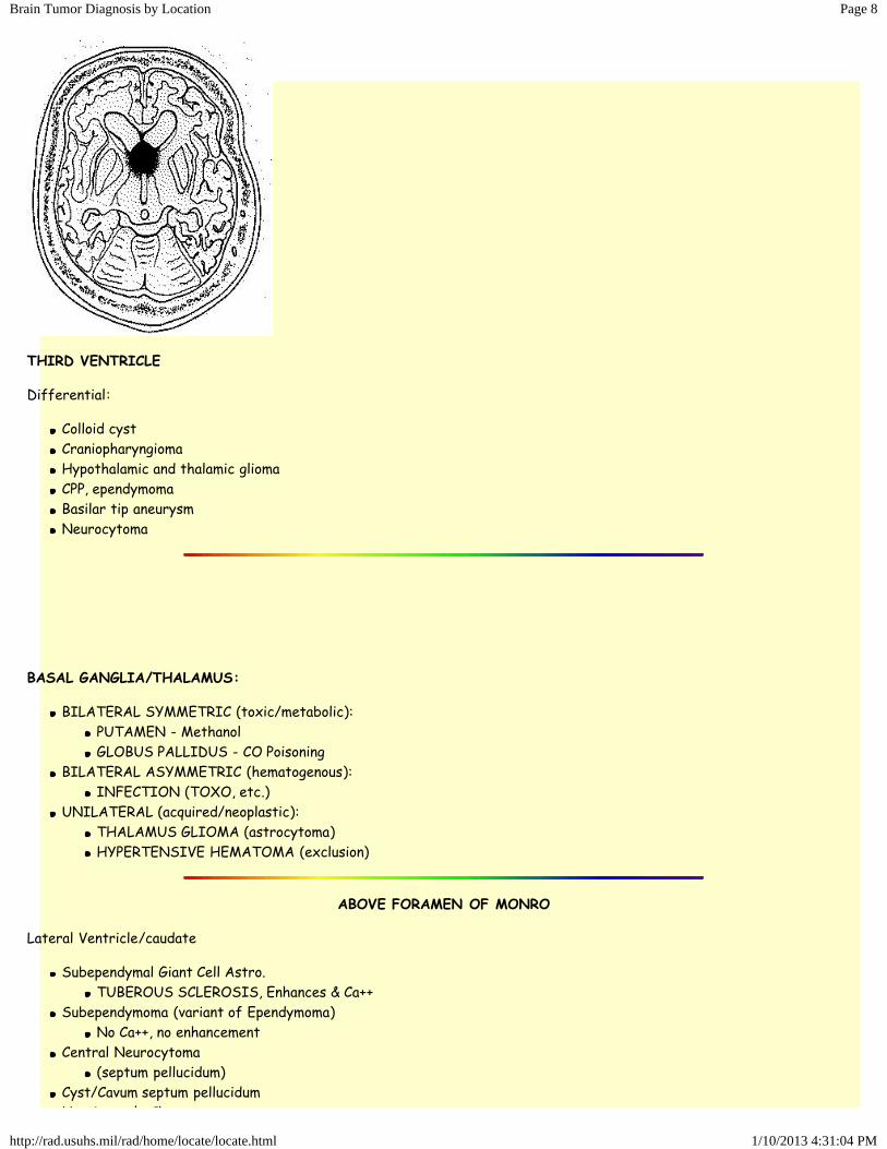
THIRD VENTRICLE
Differential:
Colloid cystCraniopharyngiomaHypothalamic and thalamic gliomaCPP, ependymomaBasilar tip aneurysmNeurocytoma
BASAL GANGLIA/THALAMUS:
BILATERAL SYMMETRIC (toxic/metabolic):PUTAMEN - MethanolGLOBUS PALLIDUS - CO Poisoning
BILATERAL ASYMMETRIC (hematogenous):INFECTION (TOXO, etc.)
UNILATERAL (acquired/neoplastic):THALAMUS GLIOMA (astrocytoma)HYPERTENSIVE HEMATOMA (exclusion)
ABOVE FORAMEN OF MONRO
Lateral Ventricle/caudate
Subependymal Giant Cell Astro.TUBEROUS SCLEROSIS, Enhances & Ca++
Subependymoma (variant of Ependymoma)No Ca++, no enhancement
Central Neurocytoma(septum pellucidum)
Cyst/Cavum septum pellucidumHuntington's Chorea
Page 9Brain Tumor Diagnosis by Location
1/10/2013 4:31:04 PMhttp://rad.usuhs.mil/rad/home/locate/locate.html
Huntington's ChoreaATROPHY
INTRAVENTRICULAR NEOPLASMS:
Ependymoma (and subependymoma)Choroid plexus papillomaSubependymal giant cell astro.MeningiomaColloid cyst (3rd)Dermoid/epidermoidCentral neurocytomaMedulloblastoma (4th)Mets, lymphoma, Germ Cell
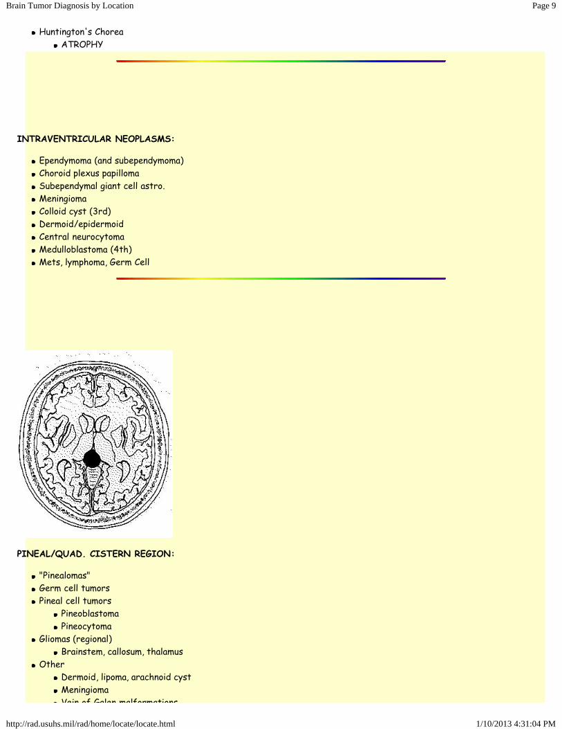
PINEAL/QUAD. CISTERN REGION:
"Pinealomas"Germ cell tumorsPineal cell tumors
PineoblastomaPineocytoma
Gliomas (regional)Brainstem, callosum, thalamus
OtherDermoid, lipoma, arachnoid cystMeningiomaVein of Galen malformations
Page 10Brain Tumor Diagnosis by Location
1/10/2013 4:31:04 PMhttp://rad.usuhs.mil/rad/home/locate/locate.html
Vein of Galen malformations
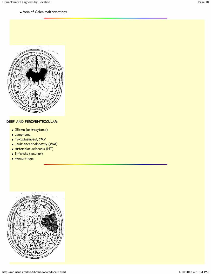
DEEP AND PERIVENTRICULAR:
Glioma (astrocytoma)LymphomaToxoplasmosis, CMVLeukoencephalopathy (WM)Arteriolar sclerosis (HT)Infarcts (lacunar)Hemorrhage
Page 11Brain Tumor Diagnosis by Location
1/10/2013 4:31:04 PMhttp://rad.usuhs.mil/rad/home/locate/locate.html
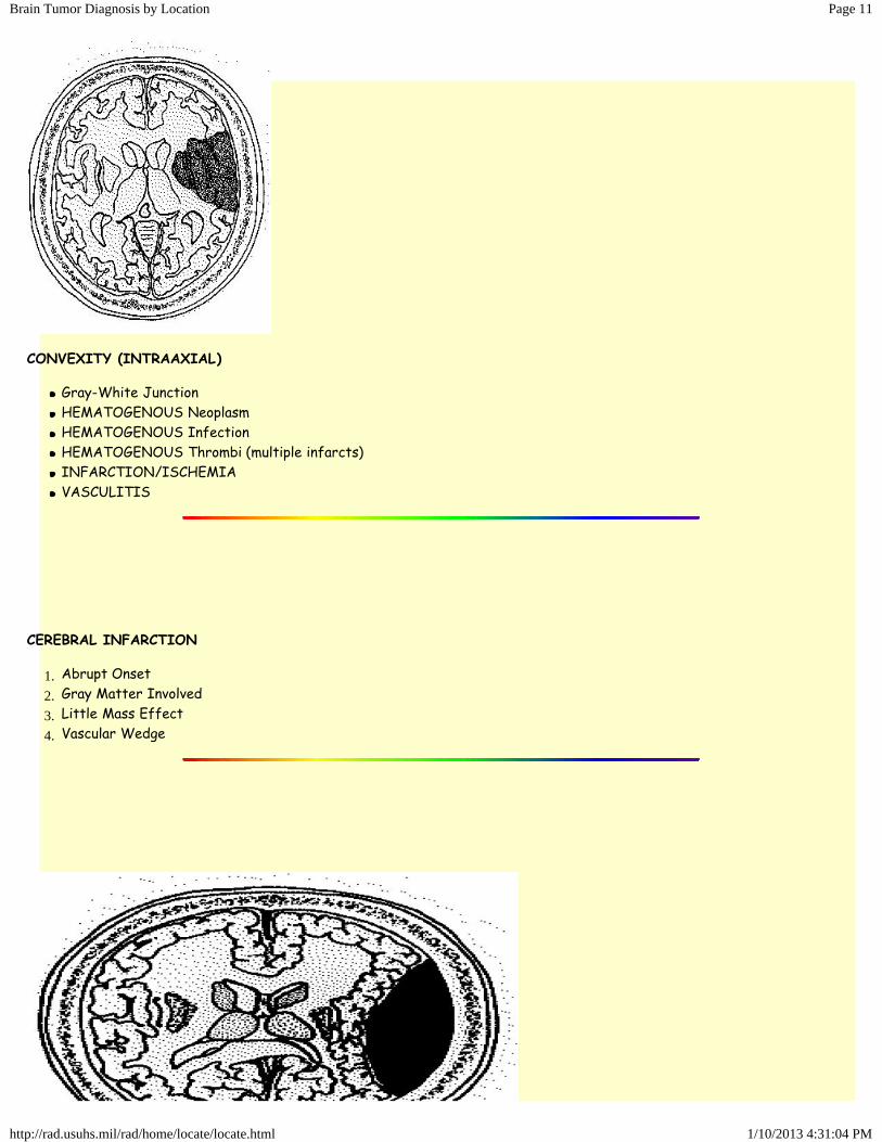
CONVEXITY (INTRAAXIAL)
Gray-White JunctionHEMATOGENOUS NeoplasmHEMATOGENOUS InfectionHEMATOGENOUS Thrombi (multiple infarcts)INFARCTION/ISCHEMIAVASCULITIS
CEREBRAL INFARCTION
1. Abrupt Onset2. Gray Matter Involved3. Little Mass Effect4. Vascular Wedge
Page 12Brain Tumor Diagnosis by Location
1/10/2013 4:31:04 PMhttp://rad.usuhs.mil/rad/home/locate/locate.html
Home » rad » home » locate » locate.html Turbo Search: Go!
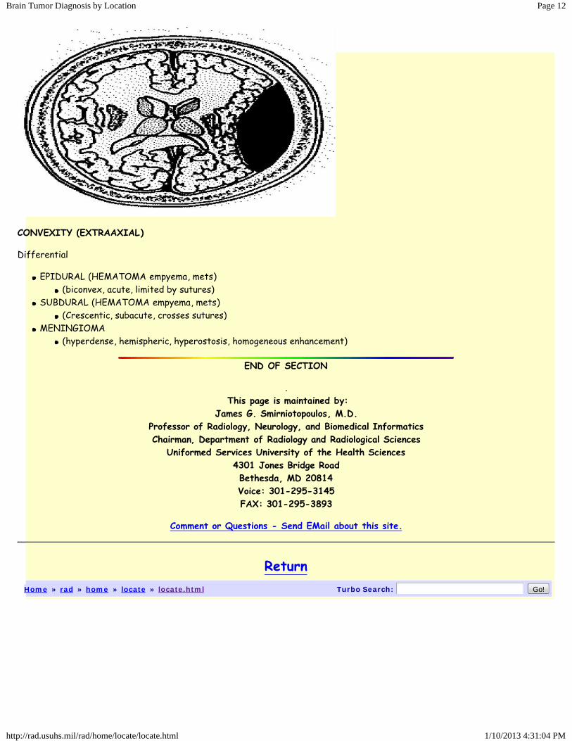
CONVEXITY (EXTRAAXIAL)
Differential
EPIDURAL (HEMATOMA empyema, mets)(biconvex, acute, limited by sutures)
SUBDURAL (HEMATOMA empyema, mets)(Crescentic, subacute, crosses sutures)
MENINGIOMA(hyperdense, hemispheric, hyperostosis, homogeneous enhancement)
END OF SECTION
.This page is maintained by:
James G. Smirniotopoulos, M.D.Professor of Radiology, Neurology, and Biomedical InformaticsChairman, Department of Radiology and Radiological SciencesUniformed Services University of the Health Sciences
4301 Jones Bridge RoadBethesda, MD 20814Voice: 301-295-3145FAX: 301-295-3893
Comment or Questions - Send EMail about this site.
Return