Embed Size (px)
Citation preview
7/27/2019 Brain Tumor Identification Using K-Means Clustering
http://slidepdf.com/reader/full/brain-tumor-identification-using-k-means-clustering 1/4
International Journal of Engineering Trends and Technology- Volume4Issue3- 2013
ISSN: 2231-5381 http://www.internationaljournalssrg.org Page 354
Brain Tumor Identification Using K-Means
ClusteringManali Patil Samata Prabhu Sonal Patil Sunilka Patil
Mrs.Prachi Kshirsagar
I. T. Department,Padmabhushan Vasantdada Patil Pratishthan’s College Of Engineering.
Sion (East), Mumbai-400 022, India.
Abstract
The project is entitled as “BRAIN TUMOR
IDENTIFICATION”. The idea behind choosing this topic
was to simplify the process of tumor identification. The
system will be computerized and hence time consumed
will be less. The system will also keep records of patientswho are affected by tumor and who are not. So the
doctors can schedule the further treatments for the
patients.
KeywordsTumor, Brain, Clustering, MRI image, identifying tumor.
I. INTRODUCTION
A brain Image consists of four regions i.e. gray matter (GM),
white matter (WM), cerebrospinal fluid (CSF) and background. These regions can be considered as four different classes. Therefore, an input image needs to be
divided into these four classes. In order to avoid the chances
of misclassification, the outer elliptical shaped object should
be removed. By removing this object we will get rid of non brain tissues and will be left with only soft tissues. Brain
tumor identification is used to identify tumor from particular
image. Brain tumor identification image application is
typically based on clustering concept of image pixels matrix. Brain tumor identification is used to identify tumor affected
image based on clustering and centroid concept.
The basic concept is that local textures in the images can
reveal the typical regularities of the biological structures.
Thus, the textural features have been extracted using a co-
occurrence matrix approach. The level of recognition, amongthree possible types of image areas: tumor, non-tumor and
back ground.
The main objective of this project is to study the design of acomputer system able to detect the presence of a tumor in the
digital images of the brain, and to accurately define its
borderlines
A. Images clustering
Convert 2-D report into 3-D images because brain is mass
body to calculate centroid so it has 3 dimensions and we need to calculate centroid, as also we can get more accurate results
with 3 dimensions.
B. Algorithm k-means
1. Place K points into the space represented by the objects
that are being clustered.2. These points represent initial group of centroids.
3. Assign each object to the group that has the closest
centroid.
When all objects have been assigned, recalculate the
positions of the K centroids.Repeat Steps 2 and 3 until the centroids no longer move. This
produces a separation of the objects into groups from which
the metric to be minimized can be calculated.

7/27/2019 Brain Tumor Identification Using K-Means Clustering
http://slidepdf.com/reader/full/brain-tumor-identification-using-k-means-clustering 2/4
International Journal of Engineering Trends and Technology- Volume4Issue3- 2013
ISSN: 2231-5381 http://www.internationaljournalssrg.org Page 355
II.PROPOSED SYSTEM
In our project, first the MRI report of the patient is scanned
and made into computerized form. As it becomes in computerized form, detection of the tumor becomes simpler
as clustering is done on that MRI image and manual checking
by doctors is avoided. So the results generated are morespecific.
The major role of this application is to identify the tumor inthe brain image and reconstruct the area which the tumor is
affected and based on the threshold value the system will
identify whether the image is affected by the tumor or not.Following are the functionality which is involved in the
tumor identification module.
Identification Reconstruction
Testimony
A. Identification
K-Means algorithm is used to implement the Identification
of the MRI brain image. Clustering can be considered the
most important unsupervised learning problem; so, as everyother problem of this kind, it deals with finding a structure in
a collection of Unlabeled data. A loose definition of
clustering could be “the process of organizing objects intogroups whose members are similar in some way”. A cluster is therefore a collection of objects which are “similar”
between them and are “dissimilar” to the objects belonging to
other clusters.
We are implementing the k-means algorithm with brain
image. Algorithm will cluster the brain image and
differentiate the cells into the affected cluster region and unaffected cluster region.
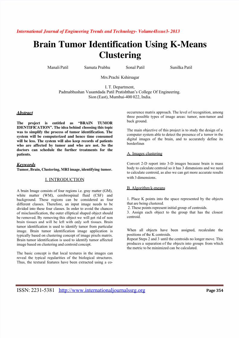
B.Reconstruction
The affected area will be selected and as a cluster and constructed as an image and it is displayed in the label. Based
on the constructed area threshold values will calculated and
the tumor identification process will performed based on the
threshold values. Our system will show the option panemessage dialog contain the image affected or not.
_ _ +
Fig: K-means clustering
C.Testimony
The report is generated based on the affected and theunaffected image. The users have to select the option the
affected or un affected patients. The reports contain the
patient id and the name of the candidate.
III.STEPS OF IMPLEMENTATION
1. Register patient details with image of his/her brain.
2. Select operation.
3. Select patient ID for identification of image.4. Select clustering for image clustering.
5. Reconstruction of image with proper result.
A. Block Diagram
DatabaseUser Login
Clustering of
3D MRI image
using threshold
value
Report
generation
No of
clusters
centroid
Distance objects to centroid
endNo obj
move
Start
Grouping based on min
distance

7/27/2019 Brain Tumor Identification Using K-Means Clustering
http://slidepdf.com/reader/full/brain-tumor-identification-using-k-means-clustering 3/4
7/27/2019 Brain Tumor Identification Using K-Means Clustering
http://slidepdf.com/reader/full/brain-tumor-identification-using-k-means-clustering 4/4
International Journal of Engineering Trends and Technology- Volume4Issue3- 2013
ISSN: 2231-5381 http://www.internationaljournalssrg.org Page 357
V.CONCLUSION
The “BRAIN TUMOUR IDENTIFICATION” has beendeveloped to satisfy all proposed requirements.
The system is highly scalable and user friendly. Almost allthe system objectives have been met. The system has beentested under all criteria. The system minimizes the problem
arising in the existing manual system and it eliminates the
human errors to zero level.
The design of the database is flexible ensuring that the
system can be implemented. It is implemented and gone
through all validation.
VI.REFERENCES
[1]. Gerig, G., Martin, J., Kikinis, R., Kubler, O., Shenton,
M., Jolesz, F.: Automating Identification of dual-echo MR head data. In: IPMI.
Volume 511. (1991) 175{185
[2]. Kjaer, L., Ring, P., Thomson, C., Henriksen, O.: Texture
analysis in quantitative MR imaging: Tissue characterization
of normal brainand intracranial tumors at 1.5 T. Acta Radiologic (1995)
[3]. War_eld, S., Dengler, J., Zaers, J., Guttman, C., Wells,W., Ettinger, G., Hiller, J., Kikinis, R.: Automatic
identi_cation of gray matter structures from MRI to improve the Identification of white
matter lesions. Journal of Image Guided Surgery 1 (1995)326{338
[4]. Vinitski, S., Gonzales, C., Mohamed, F., Iwanaga, T.,Knobler, R., Khalili, K., Mack, J.: Improved intracranial
lesion characterization
by tissue Identification based on a 3D feature map. Mag Re
Med (1997) 457{469
[5]. Zhu, Y., Yan, H.: Computerized tumor boundary
detection using a hop_eld neural network. IEEE-TMI (1997)55{67
[6]. Dickson, S., Thomas, B.: Using neural networks to
automatically detect brain tumours in MR images. Int J Neural Syst (1997) 91{99
[7]. Just, M., Thelen, M.: Tissue characterization with T1,
T2, and proton density values: Results in 160 patients with brain tumors.
[8]. O’reilly, Java Swings, Tata McGraw Hill, Fifth Edition,2002
[9]. Java Handbook, Patrick Naughton & Michael Morrison,
McGraw Hill, 0-078-82199-1,1996
[10]. The Java AWT Reference, O'Reilly & Associates, Inc. ,John Zukowski,1-565-92240-9,1997