Embed Size (px)
Citation preview
Brain Tumors:Neurosurgical treatment (including Gamma Knife)
An Introduction
The LSU-Shreveport Department of Neurosurgery
Presenting Authors: Neurosurgery Residents & Faculty
Brain Tumors: Classification
• Categorized & Considered by cell layer of origin
• World Health Organization (WHO) Classification:– Tumors of Neuroepithelial tissue– Tumors of Meninges– Tumors of Craniospinal Nerves– Hematopoietic Neoplasms– Germ Cell Tumors– Sellar Tumors– Metastatic Tumors
Common Intracranial Tumors• Metastatic tumors to brain (lung, breast, renal, GI most
freqeuent; melanoma has highest affinity for brain)• Gliomas (astrocytoma, anaplastic astrocytoma,
glioblastoma multiforme)• Meningioma & Hemangiopericytoma• Pituitary adenoma• Craniopharyngioma• Vestibular schwannoma (a.k.a. acoustic neuroma) &
Neurofibroma• Lymphoma• Pineal region tumors• Cystic lesions which mimic intracranial tumors• Inflammatory lesions which mimic intracranial tumors• Infectious lesions which mimic intracranial tumors
Brain Tumors in General• Presentation
– Headache, seizure, neurological deficit• History
– Progressive onset of symptoms (weeks to months)• Physical Exam
– Depends upon brain region affected• Diagnostic Imaging Studies
– CT, MRI, PET (if r/o necrosis)• Diagnostic Laboratory Studies
– Important for systemic tumors, e.g. leukemia• Treatment
– Varies per histology of specific tumors• Prognosis
– Varies per histology of specific tumors
http://www.neurobc.com/conditions/Brain_metastases.htm
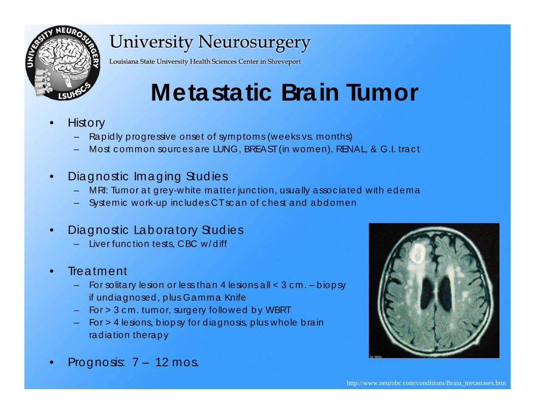
Metastatic Brain Tumor• History
– Rapidly progressive onset of symptoms (weeks vs. months)– Most common sources are LUNG, BREAST (in women), RENAL, & G.I. tract
• Diagnostic Imaging Studies– MRI: Tumor at grey-white matter junction, usually associated with edema– Systemic work-up includes CT scan of chest and abdomen
• Diagnostic Laboratory Studies– Liver function tests, CBC w/diff
• Treatment– For solitary lesion or less than 4 lesions all < 3 cm. – biopsy
if undiagnosed, plus Gamma Knife– For > 3 cm. tumor, surgery followed by WBRT– For > 4 lesions, biopsy for diagnosis, plus whole brain
radiation therapy
• Prognosis: 7 – 12 mos.http://www.neurobc.com/conditions/Brain_metastases.htm
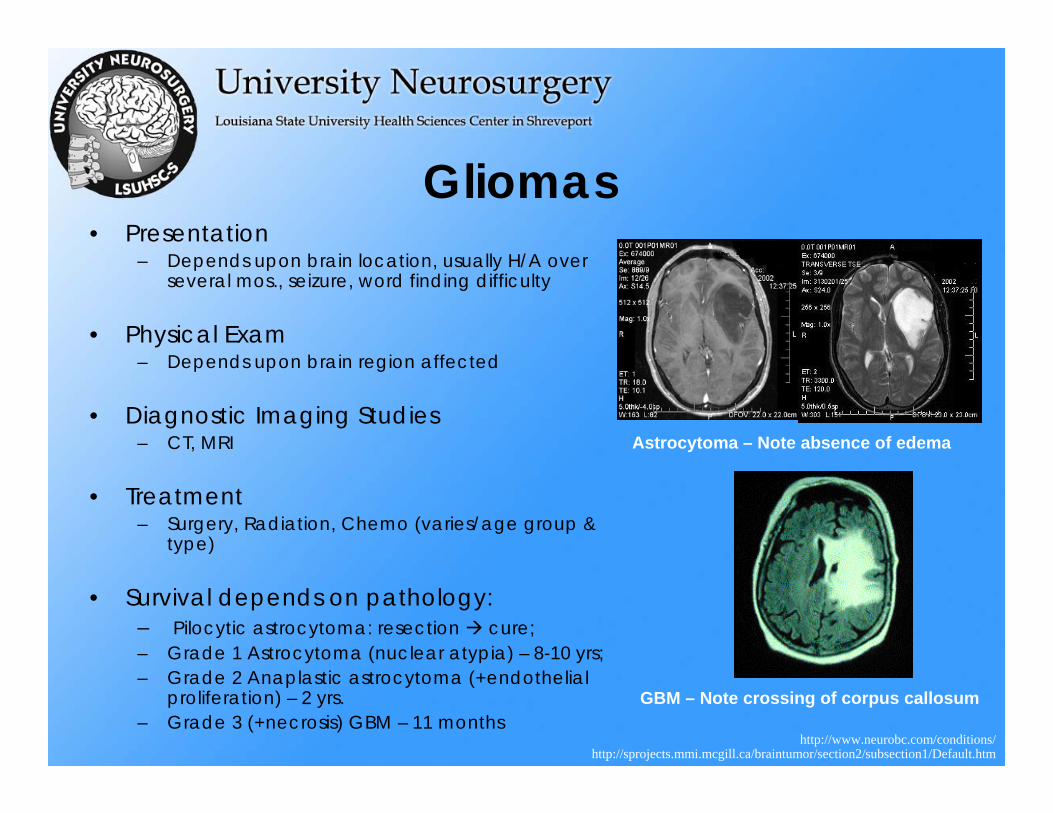
Gliomas• Presentation
– Depends upon brain location, usually H/A over several mos., seizure, word finding difficulty
• Physical Exam– Depends upon brain region affected
• Diagnostic Imaging Studies– CT, MRI
• Treatment– Surgery, Radiation, Chemo (varies/age group &
type)
• Survival depends on pathology:– Pilocytic astrocytoma: resection cure;– Grade 1 Astrocytoma (nuclear atypia) – 8-10 yrs;– Grade 2 Anaplastic astrocytoma (+endothelial
proliferation) – 2 yrs.– Grade 3 (+necrosis) GBM – 11 months
http://www.neurobc.com/conditions/
Astrocytoma – Note absence of edema
GBM – Note crossing of corpus callosum
http://sprojects.mmi.mcgill.ca/braintumor/section2/subsection1/Default.htm
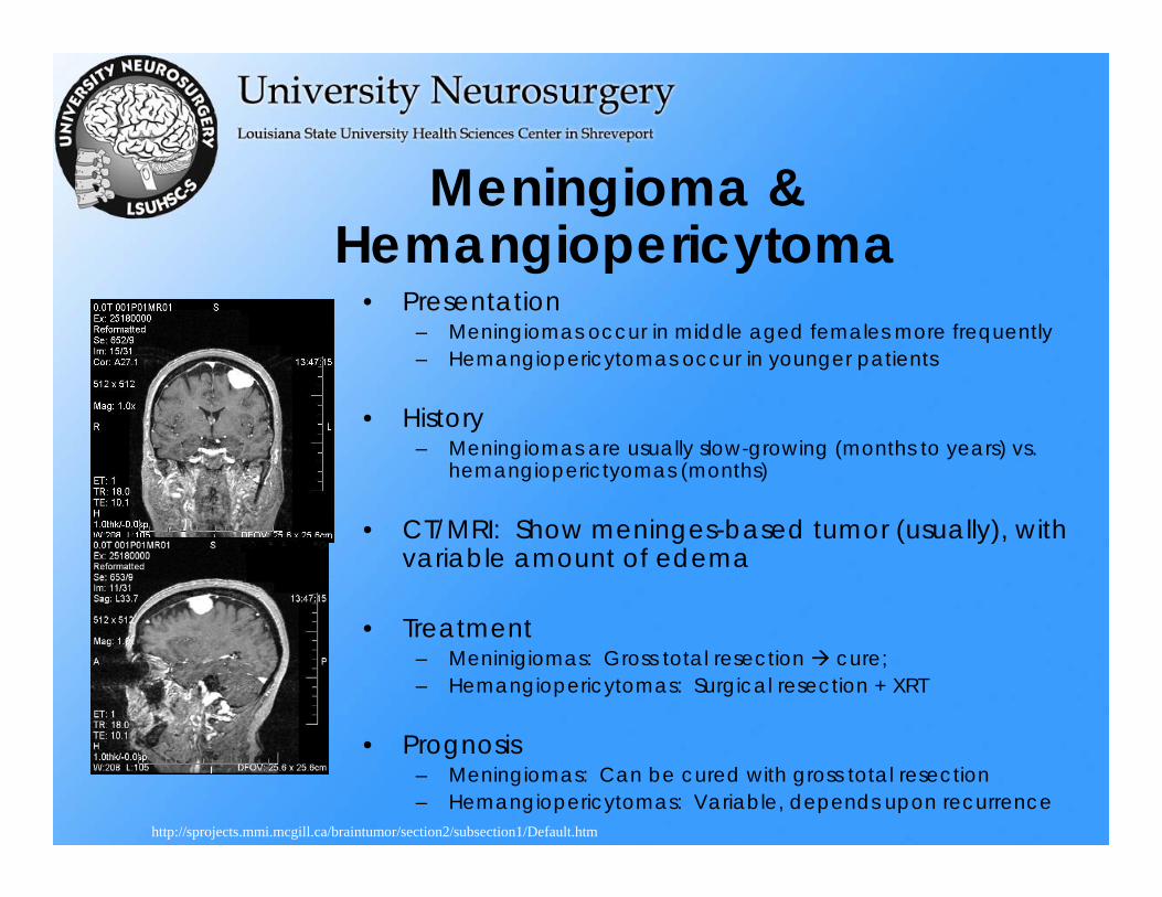
Meningioma & Hemangiopericytoma
• Presentation– Meningiomas occur in middle aged females more frequently– Hemangiopericytomas occur in younger patients
• History– Meningiomas are usually slow-growing (months to years) vs.
hemangioperictyomas (months)
• CT/MRI: Show meninges-based tumor (usually), with variable amount of edema
• Treatment– Meninigiomas: Gross total resection cure;– Hemangiopericytomas: Surgical resection + XRT
• Prognosis– Meningiomas: Can be cured with gross total resection– Hemangiopericytomas: Variable, depends upon recurrence
http://sprojects.mmi.mcgill.ca/braintumor/section2/subsection1/Default.htm
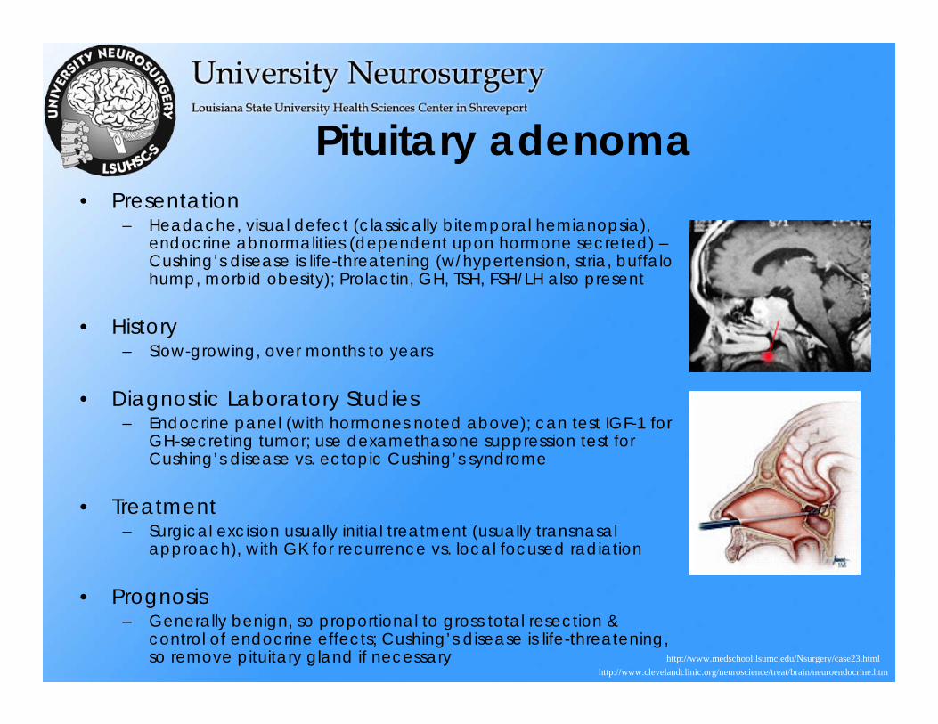
Pituitary adenoma• Presentation
– Headache, visual defect (classically bitemporal hemianopsia), endocrine abnormalities (dependent upon hormone secreted) –Cushing’s disease is life-threatening (w/hypertension, stria, buffalo hump, morbid obesity); Prolactin, GH, TSH, FSH/LH also present
• History– Slow-growing, over months to years
• Diagnostic Laboratory Studies– Endocrine panel (with hormones noted above); can test IGF-1 for
GH-secreting tumor; use dexamethasone suppression test for Cushing’s disease vs. ectopic Cushing’s syndrome
• Treatment– Surgical excision usually initial treatment (usually transnasal
approach), with GK for recurrence vs. local focused radiation
• Prognosis– Generally benign, so proportional to gross total resection &
control of endocrine effects; Cushing’s disease is life-threatening, so remove pituitary gland if necessary
http://www.clevelandclinic.org/neuroscience/treat/brain/neuroendocrine.htmhttp://www.medschool.lsumc.edu/Nsurgery/case23.html
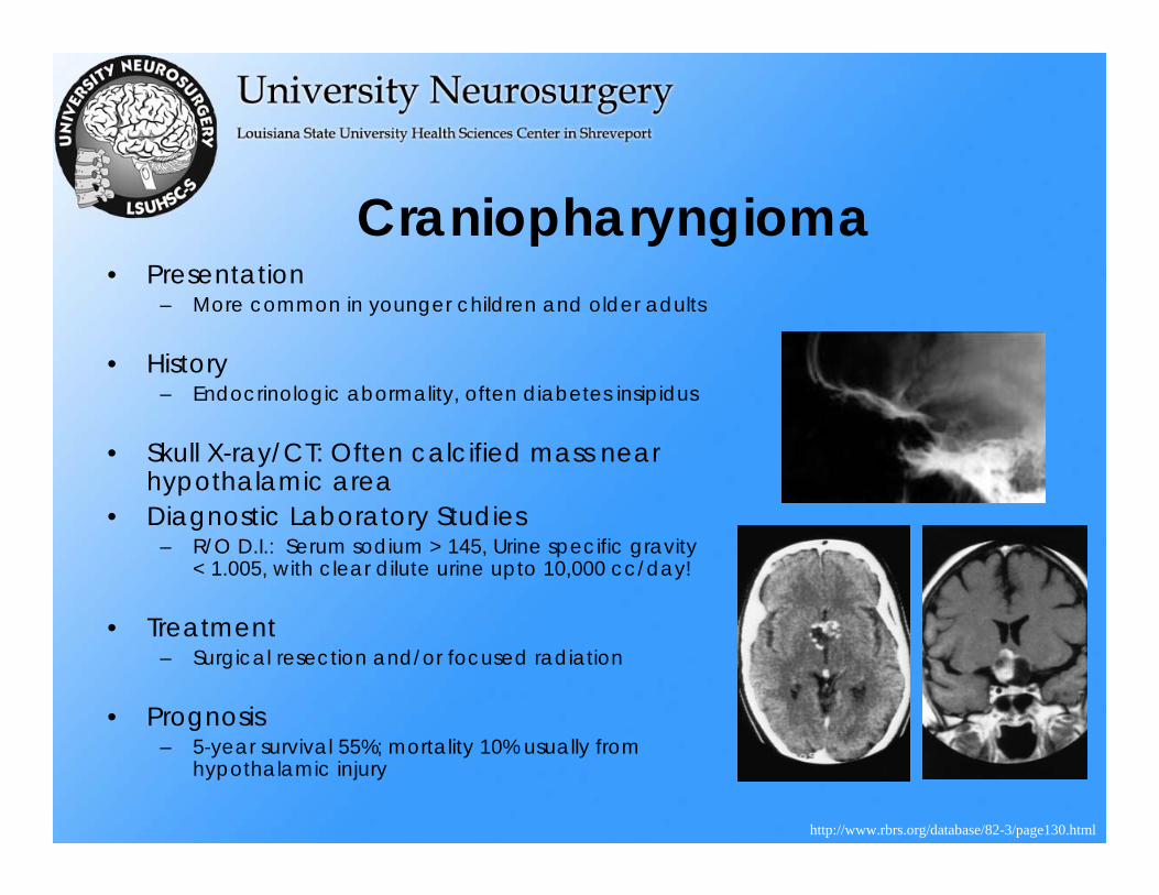
Craniopharyngioma• Presentation
– More common in younger children and older adults
• History– Endocrinologic abormality, often diabetes insipidus
• Skull X-ray/CT: Often calcified mass near hypothalamic area
• Diagnostic Laboratory Studies– R/O D.I.: Serum sodium > 145, Urine specific gravity
< 1.005, with clear dilute urine upto 10,000 cc/day!
• Treatment– Surgical resection and/or focused radiation
• Prognosis– 5-year survival 55%; mortality 10% usually from
hypothalamic injury
http://www.rbrs.org/database/82-3/page130.html
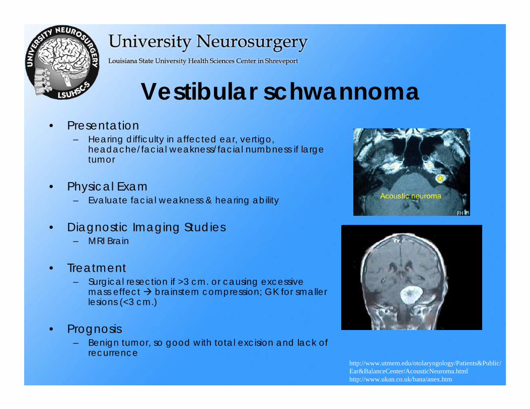
Vestibular schwannoma• Presentation
– Hearing difficulty in affected ear, vertigo, headache/facial weakness/facial numbness if large tumor
• Physical Exam– Evaluate facial weakness & hearing ability
• Diagnostic Imaging Studies– MRI Brain
• Treatment– Surgical resection if >3 cm. or causing excessive
mass effect brainstem compression; GK for smaller lesions (<3 cm.)
• Prognosis– Benign tumor, so good with total excision and lack of
recurrencehttp://www.utmem.edu/otolaryngology/Patients&Public/Ear&BalanceCenter/AcousticNeuroma.htmlhttp://www.ukan.co.uk/bana/anex.htm
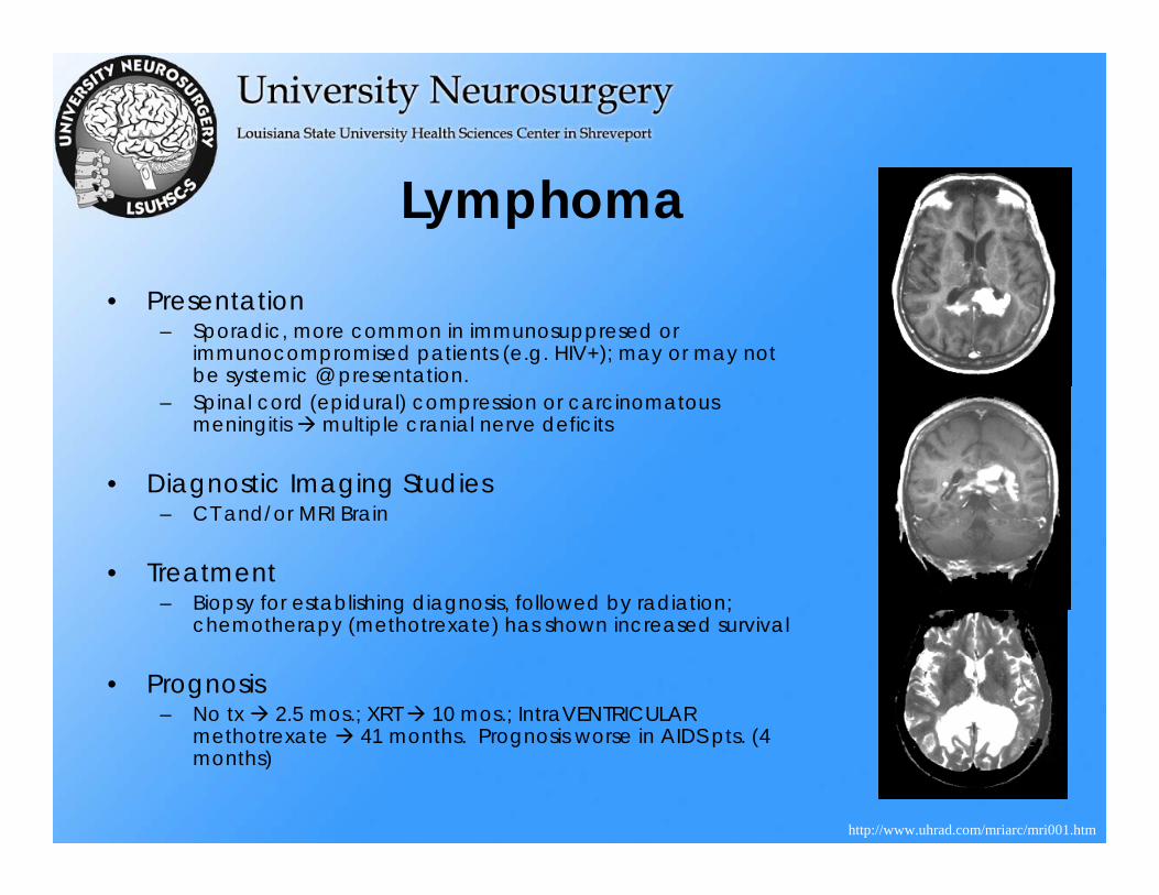
Lymphoma• Presentation
– Sporadic, more common in immunosuppresed or immunocompromised patients (e.g. HIV+); may or may not be systemic @ presentation.
– Spinal cord (epidural) compression or carcinomatousmeningitis multiple cranial nerve deficits
• Diagnostic Imaging Studies– CT and/or MRI Brain
• Treatment– Biopsy for establishing diagnosis, followed by radiation;
chemotherapy (methotrexate) has shown increased survival
• Prognosis– No tx 2.5 mos.; XRT 10 mos.; IntraVENTRICULAR
methotrexate 41 months. Prognosis worse in AIDS pts. (4 months)
http://www.uhrad.com/mriarc/mri001.htm
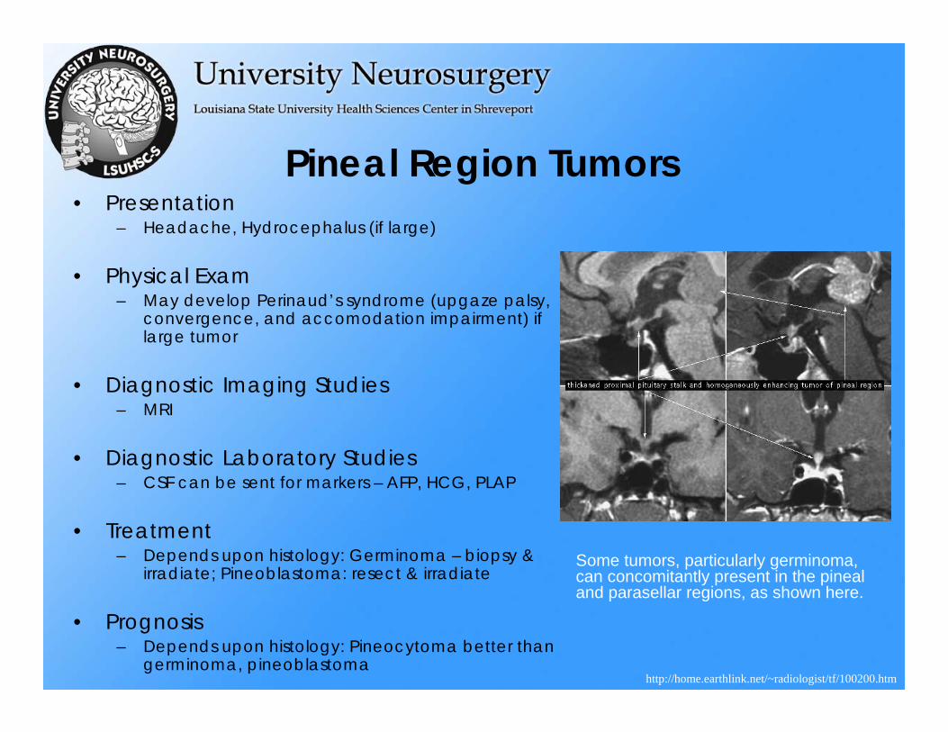
Pineal Region Tumors• Presentation
– Headache, Hydrocephalus (if large)
• Physical Exam– May develop Perinaud’s syndrome (upgaze palsy,
convergence, and accomodation impairment) if large tumor
• Diagnostic Imaging Studies– MRI
• Diagnostic Laboratory Studies– CSF can be sent for markers – AFP, HCG, PLAP
• Treatment– Depends upon histology: Germinoma – biopsy &
irradiate; Pineoblastoma: resect & irradiate
• Prognosis– Depends upon histology: Pineocytoma better than
germinoma, pineoblastoma
Some tumors, particularly germinoma, can concomitantly present in the pineal and parasellar regions, as shown here.
http://home.earthlink.net/~radiologist/tf/100200.htm
Pediatric tumors• Will be covered in lecture on
“Pediatric Neurosurgery”
• Include medulloblastoma, ependymoma, pilocytic astrocytoma, choroid plexus papilloma & carcinoma, and DNET
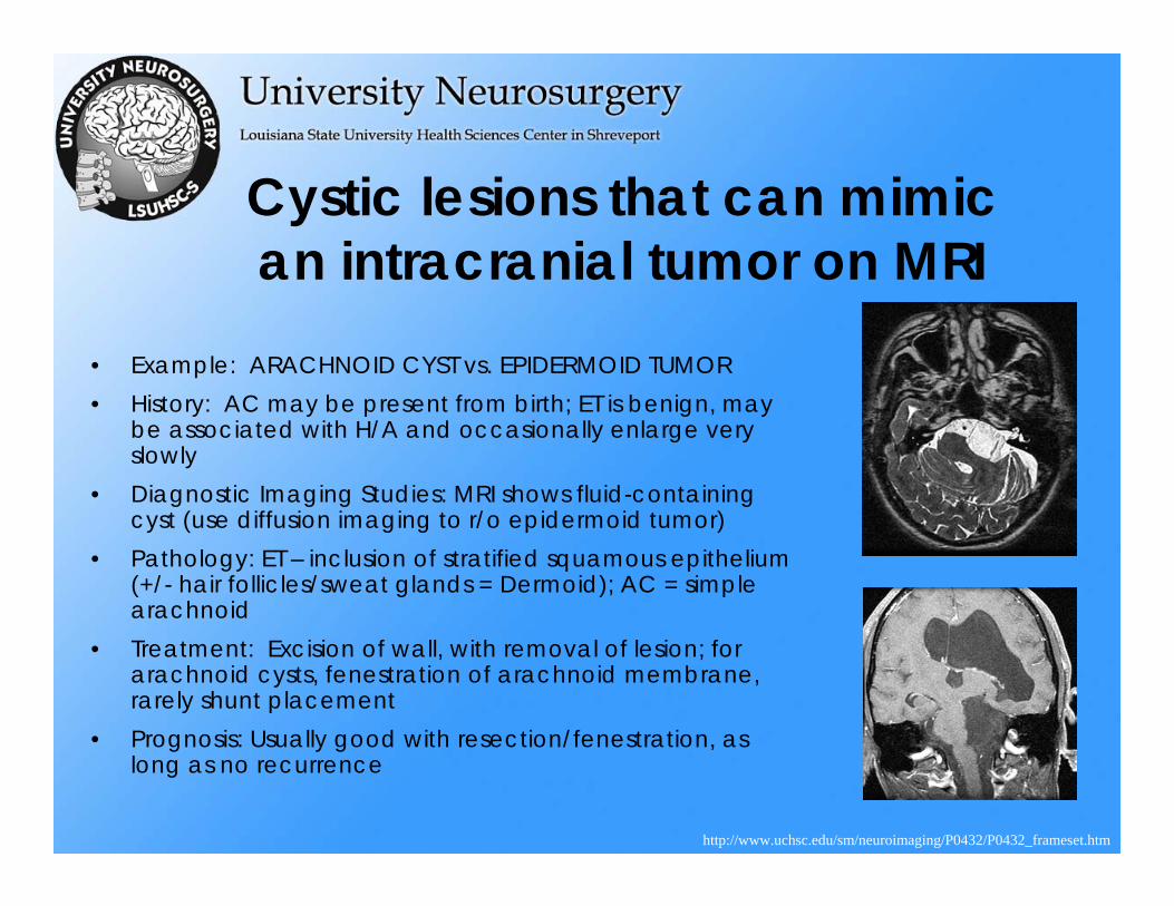
Cystic lesions that can mimican intracranial tumor on MRI
• Example: ARACHNOID CYST vs. EPIDERMOID TUMOR• History: AC may be present from birth; ET is benign, may
be associated with H/A and occasionally enlarge very slowly
• Diagnostic Imaging Studies: MRI shows fluid-containing cyst (use diffusion imaging to r/o epidermoid tumor)
• Pathology: ET – inclusion of stratified squamous epithelium (+/- hair follicles/sweat glands = Dermoid); AC = simple arachnoid
• Treatment: Excision of wall, with removal of lesion; for arachnoid cysts, fenestration of arachnoid membrane, rarely shunt placement
• Prognosis: Usually good with resection/fenestration, as long as no recurrence
http://www.uchsc.edu/sm/neuroimaging/P0432/P0432_frameset.htm
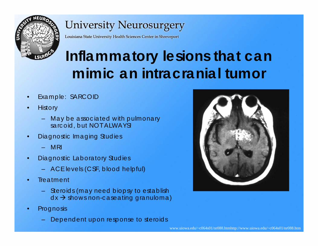
• Example: SARCOID• History
– May be associated with pulmonary sarcoid, but NOT ALWAYS!
• Diagnostic Imaging Studies– MRI
• Diagnostic Laboratory Studies– ACE levels (CSF, blood helpful)
• Treatment– Steroids (may need biopsy to establish
dx shows non-caseating granuloma)• Prognosis
– Dependent upon response to steroids
Inflammatory lesions that canmimic an intracranial tumor
www.uiowa.edu/~c064s01/nr088.htmhttp://www.uiowa.edu/~c064s01/nr088.htm
Infectious lesions canmimic an intracranial tumor
• Discussed under section of “Vascular, Critical Care, & CNS Infections”
Care of the Brain Tumor Patient• Steroids
– Help resolve edema and symptoms associated with it– Can confuse the issue of lymphoma, because rapid
“disappearance” is seen for lymphoma, followed by recurrence
• Anti-convulsants– Recommended particularly for epileptogenic areas, e.g.
mesial temporal lobe, and with past h/o seizures
Treatment Options in generalfor Intracranial Tumors
• Sometimes need Biopsy for identification… then one or combination of:– Surgery
– Radiation Therapy
• Whole brain• Focused beam• Gamma Knife Stereotactic Radiosurgery
– Chemotherapy• Systemic• Local
Gamma Knife Radiosurgery• Indications
– Tumors (Benign & Malignant, Primary & Metastatic)– Arteriovenous malformations– Trigeminal neuralgia– Functional neurosurgery, to create lesions (controversial)
• Success Rate– Comparable success rate for tumors vs. surgery/conventional
radiation, with fewer side effects/morbidity/mortality
• Limitations– Tumors must be smaller (<3 cm.), and once a lesion is made,
it cannot be undone (irreversible)
• Side Effects/Complications– Rare, can include edema post-procedure
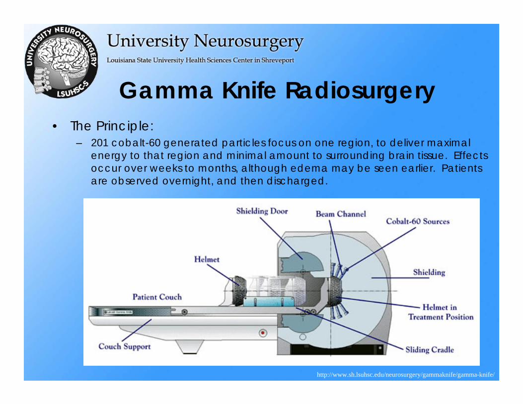
Gamma Knife Radiosurgery• The Principle:
– 201 cobalt-60 generated particles focus on one region, to deliver maximal energy to that region and minimal amount to surrounding brain tissue. Effects occur over weeks to months, although edema may be seen earlier. Patients are observed overnight, and then discharged.
http://www.sh.lsuhsc.edu/neurosurgery/gammaknife/gamma-knife/
The End


![Metastatic Intracranial Hemangiopericytoma to the Spinal ... · 128 INTRODUCTION Intracranial hemangiopericytoma (HPC) is a rare tumor with malignant features [1], of which incidence](https://img.pdfslide.net/doc/110x75/5d4d5a7488c993a90e8bc971/metastatic-intracranial-hemangiopericytoma-to-the-spinal-128-introduction.jpg)


![Foramen magnum meningiomas: detailed surgical ......Meningiomas are common neoplasms representing 14.3 to 19% of all intracranial tumors [63]. Among all the meningiomas, only 1.8 to](https://img.pdfslide.net/doc/110x75/60aa2d3285131731732f9abe/foramen-magnum-meningiomas-detailed-surgical-meningiomas-are-common-neoplasms.jpg)