Embed Size (px)
Citation preview
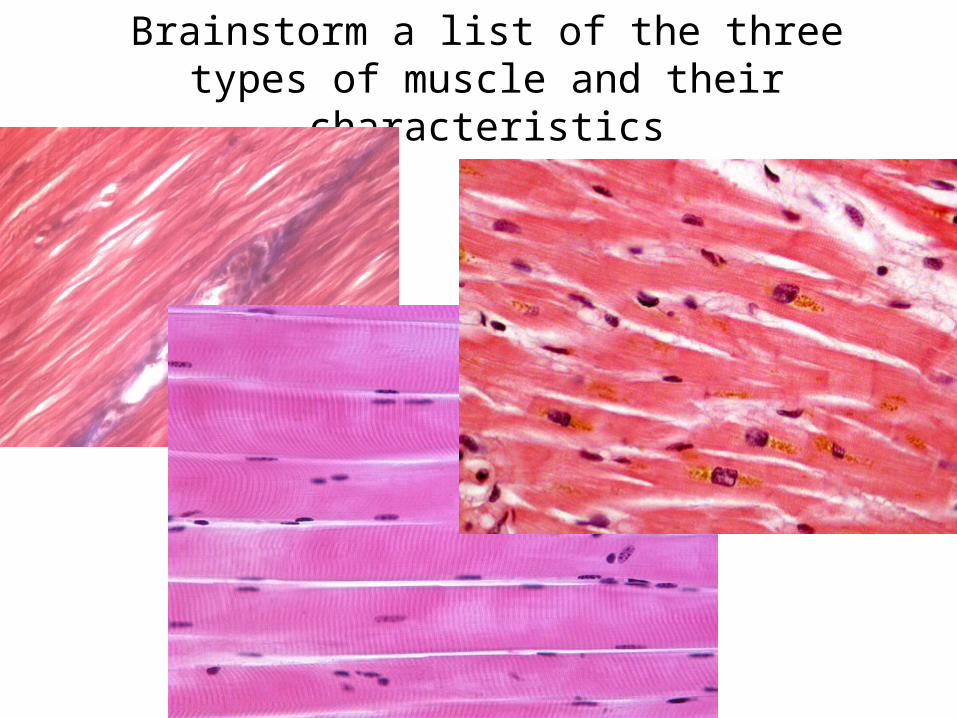
Brainstorm a list of the three types of muscle and their characteristics
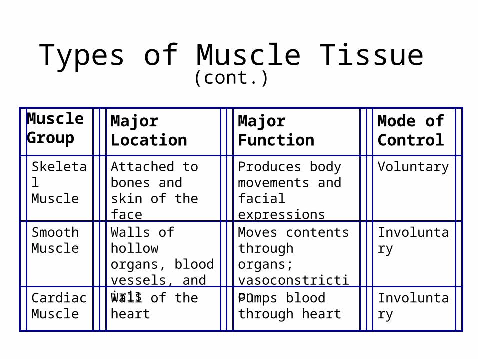
Types of Muscle Tissue (cont.)
Muscle Group
Major Location
Major Function
Mode of Control
Skeletal Muscle
Attached to bones and skin of the face
Produces body movements and facial expressions
Voluntary
Smooth Muscle
Walls of hollow organs, blood vessels, and iris
Moves contents through organs; vasoconstriction
Involuntary
Cardiac Muscle
Wall of the heart Pumps blood through heart
Involuntary
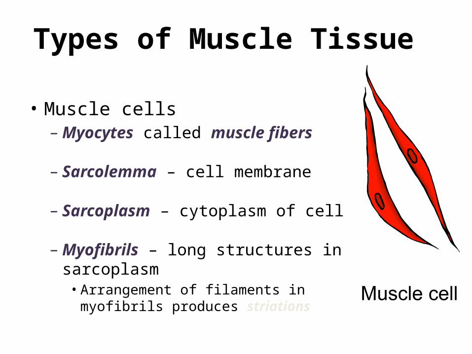
Types of Muscle Tissue
• Muscle cells– Myocytes called muscle fibers
– Sarcolemma – cell membrane
– Sarcoplasm – cytoplasm of cell
– Myofibrils – long structures in sarcoplasm
• Arrangement of filaments in myofibrils produces striations
Skeletal Muscle
Structure & Function
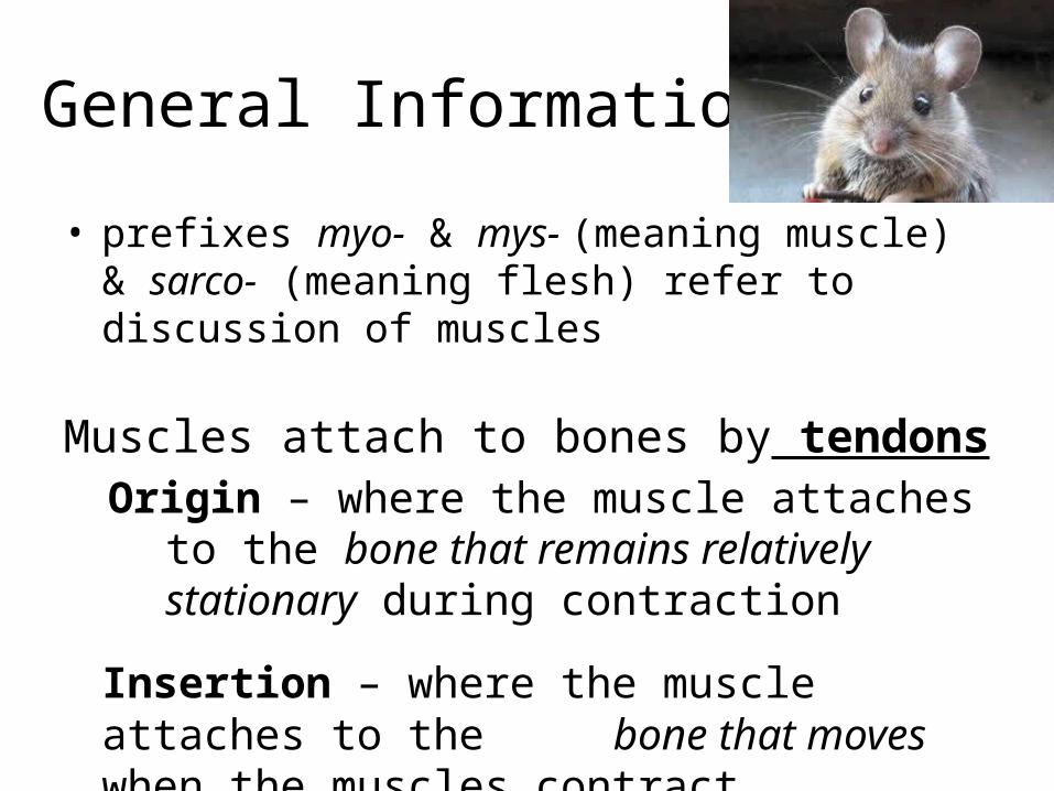
General Information
• prefixes myo- & mys- (meaning muscle) & sarco- (meaning flesh) refer to discussion of muscles
Muscles attach to bones by tendonsOrigin – where the muscle attaches to the bone
that remains relatively stationary during contraction
Insertion – where the muscle attaches to the bone that moves when the muscles
contract
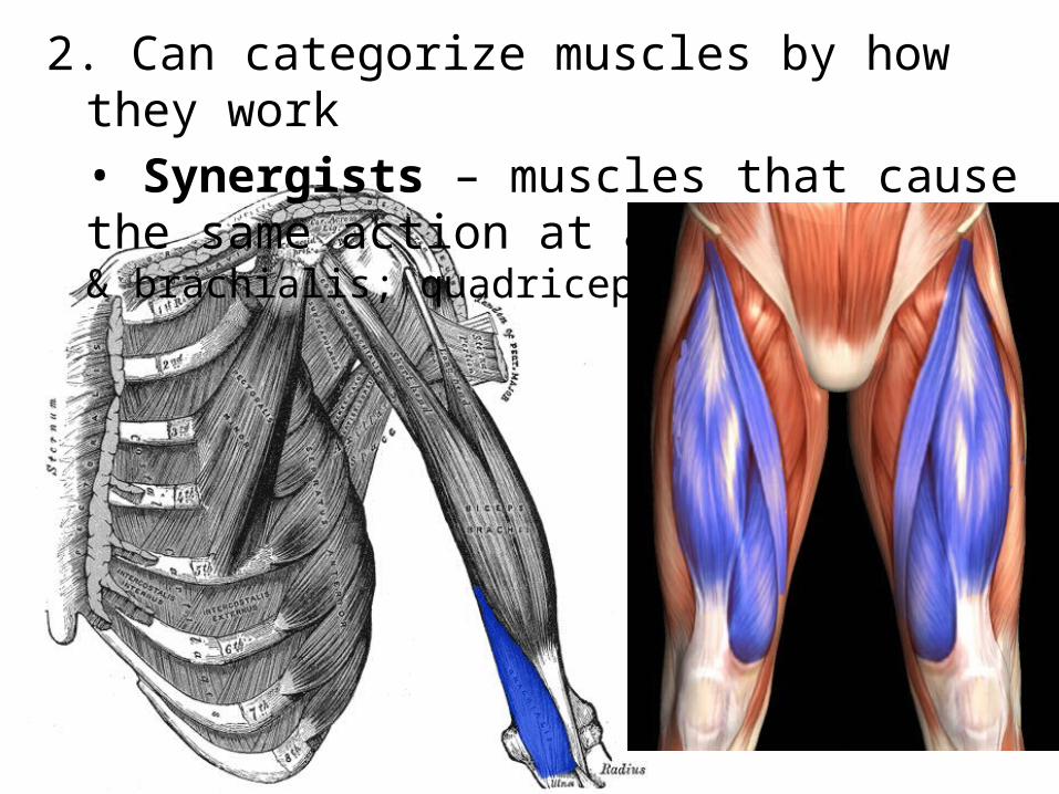
2. Can categorize muscles by how they work• Synergists – muscles that cause the same action at a joint (biceps & brachialis; quadriceps group)
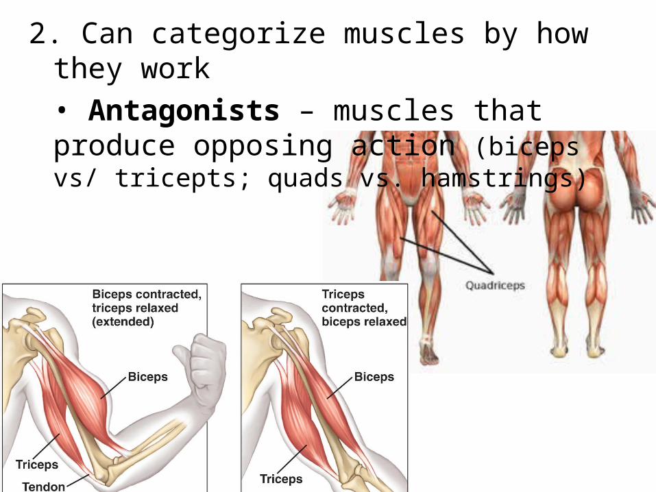
2. Can categorize muscles by how they work• Antagonists – muscles that produce opposing action (biceps vs/ tricepts; quads vs. hamstrings)
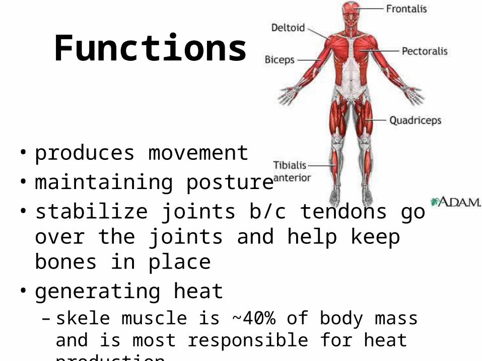
Functions
• produces movement• maintaining posture• stabilize joints b/c tendons go over the joints
and help keep bones in place• generating heat
– skele muscle is ~40% of body mass and is most responsible for heat production
Skeletal Muscle• Made of muscle fibers (myofibril) packed into sheets of
muscle – Muscle fibers are cigar-shaped & have several nuclei– Individual muscle fibers are made of smaller pieces…
we’ll get to that• Some muscle fibers are about 1 ft in length• Have visible stripes • Voluntary• can contract quickly & w/ large force but tires easily and
must be rested after use• individual cells are fragile but muscles are strong…why?

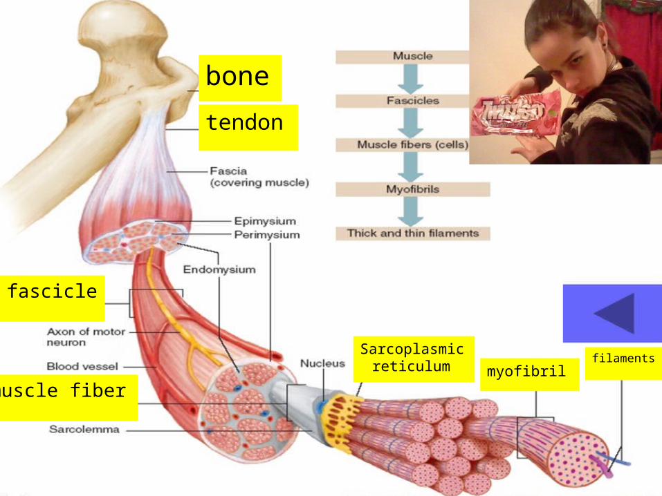
tendon
bone
fascicle
muscle fibermyofibril
Sarcoplasmic reticulum filaments
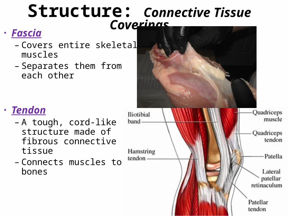
Structure: Connective Tissue Coverings• Fascia
– Covers entire skeletal muscles
– Separates them from each other
• Tendon– A tough, cord-like structure
made of fibrous connective tissue
– Connects muscles to bones
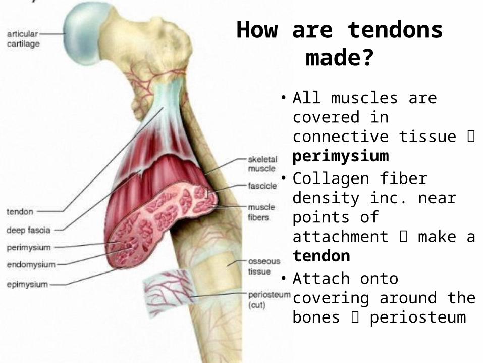
How are tendons made?
• All muscles are covered in connective tissue perimysium
• Collagen fiber density inc. near points of attachment make a tendon
• Attach onto covering around the bones periosteum
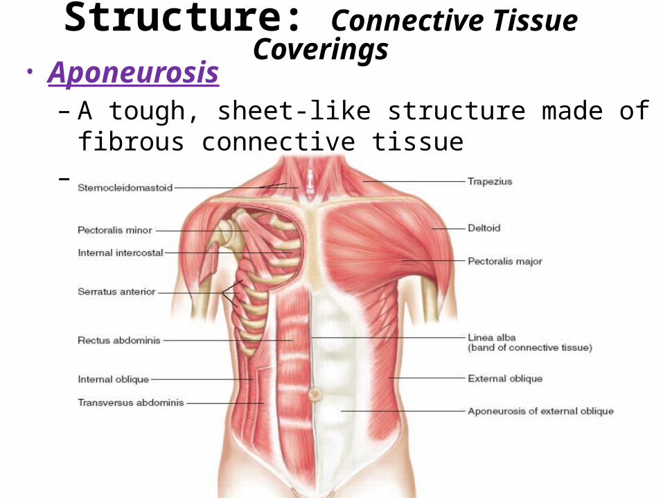
Structure: Connective Tissue Coverings• Aponeurosis
– A tough, sheet-like structure made of fibrous connective tissue
– Attaches muscles to other muscles
tendon
bone
fascicle
muscle fibermyofibril
Sarcoplasmic reticulum filaments
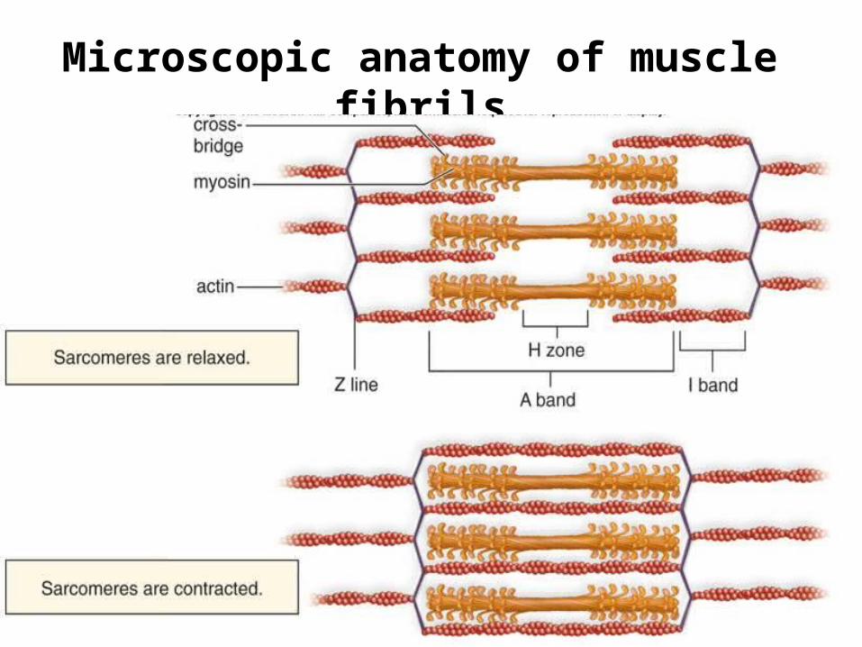
Microscopic anatomy of muscle fibrils
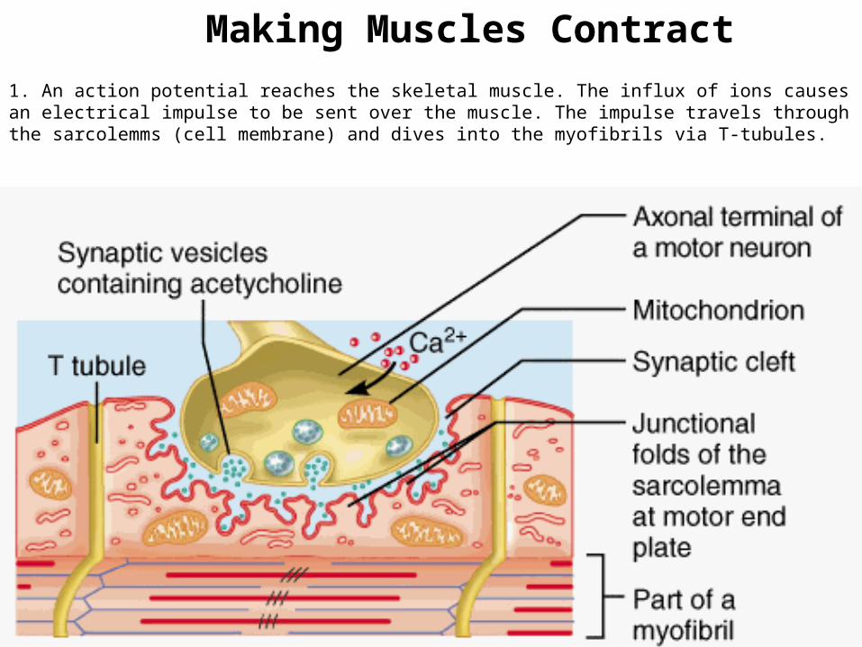
1. An action potential reaches the skeletal muscle. The influx of ions causes an electrical impulse to be sent over the muscle. The impulse travels through the sarcolemms (cell membrane) and dives into the myofibrils via T-tubules.
Making Muscles Contract
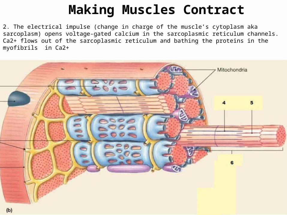
2. The electrical impulse (change in charge of the muscle’s cytoplasm aka sarcoplasm) opens voltage-gated calcium in the sarcoplasmic reticulum channels. Ca2+ flows out of the sarcoplasmic reticulum and bathing the proteins in the myofibrils in Ca2+
Making Muscles Contract
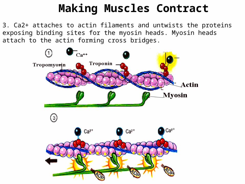
3. Ca2+ attaches to actin filaments and untwists the proteins exposing binding sites for the myosin heads. Myosin heads attach to the actin forming cross bridges.
Making Muscles Contract
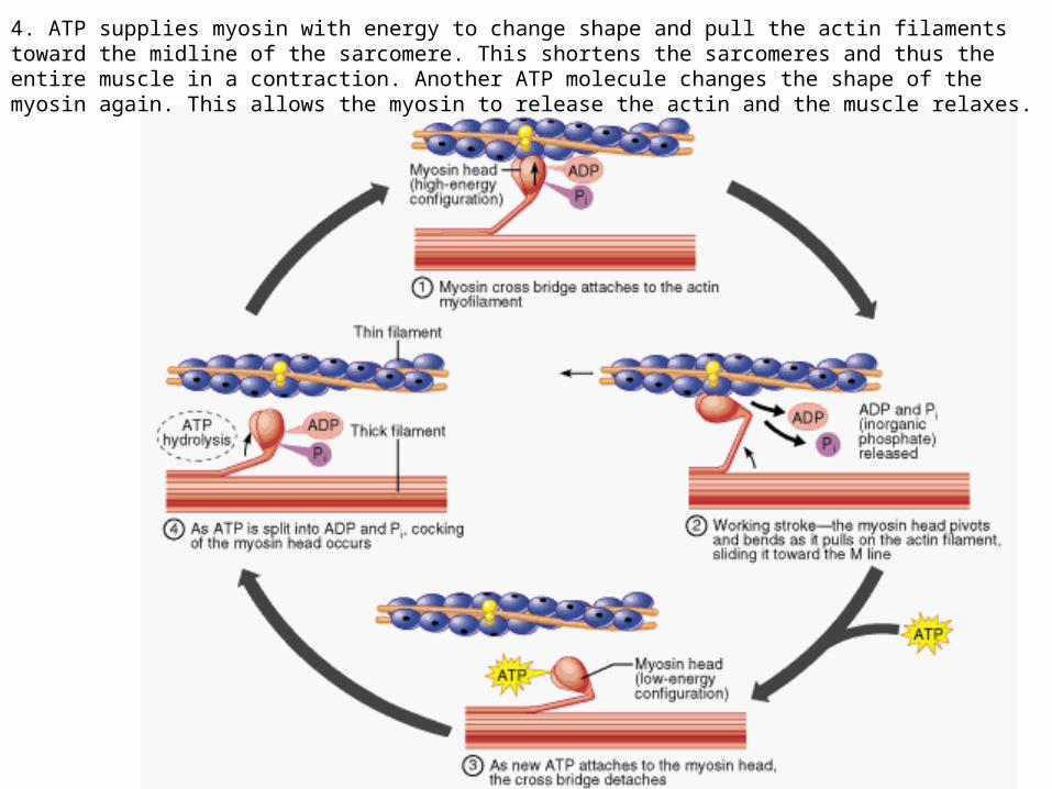
4. ATP supplies myosin with energy to change shape and pull the actin filaments toward the midline of the sarcomere. This shortens the sarcomeres and thus the entire muscle in a contraction. Another ATP molecule changes the shape of the myosin again. This allows the myosin to release the actin and the muscle relaxes.
How Skeletal Muscles Move• Stimulated by nerve impulses• Axon terminal and the sarcolemma (plasma
membrane of muscle cells) are close together with a synaptic cleft in between
• Neuron releases ACh across the cleft attaches to receptors on the sarcolemma allows Na+ to enter making cell more + making an action potential causes contraction of the sarcomeres and thus the muscle http://highered.mcgraw-hill.com/sites/0072495855/student_view0/chapter10/
animation__action_potentials_and_muscle_contraction.html
Question 1
• What structure is used to connect actin and myosin during muscle contraction.
• Myosin heads AKA “paddles”
Question 2
• Compare/contrast the insertion and origin?
• Answer– Insertion- usually more distal (limbs)insertion
point moves toward the origin– Origin- where a muscle attaches to a bone that
does not move during muscle movement
Question 3
• Put the following parts of a muscle in order from largest to smallest and explain what each is– Myofibril– Fascicle– Myofibril– Sarcomere– Myosin head AKA “paddles”
• Answer– Fascicle myofibril sarcomere myofibril myosin heads
Question 4
• After an action potential is sent to the muscle what two things (ions, molecules, etc) a required for muscle contraction? What does each one do?
• Answer– ATP- allows the myosin heads to pull the Z disc in towards
the center of the cell for muscle contraction – Calcium- changes the shape of actin to expose binding
sites for the myosin heads
Question 5
• What structure gets pulled in towards the center of the sarcomere in muscle contraction?
• Answer: z-lines or z proteins
Whisper Down the Lane
• You are sitting and you want to stand. Collectively you must describe the progression of events that will occur in order to get the “move” message to your skeletal muscles (focus on just ONE muscle). Tell the person next to you what is going to happen first and they are going to share what you told them plus the next correct, logical step in this process.