Embed Size (px)
Citation preview

Branding - Fusion of Meta Data and Musculoskeletal Radiographs for
Multi-modal Diagnostic Recognition
Obioma Pelka1,2, Felix Nensa2, and Christoph M. Friedrich1,3
1Department of Computer Science, University of Applied Sciences and Arts Dortmund
{obioma.pelka, christoph.friedrich}@fh-dortmund.de2Department of Diagnostic and Interventional Radiology and Neuroradiology
University Hospital Essen, Germany
3Institute for Medical Informatics, Biometry and Epidemiology (IMIBE)
University Hospital Essen, Germany
Abstract
Data fusion techniques provide opportunities for com-
bining information from multiple domains, such as meta
and medical report data with radiology images. This
helps to obtain knowledge of enriched quality. The objec-
tive of this paper is to fuse automatically generated im-
age keywords with radiographs, enabling multi-modal im-
age representations for body part and abnormality recog-
nition. As manual annotation is often impractical, time-
consuming and prone to errors, automatic visual recogni-
tion and annotation of radiographs is a fundamental step
towards computer-aided interpretation. As the number of
digital medical images taken daily rapidly increases, there
is a need to create systems capable of appropriately de-
tecting and classifying anatomy and abnormality in radiol-
ogy images. The Long Short-Term Memory (LSTM) based
Recurrent Neural Network (RNN) Show-and-Tell model is
adopted for keyword generation. The presented work fuses
multi-modal information by incorporating automatically
generated keywords into radiographs via augmentation.
This leads to enriched sufficient features, with which deep
learning systems are trained. To demonstrate the proposed
approach, evaluation is computed on the Musculoskele-
tal Radiographs (MURA) using two classification schemes.
Prediction accuracy was higher for all tested datasets using
the proposed approach with 95.93 % for anatomic regions
and 81.5 % for abnormality classification, respectively.
1. Introduction
Due to the fast development in hardware, software, and
digital imaging technologies in the medical domain, the
amount of information collected per patient scan has rapidly
increased [1, 2]. To decrease the burden on radiologists and
maintain the maximum interpretation of these radiology re-
ports, the need to implement systems capable of fusing dif-
ferent input domains and thereby obtaining a consolidated
data representation, has become more urgent cause. Ex-
amples for the medical domain include, incorporating text
reports and meta data into radiology images, alongside with
combining ECG audio files with medical report findings.
There is no restriction to the usage of the fused data, as it
can be applied for image classification and annotation [3].
To create effective classification and recognition sys-
tems, the selection and fusion of features for a sufficient
image representation is essential. As shown in [4, 5, 6, 7, 8],
multi-modal representation achieve higher prediction rates
in biomedical annotation tasks. The combination of visual
and text representation is aimed to sufficiently represent
biomedical images. The achieved text features are further
adopted for visual recognition tasks, such as body part clas-
sification and semantic tagging for substantial structuring
of radiographs, as well as abnormality recognition as over
1.7 billion patient worldwide suffer from musculoskeletal
problems [11].
However, for real clinical cases and some image clas-
sification tasks such as the Musculoskeletal Radiographs
(MURA) dataset [9], corresponding text representations are
not available. In this paper, the Radiology Objects in COn-
text (ROCO) dataset [10] is utilized to automatically gener-
ate keywords, which are fused with visual features to obtain
multi-modal radiology image representations. Built on the
presented work in [8], we extend the fused image represen-
tation using a different technique and evaluate the proposed
approach for clinical diagnostic recognition.
The presented approach can be adopted to combine dif-
ferent meta data, as well as medical report findings with
radiology images to optimize classification prediction accu-
racy.
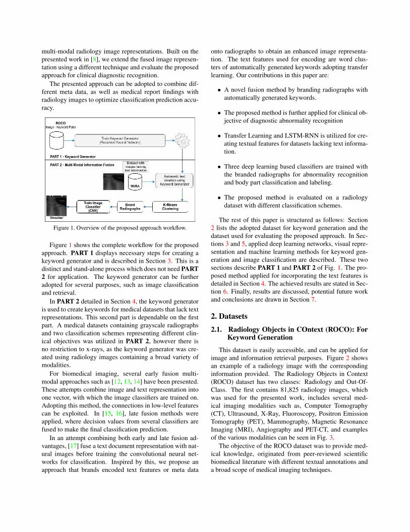
Figure 1. Overview of the proposed approach workflow.
Figure 1 shows the complete workflow for the proposed
approach. PART 1 displays necessary steps for creating a
keyword generator and is described in Section 3. This is a
distinct and stand-alone process which does not need PART
2 for application. The keyword generator can be further
adopted for several purposes, such as image classification
and retrieval.
In PART 2 detailed in Section 4, the keyword generator
is used to create keywords for medical datasets that lack text
representations. This second part is dependable on the first
part. A medical datasets containing grayscale radiographs
and two classification schemes representing different clin-
ical objectives was utilized in PART 2, however there is
no restriction to x-rays, as the keyword generator was cre-
ated using radiology images containing a broad variety of
modalities.
For biomedical imaging, several early fusion multi-
modal approaches such as [12, 13, 14] have been presented.
These attempts combine image and text representation into
one vector, with which the image classifiers are trained on.
Adopting this method, the connections in low-level features
can be exploited. In [15, 16], late fusion methods were
applied, where decision values from several classifiers are
fused to make the final classification prediction.
In an attempt combining both early and late fusion ad-
vantages, [17] fuse a text document representation with nat-
ural images before training the convolutional neural net-
works for classification. Inspired by this, we propose an
approach that brands encoded text features or meta data
onto radiographs to obtain an enhanced image representa-
tion. The text features used for encoding are word clus-
ters of automatically generated keywords adopting transfer
learning. Our contributions in this paper are:
• A novel fusion method by branding radiographs with
automatically generated keywords.
• The proposed method is further applied for clinical ob-
jective of diagnostic abnormality recognition
• Transfer Learning and LSTM-RNN is utilized for cre-
ating textual features for datasets lacking text informa-
tion.
• Three deep learning based classifiers are trained with
the branded radiographs for abnormality recognition
and body part classification and labeling.
• The proposed method is evaluated on a radiology
dataset with different classification schemes.
The rest of this paper is structured as follows: Section
2 lists the adopted dataset for keyword generation and the
dataset used for evaluating the proposed approach. In Sec-
tions 3 and 5, applied deep learning networks, visual repre-
sentation and machine learning methods for keyword gen-
eration and image classification are described. These two
sections describe PART 1 and PART 2 of Fig. 1. The pro-
posed method applied for incorporating the text features is
detailed in Section 4. The achieved results are stated in Sec-
tion 6. Finally, results are discussed, potential future work
and conclusions are drawn in Section 7.
2. Datasets
2.1. Radiology Objects in COntext (ROCO): ForKeyword Generation
This dataset is easily accessible, and can be applied for
image and information retrieval purposes. Figure 2 shows
an example of a radiology image with the corresponding
information provided. The Radiology Objects in Context
(ROCO) dataset has two classes: Radiology and Out-Of-
Class. The first contains 81,825 radiology images, which
was used for the presented work, includes several med-
ical imaging modalities such as, Computer Tomography
(CT), Ultrasound, X-Ray, Fluoroscopy, Positron Emission
Tomography (PET), Mammography, Magnetic Resonance
Imaging (MRI), Angiography and PET-CT, and examples
of the various modalities can be seen in Fig. 3.
The objective of the ROCO dataset was to provide med-
ical knowledge, originated from peer-reviewed scientific
biomedical literature with different textual annotations and
a broad scope of medical imaging techniques.

Figure 2. Example of a radiology image with corresponding cap-
tion, keywords, semantic concepts and types. The ultrasound scan
was randomly chosen from the training set of the ROCO dataset
[10].
Figure 3. Examples of radiology images contained in the ROCO
dataset, illustrating the variety of medical imaging modalities. All
images were randomly chosen from the ’Radiology’ subset.
From the PubMed [18] Open Access subset, a total
number of 6,031,814 image - caption pairs were extracted,
which were further for non-compound and radiology im-
ages using deep learning systems. Semantic knowledge
of object interplay present in the images were extracted in
form of UMLS Semantic types and Concept. In addition,
the captions were reduced to only nouns and adjectives,
which is distributed as Keywords.
2.2. Musculoskeletal Radiographs (MURA) : ForAnatomy and Abnormality ClassificationEvaluation
The MURA dataset of musculoskeletal radiographs pre-
sented in [9] consists of 14,863 studies from 12,173 pa-
tients. As each study contains one or more views (images),
a total number of 40,561 multi-view radiographic images
were assembled.
Each of the radiographs belong to one of the seven stan-
dard upper extremity radiographic study types: elbow, fin-
ger, forearm, hand, humerus, shoulder, and wrist [9]. Fig-
ure 4 shows radiographs representing each of the seven
anatomy classes. All studies were labeled as normal or
abnormal by board-certified radiologists from the Stanford
Hospital, between 2011 and 2012, at the interpretation time
in the diagnostic radiology environment [9].
Figure 4. Examples of radiographs representing the anatomy clas-
sification schemes. The images ’wrist’, ’humerus’, ’shoulder’, ’el-
bow’ and ’finger’ belong in addition to the abnormality positive
class. Whereas the images ’forearm’ and ’hand’ belong to the ab-
normality negative. All images were randomly chosen from the
MURA training set [9].
For comparison and research advance purposes, the
dataset was split into training (11,184 patients, 13,457 stud-
ies, 36,808 images), validation (783 patients, 1,199 studies,
3,197 images), and test (206 patients, 207 studies, 556 im-
ages) sets. The explorative analysis regarding class distri-
bution computed on the 9,045 normal and 5,818 abnormal
musculoskeletal radiographic studies is shown in Fig. 5. All
images in the training and validation sets are both annotated
with corresponding abnormality and body parts labels [9].
For evaluation of the proposed work, the validation set is
adopted as the official test set is not public accessible.
Figure 5. Explorative analysis on the distribution of the Muscu-
loskeletal Radiographs (MURA) dataset. The flow chart shows
the total distribution on both training and validation sets.
3. Keyword Generation
As deep learning techniques [19] have improved predic-
tion accuracies in object detection [20], speech recognition
[21] and in domain specific applications such as medical
imaging [22, 23], a deep learning architecture is used to cre-
ate the keyword generation model.
Deep Convolutional Neural Networks (dCNN) [24] are
applied to encode the medical images to a feature represen-
tation which is decoded using a Long Short-Term Memory
(LSTM) [25] based Recurrent Neural Network (RNN) [26]
to generate appropriate keywords for a given radiograph.
This approach, also known as Show-And-Tell model was
proposed in [27] and further improved in [28].
To produce rich visual representations of the images,
CNN is used as an image encoder by pre-training it for an
image classification task. The LSTM-RNN utilized as cap-
tion decoder generates the image keywords, using the CNN
last hidden layer as input [27].
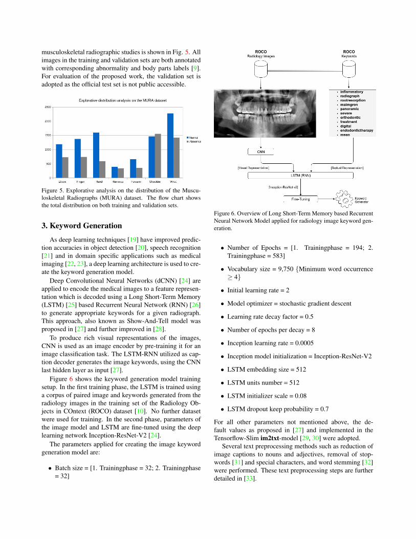
Figure 6 shows the keyword generation model training
setup. In the first training phase, the LSTM is trained using
a corpus of paired image and keywords generated from the
radiology images in the training set of the Radiology Ob-
jects in COntext (ROCO) dataset [10]. No further dataset
were used for training. In the second phase, parameters of
the image model and LSTM are fine-tuned using the deep
learning network Inception-ResNet-V2 [24].
The parameters applied for creating the image keyword
generation model are:
• Batch size = [1. Trainingphase = 32; 2. Trainingphase
= 32]
Figure 6. Overview of Long Short-Term Memory based Recurrent
Neural Network Model applied for radiology image keyword gen-
eration.
• Number of Epochs = [1. Trainingphase = 194; 2.
Trainingphase = 583]
• Vocabulary size = 9,750 {Minimum word occurrence
≥ 4}
• Initial learning rate = 2
• Model optimizer = stochastic gradient descent
• Learning rate decay factor = 0.5
• Number of epochs per decay = 8
• Inception learning rate = 0.0005
• Inception model initialization = Inception-ResNet-V2
• LSTM embedding size = 512
• LSTM units number = 512
• LSTM initializer scale = 0.08
• LSTM dropout keep probability = 0.7
For all other parameters not mentioned above, the de-
fault values as proposed in [27] and implemented in the
Tensorflow-Slim im2txt-model [29, 30] were adopted.
Several text preprocessing methods such as reduction of
image captions to nouns and adjectives, removal of stop-
words [31] and special characters, and word stemming [32]
were performed. These text preprocessing steps are further
detailed in [33].

4. Radiograph Branding
Utilizing the keyword generation model based on the
ROCO dataset and described in subsection 2, keywords
were generated for all radiology images in the MURA
dataset. Figure 7 show these keywords for two randomly
chosen radiographs from the MURA Training Set. No fur-
ther text preprocessing methods were applied to the gener-
ated keywords, as this was done before creating the key-
word generation model.
Figure 7. Examples of keywords generated. All images were ran-
domly chosen from the Musculosketal Radiograph MURA Train-
ing Set. Keyword generation model was created using the all im-
ages from the ROCO radiology class.
Figure 8 shows a subset of the vocabulary obtained with
the generated keywords for the MURA dataset. The key-
words shown were predicted for images of the abnormality
class ’positive’.
Figure 8. Automatically generated keywords for the radiology im-
ages in the MURA dataset. All keywords were predicted for im-
ages belonging to the abnormality class ’positive’.
To reduce the keywords to semantic concepts, the words
in the vocabularies were further grouped to k = 25 clusters
using the Natural Language Toolkit (NLTK) [31] k-means
clustering method [34, 35]. Finally the radiographs (image
size [299x299]) are branded by marking the cluster position
of the generated keywords on the image, as shown in Fig. 9.
The presence of the clusters is incorporated with a [10x10]pixel marker. The complete implementation was done in
python.
Figure 9. Overview of the complete procedure applied for the ra-
diograph branding. The image was randomly chosen from the
MURA training set.
5. Classification
For the dCNNs, TensorFlow-Slim (TF-slim), a
lightweight package for defining, training and evalu-
ating models in TensorFlow [29] with pre-trained models,
was adopted. To optimize prediction performance, the
models were fine-tuned with all trainable weights and best
configuration in the second training phase.
Inception-v3 The pre-trained model Inception-v3 [36]
which was trained for the ImageNet [37] Large Visual
Recognition Challenge 2012 [38], was used to fine-tune
the classification model. For the Inception-v3 classification
models, the following parameters were applied:
• Optimizer: Root Mean Square Propagation (rmsprop)
• Number of epochs: [1. Trainingphase = 2.5; 2. Train-
ingphase = 25]
• Number of steps: [1. Trainingphase = 1,000; 2. Train-
ingphase = 10,000]
• Batch size: 32
• Learning rate: 0.01
• Learning rate decay type: [1. Trainingphase = fixed; 2.
Trainingphase = exponential]
• Weight decay: 0.00004
• Model name: Inception-v3
For all other parameters not mentioned above, the default
values as proposed in TF-Slim [29] were adopted.

Inception-v4 The pre-trained model Inception-v4 [24]
which is a variation of the Inception-v3, having a more
uniform simplified architecture and more inception mod-
ules, was used to train and fine-tune the second classifica-
tion model. The following parameters were applied for the
Inception-v4 classifier:
• Optimizer: Root Mean Square Propagation (rmsprop)
• Number of epochs: [1. Trainingphase = 2.5; 2. Train-
ingphase = 25]
• Number of steps: [1. Trainingphase = 1,000; 2. Train-
ingphase = 10,000]
• Batch size: 32
• Learning rate: 0.01
• Learning rate decay type: [1. Trainingphase = fixed; 2.
Trainingphase = exponential]
• Weight decay: 0.00004
• Model name: Inception-v4
For all other parameters not mentioned above, the default
values as proposed in TF-Slim [29] were adopted.
Inception-ResNet-v2 The pre-trained model Inception-
ResNet-v2 [24] which is a variation of the Inception-v3 us-
ing the ideas presented in [39, 40], was used to train and
fine-tune the third classification model. For the Inception-
ResNet-v2 classification models, the following parameters
were applied:
• Optimizer: Root Mean Square Propagation (rmsprop)
• Number of epochs: [1. Trainingphase = 2.5; 2. Train-
ingphase = 25]
• Number of steps: [1. Trainingphase = 1,000; 2. Train-
ingphase = 10,000]
• Batch size: 32
• Learning rate: 0.01
• Learning rate decay type: [1. Trainingphase = fixed; 2.
Trainingphase = exponential]
• Weight decay: 0.00004
• Model name: Inception-ResNet-v2
For all other parameters not mentioned above, the default
values as proposed in TF-Slim [29] were adopted.
Classification Schemes To evaluate the proposed ap-
proach, two classification schemes from different datasets
were applied. Both classification schemes annotate radio-
graphs according to the body parts examined.
• Musculosketal Radiograph (MURA): Anatomy
1. Elbow
2. Finger
3. Forearm
4. Hand
5. Humerus
6. Shoulder
7. Wrist
• Musculosketal Radiograph (MURA): Abnormality
1. Positive
2. Negative
6. Results
Using the proposed method, increased body parts predic-
tion and abnormality recognition accuracies are obtained on
the MURA dataset, which are shown in Table 1 and 2, re-
spectively. Both tables display the performance of the three
applied deep learning systems, baseline models, as well as
visual, textual and multi-modal results.
The baseline models for the MURA dataset are not com-
parable with the accuracies in Table 1 and 2, as these were
computed using the official test set. For body parts classi-
fication, 70.50 % was achieved with a 169-layer DenseNet
convolutional neural network [41, 9]. The baseline model
for abnormality recognition with 77.80 % was obtained by
asking six board-certified radiologists to manually annotate
the test images. In addition, Random Forest [42] models
were trained with Bag-of-Word (BoW) [43] representations
of the generated keywords to show the performance gain of
the presented approach.
Table 1. Prediction accuracies obtained using the different visual
and text representations and classifier setup, as well as the baseline
accuracy presented in [9]. Evaluation was done for body parts
classification on Musculosketal Radiographs (MURA) validation
set with 3,197 radiographs.
Classifier Setup Visual Branded Textual Multi
Inception-v3 94.09 % 95.93 % - -
Inception-v4 92.39 % 94.72 % - -
Inception-ResNet-v2 91.84 % 95.00 % - -
Random Forest + BoW - - 35.11 % -
Decaf 85.23 % - - -
Decaf + BoW - - - 86.23 %

Table 2. Prediction accuracies obtained using the different visual
and text representations and classifier setup, as well as the baseline
accuracy presented in [9]. Evaluation was done for abnormality
recognition on Musculosketal Radiographs (MURA) validation set
with 3,197 radiographs.
Classifier Setup Visual Branded Textual Multi
Inception-v3 79.85 % 81.55 % - -
Inception-v4 74.27 % 76.48 % - -
Inception-ResNet-v2 78.97 % 79.77 % - -
Random Forest + BoW - - 54.24 % -
Decaf 70.98 % - - -
Decaf + BoW - - - 73.20 %
Results obtained with the original radiographs and solely
visual features are listed in the first column of the tables.
The prediction accuracies obtained with the branded im-
ages, denoting the fused text and visual information are
shown in the second column and outperform other fea-
ture representations. This is observed for both classifica-
tion schemes. Using the automatically generated keywords
without visual representation achieved the poorest predic-
tion rate, which is shown in the third column. The fourth
column ’Multi’ shows accuracies obtained when combin-
ing visual representation from the original image with the
automatically generated keywords.
Inception-v3 proved to be the best deep learning system
for tackling the two clinical objectives. For body anatomy
classification and abnormality recognition, highest accuracy
rates were obtained using Inception-v3.
7. Conclusion
This work presents an approach to combine automati-
cally generated keywords with radiographs. Data fusion is
achieved by incorporating the textual features by augmenta-
tion of the image termed branding. This process enables an
enriched multi-modal image representation which is used
for body parts classification tasks, as multi-modal image
representation has proven to obtain higher prediction results
and some image dataset lack text representation. The con-
solidated multi-modal image representation is further ap-
plied for diagnostic recognition of abnormality, as muscu-
loskeletal conditions cause severe and long-time pain.
To create a keyword generation model, image-keywords
pairs from the training set of the Radiology Objects in COn-
text (ROCO) dataset was adopted to train Long Short-Term
Memory based Recurrent Neural Network models. Uti-
lizing the keyword generation model, text representations
were created for the radiology dataset: Musculosketal Ra-
diograph (MURA), with two classification schemes.
These automatically generated keywords were grouped
into k-means clusters and incorporated by augmentation
into the radiographs by branding the presence of each clus-
ter in the images.
For both classification schemes, the prediction accura-
cies obtained with our proposed multi-modal image repre-
sentation outperformed those achieved just solely visual and
textual features, as well other feature fusion methods. The
proposed work can be further enhanced by exploiting other
word embedding methods, as well as other branding meth-
ods, and precedes the way of combining several features of
different heterogeneous modalities.
As there are several input sources in the medical domain,
the proposed work provides perspective to other data fu-
sion techniques, such as combining meta and medical report
findings with other medical imaging modalities.
Acknowledgment
The work of Obioma Pelka was partially funded by a
PhD grant from University of Applied Sciences and Arts
Dortmund, Germany.
References
[1] M. M. Rahman, P. Bhattacharya, and B. C. Desai, “A Frame-
work for Medical Image Retrieval Using Machine Learning
and Statistical Similarity Matching Techniques With Rele-
vance Feedback,” IEEE Transactions on Information Tech-
nology in Biomedicine, vol. 11, no. 1, pp. 58–69, 2007. [On-
line]. Available: https://doi.org/10.1109/TITB.2006.884364
1
[2] H. D. Tagare, C. C. Jaffe, and J. S. Duncan, “Synthesis
of research: Medical image databases: A content-based
retrieval approach,” Journal of the American Medical
Informatics Association JAMIA, vol. 4, no. 3, pp. 184–198,
1997. [Online]. Available: https://doi.org/10.1136/jamia.
1997.0040184 1
[3] M. Ilyas, A. Othmani, and A. Nait-Ali, “Prediction of hear-
ing loss based on auditory perception: A preliminary study,”
in First International Workshop on PRedictive Intelligence in
MEdicine (PRIME) 2018, Held in Conjunction with MICCAI
2018, Granada, Spain, September 16, 2018, Proceedings,
I. Rekik, G. Unal, E. Adeli, and S. H. Park, Eds. Cham:
Springer International Publishing, 2018, pp. 34–41. 1
[4] N. Codella, J. Connell, S. Pankanti, M. Merler, and J. R.
Smith, “Automated medical image modality recognition by
fusion of visual and text information,” in Medical Image
Computing and Computer-Assisted Intervention–MICCAI
2014. Springer, 2014, pp. 487–495. 1
[5] L. Valavanis, S. Stathopoulos, and T. Kalamboukis, “IPL
at CLEF 2016 Medical Task,” in Working Notes of CLEF
2016 - Conference and Labs of the Evaluation forum,
Evora, Portugal, 5-8 September, 2016., 2016, pp. 413–420.
[Online]. Available: http://ceur-ws.org/Vol-1609/16090413.
pdf 1
[6] J. Kalpathy-Cramer, A. G. S. de Herrera, D. Demner-
Fushman, S. K. Antani, S. Bedrick, and H. Muller,

“Evaluating performance of biomedical image retrieval
systems - An overview of the medical image retrieval task
at ImageCLEF 2004-2013,” Computerized Medical Imaging
and Graphics, vol. 39, pp. 55–61, 2015. [Online]. Available:
https://doi.org/10.1016/j.compmedimag.2014.03.004 1
[7] O. Pelka and C. M. Friedrich, “Modality prediction of
biomedical literature images using multimodal feature
representation ,” GMS Medizinische Informatik, Biometrie
und Epidemiologie, vol. 12, no. 2, pp. 1345–1359, 2016.
[Online]. Available: https://www.egms.de/static/de/journals/
mibe/2016-12/mibe000166.shtml 1
[8] O. Pelka, F. Nensa, and C. M. Friedrich, “Variations on
branding with text occurrence for optimized body parts clas-
sification,” in Proceedings of the 41th Annual International
Conference of the IEEE Engineering in Medicine and Biol-
ogy Society EMBC 2019, Berlin, Germany, July 23-27, 2019,
2019. 1, 2
[9] P. Rajpurkar, J. Irvin, A. Bagul, D. Ding, T. Duan, H. Mehta,
B. Yang, K. Zhu, D. Laird, R. L. Ball, C. Langlotz,
K. Shpanskaya, M. P. Lungren, and A. Y. Ng, “MURA
dataset: Towards radiologist-level abnormality detection
in musculoskeletal radiographs,” in Proceedings of the
1st Medical Imaging with Deep Learning, (MIDL) 2018,
Amsterdam, Netherlands, July 04-06, 2018., 2018. [Online].
Available: https://openreview.net/forum?id=r1Q98pjiG 1, 3,
4, 6, 7
[10] O. Pelka, S. Koitka, J. Ruckert, F. Nensa, and C. M.
Friedrich, “Radiology objects in context (ROCO): A
multimodal image dataset,” in Intravascular Imaging and
Computer Assisted Stenting - and - Large-Scale Annotation
of Biomedical Data and Expert Label Synthesis - 7th
Joint International Workshop, CVII-STENT 2018 and Third
International Workshop, LABELS 2018, Held in Conjunction
with MICCAI 2018, Granada, Spain, September 16, 2018,
Proceedings, 2018, pp. 180–189. [Online]. Available:
https://doi.org/10.1007/978-3-030-01364-6 20 1, 3, 4
[11] “The burden of musculoskele-
tal diseases in the united states,”
https://www.boneandjointburden.org/print/book/export/html/43.
1
[12] O. Pelka, F. Nensa, and C. M. Friedrich, “Optimizing body
region classification with deep convolutional activation fea-
tures,” in Computer Vision ECCV 2018 Workshops, ser. Lec-
ture Notes in Computer Science. Springer Nature Switzer-
land AG, 2019, pp. 1–6. 2
[13] ——, “Adopting semantic information of grayscale radio-
graphs for image classification and retrieval,” in Proceedings
of the 11th International Joint Conference on Biomedical En-
gineering Systems and Technologies (BIOSTEC 2018) - Vol-
ume 2: BIOIMAGING, Funchal, Madeira, Portugal, January
19-21, 2018., 2018, pp. 179–187. 2
[14] V. Andrearczyk and H. Muller, “Deep multimodal classi-
fication of image types in biomedical journal figures,” in
Experimental IR Meets Multilinguality, Multimodality, and
Interaction - 9th International Conference of the CLEF As-
sociation, CLEF 2018, Avignon, France, September 10-14,
2018, Proceedings, 2018, pp. 3–14. [Online]. Available:
https://doi.org/10.1007/978-3-319-98932-7 1 2
[15] O. Pelka and C. M. Friedrich, “FHDO Biomedical
Computer Science Group at Medical Classification Task
of ImageCLEF 2015,” in Working Notes of CLEF 2015 -
Conference and Labs of the Evaluation forum, Toulouse,
France, September 8-11, 2015., 2015. [Online]. Available:
http://ceur-ws.org/Vol-1391/14-CR.pdf 2
[16] S. Koitka and C. M. Friedrich, “Optimized convolutional
neural network ensembles for medical subfigure classifica-
tion,” in Experimental IR Meets Multilinguality, Multimodal-
ity, and Interaction at the 8th International Conference of
the CLEF Association, Dublin, Ireland, September 11-14,
2017, Lecture Notes in Computer Science (LNCS) 10456,
G. J. Jones, S. Lawless, J. Gonzalo, L. Kelly, L. Goeuriot,
T. Mandl, L. Cappellato, and N. Ferro, Eds. Cham: Springer
International Publishing, 2017, pp. 57–68. 2
[17] I. Gallo, A. Calefati, S. Nawaz, and M. K. Janjua,
“Image and encoded text fusion for multi-modal classi-
fication,” in International Conference on Digital Image
Computing: Techniques and Applications (DICTA 2018),
10-13 December 2018 in Canberra, Australia, 2018, pp.
3–14. [Online]. Available: http://artelab.dista.uninsubria.it/
res/research/papers/2018/2018-DICTA-Gallo.pdf 2
[18] R. J. Roberts, “PubMed Central: The GenBank of the pub-
lished literature,” Proceedings of the National Academy of
Sciences of the United States of America, vol. 98, no. 2, pp.
381–382, Jan. 2001. 3
[19] Y. LeCun, Y. Bengio, and G. E. Hinton, “Deep Learning,”
Nature, vol. 521, no. 7553, pp. 436–444, 2015. 4
[20] G. Huang, Z. Liu, L. van der Maaten, and K. Q. Weinberger,
“Densely Connected Convolutional Networks,” in Proceed-
ings of the IEEE Conference on Computer Vision and Pattern
Recognition, CVPR, Honolulu, USA, July 22-25, 2017, 2017.
4
[21] G. Hinton, L. Deng, D. Yu, G. Dahl, A.-a. Mohamed,
N. Jaitly, A. Senior, V. Vanhoucke, P. Nguyen, T. Sainath,
and B. Kingsbury, “Deep Neural Networks for Acoustic
Modeling in Speech Recognition: The Shared Views of
Four Research Groups,” IEEE Signal Processing Magazine,
vol. 29, no. 6, pp. 82–97, Nov. 2012. 4
[22] M. S. Abrao, M. O. d. C. Goncalves, J. A. Dias Jr,
S. Podgaec, L. P. Chamie, and R. Blasbalg, “Comparison
between clinical examination, transvaginal sonography and
magnetic resonance imaging for the diagnosis of deep en-
dometriosis,” Human Reproduction, vol. 22, no. 12, pp.
3092–3097, 2007. 4
[23] Y. Xu, T. Mo, Q. Feng, P. Zhong, M. Lai, and E. I. Chang,
“Deep learning of feature representation with multiple in-
stance learning for medical image analysis,” in IEEE Inter-
national Conference on Acoustics, Speech and Signal Pro-
cessing, ICASSP 2014, Florence, Italy, May 4-9, 2014, 2014,
pp. 1626–1630. 4
[24] C. Szegedy, S. Ioffe, V. Vanhoucke, and A. A. Alemi,
“Inception-v4, Inception-ResNet and the Impact of Resid-

ual Connections on Learning,” in Proceedings of the Thirty-
First AAAI Conference on Artificial Intelligence, February
4-9, 2017, San Francisco, California, USA., 2017, pp. 4278–
4284. 4, 6
[25] S. Hochreiter and J. Schmidhuber, “Long Short-Term
Memory,” Neural Computation, vol. 9, no. 8, pp. 1735–
1780, 1997. [Online]. Available: https://doi.org/10.1162/
neco.1997.9.8.1735 4
[26] Y. Bengio, P. Y. Simard, and P. Frasconi, “Learning
long-term dependencies with gradient descent is difficult,”
IEEE Transactions on Neural Networks, vol. 5, no. 2, pp.
157–166, 1994. [Online]. Available: https://doi.org/10.1109/
72.279181 4
[27] O. Vinyals, A. Toshev, S. Bengio, and D. Erhan, “Show and
Tell: A neural image caption generator,” in IEEE Conference
on Computer Vision and Pattern Recognition, CVPR 2015,
Boston, MA, USA, June 7-12, 2015, 2015, pp. 3156–3164. 4
[28] ——, “Show and Tell: Lessons Learned from the 2015
MSCOCO Image Captioning Challenge,” IEEE Transactions
on Pattern Analysis and Machine Intelligence, vol. 39, no. 4,
pp. 652–663, 2017. 4
[29] Abadi, A. Agarwal, P. Barham, E. Brevdo, Z. Chen, C. Citro,
G. S. Corrado, A. Davis, J. Dean, M. Devin, S. Ghe-
mawat, I. Goodfellow, A. Harp, G. Irving, M. Isard, Y. Jia,
R. Jozefowicz, L. Kaiser, M. Kudlur, J. Levenberg, D. Mane,
R. Monga, S. Moore, D. Murray, C. Olah, M. Schuster,
J. Shlens, B. Steiner, I. Sutskever, K. Talwar, P. Tucker,
V. Vanhoucke, V. Vasudevan, F. Viegas, O. Vinyals, P. War-
den, M. Wattenberg, M. Wicke, Y. Yu, and X. Zheng, “Ten-
sorflow: A system for large-scale machine learning,” in Pro-
ceedings of the 12th USENIX Conference on Operating Sys-
tems Design and Implementation. Berkeley, CA, USA:
USENIX Association, 2016. 4, 5, 6
[30] C. Shallue. (2018) Im2txt github. [Online]. Avail-
able: https://github.com/tensorflow/models/tree/master/
research/im2txt 4
[31] S. Bird, E. Klein, and E. Loper, Natural Language Pro-
cessing with Python. O’Reilly, 2009. [Online]. Available:
http://www.oreilly.de/catalog/9780596516499/index.html 4,
5
[32] M. Porter, “An algorithm for suffix stripping,” Program-
electronic Library and Information Systems, vol. 14, pp.
130–137, 1980. 4
[33] O. Pelka and C. M. Friedrich, “Keyword Generation for
Biomedical Image Retrieval with Recurrent Neural Net-
works,” in Working Notes of CLEF 2017 - Conference and
Labs of the Evaluation Forum, Dublin, Ireland, September
11-14, 2017. CEUR-WS Proceedings Notes, Volume 1866,
2017. 4
[34] J. A. Hartigan and M. A. Wong, “A k-means clustering al-
gorithm,” JSTOR: Applied Statistics, vol. 28, no. 1, pp. 100–
108, 1979. 5
[35] S. Lazebnik, C. Schmid, and J. Ponce, “Beyond Bags of Fea-
tures: Spatial Pyramid Matching for Recognizing Natural
Scene Categories,” in Proceedings of the 2006 IEEE Com-
puter Society Conference on Computer Vision and Pattern
Recognition - Volume 2, ser. CVPR ’06, 2006, pp. 2169–
2178. 5
[36] C. Szegedy, V. Vanhoucke, S. Ioffe, J. Shlens, and
Z. Wojna, “Rethinking the Inception Architecture for
Computer Vision,” in 2016 IEEE Conference on Computer
Vision and Pattern Recognition, CVPR 2016, Las Vegas, NV,
USA, June 27-30, 2016, 2016, pp. 2818–2826. [Online].
Available: https://doi.org/10.1109/CVPR.2016.308 5
[37] A. Krizhevsky, I. Sutskever, and G. E. Hinton, “Imagenet
classification with deep convolutional neural networks,”
in Proceedings of the 25th International Conference on
Neural Information Processing Systems - Volume 1, ser.
NIPS’12. USA: Curran Associates Inc., 2012, pp. 1097–
1105. [Online]. Available: http://dl.acm.org/citation.cfm?
id=2999134.2999257 5
[38] O. Russakovsky, J. Deng, H. Su, J. Krause, S. Satheesh,
S. Ma, Z. Huang, A. Karpathy, A. Khosla, M. Bernstein,
A. C. Berg, and L. Fei-Fei, “ImageNet Large Scale Visual
Recognition Challenge,” International Journal of Computer
Vision (IJCV), vol. 115, no. 3, pp. 211–252, 2015. 5
[39] K. He, X. Zhang, S. Ren, and J. Sun, “Deep residual learning
for image recognition,” in Conference on Computer Vision
and Pattern Recognition CVPR. IEEE Computer Society,
2016, pp. 770–778. 6
[40] ——, “Identity mappings in deep residual networks,” in Eu-
ropean Conference on Computer Vision ECCV, ser. Lecture
Notes in Computer Science, vol. 9908. Springer, 2016, pp.
630–645. 6
[41] G. Huang, Z. Liu, L. v. d. Maaten, and K. Q. Weinberger,
“Densely connected convolutional networks,” in 2017 IEEE
Conference on Computer Vision and Pattern Recognition
(CVPR), July 2017, pp. 2261–2269. 6
[42] L. Breiman, “Random Forests,” Machine Learning, vol. 45,
no. 1, pp. 5–32, 2001. [Online]. Available: https:
//doi.org/10.1023/A:1010933404324 6
[43] G. Salton and M. J. McGill, Introduction to Modern Infor-
mation Retrieval, ser. McGraw-Hill computer science series.
New York: McGraw-Hill, 1983. 6