Embed Size (px)
Citation preview
DOI 10.1007/978-94-007-0189-2_8, © Springer Science+Business Media B.V. 2011
Chapter 8
GENOMIC AND NON-GENOMIC EVENTS
PROMOTED PLANT CELL GROWTH
A.B. PEREIRA-NETTO Department of Botany-SCB, Centro Politecnico-UFPR, C.P. 19031 Curitiba, PR, 81531-970, Brazil
Abstract: In all multicellular organisms growth and morphogenesis must be coordinated. In plants, coordinate control of growth is regulated by both external stimuli and internal mechanisms. In most multicellular organisms, steroids act as internal mediators for physiological and developmental regulation. Brassinosteroids (BRs) are steroids known to induce a broad spectrum of responses in plants; however, promotion of cell growth is a major biological effect of BRs. In this chapter, an insight into the genomic and non-genomic events involved in the
Key words: cell expansion, cell division, cell weight, brassinazole, uniconazole
1. INTRODUCTION
Because plants are sessile, the proper functioning of developmental programs that control growth patterns is critical for their survival. Coordinated plant growth is modulated through networked actions of plant growth regulators which results in orderly cell division and tightly regulated cell expansion (Azpiroz et al., 1998; Kim et al., 2006).
Brassinosteroids (BRs) comprise a specific class of low-abundance plant steroids of ubiquitous occurrence in plants (Fujioka 1999; Belkhadir and Chory, 2006). BRs induce a broad spectrum of responses (Clouse and Sasse,
S. Hayat and A. Ahmad (eds), Brassinosteroids: A Class of Plant Hormone, 243
INVOLVED IN THE BRASSINOSTEROID-
brassinosteroids-promoted plant cell growth is provided.
1998), however, stimulation of growth via cell elongation and cell division is a major biological effect of BRs (Zurek et al., 1994; Hu et al., 2000). Genetic and biochemical approaches have contributed to an impressive progress in our understanding of the BRs metabolism and its regulation (Fujioka and Sakurai, 1997; Choe et al., 1999; Noguchi et al., 2000; Yokota, 1997), as well as of the BR-induced signaling, including the identification of BR receptors, key signaling elements, and BR-induced gene expression (Choe et al., 2002; Clouse et al., 1996; Friedrichsen et al., 2000; Geldner et al., 2007; Hu et al., 2000; Li and Nam, 2002; Li et al., 2002; Mora-García et al., 2004; Pérez-Pérez et al., 2002; Schumacher et al., 1999; Wang et al., 2001, 2002; Yin et al., 2002). In contrast with the fast progress in understanding how BRs are transduced to affect gene expression, little is known about the downstream events that result on the establishment of plant cell growth patterns. This chapter is focused on the genomic and non-genomic events
of the involvement of BRs on the regulation of expression of cell wall enzymes, D-type cyclins, and regulation of activity of proteins such as vacuolar H+-ATPase and aquaporins, among others important for the control of cell growth are discussed. Although BRs have been shown to interact with several other plant growth regulators on the determination of plant cell growth patterns, possible cross talk between BRs and other plant growth regulators, important for cell growth, will not be discussed in this chapter due to space limitations.
2. CONTROL OF CELL EXPANSION
A distinguishing feature of plant cells is the presence of a cell wall outside the plasmalemma. It has been accepted for quite some time that the cell wall is not only a complex network of polysaccharides and other high molecular weight molecules but also a dynamic structure with key roles in plant growth (Wu et al., 1996; Jauneau et al., 1998). Plant growth is largely accomplished by cell expansion, which depends on the de novo bio-synthesis and modification of cell wall components (activated mainly by gene expression), the deposit orientation of nascent cellulose microfibrils (determined by cortical microtubules), and the internal turgor pressure (generated by water intake). Cell expansion, a developmental process regulated by environmental cues such as light and internal growth regulators, including brassinosteroids (BRs), is of much greater importance for plants than for most of the other organisms and the final size reached by all plant organs depends upon a period of significant cell expansion. This period of cell expansion usually follows an active period of cell division (Azpiroz et al., 1998). Before reaching
244 A.B. Pereira-Netto
closely involved in the brassinosteroids-induced plant cell growth. Aspects
their final size, plant cells usually enlarge 10- to 1000-fold in volume by a process that entails massive vacuolation and irreversible expansion of the cell wall (Cosgrove, 1997).
Cellulose comprises the main structural component of cell walls (Aspinall, 1980). Cellulose is essentially a linear homopolymer of -4-linked D-glucosyl residues. These polymers bond together to form crystalline structures, and amorphous regions, which are more prone to enzymatic degradation (Malberg et al., 1992; Beguin and Aubert, 1994). Except for grasses and cereals, all flowering plants have type I wall, in which the principal cellulose cross-
All xyloglucans, consist of a cellulose-like backbone carrying single -D-xylopyranosyl units attached to O-6. And, some xylosyl residues are further substituted at O-2 by -D-galactopyranosyl units (Fry, 1989a; Carpita and Gibeaut, 1993). In the type II cell wall of the grasses and cereals, the major glycans cross linking the cellulose microfibrils are glucuronoarabinoxylan and (1,3)(1,4)--D-glucan (Buckeridge et al., 2004). Type II cell walls contain a relatively low amount of xyloglucan, which could, nevertheless, be very important for the establishment of the expansion rate (Yokoyama et al., 2004). It is considered that structural changes in these networks are regulated by enzymatic modification, and therefore wall-modifying enzymes would be expected to play an important role in regulating the plasticity of the cell walls.
Cell expansion, critical for growth in all plant organs, is controlled by coordinated alterations in wall mechanical properties, cell hydraulics, biochemical processes and gene expression (Cosgrove, 1997). The water intake is the driving force for cell expansion. In order for turgor-driven cell expansion to proceed, the cell wall must transiently go through relaxation or loosening, by slippage or breakage of the hemicellulose tethers, and proper incorporation of new polymers into the expanding wall to maintain cell wall thickness and integrity (Roberts, 1994; Clouse and Sasse, 1998; Clouse, 2002; Rose et al., 2002). Xyloglucan and other wall polymers are constantly being secreted and incorporated into the wall and the precise balance between xyloglucan incorporation and breakdown determine the expansion rate (Takeda et al., 2002).
Various proteins with possible roles in cell wall modification processes have been reported, including glucanases, expansins and xyloglucan endotransglycosylase/hydrolase (XTHs), the later, a well-defined class of enzymes that exhibit xyloglucan endohydrolase (XEH) and xyloglucan endo-transglucosylase activities (XET) (Campbell and Braam, 1999; Cosgrove, 1997; Rose et al., 2002). Korrigan glucanase, for example, is considered to be critical for wall assembly, cell expansion, and cytokinesis, besides acting as a cellulase whose activity is required for cellulose synthesis (reviewed in He et al., 2003), while expansin loosens the cell wall by disrupting hydrogen
245
linking glycan is xyloglucan (Carpita and Gibeaut, 1993; Cosgrove, 1997).
Genomic & Non-Genomic Events in BR-Promoted Plant Cell Growth
bonding (Cosgrove, 2000; Lee et al., 2001). It has been pointed out that expansins might be primarily responsible for wall relaxation, but glucanases and xyloglucan endotransglucosylase (XETs) would affect the extent of expansin activity by changing the viscosity of the hemi-cellulose matrix (Cosgrove, 1997). Thus, by altering the viscosity of the matrix, glucanases, other wall hydrolases, and XET affect the amount of wall enlargement that results from expansin activity (Cosgrove, 1997).
In dicotyledonous plants, the major cell wall load-bearing network consisted of cellulose microfibrils coated and cross-linked by hydrogen-bonded xyloglucan is considered by many to be the main component endowing the wall with its mechanical properties (Fry, 1989b; Carpita and Gibeaut, 1993; Carpita and McCann, 2000). Xyloglucan endohydrolase (XEH) catalyzes an endolytic cleavage of a cross-linking xyloglucan polymer at any of the non-xylosylated Glc residues (Fanutti et al., 1993) allowing cellulose microfibrils to separate and the cell to expand while xyloglucan endotransglucosylase activities (XET) cleaves a xyloglucan chain (=donor substrate) endolytically and then transfers the newly generated terminus on to the non-reducing end of an acceptor substrate (reviewed in Johansson et al., 2004 and Takeda et al., 2008). Cell wall loosening is a temporary requirement for cell expansion that must be followed by rapid reinforcement of the wall structure (Johansson et al., 2004; Knox, 2008). XET activity is largely responsible for cutting and rejoining xyloglucan chains within the cell wall matrix, thereby controlling wall plasticity (Fry et al., 1992). In fact, a purified XTH displaying exclusively XET activity, was shown to stimulate cell wall expansion, being the enzyme also considered to act as a primary cell wall-loosening agent (Van Sandt et al., 2007). Using split pea (Pisum sativum) stem segments, Takeda and co-workers (2002) demonstrated that the integration of whole exogenous xyloglucan in stem cell walls, mediated by the action of wall-bound XET, resulted in increased rigidity and suppression of elongation of pea stems. In addition, those authors found that the whole xyloglucan was incorporated into the cell wall and induced the rearrangement of cortical microtubules from transverse to longitudinal orientation. As expected, suppression of elongation of pea stems was related to integrations of whole xyloglucan. Conversely, integration of a xyloglucan-derived oligosaccharide in cell walls of pea stems resulted in wall loosening. This wall loosening oligosaccharide was shown to solubilize xyloglucan from the cell wall and to keep the microtubules in a transverse orientation. Not surprisingly, enhanced elongation of pea stems was related to integrations of the xyloglucan oligosaccharide. More recently, Cavalier and co-workers (2008) demonstrated that in mutants of Arabidopsis thaliana impaired in xyloglucan biosynthesis, the cell wall mechanical strength is reduced.
246 A.B. Pereira-Netto
Light and electron microscopy analysis of cell in wild-type and various Arabidopsis BR mutants have provided direct physical evidence that cell expansion is considerably reduced in BR mutants (Azpiroz et al., 1998; Kauschmann et al., 1996; Szekeres et al., 1996; Takahashi et al., 1995). In addition, analysis of the mechanical features of cell walls demonstrated that BRs promote wall loosening in soybean epicotyls (Zurek et al., 1994) and hypocotyls of Brassica chinensis (Wang et al., 1993) and Cucurbita maxima (Tominaga et al., 1994).
3. THE ROLE OF CELL WALL ENZYMES
CELL EXPANSION
It is now well established that BRs promote cell elongation through various events, including changes in transcript levels of genes encoding cell wall remodeling enzymes required for cell expansion such as xyloglucan endotransglycosylase/hydrolase (XTH), Pectin Lyase-like (PLL), glucanases (KOR) and expansins (EXP). And, mounting reports are clarifying the role of these cell wall enzymes in the BR-induced cell expansion. For example, the expression of the Korrigan (kor) gene, which encodes a membrane-bond endo--1,4-glucanase required for the correct assembly of the walls of elongating cells, has been shown to be unaffected by auxin, gibberellin, or ethylene (Nicol et al., 1998). However, KOR expression is reduced in the Arabidopsis BR-deficient mutants det2 (He et al., 2003; Nicol et al., 1998), dwf4 and dim1 (He et al., 2003), while it is up-regulated by BL (He et al., 2003).
XETs are encoded by a large multi-gene family in Arabidopsis and only a subset of which is regulated by BR (Xu et al., 1996). In elongating soybean (Glycine max) epicotyls, BR application resulted in increased plastic extensibility of the walls with a concomitant enhancement in the mRNA level of bru1 (Zurek et al., 1994). The regulation of bru1 expression under these conditions is specific to BR and occurs at the post-transcriptional level (Zurek and Clouse, 1994). Recombinant BRU1 protein exhibits xyloglucan-specific transglycosylation in vitro, and shares significant sequence identity with numerous XETs from several plant species. In addition, increasing concentrations of applied BR during early stages of elongation resulted in a linear enhancement of extractable XET activity in the soybean epicotyls (Oh et al., 1998). In situ hybridization in cross-sections of apical epicotyl revealed that bru1 was more highly expressed in inner vs. outer stem tissue, particularly in phloem cells, in parenchyma cells surrounding the xylem elements and in the starch sheath layer (Oh et al., 1998). Besides soybean,
247
IN THE BRASSIONOSTEROIDS-PROMOTED
Genomic & Non-Genomic Events in BR-Promoted Plant Cell Growth
BR-regulated XETs have also been identified in various plants such as Arabidopsis (Xu et al., 1995; Goda et al., 2002; He et al., 2003), tobacco (Vissenberg et al., 2000, 2005) and tomato (Lycopersicon lycopersicum) (Catala et al., 1997; Koka et al., 2000).
The Arabidopsis tch4 gene, which encodes an XET with sequence similarity to bru1 (Xu et al., 1995), was shown to be up-regulated by BR (Xu et al., 1995; He et al., 2003). In addition, tch4 was also shown to be strongly expressed in expanding tissues, particularly in dark-grown hypocotyls, and in organs that undergo cell wall modification such as vascular elements. In rice (Oryza sativa), internodal elongation accompanies panicle formation, and, in the internode, analysis of the expression pattern demonstrated that two BR-upregulated XTH genes, osxtr1 and osxtr3, were preferentially expressed in the elongating zone (Uozo et al., 2000), which suggests a role for these genes in rice cell expansion.
Various reports on genome-wide microarray analysis of BR-regulated genes (Goda et al., 2002, 2004; Mussig et al., 2002; Nemhauser et al., 2004) showed or confirmed that several genes involved in cell expansion or cell wall organization such as genes encoding XETs, pectin lyase-like and expansins were up-regulated by BRs. And, especially in the case of expansin genes, the kinetics of induction of these genes was similar to those of BR-induced cell expansion (Goda et al., 2002).
Creep measurements of internode segments of the lkb mutant, a pea (Pisum sativum L.) BR biosynthesis dwarf mutant (Nomura et al., 1997, 1999), and wild type plants showed that the yield threshold was distinctly lower in the wild type than in lkb which suggests that some load-bearing bonds in the cell wall of LKB are more easily broken than in lkb. Although no statistical difference was found for cell wall extensibility for wild type, lkb and BR-treated lkb, the data from creep measurements suggested that BR is required for acid-induced cell wall loosening and that BR modulates the activity of wall loosening enzymes which are limiting the yield threshold (Wadaa and Katsumi, 2005).
Accumulating evidences indicate that cell wall enzymes, especially XETs and expansins, play important role(s) in the BR promoted cell expansion. However, further studies, especially at the post-transcription level, are necessary to fully elucidate the role of these enzymes in the BR promoted cell expansion.
248 A.B. Pereira-Netto
BRI1 Receptor complex
BR
V-ATPase
Enhanced turgor
CELL EXPANSION
Aquaporins
Enhanced gene expression:
XETs, expansins, glucanase, pectin lyase-like
actin, tubulins
Change in Microtubules orientation
PATL2(?)Brz
BR biosynthesis
4. THE ROLE OF VACUOLAR H+-ATPASE IN THE CONTROL OF CELL EXPANSION BY BRASSIONOSTEROIDS
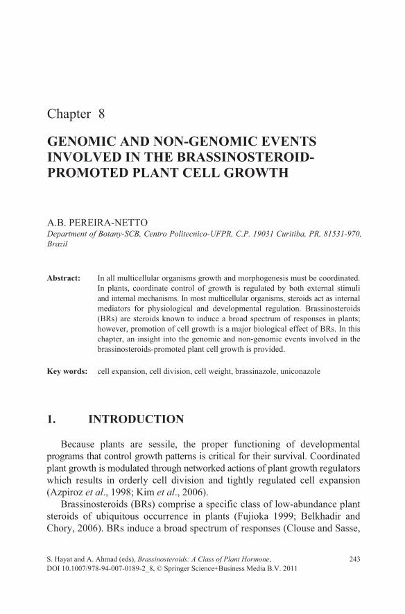
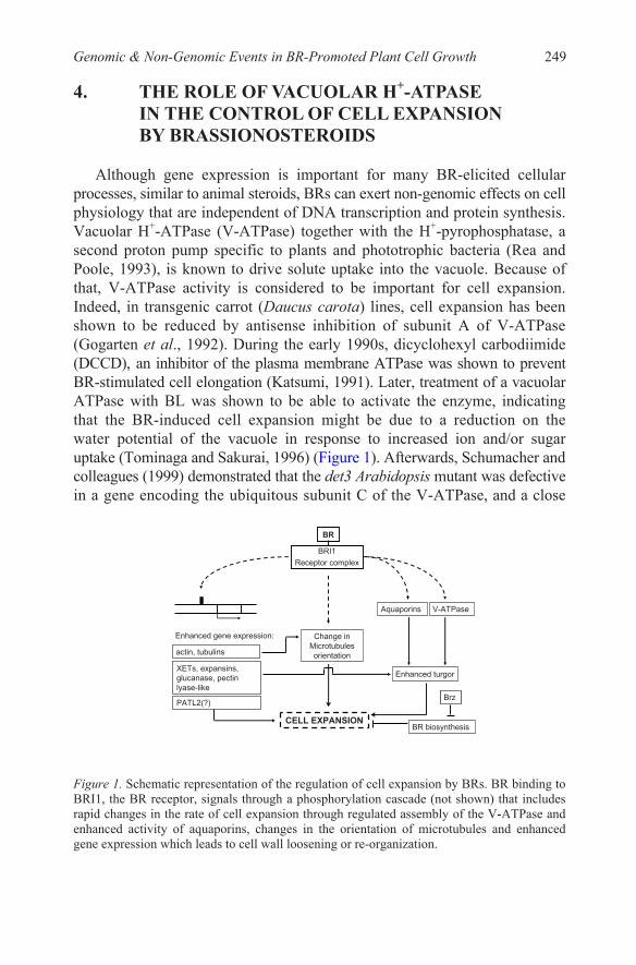
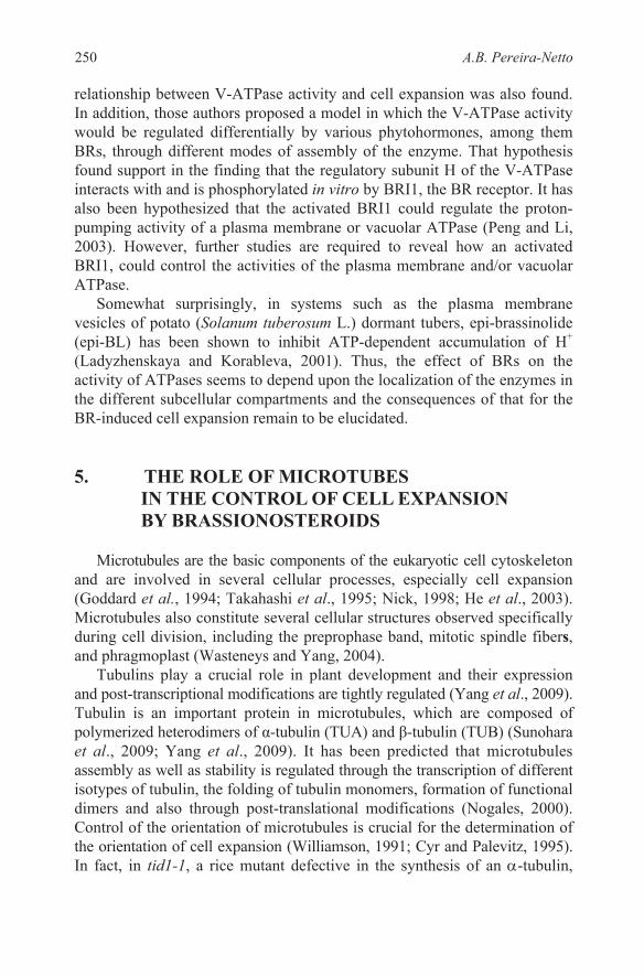
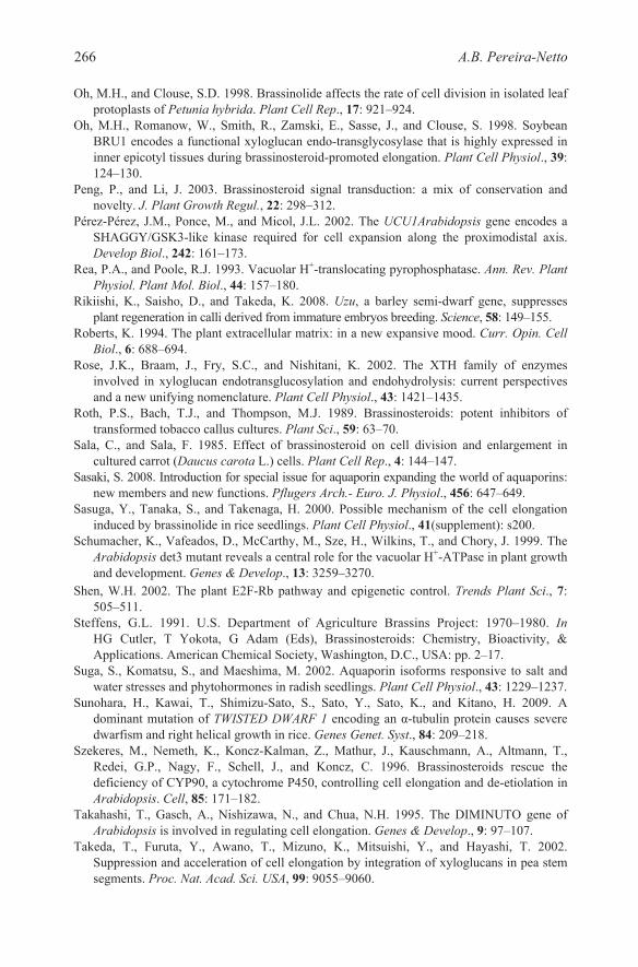
Although gene expression is important for many BR-elicited cellular processes, similar to animal steroids, BRs can exert non-genomic effects on cell physiology that are independent of DNA transcription and protein synthesis. Vacuolar H+-ATPase (V-ATPase) together with the H+-pyrophosphatase, a second proton pump specific to plants and phototrophic bacteria (Rea and Poole, 1993), is known to drive solute uptake into the vacuole. Because of that, V-ATPase activity is considered to be important for cell expansion. Indeed, in transgenic carrot (Daucus carota) lines, cell expansion has been shown to be reduced by antisense inhibition of subunit A of V-ATPase (Gogarten et al., 1992). During the early 1990s, dicyclohexyl carbodiimide (DCCD), an inhibitor of the plasma membrane ATPase was shown to prevent BR-stimulated cell elongation (Katsumi, 1991). Later, treatment of a vacuolar ATPase with BL was shown to be able to activate the enzyme, indicating that the BR-induced cell expansion might be due to a reduction on the water potential of the vacuole in response to increased ion and/or sugar uptake (Tominaga and Sakurai, 1996) (Figure 1). Afterwards, Schumacher and
Figure 1. Schematic representation of the regulation of cell expansion by BRs. BR binding to BRI1, the BR receptor, signals through a phosphorylation cascade (not shown) that includes rapid changes in the rate of cell expansion through regulated assembly of the V-ATPase and enhanced activity of aquaporins, changes in the orientation of microtubules and enhanced gene expression which leads to cell wall loosening or re-organization.
colleagues (1999) demonstrated that the det3 Arabidopsis mutant was defective in a gene encoding the ubiquitous subunit C of the V-ATPase, and a close
249Genomic & Non-Genomic Events in BR-Promoted Plant Cell Growth
relationship between V-ATPase activity and cell expansion was also found. In addition, those authors proposed a model in which the V-ATPase activity would be regulated differentially by various phytohormones, among them BRs, through different modes of assembly of the enzyme. That hypothesis found support in the finding that the regulatory subunit H of the V-ATPase interacts with and is phosphorylated in vitro by BRI1, the BR receptor. It has also been hypothesized that the activated BRI1 could regulate the proton-pumping activity of a plasma membrane or vacuolar ATPase (Peng and Li, 2003). However, further studies are required to reveal how an activated BRI1, could control the activities of the plasma membrane and/or vacuolar ATPase.
Somewhat surprisingly, in systems such as the plasma membrane vesicles of potato (Solanum tuberosum L.) dormant tubers, epi-brassinolide (epi-BL) has been shown to inhibit ATP-dependent accumulation of H+ (Ladyzhenskaya and Korableva, 2001). Thus, the effect of BRs on the activity of ATPases seems to depend upon the localization of the enzymes in the different subcellular compartments and the consequences of that for the BR-induced cell expansion remain to be elucidated.
5. THE ROLE OF MICROTUBES IN THE CONTROL OF CELL EXPANSION BY BRASSIONOSTEROIDS
Microtubules are the basic components of the eukaryotic cell cytoskeleton and are involved in several cellular processes, especially cell expansion (Goddard et al., 1994; Takahashi et al., 1995; Nick, 1998; He et al., 2003). Microtubules also constitute several cellular structures observed specifically during cell division, including the preprophase band, mitotic spindle fibers, and phragmoplast (Wasteneys and Yang, 2004).
Tubulins play a crucial role in plant development and their expression
Tubulin is an important protein in microtubules, which are composed of polymerized heterodimers of α-tubulin (TUA) and β-tubulin (TUB) (Sunohara
assembly as well as stability is regulated through the transcription of different isotypes of tubulin, the folding of tubulin monomers, formation of functional dimers and also through post-translational modifications (Nogales, 2000). Control of the orientation of microtubules is crucial for the determination of the orientation of cell expansion (Williamson, 1991; Cyr and Palevitz, 1995). In fact, in tid1-1, a rice mutant defective in the synthesis of an -tubulin,
250 A.B. Pereira-Netto
and post-transcriptional modifications are tightly regulated (Yang et al., 2009).
et al., 2009; Yang et al., 2009). It has been predicted that microtubules
abnormalities in cell expansion and division were associated with the aberrant orientation of cortical microtubules (Sunohara et al., 2009).
Reorganization of cortical microtubules is known to be important for BR-induced plant growth and it is thought that microtubule-membrane interaction is involved in this process (Mayumi and Shibaoka, 1995). BRs are known for many years to change re-configuration of microtubules to transverse orientation, in order to allow longitudinal growth. For example, in epicotyls of azuki beans (Vigna angularis), it has been demonstrated that inhibitors of cellulose biosynthesis or microtubules re-orientation inhibit BR promoted stem elongation. In addition, BL was shown to enhance the percentage of cortical microtubules transversally oriented (Mayumi and Shibaoka, 1995), which contributes to the establishment of the growth direction once the orientation of cortical microtubules usually correlates with the orientation of microfibrils (Clouse and Sasse, 1998). In another example, in the bul1/dwf7-3 mutant, a BR-deficient mutant, BR treatment induces cortical microtubule orientation and restores cell expansion (Mussig and Altmann, 2003).
Takahashi et al. (1995) and Szekeres et al. (1996) demonstrated that the dwarf phenotype of the Arabidopsis BR-deficient (Klahre et al., 1998) mutant dim could be restored to that of the wild type by treatment with exogenous BL. Interestingly, in this mutant, expression of 6α-tubulins and 5-tubulins were similar to that of the wild type, while a specific -tubulin gene, tub1, was reduced (Takahashi et al., 1995). In addition, expression of a putative -tubulin gene in chick pea (Cicer arietinum) correlated with BL-induced growth (Munoz et al., 1998). Studies using a rice BR-insensitive (Yamamuro et al., 2000) and an Arabidopsis BR-deficient mutant, bul1-1
cortical microtubules during cell elongation. Indirect immunofluorescence of
of elongating cells in the wild type was missing in the mutant. Following BR treatment, microtubules reorganized and became correctly oriented, which
molecular analyses in the study with the bul1-1 mutant showed that total tubulin was slightly lower in the mutant, compared to the wild type, although BR treatment induced no significant change in total tubulin for the mutant or for the wild type plants. At the transcriptional level, Northern blot analysis using independent probes showed a considerable reduction in 1-tubulin mRNA (TUB1) for the mutant, however, BR treatment increased and -tubulin transcript levels only in the wild type plants. Similar results were also found for another Arabidopsis BR-deficient mutant dim (Catterou et al., 2001). Thus, the microtubules reorganization observed in BR-treated bul1-1 plants was shown not to result either from an up regulation of tubulin gene
251
suggested the involvement of BRs in microtubules organization. Furthermore,
α-tubulin demonstrated that very few microtubules were present in the bul1-
(Catterou et al., 2001), confirmed the effect of BR on the organization of
1 mutant, and also that the parallel microtubules organization that is typical
Genomic & Non-Genomic Events in BR-Promoted Plant Cell Growth
expression, or from an enhancement in tubulin content. Instead, the BR promoted microtubules nucleation/organization was proposed to result from the effect of BRs on other factors such as microtubule-associated proteins (MAPs), proteins known to be important for the polymerization (Desai and Mitchison, 1997) and stabilization (Yang et al., 2009) of microtubules. In fact, plant microtubules are highly dynamic and their stability depends on the activity of various MAPs (Yang et al., 2009). Thus, BRs could, indeed, change microtubules dynamics and stability through changes in the activity of MAPs, however, that remains to be demonstrated. The expression of a -tubulin also shown to be up-regulated by BL in wild-type Arabidopsis, is reduced in the Arabidopsis insensitive mutant bri1, which indicates that - tubulin is not directly controlled by BR signaling (He et al., 2003). Although studies involving the identification of BR-regulated genes/proteins have been demonstrating that BRs promote the expression of both, -tubulin and -tubulins, accumulating evidence indicate that the BR promoted cell wall expansion might not require increased tubulin expression.
The actin cytoskeleton in plants is essential for plant growth (Muday et al., 2000). Actin is associated with microtubules to determine their orientation (Bishop and Yokota, 2001). BR-upregulated actin genes are known for quite some time (Goda et al., 2004). And, more recently, a protemics study on plasma membrane proteins using Two-dimensional Electrophoresis and Image Scanning (2-D DIGE), revealed a BR-upregulated actin, 24 hours after BR treatment (Tang et al., 2008). bru2, a BR-upregulated gene that may encode an actin effector protein that controls polymerization of actin molecules was isolated from BL-treated rice seedlings (Sasuga et al., 2000). Thus, the BR-induced cell expansion might be, at least partially, dependent on the regulation of the orientation of microtubules by promoting the synthesis of an actin effector protein. Also, the formation of cortical F-actin has been demonstrated to be under the control ROP (for Rho-of-plant) GTPases (Fu et al., 2002). And, Li and co-workers (2005) demonstrated that BR increases expression of ROP GTPases, which implicates BR in the establishment of F-actin assembly/reorganization patterns.
A proteomics study of BR-regulated proteins in Arabidopsis identified 42 BR-regulated proteins, among them three BR-induced cytoskeleton proteins, including actin 2 (At3g18780), tubulin -6 chain (At4g14960), and tubulin -4 chain (At5g44340) (Deng et al., 2007). Although the importance of the tubulin genes for the BR-induced microtubules organization is still under debate (Bishop and Yokota, 2001; Catterou et al., 2001; Munoz et al., 1998; Takahashi et al., 1995), the finding that various tubulins and especially an actin are BR-regulated proteins, provide further evidence that BR promoted cell expansion depends on changes in the pattern of microtubules organization. In addition to the cytoskeleton proteins, a member of the Sec14-like proteins,
252 A.B. Pereira-Netto
PATL2, was also shown to be BR-regulated (Deng et al., 2007). Although the precise function of the PATLs in Arabidopsis remains to be elucidated, there is evidence that supports their role in cell wall formation. More recently, another protemics study in Arabidopsis revealed PATL1 and confirmed PATL2 as BR-upregulated proteins (Tang et al., 2008). PATL2 had been previously identified in microarray studies and had also shown to be affected in bri1-116 and bzr1-1D mutants (Deng et al., 2007), which indicates that the BR promoted cell expansion might rely, partially, in changes in PATLs’s activity.
Finally, it has also been hypothesized that the activated BR receptor, BRI1, could affect the reorganization of cortical microtubules (Peng and Li, 2003). However, further studies are required to reveal how an activated BRI1 could control the nucleation/organization of the microtubules.
6. THE ROLE OF AQUAPORINS IN THE CONTROL OF CELL EXPANSION BY BRASSINOSTEROIDS
Water uptake and flow across membranes is a critical requirement for plant cell expansion. Aquaporins are proteinaceous channels formed by a superfamily of proteins containing over 450 members (Sasaki, 2008). Aquaporins in the plasma and vacuolar membranes effectively facilitate the intercellular and intracellular water transport in plants (Suga et al., 2002). Therefore, aquaporins are considered to be involved in the cell expansion process in plants. Analysis of the aquaporin activities of Arabidopsis wild type and BR biosynthetic and insensitive mutants, cpd and bri1, respectively, demonstrated that protoplasts from both mutants presented significantly lower water permeability when compared to the wild-type. In addition, BL treatment enhanced osmotic permeability of hypocotyl protoplasts from the cpd mutant significantly, though BL had no effect on the bri1 mutant (Morillon et al., 2001). In radish (Raphanus sativus), mRNA and protein levels of aquaporin isoforms in root and shoot were unaffected by BL treatment (Suga et al., 2002). This finding is consistent with various reports on microarray analysis of BR-regulated genes in which no BR-regulated aquaporin gene was found (Goda et al., 2002, 2004; Mussig et al., 2002), except for a single putatively identified aquaporin gene (Goda et al., 2004). In addition, although a proteomics study of BR-regulated proteins in Arabidopsis identified 42 BR-regulated proteins, none of them was an aquaporin gene (Deng et al., 2007). Quantitative regulation of aquaporins is assumed to be a critical step on the control of the water flow and its pathway in plant tissues. This assumption, along with data discussed in this chapter, suggests that more likely the BR
253Genomic & Non-Genomic Events in BR-Promoted Plant Cell Growth
promoted cell expansion might rely, at least partially, in changes in aquaporins activity rather than changes in aquaporins at the transcription level.
7. CONTROLL OF CELL DIVISION BY BRASSIONOSTEROIDS
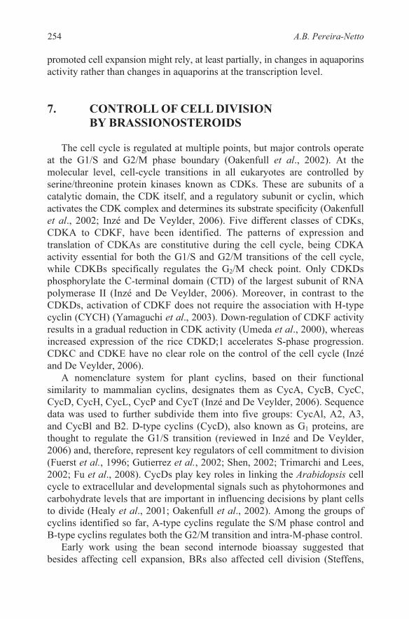
The cell cycle is regulated at multiple points, but major controls operate at the G1/S and G2/M phase boundary (Oakenfull et al., 2002). At the molecular level, cell-cycle transitions in all eukaryotes are controlled by serine/threonine protein kinases known as CDKs. These are subunits of a catalytic domain, the CDK itself, and a regulatory subunit or cyclin, which activates the CDK complex and determines its substrate specificity (Oakenfull et al., 2002; Inzé and De Veylder, 2006). Five different classes of CDKs, CDKA to CDKF, have been identified. The patterns of expression and translation of CDKAs are constitutive during the cell cycle, being CDKA activity essential for both the G1/S and G2/M transitions of the cell cycle, while CDKBs specifically regulates the G2/M check point. Only CDKDs phosphorylate the C-terminal domain (CTD) of the largest subunit of RNA polymerase II (Inzé and De Veylder, 2006). Moreover, in contrast to the CDKDs, activation of CDKF does not require the association with H-type cyclin (CYCH) (Yamaguchi et al., 2003). Down-regulation of CDKF activity results in a gradual reduction in CDK activity (Umeda et al., 2000), whereas increased expression of the rice CDKD;1 accelerates S-phase progression. CDKC and CDKE have no clear role on the control of the cell cycle (Inzé and De Veylder, 2006).
A nomenclature system for plant cyclins, based on their functional similarity to mammalian cyclins, designates them as CycA, CycB, CycC, CycD, CycH, CycL, CycP and CycT (Inzé and De Veylder, 2006). Sequence data was used to further subdivide them into five groups: CycAl, A2, A3, and CycBl and B2. D-type cyclins (CycD), also known as G1 proteins, are thought to regulate the G1/S transition (reviewed in Inzé and De Veylder, 2006) and, therefore, represent key regulators of cell commitment to division (Fuerst et al., 1996; Gutierrez et al., 2002; Shen, 2002; Trimarchi and Lees, 2002; Fu et al., 2008). CycDs play key roles in linking the Arabidopsis cell cycle to extracellular and developmental signals such as phytohormones and carbohydrate levels that are important in influencing decisions by plant cells to divide (Healy et al., 2001; Oakenfull et al., 2002). Among the groups of cyclins identified so far, A-type cyclins regulate the S/M phase control and B-type cyclins regulates both the G2/M transition and intra-M-phase control.
Early work using the bean second internode bioassay suggested that besides affecting cell expansion, BRs also affected cell division (Steffens,
254 A.B. Pereira-Netto
1991). However, whether BR played a role in cell division was an open question for quite some time because of the contradictory results reported. For example, treatment of parenchyma cell cultures of Jerusalem artichoke (Helianthus tuberosus) with nanomolar concentrations of BR, in the presence of cytokinin and auxin, led to an enhancement of at least 50% in the total number of cells (Clouse and Zurek, 1991). In addition, in protoplasts of Chinese cabbage (Brassica rapa), BR promoted cell division in a dose-dependent manner and enhanced formation of cell clusters when applied with 2,4-dichlorophenoxyacetic acid (2,4-D) and kinetin (Nakajima et al., 1996). Similar results were reported later for Petunia protoplast cultures (Oh and Clouse, 1998). However, studies carried out on carrot cell cultures (Sala and Sala, 1985), and with hormone autonomous callus or suspension cultures of Agrobacterium-transformed tobacco (Roth et al., 1989), demonstrated that BR had no stimulatory effect on cell division. Furthermore, microscopy analysis of BR-deficient and BR-insensitive Arabidopsis mutants showed that the dwarfism was mainly due to reduction in cell size rather than in cell number (Kauschmann et al., 1996). Because several of the reports on the effects of BRs on cell division originated from in vitro-grown tissue or protoplasts, and, because the effects of mitogenic factors on cell division and their interaction are very complex, the contradictory reports on the effects of BRs on cell division were suggested to be due to unbalanced concentration or combination of phytohormones in the culture media (Nakajima et al., 1996). Indeed, it became apparent that a high BR concentration in the culture medium was less effective, or even inhibitory rather than stimulatory for cell division, and that interaction with auxin(s) and cytokinin(s) was also critical in determining BR effect (Oh and Clouse, 1998). In fact, at the biochemical level, BRs are known for a long time to change endogenous cytokinin concentrations in various plant species. For example, when provided via a culture medium containing growth-limiting level of auxin, epi-BL increased the endogenous predominant cytokinins N-6-(δ-2-isopentenyl) adenine and trans-zeatin in tobacco callus tissue (Gaudinova et al., 1995). Thus, BRs might stimulate cell division by itself but also through a BR-driven enhancement of the endogenous levels of cytokinins. In a later study, Bajguz demonstrated that in synchronously dividing cultures of the alga Chlorella vulgaris, accelerated increases in cell number following BR treatment were related to increased nucleic acid and protein content (Bajguz, 2000). Furthermore, in roots of wheat (Triticum aestivum), similarly to what had been found after cytokinin treatments, increased mitotic rate and nucleoli volumes were related to treat-ment with epi-BL (Fatkhutdinova et al., 2002). Howell et al. (2007) observed great increase in the number of cells in prophase and telophase within 48 h of exposure to epiBL. More recently, a 2.8-fold increase in the mitotic
255Genomic & Non-Genomic Events in BR-Promoted Plant Cell Growth
index was found for Hordeum vulgare cv. Zafer-160 root cells originated from seeds treated with 0.5 M homobrassinolide (Kartal et al., 2009).
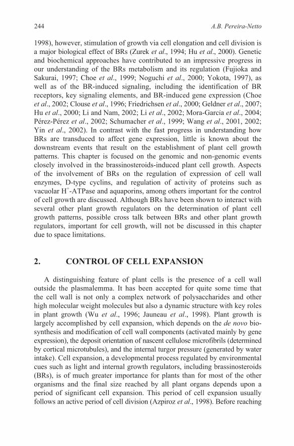
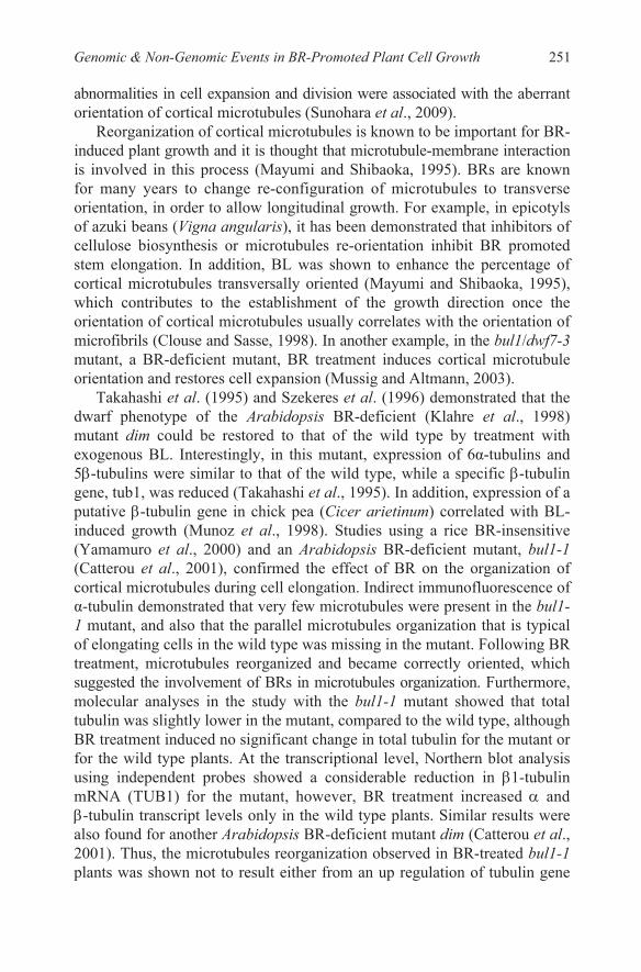
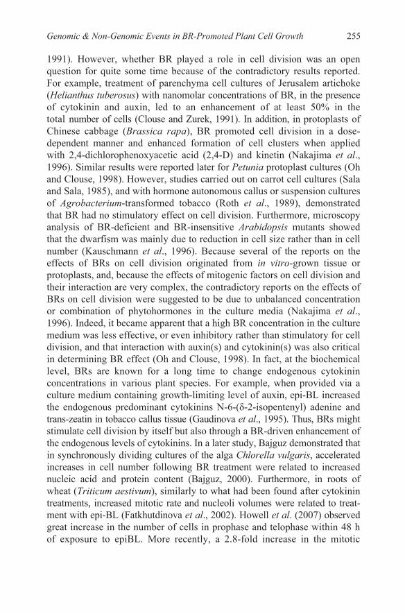
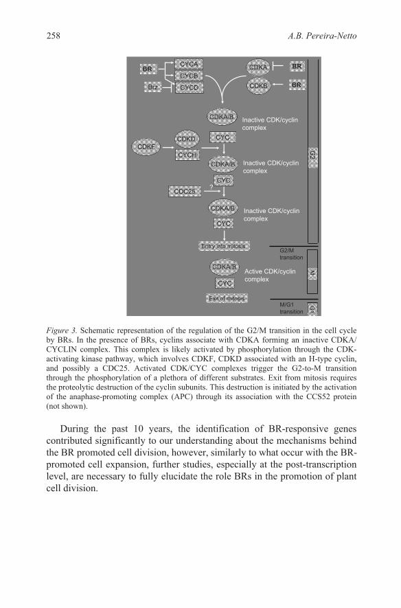
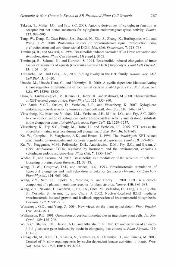
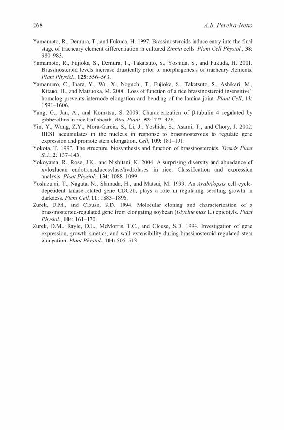
In leaves of Arabidopsis, overexpression of cycd3;1 leads to a consi-derably enhanced number of smaller cells, and cell division replaces cell expansion as the driven mechanism for leaf growth (Dewitte et al., 2003). The identification of BR-regulated genes involved in cell division such as cycd3 (Hu et al., 2000) pointed to cell division as a further mechanism, besides cell expansion, contributing to BR promoted growth, being the coordination of these diverse processes a result, at least partially, from interactions between BRs and other phytohormones (Lisso et al., 2005). During the late 90’s, evidence on the effect of BRs on cell division started to accumulate at the molecular level. In plants, CDKs are often given the nonspecific designation cdc2 because they share high amino acid sequence identity with cdc2 of fission yeast. In Arabidopsis, a cdc2b was found to be BR-induced in darkness; however, cdc2b was demonstrated to play a role in various physiological events but not in cell-cycle control (Yoshizumi et al., 1999). After that, Hu and co-workers (2000) identified BR-responsive genes in cell suspension cultures of det2, an Arabidopsis mutant deficient on BRs biosynthesis, and found that epi-BL upregulated transcription of the cycd3, a cyclin gene involved in the regulation of the G1/S transition (Figures 2 and 3). Cytokinin activates cell division through cycd3 and it is quite interesting that epi-BL was also shown to be able to effectively replace zeatin in culturing of Arabidopsis callus and suspension cells. Those findings led Hu and co-workers to conclude that epi-BL stimulated cell division through the induction of cycd3. The epi-BL-driven induction of cycd3 was further demonstrated to involve de novo protein synthesis, but no protein phosphorylation or dephosphorylation. Since this finding was apparently inconsistent with the BRI1 signal transduction pathway, Hu and co-workers performed an RNA gel-blot analysis in cell suspension culture of bri1, a BR-mutant in which the BRI1 pathway is blocked (Li and Chory, 1997). When treated with BL, bri1 cells accumulated cycd3 mRNA in a dose-dependent way similar to that of a wild-type. That finding suggested that BR-driven induction of cycd3 involves a BR signalling pathway that differs from the BRI1 pathway.
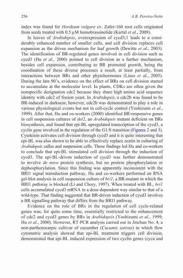
Evidence on the role of BRs in the regulation of cell cycle-related genes was, for quite some time, essentially restricted to the enhancement of cdc2 and cycd3 genes by BRs in Arabidopsis (Yoshizumi et al., 1999; Hu et al., 2000). However, RT-PCR analysis carried out in Jinchun No. 4, a non-parthenocarpic cultivar of cucumber (Cucumis sativus) in which flow cytometric analysis showed that epi-BL treatment triggers cell division, demonstrated that epi-BL induced expression of two cyclin genes (cyca and
256 A.B. Pereira-Netto
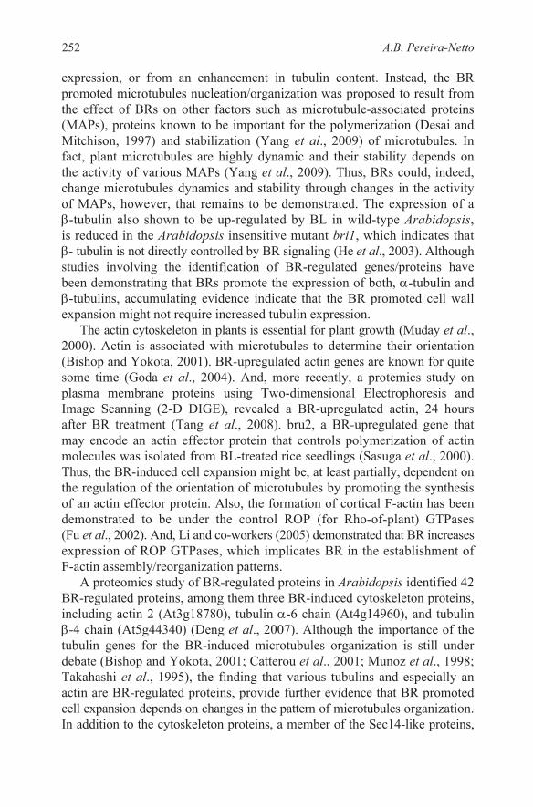
of another cdk gene, cdka, was slightly down-regulated after treatment with epi-BL (Fu et al., 2008) (Figure 3).
Figure 2. Schematic representation of the regulation of the G1/S transition in the cell cycle by BRs. In the presence of BRs, cyclins associate with CDKA forming an inactive CDKA/CYCLIN complex. This complex is likely activated by phosphorylation through the CDK-activating kinase pathway, which involves CDKF and CDKD associated with an H-type cyclin. CDKA/CYCLIN complex trigger the G1/S transition through changes in the activity of transcriptional repressor complexes. Dynamics of the transcriptional factors is not shown.
Brassinazole (Brz) is a triazole derivative known to selectively and directly binds to the DWF4 protein, a cytochrome P450 monooxygenase that catalyzes the hydroxylation of the 22-position of the side chains of BRs, an BR activation step (Choe et al., 1998; Asami et al., 2000, 2001, 2003; Yamamoto et al., 2001). Northern blot analysis showed that Brz treatment considerably repressed the expression of cscycd3;1 and cscycd3;2 in cucumber, while accumulation of cscycd3;1 transcripts after Brz treatment could be rescued by epi-BL treatment. Accumulation of the cscycd3;2 transcripts followed a similar trend (Fu et al., 2008). In a study aimed to elucidate the involvement of BRs in the progression of tracheary element differentiation in cultured Zinnia elegans cells, uniconazole was shown to suppress the accumulation of transcripts for genes that are involved in secondary wall formation. Somewhat surprisingly, the inhibitor of BRs biosynthesis did not inhibit cell division in Zinnia cells. The suppression of transcript accumulation was recoverable upon BL application (Yamamoto et al., 1997).
257Genomic & Non-Genomic Events in BR-Promoted Plant Cell Growth
cycb) besides cycd3;1, cycd3;2 and cdkb after anthesis, although the expression
G2
CYC
Entry into mitosisG2/M transition
CDKA/B
BR
CDKDCDKF
CYCA CDKA BR
CYCH
Inactive CDK/cyclin complex
Brz
CYCB
CYCD CDKB
Active CDK/cyclin complex
CYC
CDKA/B
CYC
CDKA/B
Inactive CDK/cyclin complex
CYC
CDKA/B
Exit of mitosis
M
M/G1 transition
Inactive CDK/cyclin complex
G1
CDC25?
BR
Figure 3. Schematic representation of the regulation of the G2/M transition in the cell cycle by BRs. In the presence of BRs, cyclins associate with CDKA forming an inactive CDKA/ CYCLIN complex. This complex is likely activated by phosphorylation through the CDK-activating kinase pathway, which involves CDKF, CDKD associated with an H-type cyclin, and possibly a CDC25. Activated CDK/CYC complexes trigger the G2-to-M transition through the phosphorylation of a plethora of different substrates. Exit from mitosis requires the proteolytic destruction of the cyclin subunits. This destruction is initiated by the activation of the anaphase-promoting complex (APC) through its association with the CCS52 protein (not shown).
During the past 10 years, the identification of BR-responsive genes
contributed significantly to our understanding about the mechanisms behind
promoted cell expansion, further studies, especially at the post-transcription level, are necessary to fully elucidate the role BRs in the promotion of plant cell division.
258 A.B. Pereira-Netto
the BR promoted cell division, however, similarly to what occur with the BR-
8. CONTROL OF CELL WEIGHT ACCUMULATION BY BRASSINOSTEROIDS
In Wolffia arrhiza, an aquatic monocotyledon of the duckweed-family, treatment with 10−9 M epi-BL results in 33% enhancement in fresh weight, relative to the control (Bajguz and Asami, 2005). This result indicates that limited BR supply constrain cell fresh weight accumulation in W. arrhiza.
uzu is known as a major semi-dwarf, single gene, a recessive natural mutation on the BR receptor kinase gene hvbri1 in barley (Hordeum vulgare L.). Somewhat surprisingly, in the “Ryohu” isogenic line of barley, fresh weight accumulation of 1 M 2,4dichlorophenoxy acetic (2,4-D) acid-grown calli were higher in the uzu line, compared to the non-mutant line (Rikiishi et al., 2008). However, in the “Hoshimasari” isogenic lines, no significant difference in fresh weight accumulation was found for 1 M 2,4-D-grown calli of either uzu or non-mutant line (Rikiishi et al., 2008). The reason(s) for the differential responses, towards fresh weight accumulation, found for the uzu lines and non-mutant lines of the “Ryohu” and “Hoshimasari” isogenic lines remain to be elucidated. However, the results reported for uzu line and non-mutant lines of the “Ryohu” and “Hoshimasari” isogenic lines of barley indicate that an impaired ability to sense BRs is not critical for fresh weight accumulation in barley calli.
9. CONTROL OF CELL EXPANSION BY INHIBITORS OF BR BIOSYNTHESIS
Mutants deficient in BRs biosynthesis or response have significantly contributed to increase the knowledge about BRs and their actions (Clouse and Sasse, 1998; Li and Chory, 1999; Bishop and Yokota, 2001; Clouse, 2002). However, the use of specific biosynthesis inhibitors is an alternative way for the determination of physiological functions of BRs. From the morphological standpoint, brassinazole (Brz)-treated plants, such as Arabidopsis and Lepidium sativum, display features similar to those presented by BR-deficient mutants,
treated plants, whereas no differences can be detected in the number of cells between the brassinazole-treated and non-treated plants (Asami et al., 2000). Furthermore, morphological changes induced by brassinazole are rescued by
Uniconazole, a triazole-type plant-growth retardant (Iwasaki and Shibaoka, 1991), is known to inhibit BRs biosynthesis, besides inhibiting gibberellins biosynthesis. More specifically, and similarly to Brz, uniconazole blocks the
259Genomic & Non-Genomic Events in BR-Promoted Plant Cell Growth
including dwarfism in the light (Asami et al., 2000, 2001). In contrast to non-treated Arabidopsis, hypocotyl cell expansion is reduced in the brassinazole-
the addition of exogenous BL but not by gibberellin.
step catalyzed by DWF4 in BR biosynthesis (Asami et al., 2001). In isolated Zinnia mesophyll cells, exogenously supplied uniconazole prevents uncommitted cells from trans-differentiating into tracheary elements without inhibiting cell division, being BL, but not gibberellins, able to overcome the effect (Iwasaki and Shibaoka, 1991). When the synthesis of BRs is blocked by uniconazole, not only hypocotyl elongation (Asami et al., 2000) but also preceding actin filament aggregation and microtubule bundling are inhibited (Iwasaki and Shibaoka, 1991), indicating that not proper assembly or orient-ation of components of the plant cell cytoskeleton might be involved in the uniconazole-induced inhibition of hypocotyl elongation.
10. CONTROL OF CELL WEIGHT BY INHIBITORS OF BR BIOSYNTHESIS
Brz, applied as a diastereomeric mixture of Brz2001 has been shown to inhibit fresh weight accumulation in Chlorella vulgaris, an alga, while, co-treatment with Brz2001 and BL results in normal growth in light-grown cells (Bajguz and Asami, 2004). Similarly to what had been found for C. vulgaris cells, Brz2001 was shown to reduce fresh weight accumulation, in a concentration-dependent way, in cultures of Wolffia arrhiza (Bajguz and Asami, 2005). However, co-treatment with 10−6
–5.10−6 M Brz2001 and 10−9 M epi-BL showed a weaker promotive effect on fresh weight accumulation, compared to treatment with epiBL alone. It seems to be straightforward that Brz is able to reduce fresh weight accumulation in plant cells, and this
inhibition of cell expansion. However, it remains to be elucidated.
11. CONCLUSION
Data presented in this chapter demonstrate that BR control of cell growth rely in a broad variety of mechanisms. These mechanisms are starting to be understood at the transcriptional level. However, further studies, especially at post-transcriptional level, are necessary in order to fully elucidate the
Since plant architecture is largely determined by the pattern of cell growth, increased knowledge about the networked pathways for the BRs biosynthesis and BR signaling, along with the use of selected or structurally modified BRs is expected to make it possible for us to effectively: (1) Control excessive vegetative growth in young orchards, or conversely, to promote shooting in producing orchards; (2) Inhibit lateral growth in forestry; (3) Produce plants
260 A.B. Pereira-Netto
inhibitory effect is possibly due, at least partially, to an eventual Brz-induced
mechanism(s) behind the BR-promoted cell growth.
more resistant to damage by wind and rain; 3. Improve clonal propagation techniques not only for conventional commercial applications, but also for the fastly increasing number of desirable genetically transformed plants; among many other practical applications.
12. ACKNOWLEDGEMENTS
The author thanks CNPq-Brazil for financial support.
13. REFERENCES
Asami, T., Min, Y.K., Nagata, N., Yamagishi, K., Takatsuto, S., Fujioka, S., Murofushi, N., Yamaguchi, I., and Yoshida, S. 2000. Characterization of brassinazole, a triazole-type brassinosteroid biosynthesis inhibitor. Plant Physiol., 123: 93–100.
Asami, T., Mizutani, M., Fujioka, S., Goda, H., Min, Y.K., Shimada, Y., Nakano, T., Takatsuto, S., Matsuyama, T., Nagata, N., Sakata, K., and Yoshida, S. 2001. Selective interaction of triazole derivatives with dwf4, a cytochrome p450 monooxygenase of the brassinosteroid biosynthetic pathway, correlates with brassinosteroid deficiency in planta. J. Biol. Chem., 276: 25687–25691.
Asami, T., Nakano, T., Nakashita, H., Sekimata, K., Shimada, Y., and Yoshida, S. 2003. The influence of chemical genetics on plant science: shedding light on functions and mechanism of action of brassinosteroids using biosynthesis inhibitors. J. Plant Growth Reg., 22: 336–349.
Aspinall, G.O. 1980. Chemistry of cell wall polysaccharides. In J Preiss J (Ed), Carbohydrates: Structure and Function. The Biochemistry of Plants. vol. 3. Academic Press, New York, USA: pp. 473–500.
Azpiroz, R., Wu, Y., LoCascio, J.C., and Feldmann, K.A., 1998. An Arabidopsis brassinosteroid-dependent mutant is blocked in cell elongation. Plant Cell, 10: 219–230.
Bajguz, A. 2000. Effect of brassinosteroids on nucleic acids and protein content in cultured cells of Chlorella vulgaris. Plant Physiol. Biochem., 38: 209–215.
Bajguz, A., and Asami, T. 2004. Effects of brassinazole an inhibitor of brassinosteroid biosynthesis on light- and dark-grown Chlorella vulgaris. Planta, 218: 869–877.
Bajguz, A., and Asami, T. 2005. Suppression of Wolffia arrhiza growth by brassinazole, an inhibitor of brassinosteroid biosynthesis and its restoration by endogenous 24-epibrassinolide. Phytochem., 66: 1787–1796.
Beguin, P., and Aubert, J.P. 1994. The biological degradation of cellulose. FEMS Microbiol. Rev., 13: 25–28.
Belkhadir, Y., and Chory, J. 2006. Brassinosteroid Signaling: a paradigm for steroid hormone signaling from the cell surface. Science, 314: 1410–1411.
Bishop, G.J., and Yokota, T. 2001. Plants steroid hormones, brassinosteroids: current highlights of molecular aspects on their synthesis/metabolism, transport, perception and response. Plant, Cell Physiol., 42: 114–120.
Buckeridge, M.S., Rayon, C., Urbanowics, B., Tine, A.M.A.S., and Carpita, N.C. 2004. Mixed linkage (1 3), (1 4) -D-glucans of grasses. Cereal Chem., 18: 115–127.
Campbell, P., and Braam, J. 1999. Xyloglucan endotransglycosylases: diversity of genes, enzymes and potential wall-modifying functions. Trends Plant Sci., 4: 361–366.
261Genomic & Non-Genomic Events in BR-Promoted Plant Cell Growth
Carpita, N.C., and Gibeaut, D.M. 1993. Structural models of primary cell walls in flowering plants: consistency of molecular structure with the physical properties of the walls during growth. Plant J., 3: 1–30.
Carpita, N., and McCann, M. 2000. The cell wall. In BB Buchanan, W Gruissem, RL Jones (Eds), Biochemistry and Molecular Biology of Plants. American Society of Plant Physiologists, Rockville, USA: pp. 52–108.
Catala, C., Rose, J.K.C., and Bennett, A. 1997. Auxin-regulation and spatial localization of an endo-1,4--D-glucanase and a xyloglucan endotransglycosylase in expanding tomato hypocotyls. Plant J., 12: 417–426.
Catterou, M., Dubois, F., Schaller, H., Aubanelle, L., Vilcot, B., Sangwan-Norreel, B.S., and Sangwan, R.S. 2001. Brassinosteroids, microtubules and cell elongation in Arabidopsis thaliana. II. Effects of brassinosteroids on microtubules and cell elongation in the bul1 mutant. Planta, 212: 673–683.
Cavalier, D.M., Lerouxel, O., Neumetzler, L., Yamauchi, K., Reinecke, A., Freshour, G., Zabotina, O.A., Hahn, M.G., Burgert, I., Pauly, M., Raikhel, N.V., and Keegstra, K. 2008. Disrupting two Arabidopsis thaliana xylosyltransferase genes results in plants deficient in xyloglucan, a major primary cell wall component. Plant Cell, 20: 1519–1537.
Choe, S., Dilkes, B.P., Fujioka, S., Takatsuto, S., Sakurai, A., and Feldmann, K.A. 1998. The DWF4 gene of Arabidopsis encodes a cytochrome P450 that mediates multiple 22-hydroxylation steps in brassinosteroid biosynthesis. Plant Cell, 10: 231–243.
Choe, S., Dilkes, B.P., Gregory, B.D., Ross, A.S., Yuan, H., Noguchi, T., Fujioka, S., Takatsuto, S., Tanaka, A., Yoshida, S., Tax, F.E., and Feldmann, K.A. 1999. The Arabidopsis dwarf1 mutant is defective in the conversion of 24-methylenecholesterol to campesterol in brassinosteroid biosynthesis. Plant Physiol., 119: 897–907.
Choe, S., Schmitz, R.J., Fujioka, S., Takatsuto, S., Lee, M.O., and Yoshida, S. 2002. Arabidopsis brassinosteroid insensitive dwarf12 mutants are semidominant and defective in a glycogen synthase kinase 3-like kinase. Plant Physiol., 130: 1506–1515.
Chory, J. 2001. Light, brassinosteroids, and Arabidopsis development. Proceedings of the Symposium: Plant Physiology 2000 and Beyond: Breaking the Mold, Plant Biology 2001-ASPP, Providence, Rhode Island, USA: Abstract 30005.
Clouse, S.D. 1997. Molecular genetic analysis of brassinosteroid action. Physiol. Plant., 100: 702–709.
Horm, 65: 195–223. Clouse, S.D., and Sasse, J.M. 1998. Brassinosteroids: essential regulators of plant growth and
development. Ann. Rev. Plant Phys. Plant Mol. Biol., 49: 427–451. Clouse, S.D, and Zurek, D. 1991. Molecular analysis of brassinolide action in plant growth
and development. In HG Cutler, T Yokota, G Adam (Eds), Brassinosteroids: Chemistry, Bioactivity and Applications. American Chemical Society, Washington, D.C., USA: pp. 122–140.
Clouse, S.D., Langford, M., and McMorris, T.C. 1996. A brassinosteroid-insensitive mutant in Arabidopsis thaliana exhibits multiple defects in growth and development. Plant Physiol., 111: 671–678.
Cosgrove, D. 1997. Relaxation in a high-stress environment: the molecular basis of extensible cell walls and enlargement. Plant Cell, 9: 1031–1041.
Cosgrove, D.J. 2000. Loosening of plant cell walls by expansins. Nature, 407: 321–326. Cyr, R.J., and Palevitz, B.A. 1995. Organization of cortical microtubules in plant cells. Curr.
Opin. Cell Biol., 7: 65–71.
262 A.B. Pereira-Netto
Clouse, S.D. 2002. Brassinosteroids: plant counterparts to animal steroid hormones? Vitam
Deng, Z., Zhang, X., Tang, W., Oses-Prieto, J.A., Suzuki, N., Gendron, J.M., Chen, H., Guan, S., Chalkley, R.J., Peterman, T.K., Burlingame, A.L., and Wang, Z.-Y. 2007. A proteomic study of brassinosteroid response in Arabidopsis. Mol. Cell. Proteomics, 6: 2058–2071.
Desai, A., and Mitchison, T.J. 1997. Microtubule polymerization dynamics. Ann. Rev. Cell Dev. Biol., 13: 83–117.
Dewitte, W., Riou-Khamlichi, C., Scofield, S., Healy, J.M.S., Jacqmard, A., Kilby, N.J., and Murray, J.A.H. 2003. Altered cell cycle distribution, hyperplasia, and inhibited differentiation in Arabidopsis caused by the D-type cyclin CYCD3. Plant Cell, 15: 79–92.
Fanutti, C., Gidley, M.J., and Reid, J.S.G. 1993. Action of a pure xyloglucan endo-transglycosylase (formerly called xyloglucan-specific endo-(1 4)--D-glucanase) from the cotyledons of germinated nasturtium seeds. Plant J., 3: 691–700.
Fatkhutdinova, R.A., Shakirova, F.M., Chemeris, A.V., Sabirzhanov, B.E., and Vakhitov, V.A. 2002. NOR activity in wheat species with different ploidy levels treated with phytohormones. Russ. J. Genet., 38: 1335–1338.
Friedrichsen, D.M., Joazeiro, C.A.P., Li, J., Hunter, T., and Chory, J. 2000. Brassinosteroid-insensitive-1 is a ubiquitously expressed leucine-rich repeat receptor serine/threonine kinase. Plant Physiol., 123: 1247–1256.
Fry, S.C. 1989a. The structure and functions of xyloglucan. J. Exp. Bot., 40: 1–11. Fry, S.C. 1989b. Cellulases, hemicelluloses and auxin-stimulated growth: a possible
relationship. Physiol. Plant., 75: 532–536. Fry, S.C., Smith, R.C., Renwick, K.F., Martin, D.J., Hodge, S.K., and Matthews, K.J. 1992.
Xyloglucan endotransglycosylase, a new wall-loosening enzyme activity from plants. Biochem. J., 282: 821–828.
Fu, F.Q., Mao, W.H., Shi, K., Zhou, Y.H., Asami, T., and Yu, J.Q. 2008. A role of brassinosteroids in early fruit development in cucumber. J. Exp. Bot., 59: 2299–2308.
Fu, Y., Li, H., and Yang, Z. 2002. The ROP2 GTPase controls the formation of cortical fine F-actin and the early phase of directional cell expansion during Arabidopsis organogenesis. Plant Cell, 14: 777–794.
Fuerst, R.A.U.A., Soni, R., Murray, J.A.H., and Lindsey, K., 1996. Modulation of cyclin transcript levels in cultured cells of Arabidopsis thaliana. Plant Physiol., 112: 1023–1033.
Fujioka, S. 1999. Natural occurrence of brassinosteroids in the plant kingdom. In A Sakurai, T Yokota, SD Clouse (Eds), Brassinosteroids: Steroidal Plant Hormones. Springer Verlag, Tokyo: pp. 21–45.
Fujioka, S., and Sakurai, A. 1997. Biosynthesis and metabolism of brassinosteroids. Physiol. Plant., 100: 710–715.
Gaudinova, A., Sussenbekova, H., Vojtechova, M., Kaminek, M., Eder, J., and Kohout, L. 1995. Different effects of 2 brassinosteroids on growth, auxin and cytokinin content in tobacco callus-tissue. Plant Growth Reg., 17: 121–126.
Geldner, N., Hyman, D.L., Wang, X., Schumacher, K., and Chory, J. 2007. Endosomal signaling of plant steroid receptor kinase BRI1. Genes & Develop., 21: 1598–1602.
Goda, H., Sawa, S., Asami, T., Fujioka, S., Shimada, Y., and Yoshida, S. 2004. Comprehensive comparison of auxin-regulated and brassinosteroid-regulated genes in Arabidopsis. Plant Physiol., 134: 1555–1573.
Goda, H., Shimada, Y., Asami, T., Fujioka, S., and Yoshida, S. 2002. Microarray analysis of brassinosteroid-regulated genes in Arabidopsis. Plant Physiol., 130: 1319–1334.
Goddard, R.H., Wick, S.M., Silflow, C.D., and Snustad, D.P. 1994. Microtubule components of the plant cell cytoskeleton. Plant Physiol., 104: 1–6.
263Genomic & Non-Genomic Events in BR-Promoted Plant Cell Growth
Gogarten, J.P., Fichmann, J., Braun, Y., Morgan, L., Styles, P., and Taiz, S.L., 1992. The use of antisense mRNA to inhibit the tonoplast H+ ATPase in carrot. Plant Cell., 4: 851–864.
Gutierrez, C., Ramirez-Parra, E., Castellano, M.M., and del Pozo, J.C. 2002. G(1) to S transition: more than a cell cycle engine switch. Curr. Opin. Plant Biol., 5: 480–486.
He, J.-X., Fujioka, S., Li, T.-C., Kang, S.G., Seto, H., Takatsuto, S., Yoshida, S., and Jang, J.-C. 2003. Sterols regulate development and gene expression in Arabidopsis. Plant Physiol., 131: 1258–1269.
Healy, J.M.S., Menges, M., Doonan, J.H., and Murray, J.A.H. 2001. The Arabidopsis D-type cyclins CycD2 and CycD3 both interact in vivo with the PSTAIRE cyclin-dependent Kinase Cdc2a but are differentially controlled. J. Biol. Chem., 276: 7041–7047.
Howell, W.M., Keller, G.E. III, Kirkpatrick, J.D., Jenkins, R.L., Hunsinger, R.N., and McLaughlin, E.W. 2007. Effects of the plant steroidal hormone, 24-epibrassinolide, on the mitotic index and growth of onion (Allium cepa) root tips. Genet. Mol. Res., 6: 50–58.
Hu, Y., Bao, F., and Li, J., 2000. Promotive effect of brassinosteroids on cell division involves a distinct CycD3-induction pathway in Arabidopsis. Plant J., 24: 693–701.
Inzé, D., and De Veylder, L. 2006. Cell cycle regulation in plant development. Ann. Rev. Genet., 40: 77–105.
Iwasaki, T., and Shibaoka, H. 1991. Brassinosteroids act as regulators of tracheary-element differentiation in isolated Zinnia mesophyll cells. Plant Cell Physiol., 32: 1007–1014.
Jauneau, A., Roy, S., Reis, D., and Vian, B. 1998. Probes and microscopical methods for the localization of pectins in plant cells. Int. J. Plant Sci., 159: 1–13.
Johansson, P., Brumer, III H., Baumann, M.J., Kallas, A.M., Henriksson, H., Denman, S.E., Teeri, T.T., and Jonesa, T.A. 2004. Crystal structures of a poplar xyloglucan endotransglycosylase reveal details of transglycosylation acceptor binding. Plant Cell., 16: 874–886.
Kartal, G., Temel, A., Arican, E., and Gozukirmizi, N. 2009. Effects of brassinosteroids on barley root growth, antioxidant system and cell division. Plant Growth Regul., 58: 261–267.
Katsumi, M. 1991. Physiological modes of brassinolide action in cucumber hypocotyls growth. In HG Cutler, T Yokota, G Adam (Eds), Brassinosteroids: Chemistry, Bioactivity, and Application. ACS Symp Ser 474. American Chemical Society, Washington, DC, USA: pp. 246–254.
Kauschmann, A., Jessop, A., Knocz, C., Szekeres, M., Willmitzer, L., and Altmann, T. 1996. Genetic evidence for an essential role of brassinosteroids in plant development. Plant J., 9: 701–713.
Kim, H.B., Kwon, M., Ryu, H., Fujioka, S., Takatsuto, S., Yoshida, S., An, C.S., Lee, I., Hwang, I., and Choe, S. 2006. The regulation of DWARF4 expression is likely a critical mechanism in maintaining the homeostasis of bioactive brassinosteroids in Arabidopsis. Plant Physiol., 140: 548–557.
Klahre, U., Noguchi, T., Fujioka, S., Takatsuto, S., Yokota, T., Nomura, T., Yoshida, S., and Chua N.-H. 1998. The Arabidopsis DIMINUTO/DWARF1 gene encodes a protein involved in steroid synthesis. Plant Cell, 10: 1677–1690.
Knox, J.P. 2008. Revealing the structural and functional diversity of plant cell walls. Curr. Opin. Plant Biol., 11: 308–313.
Koka, C.V., Cerny, R.E., Gardner, R.G., Noguchi, T., Fujioka, S., Takatsuto, S., Yoshida, S., and Clouse, S.D. 2000. A putative role for the tomato genes DUMPY and CURL-3 in brassinosteroid biosynthesis and response. Plant Physiol., 122: 85–98.
Ladyzhenskaya, E.P., and Korableva, N.P. 2001. Effects of growth regulators on H+ translocation across the membranes of plasma membrane vesicles from potato tuber cells. Appl. Biochem. Microbiol., 37: 521–523.
264 A.B. Pereira-Netto
Lee, Y., Choi, D., and Kende, H. 2001. Expansins: ever-expanding numbers and functions. Curr. Opin. Plant Biol., 4: 527–532.
Li, J., and Chory, J. 1997. A putative leucine-rich receptor kinase involved in brassinosteroid signal transduction. Cell, 90: 929–938.
Li, J., and Chory, J. 1999. Brassinosteroid actions in plants. J. Exp. Bot., 50: 275–282. Li, J., and Nam, K.H. 2002. Regulation of brassinosteroid singling by a GSK3/SHAGGY-like
kinase. Science, 295: 1299–1301.
Li, J., Wen, J., Lease, K.A., Doke, J.T., Tax, F.E., and Walker, J.C. 2002. BAK1, an Arabidopsis LRR receptor-like protein kinase, interacts with BRI1 and modulates brassinosteroid signaling. Cell, 110: 213–222.
Li, L., Xu, J., Xu, Z.-H., and Xue, H.-W. 2005. Brassinosteroids stimulate plant tropisms
2738–2753. Lisso, J., Steinhauser, D., Altmann, T., Kopka, J., and Mussig, C. 2005. Identification of
brassinosteroid-related genes by means of transcript co-response analyses. Nucleic Acids Res., 33: 2685–2696.
Malberg, L.M., Tamblyn Lee J.M., and Forsberg, C.W. 1992. Degradation of cellulose and hemicelluloses by rumen microorganisms. In G Winkelmann (Ed), Microbial Degradation of Natural Products. Weinheim, VCH Verlagsgesellschaft mbH: pp. 1127–1159.
Mayumi, K., and Shibaoka, H. 1995. A possible double role for brassinolide in the reorientation of cortical microtubules in the epidermal cells of Azuki bean epicotyls. Plant Cell Physiol., 36: 173–181.
Mora-García, S., Vert, G., Yin, Y., Caño-Delgado, A., Cheong, H., and Chory, J. 2004. Nuclear protein phosphatases with Kelch-repeat domains modulate the response to brassinosteroids in Arabidopsis. Genes & Develop., 18: 448–460.
Morillon, R., Catterou, M., Sangwan, R.S., Sangwan, B.S., and Lassalles, J.P. 2001. Brassinolide may control aquaporin activities in Arabidopsis thaliana. Planta, 212: 199–204.
Muday, G.K., Hu, S., and Brady, R. 2000. The actin cytoskeleton may control the polar distribution of an auxin transport protein. Gravit. Space Biol. Bull., 13: 75–83.
Munoz, F.J., Labrador, E., and Dopico, B. 1998. Brassinolides promote the expression of a new Cicer arietinum tubulin gene involved in the epicotyl elongation. Plant Mol. Biol., 37: 807–817.
Mussig, C., and Altmann, T. 2003. Genomic brassinosteroid effects. J. Plant Growth Regul., 22: 313–324.
Mussig, C., Fischer, S., and Altmann, T. 2002. Brassinosteroid-regulated gene expression. Plant Physiol., 129: 1241–1251.
Nakajima, N., Shida, A., and Toyama, S. 1996. Effects of brassinosteroid on cell division and colony formation of Chinese cabbage mesophyll protoplasts. Japan J. Crop Sci., 65: 114–118.
Nemhauser, J.L., Mockler, T.C., and Chory, J. 2004. Interdependency of brassinosteroid and auxin signaling in Arabidopsis. PLoS Biol. 2: e258.
Nick, P. 1998. Signaling to the microtubular cytoskelton in plants. Int. Rev. Cytol., 184: 33–80. Nicol, F., His, I., Jauneau, A., Vernhettes, S., Canut, H., and Hofte, H. 1998. A plasma
membrane-bound putative endo-1,4--Dglucanase is required for normal wall assembly and cell elongation in Arabidopsis. EMBO J., 17: 5563–5576.
265Genomic & Non-Genomic Events in BR-Promoted Plant Cell Growth
Nogales, E. 2000. Structural insights into microtubule function. Ann. Rev. Biochem., 69: 277–302. Noguchi, T., Fujioka, S., Choe, S., Takatsuto, S., Tax, F.E., Yoshida, S., and Feldmann, K.A.
2000. Biosynthetic pathways of brassinolide in Arabidopsis. Plant Physiol., 124: 201–209. Nomura, T., Nakayama, M., Reid, J.B., Takeuchi, Y., and Yokota, T. 1997. Blockage of
brassinosteroid biosynthesis and sensitivity causes dwarfism in garden pea. Plant Physiol., 113: 31–37.
Nomura, T., Kitasaka, Y., Takatsuto, S., Reid, J.B., Fukami, M., and Yokota, T. 1999. Brassinosteroid/sterol synthesis and plant growth as affected by lka and lkb mutations of pea. Plant Physiol., 119: 1517–1526.
Oakenfull, E.A., Riou-Khamlichi, C., and Murray, J.A.H. 2002. Plant D-type cyclins and the control of G1 progression. Philos. Transac. Royal Soc. Biol. Series, 357: 749–760.
through modulation of polar auxin transport in Brassica and Arabidopsis. Plant Cell, 17:
Oh, M.H., and Clouse, S.D. 1998. Brassinolide affects the rate of cell division in isolated leaf protoplasts of Petunia hybrida. Plant Cell Rep., 17: 921–924.
Oh, M.H., Romanow, W., Smith, R., Zamski, E., Sasse, J., and Clouse, S. 1998. Soybean BRU1 encodes a functional xyloglucan endo-transglycosylase that is highly expressed in inner epicotyl tissues during brassinosteroid-promoted elongation. Plant Cell Physiol., 39: 124–130.
Peng, P., and Li, J. 2003. Brassinosteroid signal transduction: a mix of conservation and novelty. J. Plant Growth Regul., 22: 298–312.
Pérez-Pérez, J.M., Ponce, M., and Micol, J.L. 2002. The UCU1Arabidopsis gene encodes a SHAGGY/GSK3-like kinase required for cell expansion along the proximodistal axis. Develop Biol., 242: 161–173.
Rea, P.A., and Poole, R.J. 1993. Vacuolar H+-translocating pyrophosphatase. Ann. Rev. Plant Physiol. Plant Mol. Biol., 44: 157–180.
Rikiishi, K., Saisho, D., and Takeda, K. 2008. Uzu, a barley semi-dwarf gene, suppresses plant regeneration in calli derived from immature embryos breeding. Science, 58: 149–155.
Roberts, K. 1994. The plant extracellular matrix: in a new expansive mood. Curr. Opin. Cell Biol., 6: 688–694.
Rose, J.K., Braam, J., Fry, S.C., and Nishitani, K. 2002. The XTH family of enzymes involved in xyloglucan endotransglucosylation and endohydrolysis: current perspectives and a new unifying nomenclature. Plant Cell Physiol., 43: 1421–1435.
Roth, P.S., Bach, T.J., and Thompson, M.J. 1989. Brassinosteroids: potent inhibitors of transformed tobacco callus cultures. Plant Sci., 59: 63–70.
Sala, C., and Sala, F. 1985. Effect of brassinosteroid on cell division and enlargement in cultured carrot (Daucus carota L.) cells. Plant Cell Rep., 4: 144–147.
Sasaki, S. 2008. Introduction for special issue for aquaporin expanding the world of aquaporins: new members and new functions. Pflugers Arch.- Euro. J. Physiol., 456: 647–649.
Sasuga, Y., Tanaka, S., and Takenaga, H. 2000. Possible mechanism of the cell elongation induced by brassinolide in rice seedlings. Plant Cell Physiol., 41(supplement): s200.
Schumacher, K., Vafeados, D., McCarthy, M., Sze, H., Wilkins, T., and Chory, J. 1999. The Arabidopsis det3 mutant reveals a central role for the vacuolar H+-ATPase in plant growth and development. Genes & Develop., 13: 3259–3270.
266 A.B. Pereira-Netto
Shen, W.H. 2002. The plant E2F-Rb pathway and epigenetic control. Trends Plant Sci., 7: 505–511.
Steffens, G.L. 1991. U.S. Department of Agriculture Brassins Project: 1970–1980. In HG Cutler, T Yokota, G Adam (Eds), Brassinosteroids: Chemistry, Bioactivity, & Applications. American Chemical Society, Washington, D.C., USA: pp. 2–17.
Suga, S., Komatsu, S., and Maeshima, M. 2002. Aquaporin isoforms responsive to salt and water stresses and phytohormones in radish seedlings. Plant Cell Physiol., 43: 1229–1237.
Sunohara, H., Kawai, T., Shimizu-Sato, S., Sato, Y., Sato, K., and Kitano, H. 2009. A dominant mutation of TWISTED DWARF 1 encoding an α-tubulin protein causes severe dwarfism and right helical growth in rice. Genes Genet. Syst., 84: 209–218.
Szekeres, M., Nemeth, K., Koncz-Kalman, Z., Mathur, J., Kauschmann, A., Altmann, T., Redei, G.P., Nagy, F., Schell, J., and Koncz, C. 1996. Brassinosteroids rescue the deficiency of CYP90, a cytochrome P450, controlling cell elongation and de-etiolation in Arabidopsis. Cell, 85: 171–182.
Takahashi, T., Gasch, A., Nishizawa, N., and Chua, N.H. 1995. The DIMINUTO gene of Arabidopsis is involved in regulating cell elongation. Genes & Develop., 9: 97–107.
Takeda, T., Furuta, Y., Awano, T., Mizuno, K., Mitsuishi, Y., and Hayashi, T. 2002. Suppression and acceleration of cell elongation by integration of xyloglucans in pea stem segments. Proc. Nat. Acad. Sci. USA, 99: 9055–9060.
Takeda, T., Miller, J.G., and Fry, S.C. 2008. Anionic derivatives of xyloglucan function as acceptor but not donor substrates for xyloglucan endotransglucosylase activity. Planta, 227: 893–905.
Tang, W., Deng, Z., Oses-Prieto, J.A., Suzuki, N., Zhu, S., Zhang, X., Burlingame, A.L., and Wang, Z.-Y. 2008. Proteomics studies of brassinosteroid signal transduction using prefractionation and two-dimensional DIGE. Mol. Cell. Proteomics, 7: 728–738.
Tominaga, R., and Sakurai, N. 1996. Brassinolide induces vacuolar H+-ATPase activation and stem elongation. Plant Cell Physiol., 37(Suppl.): S152.
Tominaga, R., Sakurai, N., and Kuraishi, S. 1994. Brassinolide-induced elongation of inner tissues of segments of squash (Cucurbita maxima Duch.) hypocotyls. Plant Cell Physiol., 35: 1103–1106.
Trimarchi, J.M., and Lees, J.A., 2002. Sibling rivalry in the E2F family. Nature. Rev. Mol. Cell Biol., 3: 11–20.
Umeda, M., Umeda-Hara, C., and Uchimiya, H. 2000. A cyclin-dependent kinaseactivating kinase regulates differentiation of root initial cells in Arabidopsis. Proc. Nat. Acad. Sci. USA, 97: 13396–13400.
Uozo, S., Tanaka-Ueguchi, M., Kitano, H., Hattori, K., and Matsuoka, M. 2000. Characterization of XET-related genes of rice. Plant Physiol., 122: 853–860.
Van Sandt, V.S.T., Suslov, D., Verbelen, J.-P., and Vissenberg, K. 2007. Xyloglucan endotransglucosylase activity loosens a plant cell wall. Ann. Bot., 100: 1467–1473.
Vissenberg, K., Martinez-Vilchez, I.M., Verbelen, J.P., Miller, J.G., and Fry, S.C. 2000. In vivo colocalization of xyloglucan endotransglycosylase activity and its donor substrate
Vissenberg, K., Fry, S.C., Pauly, M., Hofte, H., and Verbelen, J.P. 2005. XTH acts at the microfibril-matrix interface during cell elongation. J. Exp. Bot., 56: 673–683.
Xu, W., Campbell, P., Vargheese, A.K., and Braam, J. 1996. The Arabidopsis XET-related gene family: environmental and hormonal regulation of expression. Plant J., 9: 879–889.
Xu, W., Prugganan, M.M., Polisensky, D.H., Antosiewicz, D.M., Fry, S.C., and Braam, J. 1995. Arabidopsis TCH4, regulated by hormones and the environment, encodes a xyloglucan endotransglycosylase. Plant Cell, 7: 1555–1567.
267Genomic & Non-Genomic Events in BR-Promoted Plant Cell Growth
Wadaa, Y., and Katsumi, M. 2005. Brassinolide as a modulator of the activities of cell wall loosening proteins. Plant Biotech., 22: 33–38.
Wang, T.-W., Cosgrove, D.J., and Arteca, R.N. 1993. Brassinosteroid stimulation of hypocotyl elongation and wall relaxation in pakchoi (Brassica chinensis cv Lei-choi). Plant Physiol., 101: 965–968.
Wang, Z.Y., Seto, H., Fujioka, S., Yoshida, S., and Chory, J. 2001. BRI1 is a critical component of a plasma-membrane receptor for plant steroids. Nature, 410: 380–383.
Wang, Z.Y., Nakano, T., Gendron, J., He, J.X., Chen, M., Vafeados, D., Yang, Y.L., Fujioka, S., Yoshida, S., Asami, T., and Chory, J. 2002. Nuclear-localized BZR1 mediates brassinosteroid-induced growth and feedback suppression of brassinosteroid biosynthesis. Develop. Cell, 2: 505–513.
Wasteneys, G.O., and Yang, Z. 2004. New views on the plant cytoskeleton. Plant Physiol. 136: 3884–3891.
Williamson, R.E. 1991. Orientation of cortical microtubules in interphase plant cells. Int. Rev. Cytol., 129: 135–206.
Wu, S.C., Blumer, J.M., Darvill, A.G., and Albersheim, P. 1996. Characterization of an endo--1,4-glucanase gene induced by auxin in elongating pea epicotyls. Plant Physiol., 110: 163–170.
Yamaguchi, M., Kato, H., Yoshida, S., Yamamura, S., Uchimiya, H., and Umeda, M. 2003. Control of in vitro organogenesis by cyclin-dependent kinase activities in plants. Proc. Nat. Acad. Sci. USA, 100: 8019–8023.
in the elongation zone of Arabidopsis roots. Plant Cell, 12: 1229–1237.
Yamamoto, R., Demura, T., and Fukuda, H. 1997. Brassinosteroids induce entry into the final stage of tracheary element differentiation in cultured Zinnia cells. Plant Cell Physiol., 38: 980–983.
Yamamoto, R., Fujioka, S., Demura, T., Takatsuto, S., Yoshida, S., and Fukuda, H. 2001. Brassinosteroid levels increase drastically prior to morphogenesis of tracheary elements. Plant Physiol., 125: 556–563.
Yamamuro, C., Ihara, Y., Wu, X., Noguchi, T., Fujioka, S., Takatsuto, S., Ashikari, M., Kitano, H., and Matsuoka, M. 2000. Loss of function of a rice brassinosteroid insensitive1 homolog prevents internode elongation and bending of the lamina joint. Plant Cell, 12: 1591–1606.
Yang, G., Jan, A., and Komatsu, S. 2009. Characterization of β-tubulin 4 regulated by gibberellins in rice leaf sheath. Biol. Plant., 53: 422–428.
Yin, Y., Wang, Z.Y., Mora-Garcia, S., Li, J., Yoshida, S., Asami, T., and Chory, J. 2002. BES1 accumulates in the nucleus in response to brassinosteroids to regulate gene expression and promote stem elongation. Cell, 109: 181–191.
Yokota, T. 1997. The structure, biosynthesis and function of brassinosteroids. Trends Plant Sci., 2: 137–143.
Yokoyama, R., Rose, J.K., and Nishitani, K. 2004. A surprising diversity and abundance of xyloglucan endotransglucosylase/hydrolases in rice. Classification and expression analysis. Plant Physiol., 134: 1088–1099.
Yoshizumi, T., Nagata, N., Shimada, H., and Matsui, M. 1999. An Arabidopsis cell cycle-dependent kinase-related gene CDC2b, plays a role in regulating seedling growth in darkness. Plant Cell, 11: 1883–1896.
Zurek, D.M., and Clouse, S.D. 1994. Molecular cloning and characterization of a brassinosteroid-regulated gene from elongating soybean (Glycine max L.) epicotyls. Plant Physiol., 104: 161–170.
Zurek, D.M., Rayle, D.L., McMorris, T.C., and Clouse, S.D. 1994. Investigation of gene expression, growth kinetics, and wall extensibility during brassinosteroid-regulated stem elongation. Plant Physiol., 104: 505–513.
268 A.B. Pereira-Netto


![Brassinosteroids: Multidimensional Regulators of Plant Growth, … · Brassinosteroids: Multidimensional Regulators of Plant Growth, Development, and Stress Responses[OPEN] Trevor](https://img.pdfslide.net/doc/110x75/5ea1fb104170e7303673d39e/brassinosteroids-multidimensional-regulators-of-plant-growth-brassinosteroids.jpg)
![Reactive Oxygen Species Are Involved in Brassinosteroid-Induced … · Reactive Oxygen Species Are Involved in Brassinosteroid-Induced Stress Tolerance in Cucumber1[W] ... Vert and](https://img.pdfslide.net/doc/110x75/60ebd8840c3a8322ad22a20e/reactive-oxygen-species-are-involved-in-brassinosteroid-induced-reactive-oxygen.jpg)