Embed Size (px)
Citation preview

4/3/2018
1
Breast Biopsy from the Physicists PerspectiveBILL GEISER, MS DABRAAPM SPRING CLINICAL MEETING 2018
Conflict of Interest Statement
Member of ACR Mammography Physics Subcommittee Member of ACR Committee on Practice Parameters and Technical
Standards – Physics
Introduction Methods of Breast Biopsy
The Old Stereotactic Breast Biopsy
Ultrasound Breast Biopsy and FNA
MRI Guided Biopsy
The New Tomosynthesis Guided Breast Biopsy
The Future Molecular Breast Imaging Guided Biopsy
Contrast Enhanced Mammography Guided Biopsy

4/3/2018
2
Introduction
ACR Accreditation Program Requirements Stereotactic Breast Biopsy
Breast Ultrasound
Breast MRI
Nuclear Medicine
Breast Imaging Center of Excellence
Accreditation Programs Required for BICOE
Mammography State or ACR
Required by MQSA
FFDM Accreditation (ACR August 2016)
Physics annual testing required
Stereotactic Breast Biopsy Standard accreditation of prone or upright add on systems
No requirement for tomosynthesis guided systems yet!
Physics annual testing required
ACR Accreditation References
BICOE: (https://www.acraccreditation.org/Breast-Imaging-Center-of-Excellence)
Mammography: (https://www.acraccreditation.org/Modalities/Mammography)
https://www.acraccreditation.org/-/media/ACRAccreditation/Documents/Mammography/Requirements.pdf?la=en
Breast Ultrasound: (https://www.acraccreditation.org/Modalities/Breast-Ultrasound)
https://www.acraccreditation.org/-/media/ACRAccreditation/Documents/Breast-Ultrasound/Requirements.pdf?la=en
Stereotactic Breast Biopsy: (https://www.acraccreditation.org/Modalities/Stereotactic-Breast-Biopsy)
https://www.acraccreditation.org/-/media/ACRAccreditation/Documents/Stereotactic/Requirements.pdf?la=en

4/3/2018
3
ACR Accreditation References
Breast MRI (https://www.acraccreditation.org/Modalities/Breast-MRI)
https://www.acraccreditation.org/-/media/ACRAccreditation/Documents/Breast-MRI/Requirements.pdf?la=en
Nuclear Medicine (https://www.acraccreditation.org/Modalities/Nuclear-Medicine-and-PET)
https://www.acraccreditation.org/-/media/ACRAccreditation/Documents/NucMed-PET/Nuclear-Medicine-Requirements.pdf?la=en
Stereotactic Breast Biopsy Program Requirements Physicist
Stereotactic Breast Biopsy Annual Tests
Unit Assembly Evaluation Collimation Assessment Focal Spot Performance and System Limiting Spatial Resolution kVp Accuracy and Reproducibility Beam Quality Assessment (Half Value Layer) Automatic Exposure Control System or Manual Exposure
Performance Assessment

4/3/2018
4
Stereotactic Breast Biopsy Annual Tests (continued)
Breast Entrance Exposure, Average Glandular Dose and Exposure Reproducibility
Image Quality Evaluation Artifact Evaluation Localization Accuracy
Stereotactic Breast Biopsy Critical Tests
Half Value Layer Automatic Exposure Control Average Glandular Dose
< 300 mrad (3 mGy)
Localization Accuracy Technologist competency
Breast Ultrasound Accreditation Program Requirements
The QC tests listed in the table below are required (unless they are designated as optional) and must be performed at least annually on all machines and transducers in routine clinical use.

4/3/2018
5
Breast Ultrasound Accreditation Physics Tests
Breast MRI Accreditation Program Requirements Physicist
Breast MRI ACR Accreditation Equipment Requirements
There is no requirement for minimum field strength. However, the MR equipment must: Have a dedicated, bilateral breast coil
Be capable of simultaneous, bilateral, imaging
Meet all state and federal performance requirements, including those for: - Maximum static magnetic field strength
- Maximum rate of change of magnetic field strength (dB/dt)
- Maximum radiofrequency power deposition (specific absorption rate)
- Maximum auditory noise levels
4/3/2018
6
Breast MRI Accreditation Requirements
The medical physicist/MR scientist must perform the QC tests listed in the table below when the equipment is installed and at least annually thereafter2.
Breast MRI Accreditation Physics Tests
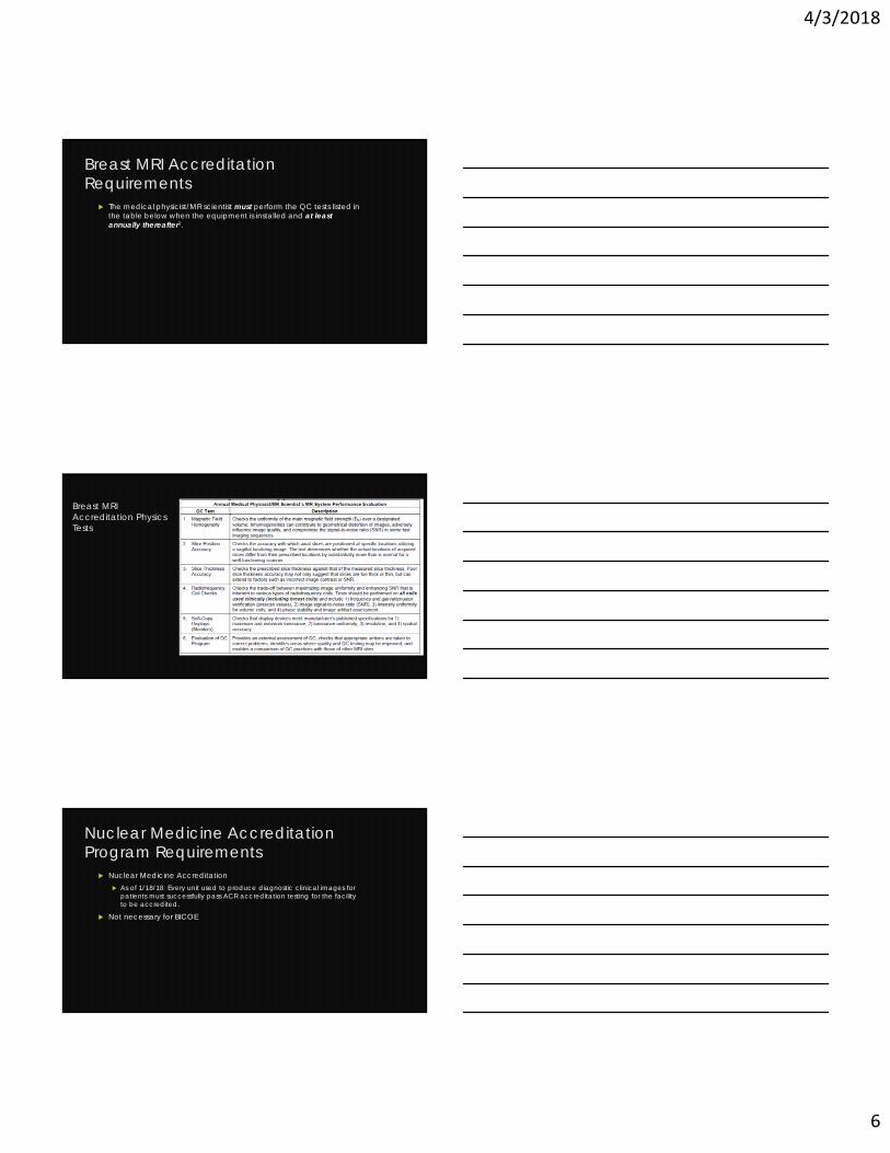
Nuclear Medicine Accreditation Program Requirements
Nuclear Medicine Accreditation As of 1/18/18: Every unit used to produce diagnostic clinical images for
patients must successfully pass ACR accreditation testing for the facility to be accredited.
Not necessary for BICOE

4/3/2018
7
Nuclear Medicine Program Requirements -Physicist
Nuclear Medicine Required Tests
Intrinsic Uniformity System Uniformity Intrinsic or System Spatial Resolution Relative Sensitivity Intrinsic or System Spatial Resolution Relative Sensitivity Energy Resolution Count Rate Parameters Monitor Evaluation System Interlocks Dose Calibrators
Clinical Cases
Stereotactic Breast Biopsy Tomosynthesis Guided Biopsy Ultrasound Guided Biopsy MRI Guided Biopsy

4/3/2018
8
Stereotactic Breast Biopsy
61 y.o. female indeterminint calcifications in the left breast on screening – BIRADS 0 additional imaging needed
Diagnostic mammogram – magnification views show suspicious calcifications recommend stereotactic biopsy
Performed biopsy
CC View
Suspicious Calcifications
BIRADS 0 additional imaging
Magnification viewsSuspicious Calcifications BIRADS 4

4/3/2018
9
Stereo Pair
-15 degrees + 15 degrees
Specimen Radiograph
Clip Placement

4/3/2018
10
No calcifications left
Pathology Results
Fibroadenoma, fibroadneomatous change and benign breast tissue with associated calcifications
Tomosynthesis Guided Biopsy –Clinical Case
64 YO female with prior left mastectomy with new 4 mm oval mass in right breast not seen on previous mammogram
Possible Cyst: Send to ultrasound Ultrasound
Irregular mass corresponding to mass on mammography
Recommend stereotactic core biopsy
Biopsy: converted from stereotactic guided to tomosynthesis guided Pathology results:
Infiltrating ductal carcinoma and DCIS

4/3/2018
11
2D CC View
30 kVp
122 mAs
W/Ag Anode/Filter
Thickness: 72 mm
Tomosynthesis slice of interest
35 kVp
74 mAs
W/Al Anode/Filter
Thickness: 72 mm
Slice 35
Slice 34RCC
2D RMLO
4/3/2018
12
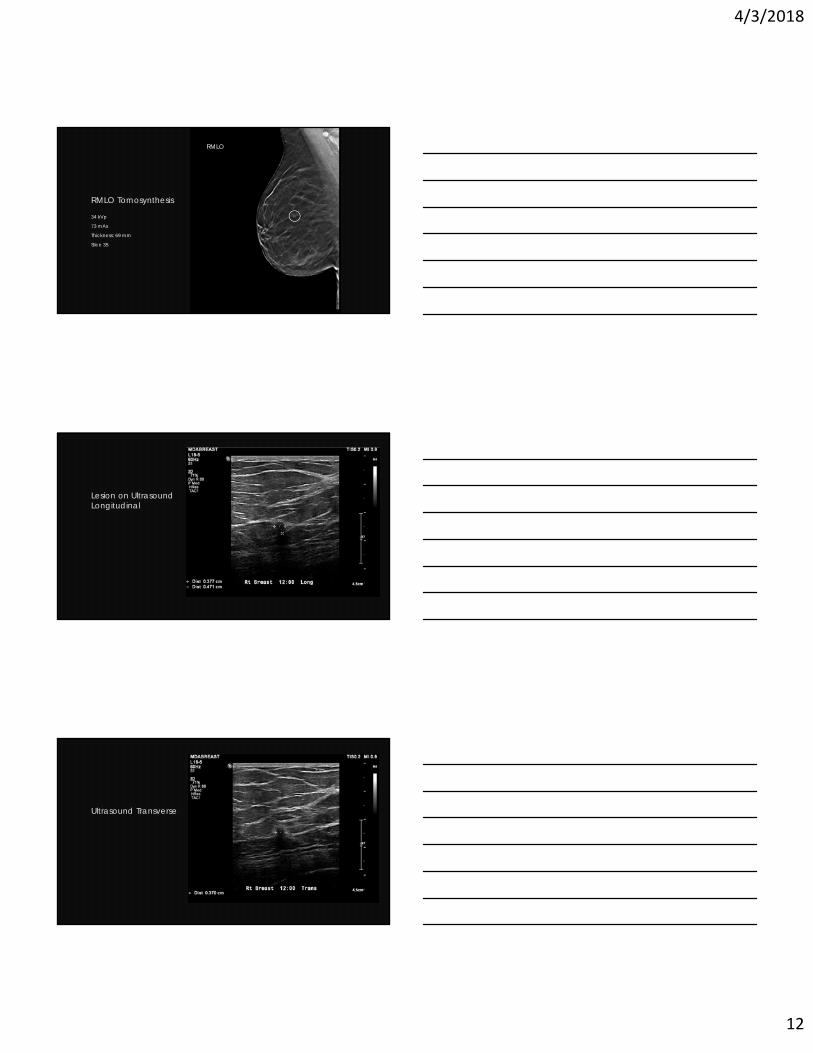
RMLO Tomosynthesis
34 kVp
73 mAs
Thickness: 69 mm
Slice 35
RMLO
Lesion on Ultrasound Longitudinal
Ultrasound Transverse

4/3/2018
13
Slice 1 of 68
Slice 35 of 68
Clip Placement

4/3/2018
14
Specimen X-ray
Ultrasound Guided Biopsy
81 year old male patient presented with palpable finding in left breast
Digital Unilateral Mammogram performed 1 cm irregular mass noted
Left breast ultrasound with possible biopsy recommended
Ultrasound performed and biopsy recommended Biopsy performed Invasive and in situ papillary carcinoma no calcifications, no
necrosis
LCC
31 kVp
55 mAs
Mo/Mo - Anode/Filter
Thickness: 61 mm

4/3/2018
15
LMLO
31 kVp
68.4 mAs
Mo/Mo - Anode Filter
Thickness: 61 mm
Ultrasound
Biopsy

4/3/2018
16
Clip Placement
MRI Guided Biopsy
65 Y.O. patient BRCA 2 positive under high risk surveillance Screening mammogram with tomosynthesis – no changes from
previous year Screening MRI – New area of enhancement
MRI guided biopsy of areas of enhancement requested
Biopsy performed with following results Predominantly Ductal Carcinoma In Situ (DCIS), high grade with
comedonecrosis and microinvasive ductal carcinoma
Prior Mammograms 2015 and 2016

4/3/2018
17
MRI Guided Biopsy
MRI Guided Biopsy

4/3/2018
18
MRI Guided Biopsy – clip placement
MBI (Molecular Breast Imaging)
The procedure Inject 8 mCi Technicium 99m Sestimibi IV
Wait a few minutes
Position patient on imaging device
Approximately 10 minutes per image (CC and MLO)

4/3/2018
19
Dilon Biopsy Device
Gamma Imaging – Guided Minimally Invasive Breast Biopsy Initial Clinical Experience, Rachel Brem et. al. AJR, March 2018
Clin Transl Imaging 2016 4-5: 367 -376
GE add on approved by FDA in November 2016

4/3/2018
20
Contrast Enhanced Biopsy
No word yet from the vendors! Most likely some form of stereotactic breast biopsy using contrast
enhanced digital mammography.