Embed Size (px)
Citation preview
Breast Cancer Early Detection
The importance of finding breast cancer early The goal of screening exams for breast cancer is to find cancers before they start to cause symptoms (like a lump that can be felt). Screening refers to tests and exams used to find a disease, such as cancer, in people who do not have any symptoms. Early detection means using an approach that lets breast cancer get diagnosed earlier than otherwise might have occurred.
Breast cancers that are found because they are causing symptoms tend to be larger and are more likely to have already spread beyond the breast. In contrast, breast cancers found during screening exams are more likely to be smaller and still confined to the breast. The size of a breast cancer and how far it has spread are some of the most important factors in predicting the prognosis (outlook) of a woman with this disease.
Most doctors feel that early detection tests for breast cancer save thousands of lives each year, and that many more lives could be saved if even more women and their health care providers took advantage of these tests. Following the American Cancer Society’s guidelines for the early detection of breast cancer improves the chances that breast cancer can be diagnosed at an early stage and treated successfully.
What are the risk factors for breast cancer? A risk factor is anything that affects your chance of getting a disease, such as cancer. Different cancers have different risk factors. For example, exposing skin to strong sunlight is a risk factor for skin cancer. Smoking is a risk factor for cancers of the lung, mouth, larynx (voice box), bladder, kidney, and several other organs.
But risk factors don’t tell us everything. Having a risk factor, or even several, does not mean that you will get the disease. Most women who have one or more breast cancer risk factors never develop the disease, while many women with breast cancer have no
apparent risk factors (other than being a woman and growing older). Even when a woman with risk factors develops breast cancer, it is hard to know just how much these factors might have contributed to her cancer.
Some risk factors, like a person’s age or race, can’t be changed. Others are related to personal behaviors such as smoking, drinking, and diet. Still others are linked to cancer-causing factors in the environment. Some factors influence risk more than others, and your risk for breast cancer can change over time, due to factors such as aging or lifestyle changes.
Breast cancer risk factors you cannot change
Gender
Simply being a woman is the main risk factor for developing breast cancer. Men can develop breast cancer, but this disease is about 100 times more common among women than men. This is probably because men have less of the female hormones estrogen and progesterone, which can promote breast cancer cell growth.
Aging
Your risk of developing breast cancer increases as you get older. About 1 out of 8 invasive breast cancers are found in women younger than 45, while about 2 of 3 invasive breast cancers are found in women age 55 or older.
Genetic risk factors
About 5% to 10% of breast cancer cases are thought to be hereditary, meaning that they result directly from gene defects (called mutations) inherited from a parent.
BRCA1 and BRCA2: The most common cause of hereditary breast cancer is an inherited mutation in the BRCA1 and BRCA2 genes. In normal cells, these genes help prevent cancer by making proteins that help keep the cells from growing abnormally. If you have inherited a mutated copy of either gene from a parent, you have a high risk of developing breast cancer during your lifetime.
Although in some families with BRCA1 mutations the lifetime risk of breast cancer is as high as 80%, on average this risk seems to be in the range of 55 to 65%. For BRCA2 mutations the risk is lower, around 45%.
Breast cancers linked to these mutations occur more often in younger women and more often affect both breasts than cancers not linked to these mutations. Women with these inherited mutations also have an increased risk for developing other cancers, particularly ovarian cancer.
In the United States, BRCA mutations are more common in Jewish people of Ashkenazi (Eastern Europe) origin than in other racial and ethnic groups, but they can occur in anyone.
Changes in other genes: Other gene mutations can also lead to inherited breast cancers. These gene mutations are much rarer and often do not increase the risk of breast cancer as much as the BRCA genes. They are not frequent causes of inherited breast cancer.
• ATM: The ATM gene normally helps repair damaged DNA. Inheriting 2 abnormal copies of this gene causes the disease ataxia-telangiectasia. Inheriting one abnormal copy of this gene has been linked to a high rate of breast cancer in some families.
• TP53: The TP53 gene gives instructions for making a protein called p53 that helps stop the growth of abnormal cells. Inherited mutations of this gene cause the Li-Fraumeni syndrome. People with this syndrome have an increased risk of breast cancer, as well as several other cancers such as leukemia, brain tumors, and sarcomas (cancers of bones or connective tissue). This is a rare cause of breast cancer.
• CHEK2: The Li-Fraumeni syndrome can also be caused by inherited mutations in the CHEK2 gene. Even when it does not cause this syndrome, it can increase breast cancer risk about twofold when it is mutated.
• PTEN: The PTEN gene normally helps regulate cell growth. Inherited mutations in this gene cause Cowden syndrome, a rare disorder in which people are at increased risk for both benign and malignant breast tumors, as well as growths in the digestive tract, thyroid, uterus, and ovaries. Defects in this gene can also cause a different syndrome called Bannayan-Riley-Ruvalcaba syndrome that is not thought to be linked to breast cancer risk. The syndromes caused by mutations in PTEN can be grouped together as PTEN Tumor Hamartoma Syndrome.
• CDH1: Inherited mutations in this gene cause hereditary diffuse gastric cancer, a syndrome in which people develop a rare type of stomach cancer at an early age. Women with mutations in this gene also have an increased risk of invasive lobular breast cancer.
• STK11: Defects in this gene can lead to Peutz-Jeghers syndrome. People affected with this disorder develop pigmented spots on their lips and in their mouths, polyps in the urinary and gastrointestinal tracts, and have an increased risk of many types of cancer, including breast cancer.
• PALB2: The PALB2 gene makes a protein that interacts with the protein made by the BRCA2 gene. Defects (mutations) in this gene can lead to an increased risk of breast cancer. It isn’t yet clear if PALB2 gene mutations also increase the risk for ovarian cancer and male breast cancer.
Genetic testing: Genetic testing can be done to look for mutations in the BRCA1 and BRCA2 genes (or less commonly in other genes such as PTEN or TP53). Although testing can be helpful in some situations, the pros and cons need to be considered carefully.
If you are considering genetic testing, it’s strongly recommended that first you talk to a genetic counselor, nurse, or doctor qualified to explain and interpret the results of these tests. It’s very important to understand what genetic testing can and can’t tell you, and to carefully weigh the benefits and risks of genetic testing before these tests are done. Testing is expensive and might not be covered by some health insurance plans.
For more information, see the American Cancer Society document Genetic Testing: What You Need to Know. You might also want to visit the National Cancer Institute Web site.
Family history of breast cancer
Breast cancer risk is higher among women whose close blood relatives have this disease.
Having a first-degree relative (mother, sister, or daughter) with breast cancer almost doubles a woman’s risk. Having 2 first-degree relatives increases her risk about 3-fold.
Although the exact risk is not known, women with a family history of breast cancer in a father or brother also have an increased risk of breast cancer.
Overall, less than 15% of women with breast cancer have a family member with this disease. This means that most (85%) women who get breast cancer do not have a family history of this disease.
Personal history of breast cancer
A woman with cancer in one breast has a 3- to 4-fold increased risk of developing a new cancer in the other breast or in another part of the same breast. This is different from a recurrence (return) of the first cancer.
Race and ethnicity
Overall, white women are slightly more likely to develop breast cancer than are African-American women, but African-American women are more likely to die of this cancer. In women under 45 years of age, however, breast cancer is more common in African-American women. Asian, Hispanic, and Native American women have a lower risk of developing and dying from breast cancer.
Dense breast tissue
Breasts are made up of fatty tissue, fibrous tissue, and glandular tissue. Someone is said to have dense breasts (on a mammogram) when they have more glandular and fibrous
tissue and less fatty tissue. Women with dense breasts on mammogram have a risk of breast cancer that is 1.2 to 2 times that of women with average breast density. Unfortunately, dense breast tissue can also make mammograms less accurate.
A number of factors can affect breast density, such as age, menopausal status, the use of certain drugs (including menopausal hormone therapy), pregnancy, and genetics.
Certain benign breast conditions
Women diagnosed with certain benign breast conditions may have an increased risk of breast cancer. Some of these conditions are more closely linked to breast cancer risk than others. Doctors often divide benign breast conditions into 3 general groups, depending on how they affect this risk.
Non-proliferative lesions: These conditions are not associated with overgrowth of breast tissue. They do not seem to affect breast cancer risk, or if they do, it’s to a very small extent. They include:
• Fibrosis and/or simple cysts (sometimes called fibrocystic changes or disease)
• Mild hyperplasia
• Adenosis (non-sclerosing)
• Phyllodes tumor (benign)
• A single papilloma
• Fat necrosis
• Duct ectasia
• Periductal fibrosis
• Squamous and apocrine metaplasia
• Epithelial-related calcifications
• Other benign tumors (lipoma, hamartoma, hemangioma, neurofibroma, adenomyoepthelioma)
Mastitis (infection of the breast) is not a lesion, but is a condition that can occur that does not increase the risk of breast cancer.
Proliferative lesions without atypia: These conditions show excessive growth of cells in the ducts or lobules of the breast tissue. They seem to raise a woman’s risk of breast cancer slightly (1½ to 2 times normal). They include:
• Usual ductal hyperplasia (without atypia)
• Fibroadenoma
• Sclerosing adenosis
• Several papillomas (called papillomatosis)
• Radial scar
Proliferative lesions with atypia: In these conditions, there is excessive growth of cells in the ducts or lobules of the breast tissue, with some of the cells no longer appearing normal. They have a stronger effect on breast cancer risk, raising it 3½ to 5 times higher than normal. These types of lesions include:
• Atypical ductal hyperplasia (ADH)
• Atypical lobular hyperplasia (ALH)
Women with a family history of breast cancer and either hyperplasia or atypical hyperplasia have an even higher risk of developing a breast cancer.
For more information on these conditions, see the separate American Cancer Society document Non-cancerous Breast Conditions.
Lobular carcinoma in situ
In lobular carcinoma in situ (LCIS) cells that look like cancer cells are growing in the lobules of the milk-producing glands of the breast, but they do not grow through the wall of the lobules. LCIS (also called lobular neoplasia) is sometimes grouped with ductal carcinoma in situ (DCIS) as a non-invasive breast cancer, but it differs from DCIS in that it doesn’t seem to become invasive cancer if it isn’t treated.
Women with lobular carcinoma in situ (LCIS) have a 7- to 11-fold increased risk of developing cancer in either breast.
Menstrual periods
Women who have had more menstrual cycles because they started menstruating early (before age 12) and/or went through menopause later (after age 55) have a slightly higher risk of breast cancer. The increase in risk may be due to a longer lifetime exposure to the hormones estrogen and progesterone.
Previous chest radiation
Women who as children or young adults were treated with radiation therapy to the chest area for another cancer (such as Hodgkin disease or non-Hodgkin lymphoma) have a significantly increased risk for breast cancer. This varies with the patient’s age when they
got radiation. If chemotherapy was also given, it might have stopped ovarian hormone production for some time, lowering the risk. The risk of developing breast cancer from chest radiation is highest if the radiation was given during adolescence, when the breasts were still developing. Radiation treatment after age 40 does not seem to increase breast cancer risk.
Diethylstilbestrol (DES) exposure
From the 1940s through the early 1970s some pregnant women were given an estrogen-like drug called DES because it was thought to lower their chances of losing the baby (miscarriage). These women have a slightly increased risk of developing breast cancer. Women whose mothers took DES during pregnancy may also have a slightly higher risk of breast cancer. For more information on DES see our document DES Exposure: Questions and Answers.
Lifestyle-related risk factors for breast cancer
Having children
Women who have not had children or who had their first child after age 30 have a slightly higher breast cancer risk overall. Having many pregnancies and becoming pregnant at an early age reduces breast cancer risk overall. Still, the effect of pregnancy is different for different types of breast cancer. For a certain type of breast cancer known as triple-negative, pregnancy seems to increase risk.
Birth control
Oral contraceptives: Studies have found that women using oral contraceptives (birth control pills) have a slightly greater risk of breast cancer than women who have never used them. Over time, this risk seems to go back to normal once the pills are stopped. Women who stopped using oral contraceptives more than 10 years ago do not appear to have any increased breast cancer risk. When thinking about using oral contraceptives, women should discuss their other risk factors for breast cancer with their health care team.
Depot-medroxyprogesterone acetate (DMPA; Depo-Provera) is an injectable form of progesterone that is given once every 3 months as birth control. A few studies have looked at the effect of DMPA on breast cancer risk. Women currently using DMPA seem to have an increase in risk, but the risk doesn’t seem to be increased if this drug was used more than 5 years ago.

Hormone therapy after menopause
Hormone therapy using estrogen (often combined with progesterone) has been used for many years to help relieve symptoms of menopause and to help prevent osteoporosis (thinning of the bones). Earlier studies suggested it might have other health benefits as well, but those benefits have not been found in more recent, better designed studies. This treatment goes by many names, such as post-menopausal hormone therapy (PHT), hormone replacement therapy (HRT), and menopausal hormone therapy (MHT).
There are 2 main types of hormone therapy. For women who still have a uterus (womb), doctors generally prescribe estrogen and progesterone (known as combined hormone therapy or HT). Progesterone is needed because estrogen alone can increase the risk of cancer of the uterus. For women who’ve had a hysterectomy (those who no longer have a uterus), estrogen alone can be prescribed. This is commonly known as estrogen replacement therapy (ERT) or just estrogen therapy (ET).
Combined hormone therapy (HT): Use of combined post-menopausal hormone therapy increases the risk of getting breast cancer. It may also increase the chances of dying from breast cancer. This increase in risk can be seen with as little as 2 years of use. Large studies have found that there is an increased risk of breast cancer related to the use of combined HT. Combined HT also increases the likelihood that the cancer may be found at a more advanced stage.
The increased risk from combined HT appears to apply only to current and recent users. A woman’s breast cancer risk seems to return to that of the general population within 5 years of stopping treatment.
The word bioidentical is sometimes used to describe versions of estrogen and progesterone with the same chemical structure as those found naturally in people. The use of these hormones has been marketed as a safe way to treat the symptoms of menopause. It’s important to realize that although there are few studies comparing “bioidentical” or “natural” hormones to synthetic versions of hormones, there is no evidence that they are safer or more effective. The use of these bioidentical hormones should be assumed to have the same health risks as any other type of hormone therapy.
Estrogen therapy (ET): The use of estrogen alone after menopause does not appear to increase the risk of developing breast cancer significantly, if at all. But when used long term (for more than 10 years), ET has been found to increase the risk of ovarian and breast cancer in some studies.
At this time there appear to be few strong reasons to use post-menopausal hormone therapy (either combined HT or ET), other than possibly for the short-term relief of menopausal symptoms. Along with the increased risk of breast cancer, combined HT also appears to increase the risk of heart disease, blood clots, and strokes. It does lower the risk of colorectal cancer and osteoporosis, but this must be weighed against the possible harms, especially since there are other effective ways to prevent and treat osteoporosis.
Although ET does not seem to increase breast cancer risk, it does increase the risk of stroke.
The decision to use HT should be made by a woman and her doctor after weighing the possible risks and benefits (including the severity of her menopausal symptoms), and considering her other risk factors for heart disease, breast cancer, and osteoporosis. If a woman and her doctor decide to try HT for symptoms of menopause, it’s usually best to use it at the lowest dose that works for her and for as short a time as possible.
Breastfeeding
Some studies suggest that breastfeeding may slightly lower breast cancer risk, especially if it is continued for 1½ to 2 years. But this has been a difficult area to study, especially in countries such as the United States, where breastfeeding for this long is uncommon.
The explanation for this possible effect may be that breastfeeding reduces a woman’s total number of lifetime menstrual cycles (the same as starting menstrual periods at a later age or going through early menopause).
Drinking alcohol
Consumption of alcohol is clearly linked to an increased risk of developing breast cancer. The risk increases with the amount of alcohol consumed. Compared with non-drinkers, women who consume 1 alcoholic drink a day have a very small increase in risk. Those who have 2 to 5 drinks daily have about 1½ times the risk of women who don’t drink alcohol. Excessive alcohol consumption is also known to increase the risk of developing several other cancers. The American Cancer Society recommends that women have no more than 1 alcoholic drink a day.
Being overweight or obese
Being overweight or obese after menopause increases breast cancer risk. Before menopause your ovaries produce most of your estrogen, and fat tissue produces a small amount of estrogen. After menopause (when the ovaries stop making estrogen), most of a woman’s estrogen comes from fat tissue. Having more fat tissue after menopause can increase your chance of getting breast cancer by raising estrogen levels. Also, women who are overweight tend to have higher blood insulin levels. Higher insulin levels have also been linked to some cancers, including breast cancer.
The connection between weight and breast cancer risk is complex, however. For example, risk appears to be increased for women who gained weight as an adult but may not be increased among those who have been overweight since childhood. Also, excess fat in the waist area may affect risk more than the same amount of fat in the hips and thighs. Researchers believe that fat cells in various parts of the body have subtle differences that may explain this.
The American Cancer Society recommends you maintain a healthy weight throughout your life by balancing your food intake with physical activity and avoiding excessive weight gain.
Physical activity
Evidence is growing that physical activity in the form of exercise reduces breast cancer risk. The main question is how much exercise is needed. In one study from the Women’s Health Initiative, as little as 1¼ to 2½ hours per week of brisk walking reduced a woman’s risk by 18%. Walking 10 hours a week reduced the risk a little more.
To reduce your risk of breast cancer, the American Cancer Society recommends that adults get at least 150 minutes of moderate intensity or 75 minutes of vigorous intensity activity each week (or a combination of these), preferably spread throughout the week.
Factors with unclear effects on breast cancer risk
Diet and vitamin intake
Many studies have looked for a link between certain diets and breast cancer risk, but so far the results have been conflicting. Some studies have indicated that diet may play a role, while others found no evidence that diet influences breast cancer risk. For example, a recent study found a higher risk of breast cancer in women who ate more red meat.
Studies have also looked at vitamin levels, again with inconsistent results. Some studies actually found an increased risk of breast cancer in women with higher levels of certain nutrients. So far, no study has shown that taking vitamins reduces breast cancer risk. This is not to say that there is no point in eating a healthy diet. A diet low in fat, low in red meat and processed meat, and high in fruits and vegetables may have other health benefits.
Most studies have found that breast cancer is less common in countries where the typical diet is low in total fat, low in polyunsaturated fat, and low in saturated fat. But many studies of women in the United States have not linked breast cancer risk to fat in the diet. Researchers are still not sure how to explain this apparent disagreement. It may be at least partly due to the effect of diet on body weight (see below). Also, studies comparing diet and breast cancer risk in different countries are complicated by other differences (such as activity level, intake of other nutrients, and genetic factors) that might also alter breast cancer risk.
More research is needed to better understand the effect of the types of fat eaten on breast cancer risk. But it is clear that calories do count, and fat is a major source of calories. High-fat diets can lead to being overweight or obese, which is a breast cancer risk factor. A diet high in fat has also been shown to influence the risk of developing several other types of cancer, and intake of certain types of fat is clearly related to heart disease risk.
Chemicals in the environment
A great deal of research has been reported and more is being done to understand possible environmental influences on breast cancer risk.
Compounds in the environment that have estrogen-like properties are of special interest. For example, substances found in some plastics, certain cosmetics and personal care products, pesticides, and PCBs (polychlorinated biphenyls) seem to have such properties. These could in theory affect breast cancer risk.
This issue understandably invokes a great deal of public concern, but at this time research does not show a clear link between breast cancer risk and exposure to these substances. Unfortunately, studying such effects in humans is difficult. More research is needed to better define the possible health effects of these and similar substances.
Tobacco smoke
For a long time, studies found no link between cigarette smoking and breast cancer. In recent years though, more studies have found that long-term heavy smoking is linked to a higher risk of breast cancer. Some studies have found that the risk is highest in certain groups, such as women who started smoking when they were young. In 2009, the International Agency for Research on Cancer concluded that there is limited evidence that tobacco smoking causes breast cancer.
An active focus of research is whether secondhand smoke increases the risk of breast cancer. Both mainstream and secondhand smoke contain chemicals that, in high concentrations, cause breast cancer in rodents. Chemicals in tobacco smoke reach breast tissue and are found in breast milk.
The evidence on secondhand smoke and breast cancer risk in human studies is controversial, at least in part because the link between smoking and breast cancer is also not clear. One possible explanation for this is that tobacco smoke may have different effects on breast cancer risk in smokers compared to those who are just exposed to secondhand smoke.
A report from the California Environmental Protection Agency in 2005 concluded that the evidence about secondhand smoke and breast cancer is “consistent with a causal association” in younger, mainly pre-menopausal women. The 2006 US Surgeon General's report, The Health Consequences of Involuntary Exposure to Tobacco Smoke, concluded that there is “suggestive but not sufficient” evidence of a link at this point. In any case, this possible link to breast cancer is yet another reason to avoid secondhand smoke.
Night work
Several studies have suggested that women who work at night, such as nurses on a night shift, may have an increased risk of developing breast cancer. This is a fairly recent finding, and more studies are looking at this issue. Some researchers think the effect may be due to changes in levels of melatonin, a hormone whose production is affected by the body’s exposure to light, but other hormones are also being studied.
Disproven or controversial breast cancer cancer risk factors
Antiperspirants
Internet and e-mail rumors have suggested that chemicals in underarm antiperspirants are absorbed through the skin, interfere with lymph circulation, and cause toxins to build up in the breast, eventually leading to breast cancer.
Based on the available evidence (including what we know about how the body works), there is little if any reason to believe that antiperspirants increase the risk of breast cancer. For more information, see our document Antiperspirants and Breast Cancer Risk.
Bras
Internet and e-mail rumors and at least one book have suggested that bras cause breast cancer by obstructing lymph flow. There is no good scientific or clinical basis for this claim, and a recent study of more than 1,500 women found no association of bra use with breast cancer risk.
Induced abortion
Several studies have provided very strong data that neither induced abortions nor spontaneous abortions (miscarriages) have an overall effect on the risk of breast cancer. For more detailed information, see our document Is Abortion Linked to Breast Cancer?
Breast implants
Several studies have found that breast implants do not increase the risk of breast cancer, although silicone breast implants can cause scar tissue to form in the breast. Implants make it harder to see breast tissue on standard mammograms, but additional x-ray pictures called implant displacement views can be used to examine the breast tissue more completely.
Breast implants may be linked to a rare type of lymphoma called anaplastic large cell lymphoma. This lymphoma has rarely been found in the breast tissue around the implants.
So far, though, there are too few cases to know if the risk of this lymphoma is really higher in women with implants.
Signs and symptoms of breast cancer Widespread use of screening mammograms has increased the number of breast cancers found before they cause any symptoms. Still some breast cancers are not found by mammograms, either because the test was not done or because even under ideal conditions mammograms do not find every breast cancer.
The most common symptom of breast cancer is a new lump or mass. A mass that is painless, hard, and has irregular edges is more likely to be cancerous, but breast cancers can be tender, soft, or rounded. They can even be painful. For this reason, it is important to have any new breast mass or lump, or breast change checked by a health care professional experienced in diagnosing breast diseases.
Other possible signs of breast cancer include:
• Swelling of all or part of a breast (even if no distinct lump is felt)
• Skin irritation or dimpling
• Breast or nipple pain
• Nipple retraction (turning inward)
• Redness, scaliness, or thickening of the nipple or breast skin
• A nipple discharge other than breast milk
Sometimes a breast cancer can spread to lymph nodes under the arm or around the collar bone and cause a lump or swelling there, even before the original tumor in the breast tissue is large enough to be felt.
Although any of these symptoms can be caused by things other than breast cancer, if you have them, they should be reported to your doctor so that he or she can find the cause.
American Cancer Society recommendations for early breast cancer detection in women without breast symptoms Women age 40 and older should have a mammogram every year and should continue to do so for as long as they are in good health.
• Current evidence supporting mammograms is even stronger than in the past. In particular, recent evidence has confirmed that mammograms offer substantial benefit for women in their 40s. Women can feel confident about the benefits associated with regular mammograms for finding cancer early. However, mammograms also have limitations. A mammogram can miss some cancers, and it may lead to follow up of findings that are not cancer.
• Women should be told about the benefits and limitations linked with yearly mammograms. But despite their limitations, mammograms are still a very effective and valuable tool for decreasing suffering and death from breast cancer.
• Mammograms should be continued regardless of a woman’s age, as long as she does not have serious, chronic health problems such as congestive heart failure, end-stage renal disease, chronic obstructive pulmonary disease, and moderate to severe dementia. Age alone should not be the reason to stop having regular mammograms. Women with serious health problems or short life expectancies should discuss with their doctors whether to continue having mammograms.
Women in their 20s and 30s should have a clinical breast exam (CBE) as part of a periodic (regular) health exam by a health professional preferably every 3 years. Starting at age 40, women should have a CBE by a health professional every year.
• CBE is done along with mammograms and offers a chance for women and their doctor or nurse to discuss changes in their breasts, early detection testing, and factors in the woman’s history that might make her more likely to have breast cancer.
• There may be some benefit in having the CBE shortly before the mammogram. The exam should include instruction for the purpose of getting more familiar with your own breasts. Women should also be given information about the benefits and limitations of CBE and breast self-exam (BSE). The chance of breast cancer occurring is very low for women in their 20s and gradually increases with age. Women should be told to promptly report any new breast symptoms to a health professional.
Breast self-exam (BSE) is an option for women starting in their 20s. Women should be told about the benefits and limitations of BSE. Women should report any breast changes to their health professional right away.
• Research has shown that BSE plays a small role in finding breast cancer compared with finding a breast lump by chance or simply being aware of what is normal for each woman. Some women feel very comfortable doing BSE regularly (usually monthly after their period) which involves a systematic step-by-step approach to examining the look and feel of one’s breasts. Other women are more comfortable simply feeling their breasts in a less systematic approach, such as while showering or getting dressed or doing an occasional thorough exam.
• Sometimes, women are so concerned about “doing it right” that they become stressed over the technique. Doing BSE regularly is one way for women to know how their breasts normally look and feel and to notice any changes. The goal, with or without BSE, is to report any breast changes to a doctor or nurse right away.
• Women who choose to use a step-by-step approach to BSE should have their BSE technique reviewed during their physical exam by a health professional. It is okay for women to choose not to do BSE or not to do it on a regular schedule such as once every month. However, by doing the exam regularly, you get to know how your breasts normally look and feel and you can more readily find any changes. If a change occurs, such as development of a lump or swelling, skin irritation or dimpling, nipple pain or retraction (turning inward), redness or scaliness of the nipple or breast skin, or a discharge other than breast milk (such as staining of your sheets or bra), you should see your health care professional as soon as possible for evaluation. Remember that most of the time, however, these breast changes are not cancer.
Women who are at high risk for breast cancer based on certain factors should get an MRI and a mammogram every year.
This includes women who:
• Have a lifetime risk of breast cancer of about 20% to 25% or greater, according to risk assessment tools that are based mainly on family history (such as the Claus model - see below)
• Have a known BRCA1 or BRCA2 gene mutation
• Have a first-degree relative (parent, brother, sister, or child) with a BRCA1 or BRCA2 gene mutation, and have not had genetic testing themselves
• Had radiation therapy to the chest when they were between the ages of 10 and 30 years
• Have Li-Fraumeni syndrome, Cowden syndrome, or Bannayan-Riley-Ruvalcaba syndrome, or have first-degree relatives with one of these syndromes
The American Cancer Society recommends against MRI screening for women whose lifetime risk of breast cancer is less than 15%.
There is not enough evidence to make a recommendation for or against yearly MRI screening for women who have a moderately increased risk of breast cancer (a lifetime risk of 15% to 20% according to risk assessment tools that are based mainly on family history) or who may be at increased risk of breast cancer based on certain factors, such as:
• Having a personal history of breast cancer, ductal carcinoma in situ (DCIS), lobular carcinoma in situ (LCIS), atypical ductal hyperplasia (ADH), or atypical lobular hyperplasia (ALH)
• Having dense breasts (“extremely” or “heterogeneously” dense) as seen on a mammogram
If MRI is used, it should be in addition to, not instead of, a screening mammogram. This is because although an MRI is a more sensitive test (it’s more likely to detect cancer than a mammogram), it may still miss some cancers that a mammogram would detect.
For most women at high risk, screening with MRI and mammograms should begin at age 30 years and continue for as long as a woman is in good health. But because the evidence is limited about the best age at which to start screening, this decision should be based on shared decision-making between patients and their health care providers, taking into account personal circumstances and preferences.
Several risk assessment tools, with names such as the Gail model, the Claus model, and the Tyrer-Cuzick model, are available to help health professionals estimate a woman’s breast cancer risk. These tools give approximate, rather than precise, estimates of breast cancer risk based on different combinations of risk factors and different data sets.
Because the different tools use different factors to estimate risk, they may give different risk estimates for the same woman. For example, the Gail model bases its risk estimates on certain personal risk factors, like current age, age at menarche (first menstrual period) and history of prior breast biopsies, along with any history of breast cancer in first-degree relatives. In contrast, the Claus model estimates risk based only on family history of breast cancer in both first and second-degree relatives. These 2 models could easily give different estimates for the same person.
Risk assessment tools (like the Gail model, for example) that are not based mainly on family history are not appropriate to use with the ACS guidelines to decide if a woman should have MRI screening. The use of any of the risk assessment tools and its results should be discussed by a woman with her doctor.
It is recommended that women who get a screening MRI do so at a facility that can do an MRI-guided breast biopsy at the same time if needed. Otherwise, the woman will have to have a second MRI exam at another facility when she has the biopsy.
There is no evidence right now that MRI is an effective screening tool for women at average risk. While MRI is more sensitive than mammograms, it also has a higher false-positive rate (it is more likely to find something that turns out not to be cancer). This would lead to unneeded biopsies and other tests in many of the women screened, which can lead to a lot of worry and anxiety.
The American Cancer Society believes the use of mammograms, MRI (in women at high risk), clinical breast exams, and finding and reporting breast changes early, according to
the recommendations outlined above, offers women the best chance to reduce their risk of dying from breast cancer. This approach is clearly better than any one exam or test alone.
Without question, a physical exam of the breast without a mammogram would miss the opportunity to detect many breast cancers that are too small for a woman or her doctor to feel but can be seen on mammograms. Mammograms are a sensitive screening method, but a small percentage of breast cancers do not show up on mammograms but can be felt by a woman or her doctors. For women at high risk of breast cancer, such as those with BRCA gene mutations or a strong family history, both MRI and mammogram exams of the breast are recommended.
Mammograms A mammogram is an x-ray of the breast. A diagnostic mammogram is used to diagnose breast disease in women who have breast symptoms or an abnormal result on a screening mammogram. Screening mammograms are used to look for breast disease in women who are asymptomatic; that is, those who appear to have no breast problems. Standard screening mammograms take 2 views (x-ray pictures taken from different angles) of each breast, while diagnostic mammograms may take more views of the breast. Women who are breastfeeding can still get mammograms, although these are probably not quite as accurate because the breast tissue tends to be dense.
For some women, such as those with breast implants, additional pictures may be needed to include as much breast tissue as possible. Breast implants make it harder to see breast tissue on standard mammograms, but additional x-ray pictures with implant displacement and compression views can be used to more completely examine the breast tissue. If you have implants, it is important that you have your mammograms done by someone skilled in the techniques used for women with implants.
Strict guidelines ensure that mammogram equipment is safe and uses the lowest dose of radiation possible. Many people are concerned about the exposure to x-rays, but the level of radiation used in modern mammograms is not thought to significantly increase the risk for breast cancer.
Modern mammogram equipment uses very low levels of radiation, on average a total dose of about 0.4 mSv for a typical mammogram with 2 views of both breasts (a mSv is a measure of radiation dose).
To put dose into perspective, people in the US are normally exposed to an average of about 3 mSv of radiation each year just from their natural surroundings (this is called background radiation). This means that the dose from a mammogram is the same as about 7 weeks of background radiation.
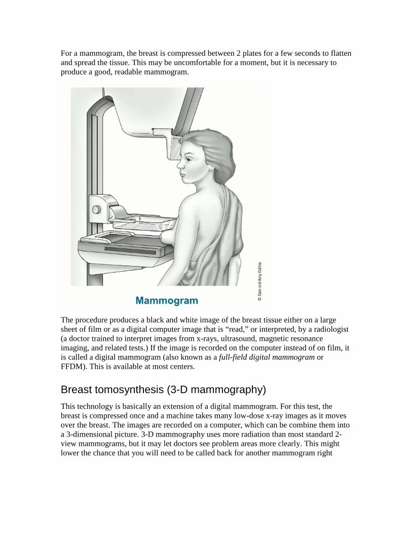
For a mammogram, the breast is compressed between 2 plates for a few seconds to flatten and spread the tissue. This may be uncomfortable for a moment, but it is necessary to produce a good, readable mammogram.
The procedure produces a black and white image of the breast tissue either on a large sheet of film or as a digital computer image that is “read,” or interpreted, by a radiologist (a doctor trained to interpret images from x-rays, ultrasound, magnetic resonance imaging, and related tests.) If the image is recorded on the computer instead of on film, it is called a digital mammogram (also known as a full-field digital mammogram or FFDM). This is available at most centers.
Breast tomosynthesis (3-D mammography)
This technology is basically an extension of a digital mammogram. For this test, the breast is compressed once and a machine takes many low-dose x-ray images as it moves over the breast. The images are recorded on a computer, which can be combine them into a 3-dimensional picture. 3-D mammography uses more radiation than most standard 2-view mammograms, but it may let doctors see problem areas more clearly. This might lower the chance that you will need to be called back for another mammogram right
away. It may also be able to find more cancers. Breast tomosynthesis is not widely available, and its role in screening and diagnosing breast cancer is still not clear.
What the doctor looks for on your mammogram
The doctor reading your mammogram will look for several types of changes:
Calcifications are tiny calcium deposits within the breast tissue that appear as small white spots on the films. They may or may not be caused by cancer. There are 2 types of calcifications, macrocalcifications which are coarse (larger) and are linked with benign (non-cancerous) conditions, and microcalcifications which are smaller and can sometimes mean that cancer is present. If microcalcifications look suspicious for cancer based on their shape and layout, a biopsy will be recommended.
A mass, which may occur with or without calcifications, is another important change seen on a mammogram. Masses are areas that look abnormal and they can be many things, including cysts (non-cancerous, fluid-filled sacs), cancers, and non-cancerous solid tumors (such as fibroadenomas).
Cysts can be simple fluid-filled sacs (known as simple cysts) or can be partially solid (known as complex cysts). Simple cysts are benign and don’t need to be biopsied. Any other type of mass (such as a complex cyst or a solid tumor) might need to be biopsied to be sure it isn’t cancer.
• A cyst and a tumor can feel alike on a physical exam. They can also look the same on a mammogram. To confirm that a mass is really a cyst, a breast ultrasound is often done. Sometimes the fluid in the cyst is removed with a thin, hollow needle.
• If a mass is not a simple cyst (that is, if it is at least partly solid), then you might need to have more imaging tests. Some masses can be watched with periodic mammograms, while others may need a biopsy. The size, shape, and margins (edges) of the mass help the radiologist determine if cancer is likely to be present.
Having your previous mammograms available for the radiologist is very important. They can help show that a mass or calcification has not changed for many years. This would mean that it is probably a benign condition and a biopsy is not needed.
The radiologist will also assess your breast density. Breast density is based on how much of your breast is made up fatty tissue vs. how much is made up of fibrous and glandular tissue.
If your breast density is higher than average, the letter you get about your mammogram results may say that you have dense breasts. Dense breasts are very common and don’t mean that there is anything wrong. Almost half of women have dense breasts on a screening mammogram. Although dense breast tissue can make it harder to find cancers on a mammogram, at this time, experts do not agree what other tests, if any, should be
done in addition to mammograms in women with dense breasts who are not high risk based on other factors.
Limitations of mammograms
A mammogram cannot prove that an abnormal area is cancer. To confirm whether cancer is present, a small amount of tissue must be removed and looked at under a microscope. This procedure is called a biopsy. For more information, see our document For Women Facing a Breast Biopsy.
You should also be aware that mammograms are done to find cancers that can’t be felt. If you have a breast lump, you should have it checked by your doctor, who may recommend a biopsy even if your mammogram result is normal.Mammograms are not perfect at finding breast cancer. They do not work as well in women with dense breasts, since dense breasts can hide a tumor. Dense breasts are more common in younger women, pregnant women, and women who are breastfeeding, but any woman can have dense breasts.
This can be a problem for younger women who need breast screening because they are at high risk for breast cancer (because of gene mutations, a strong family history of breast cancer, or other factors). This is one of the reasons that the American Cancer Society recommends MRI scans in addition to mammograms for screening in these women.
At this time, American Cancer Society guidelines do not contain recommendations for additional testing to screen women with dense breasts who aren’t at high risk of breast cancer based on other risk factors.
For more information about mammograms, see our document Mammograms and Other Breast Imaging Procedures.
Tips for having a mammogram
Here are some useful suggestions for making sure that you receive a quality mammogram:
• If it is not posted in a place you can see it near the receptionist’s desk, ask to see the FDA certificate that is issued to all facilities that offer mammography. The FDA requires all facilities to meet high professional standards of safety and quality in order to be a provider of mammography services. A facility may not provide mammography without certification.
• Use a facility that either specializes in mammography or does many mammograms a day.
• If you are satisfied that the facility is of high quality, continue to go there on a regular basis so that your mammograms can be compared from year to year.
• If you are going to a facility for the first time, bring a list of the places, dates of mammograms, biopsies, or other breast treatments you have had before.
• If you have had mammograms at another facility, you should make every attempt to get those mammograms to bring with you to the new facility (or have them sent there) so that they can be compared to the new ones.
• Try to schedule your mammogram at a time of the month when your breasts are not tender or swollen to help reduce discomfort and assure a good picture. Try to avoid the week right before your period.
• On the day of the exam, don’t wear deodorant or antiperspirant. Some of these contain substances that can interfere with the reading of the mammogram by appearing on the x-ray film as white spots.
• You may find it easier to wear a skirt or pants, so that you’ll only need to remove your blouse for the exam.
• Always describe any breast symptoms or problems that you are having to the technologist who is doing the mammogram. Be prepared to describe any medical history that could affect your breast cancer risk − such as surgery, hormone use, or family or personal history of breast cancer. Also discuss any new findings or problems in your breasts with your doctor or nurse before having a mammogram.
• If you do not hear from your doctor within 10 days, do not assume that your mammogram result was normal. Call your doctor or the facility.
What to expect when you get a screening mammogram
• To have a mammogram you must undress above the waist. The facility will give you a wrap to wear.
• A technologist will be there to position your breasts for the mammogram. Most technologists are women. You and the technologist are the only ones in the room during the mammogram.
• To get a high-quality mammogram picture, it is necessary to flatten the breast slightly. The technologist places the breast on the mammogram machine’s lower plate, which is made of metal and has a drawer to hold the x-ray film or the camera to produce a digital image. The upper plate, made of plastic, is lowered to compress the breast for a few seconds while the picture is taken.
• The whole procedure takes about 20 minutes. The actual breast compression only lasts a few seconds.
• You may feel some discomfort when your breasts are compressed, and for some women compression can be painful. Try not to schedule a mammogram when your breasts are likely to be tender, as they may be just before or during your period.
• Although usually 2 views of each breast are usually taken for a screening mammogram, for some women, such as those with breast implants, additional pictures may be needed.
• All mammogram facilities are now required to send a copy of your results in simple language to you within 30 days. Generally, you will be contacted within 5 working days if there is a problem with the mammogram.
• Being called back for more testing does not mean that you have cancer. In fact, less than 10% of women who are called back for more tests are found to have breast cancer. Being called back occurs fairly often, and it usually just means an additional image or an ultrasound needs to be done to look at an area more clearly. This is more common for first mammograms (or when there is no previous mammogram to look at) and in mammograms done in women before menopause. It may be slightly less common for digital mammograms.
• Only 2 to 4 screening mammograms of every 1,000 lead to a diagnosis of cancer.
If you are a woman 40 or over, you should get a mammogram every year. You can schedule the next one while you’re there at the facility. Or, you can ask for a reminder to schedule it as the date gets closer.
For more information on mammograms and other imaging tests for early detection and diagnosis of breast diseases, see our document Mammograms and Other Breast Imaging Procedures.
Magnetic resonance imaging For certain women at high risk for breast cancer, the American Cancer Society recommends screening magnetic resonance imaging (MRI) along with a yearly mammogram. MRI is not recommended as a screening tool by itself, because although it is a sensitive test, it can still miss some cancers that mammograms would detect. Breast MRI is also sometimes be used in other situations, such as to better examine suspicious areas found by a mammogram or to look more closely at the breast in someone who hase already been diagnosed with breast cancer.
MRI scans use magnets and radio waves instead of x-rays to produce very detailed, cross-sectional images of the body. The most useful MRI exams for breast imaging use a contrast material (called gadolinium) that is injected into a catheter in a vein (IV) in the arm before or during the exam. This improves the ability of the MRI to clearly show breast tissue details.
Although MRI can find some cancers not seen on than mammogram, it is also more likely to find something that turns out not to be cancer (called a false positive). False-positive findings have to be checked out to know that cancer isn’t present, which means coming back for further tests and/or biopsies. This is why MRI is not recommended as a screening test for women at average risk of breast cancer, as it would result in unneeded biopsies and other tests in a large portion of these women.
Just as mammography uses x-ray machines that are specially designed to image the breasts, breast MRI also requires special equipment. However, not all hospitals and imaging centers have dedicated breast MRI equipment available. It is also important that you have your screening MRIs at facilities that can perform an MRI-guided breast biopsy. Otherwise, the entire scan will need to be repeated at another facility when the biopsy is done.
Because breast MRI is expensive, it often needs to be approved by an insurance company before the scan is done. Most private insurance that pays for mammogram screening will probably also pay for MRI for screening tests if a woman can be shown to be at high risk. It can help to go to a center with a high-risk clinic, where the staff has experience getting approval for breast MRIs.
What to expect when you get a breast MRI
MRI scans can take a long time—often up to an hour. For a breast MRI, you have to lie inside a narrow tube, face down, on a platform specially designed for the procedure. The platform has openings for each breast that allow them to be imaged without being compressed. The platform contains the sensors needed to capture the MRI image. It is important to stay very still throughout the exam.
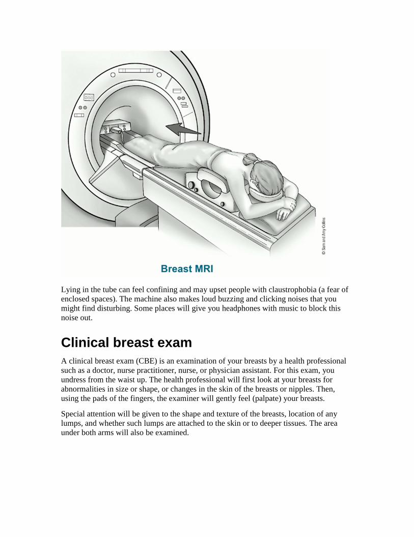
Lying in the tube can feel confining and may upset people with claustrophobia (a fear of enclosed spaces). The machine also makes loud buzzing and clicking noises that you might find disturbing. Some places will give you headphones with music to block this noise out.
Clinical breast exam A clinical breast exam (CBE) is an examination of your breasts by a health professional such as a doctor, nurse practitioner, nurse, or physician assistant. For this exam, you undress from the waist up. The health professional will first look at your breasts for abnormalities in size or shape, or changes in the skin of the breasts or nipples. Then, using the pads of the fingers, the examiner will gently feel (palpate) your breasts.
Special attention will be given to the shape and texture of the breasts, location of any lumps, and whether such lumps are attached to the skin or to deeper tissues. The area under both arms will also be examined.
The CBE is a good time for women who don’t know how to examine their breasts to learn the right way to do it from their health care professionals. Ask your doctor or nurse to teach you and watch your technique.
Breast awareness and self-exam Beginning in their 20s, women should be told about the benefits and limitations of breast self-exam (BSE). Even those who choose not to do BSE should be aware of how their breasts normally look and feel and report any new breast changes to a health professional as soon as they are found. Finding a breast change does not necessarily mean there is a cancer.
A woman can notice changes by knowing how her breasts normally look and feel and feeling her breasts for changes (breast awareness), or by choosing to use a step-by-step approach (with a BSE) and using a specific schedule to examine her breasts.
Women with breast implants can do BSE. It may be useful to have the surgeon help identify the edges of the implant so that you know what you are feeling. There is some thought that the implants push out the breast tissue and may make it easier to examine.
Women who are pregnant or breastfeeding can also choose to examine their breasts regularly.
It is acceptable for women to choose not to do BSE or to do BSE occasionally. Women who choose not to do BSE should still know how their breasts normally look and feel and report any changes to their doctor right away.
If you choose to do BSE, the following information provides a step-by-step approach for the exam. The best time for a woman to examine her breasts is when they are not tender or swollen. Women who examine their breasts should have their technique reviewed during their periodic health exams by their health care professional.
How to examine your breasts
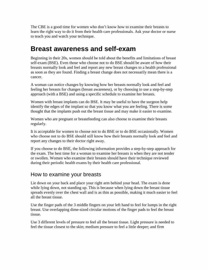
Lie down on your back and place your right arm behind your head. The exam is done while lying down, not standing up. This is because when lying down the breast tissue spreads evenly over the chest wall and is as thin as possible, making it much easier to feel all the breast tissue.
Use the finger pads of the 3 middle fingers on your left hand to feel for lumps in the right breast. Use overlapping dime-sized circular motions of the finger pads to feel the breast tissue.
Use 3 different levels of pressure to feel all the breast tissue. Light pressure is needed to feel the tissue closest to the skin; medium pressure to feel a little deeper; and firm
pressure to feel the tissue closest to the chest and ribs. It is normal to feel a firm ridge in the lower curve of each breast, but you should tell your doctor if you feel anything else out of the ordinary. If you’re not sure how hard to press, talk with your doctor or nurse. Use each pressure level to feel the breast tissue before moving on to the next spot.
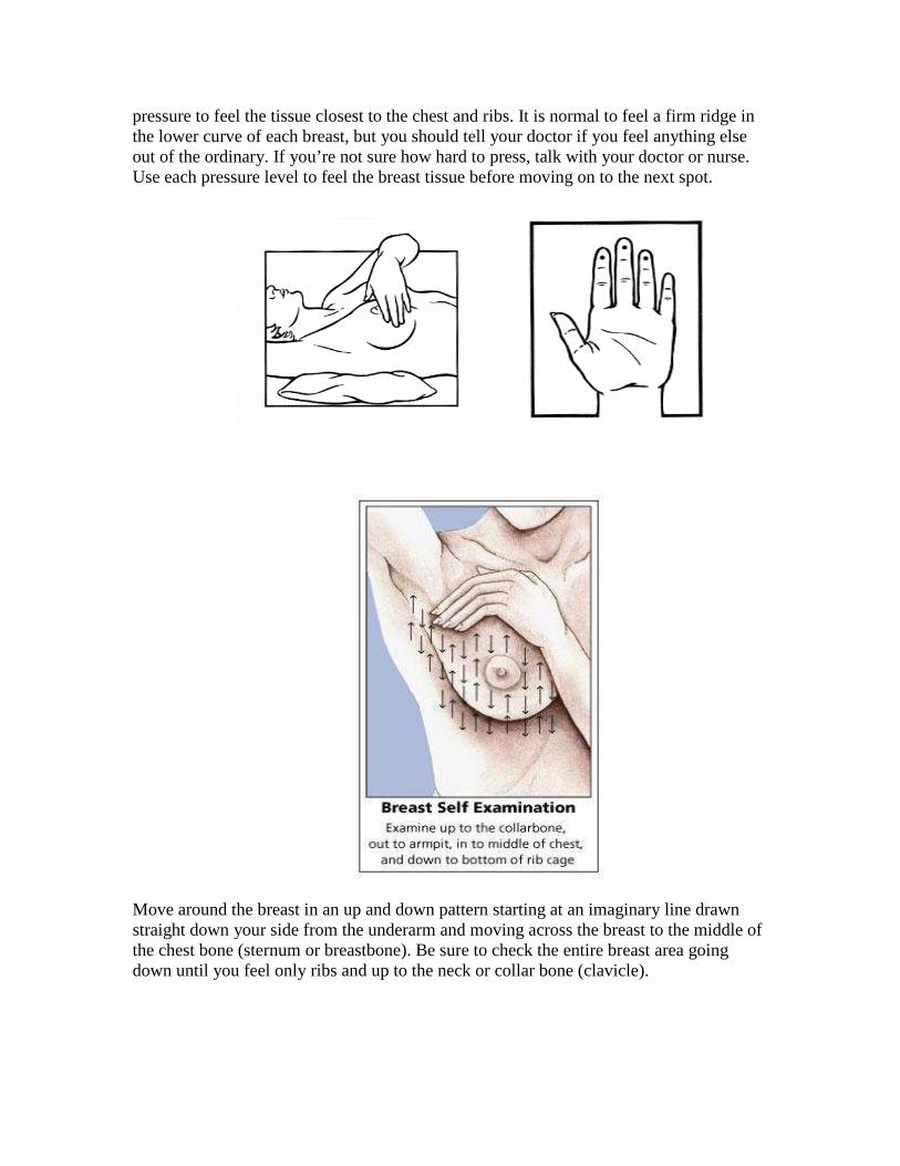
Move around the breast in an up and down pattern starting at an imaginary line drawn straight down your side from the underarm and moving across the breast to the middle of the chest bone (sternum or breastbone). Be sure to check the entire breast area going down until you feel only ribs and up to the neck or collar bone (clavicle).
There is some evidence to suggest that the up-and-down pattern (sometimes called the vertical pattern) is the most effective pattern for covering the entire breast without missing any breast tissue.
Repeat the exam on your left breast, putting your left arm behind your head and using the finger pads of your right hand to do the exam.
While standing in front of a mirror with your hands pressing firmly down on your hips, look at your breasts for any changes of size, shape, contour, or dimpling, or redness or scaliness of the nipple or breast skin. (The pressing down on the hips position contracts the chest wall muscles and enhances any breast changes.)
Examine each underarm while sitting up or standing and with your arm only slightly raised so you can easily feel in this area. Raising your arm straight up tightens the tissue in this area and makes it harder to examine.
This procedure for doing breast self-exam is different from previous recommendations. These changes represent an extensive review of the medical literature and input from an expert advisory group. There is evidence that this position (lying down), the area felt, pattern of coverage of the breast, and use of different amounts of pressure increase a woman’s ability to find abnormal areas.
Breast ultrasound Ultrasound, also known as sonography, is an imaging method using sound waves to look inside a part of the body. In the most common version of this test, a small, microphone-like instrument called a transducer is placed on the skin (which is often first lubricated with ultrasound gel). It emits sound waves and picks up the echoes as they bounce off body tissues. The echoes are converted by a computer into a black and white image on a computer screen. This test is painless and does not expose you to radiation.
Breast ultrasound is often used to evaluate breast problems that are found during a screening or diagnostic mammogram or on physical exam. Ultrasound helps distinguish between cysts (fluid-filled sacs) and solid masses. In someone with a breast mass, it can be used to look for enlarged lymph nodes. Breast ultrasound is often used to guide a needle to biopsy breast lesions and enlarged lymph nodes. It can also be used to guide a needle to draw fluid from cysts.
The use of ultrasound instead of mammograms for breast cancer screening is not recommended. But it is sometimes used in addition to mammogram to screen certain women, such as those with dense breasts (for whom mammography may not be as helpful). When it is used for screening, a newer version of breast ultrasound can be used that uses a large transducer that fits in place over most or all of the breast at once. This allows the whole breast to be scanned in a much shorter time.
Other breast cancer screening tests Mammography is the current standard test for breast cancer screening. MRI is also recommended along with mammograms for some women at high risk for breast cancer.
Other tests may be useful for some women, but they are not used often and have not yet been found to be helpful in diagnosing breast cancer in most women. These include scintimammography, thermography, ductogram, nipple discharge exam, nipple aspiration, and ductal lavage. These tests are discussed in more detail in our documents Breast Cancer and Mammograms and Other Breast Imaging Procedures.
Talk to your doctor
If you think you are at higher risk for developing breast cancer, talk to your doctor about what is known about these tests and their potential benefits, limitations, and harms. Then decide together what is best for you.
For more information on imaging tests for early detection and diagnosis of breast diseases, see our document Mammograms and Other Breast Imaging Procedures.
Paying for breast cancer screening This section gives a brief overview of the laws that require private health plans, Medicaid, and Medicare to cover early detection services for breast cancer screening.
Federal law
Coverage of mammograms for breast cancer screening is mandated by the Affordable Care Act, which provides that these be given without a co-pay or deductible in plans that started after August 1, 2012. This doesn’t apply to health plans that were in place before the law was passed (called grandfathered plans). You can find out the date your insurance plan started by contacting your health insurance plan administrator. Even grandfathered plans may still have coverage requirements based on state laws, which vary, and other federal laws.
State efforts to ensure private health insurance coverage of mammography
Many states require that private insurance companies, Medicaid, and public employee health plans provide coverage and reimbursement for specific health services and procedures. The American Cancer Society (ACS) supports these kinds of patient
protections, particularly when it comes to evidence-based cancer prevention, early detection, and treatment services.
The only state without a law ensuring that private health plans cover or offer coverage for screening mammograms is Utah. Of the remaining 49 states, only 22 cover mammograms yearly for women 40 and older. The remaining 27 have less generous coverage.
Laws on coverage may vary slightly from state to state, so check with your insurer to see what’s covered. Note that state laws don’t affect self-insured (self-funded) health plans.
Source: National Women’s Law Center. Screening Coverage Mandates: Mammogram. Accessed at http://hrc.nwlc.org/policy-indicators/mammogram on September 10, 2014.
Self-insured plans
Self-insured or self-funded plans do not have to follow state laws about breast cancer screening. They are governed by the Affordable Care Act (ACA), and are required to cover breast cancer screening. The exception is any self-insured plan that was in effect before the ACA was passed. These plans are called grandfathered, and they don’t have to provide coverage based on what the ACA says.
Many employers offer self-insured plans. These plans pay employee health care costs from the employer’s own funds, even though they usually contract with another company to track and pay claims. You can find out if your health plan is self-insured by contacting your insurance administrator at work or reading your Summary of Plan Benefits. Women covered by self-insured employer plans should check with their health insurance administrator to see what breast cancer early detection services are covered.
Medicaid
All state Medicaid programs plus the District of Columbia cover screening mammograms. This coverage may or may not conform to American Cancer Society guidelines. State Medicaid offices should be able to provide screening coverage information to interested individuals. The Medicaid programs are governed by state legislation and regulation, so assured coverage is not always apparent in legislative bills.
In addition, all 50 states plus the District of Columbia have opted to provide Medicaid coverage for all women diagnosed with breast cancer through the Centers for Disease Control and Prevention’s (CDC’s) National Breast and Cervical Cancer Early Detection Program (see the next section), so that they may receive cancer treatmentStates vary in the age, income and other requirements that women must meet in order to qualify for treatment through the Medicaid program. (All 50 states, 4 U.S. territories, the District of Columbia, and 13 American Indian/Alaska Native organizations participate in the National Breast and Cervical Cancer Early Detection Program.)
National Breast and Cervical Cancer Early Detection Program
States are making breast cancer screening more available to medically underserved women through the National Breast and Cervical Cancer Early Detection Program (NBCCEDP). The NBCCEDP attempts to reach as many women in medically underserved communities as possible, including older women, women without health insurance, and women who are members of racial and ethnic minorities. Age and income requirements vary by state.
The program provides both screening and diagnostic services to low-income, uninsured, and underserved women for free or at very low cost, including:
• Clinical breast exams
• Mammograms
• Pap tests
• Diagnostic testing for women whose screening results are abnormal
• Surgical consultations
• Referrals to treatment
Though the program is administered within each state, tribe, or territory, the Centers for Disease Control and Prevention (CDC) matches funds and support to each program.
Since 1991 when the program began, it has provided millions of screening exams to underserved women and diagnosed more than 60,000 breast cancers. Due to limited resources, though, less than 1 in 8 eligible women aged 40 to 64 is able to be screened for breast cancer through this program nationwide.
In 2000, Congress passed the Breast and Cervical Cancer Prevention and Treatment Act, giving states the option to offer women in the NBCCEDP access to treatment through Medicaid. All 50 states plus the District of Columbia provide Medicaid coverage for women diagnosed with breast cancer through the NBCCEDP, so that they have a way to pay for treatment.
Each state’s Department of Health will have information on how to contact the nearest CDC screening and early detection program in your area. For more information, please contact the CDC at 1-800-CDC-INFO (1-800-232-4636) or through their website at www.cdc.gov/cancer.
Medicare
As a part of the Affordable Care Act, Medicare covers the full cost of a screening mammogram once every 12 months for all women with Medicare aged 40 and over. Diagnostic mammograms are covered with a 20% copay after the part B deductible is met. (Medicare also pays for a clinical breast exam when it is done for screening or prevention.
To learn more about breast cancer early detection
More information from your American Cancer Society
Here is more information you might find helpful. You also can order free copies of our documents from our toll-free number, 1-800-227-2345, or read them on our website, www.cancer.org.
Breast Cancer (also available in Spanish)
Breast Cancer Dictionary (also available in Spanish)
Breast Cancer in Men
Is Abortion Linked to Breast Cancer?
DES Exposure: Questions and Answers
For Women Facing a Breast Biopsy
Genetic Testing: What You Need to Know
Mammograms and Other Breast Imaging Procedures
Medicines to Reduce Breast Cancer Risk
Non-cancerous Breast Conditions (also available in Spanish)
National organizations and websites*
Along with the American Cancer Society, other sources of information and support include:
Centers for Disease Control and Prevention (CDC) Cancer Prevention and Control Program
Toll-free number: 1-800-232-4636 (1-800-CDC-INFO) Website: www.cdc.gov/cancer
Information about the National Breast and Cervical Cancer Early Detection Program
National Cancer Institute (NCI) Toll-free number: 1-800-4-CANCER (1-800-422-6237) Website: www.cancer.gov
General breast cancer information
*Inclusion on this list does not imply endorsement by the American Cancer Society.
No matter who you are, we can help. Contact us any time, day or night, for information and support. Call us at 1-800-227-2345 or visit www.cancer.org.
References: Breast cancer early detection American Cancer Society. Detailed Guide: Breast Cancer. 2014. Accessed at http://www.cancer.org/Cancer/BreastCancer/DetailedGuide/index on August 24, 2012.
Centers for Disease Control and Prevention. National Breast and Cervical Cancer Early Detection Program. Accessed at: www.cdc.gov/cancer/nbccedp/about.htm on September 10, 2014.
Pisano ED, Gatsonis C, Hendrick E, et al. Diagnostic performance of digital versus film mammography for breast-cancer screening. N Engl J Med. 2005;353:1773-1783.
Saslow D, Boetes C, Burke W, et al for the American Cancer Society Breast Cancer Advisory Group. American Cancer Society guidelines for breast screening with MRI as an adjunct to mammography. CA Cancer J Clin. 2007;57:75-89.
Smith RA, Saslow D, Sawyer KA, et al. American Cancer Society guidelines for breast cancer screening: Update 2003. CA Cancer J Clin. 2003;53:141-169.
Last Medical Review: 9/10/2014 Last Revised: 9/10/2014 2014 Copyright American Cancer Society