Embed Size (px)
Citation preview

Breast Cancer
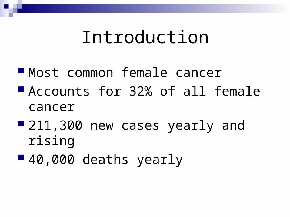
Introduction
Most common female cancer Accounts for 32% of all female cancer 211,300 new cases yearly and rising 40,000 deaths yearly
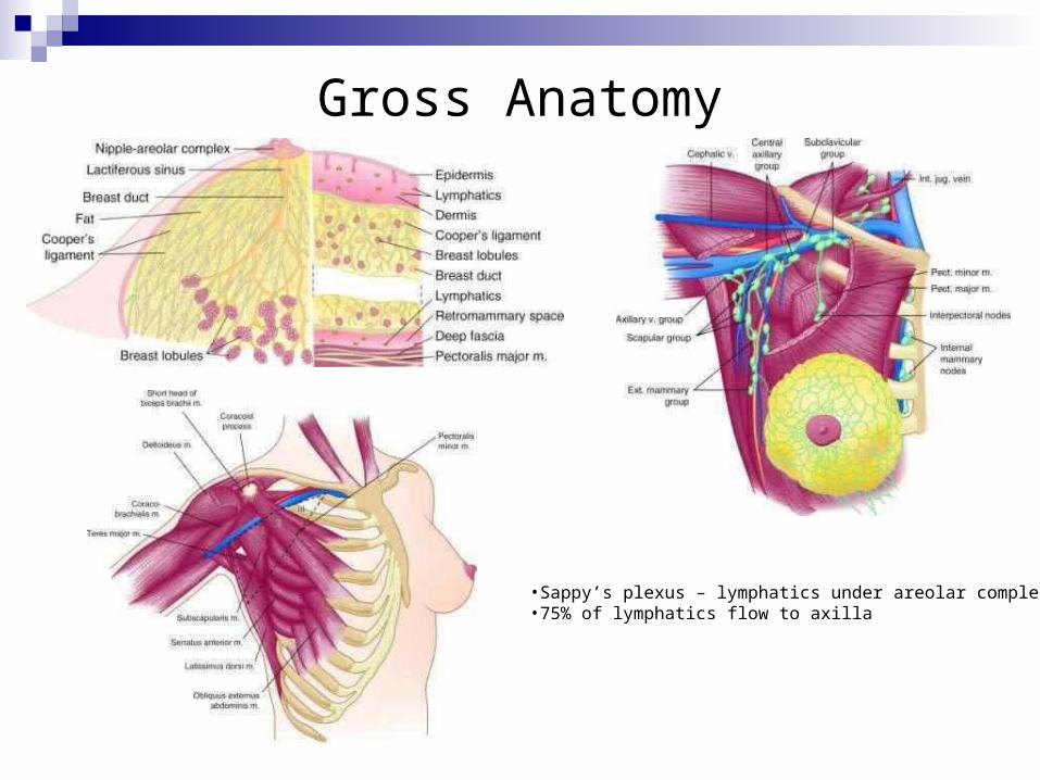
Gross Anatomy
•Sappy’s plexus – lymphatics under areolar complex•75% of lymphatics flow to axilla
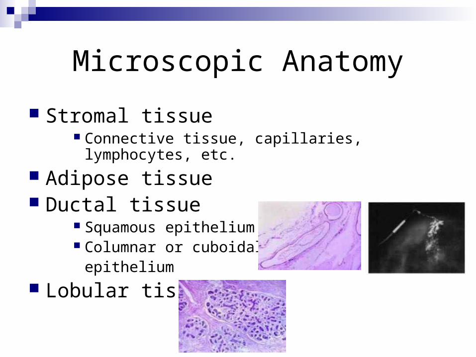
Microscopic Anatomy
Stromal tissue Connective tissue, capillaries, lymphocytes, etc.
Adipose tissue Ductal tissue
Squamous epithelium Columnar or cuboidal
epithelium
Lobular tissue
Presentation
Breast lump Abnormal mammogram Axillary lympadenopathy Metastatic disease
Familial Breast Cancer
Cause 5-10% of all cancer and 25% in women <30 y/o
BRCA2 Causes 40% of familial breast CA 50-70% - breast 15-45% - ovarian Increased risk for prostate, colon
BRCA1 50-70% - breast 20-30% - ovarian Increased risk for prostate, pancreatic, laryngeal,
Screening Mammography
Recommendations Biannually or annually in 40-49 y/o Annually in >50 y/o
15% relative risk reduction Birads
0 - Incomplete assessment; need additional imaging evaluation 1 - Negative; routine mammogram in 1 year recommended 2 - Benign finding; routine mammogram in 1 year recommended 3 - Probably benign finding; short-term follow-up suggested (3%) 4 - Suspicious abnormality; biopsy should be considered (30%) 5 - Highly suggestive of malignancy; appropriate action should be
taken (94%)
Biopsy techniques
FNA Diagnostic and therapeutic in cystic lesions
Core needle U/S guided or sterotatic 90% effective in establishing diagnosis Atypia – need excision
Sterotatic Needle localization Excision biopsy
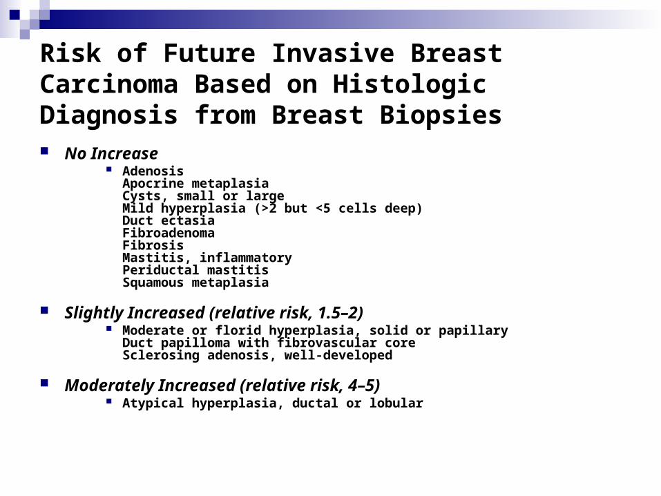
Risk of Future Invasive Breast Carcinoma Based on Histologic Diagnosis from Breast Biopsies No Increase
AdenosisApocrine metaplasiaCysts, small or largeMild hyperplasia (>2 but <5 cells deep)Duct ectasiaFibroadenomaFibrosisMastitis, inflammatoryPeriductal mastitisSquamous metaplasia
Slightly Increased (relative risk, 1.5–2) Moderate or florid hyperplasia, solid or papillary
Duct papilloma with fibrovascular coreSclerosing adenosis, well-developed
Moderately Increased (relative risk, 4–5) Atypical hyperplasia, ductal or lobular
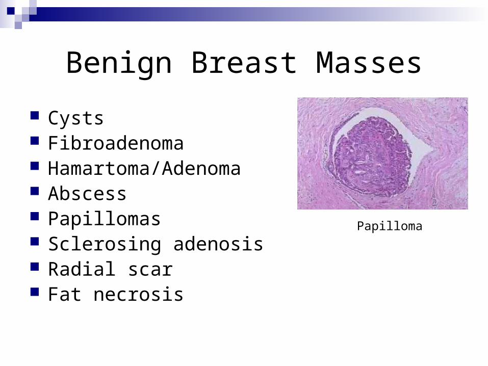
Benign Breast Masses
Cysts Fibroadenoma Hamartoma/Adenoma Abscess Papillomas Sclerosing adenosis Radial scar Fat necrosis
Papilloma
Maligant Breast Masses
Ductal carcinoma DCIS Invasive
Lobular carcinoma LCIS Invasive
Inflammatory carcinoma Paget’s disease Phyllodes tumor Angiosarcoma
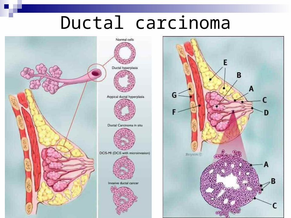
Ductal carcinoma
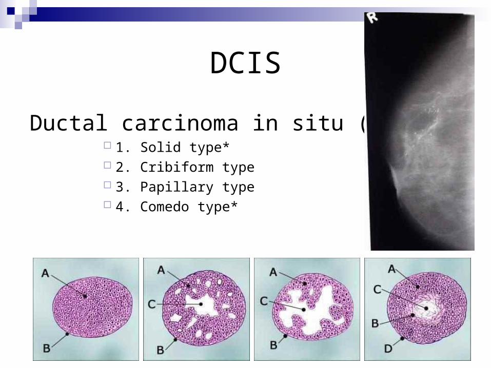
DCIS
Ductal carcinoma in situ (DCIS) 1. Solid type* 2. Cribiform type 3. Papillary type 4. Comedo type*
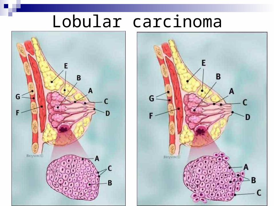
Lobular carcinoma
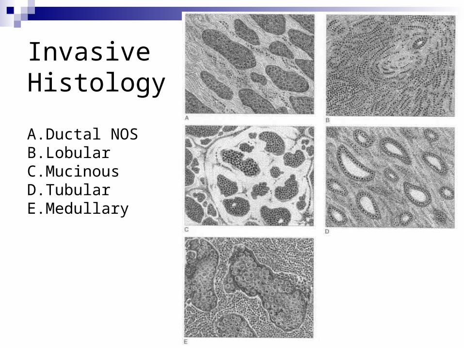
Invasive Histology
A. Ductal NOSB. LobularC.MucinousD.TubularE. Medullary
Staging
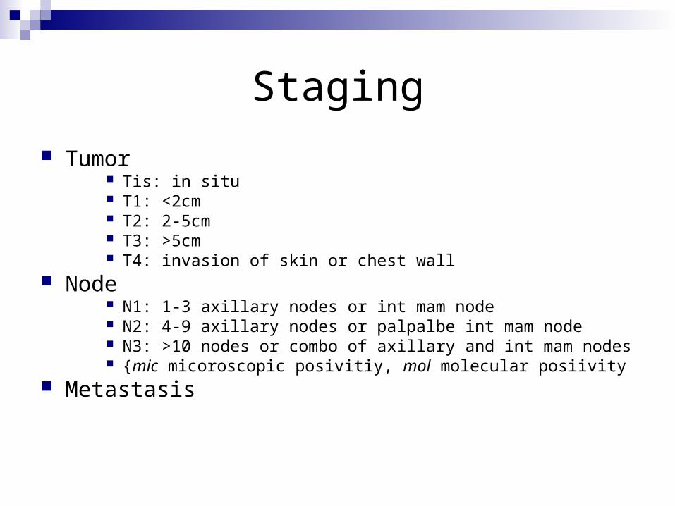
Tumor Tis: in situ T1: <2cm T2: 2-5cm T3: >5cm T4: invasion of skin or chest wall
Node N1: 1-3 axillary nodes or int mam node N2: 4-9 axillary nodes or palpalbe int mam node N3: >10 nodes or combo of axillary and int mam nodes {mic micoroscopic posivitiy, mol molecular posiivity
Metastasis

Staging
Modified Radical Mastectomy
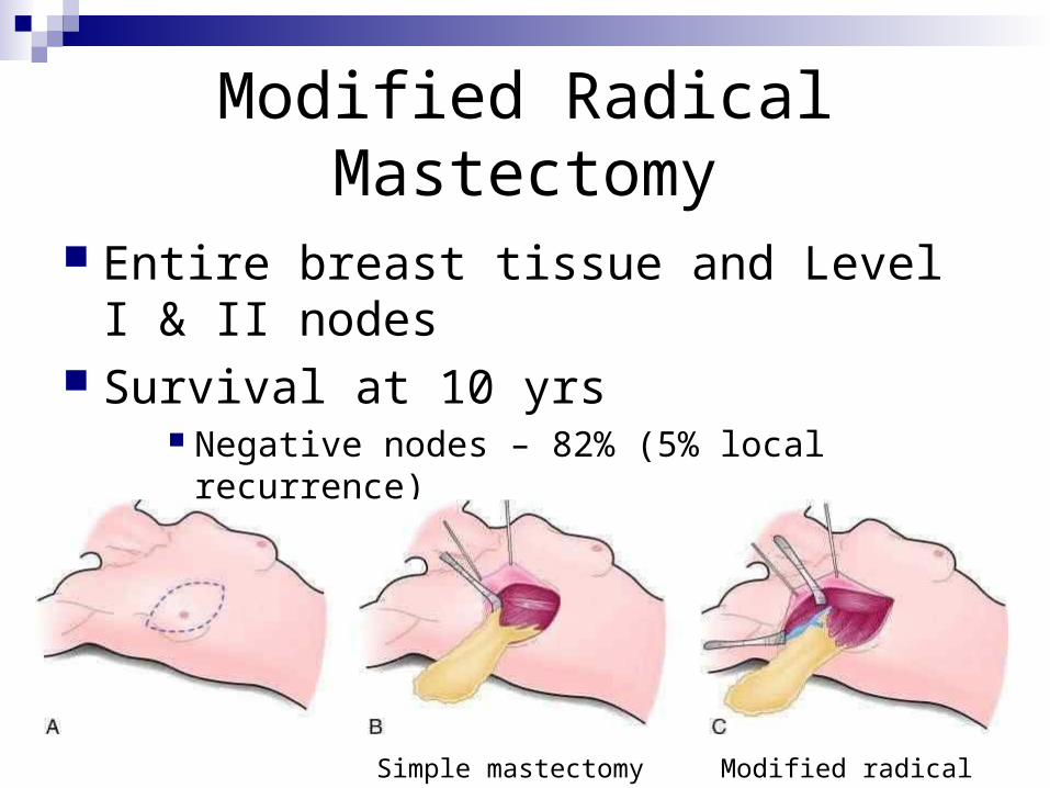
Entire breast tissue and Level I & II nodes Survival at 10 yrs
Negative nodes – 82% (5% local recurrence) Positive nodes – 48% (5% local recurrence)
Simple mastectomy Modified radical
Breast Treatment Trials
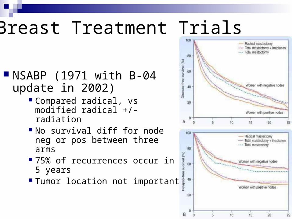
NSABP (1971 with B-04 update in 2002)
Compared radical, vs modified radical +/- radiation
No survival diff for node neg or pos between three arms
75% of recurrences occur in 5 years
Tumor location not important
Breast Treatment Trials
Ontario study All pts got lumpectomy, randomized to radiation or no radiation 25% failure rate without radiation, 5% with
NSABP B-06 Mastecomy vs lumpectomy vs lumpectomy with radiation No difference in survival 39% recur with lumpectomy, reduced to 14% with radiation, 3-4%
with mastectomy 0.5-1% per year recurrence rate for life with BCT and radiation 2-5% complication rate with radiation (rib fx, pericarditis, cosmesis)
Radiation after mastectomy?
2 Danish studies and one Britsh study Recommend in: >3 nodes positive,
aggressive/large tumors or extranodal invasion Decreased local or regional recurrence +/- survival benefit
Sentinel node biopsy
Contraindications: Clinically positive nodes, pregnant or nursing, prior axillary
surgery, locally advanced disease
False negative rate 3.1% Macrometases (>0.2cm) so recommended pathology cuts are
0.2 cm Micrometases (IHC staining) 37% death rate vs 50% of those
with macrometases If sentinel node positive 43% will have other nodes positive and
24% will have >4 nodes positive
NSABP (B-32) in progress
Treatment of DCIS
600% increase after mammography Options
Mastectomy – 1% breast ca mortality Large tumors, multicentric, positive margins after
reexcision, Lumpectomy and radiation
Radiation decreases local recurrence by 50% Of those that recur 50/50 DCIS vs Invasive 0-3% chance of dying of maligant breast ca for all
DCIS
Treatment of DCIS
Nodal involvement 3.6% of DCIS pts have positive nodes in
mastectomy specimins By definition DCIS has no access to lymphatics
Size may matter (111 DCIS tumors evaluated) <45mm – 0% microinvasion 45-55mm – 17% microinvasion >55mm – 48% microinvasion
Tamoxifen in DCIS
NSABP (B-24) Determine benefit of tamoxifen in lumpectomy plus
radiation pts 31% decrease in ipsilateral, 47% in contralateral,
31% decrease all together Retrospectively looked at ER status
75% of DCIS is ER+ 59% reduction in ER+ pts No significant reduction in ER-

Treatment for invasive breast ca
Locally advanced is likely already metastatic in most
Surgery and radiation alone make no difference on survival Chemotherapy & +/- Tamoxifen
Neoadjuvant chemotherapy 7 randomized trials
No survival benefit 50-80% response May allow for BCT in large tumors
Sentinel node before chemo
Tamoxifen
Indications ER + breast ca LCIS BRCA1/2 Increased overall risk
Benefits Decreases risk of ca in other breast by 47-80%
Draw backs Increases endometrial ca risk by 2.5, PE 3.0, DVT 1.7
Source: NSABP P-1 trial

Chemotherapy
Early Breast Cancer Trialists’ Collaborative Group
Decreases recurrence (12%) and death (11%) regardless of nodal status
Indications All patients except node negative, <10mm tumors
Regimens Multidrug combination chemotherapy Tamoxifen or aromatse inhibitor - ER positive tumors Herceptin (trastuzumab) – HER2/neu positive tumors
NSABP B-31 – 33% reduction in risk of death

Other breast cancers
Inflammatory ca Carcinoma invading lymphatic ducts Chemotherapy, mastectomy, radiation 50% survival at 5 years
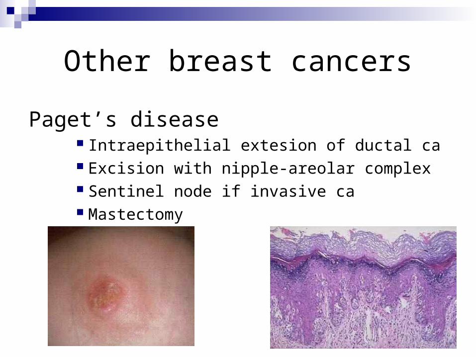
Other breast cancers
Paget’s disease Intraepithelial extesion of ductal ca Excision with nipple-areolar complex Sentinel node if invasive ca Mastectomy
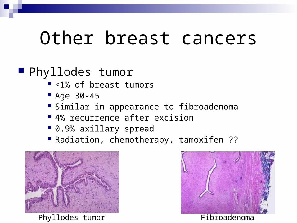
Other breast cancers
Phyllodes tumor <1% of breast tumors Age 30-45 Similar in appearance to fibroadenoma 4% recurrence after excision 0.9% axillary spread Radiation, chemotherapy, tamoxifen ??
Phyllodes tumor Fibroadenoma

Angiosarcoma Risk factors
Radiation Lymphedema
Treatment Excision, radiation
Male breast cancer
90% are invasive at time of diagnosis 80% ER+, 75% PR+, 30% HER2/neu More invade into pectoralis Treatment same as for female ca