Embed Size (px)
Citation preview

Breast Clinical Correlation
Anne T. Mancino MD
American Cancer Society 1999
Cancer Facts & Figures
Breast Cancer Facts
An estimated 178,000 new cases of female invasive breast cancer will be diagnosed
An estimated 43,500 women will die from breast cancer
Approximately 37,000 cases of female in situ breast cancer will be diagnosed
Risk Factors for Breast Cancer
• Age• Personal history - 0.5-1% per year risk new cancer• Family history
– First degree relative– Pre-menopausal risk 3-4 fold– Germline mutation (BRCA1/2) 60-85% risk
• Previous biopsy, especially with atypia• Early menses, late menopause, parity
ACS Screening Guidelines
• Screening Mammography– Yearly starting at age 40
• Clinical Breast Exam– Every 3 years age 20-39– Yearly after age 40
• Breast Self Exam– monthly after age 20

Breast Exam: Anatomy
•Variety of sizes and shapes
•Composed of fatty, fibrous and glandular tissue
•Lymph nodes are important
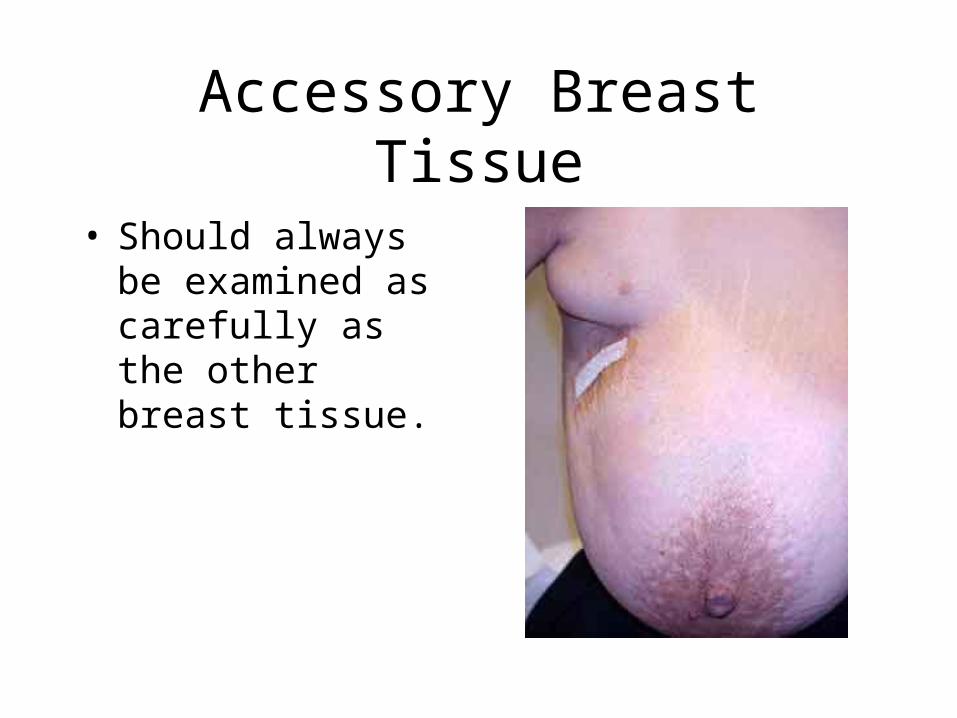
Accessory Breast Tissue
• Should always be examined as carefully as the other breast tissue.

Physical Findings Suspicious for Malignancy
• Venous patterns
• Skin edema
• Nipple inversion
• Retraction
• Scaling or ulceration of the nipple
• Inflammation
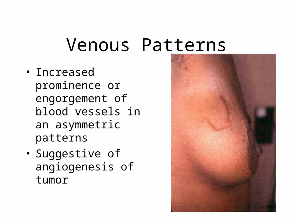
Venous Patterns• Increased prominence
or engorgement of blood vessels in an asymmetric patterns
• Suggestive of angiogenesis of tumor
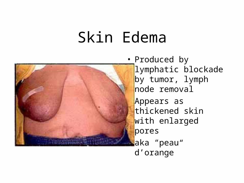
Skin Edema• Produced by
lymphatic blockade by tumor, lymph node removal
• Appears as thickened skin with enlarged pores
• aka “peau d’orange”

Nipple Inversion• Can be a normal
variant• Unilateral or bilateral• Be suspicious for
cancer in recently developed cases
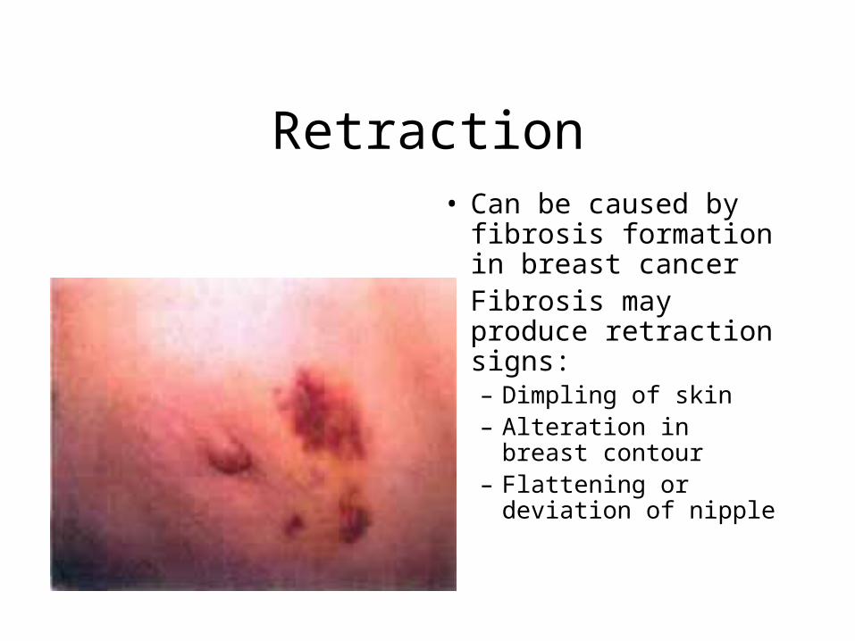
Retraction• Can be caused by
fibrosis formation in breast cancer
• Fibrosis may produce retraction signs:– Dimpling of skin– Alteration in breast
contour– Flattening or deviation
of nipple

Retraction As Seen on Mammogram
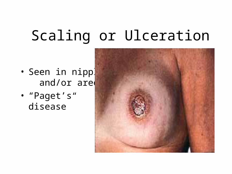
Scaling or Ulceration
• Seen in nipple and/or areola
• “Paget’s disease”
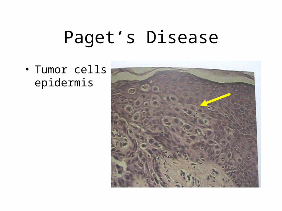
Paget’s Disease
• Tumor cells in epidermis

Inflammation - Breast Abscess
• need to distinguish from inflammatory breast cancer
• needs incision and drainage
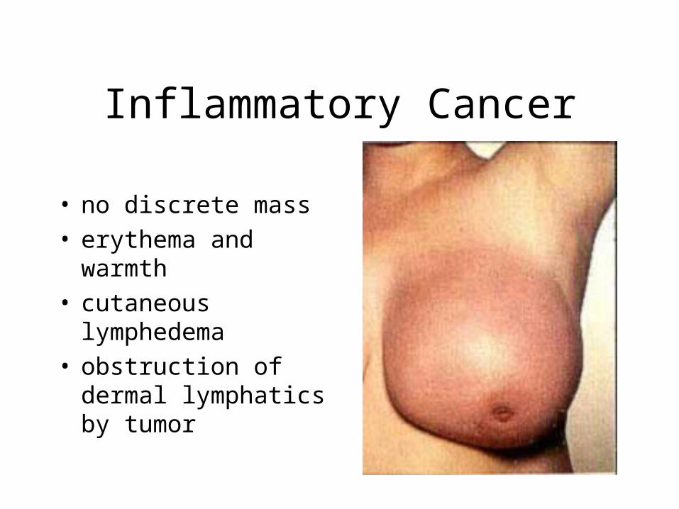
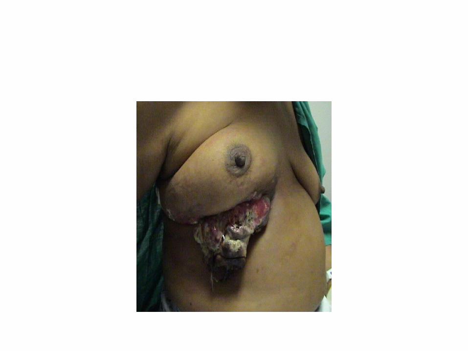
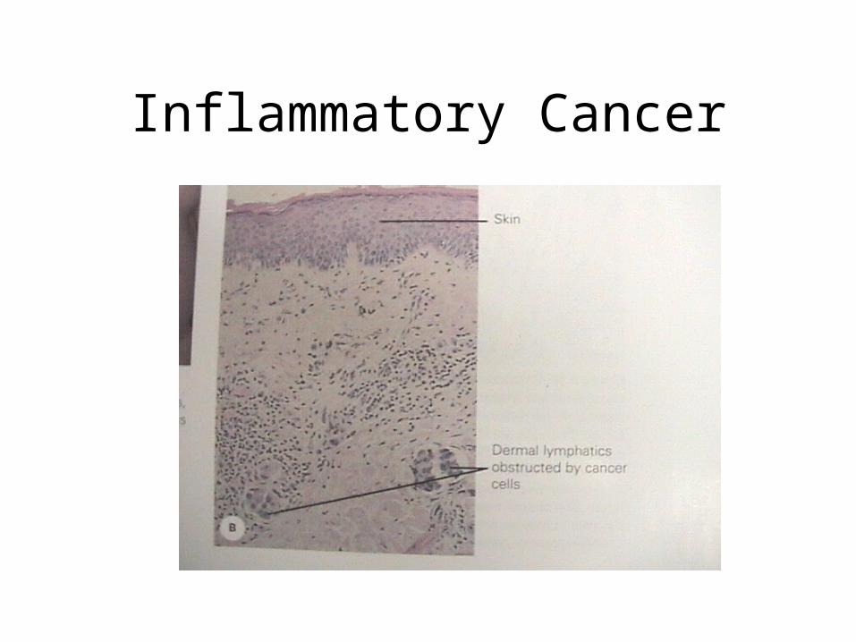
Inflammatory Cancer
• no discrete mass• erythema and warmth• cutaneous
lymphedema• obstruction of dermal
lymphatics by tumor
Inflammatory Cancer

Nipple Discharge
• Spontaneous
• Unilateral
• One Duct
• Clear, Serous, Bloody or Serosanguinous
• Green
• White or Milky
Nipple Discharge
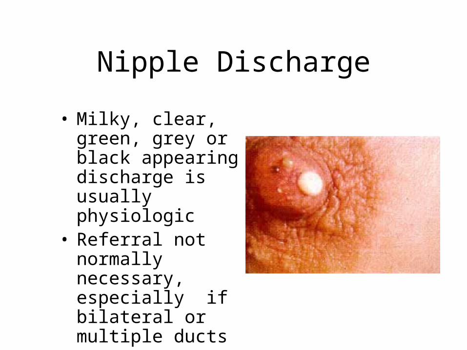
• Milky, clear, green, grey or black appearing discharge is usually physiologic
• Referral not normally necessary, especially if bilateral or multiple ducts

Nipple Discharge
• Bloody discharge• Could be a sign of
benign intraductal papilloma
• Should always be a referral to a breast specialist
Intraductal Papilloma
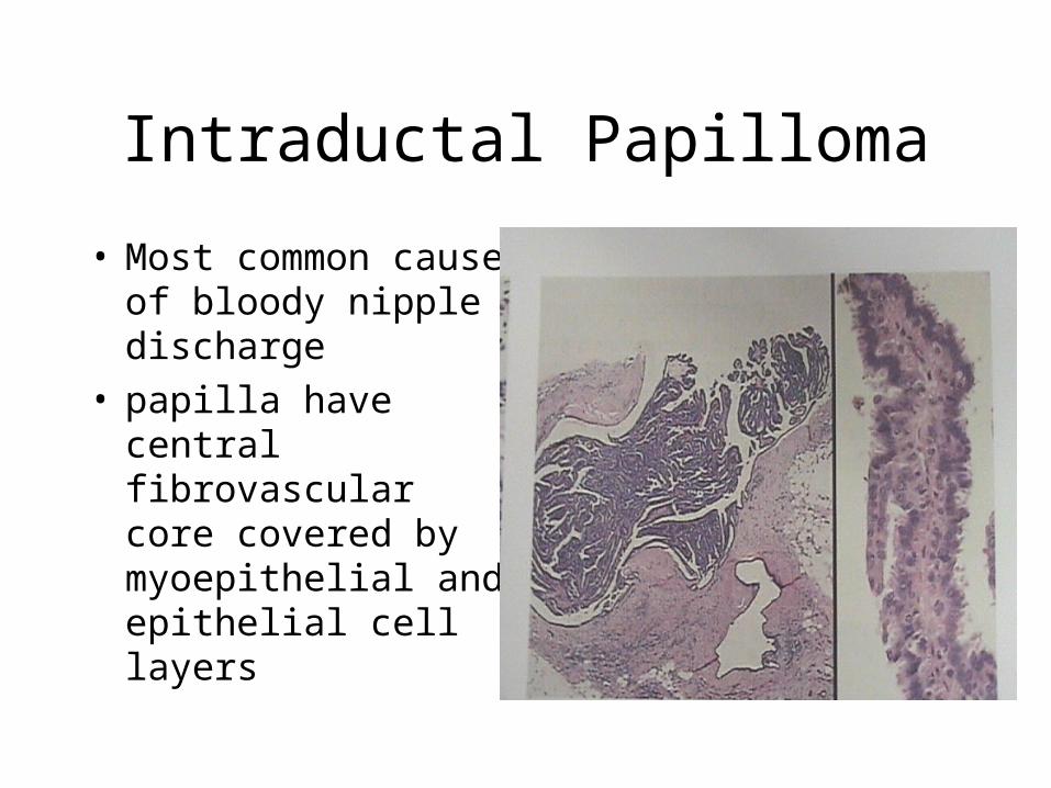
• Most common cause of bloody nipple discharge
• papilla have central fibrovascular core covered by myoepithelial and epithelial cell layers

Nipple Discharge
• Serous drainage could be a sign of duct ectasia
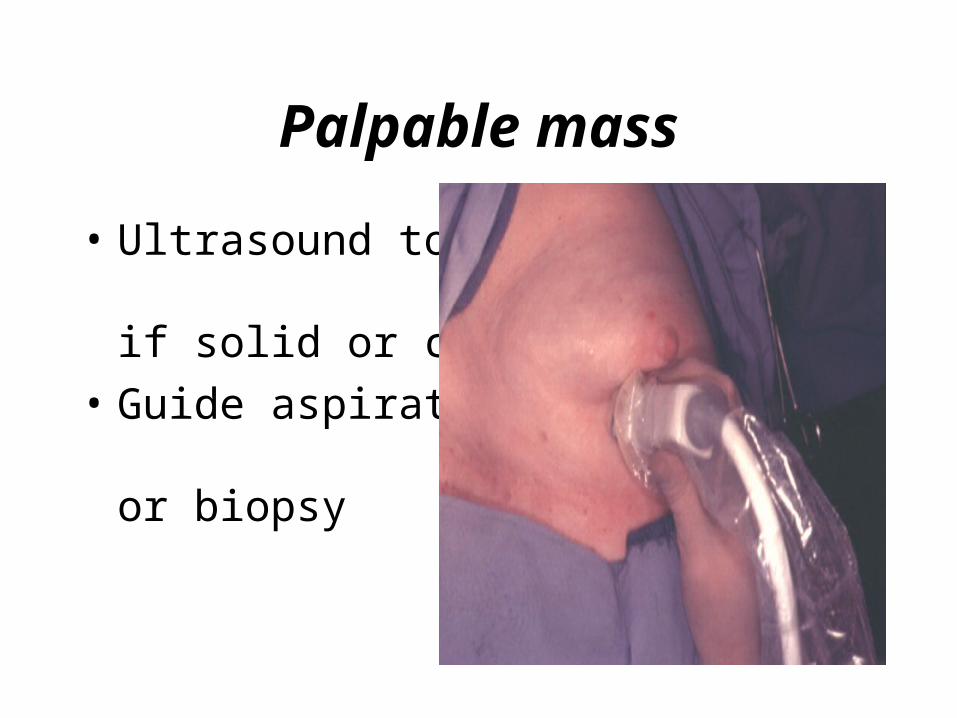
Palpable mass
• Ultrasound to see if solid or cystic
• Guide aspiration or biopsy
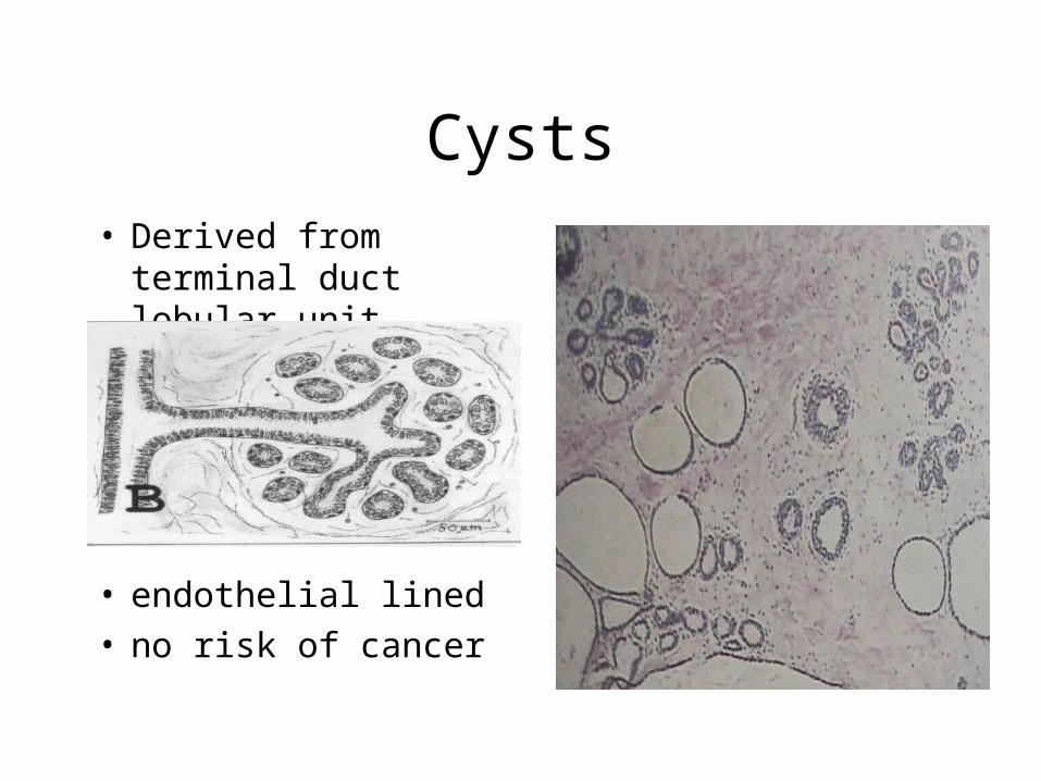
Cysts
• Derived from terminal duct lobular unit
• endothelial lined• no risk of cancer
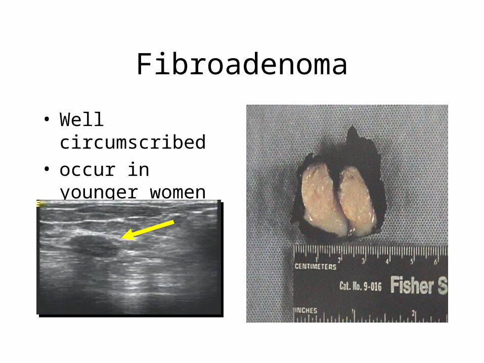
Fibroadenoma
• Well circumscribed• occur in younger
women
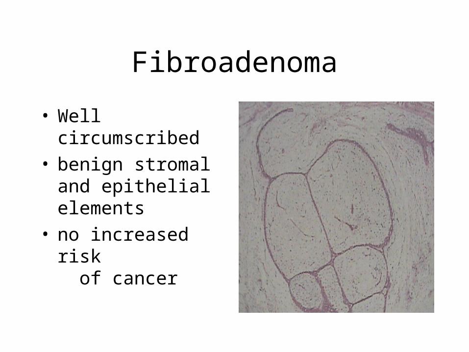
Fibroadenoma
• Well circumscribed• benign stromal and
epithelial elements• no increased risk
of cancer
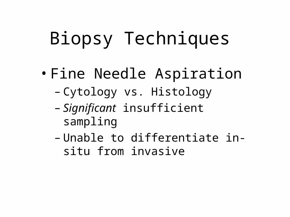
Biopsy Techniques
• Fine Needle Aspiration– Cytology vs. Histology– Significant insufficient sampling– Unable to differentiate in-situ from
invasive
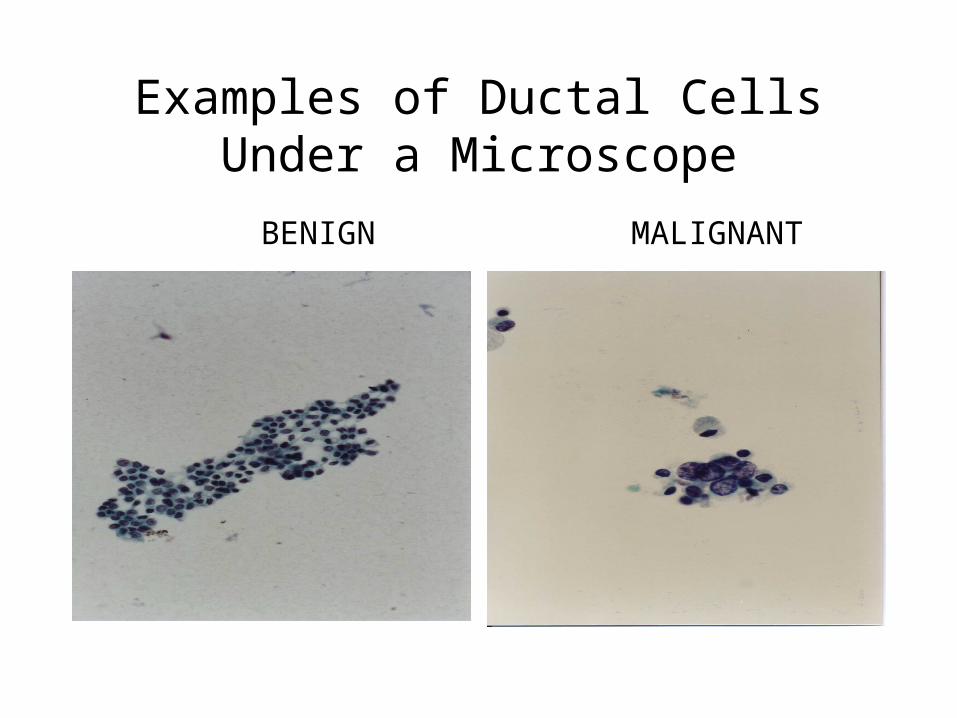
Examples of Ductal Cells Under a Microscope
BENIGN MALIGNANT
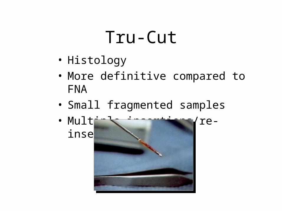
Tru-Cut• Histology• More definitive compared to FNA• Small fragmented samples• Multiple insertions/re-insertion's

Vacuum-Assisted Mammotome
• Histology• Large, contiguous
tissue samples• Single insertion• Can mark biopsy site• 2-3 mm skin incision –
sutureless
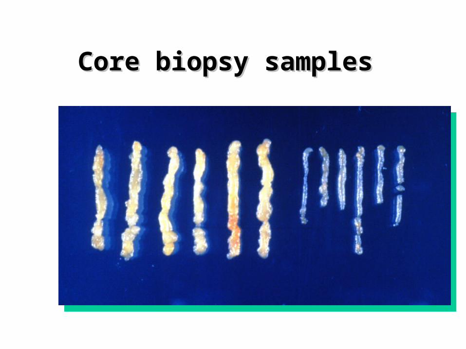
Core biopsy samples Core biopsy samples
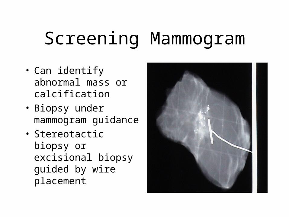
Screening Mammogram
• Can identify abnormal mass or calcification
• Biopsy under mammogram guidance
• Stereotactic biopsy or excisional biopsy guided by wire placement
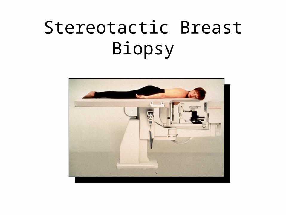
Stereotactic Breast Biopsy
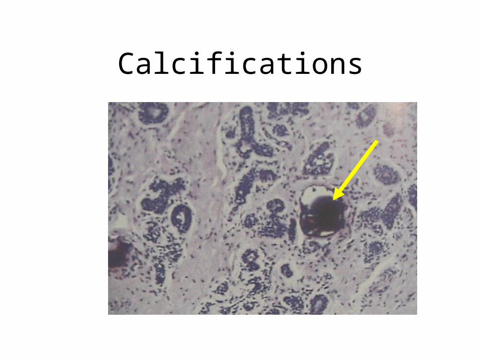
Calcifications
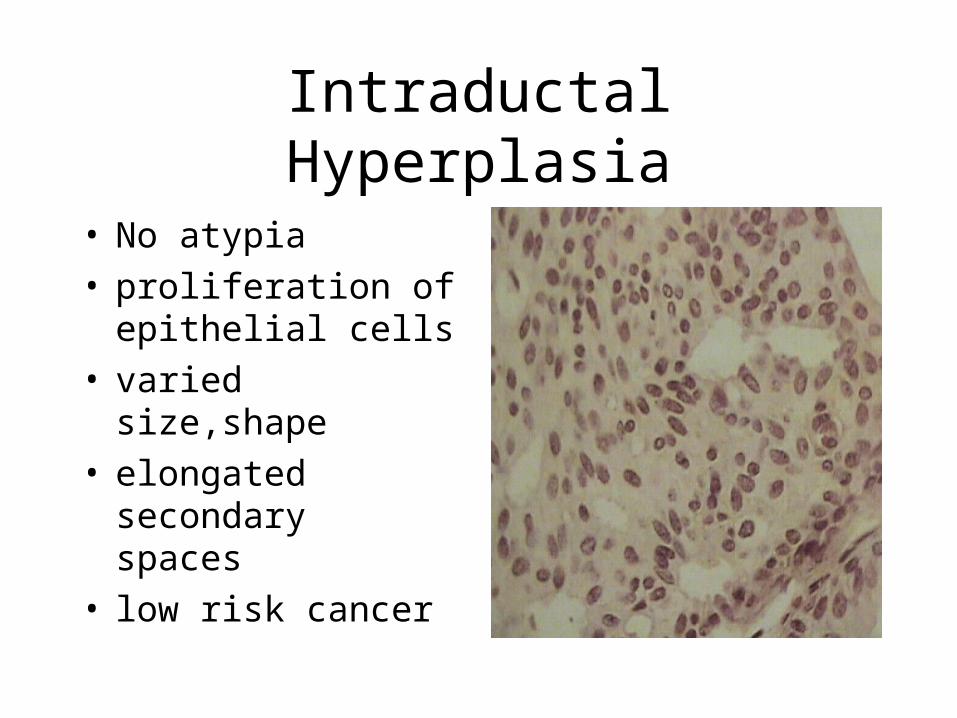
Intraductal Hyperplasia
• No atypia• proliferation of
epithelial cells• varied size,shape• elongated secondary
spaces• low risk cancer
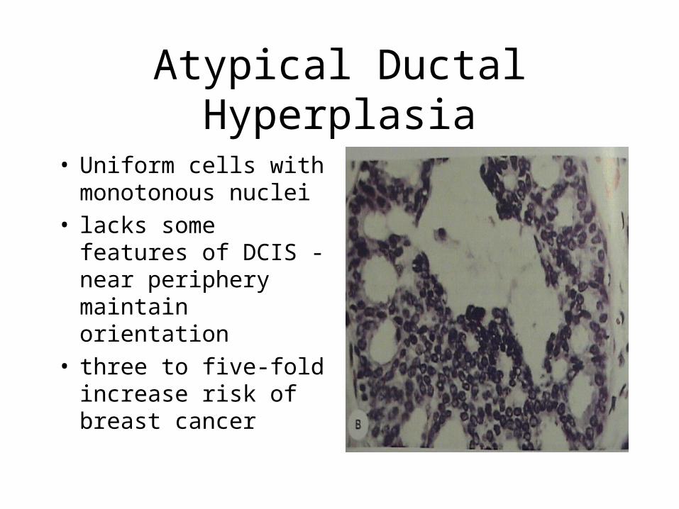
Atypical Ductal Hyperplasia
• Uniform cells with monotonous nuclei
• lacks some features of DCIS -near periphery maintain orientation
• three to five-fold increase risk of breast cancer
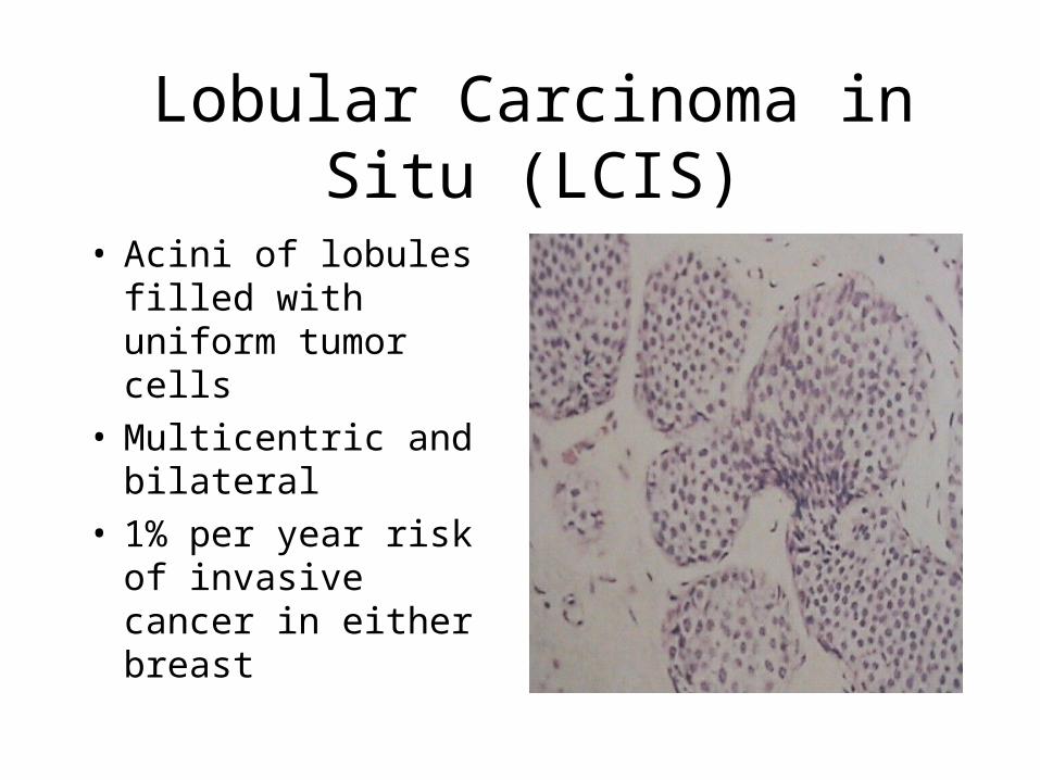
Lobular Carcinoma in Situ (LCIS)
• Acini of lobules filled with uniform tumor cells
• Multicentric and bilateral
• 1% per year risk of invasive cancer in either breast
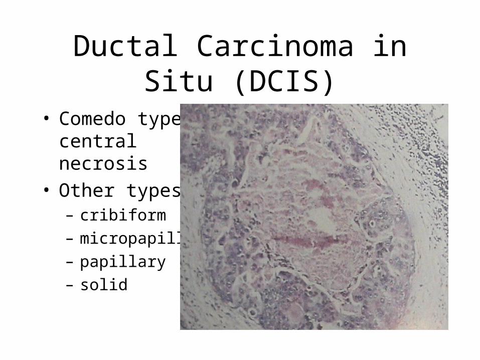
Ductal Carcinoma in Situ (DCIS)
• Comedo type - central necrosis
• Other types:– cribiform
– micropapillary
– papillary
– solid

Infiltrating Ductal Cancer
• most common type• well (gr I) to poorly
(gr III) differentiated• Gr I tumor cells grow
in glandular patterns• prognostic factors:
– ER,PR, HER-2neu,p53
– S-phase, ploidy
– angiogenesis
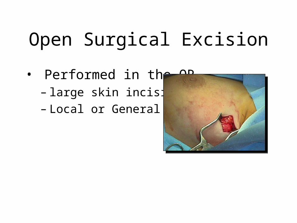
Open Surgical Excision
• Performed in the OR– large skin incision– Local or General
Anesthesia

History of Treatment• 1890’s - Halstead - Radical Mastectomy
• 1948 - Dyson and Patey - Modified Radical Mastectomy
• 1948 - McWhirter - Simple Mastectomy and radiation therapy
• 1990’s - Lumpectomy/Axillary node dissection and radiation therapy
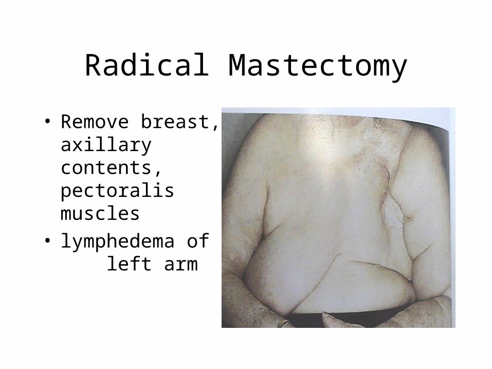
Radical Mastectomy
• Remove breast, axillary contents, pectoralis muscles
• lymphedema of left arm
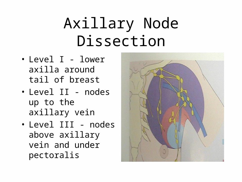
Axillary Node Dissection
• Level I - lower axilla around tail of breast
• Level II - nodes up to the axillary vein
• Level III - nodes above axillary vein and under pectoralis
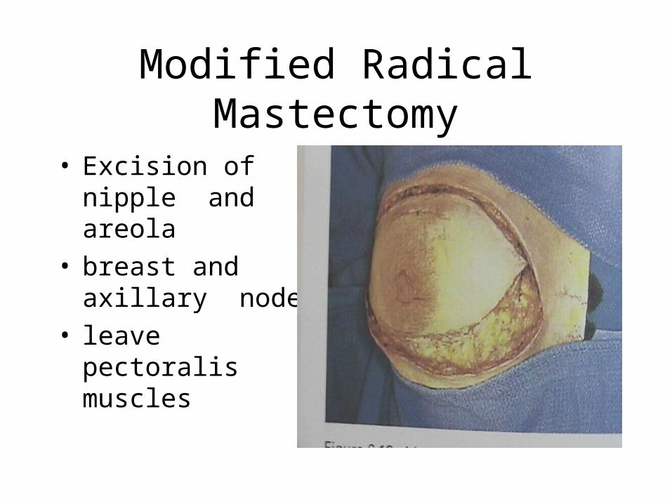
Modified Radical Mastectomy
• Excision of nipple and areola
• breast and axillary nodes
• leave pectoralis muscles

Modified Radical Mastectomy
• Axilla dissected
en bloc with the breast
Modified Radical Mastectomy
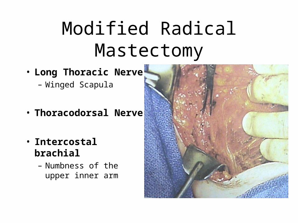
• Long Thoracic Nerve– Winged Scapula
• Thoracodorsal Nerve
• Intercostal brachial– Numbness of the upper
inner arm
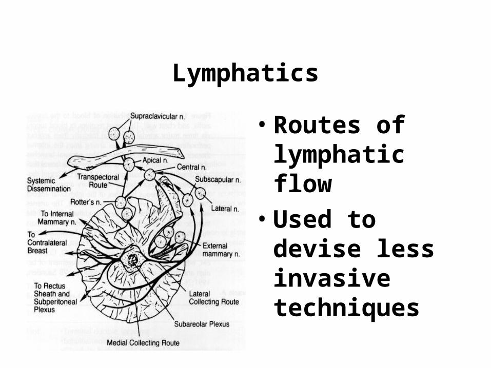
Lymphatics
• Routes of lymphatic flow
• Used to devise less invasive techniques
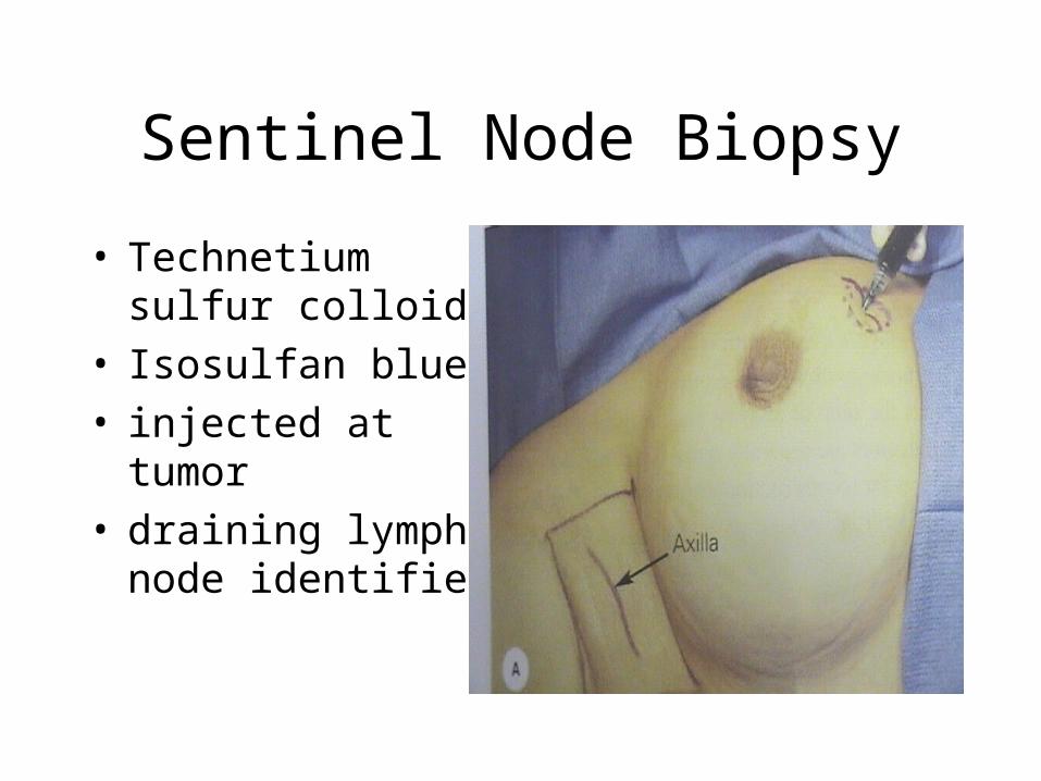
Sentinel Node Biopsy
• Technetium sulfur colloid
• Isosulfan blue• injected at tumor• draining lymph node
identified
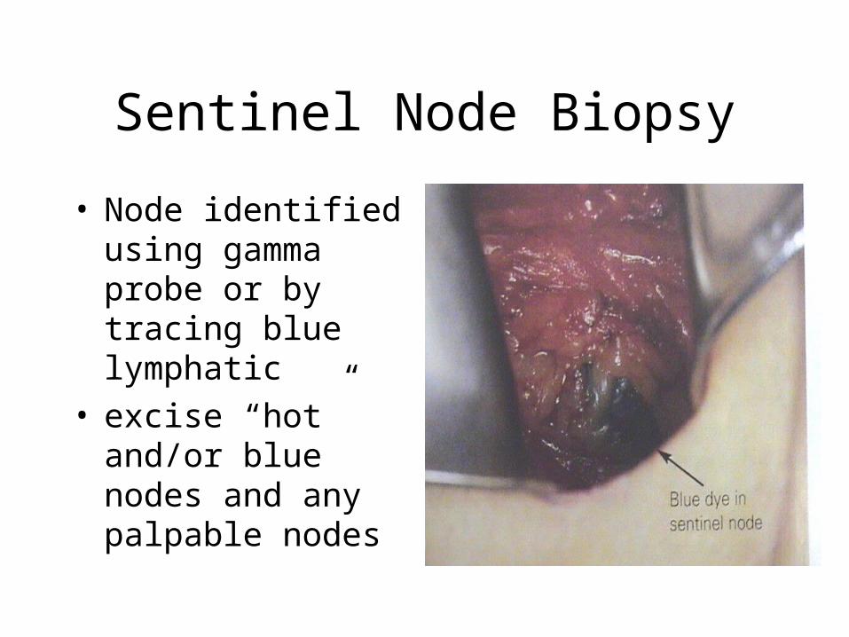
Sentinel Node Biopsy
• Node identified using gamma probe or by tracing blue lymphatic
• excise “hot” and/or blue nodes and any palpable nodes
Sentinel Node Biopsy
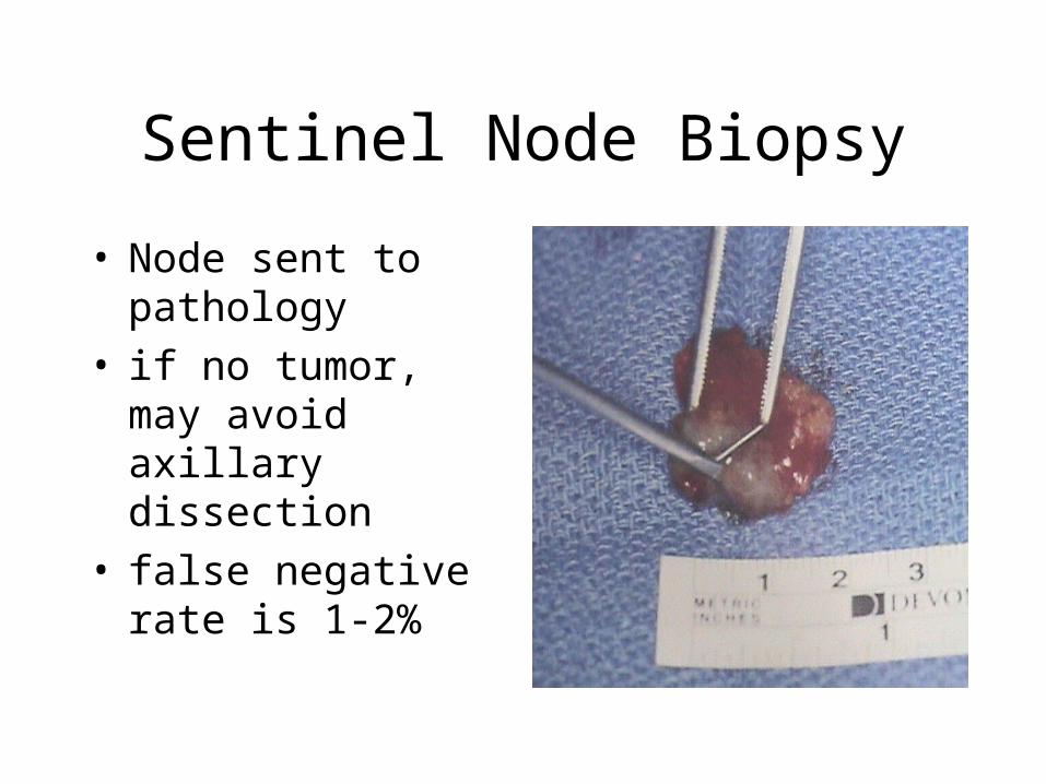
• Node sent to pathology
• if no tumor, may avoid axillary dissection
• false negative rate is 1-2%
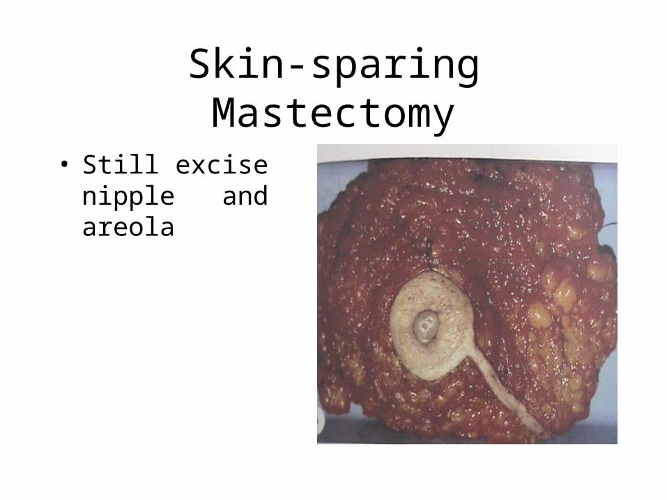
Skin-sparing Mastectomy
• Still excise nipple and areola

Skin-sparing Mastectomy
• Leaves adequate skin for immediate reconstruction
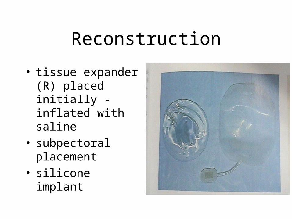
Reconstruction
• tissue expander (R) placed initially - inflated with saline
• subpectoral placement• silicone implant
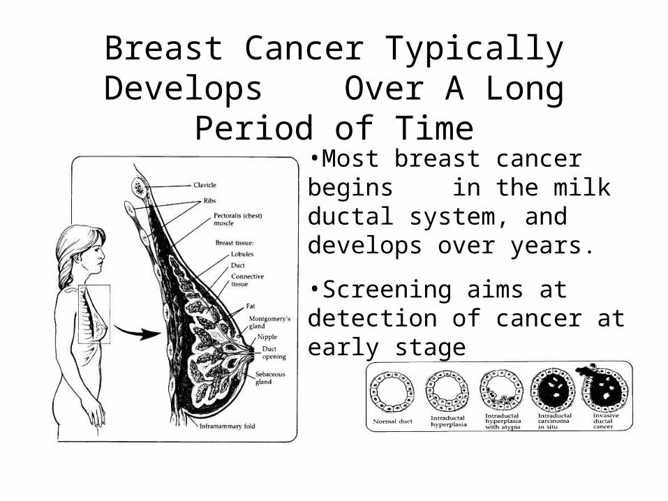
Breast Cancer Typically Develops Over A Long Period of Time
•Most breast cancer begins in the milk ductal system, and develops over years.
•Screening aims at detection of cancer at early stage