Embed Size (px)
Citation preview

Breast Disease:Diagnosis and Management
Nicole Kounalakis, MDAssistant Professor of Surgery
Christina A. Finlayson, MDProfessor of SurgeryDirector, Dianne O’Connor Thompson Breast Center

Goal of Breast Evaluation
The goal of breast evaluation is to classify findings as: normal physiologic variations clearly benign or possibly malignant

Incidence of Breast Cancer by Age
1.9
10.2
22.624.4
19.7
15.5
5.6
0
5
10
15
20
25
30
20-34 35-44 45-54 55-64 65-74 75-84 >85
Age
Per
cent
SEER database 2004-2008
Risk Factors with clinical relevance
Gail Model guidelines 1989 – Current age >50– Early menarche– Delayed child bearing >30-35 yo– Family history (1st degree relatives)– Previous biopsy
• Atypical hyperplasia• LCIS
BRCA1/BRCA2 Radiation to chest wall ↑BMI Postmenopausal use of estrogen/progestin hormone
therapy Chlebowski et al JAMA 2010 Oct 20

Risk Factors with clinical relevance
Most women who develop breast cancer have no identifiable increased risk factors

Preventing Death from Breast Cancer – Early Detection
Breast self examination– Monthly except in the highly anxious woman
Clinician examination– Always important in early detection

Screening Mammography
Should be performed annually beginning at age 40 yearsShould begin earlier in women with a first
degree relative with breast cancer or who had chest radiation as young womanDecreases the chance of dying of breast
cancer by at least 30%
Nystrom et al. Lancet 2002;359

Screening mammography
DCIS
with microcalcifications

Screening mammograhy(BI-RADS™)
Category Definition
1 Normal2 Benign3 Probably benign→
need close f/u4 Suspicious → need
tissue bx5 Cancer

Breast Self Examination (BSE)
Performed every 1-2 monthsBegin at age 20 yearsInstruction on technique needs to be
repeatedTumors detected on BSE tend to be smaller
and with fewer lymph node metastases

Breast Self Examination (BSE)
What to look for:
New lump or thickeningNew skin retraction or dimplingNew skin changes on nipple or breast

Inflammatory Breast Cancer

Clinical Breast Exam (CBE)
Should be performed at least annuallyRequires a thorough, systematic evaluation
of the breast and the draining lymph nodes14-20% of breast cancers found on CBE20-40% false negative rate

Clinical Breast Exam (CBE)
Management of a new breast mass
ObservationBreast imagingBiopsy

Clinical Breast Exam (CBE)Observation
A non-suspicious mass in a young woman can be observed over one menstrual cycle
to see if it disappears, indicating fibroglandular change
Any persistent breast mass requires a diagnosis
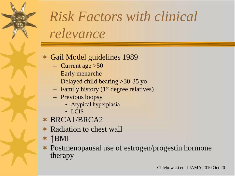
Clinical Breast Exam (CBE)Breast imaging
Ultrasound – can differentiate cystic vs. solidMammography – evaluates the entire breast
as well as characteristics of the massMRI – very sensitive, not very specific. Can
not be used to rule out cancer.
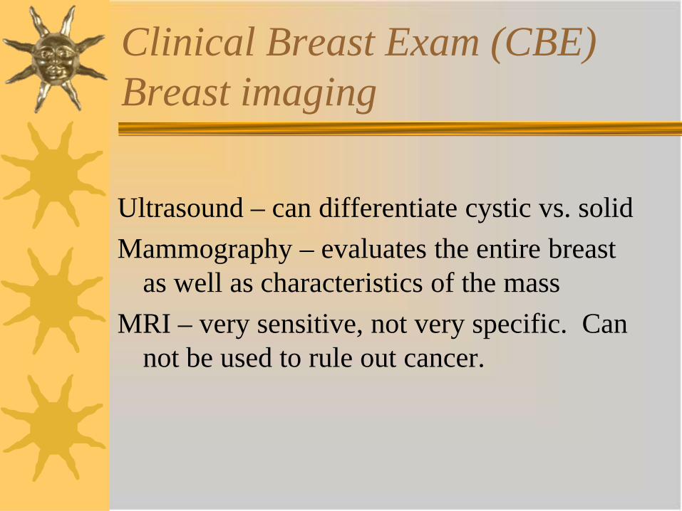
Clinical Breast Exam (CBE)Biopsy
Fine Needle Aspiration (FNA) – recovers single cells
Core needle biopsy – small pieces of tissue
Excisional biopsy – removes the lesion

Fine Needle Aspiration
Simple, accurate, low morbidityRequires skilled cytopathologic interpretationFalse negative results due to sampling errorSensitivity varies (65 to 98%)False positives are rare (about 0.2%) Insufficient or nondiagnostic FNA material:
– repeat FNA– evaluate with alternative biopsy techniques

Triple Test
Clinical examBreast imaging (mammogram and/or US)FNA cytologyIf all three components are clearly benign
and concordant, excisional biopsy is not required (accuracy = 100%)

Clinical Breast Exam (CBE)Core Biopsy

Excisional Biopsy
The highest accuracy for diagnosis in palpable lesions, although more invasive than fine needle or core techniquesSimple with low morbidity

Specific Benign Entities

Definition ofFibrocystic Condition
The clinical manifestations of breasttissue response to cyclical hormonalchanges.

Pathology ofBenign Breast Disease
Non-proliferative lesions (RR = 1.0)– cysts, mild hyperplasia of the usual type
Proliferative lesions without atypia (RR = 1.5 -2.0)– moderate or florid hyperplasia, intraductal
papilloma, sclerosing adenosis, fibroadenoma
Atypical hyperplasia (RR = 4.0 - 5.0)– atypical ductal hyperplasia (ADH), atypical
lobular hyperplasia (ALH)

Fibroadenoma
Common, especially in younger women Epithelial and stromal componentDiagnosis by FNA or core biopsyPathognomonic mammographic appearance:
clinical follow-up is appropriateEquivocal diagnosis or growth should lead to
excisional biopsy

Simple Cysts
Common, often fluctuate with menstrual cycleLess common in post-menopausal
womenDiagnosis by FNA or USSimple cysts confirmed by US may be
observedAspiration for diagnosis or symptoms

Ultrasound of Simple Cyst

Complex Cysts
Evaluation requiredCore biopsy may not sample solid
component and cyst may collapseExcisional biopsy often required for
diagnosis
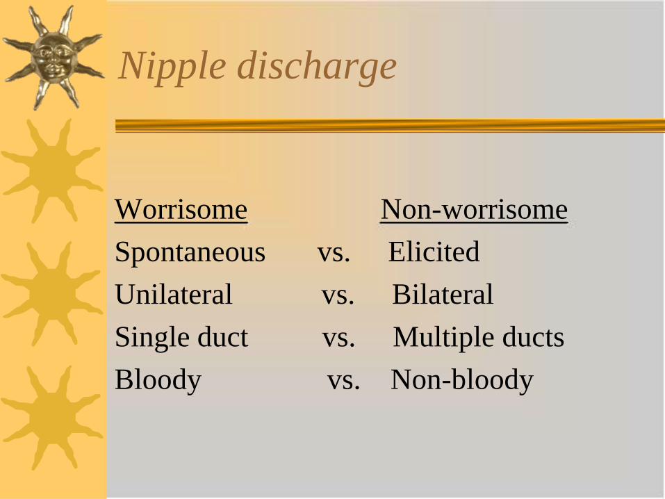
Nipple discharge
Worrisome Non-worrisomeSpontaneous vs. ElicitedUnilateral vs. BilateralSingle duct vs. Multiple ductsBloody vs. Non-bloody

Nipple dischargeTreatment
Milky, unilateral No treatment
Milky bilateral Check prolactin
Unilateral spontaneous Duct excision
Bloody Duct excision
Bilateral, multi-duct or nonbloody elicited
reassurance, avoid trauma

Breast Pain

Breast Pain
Physiologic breast pain– Cyclic– Associated with menstrual cycle
Idiopathic breast pain– Chronic– Constant

Breast Pain
Work up– H & P– Imaging if age appropriate– Focused ultrasound if focal area of pain

Breast Pain
Treatment– Support– NSAID– Evening Primrose Oil– Management of depression

Halsted radical mastectomy
Treating Breast Cancer

Treating breast cancer
Removing the tumorLumpectomy + Radiation Therapy
Mastectomy
Survival is the same

Treating breast cancer:Lumpectomy
Advantages DisadvantagesMore normal Longer treatment timeappearance Not good for all tumor types
Requires radiation
Survival is the same

Treating breast cancer:Mastectomy
Advantages DisadvantagesShorter treatment Loss of breasttime May still require radiation
Survival is the same

Distribution of Breast tissue
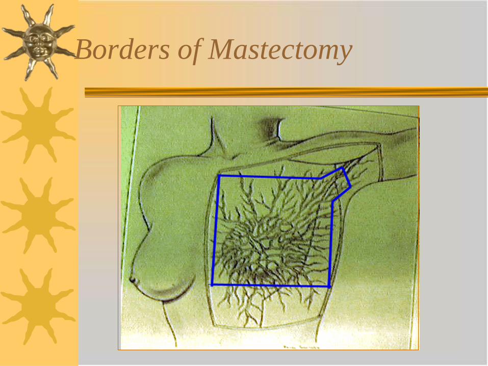
Borders of Mastectomy

Treating breast cancer:Lymph node staging
Axillary node dissection vs. Sentinel lymph node biopsy

Axillary Node Dissection

Definitions-Lymphatic Mapping

Definitions-Sentinel lymph node biopsy

Sentinel Node Biopsy
False negative rate about 8%Identifies those women with negative
lymph nodes who can avoid axillary dissectionLess lymphedemaSignificantly less parasthesiasPositive sentinel lymph nodes should go on
to axillary dissection

St. Agatha the Pure


![Benign Breast Disease[1]](https://img.pdfslide.net/doc/110x75/5571f7b649795991698bd982/benign-breast-disease1.jpg)