Embed Size (px)
Citation preview
BREAST ELASTOGRAPHY AND POWER DOPPLER VOCAL FREMITUS SONOGRAPHY (PDVF)
Lisa Smith, Nancy Infusini, Nicoleta Miclea Miron, Martine Paré
Cedars Breast Clinic
A Technologist’s Point of View
Disclosure Statement: No Conflict of Interest
May 28 – 30, 2015, Montréal, Québec
I do not have an affiliation, financial or otherwise, with a pharmaceutical company, medical device or communications organization.
I have no conflicts of interest to disclose ( i.e. no industry funding received or other commercial relationships).
I have no financial relationship or advisory role with pharmaceutical or device-making companies, or CME provider.
I will be discussing the results of ____ (“off-label” use), which is currently classified by Health Canada as investigational for the intended use.
I will not discuss or describe in my presentation at the meeting the investigational or unlabeled ("off-label") use of a medical device, product, or pharmaceutical that is classified by Health Canada as investigational for the intended use.
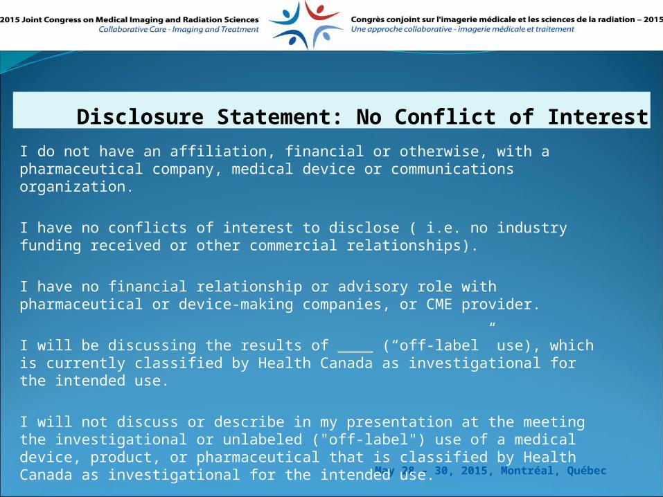
THE CEDARS BREAST CLINIC TEAM
OBJECTIVES
BREAST ANATOMYWHAT IS ELASTOGRAPHY TYPES OF ELASTOGRAPHYELASTOGRAPHY MAPSWHAT IS POWER DOPPLER VOCAL
FREMITUS (PDVF)THE EFFECTS OF THE APPLICATIONS EXAMPLES OF DIFFERENT LESIONS
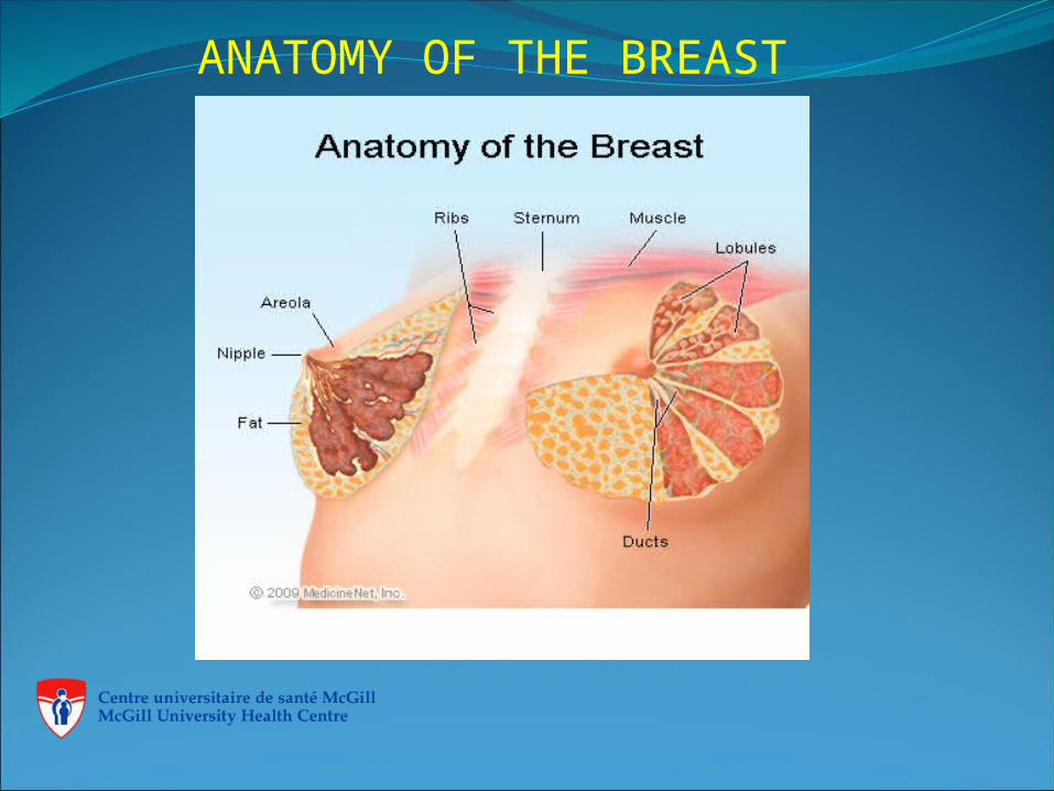
ANATOMY OF THE BREAST
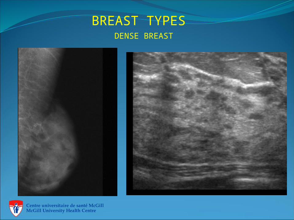
BREAST TYPESDENSE BREAST
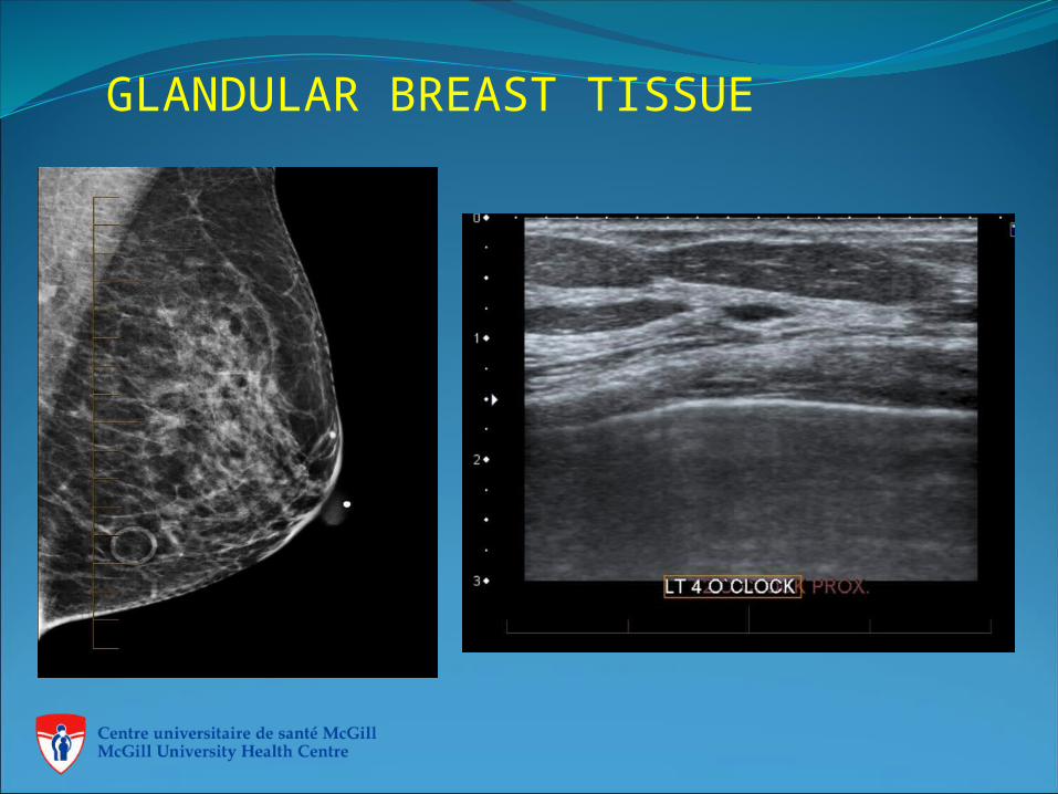
GLANDULAR BREAST TISSUE
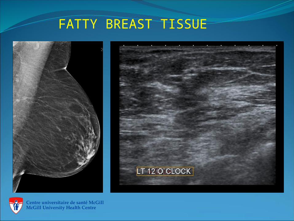
FATTY BREAST TISSUE
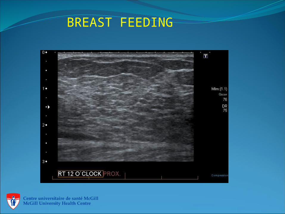
BREAST FEEDING
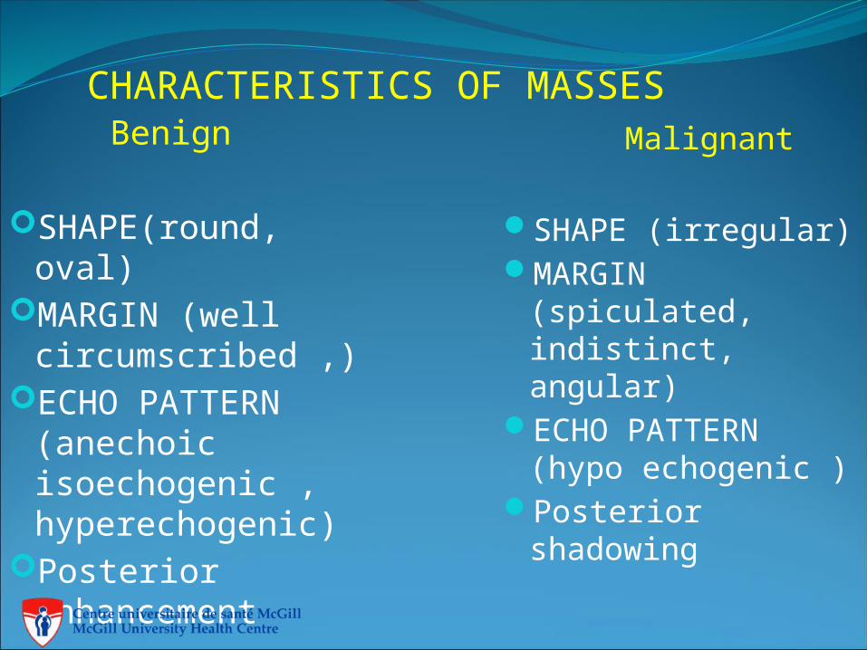
CHARACTERISTICS OF MASSES Benign
SHAPE(round, oval)
MARGIN (well circumscribed ,)
ECHO PATTERN (anechoic isoechogenic , hyperechogenic)
Posterior enhancement
Malignant
SHAPE (irregular)MARGIN
(spiculated, indistinct, angular)
ECHO PATTERN (hypo echogenic )
Posterior shadowing
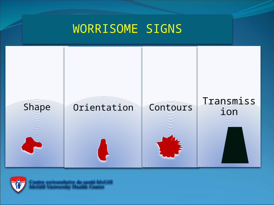
WORRISOME SIGNS
Shape Orientation ContoursTransmissi
on
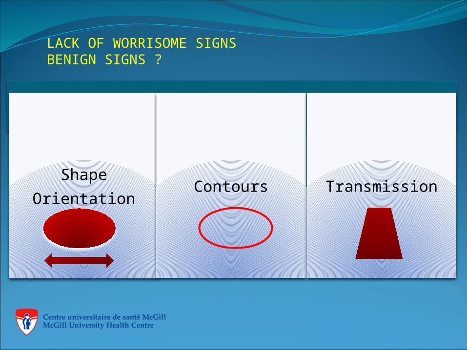
LACK OF WORRISOME SIGNSBENIGN SIGNS ?
Shape
OrientationContours Transmission
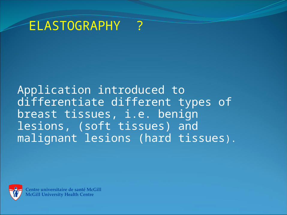
ELASTOGRAPHY ?
Application introduced to differentiate different types of breast tissues, i.e. benign lesions, (soft tissues) and malignant lesions (hard tissues).
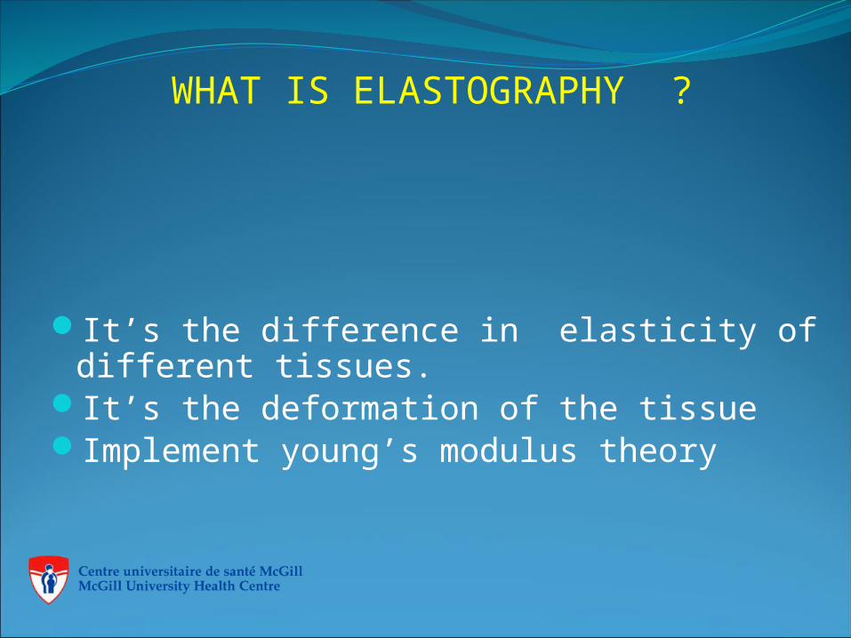
WHAT IS ELASTOGRAPHY ?
It’s the difference in elasticity of different tissues.
It’s the deformation of the tissueImplement young’s modulus theory
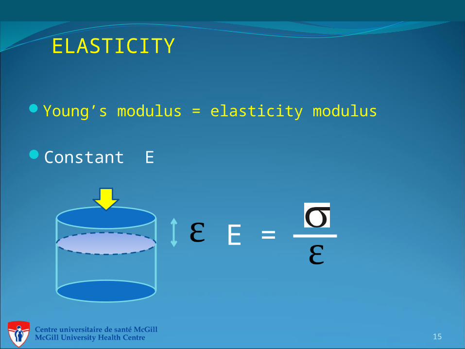
ELASTICITY
Young’s modulus = elasticity modulus
Constant E
15
E =
TYPES OF ELASTOGRAHPY
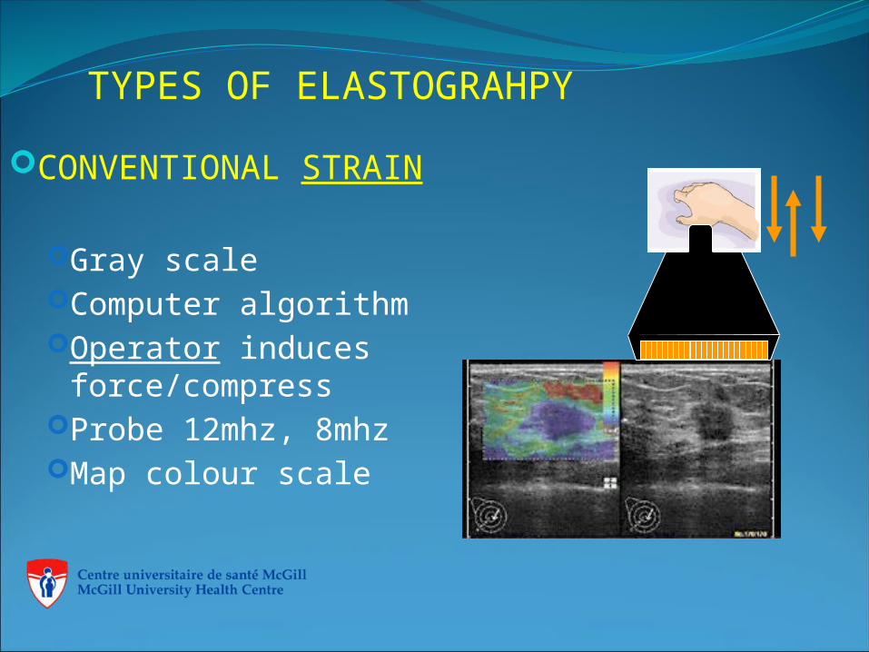
CONVENTIONAL STRAIN
Gray scaleComputer algorithmOperator induces
force/compressProbe 12mhz, 8mhzMap colour scale
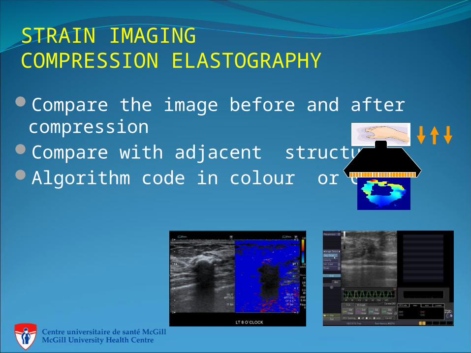
STRAIN IMAGINGCOMPRESSION ELASTOGRAPHY
Compare the image before and after compression
Compare with adjacent structureAlgorithm code in colour or Grey
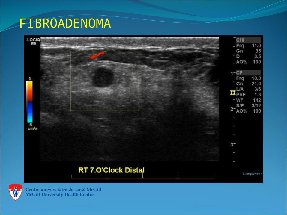
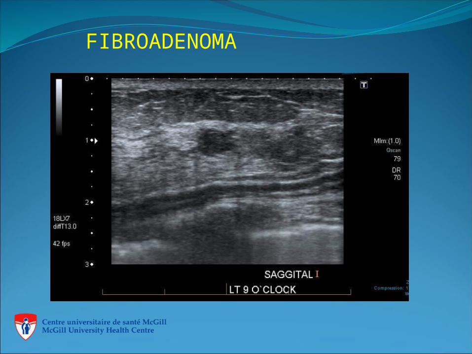
FIBROADENOMA
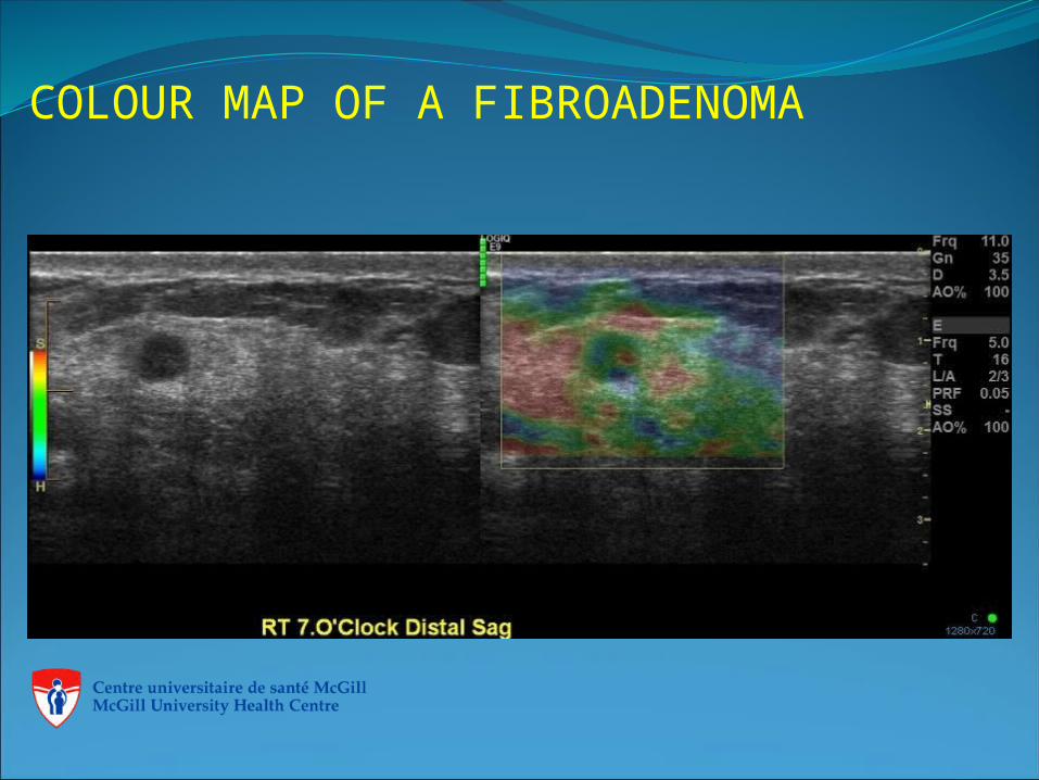
COLOUR MAP OF A FIBROADENOMA
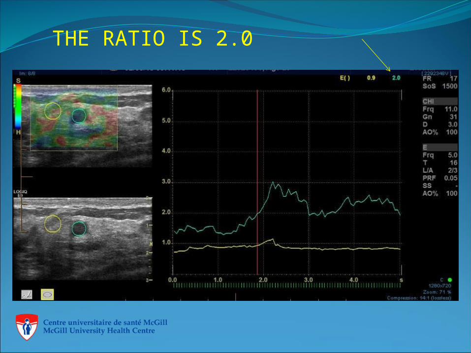
THE RATIO IS 2.0
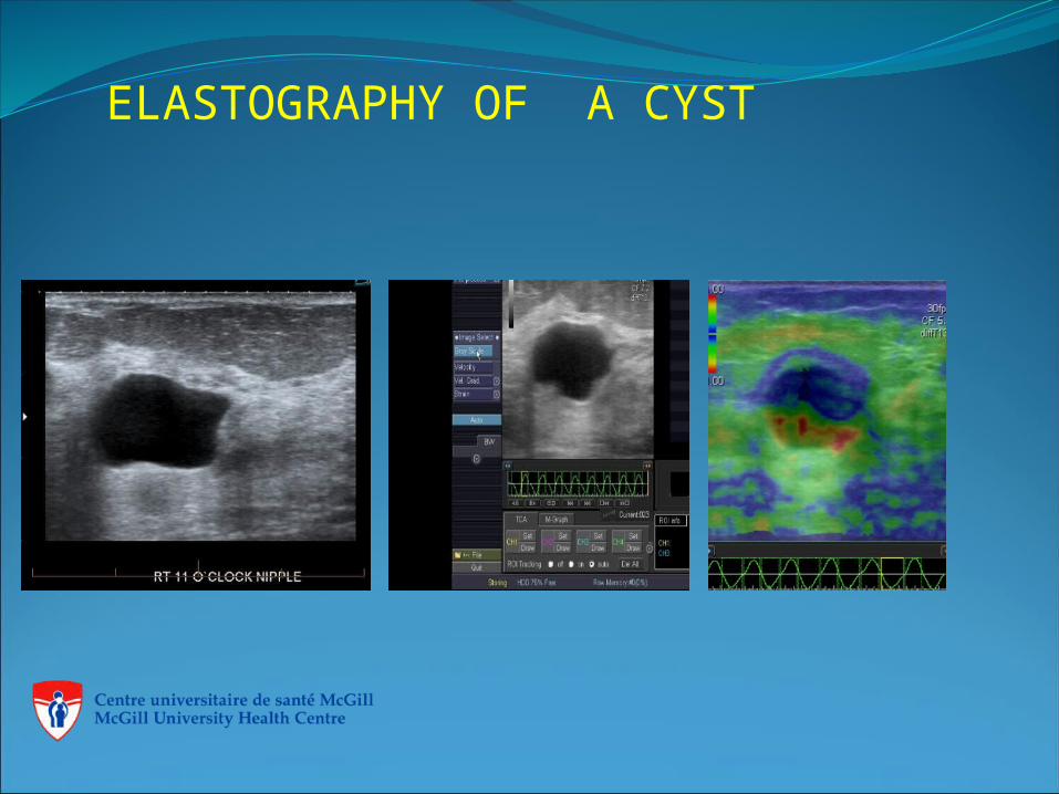
ELASTOGRAPHY OF A CYST
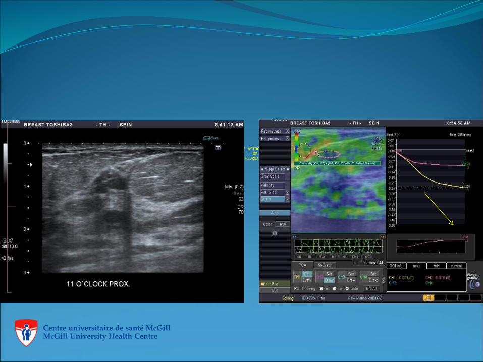
ELASTOGRAPHY OF A
FIBROADENOMA
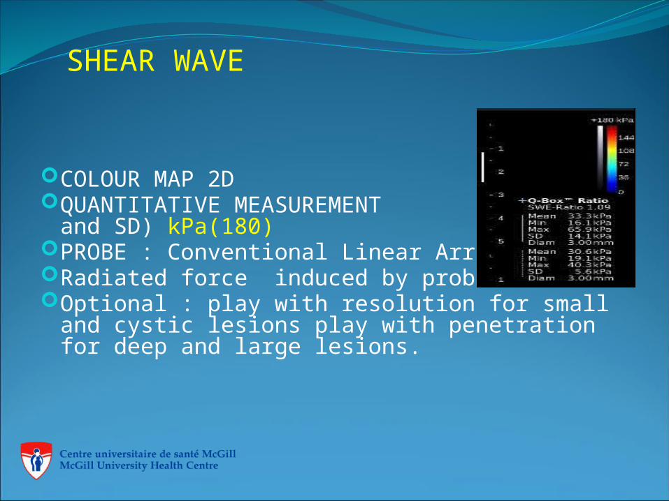
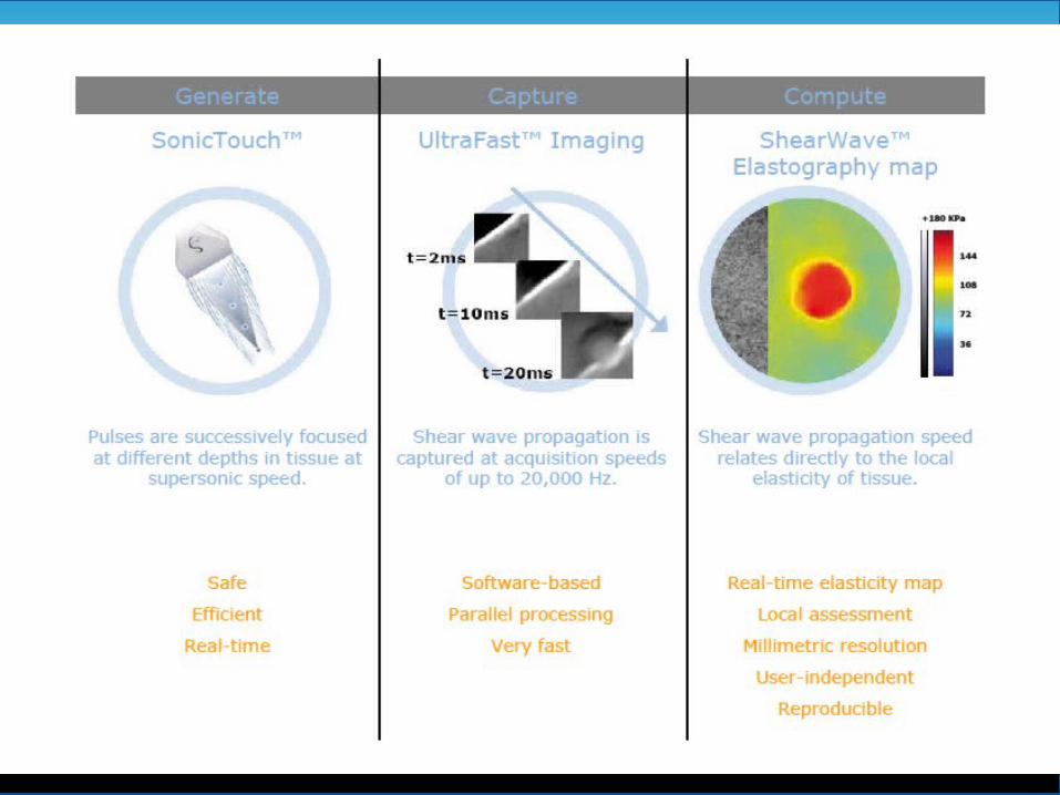
SHEAR WAVE
COLOUR MAP 2DQUANTITATIVE MEASUREMENT
(max, mean and SD) kPa(180)PROBE : Conventional Linear Array. Radiated force induced by probe.Optional : play with resolution for small and
cystic lesions play with penetration for deep and large lesions.
Step One Step Two Step Three
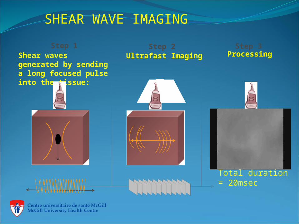
Step 1Shear waves generated by sending a long focused pulse into the tissue:
Step 2Ultrafast Imaging
Step 3Processing
Total duration = 20msec
SHEAR WAVE IMAGING
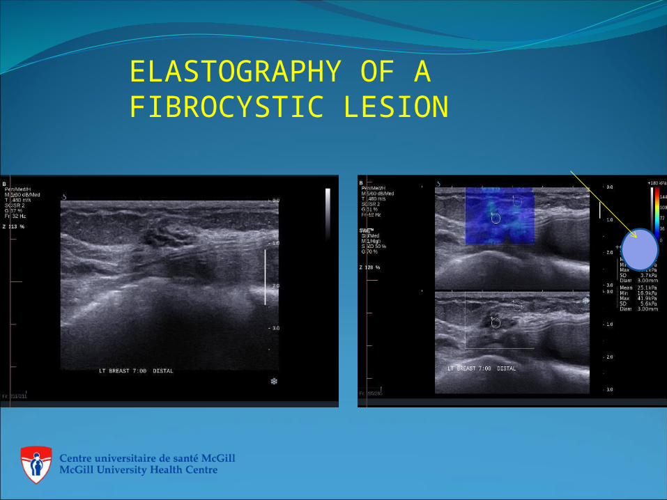
ELASTOGRAPHY OF A FIBROCYSTIC LESION
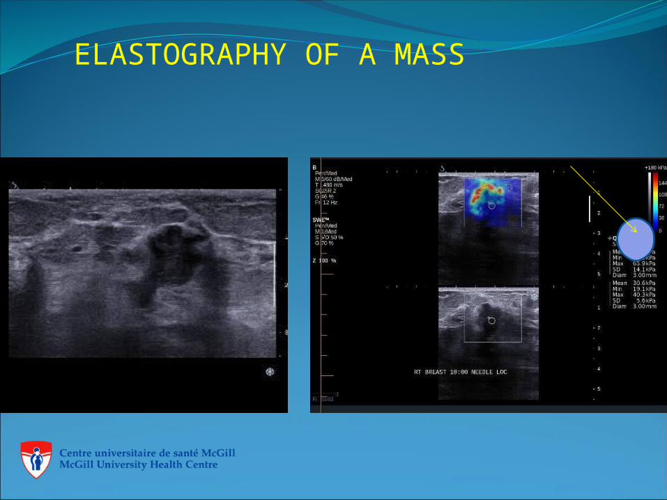
ELASTOGRAPHY OF A MASS
COMPARISON OF SOFT AND HARD LESION. FALSE NEGATIVE
It is stated that benign lesions are soft and malignant lesions are hard. But there are exceptions.
A palpable area biopsied , the result was fibrous breast tissue. Some cancers are soft hyper echogenic, like (mucinous carcinomas) and like scars are hard.
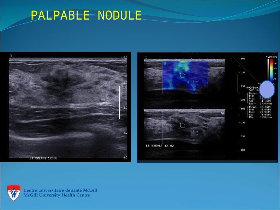
PALPABLE NODULE
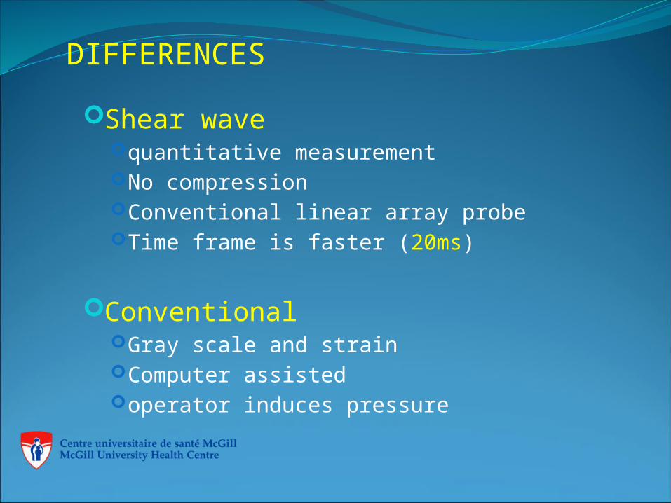
DIFFERENCES Shear wave
quantitative measurementNo compressionConventional linear array probeTime frame is faster (20ms)
ConventionalGray scale and strain Computer assistedoperator induces pressure
LIMITATIONS
Shear wave; Fatty breast with deep lesions. Mobile lesions, Breast implants
Conventional: Breast implants, Deep lesions, superficial lesions, Mobile lesions, small lesions, Excessive pressure
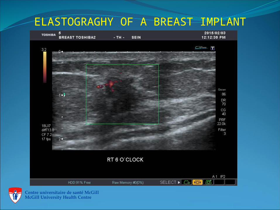
ELASTOGRAGHY OF A BREAST IMPLANT
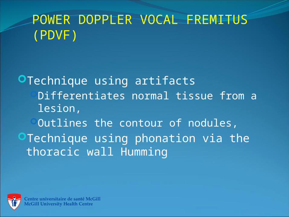
POWER DOPPLER VOCAL FREMITUS (PDVF)
Technique using artifactsDifferentiates normal tissue from a lesion,Outlines the contour of nodules,
Technique using phonation via the thoracic wall Humming
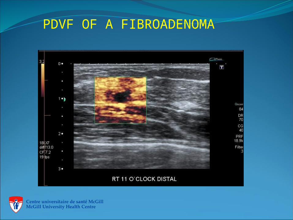
PDVF OF A FIBROADENOMA
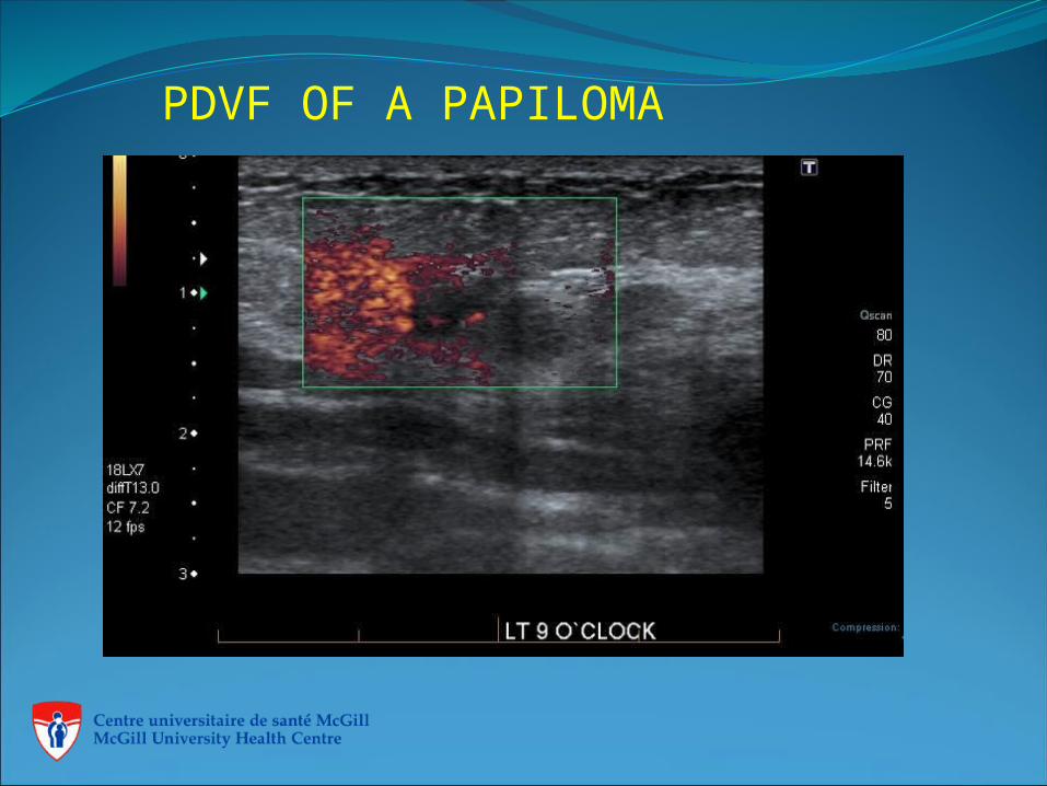
PDVF OF A PAPILOMA
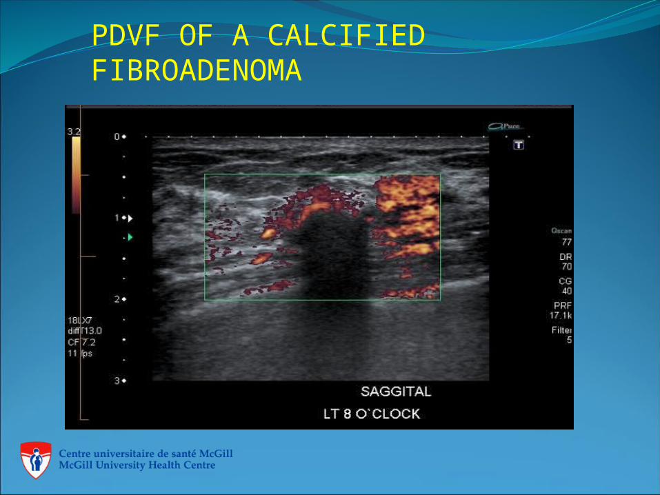
PDVF OF A CALCIFIED FIBROADENOMA
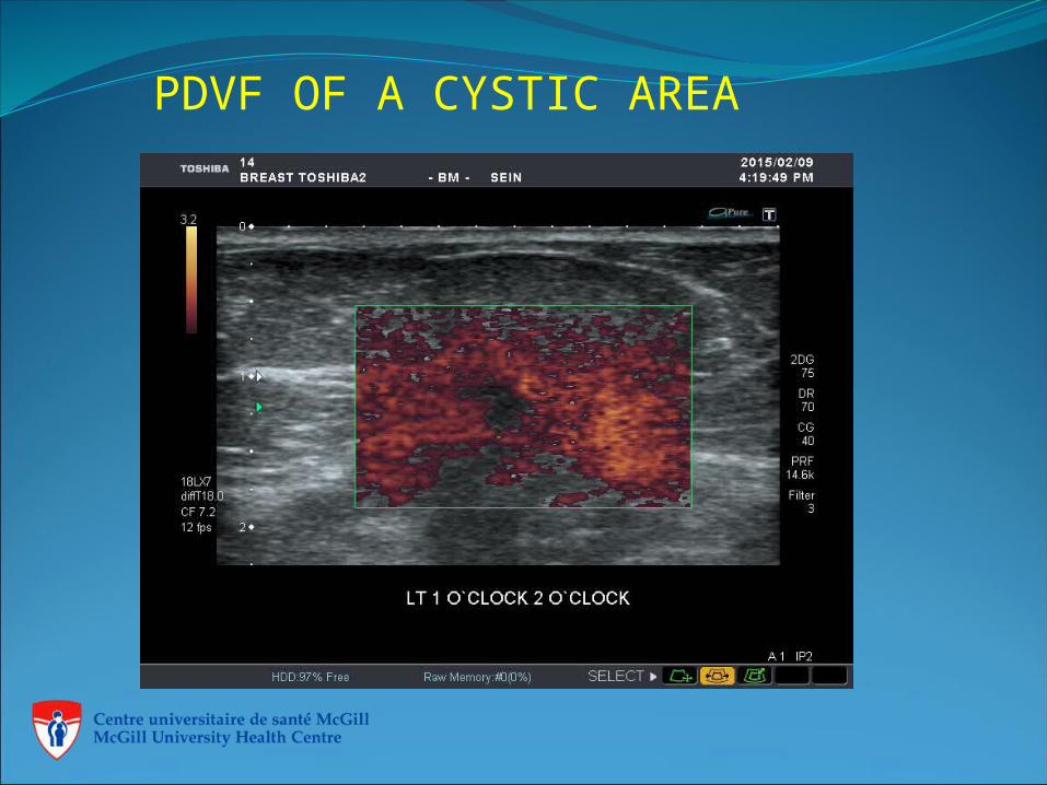
PDVF OF A CYSTIC AREA
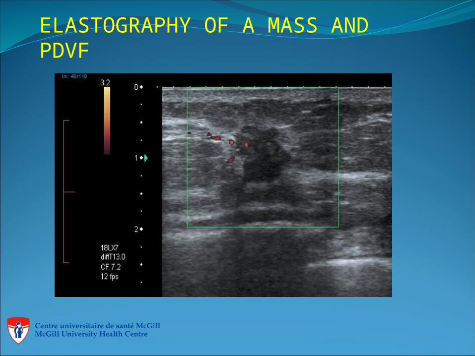
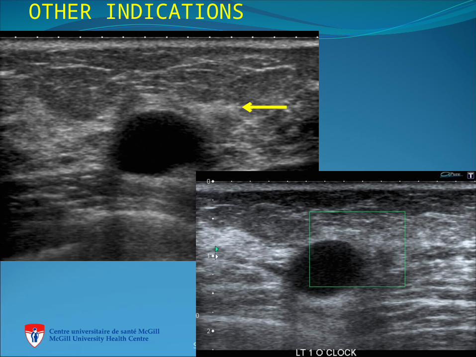
ELASTOGRAPHY OF A MASS AND PDVF
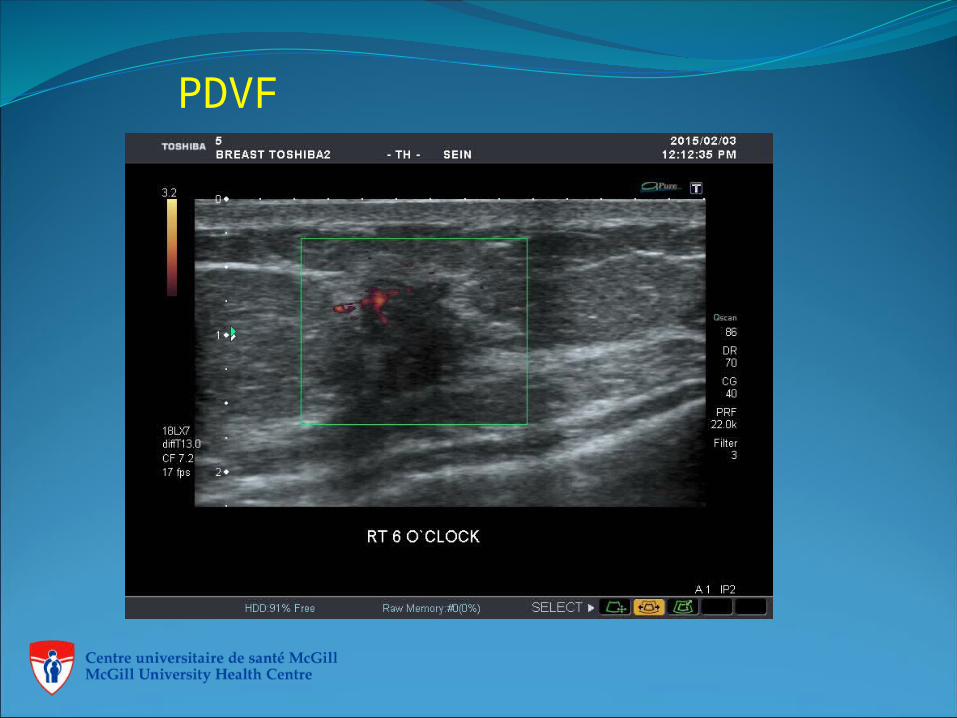
PDVF
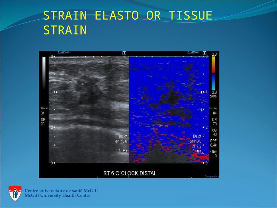
STRAIN ELASTO OR TISSUE STRAIN
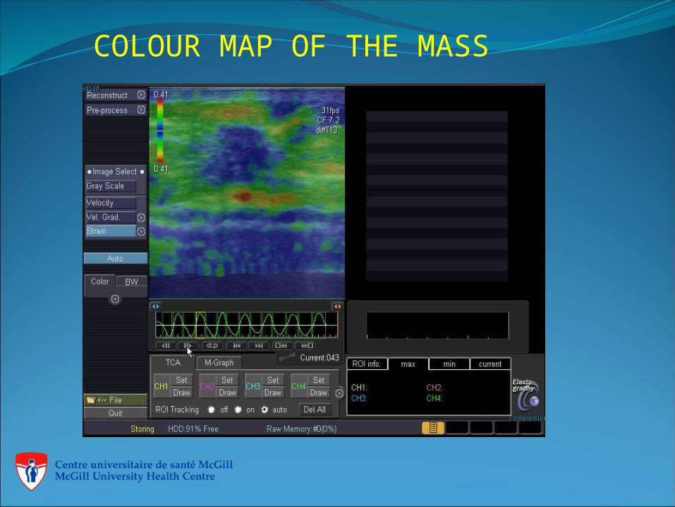
COLOUR MAP OF THE MASS
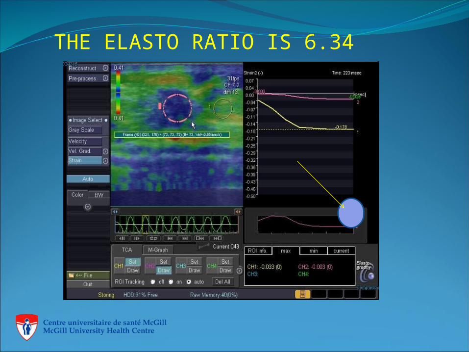
THE ELASTO RATIO IS 6.34
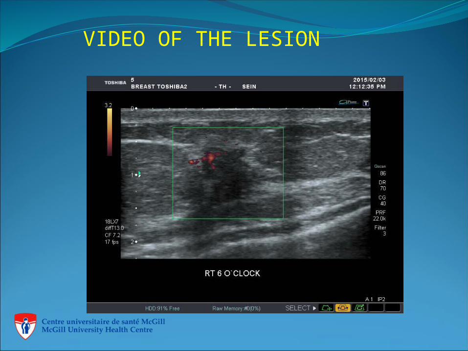
VIDEO OF THE LESION
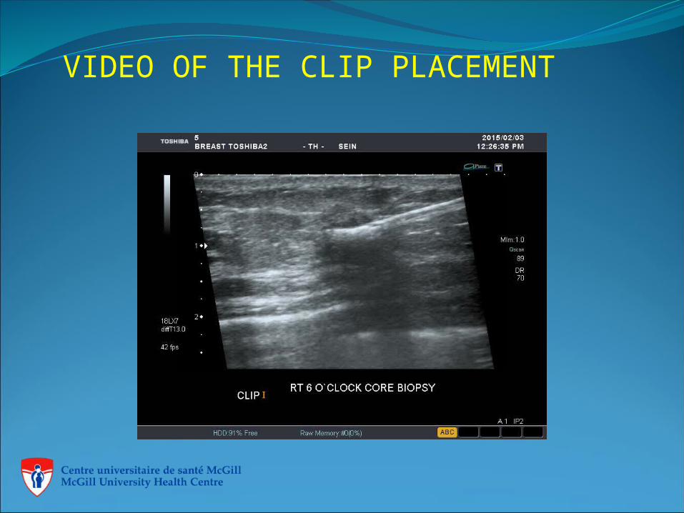
VIDEO OF THE CLIP PLACEMENT
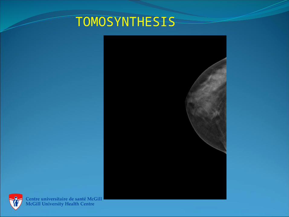
TOMOSYNTHESIS
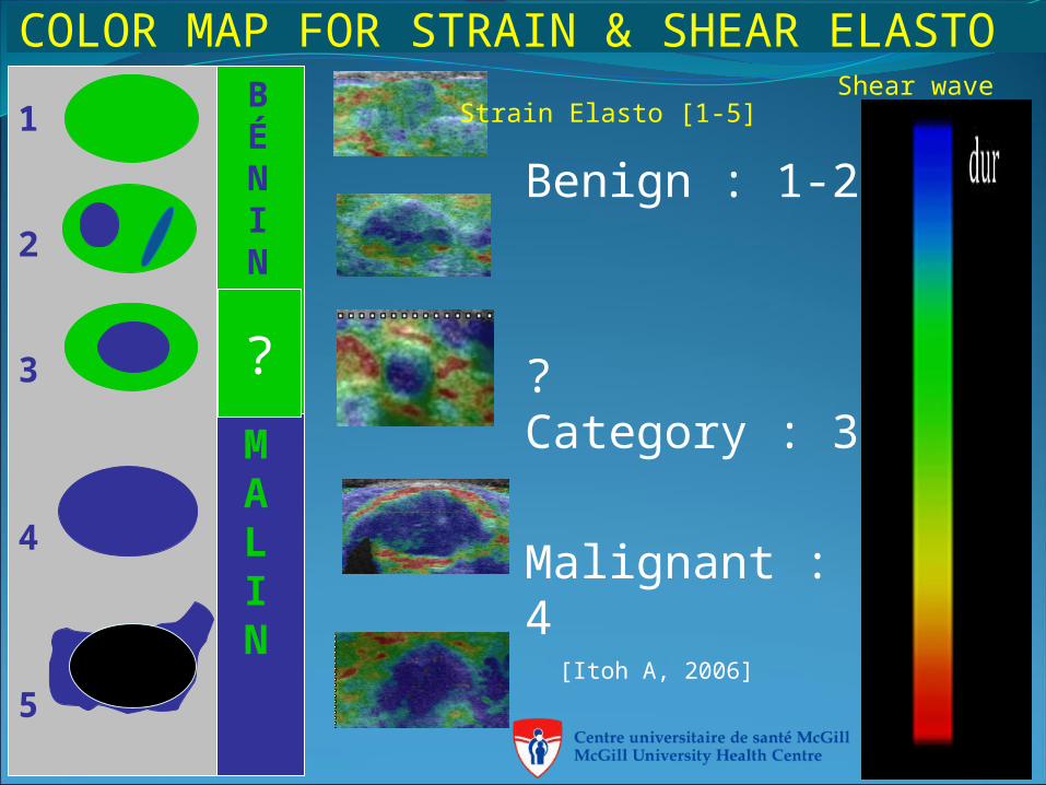
BÉNIN
MALIN
1
2
3
4
5
COLOR MAP FOR STRAIN & SHEAR ELASTO
[Itoh A, 2006]
?
Benign : 1-2
? Category : 3
Malignant : 4
Shear waveStrain Elasto [1-5]
FIBROADENOMA
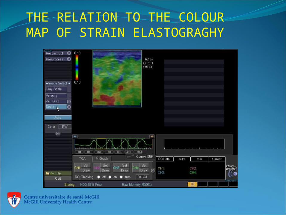
THE RELATION TO THE COLOUR MAP OF STRAIN ELASTOGRAGHY
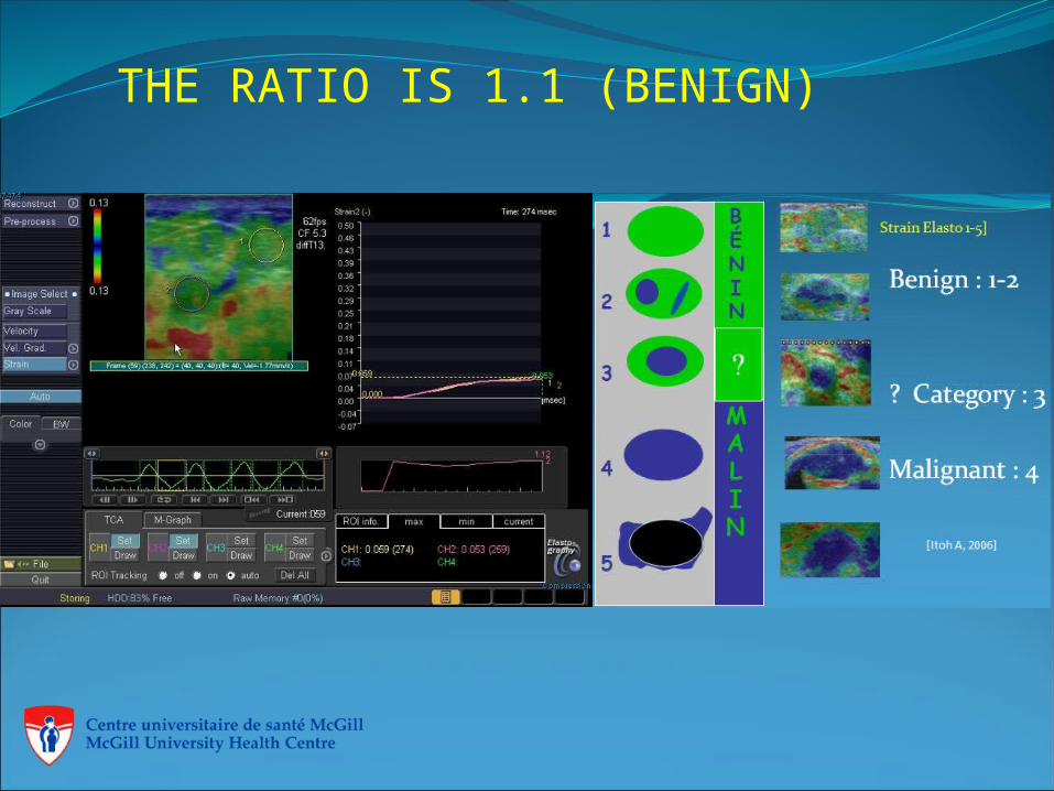
THE RATIO IS 1.1 (BENIGN)
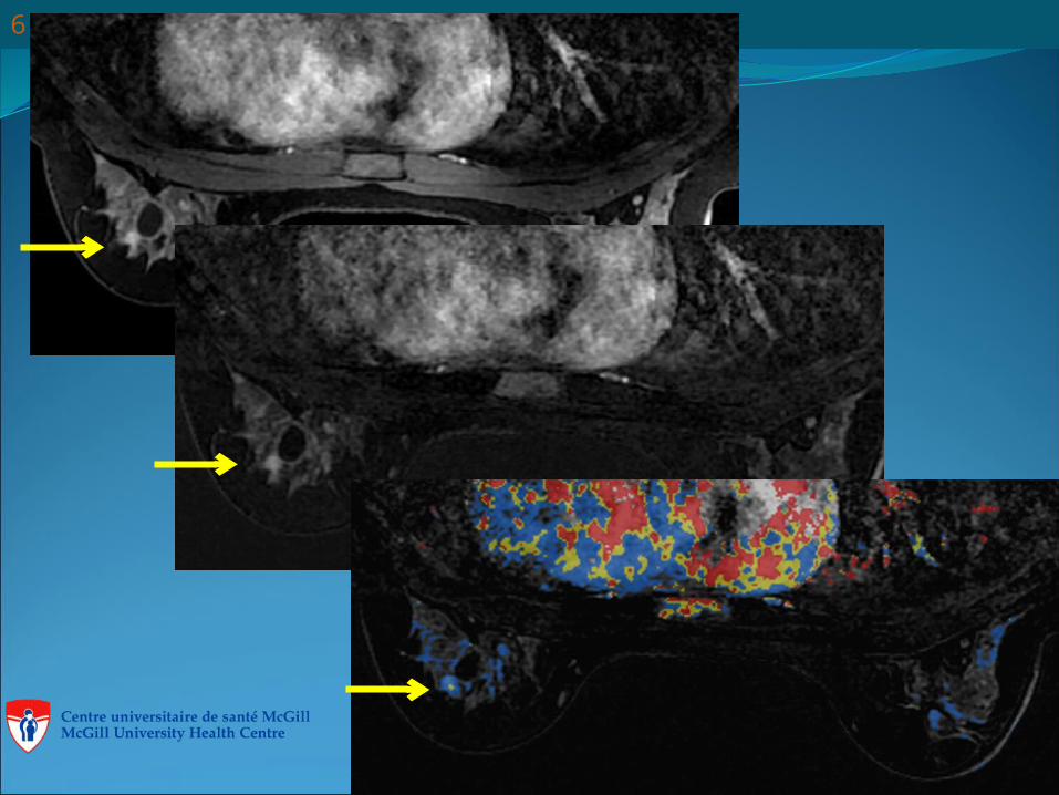
THE CORRELATION OF ULTRASOUND AND MRI
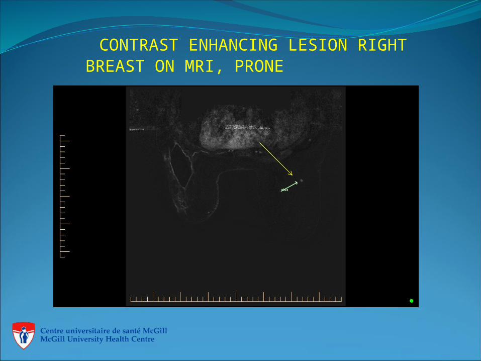
CONTRAST ENHANCING LESION RIGHT BREAST ON MRI, PRONE
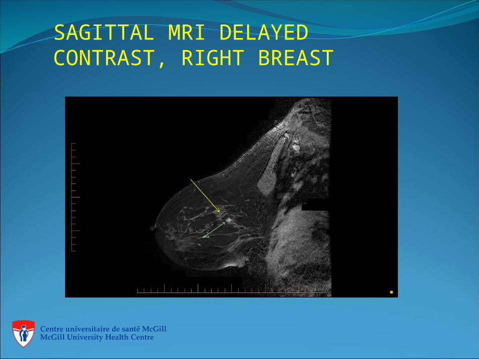
SAGITTAL MRI DELAYED CONTRAST, RIGHT BREAST
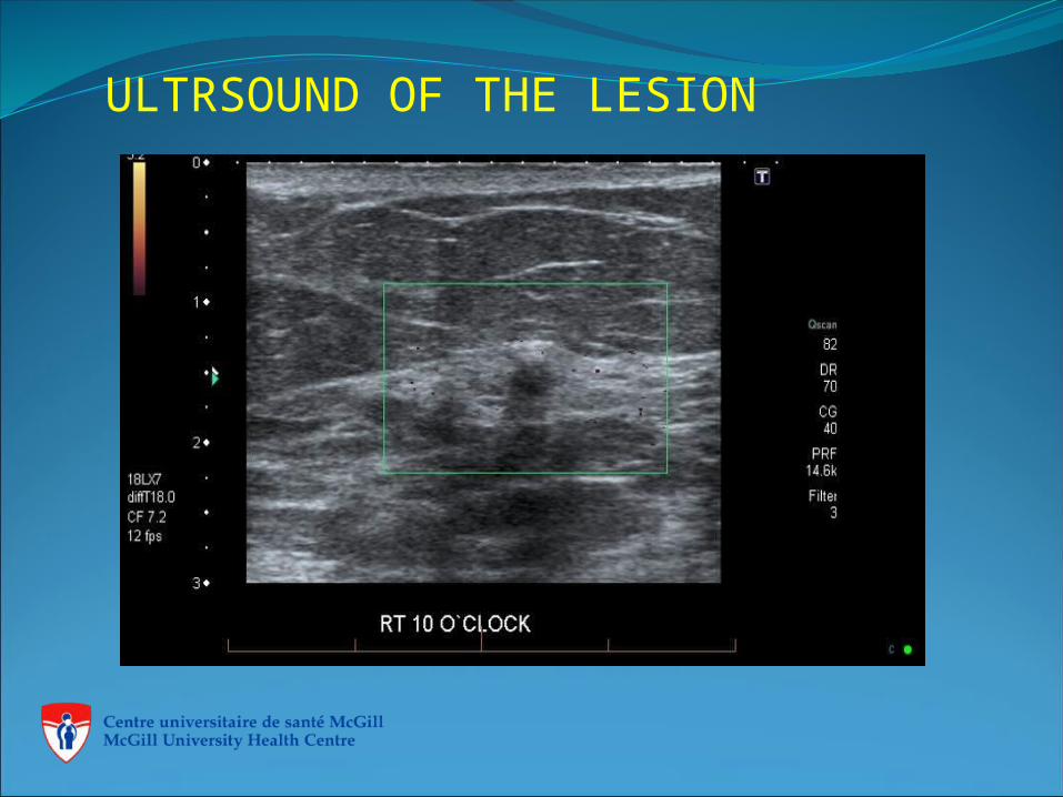
ULTRSOUND OF THE LESION
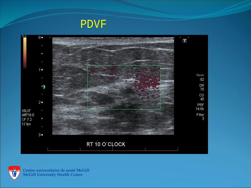
PDVF
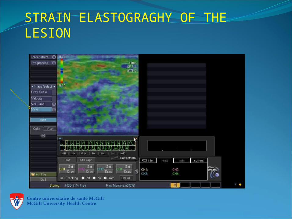
STRAIN ELASTOGRAGHY OF THE LESION
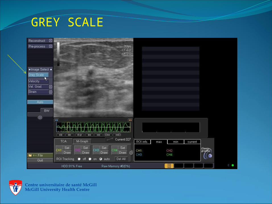
GREY SCALE
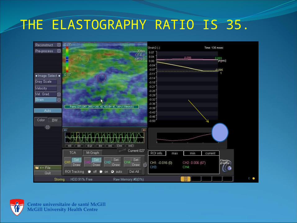
THE ELASTOGRAPHY RATIO IS 35.
IMPORTANCE OF ELASTOGRAPHY WITH SECOND LOOK ULTRASOUND
SCFR 59
6 Autres Indications
Lundi 29 octobre 2012
OTHER INDICATIONS
60
Lundi 29 octobre 2012
SCFR 61
OTHER INDICATIONS
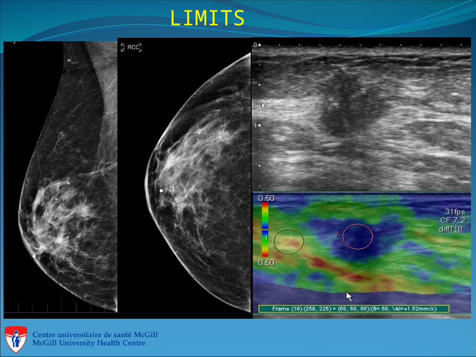
LIMITS
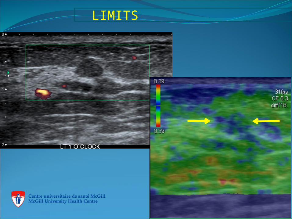
LIMITS
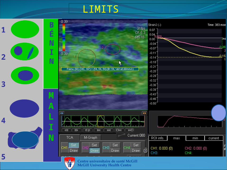
LIMITSBÉNIN
MALIN
1
2
3
4
5
CONCLUSION
Elastography, Power Doppler Vocal Fremitus ( PDVF) ultrasound along with Mammography, Magnectic resonance and Tomosynthesis can assist in the detection of lesions.
The radiologist will use the findings to assign a BI-RADS classification ( Breast imaging report and data system) (ACR).
THANK YOU