Embed Size (px)
Citation preview

Breast MRI of Invasive Cancer
Breast MRI of Invasive Breast MRI of Invasive CancerCancer
Audrey Spielmann, MDAudrey Audrey SpielmannSpielmann, MD, MDBC Surgery Oncology Breast Cancer Update BC Surgery OncologyBC Surgery Oncology Breast Cancer Update Breast Cancer Update

OutlineOutlineOutline
TechniqueIndicationsCasesPre-op Breast MRIConclusion
TechniqueTechniqueIndicationsIndicationsCasesCasesPrePre--op Breast MRIop Breast MRIConclusion Conclusion
OutlineOutlineOutline
Excluded• Screening• Post-op MRI• DCIS• Breast Implant
integrity
ExcludedExcluded•• ScreeningScreening•• PostPost--op MRIop MRI•• DCISDCIS•• Breast Implant Breast Implant
integrityintegrity
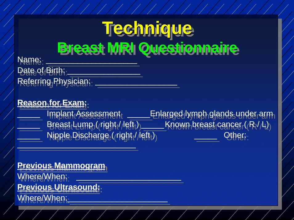
Name: ____________________Date of Birth: ________________Referring Physician: __________________
Reason for Exam:_____ Implant Assessment _____Enlarged lymph glands under arm_____ Breast Lump ( right / left ) _____Known breast cancer ( R / L)_____ Nipple Discharge ( right / left ) _____ Other: __________________________
Previous MammogramWhere/When: _______________________Previous Ultrasound:Where/When:______________________
TechniqueBreast MRI Questionnaire
TechniqueTechniqueBreast MRI Questionnaire

Previous Breast Surgery: ( yes / no )Where/When: _____________R/ L breast Benign / Malignant
Pre-menopausal ( yes / no ) First day of LMP: _____________Exam should be scheduled for Day 7-14 of cycle
Post-menopausal ( yes / no ) On HRT? _________________HRT should be stopped 3 months prior to exam—consult your physician
Do you have a family history of breast cancer (if yes, please indicate age of diagnosis)Mother _____ Sister _____ Grandmother _____ Aunt _____ Daughter ______
TechniqueBreast MRI Questionnaire
TechniqueTechniqueBreast MRI Questionnaire

TechniqueBreast MRI Questionnaire
TechniqueTechniqueBreast MRI Questionnaire
Have you had any of the following treatments(If yes, please indicate where & when)
_____ Lumpectomy _____ Mastectomy_____ Chemotherapy _____ Radiation_____ Tamoxifen ______ HRT / BCP_____ Needle biopsy
Patient Signature: ______________________________________Date: _______________
Have you had any of the following treatments(If yes, please indicate where & when)
_____ Lumpectomy _____ Mastectomy_____ Chemotherapy _____ Radiation_____ Tamoxifen ______ HRT / BCP_____ Needle biopsy
Patient Signature: ______________________________________Date: _______________

TechniqueBreast Coil
TechniqueTechniqueBreast CoilBreast Coil

Technique Positioning
Technique Technique PositioningPositioning
TechniqueSequences
TechniqueTechniqueSequencesSequences
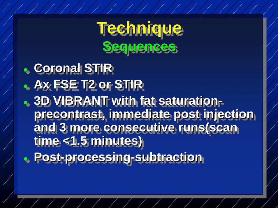
Coronal STIRAx FSE T2 or STIR3D VIBRANT with fat saturation-precontrast, immediate post injection and 3 more consecutive runs(scantime <1.5 minutes)Post-processing-subtraction
Coronal STIRCoronal STIRAx FSE T2 or STIRAx FSE T2 or STIR3D VIBRANT with fat saturation3D VIBRANT with fat saturation--precontrastprecontrast, immediate post injection , immediate post injection and 3 more consecutive and 3 more consecutive runs(scanruns(scantime <1.5 minutes)time <1.5 minutes)PostPost--processingprocessing--subtractionsubtraction
Breast MRI BI-RADS LexiconBreast MRI Breast MRI BIBI--RADS LexiconRADS Lexicon
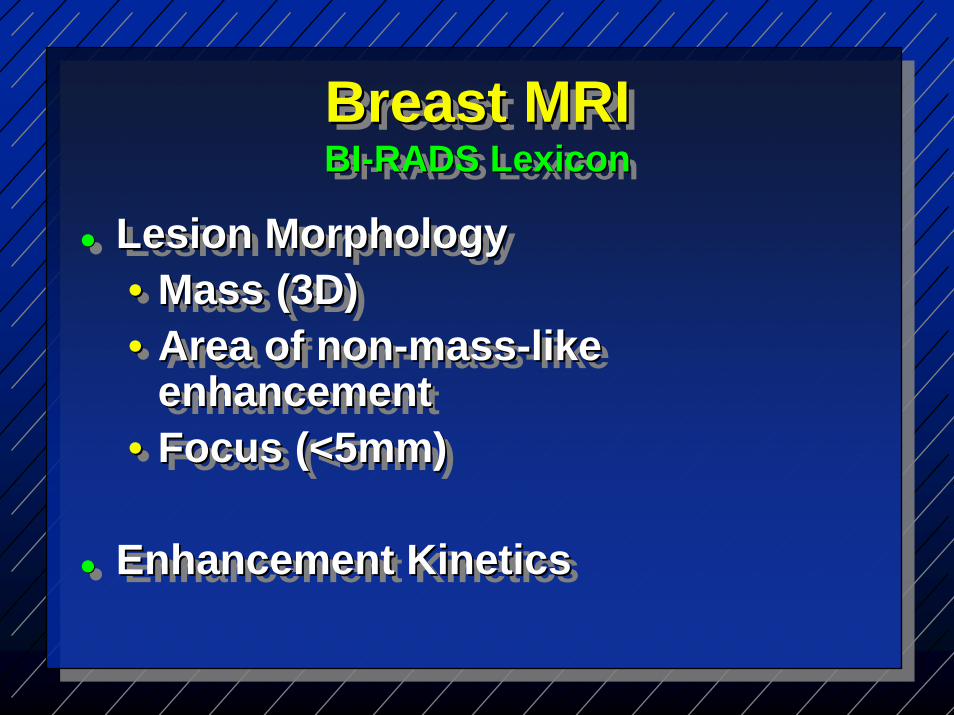
Lesion Morphology• Mass (3D)• Area of non-mass-like
enhancement• Focus (<5mm)
Enhancement Kinetics
Lesion MorphologyLesion Morphology•• Mass (3D)Mass (3D)•• Area of nonArea of non--massmass--like like
enhancementenhancement•• Focus (<5mm)Focus (<5mm)
Enhancement KineticsEnhancement Kinetics
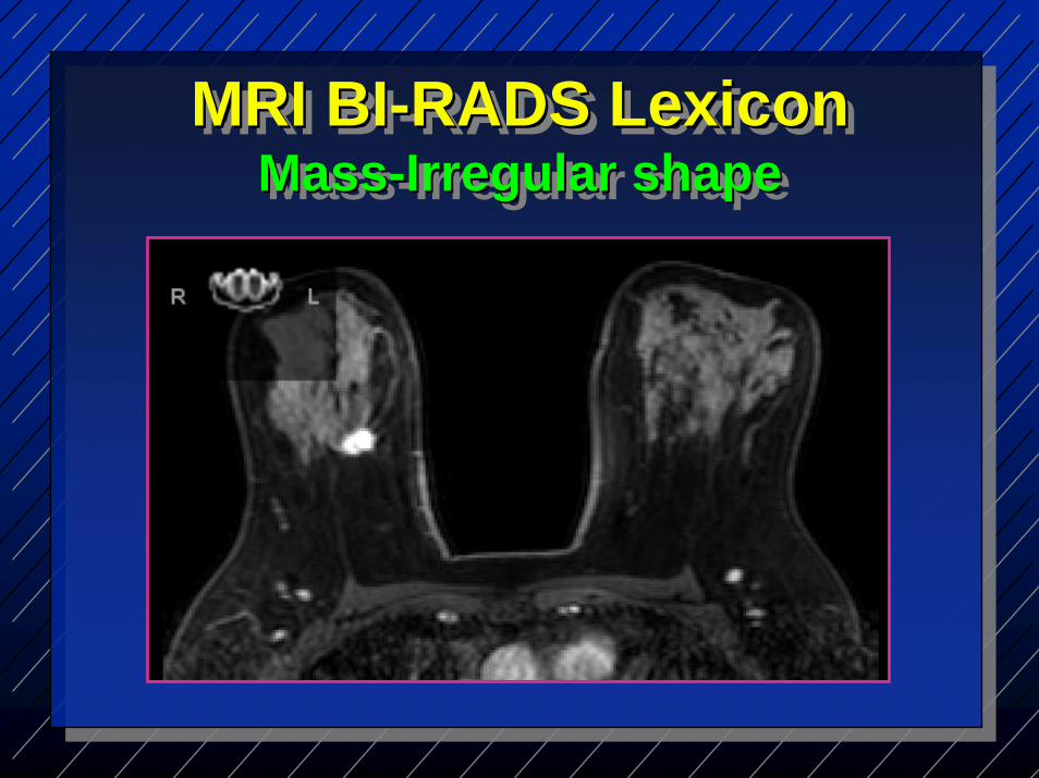
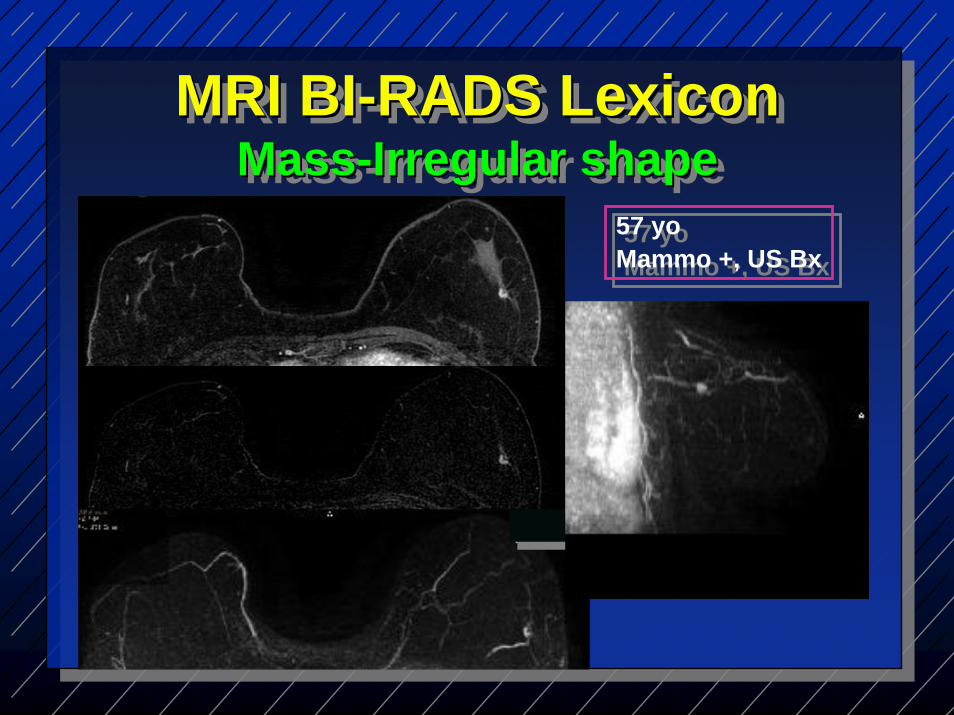
MRI BI-RADS Lexicon Mass-Irregular shape
MRI BIMRI BI--RADS Lexicon RADS Lexicon MassMass--Irregular shapeIrregular shape
MRI BI-RADS Lexicon Mass-Irregular shape
MRI BIMRI BI--RADS Lexicon RADS Lexicon MassMass--Irregular shapeIrregular shape
57 yoMammo +, US Bx
57 yoMammo +, US Bx

MRI BI-RADS Lexicon Asymmetric EnhancementMRI BIMRI BI--RADS Lexicon RADS Lexicon Asymmetric EnhancementAsymmetric Enhancement
45 yoBx DCIS
45 yoBx DCIS
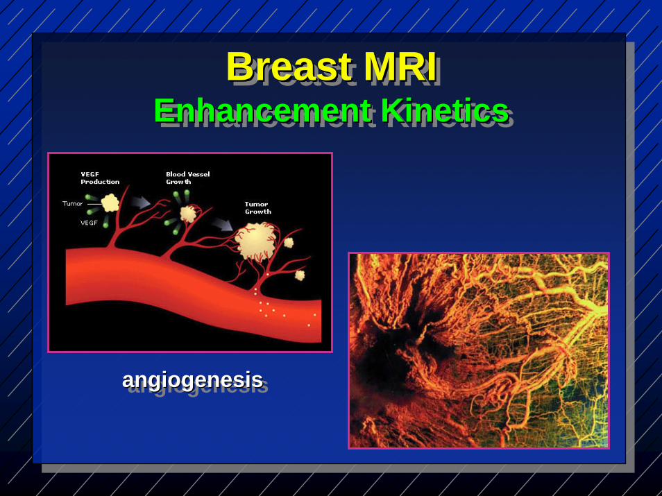
Breast MRIEnhancement Kinetics
Breast MRIBreast MRIEnhancement KineticsEnhancement Kinetics
angiogenesisangiogenesisangiogenesis
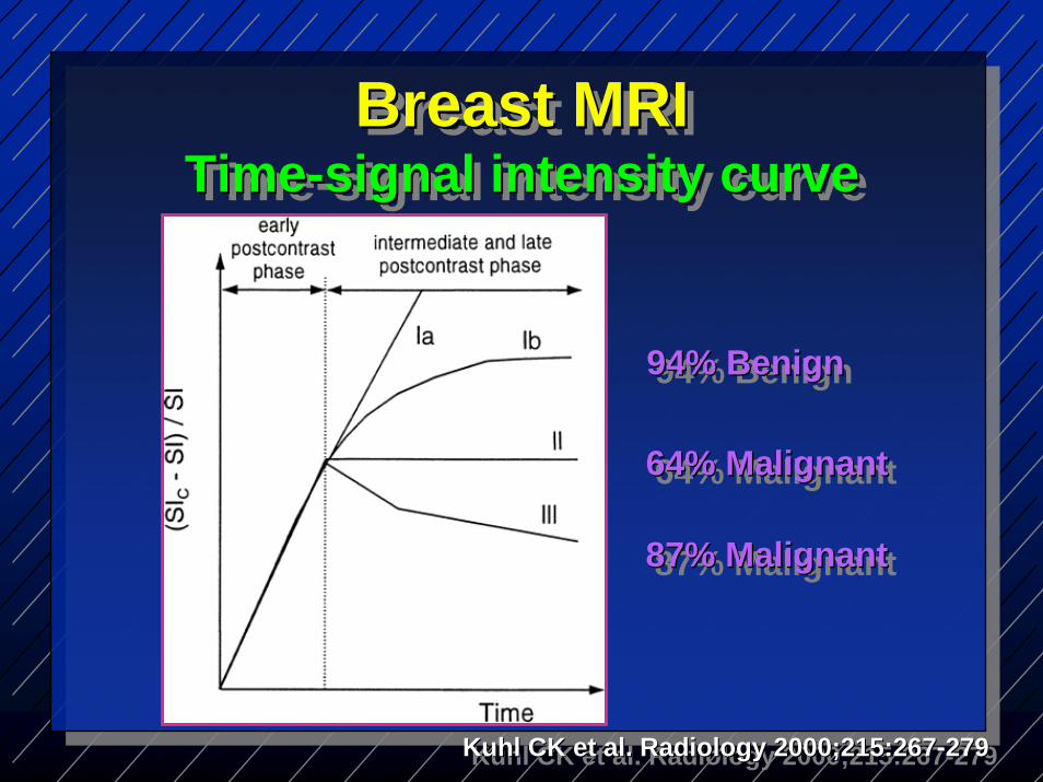
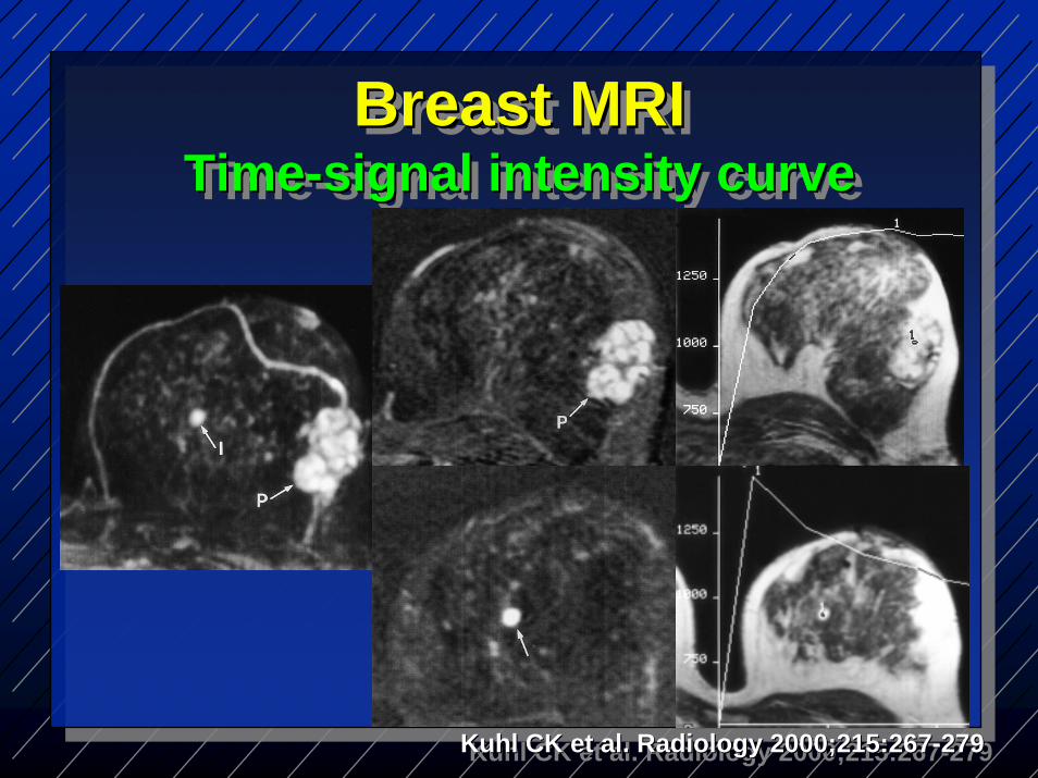
Breast MRITime-signal intensity curve
Breast MRIBreast MRITimeTime--signal intensity curvesignal intensity curve
64% Malignant64% Malignant64% Malignant
87% Malignant87% Malignant87% Malignant
94% Benign94% Benign94% Benign
Kuhl CK et al. Radiology 2000;215:267-279KuhlKuhl CK et al. Radiology 2000;215:267CK et al. Radiology 2000;215:267--279279
Breast MRITime-signal intensity curve
Breast MRIBreast MRITimeTime--signal intensity curvesignal intensity curve
Kuhl CK et al. Radiology 2000;215:267-279KuhlKuhl CK et al. Radiology 2000;215:267CK et al. Radiology 2000;215:267--279279
IndicationsACR practice guidelines (2008)
IndicationsIndicationsACR practice guidelines (2008)ACR practice guidelines (2008)
Screening• High risk patients• Contralateral breast (3-5%
occult malignancy)• Breast Augmentation
ScreeningScreening•• High risk patientsHigh risk patients•• ContralateralContralateral breast (3breast (3--5% 5%
occult malignancy)occult malignancy)•• Breast AugmentationBreast Augmentation

Indications Breast Augmentation
Indications Indications Breast AugmentationBreast Augmentation
54 y womanImplants 20 y agoPalpable mass LMammo +US bx
54 y womanImplants 20 y agoPalpable mass LMammo +US bx

IndicationsACR practice guidelines (2008)
IndicationsIndicationsACR practice guidelines (2008)ACR practice guidelines (2008)
Extent of disease• Multifocality and Multicentricity• Invasion deep to fascia• Postlumpectomy + margins• Neoadjuvant chemotherapy
Extent of diseaseExtent of disease•• MultifocalityMultifocality and and MulticentricityMulticentricity•• Invasion deep to fasciaInvasion deep to fascia•• PostlumpectomyPostlumpectomy + margins+ margins•• NeoadjuvantNeoadjuvant chemotherapychemotherapy

Additional evaluation of clinical/imaging findings• Recurrence• Occult Breast Cancer• Lesion characterization• PO tissue reconstruction• MRI-guided biopsy
Additional evaluation of Additional evaluation of clinical/imaging findingsclinical/imaging findings•• RecurrenceRecurrence•• Occult Breast CancerOccult Breast Cancer•• Lesion characterizationLesion characterization•• PO tissue reconstructionPO tissue reconstruction•• MRIMRI--guided biopsyguided biopsy
IndicationsACR practice guidelines (2008)
IndicationsIndicationsACR practice guidelines (2008)ACR practice guidelines (2008)

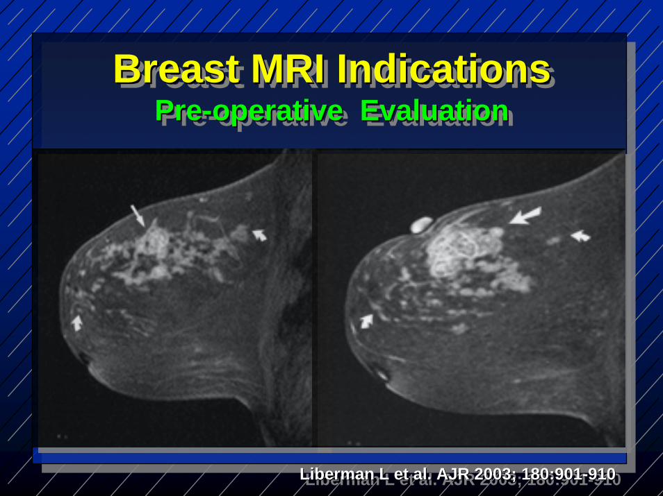
Breast MRI Indications Pre-operative Evaluation
Breast MRI Indications Breast MRI Indications PrePre--operative Evaluationoperative Evaluation
Tumour size and locationMultifocality & Multicentricity(occult disease 15-37%)Chest wall or pectoralis muscle invasion, nipple or skin invasionAxillary or internal mammary LNMetastasis
TumourTumour size and locationsize and locationMultifocalityMultifocality & & MulticentricityMulticentricity(occult disease 15(occult disease 15--37%)37%)Chest wall or Chest wall or pectoralispectoralis muscle muscle invasion, nipple or skin invasioninvasion, nipple or skin invasionAxillaryAxillary or internal mammary LNor internal mammary LNMetastasisMetastasis

Ipsilateral cancer was found on MRI in 19/70 (27%)• 20% same quadrant• 4% different quadrant• 3% same and different
Strong family Hx or infiltrating lobular histology
IpsilateralIpsilateral cancer was found on MRI cancer was found on MRI in 19/70 (in 19/70 (27%27%))•• 20% same quadrant20% same quadrant•• 4% different quadrant4% different quadrant•• 3% same and different3% same and different
Strong family Strong family HxHx or infiltrating or infiltrating lobular histologylobular histology
Liberman L et al. AJR 2003; 180:901-910LibermanLiberman L et al. AJR 2003; 180:901L et al. AJR 2003; 180:901--910910
Breast MRI Indications Pre-operative Evaluation
Breast MRI Indications Breast MRI Indications PrePre--operative Evaluationoperative Evaluation

Liberman L et al. AJR 2003; 180:901-910LibermanLiberman L et al. AJR 2003; 180:901L et al. AJR 2003; 180:901--910910
Breast MRI Indications Pre-operative Evaluation
Breast MRI Indications Breast MRI Indications PrePre--operative Evaluationoperative Evaluation
Liberman L et al. AJR 2003; 180:901-910LibermanLiberman L et al. AJR 2003; 180:901L et al. AJR 2003; 180:901--910910
Breast MRI Indications Pre-operative Evaluation
Breast MRI Indications Breast MRI Indications PrePre--operative Evaluationoperative Evaluation

50 yo woman77 yo mother dx metastatic breast
CaThickening L UOQMammo -, US - in area but lesion
in L UIQUS Bx→ infiltrating lobular Ca
50 50 yoyo womanwoman77 77 yoyo mother mother dxdx metastaticmetastatic breast breast
CaCaThickening L UOQThickening L UOQMammoMammo --, US , US -- in area but lesion in area but lesion
in L UIQin L UIQUS US BxBx→→ infiltrating lobular Ca infiltrating lobular Ca
Breast MRI Indications Pre-operative Evaluation
Breast MRI Indications Breast MRI Indications PrePre--operative Evaluationoperative Evaluation

Breast MRI Indications Pre-operative Evaluation
Breast MRI Indications Breast MRI Indications PrePre--operative Evaluationoperative Evaluation

Breast MRI Indications Pre-operative Evaluation
Breast MRI Indications Breast MRI Indications PrePre--operative Evaluationoperative Evaluation
Lobular CarcinomaLobular CarcinomaLobular Carcinoma

42 y womanPalpable L breast mass
42 y womanPalpable L breast mass
Breast MRI Indications Pre-operative Evaluation
Breast MRI Indications Breast MRI Indications PrePre--operative Evaluationoperative Evaluation

Breast MRI Indications Pre-operative Evaluation
Breast MRI Indications Breast MRI Indications PrePre--operative Evaluationoperative Evaluation

Breast MRI Indications Pre-operative Evaluation
Breast MRI Indications Breast MRI Indications PrePre--operative Evaluationoperative Evaluation
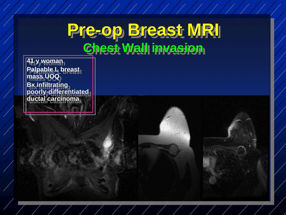
Pre-op Breast MRIChest Wall invasion
PrePre--op Breast MRIop Breast MRIChest Wall invasionChest Wall invasion
41 y womanPalpable L breast mass UOQBx infiltrating poorly-differentiated ductal carcinoma
41 y womanPalpable L breast mass UOQBxBx infiltrating infiltrating poorlypoorly--differentiated differentiated ductalductal carcinomacarcinoma

Breast MRI Indications Contralateral breast screening pre-op
Breast MRI Indications Breast MRI Indications ContralateralContralateral breast screening prebreast screening pre--opop
3-5% synchronous contralateralbreast Ca on MRI only • 30/969 (3%) contralat breast Ca• 121/969 (12.5%) Bx• 30 + bx(24.8%)
18 invasive Ca12 DCISAll node negative
33--5% synchronous 5% synchronous contralateralcontralateralbreast Ca on MRI only breast Ca on MRI only •• 30/969 (3%) 30/969 (3%) contralatcontralat breast Cabreast Ca•• 121/969 (12.5%) 121/969 (12.5%) BxBx•• 30 + bx(24.8%)30 + bx(24.8%)
18 invasive Ca18 invasive Ca12 DCIS12 DCISAll node negativeAll node negative
Lehman C et al N Engl J Med 2007; 356:1295Lehman C et al N Lehman C et al N EnglEngl J Med 2007; 356:1295J Med 2007; 356:1295

Risk of occult Ca in contralateralbreast 1 year post neg MRI 0.3%Risk of occult Ca in Risk of occult Ca in contralateralcontralateralbreast 1 year post breast 1 year post negneg MRI 0.3%MRI 0.3%
Lehman C et al N Engl J Med 2007; 356:1295Lehman C et al N Lehman C et al N EnglEngl J Med 2007; 356:1295J Med 2007; 356:1295
Breast MRI Indications Contralateral breast screening pre-op
Breast MRI Indications Breast MRI Indications ContralateralContralateral breast screening prebreast screening pre--opop

Additional breast tumour foci• 15-37% ipsilateral breast• 3-5% contralateral breastAlters clinical management 10-31%Biopsy of suspicious lesions before Δ surgical approach
Additional breast Additional breast tumourtumour focifoci•• 1515--37% 37% ipsilateralipsilateral breastbreast•• 33--5% 5% contralateralcontralateral breastbreastAlters clinical management 10Alters clinical management 10--31%31%Biopsy of suspicious lesions Biopsy of suspicious lesions before before ΔΔ surgical approachsurgical approach
Pre-op Breast MRIRadiological PerspectivePrePre--op Breast MRIop Breast MRIRadiological PerspectiveRadiological Perspective

Conflicting endorsementCleveland clinic-fully promotes pre-op breast MRI without restriction327 patients-25% pts occult but suspicious lesions13% pts occult and separate tumours75% no additional suspicious foci
Conflicting endorsementConflicting endorsementCleveland clinicCleveland clinic--fully promotes prefully promotes pre--op breast MRI without restrictionop breast MRI without restriction327 patients327 patients--25% pts occult but 25% pts occult but suspicious lesionssuspicious lesions13% pts occult and separate 13% pts occult and separate tumourstumours75% no additional suspicious foci75% no additional suspicious foci
J Crowe, The Breast Journal, Vol 15 # 1, 2009, p52-60
Pre-op Breast MRISurgical Perspective
PrePre--op Breast MRIop Breast MRISurgical PerspectiveSurgical Perspective
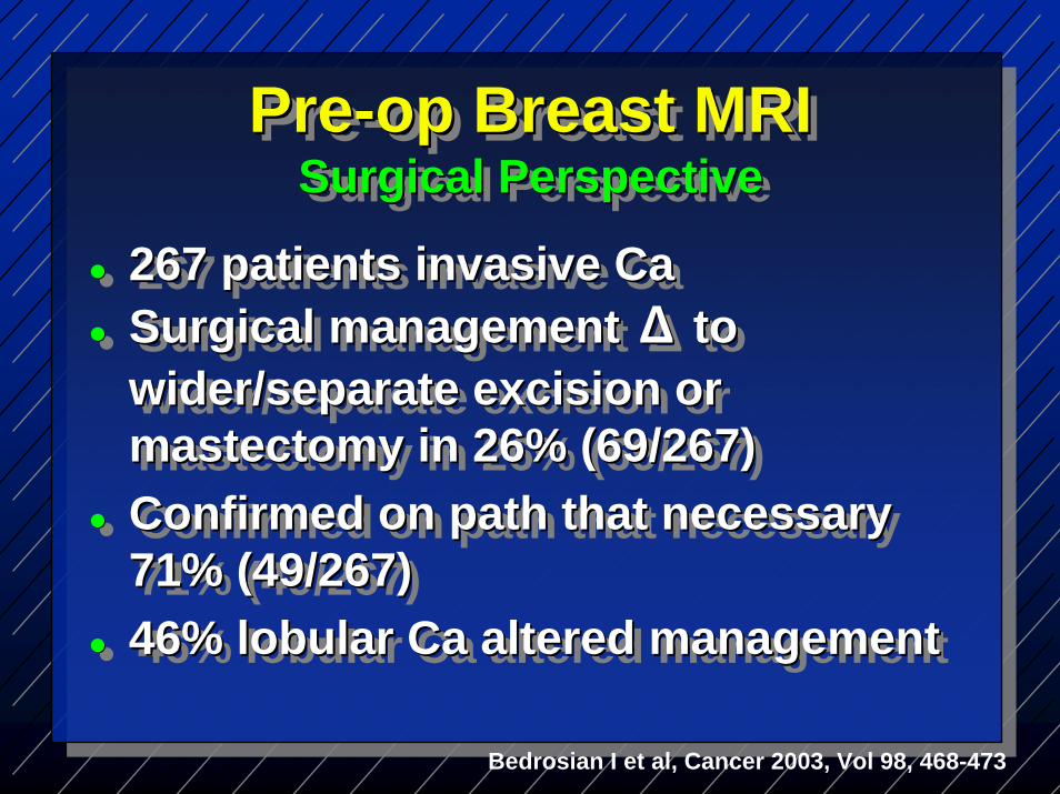
267 patients invasive CaSurgical management Δ to wider/separate excision or mastectomy in 26% (69/267)Confirmed on path that necessary 71% (49/267)46% lobular Ca altered management
267 patients invasive Ca267 patients invasive CaSurgical management Surgical management ΔΔ to to wider/separate excision or wider/separate excision or mastectomy in 26% (69/267)mastectomy in 26% (69/267)Confirmed on path that necessary Confirmed on path that necessary 71% (49/267)71% (49/267)46% lobular Ca altered management46% lobular Ca altered management
Bedrosian I et al, Cancer 2003, Vol 98, 468-473
Pre-op Breast MRISurgical Perspective
PrePre--op Breast MRIop Breast MRISurgical PerspectiveSurgical Perspective
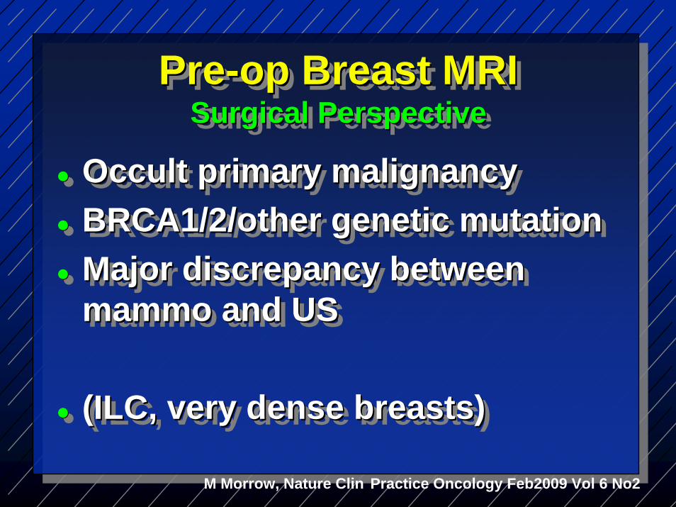
Occult primary malignancyBRCA1/2/other genetic mutationMajor discrepancy between mammo and US
(ILC, very dense breasts)
Occult primary malignancyOccult primary malignancyBRCA1/2/other genetic mutationBRCA1/2/other genetic mutationMajor discrepancy between Major discrepancy between mammomammo and USand US
(ILC, very dense breasts)(ILC, very dense breasts)
Pre-op Breast MRISurgical Perspective
PrePre--op Breast MRIop Breast MRISurgical PerspectiveSurgical Perspective
M Morrow, Nature Clin Practice Oncology Feb2009 Vol 6 No2
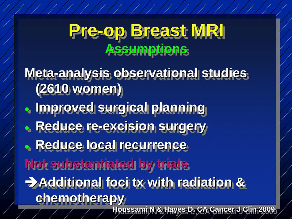
Meta-analysis observational studies (2610 women)Improved surgical planningReduce re-excision surgeryReduce local recurrence
Not substantiated by trialsAdditional foci tx with radiation & chemotherapy
MetaMeta--analysis observational studies analysis observational studies (2610 women)(2610 women)Improved surgical planningImproved surgical planningReduce reReduce re--excision surgeryexcision surgeryReduce local recurrenceReduce local recurrence
Not substantiated by trialsNot substantiated by trialsAdditional foci Additional foci txtx with radiation & with radiation & chemotherapychemotherapy
Houssami N & Hayes D, CA Cancer J Clin 2009HoussamiHoussami N & Hayes D, CA Cancer J N & Hayes D, CA Cancer J ClinClin 20092009
Pre-op Breast MRIAssumptions
PrePre--op Breast MRIop Breast MRIAssumptionsAssumptions
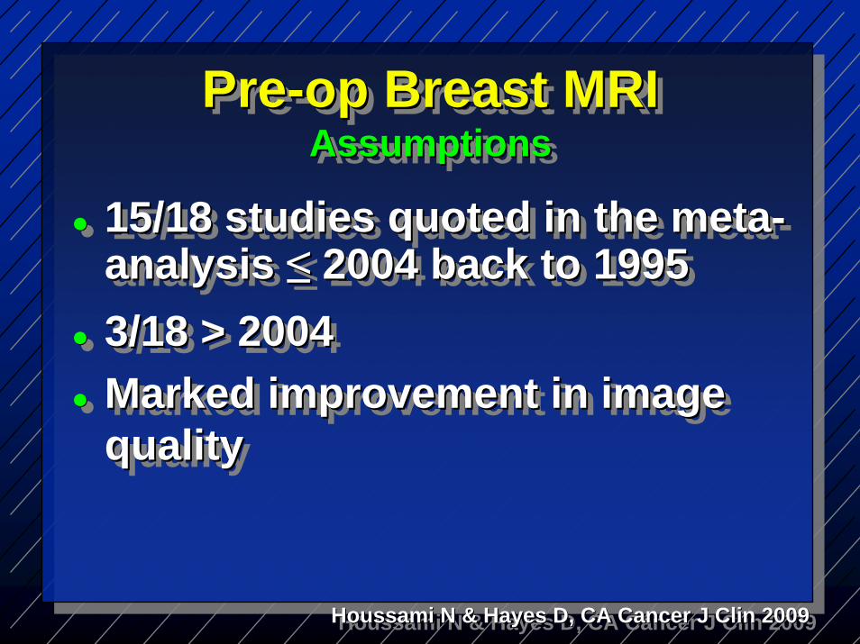
15/18 studies quoted in the meta-analysis ≤ 2004 back to 19953/18 > 2004 Marked improvement in image quality
15/18 studies quoted in the meta15/18 studies quoted in the meta--analysis analysis ≤≤ 2004 back to 19952004 back to 19953/18 > 2004 3/18 > 2004 Marked improvement in image Marked improvement in image qualityquality
Pre-op Breast MRIAssumptions
PrePre--op Breast MRIop Breast MRIAssumptionsAssumptions
Houssami N & Hayes D, CA Cancer J Clin 2009HoussamiHoussami N & Hayes D, CA Cancer J N & Hayes D, CA Cancer J ClinClin 20092009

Increased risk of recurrenceIncreased risk of recurrenceIncreased risk of recurrence
Involvement of surgical marginsExtensive cancer ID clinically or mammographicallyPresence of locally advanced Ca
Involvement of surgical marginsInvolvement of surgical marginsExtensive cancer ID clinically or Extensive cancer ID clinically or mammographicallymammographicallyPresence of locally advanced CaPresence of locally advanced Ca
Houssami N & Hayes D, CA Cancer J Clin 2009HoussamiHoussami N & Hayes D, CA Cancer J N & Hayes D, CA Cancer J ClinClin 20092009

Liberman L et al. AJR 2003; 180:901-910LibermanLiberman L et al. AJR 2003; 180:901L et al. AJR 2003; 180:901--910910
Breast MRI Indications Pre-operative Evaluation
Breast MRI Indications Breast MRI Indications PrePre--operative Evaluationoperative Evaluation
Extent of cancer underestimated clinically and mammographically
Pre-op Breast MRIPrePre--op Breast MRIop Breast MRI
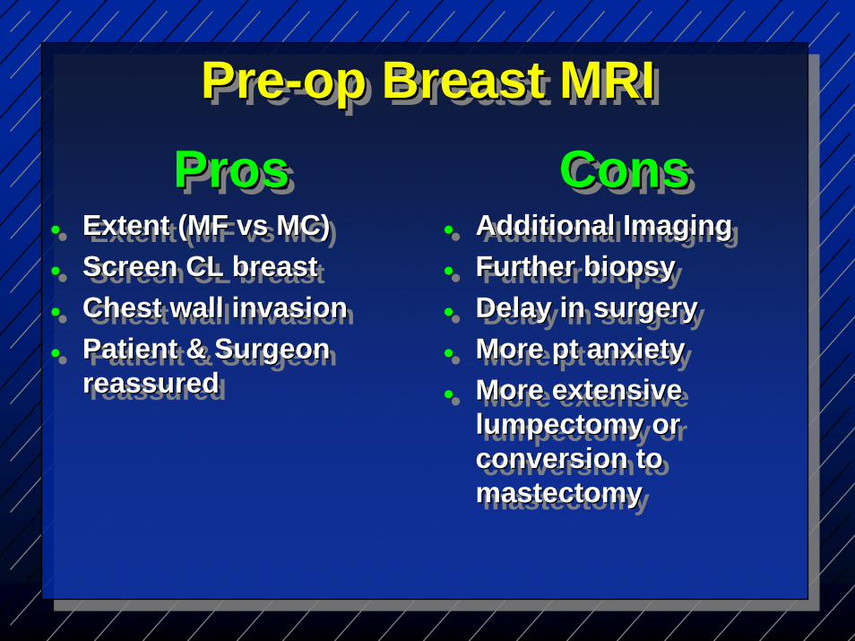
ProsProsProsExtent (MF vs MC)Screen CL breastChest wall invasionPatient & Surgeon reassured
Extent (MF Extent (MF vsvs MC)MC)Screen CL breastScreen CL breastChest wall invasionChest wall invasionPatient & Surgeon Patient & Surgeon reassuredreassured
ConsConsConsAdditional ImagingFurther biopsyDelay in surgeryMore pt anxietyMore extensive lumpectomy or conversion to mastectomy
Additional ImagingAdditional ImagingFurther biopsyFurther biopsyDelay in surgeryDelay in surgeryMore pt anxietyMore pt anxietyMore extensive More extensive lumpectomy or lumpectomy or conversion to conversion to mastectomymastectomy
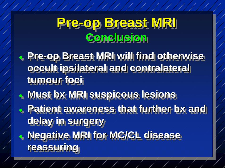
Pre-op Breast MRI will find otherwise occult ipsilateral and contralateraltumour fociMust bx MRI suspicous lesionsPatient awareness that further bx and delay in surgeryNegative MRI for MC/CL disease reassuring
PrePre--op Breast MRI will find otherwise op Breast MRI will find otherwise occult occult ipsilateralipsilateral and and contralateralcontralateraltumourtumour focifociMust Must bxbx MRI MRI suspicoussuspicous lesionslesionsPatient awareness that further Patient awareness that further bxbx and and delay in surgerydelay in surgeryNegative MRI for MC/CL disease Negative MRI for MC/CL disease reassuringreassuring
Pre-op Breast MRIConclusion
PrePre--op Breast MRIop Breast MRIConclusionConclusion

Particularly helpful• Axillary nodes-occult primary• Genetic Ca• Lobular Carcinoma• Very dense breasts/young
patients
Particularly helpfulParticularly helpful•• AxillaryAxillary nodesnodes--occult primaryoccult primary•• Genetic CaGenetic Ca•• Lobular CarcinomaLobular Carcinoma•• Very dense breasts/young Very dense breasts/young
patients patients
Pre-op Breast MRIConclusion
PrePre--op Breast MRIop Breast MRIConclusionConclusion
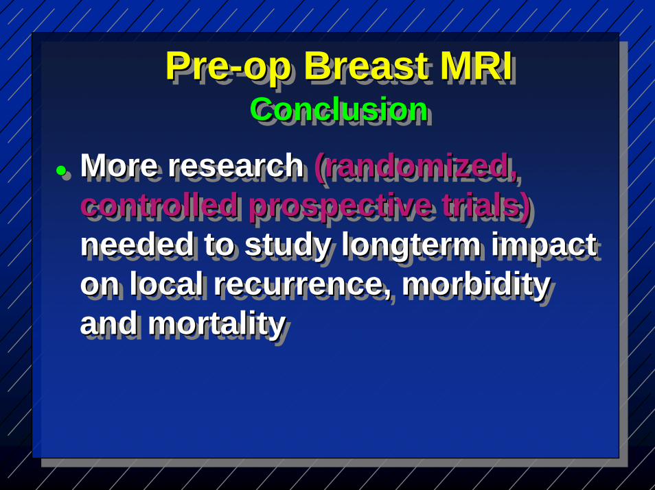
More research (randomized, controlled prospective trials)needed to study longterm impact on local recurrence, morbidity and mortality
More research More research (randomized, (randomized, controlled prospective trials)controlled prospective trials)needed to study needed to study longtermlongterm impact impact on local recurrence, morbidity on local recurrence, morbidity and mortalityand mortality
Pre-op Breast MRIConclusion
PrePre--op Breast MRIop Breast MRIConclusionConclusion

Thank You!Thank You!Thank You!