Embed Size (px)
Citation preview

4/2/2021
1
Breast UltrasoundINDICATIONS
NORMAL ANATOMY
TECHNIQUE
Annina N. Wilkes MD
Thomas Jefferson University Hospital
• Palpable mass/thickening
• Focal Pain
• Nipple discharge
Women under 30, targeted ultrasound is the first study, mammography as needed
Women over 30, bilateral mammogram is the first study, targeted ultrasound to follow
INDICATIONS
Symptoms
INDICATIONSAbnormal Mammogram
• Mass
• Developing asymmetry/focal asymmetry
• Architectural distortion
• Calcifications
INDICATIONSAbnormal Imaging
• MRI
• Nuclear medicine
• CAT Scan
• Contrast Enhanced Mammography (CEM)
• Other
INDICATIONSFollow up
• Probably benign masses (BIRADS 3) – 6 month f/u to assess for stability/2 year
• Abscess following antibiotic therapy
• Hematoma/seroma
• Trauma - bruise
INDICATIONSKnown or Suspected Malignancy (BIRADS 4,5,6)
• Determination of extent, multifocality
• Evaluation of axillary lymph nodes
• Monitoring response to neoadjuvant chemotherapy – breast, axilla and chest wall
• Screening chest wall lymph nodes after therapy

4/2/2021
2
INDICATIONSGuidance
• Cyst aspiration/FNA
• Pre biopsy needle localization/Savi scout placement
• Core biopsy
• Intra operative localization
• Tumor ablation
INDICATIONSImplants
• Silicone implants
MRI more sensitive for rupture
Almost entirely fat Heterogeneously dense
Extremely denseScattered
INDICATIONSDense Breasts
Whole Breast Screening Ultrasound
• Dense breasts- heterogeneously or extremely
• High risk women who cannot get an MRI

4/2/2021
3
29 yo pre neoadjuvant
Post treatment
Indication – palpable lump
palpable mass near chest wall
palpable lump – inframammary fold

4/2/2021
4
palpable lump
mass on the mammogram near the chest wall
Indication- abnormal mammogram
goes away on spot
compression?
2D Tomo
circumscribed mass
circumscribed mass

4/2/2021
5
obscured massmainly well circumscribed?
normal tissue
CXR in the ER
ductogram

4/2/2021
6
IMPLANTS
RUPTURE
INDICATION
Cyst or Solid Benign or Malignant

4/2/2021
7
the not so simple cyst
Cyst Not a Cyst
not a cyst not a cyst

4/2/2021
8
2016 2018
missed a year
Normal AnatomyTechnique
Artemis of Ephesus
Haagensens Diseases of the Breast 1986
NORMAL ANATOMY
• 15-20 LOBES
nipple - ducts – TDLU -lobule

4/2/2021
9
Homogeneous-fatty Heterogeneous Homogeneous-glandular
ducts
cooper’s ligaments skin

4/2/2021
10
Within or under the skin?
Claw signsebaceous cyst
nipple
stand off gel/pad
9 yo palp lump under nipple
Breast bud Infant breast hemangioma
4 month old with palpable massand overlying blue tint on skin

4/2/2021
11
male patient
Chest Wall Ultrasound - Inflammatory Breast Cancer

4/2/2021
12
TECHNIQUE TECHNIQUE
Real time, hand held transducer
7.5 – 18 MHZ
Dynamic focus
High resolution imaging
TECHNIQUE
Patient supine, semi-erect, sitting or on side
Ipsilateral arm above head
Shoulder elevated
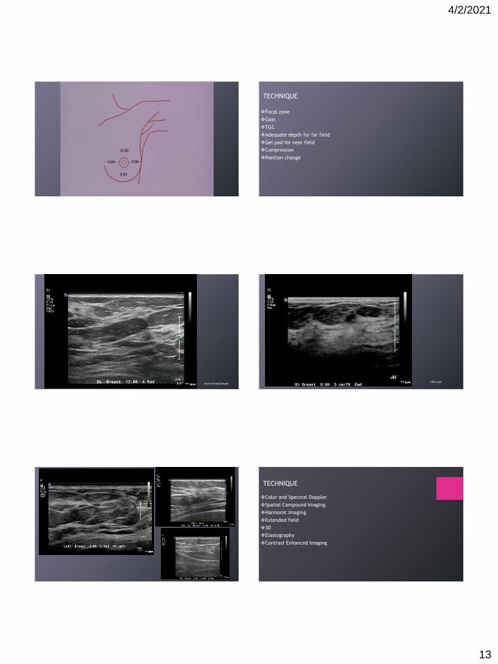
TECHNIQUE
Radial
Anti-radial
Sagittal
Transverse
Label according to mammographic clock
Label distance from nipple/areolar margin
Label area of clinical concern (palpable mass, pain)
4/2/2021
13
TECHNIQUE
Focal zone
Gain
TGC
Adequate depth for far field
Gel pad for near field
Compression
Position change
cine normal breastcine cyst
cine solid masses
TECHNIQUE
Color and Spectral Doppler
Spatial Compound Imaging
Harmonic Imaging
Extended field
3D
Elastography
Contrast Enhanced Imaging

4/2/2021
14
Gain
Posterior Acoustic Features
• no enhancement
• enhancement
• shadowing
• combined pattern
Fibroadenoma
Ca
DOPPLER – absent, internal, peripheral
Confirmation solid vs. cystic
Lymph node, abscess
Pre biopsy
Vessel Characterization – contrast agents
Intraductal debris or massPapilloma

4/2/2021
15
thick walled cyst?
cyst with debris?
Intracystic Papillary Carcinoma
color and spectral Doppler

4/2/2021
16
color/power Doppler
abscesslymph node
COMPOUND IMAGING
electronic steering of transducer array allowing for multiple scanning angles
reduction of speckle, clutter and artifacts
improves detail of mass margins
calcification
posterior enhancement less apparent
HARMONIC IMAGING
transmit at one frequency, receive at multiples of that frequency
6MHz transmit/12MHz receive
reduces clutter and reverb within cysts, increases lesion conspicuity, marginal definition
good to “clean up” cysts
EXTENDED FIELD
Panoramic image
good for locating multiple masses, correlating with mammogram
implants

4/2/2021
17
Elastography – based on tissue stiffness, resistance
Ultrasound Technology-continuing work
• Artificial Intelligence ( AI)
• Whole Breast Imaging/ABUS
• Improved Lesion Detection/Characterization –vascularity-contrast, calcifications
• Computer Aided Detection
• Guidance for tumor ablation – cryotherapy
• Biopsy devices
ACR BI-RADS Breast Imaging Data and Reporting System
Tissue Composition
Homogeneous/ fat/glandular heterogeneous

4/2/2021
18
Masses - Shape
oval, round, irregular
Masses- Orientation
• parallel
• not parallel
Masses- Margin
• Circumscribed
• Not circumscribed
indistinct
angular
microlobulated
spiculated
Echo Pattern
• Anechoic
• Hyperechoic
• Complex cystic and solid
• Hypoechoic
• Isoechoic
• Heterogeneous
Calcifications
in a mass
outside of a mass
intraductal
Associated Features
• architectural distortion
• duct changes
• skin changes thickening
retraction
• edema
• vascularityabsent
internal vascularity
vessels in rim
• elastography assessmentsoft
intermediate
hard

4/2/2021
19
Special Cases
• simple cyst
• clustered microcyst
• complicated cyst
• mass in or on skin
• foreign body including implants
• lymph nodes- intramammary and axillary
• vascular abnormalities – Mondors, AVM’s
• post surgical fluid collection
• fat necrosis
Clustered Microcysts
Assessment Categories : BI-RADS
0 Incomplete
1 Negative
2 Benign
3 Probably Benign
4 Suspicious
5 Highly Suggestive of Malignancy
6 Known Biopsy Proven Cancer
Category 0 - Incomplete
• need additional imaging evaluation: additional imaging and/or prior films
Category 1: Negative
• nothing to comment on, no change, no significant findings
Category 2: Benign
also negative, but describes a characteristically benign finding such as:
• simple cyst
• intramammary lymph node
• implant problem
• stable, probable fibroadenoma
• stable post-surgical change
4/2/2021
20
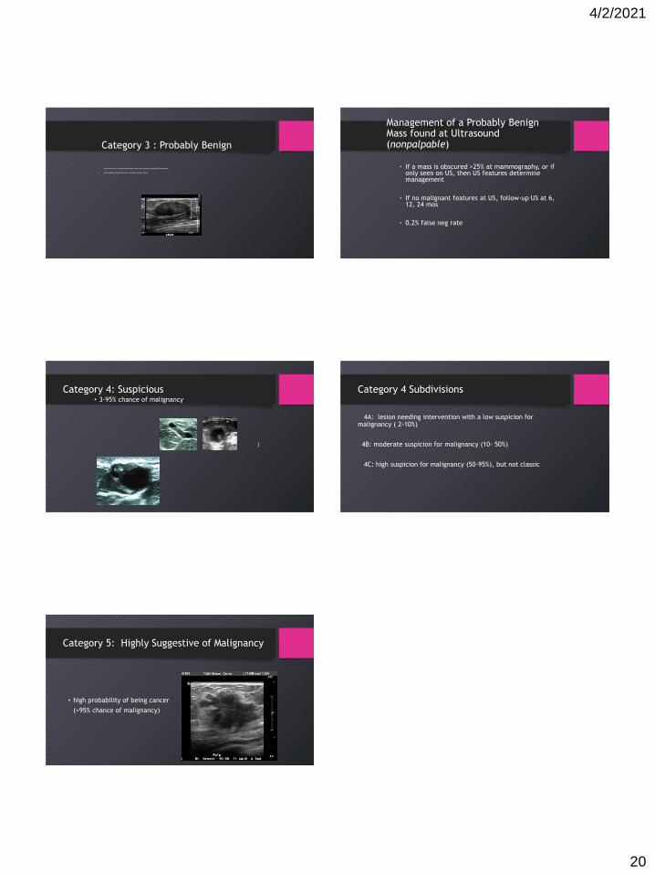
Category 3 : Probably Benign
• Solid mass with circumscribed margins, oval shape, parallel (probable fibroadenoma)
• Non palpable complicated cysts, and clustered microcysts
Management of a Probably BenignMass found at Ultrasound (nonpalpable)
• If a mass is obscured >25% at mammography, or if only seen on US, then US features determine management
• If no malignant features at US, follow-up US at 6, 12, 24 mos
• 0.2% false neg rate
Category 4: Suspicious• 3-95% chance of malignancy
)
Category 4 Subdivisions
4A: lesion needing intervention with a low suspicion for malignancy ( 2-10%)
4B: moderate suspicion for malignancy (10- 50%)
4C: high suspicion for malignancy (50-95%), but not classic
Category 5: Highly Suggestive of Malignancy
• high probability of being cancer
(>95% chance of malignancy)