Embed Size (px)
Citation preview
Brief Definitive Report
Recombinant Human Stem Cell Factor (Kit Ligand) Promotes Human Mast Cell and Melanocyte Hyperplasia and Functional Activation In Vivo B y J o h n J. Costa,** G e o r g e D. Demet r i ,g Te rence J. Harrist,*ll A n n M. Dvorak ,* Dan ie l E Hayesfl El izabeth A. Merica,g
D o r a M. Menchaca , �82 A n t h o n y J. Gr inger i , �82 L a w r e n c e B. Schwartz ,**
and S t ephen J. Galli*
From the Departments of *Pathology and Sa Medicine, Beth Israel Hospital and Harvard Medical School, Boston, Massachusetts 02215; gDepartment of Medical Oncology, Dana-Farber Cancer Institute, Boston, Massachusetts 02115; lI Pathology Services, Incorporated, Cambridge, Massachusetts 02139; �82 Incorporated, Thousand Oaks, California 91320; and the **Medical College of Virginia, Virginia Commonwealth University, Richmond, Virginia 23298
Summary
Stem cell factor (SCF), also known as mast cell growth factor, kit ligand, and Steel factor, is the ligand for the tyrosine kinase receptor (SCFtL) that is encoded by the c-kit proto-oncogene. We analyzed the effects of recombinant human SCF (r-hSCF, 5-50 btg/kg/day, injected sub- cutaneously) on mast cells and melanocytes in a phase I study of 10 patients with advanced breast carcinoma. A wheal and flare reaction developed at each r-hSCF injection site; by elec- tron microscopy, most dermal mast cells at these sites exhibited extensive, anaphylactic-type degranulation. A 14-d course ofr-hSCF significantly increased dermal mast cell density at sites distant to those injected with the cytokine and also increased both urinary levels of the major histamine metabolite, methyl-histamine, and serum levels of mast cell 0r Five subjects developed areas of persistent hyperpigmentation at r-hSCF injection sites; by light microscopy, these sites exhibited markedly increased epidermal melanization and increased numbers of me- lanocytes. The demonstration that r-hSCF can promote both the hyperplasia and the functional activation of human mast cells and melanocytes in vivo has implications for our understanding of the role of endogenous SCF in health and disease. These findings also indicate that the inter- action between SCF and its receptor represents a potential therapeutic target for regulating the numbers and functional activity of both mast cells and cutaneous melanocytes.
T he stem cell factor receptor (SCFtL) is expressed on immature hematopoietic progenitor cells, mast cells,
melanocytes, and germ cells, and both in vitro studies and analyses of experimental animals indicate that stem cell fac- tor (SCF) is critical for the survival and normal develop- ment of these cell types (1-3). Apart from its effects on mast cells, the major actions of SCF in hematopoiesis in vitro are to promote the survival of immature hematopoi- etic progenitor cells and to synergize with other hemato- poietic growth factors in promoting the proliferation and differentiation of committed progenitor cells (1-3). Accord- ingly, Escherichia coli-derived recombinant methionyl hu- man SCF (r-hSCF), which represents essentially the entire extracellular ligand domain of the native transmembrane molecule, as well as the most prevalent native soluble mol- ecule, has been introduced into phase I clinical trials; pre- liminary results indicate that r-hSCF may indeed be useful in mobilizing hematopoietic progenitors into the circula- tion and in promoting hematopoiesis (4).
However, SCF has an exceedingly complex biology; r-hSCF certainly represents the most pleiotropic of the "hematopoietic" growth factors that have been introduced into the clinic. For example, recombinant SCF can pro- mote mast cell hyperplasia in mice (5), rats (5), and nonhu- man primates (6) in vivo; human mast cell development in vitro (7-10); degranulation and mediator release from mouse dermal mast cells in vivo (11) or human cutaneous mast cells in vitro (12); and, in synergy with phorbol esters, the proliferation of human melanocytes in vitro (13).
In this study, we investigated whether r-hSCF can influ- ence human mast cell or melanocyte development or func- tion in vivo. Portions of this work have been reported in abstract form (14, 15).
Materials and Methods Patients and rohSCF Dosing Protocol. 10 women with stage IIIB
or IV breast carcinoma who had volunteered to participate in an
2681 J. Exp. Med. �9 The Rockefeller University Press �9 0022-1007/96/06/2681/06 $2.00 Volume 183 June 1996 2681-2686
Dow
nloaded from http://rupress.org/jem
/article-pdf/183/6/2681/1108224/2681.pdf by guest on 25 January 2022
open-label phase I trial of r-hSCF administered before and after up to five cycles of chemotherapy with cyclophosphamide and doxorubicin were enrolled after providing informed consent and meeting the following major eligibility requirements: no prior chemotherapy for metastatic disease, no adjuvant chemotherapy within the preceeding 6 mo, <250 mg/m 2 prior anthracyclines, age >18 yr, Karnofsky performance status I>70%, absolute neu- trophil count ~>2,000/mm 3, platelet count ~100,000/mm 3, he- moglobin I>9 g/dl, adequate organ function, and no active asthma or other significant immediate hypersensitivity disorders. The protocol, which was approved by the Institutional Review Board for Human Studies of the Dana-Farber Cancer Institute, called for patients to receive an initial 14-d course ofr-hSCF (5, 10, 25, or 50 p,g/kg/d, with each patient receiving only one of these doses) via daily subcutaneous injection in the upper arm, anterior thigh, or abdominal wall (cycle 0), followed by a 7-d pe- riod of observation before initiation of chemotherapy.
Histology. Written informed consent was obtained from each patient to procure 4-mm punch biopsies of skin after induction of local anaesthesia (0.5-1.0 ml of 1% or 2% lidocaine, injected in- tradermally at least 1 cm away from the site to be biopsied). To assess the systemic effects ofr-hSCF on dermal mast cells, biopsies (separated by at least 3 cm) were obtained from the skin over the posterior iliac crest (i.e., at a site not injected with r-hSCF) before (day -1 or 0) and on the last day (day 14) of the first period of r-hSCF dosing. Half of each biopsy was immediately placed in 10% neutral buffered formalin (NBF) and the other half into freshly prepared Carnoy's fixative. All sections were coded, to conceal each specimen's identity, before microscopic analysis. Carnoy's fixed, paraffin-embedded sections (4 ~m thick) were stained for 10-15 min with 0.5% alcian blue (Rowley Biochemi- cal Institutes, Inc., Rowley, MA) in 0.7 M HC1, rinsed, and counterstained with 0.25% safranin (Kowley Biochemicals) in 0.125 M HCI for 2 h (5), and the numbers of mast cells per square millimeter of dermis were quantified at 400 • by use of the Bioquant Morphometric System (R & M Biometrics, Inc., Nashville, TN) (6). Biopsies of hyperpignlented lesions were fixed in NBF, embedded in paraffin, and stained with the Fon- tana-Masson technique to identify melanin (16) or with hema- toxylin and eosin (H and E) to quantify melanocytes as number of basal clear cells per square millimeter of skin (17).
Electron Microscopy. Skin biopsies (performed as above) of the wheal-like lesions that developed 90-120 rain after r-hSCF injec- tion, and control biopsies of identically prepared contralateral sites (not injected with r-hSCF), were immediately immersed in a pool of freshly prepared fixative (2.0% paraformaldehyde, 2.5% glutaraldehyde, and 0.025% CaCl 2 in 0.1 M sodium cacodylate buffer, pH 7.4) at room temperature, trimmed, and then fixed and processed for transmission electron microscopy (18).
�9 Quantification of Mast Cell Mediators. Serum specimens obtained before r-hSCF dosing (day -1 ) and on day 14, just before the last r-hSCF injection, and urine specimens obtained on day - 1 and on day 14 or 15 (before or "~ d after the last r-hSCF injection), were stored as 0.5-ml aliquots at -20~ Because of histamine's short half-life in the circulation ("-q rain), urinary levels of the major histamine metabolite, methyl-histamine, are used to assess systemic changes in histamine levels (19). Urine methyl-histamine was measured with the methyl-histamine tLIA kit, exactly as specified by the manufacturer (Pharmacia Inc. Diagnostics, Co- lumbus, OH; detection range, 0.2-10.0 ~g/1). To account for in- dividual variation in urinary concentration, values were expressed as normalized methyl-histamine, i.e., micrograms of methyl-his- tamine per milligram of creatinine in the same aliquot of urine. In
all patients, the serum blood urea nitrogen and creatinine levels were stable over the 2-wk period of r-hSCF dosing.
Tryptase (stored in the cytoplasmic granules of most, if not all, human mast cells) is a more specific marker of mast cells than is histamine (20, 21). At least two tryptase genes occur in the human genome, encoding o~-tryptase and the r-'93% identical [3-tryptase (22). Serum tryptase was measured with the Tryptase RIACT kit (exactly as specified by the manufacturer [Pharmacia Diagnos- tics]); the capture antibody used in this assay (G5) recognizes pri- marily [3-tryptase, which is not detectable (<1.0 ng/ml) in serum obtained from healthy control subjects (20). Most samples were also analyzed by using the new B12 capture assay that measures both oL- and 13-tryptase and can detect oe-tryptase in blood from normal individuals (mean levels o f ~ 5 ng/ml, all values <20 ng/ ml) (20, 21).
Statistics. Serial observations for each individual were com- pared by use of the 1-tailed paired Student's t test (after first estab- lishing that the differences between the baseline and post-r-hSCF dosing values conformed to a normal distribution). Data are ex- pressed as mean _+ SD.
Results and Di scuss ion
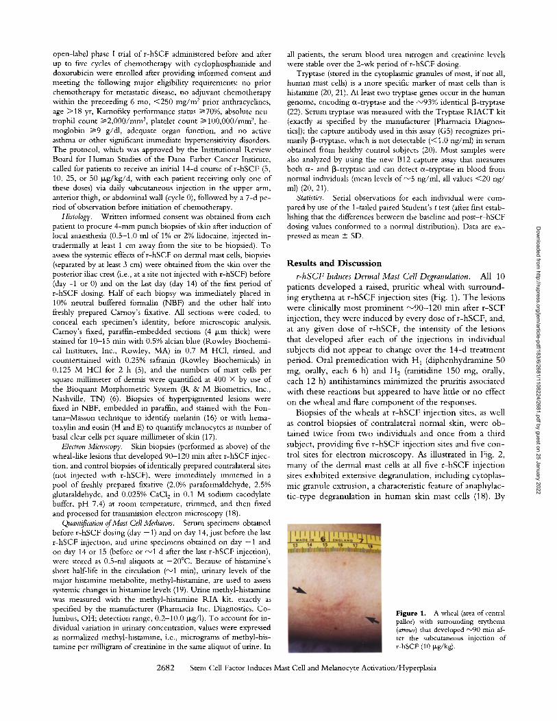
r -hSCF Induces Dermal Mast Cell Degranulation. All 10 patients developed a raised, pruritic wheal with surround- ing erythema at r -hSCF injection sites (Fig. 1). The lesions were clinically most prominent '--~90-120 rain after r -SCF injection, they were induced by every dose o f r -hSCF , and, at any given dose o f r -hSCF, the intensity o f the lesions that developed after each o f the injections in individual subjects did not appear to change over the 14-d treatment period. Oral premedicat ion with H1 (diphenhydramine 50 mg, orally, each 6 h) and H 2 (ranitidine 150 rag, orally, each 12 h) antihistamines minimized the pruritis associated with these reactions but appeared to have little or no effect on the wheal and flare component of the responses.
Biopsies of the wheals at r -hSCF injection sites, as well as control biopsies o f contralateral normal skin, were ob- tained twice from two individuals and once from a third subject, providing five r -hSCF injection sites and five con- trol sites for electron microscopy. As illustrated in Fig. 2, many of the dermal mast cells at all five r -hSCF injection sites exhibited extensive degranulation, including cytoplas- mic granule extrusion, a characteristic feature o f anaphylac- t ic-type degranulation in human skin mast cells (18). By
Figure 1. A wheal (area of central pallor) with surrounding erythema (arrows) that developed ~90 rain af- ter the subcutaneous injection of r-hSCF (10 gtg/kg).
2682 Stem Cell Factor Induces Mast Cell and Melanocyte Activation/Hyperplasia
Dow
nloaded from http://rupress.org/jem
/article-pdf/183/6/2681/1108224/2681.pdf by guest on 25 January 2022
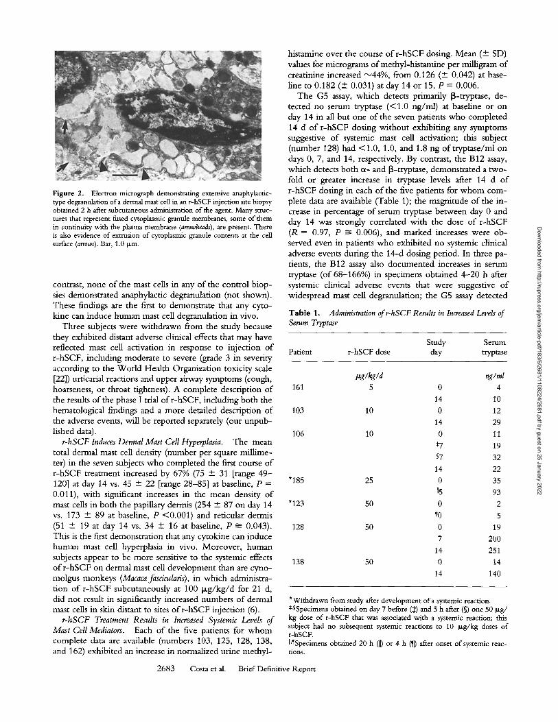
Figure 2. Electron micrograph demonstrating extensive anaphylactic- type degranulation of a dermal mast cell in an r-hSCF injection site biopsy obtained 2 h after subcutaneous administration of the agent. Many struc- tures that represent fused cytoplasmic granule membranes, some of them in continuity with the plasma membrane (arrowheads), are present. There is also evidence of extrusion of cytoplasmic granule contents at the cell surface (arrows). Bar, 1.0 Ixm.
contrast, none o f the mast cells in any o f the control biop- sies demonstrated anaphylactic degranulation (not shown). These findings are the first to demonstrate that any cyto- kine can induce human mast cell degranulation in vivo.
Three subjects were withdrawn from the study because they exhibited distant adverse clinical effects that may have reflected mast cell activation in response to injection o f r-hSCF, including moderate to severe (grade 3 in severity according to the World Health Organization toxicity scale [22]) urticarial reactions and upper airway symptoms (cough, hoarseness, or throat tightness). A complete description o f the results o f the phase I trial o f r -hSCF, including both the hematological findings and a more detailed description o f the adverse events, will be reported separately (our unpub- lished data).
r-hSCF Induces Dermal Mast Cell Hyperplasia. The mean total dermal mast cell density (number per square millime- ter) in the seven subjects who completed the first course o f r-hSCF treatment increased by 67% (75 + 31 [range 49 - 120] at day 14 vs. 45 + 22 [range 28-85] at baseline, P = 0.011), with significant increases in the mean density o f mast cells in both the papillary dermis (254 -+ 87 on day 14 vs. 173 - 89 at baseline, P <0.001) and reticular dermis (51 -+ 19 at day 14 vs. 34 + 16 at baseline, P ~- 0.043). This is the first demonstration that any cytokine can induce human mast cell hyperplasia in vivo. Moreover, human subjects appear to be more sensitive to the systemic effects o f r -hSCF on dermal mast cell development than are cyno- molgus monkeys (Macaca fascicularis), in which administra- tion o f r-hSCF subcutaneously at 100 Ixg/kg/d for 21 d, did not result in significantly increased numbers o f dermal mast cells in skin distant to sites o f r -hSCF injection (6).
r-hSCF Treatment Results in Increased Systemic Levels of Mast Cell Mediators. Each of the five patients for w h o m complete data are available (numbers 103, 125, 128, 138, and 162) exhibited an increase in normalized urine methyl-
histamine over the course o f r -hSCF dosing. Mean (+ SD) values for micrograms o f methyl-histamine per milligram o f creatinine increased "-~44%, from 0.126 ( - 0.042) at base- line to 0.182 ( - 0.031) at day 14 or 15, P = 0.006.
The G5 assay, which detects primarily 13-tryptase, de- tected no serum tryptase (<1.0 ng/ml) at baseline or on day 14 in all but one o f the seven patients who completed 14 d o f r-hSCF dosing without exhibiting any symptoms suggestive o f systemic mast cell activation; this subject (number 128) had <1.0, 1.0, and 1.8 ng of t ryptase/ml on days 0, 7, and 14, respectively. By contrast, the B12 assay, which detects both o~- and [3-tryptase, demonstrated a two- fold or greater increase in tryptase levels after 14 d o f r-hSCF dosing in each o f the five patients for w h o m com- plete data are available (Table 1); the magnitude o f the in- crease in percentage o f serum tryptase between day 0 and day 14 was strongly correlated with the dose o f r-hSCF (R = 0.97, P ------- 0.006), and marked increases were ob- served even in patients who exhibited no systemic clinical adverse events during the 14-d dosing period. In three pa- tients, the B12 assay also documented increases in serum tryptase (of 68-166%) in specimens obtained 4--20 h after systemic clinical adverse events that were suggestive o f widespread mast cell degranulation; the G5 assay detected
Table 1. Administration of r-hSCF Results in Increased Levels of Serum Tryptase
Study Serum Patient r-hSCF dose day tryptase
~glkgld nglml 161 5 0 4
14 10 103 10 0 12
14 29 106 10 0 11
*7 19
57 32 14 22
"185 25 0 35
[J5 93 "123 50 0 2
70 5 128 50 0 19
7 200 14 251
138 50 0 14
14 140
*Withdrawn from study after development of a systemic reaction. *,SSpecimens obtained on day 7 before (:I:) and 5 h after (w one 50 I~g/ kg dose of r-hSCF that was associated with a systemic reaction; this subject had no subsequent systemic reactions to 10 I~g/kg doses of r-hSCF. II.�82 obtained 20 h (IJ) or 4 h (~ after onset of systemic reac- tions.
2683 Costa et al. Brief Definitive Report
Dow
nloaded from http://rupress.org/jem
/article-pdf/183/6/2681/1108224/2681.pdf by guest on 25 January 2022
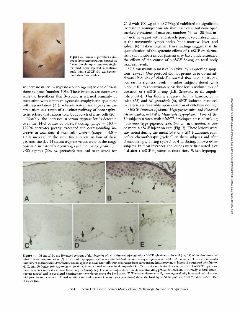
Figure 3. Areas of persistent cuta- neous hyperpigmentation (arrows) at 3 sites (on the upper anterior thigh) that had been injected subcutane- ously with r-hSCF (50 p~g/kg/site) more than 6 mo earlier.
an increase in serum tryptase (to 2.6 ng/ml) in one ofthege three subjects (number 106). These findings are consistent with the hypothesis that [3-typtase is released primarily in association with extensive, systemic, anaphylactic-type mast cell degranulation (21), whereas ot-tryptase appears in the circulation as a result o f a distinct pathway o f nonanaphy- lactic release that reflects total body levels o f mast cells (20).
Notably, the increases in serum tryptase levels detected over the 14-d course o f r -hSCF dosing (range = 100 - 1220% increase) greatly exceeded the corresponding in- creases in total dermal mast cell numbers (range = 4.5 - 100% increase) in the same five subjects; in four o f these patients, the day 14 serum tryptase values were in the range observed in naturally occurring systemic mastocytosis (i.e., > 2 0 ng/ml) (20). M. fascicularis that had been dosed for
21 d with 100 Ixg o f r - h S C F / k g / d exhibited no significant increase in noninjection-si te skin mast cells, but developed marked elevations o f mast cell numbers (4- to 128-fold in- creases) in organs with a relatively porous vasculature, such as the mesenteric lymph nodes, bone marrow, liver, and spleen (6). Taken together, these findings suggest that the quantification o f the systemic effects o f r -hSCF on dermal mast cell numbers in our patients may have underestimated the effects o f the course o f r -hSCF dosing on total body mast cell levels.
SCF can maintain mast cell survival by suppressing apop- tosis (23-25). Our protocol did not permit us to obtain ad- ditional biopsies of clinically normal skin in our patients, but serum tryptase levels in other subjects dosed with r -hSCF fell to approximately baseline levels within 2 wk of cessation of r -hSCF dosing (L.B. Schwartz et al., unpub- lished data). This finding suggests that in humans, as in mice (25) and M. fascicularis (6), rSCF- induced mast cell hyperplasia is reversible upon cessation o f cytokine dosing.
rohSCF Promotes Epidermal Hyperpigmentation and Enhanced Melaninization as Well as Melanocyte Hyperplasia. Five of the 10 subjects treated with r -hSCF developed areas o f striking cutaneous hyperpigmentat ion, 3-5 cm in diameter, at one or more r -hSCF injection sites (Fig. 3). These lesions were first noted during the initial 14 d o f r -hSCF administration before chemotherapy (cycle 0) in three subjects and after chemotherapy, during cycle 3 or 4 of dosing, in two other subjects. In most instances, the lesions were first noted 3 or 4 d after r -hSCF injection at those sites. W h e n hyperpig-
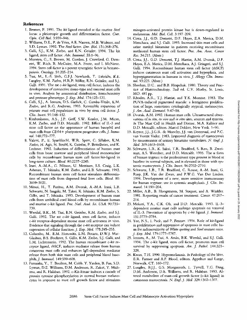
Figure 4. (A and B) H and E~stained sections of skin biopsies of (A), a site not injected with r-hSCF, obtained at the end (day 14) of the first course of r-hSCF administration, or of (B), an area of hyperpigmentation at a site that had received a single injection of r-hSCF 2 mo earlier. There are increased numbers of melanocytes (arrowheads), which appear as basal clear cells with separation from surrounding keratinocytes, in biopsy B compared with biopsy A. (C and D) Fontana-Masson~stained sections, in which melanin is stained purple-black. (C) In a biopsy obtained before the start of r-hSCF injections, melanin is present focally in basal keratinocytes (arrow). (D) The same biopsy shown in A, demonstrating prominent melanin in virtually all basal kerati- nocytes (arrows) and in occasional keratinocytes (arrowheads) above the basal layer. (E) The same biopsy as in B, showing markedly increased melanization, with prominent melanin in all basal keratinocytes and in many keratinocytes (arrowheads) above the basal layer. All biopsies are from the same patient. Bar in E, 25 p~m.
2684 Stem Cell Factor Induces Mast Cell and Melanocyte Activation/Hyperplasia
Dow
nloaded from http://rupress.org/jem
/article-pdf/183/6/2681/1108224/2681.pdf by guest on 25 January 2022

mentation developed during cycle 0, this response was ob- served at each r-hSCF injection site over the entire course of the study. The hyperpigmented skin lesions persisted for variable but generally long periods of time (2-12 mo), dur- ing which they gradually faded.
In comparison with the skin biopsies obtained from the same subjects before the initiation ofr-hSCF administration (Fig. 4 C'), the hyperpigmented lesions exhibited markedly enhanced epidermal melanization (Fig. 4 E). While some of the sites also exhibited focal perivascnlar infiltrates of lymphocytes and eosinophils and occasional basophils, few melanophages were present, indicating that the enhanced melanization at these sites is unlikely to be of a postinflam- matory etiology (26). Moreover, modest increases in epi- dermal melanization were also detected in some skin biop- sies obtained at the end (day 14) of the first cycle ofr-hSCF dosing at sites that had not been injected with r-hSCF and that clinically did not appear to be hyperpigmented (Fig. 4 D). Taken together, these findings indicate that r-hSCF can promote the functional activation of human melanocytes, as well as human mast cells.
Biopsies of both pretreatment skin and sites of persistent (2-12-mo-old) r-hSCF-induced hyperpigmentation were available in three subjects (numbers 161, 128, and 138, treated with 5, 50, or 50 b~g of r -hSCF/kg/d , respectively). The mean number of melanocytes in the hyperpigmented lesions was increased by >300% compared with that in the pretreatment biopsies (38 + 8 [range 29-44] vs. 9 +_ 2 [range 7-11] melanocytes per linear millimeter of epider- mis, respectively; P --- 0.015) (Figs. 4, A and B). None of the melanocytes appeared dysplastic. Moreover, melano- cyte numbers were not increased in hyperpigmented areas that were biopsied within 2 wk of r-hSCF injection (n = 2), indicating that r-hSCF-induced hypermelanosis can oc- cur before the onset of melanocytic hyperplasia. Nor were melanocyte numbers significantly increased after 14 d of r-hSCF dosing in skin biopsies from the iliac crest, a site distant from those directly injected with the cytokine (n = 7) (data not shown). This finding suggests that human me-
lanocytes may be less sensitive to the systemic effects of r-hSCF on proliferation than are human mast cells.
It is not clear why some subjects injected with r-hSCF did not develop clinically detectable areas of hyperpigmen- tation at the injection sites, or whether this represented a qualitative or quantitative difference in the responses of the different patients. Notably, r-hSCF does not induce human melanocyte proliferation in vitro unless a second agent (e.g., PMA) is also present (13), raising the possibility that individual subjects may vary in the levels of other factors that contribute to the expression ofr-hSCF-induced effects on melanocyte development or function.
Conclusions. Our observations provide direct evidence that r-hSCF can promote the hyperplasia and the func- tional activation of two distinct SCFR. + populations in hu- mans in vivo, mast cells and melanocytes. These findings thus not only strengthen the evidence that endogenous SCF represents a critical regulator of the development and function of mast cells and melanocytes in humans, as well as in experimental animals, but also support the view that the increased production or bioavailability of endogenous SCF may contribute to certain diseases associated with mast cell and/or melanocyte hyperplasia or hyperpigmentation. For example, our observations support the concept that changes in levels of endogenous SCF may account for two of the cardinal features of urticaria pigmentosa (27), and may also contribute to mast cell hyperplasia and/or epider- mal hyperpigmentation in other settings.
Our findings also suggest that enhancing or suppressing local levels of SCF (or other approaches for manipulating SCFR-dependent signaling) may be useful for increasing or reducing mast cell numbers or for promoting or reducing cutaneous melanization, in settings in which these effects are clinically desirable. Finally, since blood levels of c~-tryptase are elevated in both naturally occurring (20) and r -hSCF- induced iatrogenic systemic mastocytosis, et-tryptase may prove to be a practical and sensitive index for estimating total body mast cell numbers.
We thank Dr. Zhen-sheng Wang for assistance with the histological analysis, R.ita A. Monahan-Earley, and Patricia Fox for assistance with the electron microscopy, and Bernard J. Kansil, Ph.D., M.D. for assistance with the statistical analysis.
This work was supported by United States Public Health Science grants CA/AI-72074, AI/GM-23990 and AI-31982 (to S.J. GaUi), AI-33372 (to A.M. Dvorak), and AI-20487 (to L.B. Schwartz), by AMGEN, Inc., and by the Beth Israel Hospital Pathology Foundation. J.J. Costa, G.D. Demetri, and S.J. GaUi have per- formed research funded by AMGEN, Inc. and consult for AMGEN, Inc. under terms that are in accord with Beth Israel Hospital (J.J. Costa, S.J. GaUi), Dana-Farber Cancer Institute (G.D. Demetri) and Harvard Med- ical School conflict-of-interest guidelines; L.B. Schwartz performs research funded by Pharmacia under terms that are in accord with Virginia Commonwealth University's conflict-of-interest guidelines.
Address correspondence to Stephen J. Galli, M.D., Division of Experimental Pathology, Department of Pa- thology, R.N227, Beth Israel Hospital, 330 Brookline Avenue, Boston, MA 02215.
Received for publication 29January 1996 and in revised form 5 March 1996.
2685 Costa et al. Brief Definitive Report
Dow
nloaded from http://rupress.org/jem
/article-pdf/183/6/2681/1108224/2681.pdf by guest on 25 January 2022
References 1. Besmer, P. 1991. The kit ligand encoded at the murine Steel
locus: a pleiotropic growth and differentiation factor. Curr. Opin. Cell Biol. 3:939-946.
2. Williams, D.E., P. de Vries, A.E. Namen, M.B. Widmer, and S.D. Lyman. 1992. The Steel factor. Dev. Biol. 151:368-376.
3. Galli, S.J., K.M. Zsebo, and E.N. Geissler. 1994. The kit ligand, stem cell factor. Adv. Immunol. 55:1-96.
4. Morstyn, G., S. Brown, M. Gordon, J. Crawford, G. Dem- etri, W. Rich, B. McGnire, M.A. Foote, and I. McNiece. 1994. Stern cell factor is a potent synergistic factor in hemato- poiesis. Oncology. 51:205-214.
5. Tsai, M., L.-S. Shih, G.F.J. Newlands, T. Takeishi, K.E. Langley, K.M. Zsebo, H.R.P. Miller, E.N. Geissler, and S.J. Galli. 1991. The rat c-kit ligand, stem cell factor, induces the development of connective tissue-type and mucosal mast cells in vivo. Analysis by anatomical distribution, histochemistry and protease phenotype.J. Exp. Med. 174:125-131.
6. Galli, S.J., A. Iemura, D.S. Garlick, C. Gamba-Vitalo, K.M. Zsebo, and R.G. Andrews. 1993. Reversible expansion of primate mast cell populations in vivo by stem cell factor. J. Clin. Invest. 91:148-152.
7. Kirshenbaum, A.S., J.P. Goff, S.W. Kessler, J.M. Mican, K.M. Zsebo, and D.D, Metcalfe. 1992. Effect of IL-3 and stem cell factor on the appearance of human basophil and mast cells from CD34+ pluripotent progenitor cells..]. Immu- nol. 148:772-777.
8. Valent, P., E. Spanblochl, W.R. Sperr, C. Sillaber, K.M. Zsebo, H. Agis, H. Strobl, K. Geissler, P. Bettelheim, and K. Lechner. 1992. Induction of differentiation of human mast cells from bone marrow and peripheral blood mononuclear cells by recombinant human stem cell factor/kit-ligand in long-term culture. Blood. 80:2237-2245.
9. Irani, A.-M.A., G. Nilsson, U. Mettinen, S.S. Craig, L.K. Ashman, T. Ishizaka, K.M. Zsebo, and L.B. Schwartz. 1992. Recombinant human stem cell factor stimulates differentia- tion of mast cells from dispersed fetal liver cells. Blood. 80: 3009-3021.
10. Mitsui, H., T. Furitsu, A.M. Dvorak, A.-M.A. Irani, L.B. Schwartz, N. Inagaki, M. Takei, K. Ishizaka, K.M. Zsebo, S. Gillis, and T. Ishizaka. 1993. Development of human mast cells from umbilical cord blood cells by recombinant human and murine c-kit ligand. Proc. Natl. Acad. Sci. USA. 90:735- 739.
11. Wershil, B.K., M. Tsai, E.N. Geissler, K.M. Zsebo, and S.J. Galli. 1992. The rat c-kit ligand, stem cell factor, induces c-kit receptor-dependent mouse mast cell activation in vivo. Evidence that signaling through the c-kit receptor can induce expression of cellular function.J. Exp. Med. 175:245-255.
12. Columbo, M., E.M. Horowitz, L.M. Botana, D.W.J. Mac- Glashan, B.S. Bochner, S. Gillis, K.M. Zsebo, S.J. Galli, and L.M. Lichtenstein. 1992. The human recombinant c-kit re- ceptor ligand, rhSCF, induces mediator release from human cutaneous mast cells and enhances IgE-dependent mediator release from both skin mast cells and peripheral blood baso- phils.J. Immunol. 149:599-608.
13. Funasaka, Y., T. Boulton, M. Cobb, Y. Yarden, B. Fan, S.D. Lyman, D.E. Williams, D.M. Anderson, R. Zakut, Y. Mish- ima, and R. Halaban. 1992. c-Kit-kinase induces a cascade of protein tyrosine phosphorylation in normal human melano- cytes in response to mast cell growth factor and stimulates
mitogen-activated protein kinase but is down-regulated in melanomas. Mol. Biol. Cell. 3:197-209.
14. Costa, J.J., G.D. Demetri, D.F. Hayes, E.A. Merica, D.M. Menchaca, and S.J. Galli. 1993. Increased skin mast cells and urine methyl histamine in patients receiving recombinant methionyl human stem cell factor. Proc. Am. Assoc. Cancer Res. 34:211. (Abstr.)
15. Costa, J.J., G.D. Demetri, T.J. Harrist, A.M. Dvorak, D.F. Hayes, E.A. Merica, D.M. Menchaca, A.J. Gringeri, and S.J. Galli. 1994. Recombinant human stem cell factor (rhSCF) induces cutaneous mast cell activation and hyperplasia, and hyperpigrnentation in humans in vivo. J. Allergy Clin. Immu- nol. 93:225. (Abstr.)
16. Sheehan, D.C., and B.B. Hrapchak. 1980. Theory and Prac- tice of Histotechnology. 2nd ed. C.V. Mosby, St. Louis, MO. 481 pp.
17. Rhodes, A.R., T.J. Harrist, and T.K. Momtaz. 1983. The PUVA-induced pigmented macule: a lentiginous prolifera- tion of large, sometimes cytologically atypical, melanocytes.
J. Am. Acad. Dermatol. 9:47-58. 18. Dvorak, A.M. 1992. Human mast cells. Ultrastructural obser-
vations of in situ, ex vivo and in vitro sites, sources and systems. In The Mast Cell in Health and Disease. M.A. Kaliner and D.D. Metcalfe, editors. Marcel Dekker, New York. 1-90.
19. Keyzer, J.J., J.G.R.. de Monchy, J.J. van Doormaal, and P.C. van Voorst Vader. 1983. improved diagnosis of mastocytosis by measurement of urinary histamine metabolites. N. Engl. J. Med. 309:1603-1605. Schwartz, L.B., K. Sakai, T.R. Bradford, S. Ren, B. Zwei- man, A.S. Worobec, and D.D. Metcalfe. 1995. The ot form of human tryptase is the predominant type present in blood at baseline in normal subjects, and is elevated in those with sys- temic mastocytosis.J. Clin. Invest. 96:2702-2710. Schwartz, L.B., T.R. Bradford, C. Rouse, A.-M. Irani, G. Rasp, J.K. Van der Zwan, and P.W.G. Van Der Linden. 1994. Development of a new, more sensitive immunoassay for human tryptase: use in systemic anaphylaxis. J. Clin. Im- munol. 14:190-204.
22. Miller, A.B., B. Hoogstraten, M. Staquet, and A. Winkler. 1981. Reporting results of cancer treatment. Cancer. 47:207- 214.
23. Mekori, Y.A., C.K. Oh, and D.D. Metcalfe. 1993. IL-3- dependent murine mast cells undergo apoptosis on removal of IL-3. Prevention of apoptosis by c-kit ligand. J. Immunol. 151:3775-3784.
24. Yee, N.S., I. Paek, and P. Besmer. 1994. Role ofkit-ligand in proliferation and suppression of apoptosis in mast cells: ba- sis for radiosensitivity of White spotting and Steel mutant mice. J. Exp. Med. 179:1777-1787.
25. Iemura, A., M. Tsai, A. Ando, B.K. Wershil, and S.J. Galli. 1994. The c-kit ligand, stem cell factor, promotes mast cell survival by suppressing apoptosis. Am. J. Pathol. 144:321- 328.
26. Kwan, T.H. 1990. Hypermelanosis. In Pathology of the Skin. E.R. Farmer and A.F. Hood, editors. Appelton and Lange, Norwalk, CT. 160--172.
27. Longley, B.j.J., G.S. Morganroth, L. Tyrrell, T.G. Ding, D.M. Anderson, D.E. Williams, and R. Halaban. 1993. Al- tered metabolism of mast-cell growth factor (c-kit ligand) in cutaneous mastocytosis. N. Engl.J. Med. 328:1302-1307.
20.
21.
2686 Stem Cell Factor Induces Mast Cell and Melanocyte Activation/Hyperplasia
Dow
nloaded from http://rupress.org/jem
/article-pdf/183/6/2681/1108224/2681.pdf by guest on 25 January 2022