Embed Size (px)
Citation preview

ARTICLE
Received 29 Sep 2016 | Accepted 30 Jan 2017 | Published 13 Mar 2017
Brigatinib combined with anti-EGFRantibody overcomes osimertinib resistancein EGFR-mutated non-small-cell lung cancerKen Uchibori1,2, Naohiko Inase2, Mitsugu Araki3, Mayumi Kamada4, Shigeo Sato1, Yasushi Okuno3,4,
Naoya Fujita1 & Ryohei Katayama1
Osimertinib has been demonstrated to overcome the epidermal growth factor receptor
(EGFR)-T790M, the most relevant acquired resistance to first-generation EGFR–tyrosine
kinase inhibitors (EGFR–TKIs). However, the C797S mutation, which impairs the covalent
binding between the cysteine residue at position 797 of EGFR and osimertinib, induces
resistance to osimertinib. Currently, there are no effective therapeutic strategies to overcome
the C797S/T790M/activating-mutation (triple-mutation)-mediated EGFR–TKI resistance.
In the present study, we identify brigatinib to be effective against triple-mutation-harbouring
cells in vitro and in vivo. Our original computational simulation demonstrates that brigatinib
fits into the ATP-binding pocket of triple-mutant EGFR. The structure–activity relationship
analysis reveals the key component in brigatinib to inhibit the triple-mutant EGFR. The
efficacy of brigatinib is enhanced markedly by combination with anti-EGFR antibody because
of the decrease of surface and total EGFR expression. Thus, the combination therapy of
brigatinib with anti-EGFR antibody is a powerful candidate to overcome triple-mutant EGFR.
DOI: 10.1038/ncomms14768 OPEN
1 Cancer Chemotherapy Center, Japanese Foundation for Cancer Research, 3-8-31, Ariake, Koto-ku, Tokyo 135-8550, Japan. 2 Department of RespiratoryMedicine, Graduate School of Medical and Dental Sciences, Tokyo Medical and Dental University, 1-5-45 Yushima, Bunkyo-ku, Tokyo 113-8510, Japan.3 RIKEN Advanced Institute for Computational Science, 7-1-26 Minatojima-Minamimachi, Chuo-ku, Kobe, Hyogo 650-0047, Japan. 4 Graduate School ofMedicine, Kyoto University, 54 Shogoin-Kawaharacho, Sakyo-ku, Kyoto 606-8507, Japan. Correspondence and requests for materials should be addressed toR.K. (email: [email protected]).
NATURE COMMUNICATIONS | 8:14768 | DOI: 10.1038/ncomms14768 | www.nature.com/naturecommunications 1

Non-small-cell lung cancer (NSCLC) harbouring anepidermal growth factor receptor (EGFR)-activatingmutation accounts for B30–40% of NSCLC in the
Japanese population and B15% in Caucasians1. For thetreatment of EGFR-mutated NSCLC, EGFR–tyrosine kinaseinhibitors (EGFR–TKIs) that inhibit the EGFR-induceddownstream signalling pathway by binding to the ATP-bindingpocket of the EGFR–tyrosine kinase domain have been evaluatedand are currently being clinically used2–9. The use of EGFR–TKIshas improved prognoses in patients with EGFR-mutated lungcancer10.
Before clinical application of EGFR–TKIs, the overall survivalof NSCLC patients was only approximately a year, as shown inthe trials verifying the efficacy of platinum doublets chemother-apy in metastatic NSCLC11. Several clinical trials of EGFR–TKIsin EGFR-activating mutation-positive NSCLC patients haveshown improved survival of 2 to nearly 3 years12–14. Theimportance of treatment with the appropriate molecularlytargeted drugs in driver oncogene-positive NSCLC has beenincreasingly recognized. Kris et al.15 reported the benefit ofprecisely identifying the target oncogenes in cancer cells andproviding the appropriate molecularly targeted therapy.
Although the benefits of molecularly targeted drugs aresubstantial, most patients experience a recurrence of disease withinB1–2 years due to acquired resistance. The acquired resistanceagainst the first-generation EGFR–TKIs gefitinib and erlotinib hasbeen revealed to be mainly caused by a gatekeeper mutation16,17
involving substitution of threonine at position 790 with methionine(T790M), which hampers the binding of the EGFR–TKI to theATP-binding site of EGFR and accounts for 60–70% of resistantcases18,19. Many clinical trials of various agents to overcomethe acquired resistance to gefitinib or erlotinib were tried butfailed to show any clinical advantage, except for one trial inwhich afatinibþ cetuximab achieved a response rate of 29%(refs 20–25). Despite its efficacy, this combination treatmenthas not been used in the clinical setting because of its relativelysevere toxicity. To resolve this difficult situation, covalentlybinding third-generation EGFR–TKIs selectively targetingT790M have been evaluated for the treatment of patients withadvanced EGFR-mutated NSCLC26–28. Osimertinib, which wasreported to be efficacious in T790M mutation-positive EGFR-mutated NSCLC29, has been approved in the US and othercountries. Janne et al.30 reported that in a phase 1/2 trialof osimertinib, among 127 patients with confirmed EGFRT790M who could be evaluated, the response rate was 61% andthe median progression-free survival was 9.6 months, which is aslong as that of first-line EGFR–TKIs for EGFR-mutated lungcancer.
Approval of osimertinib will influence the treatment tactics forEGFR-mutated lung cancer, but, again, resistance to osimertinibwill be a major obstacle. In 2015, various mechanisms of theacquired resistance against osimertinib were independentlypublished by several groups. An EGFR mutation involvingsubstitution of cysteine at position 797 with serine (C797S) wasdetected in cell-free plasma DNA from osimertinib-refractorypatients and was shown to induce osimertinib resistance31.Ercan et al.32 reported that Ba/F3 cells with three amino acidsubstitutions, L844V, L718Q and C797S, found using theN-ethyl-N-nitrosourea mutagenesis method, are totallyrefractory to the third-generation EGFR–TKIs WZ-4002,osimertinib and CO-1686. In cases without the C797Smutation, although loss of T790M was reported as specificresistant mechanism to third-generation EGFR–TKIs, bypasspathway activation, such as c-MET activation or small-cell lungcancer (SCLC) transformation, in resistant tumours is thoughtto be the mechanism of resistance similar to that known in
first-generation EGFR–TKIs33–39. Indeed, HER2 amplification,Met amplification, BRAF mutation and SCLC transformationhave been observed in osimertinib-resistant cases40,41. Therefore,new therapeutic strategies are needed to overcome the resistanceto the third-generation EGFR–TKIs.
The osimertinib resistance due to the loss of T790M or bypasspathway activation is expected to be overcome using existingmethods, for example, exchange to or addition of a first-generation EGFR–TKI or concurrent combination therapy ofan inhibiting alternative pathway, respectively. However, we nowhave no clinically available strategy to conquer the C797S/T790M/activating-mutation (triple-mutation). Recently, Jiaet al.42 published a unique allosteric EGFR inhibitor that canovercome the EGFR–TKI resistance by EGFR-C797S/T790M/L858R mutation, but not EGFR-C797S/T790M/del19-mediatedresistance by treating in combination with cetuximab. Niederstet al.43 investigated the emergence of the C797S allele and foundthat if C797S developed in trans of the T790M allele, acombination of first- and third-generation EGFR–TKIs may beeffective enough for clinical use; however, when the C797S andT790M mutations developed in cis, all sensitivity to any of theexisting EGFR–TKIs, including the third-generation ones, waslost. However, we currently have no information on whether thein vitro efficacy of the combination of first- and third-generationEGFR–TKIs for trans C797S is clinically reproducible. The C797Smutations found in the samples obtained from participants in theosimertinib trial mentioned above were all in cis alleles except forone case of in trans31. The frequency of resistance caused byC797S emergence is not well known because of the small numberof third-generation-resistant patients, but the importance ofdeveloping treatment strategies for this group will be increasingin the near future as more and more patients with EGFR-mutatedNSCLC will be receiving osimertinib.
To investigate the therapeutic strategies for treating patientswith triple-mutant EGFR, we performed drug screening andfound brigatinib to be the promising candidate against triple-mutant EGFR with less potency against wild-type EGFRaccording to the in vitro and in vivo assays. Structure–activityrelationship analysis and computational simulation reveal the keycomponent determining the affinity and the binding mode totriple-mutant EGFR that are expected to attribute to the futuredevelopment. Finally, the combination with anti-EGFR antibodystrikingly reduces the IC50 of brigatinib and prolongs the survivalof the triple-mutant EGFR xenograft-bearing mice. Thesefindings in this study may help overcome acquired resistance tothird-generation EGFR–TKIs.
ResultsDrug resistance by EGFR-C797S/T790M/activating mutations.Currently, there are four EGFR–TKIs available in the clinicalsetting—gefitinib, erlotinib, afatinib and osimertinib. Gefitiniband erlotinib are so-called first-generation EGFR–TKIs that wereproven to be efficacious for NSCLC harbouring an EGFR muta-tion (EGFR-activating mutation; exon 19 deletion [del19] orL858R point mutation in exon 21 [L858R]). Afatinib is a second-generation EGFR–TKI irreversibly targeting the pan-HER signalpathway. Osimertinib and EGF-816 are third-generation EGFR–TKIs that covalently bind to EGFR and are effective against theT790M-mutated EGFR, the most common mechanism ofacquired resistance to first-generation EGFR–TKIs. EGF-816 isnot yet accessible except for clinical trials. All classes of EGFR–TKIs are active against the EGFR-activating mutation alone.Therefore, we evaluated the sensitivity of the EGFR–TKI-resistant mutations introduced into Ba/F3 cells (T790M/activating mutation or C797S/T790M/activating mutation
ARTICLE NATURE COMMUNICATIONS | DOI: 10.1038/ncomms14768
2 NATURE COMMUNICATIONS | 8:14768 | DOI: 10.1038/ncomms14768 | www.nature.com/naturecommunications

(triple-mutation)) to the clinically relevant EGFR–TKIs gefitinib,afatinib, osimertinib and EGF-816.
The CellTiter-Glo assay showed that gefitinib and afatinib wereeffective against the EGFR-activating mutation, as previouslydescribed, and also potent against the double mutation withC797S, which is the covalent binding site of the second- andthird-generation EGFR–TKIs (Supplementary Fig. 1a–d). How-ever, they are no longer effective against the T790M gatekeepermutation, the most relevant mechanism of resistance to the first-generation EGFR–TKIs. Osimertinib and EGF-816 showedefficacy not only against the EGFR-activating mutation alonebut also against the double mutation with T790M in vitro(Supplementary Fig. 1e,f). Although the resistance due to theT790M mutation has been shown to be overcome by the third-generation EGFR–TKIs, they lost their inhibitory activity whenthe C797S mutation occurred concurrent with the T790M in cis.The Ba/F3 cells expressing the triple-mutant EGFR were entirelyresistant to all generations of EGFR–TKIs with similar IC50 valuesas in the parental Ba/F3 cells (Supplementary Fig. 1g–i andTable 1a,b). We examined the sensitivity of PC9 cells (parental;expressing del19 alone) and resistant PC9 cells (T790M; doublemutation del19 with T790M, triple-mutant; generated byintroducing C797S/T790M/del19 (triple-del19) mutant EGFR)to the EGFR–TKIs to confirm the characteristics demonstrated inBa/F3 cells. The CellTiter-Glo assay revealed similar results in thePC9 triple-mutant cells that were also refractory to all EGFR–TKIs as seen in Ba/F3 cells (Supplementary Fig. 2a–c and
Table 1c). Interestingly, PC9 triple-mutant cells showed nosensitivity to the combination therapy of gefitinib and osimertinibthat was shown to overcome the acquired osimertinib resistancemediated by C797S and T790M mutations in trans(Supplementary Fig. 2d). These results suggest that no clinicallybeneficial drug is available for the treatment of the triple-mutantEGFR.
Brigatinib overcomes the resistance of EGFR-triple-mutant. Toinvestigate the candidates who could overcome the triple-mutantEGFR, we performed a focused drug screening to examine theirefficacy against each type of EGFR-del19 mutation in Ba/F3 cellsusing the CellTiter-Glo assay. The 30 drugs used in the focuseddrug screening comprised not only EGFR–TKIs but also kinaseinhibitors targeting other tyrosine kinases or serine/threoninekinases that are now available clinically or are being evaluated inclinical trials, referring to the report by Duong-Ly et al.44 thatshowed the potential to repurpose inhibitors against disease-associated or drug-resistant mutant kinases. Among TKIs, onlybrigatinib and ponatinib were expected to have inhibitory activityagainst EGFR-triple-del19 with B50% growth inhibition at100 nM (Fig. 1a). However, the potency of ponatinib againsttriple-del19 assessed by the CellTiter-Glo assay was disappointingwith almost the same IC50 value as that in the parental Ba/F3cells (Supplementary Fig. 3). We then evaluated the efficacyof brigatinib in T790M/del19 and triple-del19-mutated
Table 1 | IC50 values (nM) for the mutant EGFR-expressing Ba/F3 cells, PC9 cells or MGH121 cells.
(a) IC50 values of Ba/F3 cells expressing EGFR-del19 series to EGFR–TKIs
Ba/F3-EGFR- Gefitinib Afatinib Osimertinib EGF-816
Del19 5.9 o0.3 1.7 2.9T790M/del19 5,603 78.2 6.7 18.5C797S/del19 2.7 2.1 513.4 1,241C797S/T790M/del19 2,922 392.7 740.5 1,408Parent(þ IL-3) 410,000 381.3 752.4 –
(b) IC50 values of Ba/F3 cells expressing EGFR-L858R series to EGFR–TKIsBa/F3-EGFR- Gefitinib Afatinib Osimertinib EGF-816
L858R 10.4 o0.3 3 7.7T790M/L858R 5,922 39.2 5.8 15.5C797S/L858R 15.5 7.7 1,115 1,659C797S/T790M/L858R 410,000 804.2 1,171 2,278
(c) IC50 values of PC9 cells to EGFR–TKIsGefitinib Afatinib Osimertinib
PC9 parent 16.38 0.638 7.463PC9 T790M 14,684 61.94 11.31PC9 triple 410,000 1,130 3,461
(d) IC50 values of PC9 cells to ALK–TKIsBrigatinib AP26113-analog AZD3463
PC9 parent 132.8 46.18 253PC9 T790M 243.8 89.28 378.3PC9 triple 599.2 194.7 622.4
(e) IC50 values of MGH121 cells to EGFR–TKIsGefitinib Osimertinib Brigatinib
MGH121-pt 3,228 2.58 155.3MGH121-res2 410,000 7,155 592.1
EGFR, epidermal growth factor receptor; TKIs, tyrosine kinase inhibitors;(a–b) IC50 values for the Ba/F3 cells expressing EGFR-activating mutations with or without resistant mutations, the del19 series (a) and the L858R series (b), respectively, treated with the indicatedEGFR–TKIs. (c–d) IC50 values for PC9 cells (parental, T790M or C797S/T790M/del19-induced) treated with the indicated EGFR–TKIs (c) or ALK–TKIs (d). (e) IC50 values for the MGH121 parental andres-2 cells treated with the indicated TKIs.
NATURE COMMUNICATIONS | DOI: 10.1038/ncomms14768 ARTICLE
NATURE COMMUNICATIONS | 8:14768 | DOI: 10.1038/ncomms14768 | www.nature.com/naturecommunications 3

EGFR-expressing Ba/F3 cells using the CellTiter-Glo assaycompared with other clinically available EGFR–TKIs. The assaydemonstrated that only brigatinib inhibited the growth of Ba/F3cells expressing the triple-del19 mutation with a low IC50
(o100 nM; Fig. 1b). The addition of IL-3 as an originalsurvival signalling pathway activator counteracted thisinhibitory effect of brigatinib. The potency of brigatinib intriple-del19 was confirmed by western blotting that showeddecreased phosphorylation of EGFR and its downstreamsignalling pathway in a dose-dependent manner in contrast tothe lack of inhibition of EGFR phosphorylation by afatinib andosimertinib (Fig. 1c). Although brigatinib showed acceptableinhibitory activity in cell growth inhibition and EGFR signalpathway also in Ba/F3 cells expressing EGFR-del19 alone,
T790M/del19 or C797S/del19, its efficacy was only superior tothat of osimertinib against C797S/del19 (Supplementary Fig. 4).The cell growth inhibition assay and western blotting using samesetting of EGFR–TKIs in Ba/F3 cells expressing mutant EGFRactivated by L858R (L858R alone, T790M/L858R, C797S/L858Rand C797S/T790M/L858R (triple-L858R)) showed that brigatinibwas also effective in EGFR-L858R series but was less potent thanin del19, with similar pattern of efficacy observed in Ba/F3 cellsexpressing corresponding mutation types of EGFR activated bydel19 (Supplementary Fig. 5).
To evaluate whether or not brigatinib acts on EGFR throughATP competition, and to compare its activity between triple-mutant EGFR and wild-type EGFR, an in vitro kinase assay wasperformed using an ADP-Glo kit. The kinase activity inhibition
ALK EGFR Other RTKa
Criz
otin
ibC
eriti
nib
Ale
ctin
ibT
AE
684
Lorla
tinib
AS
P30
26A
ZD
3463
Brig
atin
ibA
fatin
ibE
rlotin
ibG
efiti
nib
Osi
mer
tinib
Lapa
tinib
Van
deta
nib
E70
80F
oret
inib
Cab
ozan
tinib
PH
A66
5752
AE
W54
1S
oraf
enib
Sun
itini
bB
IBF
1120
CH
5183
284
BG
J398
Imat
inib
Pon
atin
ib17
-AA
GC
EP
701
Pac
litax
elD
MS
O
Del19alone
C797S/del19
T790M/del19
C797S/T790M/del19
Cell growth inhibition rate to DMSO
100%0%
cbBa/F3 C797S/T790M/del19
GefitinibOsimertinibBrigatinib
C797S/T790M/del19
Afatinib Osimertinib Brigatinib
50
100
Non
10 100
1,00
0
10 100
1,00
0
10 100
1,00
0
pEGFR
EGFR
Drug conc.(nM)
185
185
(kD)
Concentration of drugs (nM)
Rel
ativ
e ce
ll vi
abili
ty (
% o
f con
trol
)
00 1 10 100 1,000 10,000
Akt
pAkt
pERK
ERK
pS6
60
60
45
45
32
S6
Actin(IC50: nM)
32
45
Gefitinib 3,239
Osimertinib 723.1
Brigatinib 55.5
Figure 1 | Identification of brigatinib as an EGFR-C797S/T790M/activating-mutation (triple-mutant EGFR) inhibitor. (a) The results of screening the
growth-inhibitory activity of 30 drugs in Ba/F3 cells expressing four types of EGFR-del19 with or without T790M or C797S mutations are shown in a heat
map. Ba/F3 cells expressing each EGFR mutant were treated with 100 nM of the indicated inhibitors. After 72 h of drug treatment, the cell viability was
measured using the CellTiter-Glo assay. Relative cell viability was calculated from each value divided by the DMSO control. Among the inhibitors, only
brigatinib and ponatinib were sufficiently efficacious against the triple-mutant EGFR. AZD3463 acted as a weak inhibitor to the triple mutation. (b) Growth
inhibition assessed by the CellTiter-Glo assay of EGFR-C797S/T790M/del19 (triple-del19)-mutated Ba/F3 cells treated with gefitinib, osimertinib and
brigatinib.; N¼ 3. Results are expressed as mean±s.d. IC50 values were calculated using growth inhibition assay. (c) Phosphorylation of EGFR and
downstream signals were significantly inhibited by brigatinib in Ba/F3 cells expressing triple-del19 even though afatinib and osimertinib did not suppress at
all the EGFR signalling of triple-del19.
ARTICLE NATURE COMMUNICATIONS | DOI: 10.1038/ncomms14768
4 NATURE COMMUNICATIONS | 8:14768 | DOI: 10.1038/ncomms14768 | www.nature.com/naturecommunications
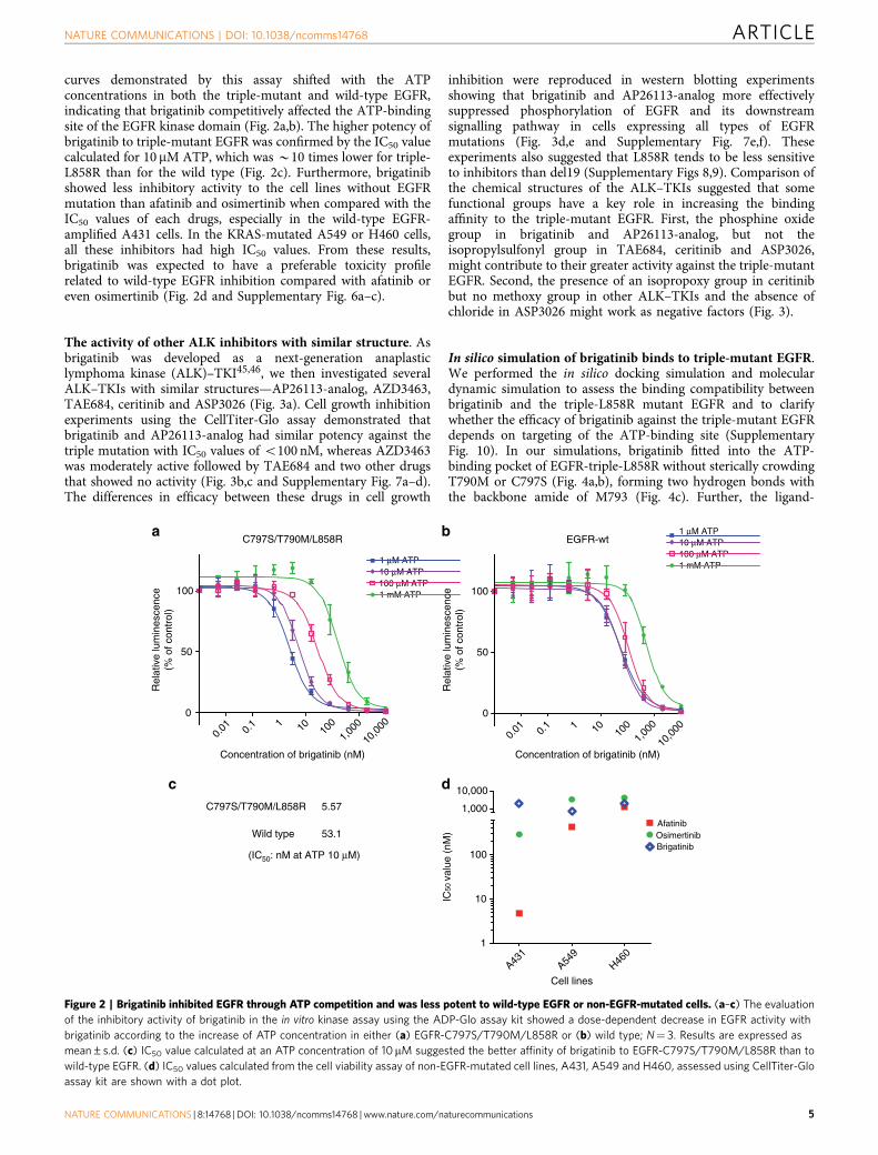
curves demonstrated by this assay shifted with the ATPconcentrations in both the triple-mutant and wild-type EGFR,indicating that brigatinib competitively affected the ATP-bindingsite of the EGFR kinase domain (Fig. 2a,b). The higher potency ofbrigatinib to triple-mutant EGFR was confirmed by the IC50 valuecalculated for 10 mM ATP, which was B10 times lower for triple-L858R than for the wild type (Fig. 2c). Furthermore, brigatinibshowed less inhibitory activity to the cell lines without EGFRmutation than afatinib and osimertinib when compared with theIC50 values of each drugs, especially in the wild-type EGFR-amplified A431 cells. In the KRAS-mutated A549 or H460 cells,all these inhibitors had high IC50 values. From these results,brigatinib was expected to have a preferable toxicity profilerelated to wild-type EGFR inhibition compared with afatinib oreven osimertinib (Fig. 2d and Supplementary Fig. 6a–c).
The activity of other ALK inhibitors with similar structure. Asbrigatinib was developed as a next-generation anaplasticlymphoma kinase (ALK)–TKI45,46, we then investigated severalALK–TKIs with similar structures—AP26113-analog, AZD3463,TAE684, ceritinib and ASP3026 (Fig. 3a). Cell growth inhibitionexperiments using the CellTiter-Glo assay demonstrated thatbrigatinib and AP26113-analog had similar potency against thetriple mutation with IC50 values of o100 nM, whereas AZD3463was moderately active followed by TAE684 and two other drugsthat showed no activity (Fig. 3b,c and Supplementary Fig. 7a–d).The differences in efficacy between these drugs in cell growth
inhibition were reproduced in western blotting experimentsshowing that brigatinib and AP26113-analog more effectivelysuppressed phosphorylation of EGFR and its downstreamsignalling pathway in cells expressing all types of EGFRmutations (Fig. 3d,e and Supplementary Fig. 7e,f). Theseexperiments also suggested that L858R tends to be less sensitiveto inhibitors than del19 (Supplementary Figs 8,9). Comparison ofthe chemical structures of the ALK–TKIs suggested that somefunctional groups have a key role in increasing the bindingaffinity to the triple-mutant EGFR. First, the phosphine oxidegroup in brigatinib and AP26113-analog, but not theisopropylsulfonyl group in TAE684, ceritinib and ASP3026,might contribute to their greater activity against the triple-mutantEGFR. Second, the presence of an isopropoxy group in ceritinibbut no methoxy group in other ALK–TKIs and the absence ofchloride in ASP3026 might work as negative factors (Fig. 3).
In silico simulation of brigatinib binds to triple-mutant EGFR.We performed the in silico docking simulation and moleculardynamic simulation to assess the binding compatibility betweenbrigatinib and the triple-L858R mutant EGFR and to clarifywhether the efficacy of brigatinib against the triple-mutant EGFRdepends on targeting of the ATP-binding site (SupplementaryFig. 10). In our simulations, brigatinib fitted into the ATP-binding pocket of EGFR-triple-L858R without sterically crowdingT790M or C797S (Fig. 4a,b), forming two hydrogen bonds withthe backbone amide of M793 (Fig. 4c). Further, the ligand-
C797S/T790M/L858R
100
1 µM ATP
1 mM ATP
10 µM ATP100 µM ATP
EGFR-wt
100
1 µM ATP
1 mM ATP
10 µM ATP100 µM ATP
ba
Rel
ativ
e lu
min
esce
nce
(% o
f con
trol
)
0
50
0.1 1 10 10
01,
000
10,0
000.
01
Rel
ativ
e lu
min
esce
nce
(% o
f con
trol
)
0
50
0.1 1 10 10
01,
000
10,0
000.
01
Concentration of brigatinib (nM) Concentration of brigatinib (nM)
AfatinibOsimertinibBrigatinib
dcC797S/T790M/L858R 5.57
Wild type 53.1
IC50
val
ue (
nM)
A431
A549
H460
1,000
10,000
1
10
100(IC50: nM at ATP 10 µM)
Cell lines
Figure 2 | Brigatinib inhibited EGFR through ATP competition and was less potent to wild-type EGFR or non-EGFR-mutated cells. (a–c) The evaluation
of the inhibitory activity of brigatinib in the in vitro kinase assay using the ADP-Glo assay kit showed a dose-dependent decrease in EGFR activity with
brigatinib according to the increase of ATP concentration in either (a) EGFR-C797S/T790M/L858R or (b) wild type; N¼ 3. Results are expressed as
mean±s.d. (c) IC50 value calculated at an ATP concentration of 10mM suggested the better affinity of brigatinib to EGFR-C797S/T790M/L858R than to
wild-type EGFR. (d) IC50 values calculated from the cell viability assay of non-EGFR-mutated cell lines, A431, A549 and H460, assessed using CellTiter-Glo
assay kit are shown with a dot plot.
NATURE COMMUNICATIONS | DOI: 10.1038/ncomms14768 ARTICLE
NATURE COMMUNICATIONS | 8:14768 | DOI: 10.1038/ncomms14768 | www.nature.com/naturecommunications 5

binding conformation in the docked complex model was quitesimilar to that in the crystal structure of EML4-ALK bound toTAE684 (PDB-ID: 2XB7). This finding is considered to be rea-sonable because of high similarities between both protein andchemical structures of the two inhibitors. Although these struc-tural analyses suggest that these two inhibitors were presumed tobind to EGFR with similar orientations, activity to triple-mutantEGFR significantly differed from each other in cell growth inhi-bition or western blotting (Fig. 3c,e). For explanation of the
difference in the inhibitory activities, the docking model hintedadvantageous substructures in brigatinib for EGFR binding. Inthe docked EGFR–brigatinib model, the phosphine oxide groupfully occupies the triphosphate-binding space in the ATP-bindingsite (Fig. 4d), concomitantly with meaningful gains of the elec-trostatic or van der Waals interaction energy for all atoms in thisgroup (Fig. 4e,f), suggesting specific interaction with EGFR. In thecase of TAE684, its substitution with the isopropylsulfonyl groupmight disturb the intermolecular interaction pattern and explain
a
AP26113-analog
Brigatinib AZD3463
CeritinibTAE684 ASP3026
200
300500
1,0001,500
b cIC50:nM Brigatinib
AP26113–analog
AZD3463 TAE684 Ceritinib ASP3026
Del19 43.7
NH
HN
NH NH
NH NH
N N
NO
NHPO
PO
N
O
NN
N
N
N N
H2N
N
Cl
N
O O OS O OS
OO OS
NN
NN
N
NN
Cl
NH NH
O
NH N
HN
HN
HN
NCl
N
N
ClN
O
N
Cl
36.9 90.0 314.4 524.3 450.1
T790M/del19
Brig
atin
ib
AP
2611
3 an
alog
AZ
D34
63
TA
E68
4
Cer
itini
b
AS
P30
26
IC50
(nM
)
0
100
C797S/del19
C797S/T790M/del19
150.3 138.6 175.4 510.0 1,007 2,165
39.9 28.4 74.4 229.3 576.6 323.9
67.2 59.1 131.5 340.7 780.5 1,508
dT790M/del19 C797S/T790M/del19
eAP26113-
analog
pEGFR
10
Brigatinib
Non
100
1,00
0
10 100
1,00
0
TA
E68
4 1
µM
Cer
itini
b 1
µM
AS
P30
26 1
µM
pEGFR
AP26113-analog
10Brigatinib
Non
100
1,00
0
10 100
1,00
0
TA
E68
4 1
µMC
eriti
nib
1 µM
AS
P30
26 1
µM
Drug conc.(nM)
Drug conc.(nM)
185(kD)
185(kD)
pAkt
Akt
EGFR
pERK
ERK
pAkt
Akt
EGFR
pERK
ERK
185
60
60
45
45
185
60
60
45
45
Actin
pS6
S6
Actin
pS6
S6
32
32
45
32
32
45
Figure 3 | Efficacy of brigatinib and similarly structured drugs in the EGFR-mutated Ba/F3 cells and their chemical structures. (a) Chemical structures
of six ALK–TKIs were very similar. (b,c) IC50 values in Ba/F3 cells expressing four mutation types of EGFR-del19 were obtained by treatment with brigatinib,
AP26113-analog, AZD3463, TAE684, ceritinib and ASP3026 for 72 h. Those of C797S/T790M/del19 were shown by bar graph (b) and those of all
mutation types were demonstrated by a table (c). The CellTiter-Glo assay was used to measure cell viability. (d,e) Ba/F3 cells expressing T790M/del19
(d) or C797S/T790M/del19 (e) were treated with the indicated concentrations of brigatinib, AP26113 analog, TAE684, ceritinib or ASP3026 for 6 h.
Phosphorylation of EGFR and its downstream signals were evaluated by western blotting with the indicated antibodies.
ARTICLE NATURE COMMUNICATIONS | DOI: 10.1038/ncomms14768
6 NATURE COMMUNICATIONS | 8:14768 | DOI: 10.1038/ncomms14768 | www.nature.com/naturecommunications
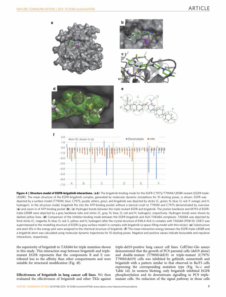
the superiority of brigatinib to TAE684 for triple mutation shownin this study. This interaction map between brigatinib and triple-mutant EGFR represents that the components B and E con-tributed less to the affinity than other compartments and weresuitable for structural modification (Fig. 4f).
Effectiveness of brigatinib in lung cancer cell lines. We thenevaluated the effectiveness of brigatinib and other TKIs against
triple-del19-positive lung cancer cell lines. CellTiter-Glo assaysdemonstrated that the growth of PC9 parental cells (del19 alone)and double-mutant (T790M/del19) or triple-mutant (C797S/T790M/del19) cells was inhibited by gefitinib, osimertinib andbrigatinib with a pattern similar to that observed in Ba/F3 cellsexpressing the corresponding mutation type (Fig. 5a–c andTable 1d). In western blotting, only brigatinib inhibited EGFRphosphorylation and its downstream signalling in PC9 triple-mutant cells. No reduction of the signal pathway in those cells
a b
c
M793
Brigatinib
d e
N
NH N
N
NH
N
O
Cl
P
OA
B
C D
EF
1 2
3
4
11
1213
14 15
19
20
2122
2425
2627
3536
1718
165
106
78
9 2328 29
3332
31
30
34
2.0Atom ID: shown in (e)
f
N37
3840
32
39
–3.0
–2.0
–1.0
0.0
1.0
1 2 3 4 5 6 7 8 9 10 11 12 13 14 15 16 17 18 19 20 21 22 23 24 25 26 27 28 29 30 31 32 33 34 35 36 37 38 39 40
–7.0
–6.0
–5.0
–4.0
A B C D E F
Electrostatic vdw
Figure 4 | Structure model of EGFR–brigatinib interactions. (a,b) The brigatinib-binding mode for the EGFR-C797S/T790M/L858R mutant (EGFR-triple-
L858R). The mean structure of the EGFR–brigatinib complex, generated by molecular dynamic simulations for 10 docking poses, is shown. EGFR was
depicted by a surface model (T790M, blue; C797S, purple; others, grey), and brigatinib was depicted by sticks (C, green; N, blue; O, red; P, orange; and H,
hydrogen). In the structure model, brigatinib fits into the ATP-binding pocket without a sterical crush to T790M and C797S demonstrated by overview
(a) and zoom-in of ATP-binding pocket (b). (c) Hydrogen bonds between the triple-mutant EGFR and brigatinib. The protein backbone and M793 of EGFR-
triple-L858R were depicted by a grey backbone tube and sticks (C, grey; N, blue; O, red and H, hydrogen), respectively. Hydrogen bonds were shown by
dashed yellow lines. (d) Comparison of the inhibitor-binding mode between the EGFR–brigatinib and ALK–TAE684 complexes. TAE684 was depicted by
thick sticks (C, magenta; N, blue; O, red; S, yellow; and H, hydrogen) after the crystal structure of EML4-ALK in complex with TAE684 (PDB-ID: 2XB7) was
superimposed to the modelling structure of EGFR (a grey surface model) in complex with brigatinib (a space-filling model with thin sticks). (e) Substructure
and atom IDs in the energy plot were assigned to the chemical structure of brigatinib. (f) The mean interaction energy between the EGFR-triple-L858R and
a brigatinib atom was calculated using molecular dynamic trajectories for 10 docking poses. Negative and positive values indicate favourable and repulsive
interactions, respectively.
NATURE COMMUNICATIONS | DOI: 10.1038/ncomms14768 ARTICLE
NATURE COMMUNICATIONS | 8:14768 | DOI: 10.1038/ncomms14768 | www.nature.com/naturecommunications 7

was yielded by afatinib and osimertinib in contrast with obser-vations of diminished EGFR signalling in PC9 parent and T790Mcells (Fig. 5f and Supplementary Fig. 11a,b). AZD3463 showed
mild activity in these PC9 cells assessed using western blotting(Supplementary Fig. 11c). Moreover, these three EGFR–TKIsshowed the same pattern of effectiveness in MGH121-parental
PC9 parent(del19)
100
GefitinibOsimertinibBrigatinib
GefitinibOsimertinib
Brigatinib
GefitinibOsimertinibBrigatinib
GefitinibOsimertinibBrigatinib
PC9 T790M(T790M/del19)
PC9 triple mut(C797S/T790M/del19)
a b c
Concentration of drugs (nM) Concentration of drugs (nM)
Rel
ativ
e ce
ll vi
abili
ty (
% o
f con
trol
)
Rel
ativ
e ce
ll vi
abili
ty (
% o
f con
trol
)0
50
100
0
50
Rel
ativ
e ce
ll vi
abili
ty (
% o
f con
trol
)
100
0
50
0 1 10 100 1,000 10,000
0 01 10 100 1,000 10,000 100 1,000 10,000
0 1 10 100 1,000 10,000Concentration of drugs (nM)
0 100 1,000 10,000
MGH121 pt(T790M/del19)
100
50
d e MGH121 resistant-2(del19/T790M/C797S)
100 BrigatinibOsimertinibGefitinib
Concentration of drugs (nM)
Rel
ativ
e ce
ll vi
abili
ty (
% o
f con
trol
)
0
Concentration of drugs (nM)
Rel
ativ
e ce
ll vi
abili
ty (
% o
f con
trol
)
0
50
f g
101,00
0
100
Gef
itini
b 1
µM
pEGFR
10
Afatinib
Non
Osimertinib
100
1,00
0
10
Brigatinib
100
1,00
0
1,00
0
AZD3463
100
10AP
2611
3-an
alog
1 µ
M
100
pEGFR
10
Afatinib
Non
1,00
0
10
Osimertinib
100
1,00
0
10
Brigatinib
100
1,00
0
AP
2611
3-an
alog
1 µ
M
Gef
itini
b 1
µM
Drug conc.(nM)
Drug conc.(nM)
MGH121 res-2PC9 triple mutant
185(kD)
185
(kD)
pAkt
Akt
EGFR
pERK
ERK
pS6
pAkt
Akt
EGFR
pERK
ERK
185
60
60
45
45
185
60
60
45
45
Actin
S6
BimActin
pS6
S6
32
32
45
32
32
45
23
Figure 5 | Inhibition of cell growth and downstream signal pathway in lung cancer cell lines by brigatinib. (a–e) PC9 (del19) (a), PC9-T790M (T790M/
del19) (b), PC9-triple mutant (C797S/T790M/del19) (c), MGH121 parent (T790M/del19) (d) and MGH121 resistant-2 (C797S/T790M/del19) (e) cells
were treated with serially diluted gefitinib, osimertinib and brigatinib for 72 h. Cell viability was measured using the CellTiter-Glo assay.; N¼ 3. Results are
expressed as mean±s.d. (f) Western blotting of PC9 triple mutant (C797S/T790M/del19) cells indicated that brigatinib and AP26113 analog, but not
afatinib or osimertinib, suppressed phosphorylation of EGFR and its downstream signalling. (g) Similar results were obtained in MGH121 resistant-2.
ARTICLE NATURE COMMUNICATIONS | DOI: 10.1038/ncomms14768
8 NATURE COMMUNICATIONS | 8:14768 | DOI: 10.1038/ncomms14768 | www.nature.com/naturecommunications

cells derived from an erlotinib-failure patient harbouring T790M/del19 and in MGH121-resistant-2 cells expressing triple-del19established as WZ-4002–resistant MGH121 cells in vitro43
(Fig. 5d,e,g, and Table 1e).
Efficacy of brigatinib to triple-mutant EGFR in vivo. To con-firm the superior activity of brigatinib against the triple-mutantEGFR, we performed in vivo experiments by administering bri-gatinib or osimertinib to nude mice into which EGFR-triple-del19expressing PC9 lung cancer (PC9-triple mutant) cells had beensubcutaneously injected. As a result, the mice treated with bri-gatinib showed significant inhibition of the growth of PC9-triplemutant cells compared with vehicle controls and osimertinib-treated group without explicit toxicity (SupplementaryFig. 12a,b). Phosphorylation of EGFR and its downstream sig-nalling were actually inhibited by brigatinib in tumour samplesobtained from mice (Supplementary Fig. 12c). This in vivo effi-cacy was confirmed in similar experiments using Ba/F3 cellsexpressing EGFR-triple-del19 instead of PC9 cells. Of note, osi-mertinib (50 mg kg� 1) and brigatinib (75 mg kg� 1) both suc-cessfully suppressed the growth of T790M/del19-expressing PC9cells (Supplementary Fig. 13). These results suggested that bri-gatinib took advantage of triple mutation regardless of cell typeand will be a promising candidate to overcome the acquiredresistance of third-generation EGFR–TKIs. To attain bettertherapeutic effect and tumour shrinkage, it would be important todevelop more potent inhibitors than brigatinib based on ourstructural analysis in future studies.
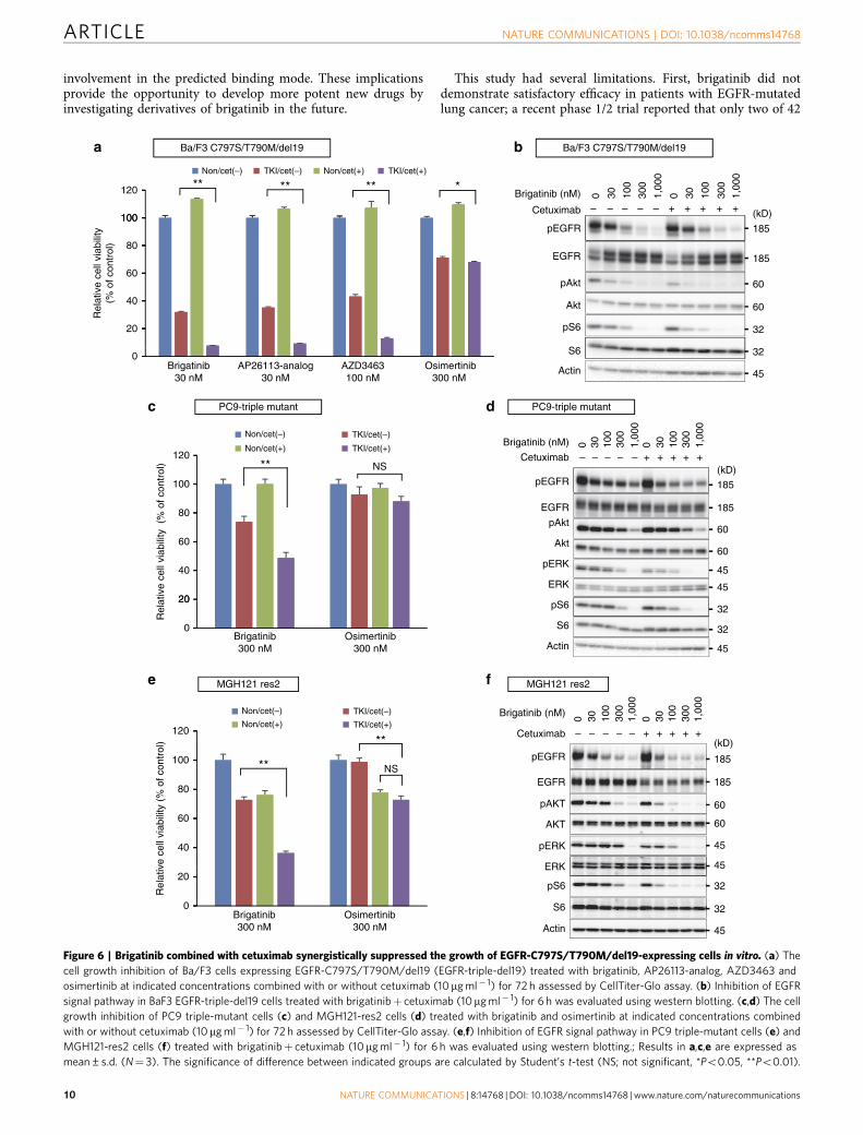
The anti-EGFR antibody enhances the efficacy of brigatinib.Referring to a previous report that stated that afatinibþ cetuximabcombination was effective for acquired resistance of first-genera-tion EGFR–TKI in preclinical models and patients25,47, weevaluated the combination of brigatinib and cetuximab orpanitumumab against the triple-mutant EGFR. The cell viabilityassay using Cell-Titer Glo kit demonstrated that cetuximabenhanced the efficacy of brigatinib or AP26113-analog againsttriple-del19 Ba/F3 cells with approximately three-fold decrease ofIC50, whereas no synergistic benefit was obtained in osimertinib(Fig. 6a). The potentiated inhibition of downstream pathway bycombination of brigatinib with cetuximab was observed usingwestern blotting (Fig. 6b). These effects were reproduced amongtriple-mutation lung cancer cell lines both in cell viability assay andwestern blotting analysis (Fig. 6c–f). The other anti-EGFRantibody, panitumumab in combination with brigatinib,indicated similar growth inhibition (Supplementary Fig. 14a–c).
To further understand the benefit of combination with anti-EGFR antibody, we evaluated the cell surface expression of EGFRin PC9 triple-mutant cells after treatment with cetuximab,brigatinib and brigatinibþ cetuximab in combination for 0, 6, 24and 48 h. EGFR expression analysis by flow cytometry of thetreated cells demonstrated a significant decrease over time in thecell surface EGFR level with brigatinibþ cetuximab and amoderate decrease with cetuximab alone, but it demonstrated noreduction with brigatinib alone (Fig. 7a). Western blot analysis ofthe corresponding treated cells showed that the decrease of totalcellular EGFR achieved with cetuximab was potentiated when thecells were treated with brigatinib and cetuximab in combinationand that the inhibition of phosphorylation of EGFR along withdownstream signalling was also enhanced by this combination(Fig. 7b). In addition, the same experiments confirmed thesuppression of total and cell surface EGFR expression usingtriple-del19-mutated EGFR-expressing MGH121-res2 cells(Fig. 7c,d). These results suggest that synergy of brigatinib andcetuximab was induced through the degeneration of EGFR on the
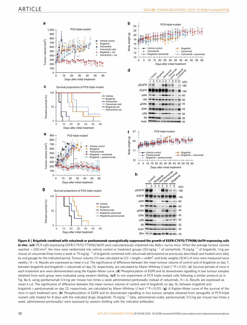
surface caused by cetuximab resulting in intensification of theefficacy of brigatinib. We then performed in vivo experiments ofPC9 triple-mutant xenograft cells as described above comparingwith vehicle control, brigatinib alone, osimertinib alone, cetuximabalone, combination of osimertinib and cetuximab and combinationof brigatinib and cetuximab. The combination of brigatinib andcetuximab demonstrated significant suppression of tumour growthwithout toxicity and achieved prolongation of survival periodscompared with other treatment groups, especially osimertinib-treated group without any superiority to control group (Fig. 8a–c).We confirmed that its efficacy depended on the inhibition ofphosphorylation of EGFR and the decreased expression of EGFRitself in western blotting of tumour samples obtained from eachtreatment group (Fig. 8d). Panitumumab, another anti-EGFRantibody used for treating patients with colorectal cancer, showed acomparable synergistic effect as cetuximab in an in vivo experiment(Fig. 8e–g). Western blotting of resected xenografts showed that thesynergy was also induced through combination with panitumu-mab, suggesting the importance of the concurrent anti-EGFRantibody regardless of its mode of action (Fig. 8h). Another EGFR-triple-mutated lung cancer xenograft model using MGH121-res2 cells showed not only a growth inhibition effect withbrigatinib but also a significant tumour shrinkage without toxicitywith a combination treatment of cetuximab and brigatinib(Fig. 9a,b).
DiscussionIn this study, we demonstrated the efficacy of brigatinib againsttriple-mutant EGFR-positive cells that acquired resistance even tothird-generation EGFR–TKIs. Engineered Ba/F3 cells overexpres-sing triple-mutant EGFR were shown to be sensitive to brigatinibnot only in vitro but also in vivo, as were the lung cancer cell lineswith the triple-mutation in vitro (Figs 1 and 5). Brigatinib alsodemonstrated growth inhibition activity in PC9 triple-mutantxenograft model and in combination with anti-EGFR antibody topotentiate the efficacy both in vitro and in vivo as shown in first-generation EGFR–TKI-resistant patients (Figs 8 and 9 andSupplementary Fig. 12). Discovery of a promising drug that iseffective against the triple mutation should be meaningfulconsidering that the approval of osimertinib, the third-generationEGFR–TKI, in the United States and other countries may lead toa rapid increase in cases of acquired resistance due to the triple-mutant EGFR in the clinical setting.
As brigatinib is now under clinical development as anALK–TKI48, we also investigated the efficacy of similarlystructured ALK–TKIs against the triple-mutant EGFR.However, no other drugs exceeded brigatinib and its analog(Figs 3 and 5 and Supplementary Figs 7–9, 11). We had doubtsabout the disparity in their activity even if it is true that brigatinibwas originally developed as a dual inhibitor of EGFR and ALK.The structure–activity relationship and computer simulationsuggested that the chloro, phosphine oxide group and methoxygroup of brigatinib worked as key elements that contribute to itssuperior efficacy for triple-mutant EGFR (Fig. 3a–c). Also, thesegroups meaningfully gained the electrostatic or van der Waalsintermolecular interaction energy in molecular simulation(Fig. 4e,f), supporting the speculation from the structure–activity relationship. The binding pose of brigatinib alsorevealed that sufficient space appears to be available forsubstitutions on the piperidine ring and a phenyl ringconnected to the phosphine oxide group, concomitantly withsmaller contributions of the two substructures to the bindingstability (Fig. 4e,f). These two functional groups (mentioned asparts B and E in Fig. 4e,f) may be suitable to be partially modifiedto achieve better binding affinity because of their lesser
NATURE COMMUNICATIONS | DOI: 10.1038/ncomms14768 ARTICLE
NATURE COMMUNICATIONS | 8:14768 | DOI: 10.1038/ncomms14768 | www.nature.com/naturecommunications 9
involvement in the predicted binding mode. These implicationsprovide the opportunity to develop more potent new drugs byinvestigating derivatives of brigatinib in the future.
This study had several limitations. First, brigatinib did notdemonstrate satisfactory efficacy in patients with EGFR-mutatedlung cancer; a recent phase 1/2 trial reported that only two of 42
ba
100
120
Non/cet(–) TKI/cet(–) Non/cet(+) TKI/cet(+)
Brigatinib (nM) 0 30 100
1,00
0
300
0 30 100
1,00
0
Cetuximab
300
– – – – – + + + + +
Ba/F3 C797S/T790M/del19 Ba/F3 C797S/T790M/del19
(kD)
****** *
20
40
60
80
100
Rel
ativ
e ce
ll vi
abili
ty(%
of c
ontr
ol)
pEGFR
EGFR
pAkt
Akt
pS6
S6
185
185
60
60
32
d
0Brigatinib
30 nMAP26113-analog
30 nMAZD3463100 nM
Osimertinib300 nM
Actin
c
120
Non/cet(–) TKI/cet(–)
Non/cet(+) TKI/cet(+) 0 30 100
1,00
0
Brigatinib (nM) 300
0 30 100
1,00
0
Cetuximab
300
– – – – – + + + + +
PC9-triple mutant PC9-triple mutant
(kD)
32
45
** NS
20
40
60
80
100 pEGFR
EGFR
pAkt
Akt
pERK
ERK
185
185
60
60
45
45
0
20
Brigatinib300 nM
Osimertinib300 nM
Rel
ativ
e ce
ll vi
abili
ty (
% o
f con
trol
)
pS6
S6
Actin
e
120
Non/cet(–) TKI/cet(–)
Non/cet(+) TKI/cet(+)
0 30 100
1,00
0
Brigatinib (nM) 300
0 30 100
1,00
0
Cetuximab
300
– – – – – + + + + +
fMGH121 res2 MGH121 res2
32
32
45
40
60
80
100 pEGFR
EGFR
pAKT
AKT
pERK
185
185
60
60
45
(kD)
**
**
NS
0
20
Brigatinib300 nM
Osimertinib300 nM
ERK
pS6
S6
Actin
Rel
ativ
e ce
ll vi
abili
ty (
% o
f con
trol
)
45
32
32
45
Figure 6 | Brigatinib combined with cetuximab synergistically suppressed the growth of EGFR-C797S/T790M/del19-expressing cells in vitro. (a) The
cell growth inhibition of Ba/F3 cells expressing EGFR-C797S/T790M/del19 (EGFR-triple-del19) treated with brigatinib, AP26113-analog, AZD3463 and
osimertinib at indicated concentrations combined with or without cetuximab (10mg ml� 1) for 72 h assessed by CellTiter-Glo assay. (b) Inhibition of EGFR
signal pathway in BaF3 EGFR-triple-del19 cells treated with brigatinibþ cetuximab (10mg ml� 1) for 6 h was evaluated using western blotting. (c,d) The cell
growth inhibition of PC9 triple-mutant cells (c) and MGH121-res2 cells (d) treated with brigatinib and osimertinib at indicated concentrations combined
with or without cetuximab (10 mg ml� 1) for 72 h assessed by CellTiter-Glo assay. (e,f) Inhibition of EGFR signal pathway in PC9 triple-mutant cells (e) and
MGH121-res2 cells (f) treated with brigatinibþ cetuximab (10 mg ml� 1) for 6 h was evaluated using western blotting.; Results in a,c,e are expressed as
mean±s.d. (N¼ 3). The significance of difference between indicated groups are calculated by Student’s t-test (NS; not significant, *Po0.05, **Po0.01).
ARTICLE NATURE COMMUNICATIONS | DOI: 10.1038/ncomms14768
10 NATURE COMMUNICATIONS | 8:14768 | DOI: 10.1038/ncomms14768 | www.nature.com/naturecommunications

cases achieved partial response48. However, the plasmaconcentrations of brigatinib at 180 mg per day in a steady statereported previously (1,694.3 nM (ref. 48) and 1,447 nM (ref. 49))were higher than the IC50 values for triple-mutant EGFRpresented in our study (Figs. 1b,3c and Table 1d,e). We expectthat a combination of brigatinib and anti-EGFR antibody wouldimprove sensitivity to the triple-mutant EGFR, resulting in betterefficacy as shown in our study of the long-term tumour stabilityin PC9 triple-mutant xenografts and significant tumour shrinkage
in MGH121-res2 xenografts; this implies that long-term ‘stabledisease’ or ‘partial response’ was achieved with the combinationtherapy, whereas only the inhibition of tumour growth wasattained with brigatinib monotherapy. Second, there is limitedevidence that brigatinib directly affects the ATP-binding site ofthe triple-mutant EGFR because of the absence of a co-crystalstructure, although our in vitro kinase assay results suggest thatbrigatinib inhibits triple-mutant EGFR in an ATP-competitivemanner, and our computer simulation demonstrated its
ba
6 h 24 h 48 hBrigatinib 500 nM Cetuximab 10 µg ml–1
PC9-triple mutant PC9-triple mutant
pEGFR
pAkt
EGFR
Non
Cet
. 10
µg m
l–1
Cet
. 10
µg m
l–1
Brig
. 500
nM
Brig
.+ c
et.
Brig
.+ c
et.
Brig
.+ c
et.
Cet
. 10
µg m
l–1
Brig
. 500
nM
Brig
. 500
nM
Brigatinib + cetuximab
185
185
60
(kD)
Akt
pERK
ERK
pS6
S6
Bim
Treated for 0 hTreated for 6 hTreated for 24 hTreated for 48 h
Cel
l cou
nts
60
45
45
32
32
dc
Actin
EGFR
6 h 24 h 48 h
MGH121 res2
Brigatinib 500 nM Cetuximab 10 µg ml–1
MGH121 res2
45
pEGFR
EGFR
Non
Brig
.+ c
et.
Brig
.+ c
et.
Brig
.+ c
et.
185
185
(kD)
Isotype control IgG
Isotype control IgG
Treated for 0 hTreated for 6 hTreated for 24 h
pAkt
Akt
pERK
ERK
pS6
S6
Cel
l cou
nts
Brigatinib + cetuximab
60
60
45
45
32
32
Actin
Bim
EGFR
45
23
23
Cet
. 10
µg m
l–1
Cet
. 10
µg m
l–1
Cet
. 10
µg m
l–1
Brig
. 500
nM
Brig
. 500
nM
Brig
. 500
nM
0
0
0103
103
103104
104
104
FL2: EGFR-PE
FL2: EGFR-PE
FL2: EGFR-PE
FL2: EGFR-PE
FL2: EGFR-PE
105
105
105106
106
1060
0
0
0 103 104 105 1060
0 103 104 105 106
0
0 103 104 105 1060
Figure 7 | Brigatinib combined with cetuximab enhanced internalization and reduced EGFR expression. (a) FACS analysis using a PE-conjugated EGFR
antibody of PC9 triple-mutant cells treated with brigatinib, cetuximab, brigatinibþ cetuximab for 0, 6, 24 and 48 h demonstrated a time-dependent marked
decrease in surface EGFR after treatment with brigatinibþ cetuximab over a period of up to 48 h, and a moderate decrease with cetuximab alone.
(b) Western blotting assessment of the cells corresponding to the treatments in a. (c,d) FACS analysis and western blotting performed with MGH121-res2
cells using the same method as with the PC9 triple-mutant cells in a,b.
NATURE COMMUNICATIONS | DOI: 10.1038/ncomms14768 ARTICLE
NATURE COMMUNICATIONS | 8:14768 | DOI: 10.1038/ncomms14768 | www.nature.com/naturecommunications 11
PC9 triple-mutant
Vehicle control Brigatinib
a b
600
700
800
900
1,000 PC9 triple-mutant
Vehicle control BrigatinibOsimertinibCetuximab (cet)
** 14
16
18
20
22
24
10
12
Contro
l
Brigat
inib
Osimer
tinib
Cetux
imab
alone Brig
atini
b
+Cet
uxim
ab
0 5 10 15 20 25 30 35 40 45 50 55 60
Bod
y w
eigh
t (g)
Days after initial treatment
Osimertinib CetuximabBrigatinib+cetuximab Osimertinib +cetuximab
d
3 41 2 5 6 7 8 9 10
0
100
200
300
400
500
0 10 20 30 40 50 60
Tum
our
volu
me
(mm
3 )
Days after initial treatment
Brigatinib + cetOsimertinib + cet
**
Survival proportions of PC9 triple mutant
50
100Vehicle
Osimertinib
Brigatinib+cetCetuximab (cet)
Brigatinib
pEGFR
pAkt
Akt
EGFR
pERK
ERK
c 185(kD)
185
60
60
45
45
Days after initial treatment
Per
cent
sur
viva
l (%
)
0 10 20 30 40 50 600
Osimertinib+cet
PC9 triple-mutante
pS6
S6Actin
f
32
3245
** 22
27 PC9 triple-mutant
Tum
our
volu
me
(mm
3 )
Vehicle controlBrigatinibPanitumumabBrigatinib + cetuximabBrigatinib + panitumumab
** 12
17
Bod
y w
eigh
t (g)
Days after initial treatment
Vehicle control BrigatinibPanitumumab Brigatinib + cetuximabBrigatinib + panitumumab
700
800
900
0
100
200
300
400
500
600
0 10 20 30 40 50 60
Days after initial treatment
pEGFR
pAkt
Akt
EGFR
1 2 4 6 8g
h
185
185
60
60
(kD)
Survival proportions of PC9 triple mutant
100
Vehicle
Actin
pERK
ERK
pS6
S6
Bim
45
45
32
32
45
23Days after initial treatment
Per
cent
sur
viva
l (%
)
0 10 20 30 40 50 600
50
BrigatinibPanitumumab
Brigatinib+panitumumabBrigatinib+cetuximab
Contro
l
Brigatin
ib
+panitumumab
Panitumumab
Brigatin
ib
605550454035302520151050
3 5 7
Figure 8 | Brigatinib combined with cetuximab or panitumumab synergistically suppressed the growth of EGFR-C797S/T790M/del19-expressing cells
in vivo. (a,b) PC9 cells expressing EGFR-C797S/T790M/del19 were subcutaneously implanted into Balb-c nu/nu mice. When the average tumour volume
reached B200 mm3, the mice were randomized into vehicle control or treatment groups (50 mg kg� 1 of osimertinib, 75 mg kg� 1 of brigatinib, 1 mg per
mouse of cetuximab three times a week or 75 mg kg� 1 of brigatinib combined with cetuximab administered as previously described) and treated once daily
by oral gavage for the indicated period. Tumour volume (V) was calculated as 0.5� length�width2, and body weights (B.W.) of mice were measured twice
weekly.; N¼ 6. Results are expressed as mean±s.d. The significance of difference between the mean tumour volume of control and of brigatinib on day 7,
between brigatinib and brigatinibþ cetuximab on day 23, respectively, are calculated by Mann–Whitney U test (**Po0.01). (c) Survival periods of mice in
each treatment arm were demonstrated using the Kaplan–Meier curve. (d) Phosphorylation of EGFR and its downstream signalling in two tumour samples
obtained from each group were evaluated using western blotting. (e,f) In vivo experiment of PC9 triple-mutant cells following a similar protocol as in
Fig. 8a–b, using panitumumab 0.5 mg per mouse two times a week administered peritoneally instead of cetuximab.; N¼6. Results are expressed as
mean±s.d. The significance of difference between the mean tumour volume of control and of brigatinib on day 16, between brigatinib and
brigatinibþ panitumumab on day 23, respectively, are calculated by Mann–Whitney U test (**Po0.01). (g) A Kaplan–Meier curve of the survival of the
mice in each treatment arm. (h) Phosphorylation of EGFR and its downstream signalling in two tumour samples obtained from xenografts of PC9-triple
mutant cells treated for 8 days with the indicated drugs (brigatinib: 75 mg kg� 1 daily, administered orally; panitumumab: 0.5 mg per mouse two times a
week, administered peritoneally) were assessed by western blotting with the indicated antibodies.
ARTICLE NATURE COMMUNICATIONS | DOI: 10.1038/ncomms14768
12 NATURE COMMUNICATIONS | 8:14768 | DOI: 10.1038/ncomms14768 | www.nature.com/naturecommunications

compatible interaction with the triple-mutant EGFR. Third, wehave not yet experienced a sufficient number of cases ofosimertinib resistance to estimate the true number of the triple-mutant EGFR. Up to the present, the prevalence of the triplemutation was estimated to be 2–4% among patients with lungcancer according to the frequency of 22% among osimertinib-resistant cases reported in WCLC2015 (ref. 50), and the C797Spopulation would be equal to that of ALK-rearranged lung cancerpatients with a similar clinical magnitude. In addition, in ourN-ethyl-N-nitrosurea mutagenesis drug screen, all clonesresistant to osimertinib mediated only the C797S mutation(unpublished data). Unless the triple-mutant EGFR occurs lessfrequently than is currently expected (B20%), beating the triple-mutant EGFR is a worthy challenge as it is highly refractory toavailable drugs, although we can hope to overcome resistance dueto other mechanisms such as T790M loss or bypass pathwayactivation using existing treatment modalities, for example,turning back to first-generation EGFR–TKIs or combinationtherapy.
In conclusion, we found brigatinib to be potent against thetriple-mutant EGFR in a focused drug-screening protocol andconfirmed its activity in in vivo and in vitro assays. The activitydepended on ATP-competitive manner with less affection towild-type EGFR. The combination therapy with anti-EGFR
antibody showed more preferable activity than monotherapyin vitro and in vivo without toxicity. The structure–activityrelationship demonstrated that the efficacy of brigatinib exceededthat of its analogs, identifying several key basic structure andfunctional groups, such as the phosphine oxide group predictedfrom the computer simulation. The simulation indicated not onlythe conformation of brigatinib binding to the triple-mutant EGFRATP-binding pocket in a compatible manner but also thepotential for developing a more potent inhibitor by partialsubstitution in future direction. While the potency of brigatinibmonotherapy does not seem to be completely satisfactory, thecombination with anti-EGFR antibody strikingly reduced IC50
against triple-mutant EGFR by inducing the degradation ofsurface and total EGFR expression, leading to tumour shrinkageof the triple-mutant EGFR-harbouring cells and significantprolongation of the survival of xenograft-bearing mice. Giventhat the development of brigatinib is now in the late phase ofclinical trials and anti-EGFR antibodies are now broadly usedclinically for several cancers other than lung cancer, the findingsof this study should help to overcome the acquired resistance tothird-generation EGFR–TKIs.
MethodsKinase inhibitors and other drugs. The drugs used in the experiments and thecompanies from which they were purchased are shown in Supplementary Table 1.
Reagents and cell culture. MGH121 (EGFR-T790M/del19 derived from a lungcancer patient), MGH121-resistant-2 (EGFR-C797S/T790M/del19), PC9 parent(amplified EGFR-del19), PC9 T790M (amplified EGFR-T790M/del19) and PC9triple-mutant (amplified EGFR-C797S/T790M/del19) cells were cultured in RPMIwith 10% serum. These cells were kindly provided by Drs. Niederst and Engelman.Ba/F3 cells harbouring EGFR mutations were cultured in low-glucose Dulbecco’sminimal essential medium (DMEM) with 10% fetal bovine serum (FBS). TheMGH121 resistant-2 cells were established from MGH121 by treating with 1 mMWZ-4002, and the PC9 T790M cells were obtained from PC9 parental cells bytreating with 1 mM gefitinib as de novo persistent resistant clone43. The PC9 triplemutant cells were generated by lentivirus infection of EGFR-triple-del19 from thePC9 parental cells. All cells were routinely tested and verified to be free ofmycoplasma contamination.
Generating lentivirus and stable expression in Ba/F3 cells. The full-lengthwild-type EGFR was synthesized from cDNA obtained from A549 cells. The EGFR-activating mutation, del19 or L858R, was obtained from the cDNA of EGFR-exon19 deletion (del19)-harbouring HCC827 cells or L858R-harbouring lung cancerspecimens, respectively. Each EGFR was amplified using polymerase chain reactionand then cloned into a pENTR vector. The T790M and/or C797S mutants weregenerated by QuikChange site-directed mutagenesis using the following primers:T790M F- 50-CCGTGCAGCTCATCATCCAGCTCATGCCCTTC-30 , and T790MR- 50-GAAGGGCATGAGCTGCATGATGAGCTGCACGG-30 , C797S F- 50-CATGCCCTTCGGCTCCCTCCTGGAGCTA-30 , and C797S R – 50-TAGTCCAGGAGGGAGCCGAAGGGCATG-30.
The resulting pENTR-EGFR mutation constructs were sequenced and used as atemplate to make the pLenti6.3 lentiviral vector using LR clonase II. The lentiviruswas made by transfecting the pLenti6.3 constructs along with helper plasmids(ViraPower) in 293FT cells. Virus production, collection and infection werecompleted following the manufacturer’s protocol. The Ba/F3 cells were selected byculturing for 1 week with 7 mM blasticidin in DMEM with interleukin-3 (IL-3)-supplemented 10% FBS and then in DMEM without IL-3 to obtain the EGFRsignalling-addicted cells.
Cell viability assays. Three-day cell viability assays were carried out by plating2,000, 1,500 and 2,000 cells per well of Ba/F3, PC9 or MGH121, respectively, intoblack transparent-bottom 96-well plates. On the same day for Ba/F3 cells and thefollowing day for PC9 and MGH121 cells, the cells were treated with each TKIacross a 10-dose range from 0.3 nmol l� 1 to 10 mmol l� 1. After 72 h of drugtreatment, cell viability was measured using the CellTiter-Glo assay (Promega).
Drug sensitivity screening. Ba/F3 cells of EGFR-del19, -T790M/del19, -C797S/del19 and -C797S/T790M/del19 were plated with 2,000 cells per well into black96-well plates and treated for 3 days with a panel of 30 inhibitors includingdimethylsulfoxide (DMSO) controls prepared in-house. After the incubation, cellviability was measured using CellTiter-Glo assay. The relative cell viability wascalculated as a ratio of each value to that of the DMSO control. Experiments were
a
600
MGH121-res2
Vehicle control Brigatinib
200
400
Tum
our
volu
me
(m
m3 )
OsimertinibCetuximabBrigatinib + cetuximab*
**
NS
NS
b
00 10 20 30 40
Days after initial treatment
25MGH121-res2
Vehicle control
10
15
20
Bod
y w
eigh
t (g) Brigatinib
OsimertinibCetuximabBrigatinib + cetuximab
0 10 20 30 40
Days after initial treatment
Figure 9 | Brigatinib combined with cetuximab synergistically suppressed
the growth of EGFR-C797S/T790M/del19-expressing lung cancer cells
in vivo. (a,b) MGH121-res2 expressing EGFR-C797S/T790M/del19 were
subcutaneously implanted into SCID-beige mice. When the average tumour
volume reached B200 mm3, the mice were randomized into vehicle control
and treatment groups (50 mg kg� 1 of osimertinib (po), 75 mg kg� 1 of
brigatinib (po), 1 mg per mouse of cetuximab two times a week and
75 mg kg� 1 of brigatinib combined with cetuximab administered as
previously described, respectively) and treated for the indicated period.
Tumour volume (V) was calculated as 0.5� length�width2, and the body
weights (B.W.) of the mice were measured twice weekly. N¼ 6. Results are
expressed as mean±s.d. The significance in difference between the mean
tumour volume of control and of osimertinib, brigatinib and cetuximab,
between cetuximab and brigatinibþ cetuximab, respectively, on day 42
are calculated by Mann–Whitney U test (NS: not significant, *Po0.05,
**Po0.01).
NATURE COMMUNICATIONS | DOI: 10.1038/ncomms14768 ARTICLE
NATURE COMMUNICATIONS | 8:14768 | DOI: 10.1038/ncomms14768 | www.nature.com/naturecommunications 13
repeated three times independently, and the average relative cell viability wascalculated and shown as a heat map (Original data are available in SupplementaryData 1).
Antibodies and western blotting. Tumour tissues or cells grown under thespecified conditions were washed with cold PBS before addition of the SDS lysisbuffer (100 mM Tris, 1% SDS, 10% glycerol). Lysates were transferred to micro-tubes, boiled for 5 min at 100 �C and then vortexed. Protein quantification wasperformed using the BCA Protein Assay Reagent (Pierce) according to themanufacturer’s protocol. Western blot analyses were conducted after separationby SDS-PAGE and transferred to polyvinylidene difluoride membranes. Afterblocking in 5% BSA with Tris-buffered saline/Tween 20 (TBS-T) or 5% skimmilk/TBS-T, membranes were incubated with phospho-EGFR antibody (Tyr1068;Abcam, ab5644, 1:1,000), total EGFR (Cell Signaling Technology, #4267, 1:2,000),phospho-Akt (Ser473; Cell Signaling Technology, #4060, 1:1,000), total Akt (CellSignaling Technology, #4691, 1:5,000), phospho-ERK (Thr202/Tyr204; CellSignaling Technology, #9101, 1:5,000), total ERK1/2 (Cell Signaling Technology,#9102, 1:2,000), phospoh-S6 (Ser240/244, Cell Signaling Technology, #5364,1:10,000), total S6 (Cell Signaling Technology, #2217, 1:2,000) or b-actin (Sigma-Aldrich, A5228, 1:10,000).
In vitro kinase assay of EGFR protein and inhibitors. The recombinant proteinsof the kinase domain of wild-type EGFR and EGFR-C797S/T790M/L858R werepurchased from Signal Chem. The inhibitors were purchased as described earlier.The appropriate amount of target proteins calculated on the basis of the ADP-Gloassay manufacturer’s protocol was incubated in 96-well half area white plates withserially diluted inhibitor over a 10-dose range from 0.0002 nM to 10 mM for 10 minat room temperature. ATP at concentration of 1, 10, 100 and 1,000 nM was mixedwith 100 mg ml� 1 substrate and added to a kinase protein–inhibitor mixture, andthen incubated for 60 min at room temperature. After the kinase reaction, an equalvolume of ADP-Glo Reagent was added to terminate the kinase reaction, and theremaining ATP was depleted. The Kinase Detection Reagent was added both toconvert ADP to ATP and to allow the newly synthesized ATP to be measured usingthe luciferase/luciferin reaction. The light generated was measured using aluminometer.
Flow cytometry. To evaluate the level of EGFR, fluorescence-activated cell sorting(FACS) analysis was performed with PE Mouse Anti-Human EGF receptor (BDBiosciences, #555997, 100 ml per 1� 106 cells) on Cytomics FC500 (BECKMANCOULTER). The assessed cells were incubated with cetuximab 5 ng for 5 minbefore the addition of the PE antibody to cancel the cross-activity with cetuximab.
In vivo evaluation of brigatinib and osimertinib. All mouse studies wereconducted through Institutional Animal Care and Use Committee-approvedanimal protocols according to the institutional guidelines. PC9-EGFR- C797S/T790M/del19 (PC9-triple-mutant) cells (6� 106) or PC9-EGFR-T790M/del19(PC9-T790M) cells (6� 106) were suspended in 100 ml of 1:2 Matrigel and sub-cutaneously implanted into Balb-c nu/nu mice (Charles River). Tumour growthwas monitored twice weekly by bilateral caliper measurement, and tumour volumewas calculated as 0.5� length�width�width (mm3). When the average tumourvolume reached B200 mm3, the mice were randomized into vehicle and treatmentgroups using the restricted randomization such that the mean tumour size of eachgroup was equivalent (control, 50 mg kg� 1 of osimertinib, or 75 mg kg� 1 ofbrigatinib, respectively). The mice whose implanted tumour size was ranked in thetop 5% and bottom 5% were excluded from randomization to minimize the var-iation of tumour sizes. The mice were treated once daily by oral gavage for theindicated period. Relative tumour volume was calculated by dividing by the tumourvolume on day 0. The body weights of the mice were measured twice weekly.Implanted tumours were resected from the mice on day 15 of drug treatment andwere fixed with formalin. The mice were euthanized when the tumour sizeexceeded 700 mm3 within several days. The investigators performing tumourmeasurements were not blinded to treatment groups. The sample size (minimumn¼ 6 per treatment group) was selected to ensure satisfactory inter-animalreproducibility. The Mann–Whitney U test was used for the statistical analysis ofthe mice experiments.
Molecular docking simulation. The genetic algorithm-docking programmeGOLD 5.2 was used to perform the molecular docking of brigatinib towards theC797S/T790M/L858R triple-mutant EGFR. The standard default settings for thegenetic algorithm were used. A protein structure for the docking simulation was setto the crystal structure of T790M-mutant EGFR in complex with WZ4002 (PDBID:3IKA), which shares the largest common basic structure with brigatinib. Thestructure of a disordered loop (residues Leu989 – Asp1003) and the side chains ofSer797 and Arg858 were modelled using the Structure Preparation module inMolecular Operating Environment (MOE, Chemical Computing Group, Montreal,Canada) version 2013.08 (ref. 51). A compound-binding site in the triple-mutantEGFR was defined to include all atoms within 10 Å of the midpoint of Leu718 Cgand Gly796 Ca atoms. Brigatinib was docked into the ATP-binding site with
positional restraint on the common basic structure, assuming that this substructurehas a similar binding geometry between brigatinib and WZ4002.
Molecular dynamics simulation of the C797S/T790M/L858R. Ten kinds ofrepresentative binding poses of brigatinib into the C797S/T790M/L858R triple-mutant EGFR were extracted by the docking simulation and used as initialstructures of the molecular dynamics (MD) simulation. Brigatinib conformationwas optimized, and the electrostatic potential was calculated at the HF/6-31G* levelusing the GAMESS programme52, after which the atomic partial charges wereobtained by the RESP approach53. The other parameters for the compound weredetermined by the general Amber force field54 using the antechamber module ofAMBER Tools 12. The Amber ff99SB-ILDN force field was used for protein andions55 and TIP3P was used for water molecules56. Water molecules were placedaround the complex model with an encompassing distance of 8 Å to form a83� 78� 68 Å3 periodic box, including roughly 13,000 water molecules. Charge-neutralizing ions were added to neutralize the system. All MD simulations werecarried out using the GROMACS 4 programme57 on High-PerformanceComputing Infrastructure (HPCI). Electrostatic interactions were calculated usingthe particle mesh Ewald method58 with a cutoff radius of 10 Å. Van der Waalsinteractions were cutoff at 10 Å. The P-LINCS algorithm was employed toconstrain all bond lengths59. After the fully solvated system was energy-minimized,the system was equilibrated for 100 ps under constant volume and run for 100 psunder constant pressure and temperature, with positional restraints on proteinheavy atoms and compound atoms. Each production run was conducted for 50 nsunder constant pressure and temperature condition without the positionalrestraints. In this procedure, the temperature was maintained at 298 K usingvelocity rescaling with a stochastic term60 and the pressure was maintained at 1 barwith the Parrinello–Rahman pressure coupling61, where the time constants for thetemperature and pressure couplings to the bath were 0.3 and 1 ps, respectively. Tenindependent MD simulations were performed with the different initial structures,and thus we carried out MD simulations of 500 ns in total. All MD runs werecarried out with time steps of 2 fs and snapshots were output every 2 ps to yield 500snapshots per nanosecond of simulation.
Data and statistical analysis. Data were analysed using GraphPad Prism software(GraphPad Software). In cell growth inhibition experiments analysis, the curveswere fitted using a nonlinear regression model with a sigmoidal dose response.Unless otherwise specified, data displayed are mean±s.d. Pairwise comparisonsbetween groups (for example, experimental versus control) were made using pairedor unpaired Student’s t-tests as appropriate. Significantprobability (P)-values are indicated as ***Po0.001, **Po0.01 and *Po0.05.
Data availability. The authors declare that all the other data supporting thefindings of this study are available within the article and its SupplementaryInformation files (The original data of Fig. 1a is presented in the SupplementaryData 1. The uncropped scans of the most important blots are shown inSupplementary Figs 15–21).
References1. Kohno, T. et al. RET fusion gene: translation to personalized lung cancer
therapy. Cancer Sci. 104, 1396–1400 (2013).2. Giaccone, G. Epidermal growth factor receptor inhibitors in the treatment of
non-small-cell lung cancer. J. Clin. Oncol. 23, 3235–3242 (2005).3. Maemondo, M. et al. Gefitinib or chemotherapy for non-small-cell lung cancer
with mutated EGFR. N. Engl. J. Med. 362, 2380–2388 (2010).4. Mok, T. S. et al. Gefitinib or carboplatin-paclitaxel in pulmonary
adenocarcinoma. N. Engl. J. Med. 361, 947–957 (2009).5. Mitsudomi, T. et al. Gefitinib versus cisplatin plus docetaxel in patients with
non-small-cell lung cancer harbouring mutations of the epidermal growthfactor receptor (WJTOG3405): an open label, randomised phase 3 trial. LancetOncol. 11, 121–128 (2010).
6. Melosky, B. Review of EGFR TKIs in metastatic NSCLC, including ongoingtrials. Front. Oncol. 4, 244 (2014).
7. Gao, G. et al. Epidermal growth factor receptor-tyrosine kinase inhibitortherapy is effective as first-line treatment of advanced non-small-cell lungcancer with mutated EGFR: a meta-analysis from six phase III randomizedcontrolled trials. Int. J. Cancer 131, E822–E829 (2012).
8. Zhou, C. et al. Erlotinib versus chemotherapy as first-line treatment for patientswith advanced EGFR mutation-positive non-small-cell lung cancer(OPTIMAL, CTONG-0802): a multicentre, open-label, randomised, phase 3study. Lancet Oncol. 12, 735–742 (2011).
9. Rosell, R. et al. Erlotinib versus standard chemotherapy as first-line treatmentfor European patients with advanced EGFR mutation-positive non-small-celllung cancer (EURTAC): a multicentre, open-label, randomised phase 3 trial.Lancet Oncol. 13, 239–246 (2012).
10. Takano, T. et al. EGFR mutations predict survival benefit from gefitinib inpatients with advanced lung adenocarcinoma: a historical comparison of
ARTICLE NATURE COMMUNICATIONS | DOI: 10.1038/ncomms14768
14 NATURE COMMUNICATIONS | 8:14768 | DOI: 10.1038/ncomms14768 | www.nature.com/naturecommunications

patients treated before and after gefitinib approval in Japan. J. Clin. Oncol. 26,5589–5595 (2008).
11. Schiller, J. H. et al. Comparison of four chemotherapy regimens for advancednon-small-cell lung cancer. N. Engl. J. Med. 346, 92–98 (2002).
12. Yang, J. C.-H. et al. Afatinib versus cisplatin-based chemotherapy for EGFRmutation-positive lung adenocarcinoma (LUX-Lung 3 and LUX-Lung 6):analysis of overall survival data from two randomised, phase 3 trials. LancetOncol. 16, 141–151 (2015).
13. Hiroshige, Y. et al. Final overall survival results of WJTOG 3405, a randomizedphase 3 trial comparing gefitinib (G) with cisplatin plus docetaxel (CD) as thefirst-line treatment for patients with non-small cell lung cancer (NSCLC)harboring mutations of the epidermal growth factor receptor (EGFR). J. Clin.Oncol. 32, 8117 (2014).
14. Inoue, A. et al. Updated overall survival results from a randomized phase IIItrial comparing gefitinib with carboplatin-paclitaxel for chemo-naive non-smallcell lung cancer with sensitive EGFR gene mutations (NEJ002). Ann. Oncol. 24,54–59 (2012).
15. Kris, M. G. et al. Using multiplexed assays of oncogenic drivers in lung cancersto select targeted drugs. JAMA 311, 1998–2006 (2014).
16. Kobayashi, S. et al. EGFR mutation and resistance of non-small-cell lung cancerto gefitinib. N. Engl. J. Med. 352, 786–792 (2005).
17. Kosaka, T. et al. Analysis of epidermal growth factor receptor gene mutation inpatients with non-small cell lung cancer and acquired resistance to gefitinib.Clin. Cancer Res. 12, 5764–5769 (2006).
18. Yu, H. A. et al. Analysis of tumor specimens at the time of acquired resistanceto EGFR-TKI therapy in 155 patients with EGFR-mutant lung cancers. Clin.Cancer Res. 19, 2240–2247 (2013).
19. Camidge, D. R., Pao, W. & Sequist, L. V. Acquired resistance to TKIs insolid tumours: learning from lung cancer. Nat. Rev. Clin. Oncol. 11, 473–481(2014).
20. Sequist, L. V. et al. Neratinib, an irreversible pan-ErbB receptor tyrosine kinaseinhibitor: results of a phase II trial in patients with advanced non-small-celllung cancer. J. Clin. Oncol. 28, 3076–3083 (2010).
21. Sequist, L. V. et al. Activity of IPI-504, a novel heat-shock protein 90 inhibitor,in patients with molecularly defined non-small-cell lung cancer. J. Clin. Oncol.28, 4953–4960 (2010).
22. Johnson, M. L. et al. Phase II trial of dasatinib for patients with acquiredresistance to treatment with the epidermal growth factor receptor tyrosinekinase inhibitors erlotinib or gefitinib. J. Thorac. Oncol. 6, 1128–1131 (2011).
23. Miller, V. A. et al. Afatinib versus placebo for patients with advanced,metastatic non-small-cell lung cancer after failure of erlotinib, gefitinib, or both,and one or two lines of chemotherapy (LUX-Lung 1): a phase 2b/3 randomisedtrial. Lancet Oncol. 13, 528–538 (2012).
24. Riely, G. J. et al. Prospective assessment of discontinuation and reinitiationof erlotinib or gefitinib in patients with acquired resistance to erlotinib orgefitinib followed by the addition of everolimus. Clin. Cancer Res. 13,5150–5155 (2007).
25. Janjigian, Y. Y. et al. Dual inhibition of EGFR with afatinib and cetuximab inkinase inhibitor-resistant EGFR-mutant lung cancer with and without T790Mmutations. Cancer Discov. 4, 1036–1045 (2014).
26. Zhou, W. et al. Novel mutant-selective EGFR kinase inhibitors against EGFRT790M. Nature 462, 1070–1074 (2009).
27. Cross, D. A. et al. AZD9291, an irreversible EGFR TKI, overcomes T790M-mediated resistance to EGFR inhibitors in lung cancer. Cancer Discov. 4,1046–1061 (2014).
28. Walter, A. O. et al. Discovery of a mutant-selective covalent inhibitor of EGFRthat overcomes T790M-mediated resistance in NSCLC. Cancer Discov. 3,1404–1415 (2013).
29. Janne, P. A. et al. Clinical activity of the mutant-selective EGFR inhibitorAZD9291 in patients (pts) with EGFR inhibitor–resistant non-small cell lungcancer (NSCLC). J. Clin. Oncol. 32, 8009 (2014).
30. Janne, P. A. et al. AZD9291 in EGFR inhibitor-resistant non-small-cell lungcancer. N. Engl. J. Med. 372, 1689–1699 (2015).
31. Thress, K. S. et al. Acquired EGFR C797S mutation mediates resistance toAZD9291 in non-small cell lung cancer harboring EGFR T790M. Nat. Med. 21,560–562 (2015).
32. Ercan, D. et al. EGFR mutations and resistance to irreversible pyrimidine-basedEGFR inhibitors. Clin. Cancer Res. 21, 3913–3923 (2015).
33. Kosaka, T., Yamaki, E., Mogi, A. & Kuwano, H. Mechanisms of resistance toEGFR TKIs and development of a new generation of drugs in non-small-celllung cancer. J. Biomed. Biotechnol. 2011, 165214 (2011).
34. Stephens, P. et al. Lung cancer: intragenic ERBB2 kinase mutations in tumours.Nature 431, 525–526 (2004).
35. Engelman, J. A. et al. Allelic dilution obscures detection of a biologicallysignificant resistance mutation in EGFR-amplified lung cancer. J. Clin. Invest.116, 2695–2706 (2006).
36. Engelman, J. A. et al. MET amplification leads to gefitinib resistance in lungcancer by activating ERBB3 signaling. Science 316, 1039–1043 (2007).
37. Yano, S. et al. Hepatocyte growth factor induces gefitinib resistance of lungadenocarcinoma with epidermal growth factor receptor-activating mutations.Cancer Res. 68, 9479–9487 (2008).
38. Ohashi, K. et al. Lung cancers with acquired resistance to EGFR inhibitorsoccasionally harbor BRAF gene mutations but lack mutations in KRAS, NRAS,or MEK1. Proc. Natl Acad. Sci. USA 109, E2127–E2133 (2012).
39. Sequist, L. V. et al. Genotypic and histological evolution of lung cancersacquiring resistance to EGFR inhibitors. Sci. Transl. Med. 3, 75ra26 (2011).
40. Planchard, D. et al. EGFR-independent mechanisms of acquired resistance toAZD9291 in EGFR T790M-positive NSCLC patients. Ann. Oncol. 26,2073–2078 (2015).
41. Ham, J. S. et al. Two cases of small cell lung cancer transformation from EGFRmutant adenocarcinoma during AZD9291 treatment. J. Thorac. Oncol. 11, e1–e4(2016).
42. Jia, Y. et al. Overcoming EGFR(T790M) and EGFR(C797S) resistance withmutant-selective allosteric inhibitors. Nature 534, 129–132 (2016).
43. Niederst, M. J. et al. The allelic context of the C797S mutation acquired upontreatment with third-generation EGFR inhibitors impacts sensitivity tosubsequent treatment strategies. Clin. Cancer Res. 21, 3924–3933 (2015).
44. Duong-Ly, K. C. et al. Kinase inhibitor profiling reveals unexpectedopportunities to inhibit disease-associated mutant kinases. Cell Rep. 14,772–781 (2016).
45. Huang, W. S. et al. Discovery of brigatinib (AP26113), a phosphine oxide-containing, potent, orally active inhibitor of anaplastic lymphoma kinase.J. Med. Chem. 59, 4948–4964 (2016).
46. Katayama, R. et al. Therapeutic strategies to overcome crizotinib resistance innon-small cell lung cancers harboring the fusion oncogene EML4-ALK. Proc.Natl Acad. Sci. USA 108, 7535–7540 (2011).
47. Regales, L. et al. Dual targeting of EGFR can overcome a major drug resistancemutation in mouse models of EGFR mutant lung cancer. J. Clin. Invest. 119,3000–3010 (2009).
48. Gettinger, S. N. et al. Activity and safety of brigatinib in ALK-rearranged non-small-cell lung cancer and other malignancies: a single-arm, open-label, phase1/2 trial. Lancet Oncol. 17, 1683–1696 (2016).
49. Zhang, S. et al. The potent ALK inhibitor brigatinib (AP26113) overcomesmechanisms of resistance to first- and second-generation ALK inhibitors inpreclinical models. Clin. Cancer Res. 22, 5527–5538 (2016).
50. Geoffrey, R. O. et al. Mechanisms of acquired resistance to AZD9291 in EGFRT790M positive lung cancer. J. Thorac. Oncol. 10, S207 (2015).
51. Goto, J., Kataoka, R., Muta, H. & Hirayama, N. ASEDock-docking based onalpha spheres and excluded volumes. J. Chem. Inf. Model. 48, 583–590 (2008).
52. Schmidt, M. W. et al. General atomic and molecular electronic structuresystem. J. Comput. Chem. 14, 1347–1363 (1993).
53. Bayly, C. I., Cieplak, P., Cornell, W. & Kollman, P. A. A well-behavedelectrostatic potential based method using charge restraints for deriving atomiccharges: the RESP model. J. Phys. Chem. 97, 10269–10280 (1993).
54. Wang, J., Wolf, R. M., Caldwell, J. W., Kollman, P. A. & Case, D. A.Development and testing of a general amber force field. J. Comput. Chem. 25,1157–1174 (2004).
55. Lindorff-Larsen, K. et al. Improved side-chain torsion potentials for the Amberff99SB protein force field. Proteins 78, 1950–1958 (2010).
56. Jorgensen, W. L., Chandrasekhar, J., Madura, J. D., Impey, R. W. & Klein, M. L.Comparison of simple potential functions for simulating liquid water. J. Chem.Phys. 79, 926–935 (1983).
57. Hess, B., Kutzner, C., van der Spoel, D. & Lindahl, E. GROMACS 4: algorithmsfor highly efficient, load-balanced, and scalable molecular simulation. J. Chem.Theory Comput. 4, 435–447 (2008).
58. Darden, T., York, D. & Pedersen, L. Particle mesh Ewald: an Nlog(N) methodfor Ewald sums in large systems. J. Chem. Phys. 98, 10089–10092 (1993).
59. Hess, B., Bekker, H., Berendsen, H. J. C. & Fraaije, J. G. E. M. LINCS: a linearconstraint solver for molecular simulations. J. Comput. Chem. 18, 1463–1472 (1997).
60. Bussi, G., Donadio, D. & Parrinello, M. Canonical sampling through velocityrescaling. J. Chem. Phys. 126, 014101 (2007).
61. Parrinello, M. & Rahman, A. Polymorphic transitions in single crystals: a newmolecular dynamics method. J. Appl. Phys. 52, 7182–7190 (1981).
AcknowledgementsWe thank Drs Jeffrey A. Engelman and Matthew N. Niederest at Massachusetts GeneralHospital Cancer Center for providing EGFR-mutated lung cancer cell lines, and Mr K. Miyataand Mr H. Ogura at Japanese Foundation for Cancer Research (JFCR) for help with the in vivoexperiments. This study was supported in part by MEXT/JSPS KAKENHI grant number15H02368 (to N.F.), 16H04715 and 15K14412 (to R.K.), the grant from the AMED grantnumber 16cm0106203h0001 (to R.K.), and the grant from the Vehicle Racing Com-memorative Foundation (to R.K.). This work was also financially supported by MEXT as‘Priority Issue 1 on Post-K computer’ (Building Innovative Drug Discovery InfrastructureThrough Functional Control of Biomolecular Systems), and FOCUS Establishing Super-computing Center of Excellence. This research used computational resources of the K
NATURE COMMUNICATIONS | DOI: 10.1038/ncomms14768 ARTICLE
NATURE COMMUNICATIONS | 8:14768 | DOI: 10.1038/ncomms14768 | www.nature.com/naturecommunications 15
computer and other computers of the HPCI system provided by the AICS through the HPCISystem Research Project (Project ID: hp160213, hp150272 and ra000018).
Author contributionsK.U. and R.K. designed the experiments; K.U., R.K. and S.S. performed cell line, in vitroand in vivo studies; M.A., M.K. and Y.O. analysed computational simulation; K.U., M.A.and R.K. wrote the manuscript; R.K., N.I. and N.F supervised the study.
Additional informationSupplementary Information accompanies this paper at http://www.nature.com/naturecommunications
Competing financial interests: The authors declare no competing financial interests.
Reprints and permission information is available online at http://npg.nature.com/reprintsandpermissions/
How to cite this article: Uchibori, K et al. Brigatinib combined with anti-EGFR antibodyovercomes osimertinib resistance in EGFR-mutated non-small-cell lung cancer. Nat.Commun. 8, 14768 doi: 10.1038/ncomms14768 (2017).
Publisher’s note: Springer Nature remains neutral with regard to jurisdictional claims inpublished maps and institutional affiliations.
This work is licensed under a Creative Commons Attribution 4.0International License. The images or other third party material in this
article are included in the article’s Creative Commons license, unless indicated otherwisein the credit line; if the material is not included under the Creative Commons license,users will need to obtain permission from the license holder to reproduce the material.To view a copy of this license, visit http://creativecommons.org/licenses/by/4.0/
r The Author(s) 2017
ARTICLE NATURE COMMUNICATIONS | DOI: 10.1038/ncomms14768
16 NATURE COMMUNICATIONS | 8:14768 | DOI: 10.1038/ncomms14768 | www.nature.com/naturecommunications