Embed Size (px)
Citation preview
1
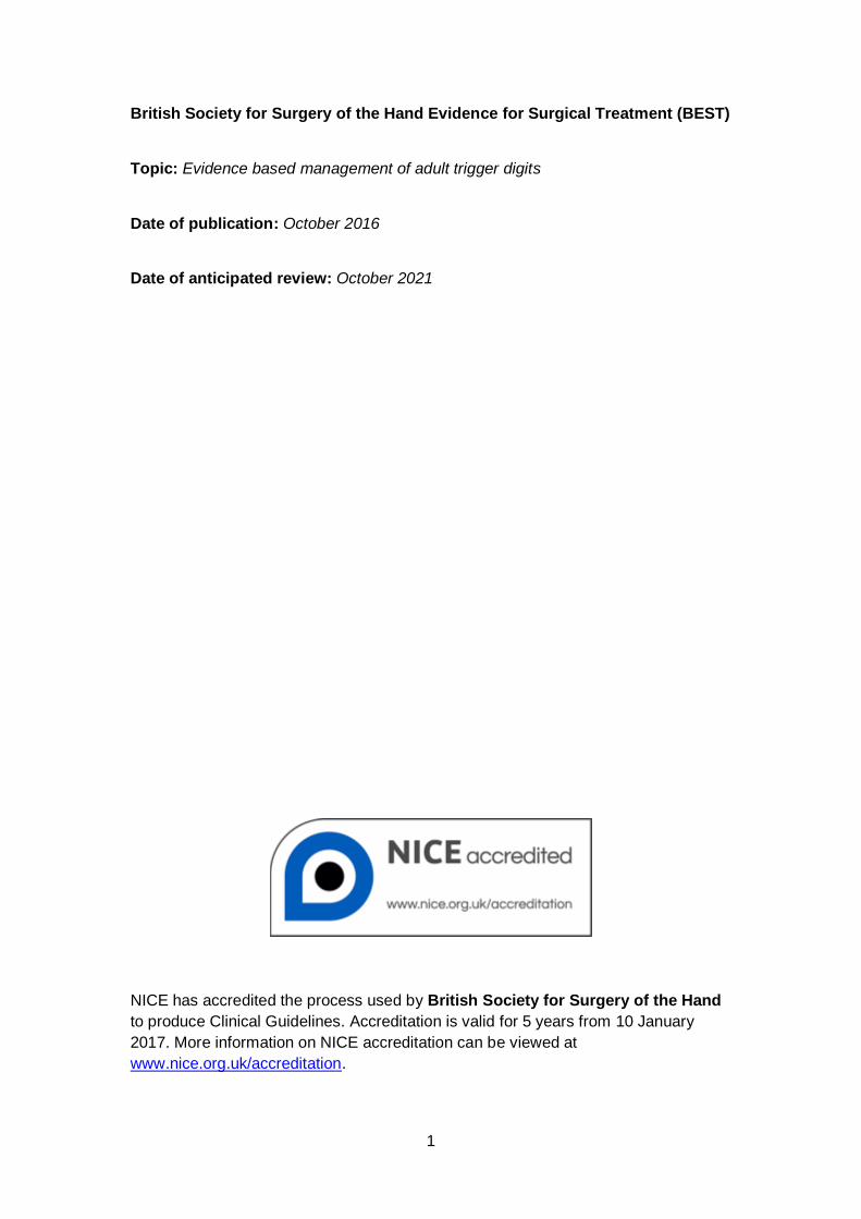
British Society for Surgery of the Hand Evidence for Surgical Treatment (BEST)
Topic: Evidence based management of adult trigger digits
Date of publication: October 2016
Date of anticipated review: October 2021
NICE has accredited the process used by British Society for Surgery of the Hand
to produce Clinical Guidelines. Accreditation is valid for 5 years from 10 January
2017. More information on NICE accreditation can be viewed at
www.nice.org.uk/accreditation.

2
Contents
AUTHORS............................................................................................................... 5
GUIDELINE DEVELOPMENT GROUP (GDG):................................................................ 5
ROLES OF AUTHORS WHO ARE NOT GDG MEMBERS .................................................. 6
ACKNOWLEDGEMENTS ........................................................................................... 6
FUNDING SOURCES USED ........................................................................................ 6
COLLABORATING ORGANISATIONS ........................................................................... 6
CONFLICTS OF INTEREST ......................................................................................... 7
DISCLAIMER ........................................................................................................... 7
PROCESS ................................................................................................................ 7
OVERALL OBJECTIVE ............................................................................................... 8
ANTICIPATED USERS ............................................................................................... 8
TARGET POPULATION ............................................................................................. 8
QUESTIONS DISCUSSED IN THIS BEST ....................................................................... 8
QUESTIONS NOT DISCUSSED IN THIS BEST ................................................................ 8
3
INCLUSION & EXCLUSION CRITERIA .......................................................................... 9
PLAIN LANGUAGE SUMMARY .................................................................................. 9
INTRODUCTION ...................................................................................................... 9
METHODS ............................................................................................................ 11
SYSTEMATIC REVIEW RESULTS ............................................................................... 13
OTHER NONOPERATIVE TREATMENT MODALITIES (NOT INCLUDED IN THE SYSTEMATIC
REVIEW): ............................................................................................................. 17
SYSTEMATIC REVIEW OVERVIEW DISCUSSION: ........................................................ 20
CLINICAL PRACTICE RECOMMENDATIONS: .............................................................. 22
GOOD PRACTICE POINTS: ...................................................................................... 22
CLINICAL AUDIT INDICATORS: ................................................................................ 22
RESOURCE IMPLICATIONS: .................................................................................... 23
FACILITATORS AND BARRIERS TO IMPLEMENTATION: .............................................. 23
FUTURE RESEARCH RECOMMENDATIONS: .............................................................. 23
STAKEHOLDERS INVITED TO PROVIDE EXTERNAL REVIEW: ........................................ 24

4
TIMELINE OF GUIDELINE: ...................................................................................... 24
APPENDIX 1: PRISMA FLOW CHARTS FOR SYSTEMATIC REVIEW ................................ 25
APPENDIX 2: EVIDENCE SUMMARY TABLE ............................................................... 27
APPENDIX 3: KEY CLINICAL PRACTICE RECOMMENDATIONS ..................................... 34
APPENDIX 4: PATIENT FLOW ALGORITHM ............................................................... 35
APPENDIX 5: SUPPORT TOOL: QUICK REFERENCE GUIDE .......................................... 36
APPENDIX 6: CHARACTERISTICS OF INCLUDED STUDIES ............................................ 38
APPENDIX 7: QUALITY OF EVIDENCE ASSESSMENT OF INCLUDED STUDIES ................. 41
APPENDIX 8: INCLUDED STUDY REFERENCES ........................................................... 44
APPENDIX 9: OTHER REFERENCES .......................................................................... 45

5
Authors
Rouin Amirfeyz MD MSc FRCS (Trauma & Orth). Consultant Hand and Upper Limb
Surgeon, Bristol Royal Infirmary, Bristol. [email protected]
Robert McNinch FRACS (Orth). Upper Limb Fellow, Bristol Royal Infirmary, Bristol.
Adam Watts FRCS (Trauma & Orth). Professor of Upper Limb Surgery, Upper Limb
Unit, Wrightington Hospital, Wigan. [email protected]
Nicole Glassey MSc. Hand therapist, Hand Unit, Queen’s Medical Centre,
Nottingham. [email protected]
Justine Bullock MCSP. Hand therapist, Hand Unit, Queen’s Medical Centre,
Nottingham. [email protected]
Jeremy Rodrigues BSc MSc PhD MRCS. Academic Clinical Fellow in Plastic
Surgery, University of Oxford. [email protected]
Tim Davis ChM, FRCS. Honorary Professor of Hand Surgery, Queen’s Medical
Centre, Nottingham. [email protected]
Guideline Development Group (GDG):
Core Stakeholders: Rouin Amirfeyz, GDG Lead
Robert McNinch, Representing Hand Surgery Trainees
Adam Watts, Representing Hand surgeons
Nicole Glassey, Representing Hand therapists
Justine Bullock, Representing Hand therapists

6
Nigel Andrews, Representing Patients
Special Stakeholders: Mike Taylor (GP), Representing Primary Care
Roles of authors who are not GDG members
Jeremy Rodrigues: Guideline development process expertise (co-author of BEST
Process manual)
Tim Davis: Guideline development process expertise (co-author of BEST Process
manual)
Acknowledgements
Peter Lanyon: President of the British Society for Rheumatology (representing
rheumatologists, whose patients might have trigger digits), provided invaluable
advice to the authors and reviewed the draft.
Funding sources used
None
Collaborating organisations
Secondary Care: University Hospitals Bristol NHS Foundation Trust
Nottingham University Hospitals NHS Foundation Trust
Wrightington, Wigan and Leigh NHS Foundation Trust
Primary Care: Bristol Primary Care

7
Conflicts of interest
There are no conflicts of interest of relevance for any of the GDG members or
document authors.
Disclaimer
This document reflects a consensus view of the British Society for Surgery of the
Hand Research Committee and Council, based on a systematic and transparent
review of evidence. All users of this document must ensure that they consider the
entirety of the document when using it, that the recommendations within this
guideline are not mandatory, and that clinical judgement and that patient-centred
decision making for all individual patients is the highest priority. Users are reminded
of their individual duties and responsibilities, professional or otherwise, to use this
guideline responsibly and that no content within this guideline overrides these duties
and responsibilities.
Process
This document has been produced by systematic reviews, with the interpretation and
development of recommendations achieved by consensus of the GDG members.

8
Overall Objective
The overall objective is to describe the management of adult patients with trigger
digits in the United Kingdom
Anticipated Users
The anticipated users are health care professionals treating patients with trigger
digits, those commissioning care for patients with trigger digits, and possibly patients
and carers of patients with trigger digits.
Target Population
Adults with trigger digits of the hand
Questions discussed in this BEST
• Which patients should be referred to hand surgeons?
• Which treatments are superior to other treatments?
• Which treatments are more cost-effective than other treatments?
• What treatments should be offered to patients?
• At what clinical stage should different treatments be offered to patients?
• What outcomes can be expected from particular treatments?
• What future research might be beneficial in clarifying optimal treatment?
Questions not discussed in this BEST
• How should paediatric trigger digits be treated?

9
Inclusion & exclusion criteria
Patients 16 years and older (adults) with trigger digit (thumb or finger) were included.
Paediatric trigger digits were not considered as part of this systematic review.
Plain Language Summary
In a trigger digit, the tendon that pulls the finger towards the palm when making a fist
does not slide easily. This leads to clicking when trying to make a fist or straighten
the fingers. If bad enough, the finger may not be able to bend or straighten at all.
Different treatments can be used. These include injecting a drug called a steroid
through the palm skin towards the tendon, to relieve the trigger digit. The injection
can also contain local anaesthetic, a drug that numbs the area. Instead, the point
where the tendon is snagging can be surgically released. This can be done using a
needle passed back and forth to release around the tendon (“percutaneous release”).
Alternatively, a knife can be used in the release, through a cut in the palm (“open
release”).
We searched for all studies that compared these kinds of treatments. A group that
included a patient, a GP, hand therapists and surgeons then formally discussed the
studies in order to agree upon recommendations of how to treat trigger digits.
The group’s recommendations are that the injection treatment is reasonable to use in
the first instance. If this is not suitable, or if the patient prefers, then the
“percutaneous” or “open” release can be used. If the trigger digit does not get better
after the injection, then either the “percutaneous” or “open” releases can be used.
Introduction
Trigger digit, or stenosing tenosynovitis, is a condition where abnormal gliding of the
flexor tendons within their flexor sheath results in snagging, or locking of the affected
digit in flexion, or occasionally, extension. “Triggering” of the affected tendon results
in difficulty in flexing or extending the finger and is frequently associated with pain in
the palm of the hand. Trigger digit may be associated with disease states such as
rheumatoid arthritis and diabetes mellitus (Wolfe 2005). Trigger digit has a reported
incidence of 28 cases per 100,000 subjects annually, or a risk of 2.6% over a lifetime
(Strom 1977). It is more common in middle-aged women (Lindner-Tons and Ingell

10
1998). The triggering is most commonly caused by jamming of the flexor tendons in
the entrance to the flexor sheath of the digit, most probably due to thickening of the
first annular (A1) pulley. It can also arise after injury or an intervention to the tendons
(such as repair) that causes it to snag under the pulley. Power grip causes high
angular loads at the distal edge of the A1 pulley (Ryzewicz and Wolf 2006).
Histologically the A1 pulley may demonstrate fibrocartilagenous metaplasia and
hypertrophy, increased glycosaminoglycan, degenerative changes and proliferation
of fibrous tissue (Sampson et al 1991). these changes can result in the wall of the
A1 pulley becoming three times thicker than normal (Ryzewicz and Wolf 2006).
Although trigger digit is frequently referred to as tenosynovitis, inflammatory changes
are not seen histologically in the tenosynovium (Moore 2000) in primary trigger
finger. Pathologic inflammatory changes may occur in the surrounding tissues but
are not observed in the pulley itself (Ryzewicz and Wolf 2006).
Treatment for trigger finger can be divided into non-operative and operative. Non-
operative management includes activity modification, NSAIDs, hand therapy,
splinting and corticosteroid injection. Operative management is by release of the A1
pulley, either percutaneously or more commonly with open surgery.
Activity modification involves avoiding positions that result in triggering, which may
allow the pathologic process to settle. For patients who do not have a
contraindication, an oral NSAID can be tried. Hand therapy treatment can include
wax therapy, ultrasound, stretching muscle exercises and massage. However, no
randomised controlled trials exist in the English literature regarding these forms of
non-operative management and so this review will mainly concentrate on
corticosteroid and local anaesthetic injection as non-operative interventions and
percutaneous and open pulley release as operative interventions of the adult trigger
digit. For completeness, these other non-operative treatment modalities, and the
evidence behind them, will be discussed.
11
Methods
Nonoperative treatment:
All randomised controlled trials and controlled clinical trials evaluating the use of
splinting, or local injection with corticosteroids, for the treatment of adult trigger
finger, were included in this review. Studies focusing on patients older than 16 years
with a clinical diagnosis of trigger digit, irrespective of the duration of symptoms,
were included. Exclusions included studies focusing on paediatric trigger finger or
paediatric trigger thumb. Cochrane Library, Medline, Embase, and Pubmed were
used for electronic database searches, coupled with a secondary search of the
references of selected articles on 22nd October 2015. Search criteria included using
the following terms in the title, abstract and subject headings (exploded); Trigger
finger(s) or trigger digit(s) or trigger thumb(s) or stenosing tenosynovitis or stenosing
flexor tenosynovitis or stenosing tenovaginitis or flexor tendon entrapment and “non-
operative treatment”, “splint or splints” or “treatment or therapeutic”, or "stretching or
muscles or muscle stretching exercises", "wax therapy", "heat or hot temperature",
"ultrasound or ultrasound, high intensity focused", "massage", "electrotherapy or
electric therapy stimulation" and "acupuncture", and were limited to English language
studies and human studies. The search results, prior the exclusions were Medline
(641), Embase (780), Pubmed (559) and Cochrane trials database (49), with a total
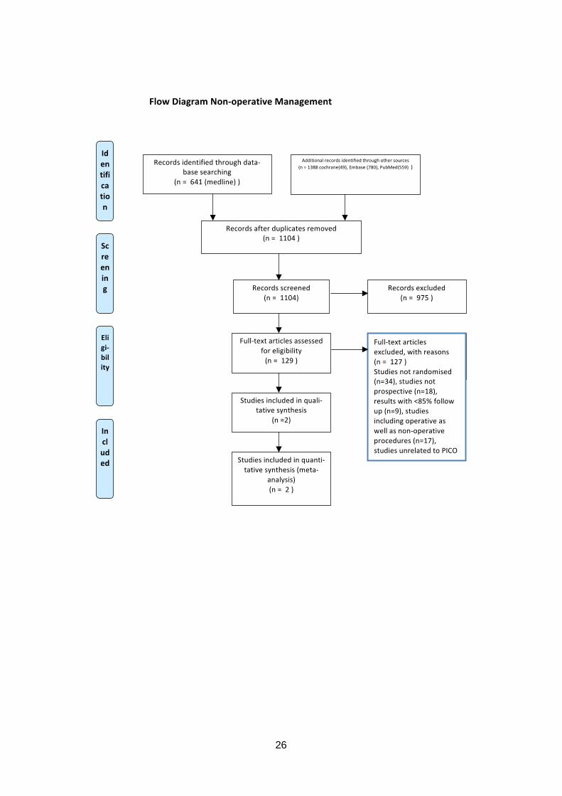
of 129 after the exclusions were applied (Appendix 1). Further specific criteria were
then applied to the 129 studies identified. These criteria were that the studies must
be randomised, prospective in nature and must have results with at least 85% follow
up.
Operative treatment:
A search was performed of the Cochrane Library, Medline, Embase, and Pubmed on
11th March 2015 using the following terms in the title, abstract and subject headings
(exploded); Trigger finger(s) or trigger digit(s) or trigger thumb(s) or stenosing
tenosynovitis or stenosing flexor tenosynovitis or stenosing tenovaginitis or flexor
tendon entrapment and surgery or operative.
The search results were as follows; Cochrane trials database (33), Medline (448),
Embase (587), PubMed (450) (figure 2). After exclusion of duplicates, case series,
cohort studies, case reports and letters eight articles were included; seven
randomised trials and one meta-analysis. A protocol was identified entitled “Surgery

12
for trigger finger” by Ventin FC et al. however no systematic review has been
published in the Cochrane Library.
Shortlisted studies were assessed using SIGN50 methodology.

13
Systematic review results
Nonoperative treatment:
Two studies were identified which met the criteria. Both of these were controlled trials
comparing the efficacy of steroid injections with lidocaine injections in trigger finger in
a secondary care setting.
Lambert et al. (1992) compared the effectiveness of intra-tendon sheath injection of
methylprednisolone (0.5 ml) combined with 1% lidocaine to 1% lidocaine alone.
Treatment success was defined as complete resolution of symptoms or sufficiently
improved that further treatment was not necessary one month after injection. Forty-
one patients were included in the study, with 20 patients allocated to the steroid and
lidocaine injection and 21 patients to an injection of lignocaine alone. The paper
states that this allocation was performed randomly, although the specifics of this are
not stated. Two patients were lost to follow up from the lidocaine group, leaving a
total of 39 patients to be included in the analysis. The outcome assessor was blinded
to the treatment group. However, concealment of allocation, blinding of the care
provider, blinding of the patients and similarity of groups at baseline regarding most
important prognostic indicators were unclear and no intention to treat analysis was
used. The results were 45% success (9/20) in the methylprednisolone + lidocaine
group and 16% (3/19) in the lidocaine alone group. The absolute risk reduction, or
the difference in treatment success between the two groups, was 0.292 (95% CI
0.017 to 0.567), the relative risk, or chance of successful outcome using
methylprednisolone combined with lidocaine rather than lidocaine alone, was 2.85
(95% CI 0.91 to 8.96) with a number needed to treat of 3.424 (95% CI 2 to 58).
These results demonstrate that a successful treatment at one month after injection is
significantly more likely using methylprednisolone combined with lidocaine than with
just lidoocaine alone. The number needed to treat demonstrates 3.4 patients would
need to be injected with methylprednisolone and lidocaine to provide one additional
successful treatment at one month compared to the control group.
Murphy et al. (1995) compared the effectiveness of 1ml of betamethasone (1ml
celestone equating to 6mg betamethasone) combined with 3ml of 1% lidocaine with
injection with 4ml of 1% lidocaine by itself. Twenty-four fingers in 24 patients were
randomised into this study with the patients being allocated to the two groups
14
depending on whether their initial presentation was on an odd or even date. Fourteen
patients were allocated to the steroid and lidocaine group and 10 to the control
group. No patients were lost to follow up. Treatment success, defined as participants
becoming asymptomatic, was assessed immediately after injection, 3 weeks and 4
months after injection. The outcome assessor and patient were blinded in this study
but the care provider was not. 3 of the 14 patients in the steroid group had unrelieved
triggering at their 3 week appointment and were reinjected with steroid. Ten of the
patients in the steroid group were asymptomatic at this stage and 1 patient had mild
triggering. Six of the 10 patients in the control group had unrelieved triggering at their
3 week appointment and were given a steroid injection at that stage. Two of the
patients in the control group were asymptomatic at this stage and 2 had mild
triggering. The involvement in the study of the patients who received an extra
treatment at 3 weeks was ended at this point and they were treated as a failure in
their original treatment for statistical purposes. Whilst an intention to treat analysis
was used, no concealment of allocation was provided. It was not clear in the study if
the two concealment groups were similar at baseline regarding prognostic indicators.
Success after 3 weeks was 71% (10/14) in the betamethasone + lidocaine group and
20% (2/10) in the lidocaine alone group. The absolute risk reduction, or the
difference in treatment success between the two groups, was 0.514 (95% CI 0.165 to
0.864), the relative risk or chance of successful outcome using betamethasone
combined with lidocaine rather than lidocaine alone, was 3.57 (95% CI 0.99 to 12.88)
with a number needed to treat of 1.946 (95% CI 1 to 6). Four months after injection
therapy treatment success was 64% (9/14) in the betamethasone + lidocaine group
and 20% (2/10) in the lidocaine alone group, resulting in a number needed to treat of
2.258 at 4 months. These results demonstrate at a successful treatment at 3 weeks
and 4 months after injection is significantly more likely using betamethasone and
lidocaine than with just lidocaine. The number needed to treat demonstrates 2
patients would need to be injected with betamethasone and lidocaine to provide one
additional successful treatment at 3 weeks and 4 months, compared to the control
group.
Neither trial specified which specific diagnostic criteria were used for the diagnosis of
trigger finger, how many cases were assessed for eligibility prior to enrolment or the
demographic and clinical characteristics their two groups. There was also no
information regarding the frequency of triggering, severity of pain and functional
status of the hands in either study. Both papers mentioned that there were no
adverse events or complications but patient satisfaction with the treatment was not

15
assessed. No power calculation was performed and no validated patient based
outcome measure was used.
Pooling of the data results in 63 participants. Corticosteroid injections with lidocaine
showed significantly more effectiveness within 4 weeks than lidocaine injection alone
(RR 3.15, 95% CI 1.34 to 7.40). (Peters-Veluthamaningal et al. 2009).
These studies demonstrate that there is moderate evidence for superiority of a
mixture of corticosteroid and lidocaine injections over injections with lidocaine alone.
It is noted that although the patients benefited from the corticosteroid and lidocaine
injection, the difference between the groups was the addition of corticosteroid. It is
unclear from the existing evidence base whether corticosteroid alone as an injection
would be effective, or whether combining corticosteroid with local anaesthetic (which
has other effects such as increasing the volume injected) is necessary.
Neither study reported any adverse effects. As the numbers are small these effects
need to be confirmed in larger, well-designed randomised trials. The results also
suggest efficacy up to four months, but long-term efficacy still remains to be clarified.
Operative treatment:
Of the randomised trials 4 were assessed as of acceptable quality to minimise bias
(Gilberts et al 2001, Chao et al 2009, Zyluk and Jagieski 2011, Sato et al 2012).
Three randomised controlled trials were excluded because the methodology was
assessed as introducing a high or uncertain risk of bias. This was because of poor
randomisation concealment, differences in the groups after randomisation, and
measurement of outcomes (Maneerit et al 2003, Dierks et al 2008,
Bamroogshawgasame 2010).
The one meta-analysis (Wang et al. 2013) was assessed as high quality with a low
probability of bias.
Percutaneous versus Open Release
Gilberts et al. (2001) conducted a randomised trial of percutaneous trigger finger
release versus open surgery in a total of 100 digits followed for 12 weeks. They
found no difference in recurrence rates, which were very low. The duration of
surgery, recovery of motor function and time to return to work were all significantly

16
shorter in the percutaneous group. A further randomised trial compared
percutaneous, open pulley release and steroid injection (Sato et al 2012) in 150
digits followed for 6 months. The study population was over the age of 15 years.
The authors reported equivalent outcome with no recurrences in the percutaneous or
open surgical arm of the trial, but percutaneous release resulted in better finger
movement in the first two post-operative months. No complications were
encountered.
Injection versus Percutaneous Release
Two studies met the inclusion criteria. Chao et al. (2009) reported on a trial in 97
thumbs (86 patients) randomised to percutaneous release with a miniscapel versus
steroid injection. Whilst the study was well designed a significant number of patients
were lost to follow up in the steroid injection cohort (32% of digits lost at 12 months
compared to no loss to follow up in the percutaneous release arm). Zyluk et al.
(2011) similarly compared percutaneous release to steroid injection in a cohort of
115 patients with trigger digit and reported recurrence rates of zero in the
percutaneous release and 12% in the injection arm of the trial. Lost to follow up
rates were also high at 22% lost from the percutaneous release arm and 13% from
the injection arm at final review at 6 months. No complications were reported.
Wang et al. (2013) conducted a meta-analysis of seven randomised controlled trials
to ascertain the best treatment method for trigger digit by determining the risk ratio of
treatment failure, level of satisfaction and complications comparing percutaneous
release, open surgery and steroid injections. A total of 397 were enrolled in
randomised trials comparing percutaneous release and open surgery. For
comparison of percutaneous release and steroid injection four trials included 417
patients. The authors found no difference in treatment failure between percutaneous
and open surgery with follow up times between 2 and 6 months. Treatment failure
rates were higher in the group who had steroid injections compared to those who had
percutaneous release who were also more likely to be satisfied with the outcome at
follow-up times of 6 to 23 months. There were no differences in complication rates
reported. It should be noted that this meta-analysis included all the trials mention
above and also some trials excluded from this review due to risk of bias.

17
Other nonoperative treatment modalities (not included in the
systematic review):
There are no robustly performed, randomised controlled trials in the English literature
on other aspects of non-operative management which fit the inclusion criteria of this
systematic review. However, the evidence for other treatment modalities shall be
reviewed.
Splinting
Proponents of splinting state that it alters the biomechanics of the flexor tendons,
which reduces friction between the tendons and pulley system, while encouraging
maximal differential tendon glide. By altering the mechanical pressures of the
proximal pulley system and encouraging maximal tendon gliding, the pathologic state
of the tendon and its sheath may be reversed in a significant number of cases
(Creighton et al 1990). Splint wear is usually advised for a 3 to 9-week period (Evans
et al 1988, Creighton et al 1990 and Cannon et al 1991). It is usually not necessary
to splint adjacent fingers (Evans et al 1988). Most studies looking at splinting to treat
trigger finger have focused on splinting of the metacarpophalangeal (MCP) joint.
However, there is some disagreement on the degree of joint positioning and there
are also advocates for splinting of the distal interphalangeal (DIP) joints (Colbourne
et al 2008).
When the MCP joint was splinted the position varied between 0 and 15 degrees of
flexion, allowing for full interphalangeal movement (Lindner-Tons and Ingell 1998,
Patel and Bassini 1992, Evans et al 1988). Studies on the efficacy of splinting report
good outcomes in 70-73% of their patients (Patel and Bassini 1992, Evans et al
1988).
In the Evans et al study the MCP joint was immobilised in 0 degrees using a volar-
based hand splint allowing full movement in the proximal and distal interphalangeal
joints. Evans et al. demonstrated a 73% success rate using a splint combined with
hook and fist exercises, in a study of 55 digits in 38 patients (1988). However,
splinting was initiated at differing times from the onset of triggering in a
heterogeneous group of patients and there was no data on recurrence following
discontinuation of the splint. The assessment and outcome measures were also
subjective and the methodology lacked detail, making it difficult to repeat. Patel and

18
Bassini reported a 70% successful outcome with splinting the MCP joint for 6 weeks
in 10-15 degrees of flexion in 40 fingers (1992). They found that splinting was
successful in 77% of the patients whose symptoms had been present six months or
less and 44% of those patients with symptoms longer than six months. Patel and
Bassini (1992), and Lindner-Tons and Ingell (1988) have suggested a higher
compliance rate with splinting in 10-15 degrees of MCP joint flexion compared with 0
degrees to allow increased function of the hand.
Colbourne et al. studied the effectiveness of an MCP joint blocking splint with the
MCP joints positioned in 10-15 degrees of flexion (2008). Patients were instructed to
wear splints day and night for 6 weeks and this was extended to 10 weeks if
triggering persisted. Subjects removed the splint three times a day to perform
passive IP joint flexion, composite full flexion, full extension plus active hook fist. 93%
of the participants reported improvements in triggering as a result of the splint.
However, 57% of subjects did not comply with splint use due to interference with
function and the majority did not follow the exercise program.
Rodgers et al. believed that the FDP tendon, either alone or in conjunction with the
FDS tendon, was instrumental in the pathogenesis of trigger finger (1998). They
used a distal interphalangeal (DIP) splint made from alumafoam taped to the dorsum
of the digit or a Stack finger splint, as well as NSAIDs in their treatment of 21 patients
with trigger finger. Their protocol consisted of full-time splint wear for at least six
weeks. They demonstrated 55% resolution of the triggering.
Splint design has been attributed to patient compliance rates. The splint should be of
the lowest profile and least restrictive design possible, since it should be worn for the
entire day for a number of weeks (Lindner-Tons and Ingell 1998). Poor design leads
to non-compliance with splint wear before it has had a chance to prove effective.
Some splints maintain the optimal finger position but bulky strapping over the dorsum
of the hand, as well as extensive palmar coverage often compromise compliance
(Evans et al 1988, Cannon et al 1991, Patel and Bassin 1992). Alternative splint
designs have been postulated to minimise interruptions with activities of daily living
(Lindner-Tons and Ingell 1998).
Although splinting to treat trigger finger has been described as inexpensive and
helpful in reducing symptoms of triggering with minimal complications (Evans et al
1988, Lindlor-Tons and Ingell 1998, Colbourne et al 2008, Rogers et al 1998) a
review of the literature shows little comprehensive or objective data to clearly support
the role of splinting. Tarbhai et al. attempted to compare different splint designs to
determine whether MCP joint or DIP joint blocking splints were more effective (2012).

19
They also attempted to compare the splint designs with respect to comfort,
compliance and usefulness during functional activities. They prospectively
randomised 30 subjects to MCP or DIP joint blocking splints and found that the MCP
joint splint provided at least partial relief of triggering and pain in 10 of 13 patients,
whereas the DIP joint splint provided at least partial relief of triggering and pain in 7
of 15 patients after 6 weeks treatment. There was statistically significant
improvement in both groups at 6 weeks, which was maintained in a minority of the
cohort for 1 year. There was little difference between the two groups with regards to
impact on function. Patients who wore the MCP joint splint reported higher rates of
comfort compared to those who wore the DIP joint blocking splint. Joint stiffness was
reported in both groups (1 of 13 in the MCP joint group and 7 of 15 in the DIP joint
group). However, this resolved once the splints were discontinued.
The European Handguide Study aimed to provide guidelines on treatment for trigger
finger based on the consensus of the Delphi group, a group of 35 experts in the field.
They found no evidence for the effectiveness of splinting in a systematic review
(Huisstede et al 2010). They felt that an MCP joint blocking splint at 0 degrees was
preferable but no consensus could be achieved on the optimal orthotic regimen
including duration of splint wear during the day or number of weeks of splint usage.
The length of time and cost required for producing and maintaining a splint compared
with the application of a single steroid injection should be considered. A patient on
average needs to seen by a qualified therapist four times and have a splint made (at
least one if no further adjustment required). This would cost £110 minimum.
Also the potential detrimental effects on adjacent digits and hand function with long
term use of splintage should not be overlooked.
Hand therapy
There is minimal evidence for the use of hand therapy techniques, such as heat,
stretching, wax therapy, ultrasound and massage, as well as other techniques such
as electrotherapy and acupuncture. Proponents of techniques involving therapeutic
heat modalities state that it increases blood flow to the area, facilitates collagen
plasticity and helps with resolution of oedema (Salim et al 2011). Combining heat
with stretching allows plastic deformation of collagen (Cameron 1999, Knight et al
2001, Recor and Johnson 2010). Massage has been claimed to ‘soften’ or remodel
tendons reducing tissue bulk at the pulleys (Evans et al 1988). The net result of
these techniques being that the tendon passes more easily through the A1 pulley.

20
Salim et al attempted to compare the effectiveness of hand therapy with
corticosteroid injections in the management of mild trigger fingers, defined as
patients with mild crepitus on flexor tendon gliding, uneven finger movements or
actively correctable triggering (2011). Eight-four patients were randomised to either
corticosteroid injection or hand therapy. The randomisation was performed
numerically, with the first five patients being allocated to hand therapy, the next five
patients to corticosteroids and so on. Ten patients were lost to follow up leaving 35
patients in the hand therapy group and 39 in the corticosteroid group. The hand
therapy regimen provided is unclear, being stated as consisting of ten sessions,
comprising wax therapy, ultrasound, muscle stretching exercises and massage. The
specific treatment, duration or technique used in these categories is not defined. The
steroid injection comprised 1ml of triamcinolone acetonide and 1ml of 2% lidocaine
injected at the A1 pulley. Whether the injection was injected deep, or superficial, to
the sheath is not clear. The patients were followed up at 6 weeks and 3 months, and
a telephone interview was used at 6 months to assess recurrence of pain and
triggering. The authors quoted an overall success rate, defined as absence of pain
and triggering, of 68.6% in the hand therapy group and 97.4% in the corticosteroid
group at 3 months. No complications were noted in either group. At 6 months no
patients in the hand therapy group had any recurrence of symptoms but 6 patients in
the corticosteroid group had developed pain and 4 had developed symptoms of
triggering. Since this was performed by telephone interview this could not be
correlated clinically.
The results of this study are compromised by the lack of detail regarding the hand
therapy treatments and the fact that multiple hand therapy techniques are used
rather than just one. This, however, is the only published study regarding the
outcome of these hand therapy techniques in trigger finger. There are no studies
which independently assess wax therapy, heat therapy, muscle stretching,
ultrasound, massage, electrotherapy or acupuncture as the primary treatment
modality in trigger finger.
Systematic review overview discussion:
Systematic review of the published literature has identified a reasonable number of
trials of acceptable quality to answer the questions outlined for operative
management but not for nonoperative management. The data indicated that open
and percutaneous releases are associated with a similar low risk of recurrence of

21
trigger finger or thumb in adults. Similarly, at six months there is no evidence for a
difference in pain scores although the initial pain scores may be lower after
percutaneous treatment. There is no evidence of an increased risk of complications
with either treatment. Local steroid injection carries a greater risk of recurrent or
persistent symptoms.
There is a paucity of quality evidence in the English literature regarding non-
operative management of trigger finger. Splinting is non-invasive and may provide
short-term relief, but evidence for its use is poor. There is no evidence for the use of
other non-invasive treatment modalities.
There is moderate evidence that corticosteroid injections are effective for the
treatment of trigger finger, but the implications for daily clinical practice may be
limited by the fact that the evidence is based on two small studies of poor quality,
performed in the setting of secondary care, and there were only data available for
effectiveness up to four months. Complications from steroid injections are rare but
may include fat necrosis, skin depigmentation and rupture of the flexor tendons
(Ryzewicz and Wolf 2006), none of which were specifically encountered or reported
in the two mentioned studies. However, corticosteroid injection is an easily applicable
treatment modality, inexpensive and less invasive than surgery. Corticosteroid and
anaesthetic injection is a reasonable first line option of treatment as the NHS cost of
trigger finger release in England is presently between £867.26 to £945.31 depending
on co-morbidities (data obtained from University Hospitals Bristol NHS Foundation
Trust, Management Accountant, Surgery Head & Neck Division, 24th February
2016). Compared with surgery, there is high evidence that local corticosteroid
injection is associated with increased rates of ongoing or recurrent symptoms at a
minimum of six months after intervention Both of the included studies were in a
secondary care setting, and this could potentially be offered in primary care, although
there would need to be considerable training for providers. As it stands, injection for
trigger finger and/or thumb is not offered by many first line services due to lack of
time, expertise or resources.
There is high quality evidence that trigger digit can be managed safely by open or
percutaneous surgical release.

22
Clinical practice recommendations:
Based on the current available evidence, it is reasonable to offer corticosteroid and
local anaesthetic injection as the first line of treatment (moderate evidence).
Percutaneous release may be offered by an appropriately trained practitioner (high
evidence).
If symptoms fail to resolve, or should the patient decide against injection, then the
next line of treatment may be either an open or percutaneous release of the
constricted pulley (high evidence). Other treatment modalities are not currently
supported.
Good practice points:
It is considered good practice that:
• in the absence of contraindication and with patient’s agreement, the first line
of treatment for an adult trigger digit should be a single injection of steroid
and local anaesthetic. However, an outpatient percutaneous release can be
offered if the practitioner is qualified and experienced in the procedure.
• a referral to the secondary care should be made if the patient prefers a
percutaneous or open release.
• a referral to secondary care for surgical treatment (percutaneous or open
depending on the available expertise) should be made it the triggering recurs
after injection.
Clinical audit indicators:
It is considered that the following could be used as clinical audit indicators:
• Recurrence (as the primary outcome)
• Patient satisfaction (one example would be Patient Global Impression of
Change)
• Pain score

23
Resource Implications:
It is believed that the clinical practice recommendations and good practice points
either align with existing NHS practice, or are less expensive than current practice
(e.g. increasing the use of steroid injection as first line treatment rather than surgery
is anticipated to reduce costs). Therefore, the resource implication of implementing
this guideline is considered minimal. However, training of clinical staff in the
technique of steroid injection ay be required in some settings.
Facilitators and barriers to implementation:
If clinical staff are not competent in injection, then training may be required. Such
training is not believed to be complex, expensive or onerous to deliver. No other
significant barriers to implementation have been identified. It is suggested that using
the quick reference as a standalone reference may be facilitator. For example, users
may wish to make the quick reference guide could be made available in clinical
areas.
Future research recommendations:
Areas for future research into the management of trigger finger include large, well
designed, randomised controlled trials of:
• surgery versus corticosteroid injections with outcomes measured beyond four
months;
• corticosteroid versus corticosteroid combined with local anaesthetic (the latter
was used in all studies which satisfied the entry criteria for our systematic
review;
• DIP joint and MCP joint splints with corticosteroid injection;
• individual hand therapy treatment modalities.
• Treatment of trigger fingers in those with rheumatoid arthritis and diabetes
mellitus
• Treatment strategies involving more than one injection containing steroid (i.e.
giving a second or even third steroid injection)

24
Stakeholders invited to provide external review:
The British Orthopaedic Association
The British Association of Plastic, Reconstructive and Aesthetic Surgeons
The British Association of Hand Therapists
The Royal College of General Practitioners
The British Society for Rheumatology
Timeline of guideline:
Date topic identified: 27/03/2013
Date GDG lead appointed: 07/04/2013
Date draft supplied by GDG authors: 08/08/2016
Date Internal review completed: 21/08/2016
Dates of public consultation: 30/08/16 – 16/09/16
Date external review completed: 22/09/16
Date published: 13/10/16

25
Appendix 1: PRISMA flow charts for systematic review
FlowDiagramOperativeManagement
Recordsidentifiedthroughdatabasesearching
(n=1518)
Recordsafterduplicatesremoved
(n=634)
Recordsscreened
(n=634)
Recordsexcluded
(n=625)
Full-Textarticlesassessedforeligibility
(n=9)
Full-textarticlesexcluded
(n=5)
Studiesincludedinqualitativesynthesis
(n=4)
26
27
Appendix 2: Evidence Summary Table
Evidence Summary: Open surgery, percutaneous release and steroid
injection. RA: Rheumatoid Arthritis, ROM: Range of Movement, VAS:
Visual Analogue Scale, F/U: Follow Up, AROM: Active Range of
Movement, RCT: Randomised Controlled Trial, RR: Risk Ratio
(Four operative primary research articles, two nonoperative primary
research articles, one meta-analysis)

28
Study details Population and setting
Method of allocation
Outcome measures and length of f/u
Results Comments
Gilberts et al (2001) The Netherlands Percutaneous vs open release
Digits Included: 18 y/o or over, trigger digit for over 1 month Excluded: Previous surgery on the same digit, connective tissue disease including RA Outpatient surgical facility
Randomisation using sealed envelopes 46 digits open surgery, 54 digits percutaneous (18 gauge needle). All using 1% lidocaine
Operation time Duration of postop pain Recovery of ROM Return to work F/u: Ten days, six weeks and Three months
Operation time 4 min shorter in percutaneous (p<0.0001) Postop pain period 1.6 days shorter in percutaneous (p=0.039) Recovery of ROM 11 days shorter in percutaneous (p <0.002) Return to work 3.6 days shorter in percutaneous (p<0.0001)
Assessors and patients not blinded No p values provided for baseline groups comparisons Duration of symptoms 6 months longer in open group compared to percutaneous
Chao et al (2009) China Percutaneous vs steroid injection
Thumbs Included: Idiopathic adult trigger thumb with uneven movement +/- intermittent locking Excluded: RA, diabetes mellitus, chronic systemic
Randomisation using sealed envelope (witnessed) 46 thumbs percutaneous (Mini Scalpel Needle) 47 thumbs injection (1 ml triamcinolone)
Success, which was defined as VAS for pain <1 and no triggering Percentage change in pain Complications Patient satisfaction F/u: One month and
At one year 44/46 in percutaneous and 12/47 in steroid groups were successfully treated. At one year percentage change for VAS pain was 89.4 for
At one year 46 thumbs in percutaneous group were assessed despite they said one loss to f/u, but only 32 in steroid group were followed despite they claimed 3 loss to f/u

29
disease Hospital outpatient
one year percutaneous and 6.8 for steroid (p<0.01) At one year 44/46 were satisfied in percutaneous versus 12/47 in steroid group (p<0.01) No complications seen in any group
Zyluk and Jagielski (2011) Poland Percutaneous vs steroid injection
Digits Included: Adult patients (youngest 19 y/o) with trigger digits (all grades) Excluded: Not mentioned Hospital outpatient
Randomisation using sealed envelope (witnessed) 55 digits percutaneous (19 gauge needle) 60 digits injection (1 ml betamethasone)
Recurrence (return to baseline grade of triggering after total or partial improvement) VAS for pain AROM Grip strength (percentage of the other side) Complication F/u: One and six months
At six months, six recurrences were seen in steroid group vs none in percutaneous (p=0.005) At six months steroid group showed 0.9 point less in Vas for pain which was statistically significant (no p value provided) At six months steroid group showed 5 degrees more in AROM which was statistically significant (no p
More severe triggering in percutaneous group 0.9 difference in VAS for pain and 5 degrees in total AROM of digit is not clinically significant

30
value provided) At six months grip strength was the same in two group At six months no complication in steroid group and one reduction in flexion in percutaneous group was reported
Sato et al (2012) Brazil Open vs percutaneous vs steroid injection
Digits Included: Age > 15 y/o with grade II-IV on Quinnell classification Excluded: Grade I triggering on Quinnell classification or previous treatment of triggering (any form) Hospital setting
Randomisation via sequentially numbered sealed envelopes. 6-sided dice was used initially for each envelope treatment allocation. 56 digits open 45 digits percutaneous (40x12 needle) 49 digits steroid (2 ml methylprednisolone 40 mg/ml) at A1 pulley within osteofibrous canal
Primary: Cessation of triggering for 6 months Secondary: Pain (A1 pulley region and joint IP/PIP) Total active motion Complication F/u: 1 & 2 weeks, 1, 2, 4 & 6 months (if second steroid injection needed then 6 months from the second injection)
100% “cure rate” in open and percutaneous vs 57% after one injection and 86% after two (p=0.004) A1 pulley site and IP/PIP joint pain was more in the open and percutaneous surgery group compared to steroid injection in the first two months (p=0.008 & 0.029) but the same after two months Lower total active motion in open surgery group in
Power calculation: 43 in each group
31
the first two months (p=0.048) but the same in all afterwards No complications in any group
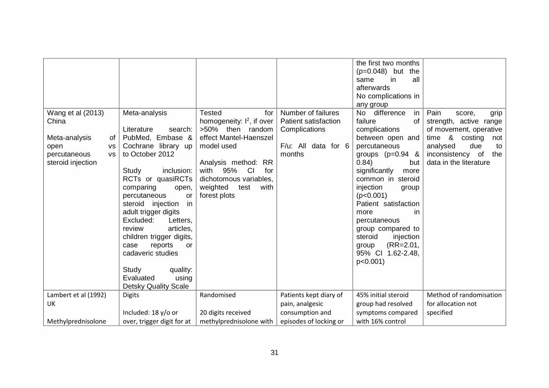
Wang et al (2013) China Meta-analysis of open vs percutaneous vs steroid injection
Meta-analysis Literature search: PubMed, Embase & Cochrane library up to October 2012 Study inclusion: RCTs or quasiRCTs comparing open, percutaneous or steroid injection in adult trigger digits Excluded: Letters, review articles, children trigger digits, case reports or cadaveric studies Study quality: Evaluated using Detsky Quality Scale
Tested for homogeneity: I2, if over >50% then random effect Mantel-Haenszel model used Analysis method: RR with 95% CI for dichotomous variables, weighted test with forest plots
Number of failures Patient satisfaction Complications F/u: All data for 6 months
No difference in failure of complications between open and percutaneous groups (p=0.94 & 0.84) but significantly more common in steroid injection group (p<0.001) Patient satisfaction more in percutaneous group compared to steroid injection group (RR=2.01, 95% CI 1.62-2.48, p<0.001)
Pain score, grip strength, active range of movement, operative time & costing not analysed due to inconsistency of the data in the literature
Lambert et al (1992) UK Methylprednisolone
Digits Included: 18 y/o or over, trigger digit for at
Randomised 20 digits received methylprednisolone with
Patients kept diary of pain, analgesic consumption and episodes of locking or
45% initial steroid group had resolved symptoms compared with 16% control
Method of randomisation for allocation not specified

32
acetate plus 1% lidocaine vs 1% lidocaine alone
least 3 months Excluded: insulin-dependent diabetics, patients with RA or eczema, patients with a concurrent infection, those who had undergone injection in the previous 3 months. Hospital outpatient
lidocaine, 21 received lidocaine by itself
clicking. Clinical assessment at follow up. F/u: 1 month following injection. Graded as a success if no further treatment required. Those with some improvement were injected with methylprednisolone, those with no improvement were listed for surgery
group (p<0.02). Subsequent steroid group had 60% resolved symptoms (p<0.02).
Trigger thumb accounted for 30% digits 2 patients in the lidocaine group lost to follow up and excluded Assessor at 1 month blinded to patient allocation
Murphy et al (1995) USA 6mg celestone (3ml) plus 1ml 1% lidocaine vs 4ml 1% lidocaine
Digits Included: 18 y/o or over, trigger digit Excluded: patients with RA, diabetes mellitus, previous tendon laceration, previous trigger finger injection or patients with unrelievable locking Hospital outpatient
Randomisation depending on day of presentation 14 digits received celestone with lidocaine, 10 digits received lidocaine by itself
Subjective grading of pain and triggering provided by the patient before and after the injection. Clinical examination by blinded examiner at f/u F/u: 3 weeks after injection, and 4 months
10 of 14 patients in the steroid group asymptomatic at 3 months, 3 had unrelieved triggering, 1 had mild triggering. 2 of the 10 placebo patients were asymptomatic at 3 months, 2 had mild triggering, 6 had no relief. At 4 months, 9 of the 10 patients in the
Duration of triggering not defined in patient groups No patient loss to follow up

33
steroid group remained asymptomatic (64% of the 14 patients) and 2 of the 10 placebo patients remained asymptomatic (20%). p< 0.05

34
Appendix 3: Key clinical practice recommendations
1. In the absence of contraindication and with patient’s agreement, the first
line of adult trigger digit should be a single steroid and local anaesthetic
injection. A percutaneous release in outpatients may be offered if the
practitioner is qualified and experienced in the procedure (moderate
evidence).
2. If the patient prefers percutaneous or open release, referral to secondary
care should be made (high evidence).
3. A referral to secondary care for surgical treatment (percutaneous or open
depending on the available expertise) should be made (high evidence) if
symptoms fail to resolve, or if there is recurrence.

35
Appendix 4: Patient flow algorithm

36
Appendix 5: Support Tool: Quick reference guide

37
BSSH Evidence for Surgical Treatment (BEST): Evidence based management
of adult trigger digits (published September 2016, valid until: September 2021)
Key clinical practice recommendations:
1. In the absence of contraindication and with patient’s agreement, the first
line of adult trigger digit should be a single steroid and local anaesthetic
injection. A percutaneous release in outpatients may be offered if the
practitioner is qualified and experienced in the procedure (moderate
evidence).
2. If the patient prefers percutaneous or open release, a referral to
secondary care should be made (high evidence).
3. A referral to secondary care for surgical treatment (percutaneous or open
depending on the available expertise) should be made (high evidence) if
symptoms fail to resolve, or if there is recurrence.

38
Appendix 6: Characteristics of included studies

39
Study details Population and setting Gilberts et al (2001) The Netherlands Percutaneous vs open release
Digits Included: 18 y/o or over, trigger digit for over 1 month Excluded: Previous surgery on the same digit, connective tissue disease including RA Outpatient surgical facility
Chao et al (2009) China Percutaneous vs steroid injection
Thumbs Included: Idiopathic adult trigger thumb with uneven movement +/- intermittent locking Excluded: RA, diabetes mellitus, chronic systemic disease Hospital outpatient
Zyluk and Jagielski (2011) Poland Percutaneous vs steroid injection
Digits Included: Adult patients (youngest 19 y/o) with trigger digits (all grades) Excluded: Not mentioned Hospital outpatient
Sato et al (2012) Brazil Open vs percutaneous vs steroid injection
Digits Included: Age > 15 y/o with grade II-IV on Quinnell classification Excluded: Grade I triggering on Quinnell classification or previous treatment of triggering (any form) Hospital setting
Wang et al (2013) China
Meta-analysis Literature search: PubMed, Embase & Cochrane library up to October 2012

40
Meta-analysis of open vs percutaneous vs steroid injection
Study inclusion: RCTs or quasiRCTs comparing open, percutaneous or steroid injection in adult trigger digits Excluded: Letters, review articles, children trigger digits, case reports or cadaveric studies Study quality: Evaluated using Detsky Quality Scale
Lambert et al (1992) UK Methylprednisolone acetate plus 1% lidocaine vs 1% lidocaine alone
Digits Included: 18 y/o or over, trigger digit for at least 3 months Excluded: insulin-dependent diabetics, patients with RA or eczema, patients with a concurrent infection, those who had undergone injection in the previous 3 months. Hospital outpatient
Murphy et al (1995) USA 6mg celestone (3ml) plus 1ml 1% lidocaine vs 4ml 1% lidocaine
Digits Included: 18 y/o or over, trigger digit Excluded: patients with RA, diabetes mellitus, previous tendon laceration, previous trigger finger injection or patients with unrelievable locking Hospital outpatient

41
Appendix 7: Quality of evidence assessment of included studies

42
Study details Design Quality Consistency Directness Overall Gilberts et al (2001) Percutaneous vs open release
Randomisation using sealed envelopes
Some concerns (assessors and patients not blinded, no p values provided for baseline groups comparisons)
No important inconsistency
Some uncertainty (duration of symptoms 6 months longer in open group compared to percutaneous)
High
Chao et al (2009) Percutaneous vs steroid injection
Randomisation using sealed envelope (witnessed)
More loss to follow up in steroid group
No important inconsistency
Some uncertainty (only thumb included)
High
Zyluk and Jagielski (2011) Percutaneous vs steroid injection
Randomisation using sealed envelope (witnessed)
Randomisation using sealed envelope (witnessed)
No important inconsistency
Potential uncertainty (no mention of exclusions)
High
Sato et al (2012) Open vs percutaneous vs steroid injection
Randomisation via sequentially numbered sealed envelopes. 6-sided dice was used initially for each envelope treatment allocation
No serious limitations
No important inconsistency
No serious indirectness
High
Wang et al (2013) Meta-analysis of open vs percutaneous vs steroid injection
Meta-analysis
No serious limitations Some uncertainty due to varied reported outcome measures in baseline studies
No serious indirectness
High
Lambert et al (1992)
Randomised Potential concerns (allocation method not
No important inconsistency
Serious concerns (very short follow up)
Medium

43
Steroid & LA vs LA specified) Murphy et al (1995) Steroid & LA vs LA
Randomised Serious concerns (allocation bias, small numbers)
No important inconsistency
Some concerns (short follow up)
Medium

44
Appendix 8: Included study references
Chao M, Wu S, Yan T. The effect of miniscalpel-needle versus steroid injection for
trigger thumb release. J Hand Surg Eur Vol. 2009 Aug;34(4):522-5.
Gilberts EC, Beekman WH, Stevens HJ, Wereldsma JC. Prospective randomized
trial of open versus percutaneous surgery for trigger digits. J Hand Surg Am. 2001
May;26(3):497-500.
Lambert M., Morton R., Sloan J., Controlled study of the use of local steroid injection in the treatment of trigger finger and thumb. The Journal of Hand Surgery 1992; 17(1): 69-70
Murphy D., Failla J., Koniuch M., Steroid versus placebo injection for trigger finger. The Journal of Hand Surgery 1995; 20(4); 628-31
Sato ES, Gomes Dos Santos JB, Belloti JC, Albertoni WM, Faloppa F. Treatment of
trigger finger: randomized clinical trial comparing the methods of corticosteroid
injection, percutaneous release and open surgery. Rheumatology (Oxford). 2012
Jan;51(1):93-9.
Wang J, Zhao JG, Liang CC. Percutaneous release, open surgery, or corticosteroid
injection, which is the best treatment method for trigger digits? Clin Orthop Relat
Res. 2013 Jun;471(6):1879-86.
Zyluk A, Jagielski G. Percutaneous A1 pulley release vs steroid injection for trigger
digit: the results of a prospective, randomized trial. J Hand Surg Eur Vol. 2011
Jan;36(1):53-6.

45
Appendix 9: Other references
Bamroongshawgasame T. A comparison of open and percutaneous pulley release in trigger digits. J Med Assoc Thai. 2010 Feb;93(2):199-204. Cameron M., Physical agents in rehabilitation: From research to practice. WB Saunders, 1999, p149-73 Cannon N., Sadler J., Alexi C., et al., Diagnosis and treatment manual for physicians and therapists, 3rd ed. Indianapolis, Ind: The Hand Centre of Indiana PC, 1991 Creighton J., Idler R., Strickland J., Trigger finger and thumb. Indiana Med. 1990; 83(4): 260-2 Colbourne J., Heath N., Manary S., Pacifico D., Effectiveness of splinting for the treatment of trigger finger. J Hand Therapy 2008, 21: 336-43 Dierks U, Hoffmann R, Meek MF. Open versus percutaneous release of the
A1-pulley for stenosing tendovaginitis: a prospective randomized trial. Tech
Hand Up Extrem Surg. 2008 Sep;12(3):183-7.
Evans B., Hunter J., Burkhalter W., Conservative management of the trigger finger: a new approach. J Hand Therapy 1988; 2: 59-68 Heuston J., Wilson W., The aetiology of trigger finger. Hand 1972; 4: 257-260 Huisstede B., van Middelkoop M., Randsdorp M., et al., Effectiveness of interventions of specific complaints of the arm, neck and/or shoulder, 3: musculoskeletal disorders of the hand - an update. Arch Phys Med Rehab 2010;91:298-314 Knight C., Rutledge C., Cox M., et al., Effect of superficial head, deep heat and active exercise warm-up on the extensibility of the plantar flexors. Phys Ther. 2001, 81: 1206-14
Lindner-Tons S., Ingell K., An alternative splint design for trigger finger. J Hand Therapy 1998; 11:206-8 Maneerit J, Sriworakun C, Budhraja N, Nagavajara P. Trigger thumb: results
of a prospective randomised study of percutaneous release with steroid
injection versus steroid injection alone. J Hand Surg Br. 2003 Dec;28(6):586-
9.

46
Moore J., Flexor tendon entrapment of the digits (trigger finger and trigger thumb). J Occup Environ Med. 2000, 42: 526-45 Patel M., Bassini L., Trigger fingers and thumb: when to splint, inject or operate. J Hand Surg 1992: 17A: 110-3 Peters-Veluthamaningal C., Van der Windt D., Winters J., Meyboom-de Jong B., Corticosteroid injection for trigger finger in adults (Review). The Cochrane Collaboration. John Wiley & Sons, Ltd 2009 Recor C., Johnson C., Hand therapy. In: Trumble T., Rayan G., Bundoff J., Baratz M (eds). Principles of hand surgery and therapy. WB Saunders, 2010, p614-20 Rodgers J., McCarthy J., Tiedeman J. Functional distal interphalangeal joint splinting for trigger finger in labourers: a review and cadaver investigation. Orthopedics 1998; 21:305-10 Ryzewicz M., Wolf JM., Trigger digits: Principles, management and complications. The Journal of Hand Surgery Vol. 31A, Jan 2006, 135-146 Salim N., Abdullah S., Sapuan J., et al., Outcome of corticosteroid injection versus physiotherapy in the treatment of mild trigger fingers. The Journal of Hand Surgery (Eur) 2011, 37E(1), 27-34 Sampson S., Badalemente M., Hurst L., et al., Pathobiology of the human A1 pulley in trigger finger. J Hand Surg Am. 1991, 16: 714-21 Sheon R., Repetitive strain injury. 2. Diagnostic and treatment tips on six common problems. The Goff Group. Postgrad Med 1997; 102: 72-8 SIGN50 http://www.sign.ac.uk/guidelines/fulltext/50/
Strom L., Trigger finger in diabetes. J Med Soc N J 1977; 74: 951-954 Sweezy R., Trigger finger splinting. Orthopedics 1999; 22(2) 180 Tarbhai K., Hannah S., von Schroeder H., Trigger finger treatment: a comparison of two splint designs. J Hand Surg 2012; 37A: 243-249 Wolfe SW. Tenosynovitis, in Green DP, Hotchkiss RN, Pederson WC, Wolfe SW (eds): Green’s Operative Hand Surgery, ed 5. Philadelphia, PA: Churchill Livingstone, 2005, 2137-2158.