Embed Size (px)
Citation preview
Thorax (1946), 1, 198.
BRONCHIECTASIS AND ATELECTASIS:TEMPORARY AND PERMANENT CHANGES
BY
F. P. LEE LANDERFrom the Brompton Hospital, London
INTRODUCTIONThe introduction of iodized oil as a contrast medium in radiology by Forestier
and Sicard made it possible for the first time to demonstrate dilated bronchi inthe living body. Thus the diagnosis of bronchiectasis could be made withcertainty during life and was no longer dependent on post-mortem verification.Cases of bronchiectasis became apparent which did not show the classicalsymptoms of long-continued ill-health, cough, and copious sputum, with clubbingof the fingers and signs of disease at the bases of the lungs. Wall and Hoyle(1933) were able to recognize cases of " dry bronchiectasis." The intimateassociation between atelectasis of the lung and bronchiectasis was recognizedand commented on by many authors-Singer and Graham (1926), Anspach(1939), Warner and Graham (1933), and Warner (1934). Finally, cases ofbronchiectasis were diagnosed within a short time of their onset, and subsequentrestoration of the bronchi to normal calibre was shown (Findlay, 1935; Jennings,1937; Lander and Davidson, 1938a; Fleischner, 1941; Ogilvie, 1941; andBlades and Dugan, 1944).
This paper presents four cases of atelectasis with bronchiectasis in whichsubsequent re-expansion of the lung was attended in three cases by return of thebronchi to normal, and in the fourth by an alteration in the bronchographicappearances with a persistence of bronchiectasis. The experimental work whichhas a bearing on this problem is reviewed, and an attempt made to explain someof the anomalies and misunderstandings which have arisen.
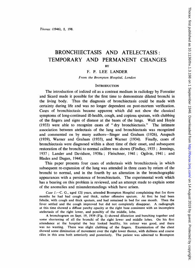
Case 1.-C. G., aged 123 years, attended Brompton Hospital complaining that for threemonths he had had cough and thick, rather offensive sputum. At first he had beenfebrile, with cough and thick sputum, and had remained in bed for one month. Then thefever settled and the cough improved but did not completely disappear. A radiographat this time showed a diffuse patchy opacity at the right base consistent with an incompleteatelectasis of the right lower, and possibly of the middle, lobe.
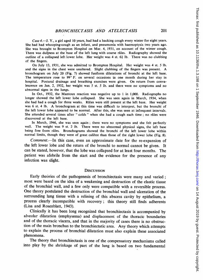
A bronchogram on Sept. 19, 1939 (Fig. 1) showed dilatation and bunching together andsome shortening of all the bronchi of the right lower and middle lobes. On his firstattendance at the hospital the boy looked healthy, his colour was good, and therewas no wasting. There was slight clubbing of the fingers. Examination of the chestshowed some diminution of movement over the right lower thorax, with dullness and coarseriles in this area both anteriorly and posteriorly. The patient was admitted to Brompton
on 14 August 2019 by guest. P
rotected by copyright.http://thorax.bm
j.com/
Thorax: first published as 10.1136/thx.1.3.198 on 1 S
eptember 1946. D
ownloaded from
BRONCHIECTASIS AND ATELECTASIS
Hospital. On admission, temperature was 98.6° F., pulse and respiration rates were normal,and sputum amounted to 1 oz. daily. Blood-count was within normal limits. A radio-graph showed a condition identical with that previously described. Throughout his sixweeks' stay in hospital he was afebrile except for two very short spells of three days each,when his temperature rose to 99° F., and on each occasion his sputum increased from atrace to 1 oz. daily. He was treated by breathing exercises and postural drainage.
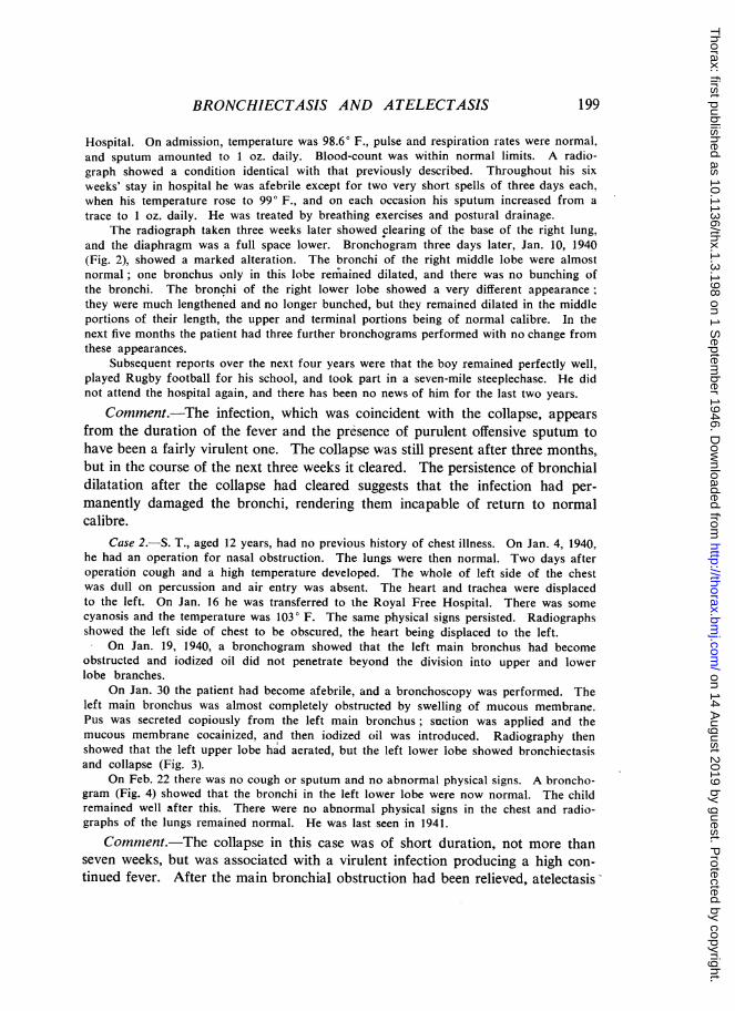
The radiograph taken three weeks later showed clearing of the base of the right lung,and the diaphragm was a full space lower. Bronchogram three days later, Jan. 10, 1940(Fig. 2), showed a marked alteration. The bronchi of the right middle lobe were almostnormal; one bronchus only in this lobe remained dilated, and there was no bunching ofthe bronchi. The bronchi of the right lower lobe showed a very different appearance;they were much lengthened and no longer bunched, but they remained dilated in the middleportions of their length, the upper and terminal portions being of normal calibre. In thenext five months the patient had three further bronchograms performed with no change fromthese appearances.
Subsequent reports over the next four years were that the boy remained perfectly well,played Rugby football for his school, and took part in a seven-mile steeplechase. He didnot attend the hospital again, and there has been no news of him for the last two years.
Comment.-The infection, which was coincident with the collapse, appearsfrom the duration of the fever and the presence of purulent offensive sputum tohave been a fairly virulent one. The collapse was still present after three months,but in the course of the next three weeks it cleared. The persistence of bronchialdilatation after the collapse had cleared suggests that the infection had per-manently damaged the bronchi, rendering them incapable of return to normalcalibre.
Case 2.-S. T., aged 12 years, had no previous history of chest illness. On Jan. 4, 1940,he had an operation for nasal obstruction. The lungs were then normal. Two days afteroperation cough and a high temperature developed. The whole of left side of the chestwas dull on percussion and air entry was absent. The heart and trachea were displacedto the left. On Jan. 16 he was transferred to the Royal Free Hospital. There was somecyanosis and the temperature was 103° F. The same physical signs persisted. Radiographsshowed the left side of chest to be obscured, the heart being displaced to the left.
On Jan. 19, 1940, a bronchogram showed that the left main bronchus had becomeobstructed and iodized oil did not penetrate beyond the division into upper and lowerlobe branches.
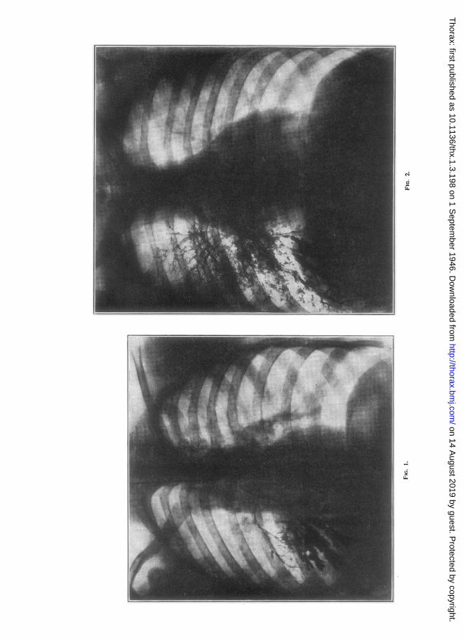
On Jan. 30 the patient had become afebrile, and a bronchoscopy was performed. Theleft main bronchus was almost completely obstructed by swelling of mucous membrane.Pus was secreted copiously from the left main bronchus; suction was applied and themucous membrane cocainized, and then iodized oil was introduced. Radiography thenshowed that the left upper lobe had aerated, but the left lower lobe showed bronchiectasisand collapse (Fig. 3).
On Feb. 22 there was no cough or sputum and no abnormal physical signs. A broncho-gram (Fig. 4) showed that the bronchi in the left lower lobe were now normal. The childremained well after this. There were no abnormal physical signs in the chest and radio-graphs of the lungs remained normal. He was last seen in 1941.
Comment.-The collapse in this case was of short duration, not more thanseven weeks, but was associated with a virulent infection producing a high con-tinued fever. After the main bronchial obstruction had been relieved, atelectasis
199
on 14 August 2019 by guest. P
rotected by copyright.http://thorax.bm
j.com/
Thorax: first published as 10.1136/thx.1.3.198 on 1 S
eptember 1946. D
ownloaded from
F. P. LEE LANDER
persisted for a time in the lower lobe. At this time the bronchi in this atelectaticlobe were shown to be grossly dilated. After the lung had become completelyre-aerated, the bronchi returned to normal calibre, without evidence of residualchanges.
Case 3.-A. A., a soldier aged 26 years, was admitted to a military hospital in June,1943, with a temperature of 102° F., pulse-rate 100, respiration-rate 28 per minute. He hadbeen feeling ill for one week with pyrexia, headache, and malaise. On admission he wascomplaining of pain in his right chest, cough, ;tnd sputum. There was dullness at the baseof the right lung with absent air entry. Sputum examination showed pneumococci andstreptococci. No tubercle bacilli were found. Leucocyte count was 14,000 per c.mm.Radiography showed an opacity at right base. He was treated with sulphapyridine (amountunknown) and the temperature subsided.
On June 23, 1943, temperature, pulse, and respiration were normal. Cough and sputumpersisted. There was dullness with loud bronchial breathing over both right lower andright middle lobes with many riles.
On July 2, 1943, he was admitted to Brompton Hospital complaining of cough, worseon lying down, with about two drachms of inoffensive, thick green sputum daily. Thefingers were not clubbed. There was dullness over the base of the right lung anteriorlyand posteriorly, with bronchial breathing and riles. Radiograph showed a high rightdiaphragm, displacement of the heart and mediastinum to the right, and an opacity at theright base suggesting collapse. Leucocyte count was 14,000 per c.mm., with 70%polymorphs.
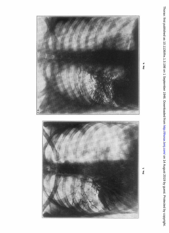
On July 7, 1943, a bronchogram (Fig. 5) showed the bronchi of the right lower andmiddle lobes to be dilated.
On July 17, 1943, a bronchoscopy was performed. The right main bronchus was inflamedand contained mucopurulent secretion. The middle lobe orifice was larger than normal,and the bronchus contained mucopus. The left bronchial tree was normal.
It was then decided to remove the middle and lower lobes on the right side, and asa preliminary measure an artificial pneumothorax was established and silver nitrate wasintroduced into the pleural cavity. The intrapleural pressures were -16-8, changingafter 250 c.cm. air had been introduced to -8-4.
On Aug. 18, 1943, a radiograph showed clearing of the shadow at the right base, underthe pneumothorax, though some atelectasis was still present. The artificial pneumothoraxwas maintained for two months, and a further injection of silver nitrate was given on Sept. 20.During this period the patient was afebrile except for the days immediately following theinjections. His symptoms had subsided, and he had only a trace of sputum.
On Sept. 17 radiography showed that the opacity at the right base had completelydisappeared, and in November a further bronchogram (Fig. 6) was performed, and all thepreviously dilated bronchi were shown to be normal.
Comment.-The collapse was attended by a severe infection, as shown by thecontinued fever and leucocytosis. It was present from June 1, possibly a few daysbefore, until some time between August 18 and September 17-that is, betweeneleven and fifteen weeks. A pneumothorax had been established seven weeksafter the onset of the collapse. The part played by this measure in the re-aerationof the lung and the prevention of permanent bronchiectasis is arguable. It isfeasible, however, that a reduction of the negative intrapleural pressure producedby the pneumothorax may have loosened the tug on the small obstructions inthe finer bronchi and allowed their ejection and consequent aeration of the lung.
200
on 14 August 2019 by guest. P
rotected by copyright.http://thorax.bm
j.com/
Thorax: first published as 10.1136/thx.1.3.198 on 1 S
eptember 1946. D
ownloaded from
BRONCHIECTASIS AND ATELECTASIS
Case 4.-J. Y., a girl aged 10 years, had had a hacking cough every winter for eight years.She had had whooping-cough as an infant, and pneumonia with haemoptysis two years ago.She was brought to Brompton Hospital on Mar. 4, 1931, on account of the winter cough.There was dullness at the-base of the left lung with coarse rales. Radiography showed theoutline of a collapsed left lower lobe. Her weight was 4 st. 61 lb. There was no clubbingof the fingers.
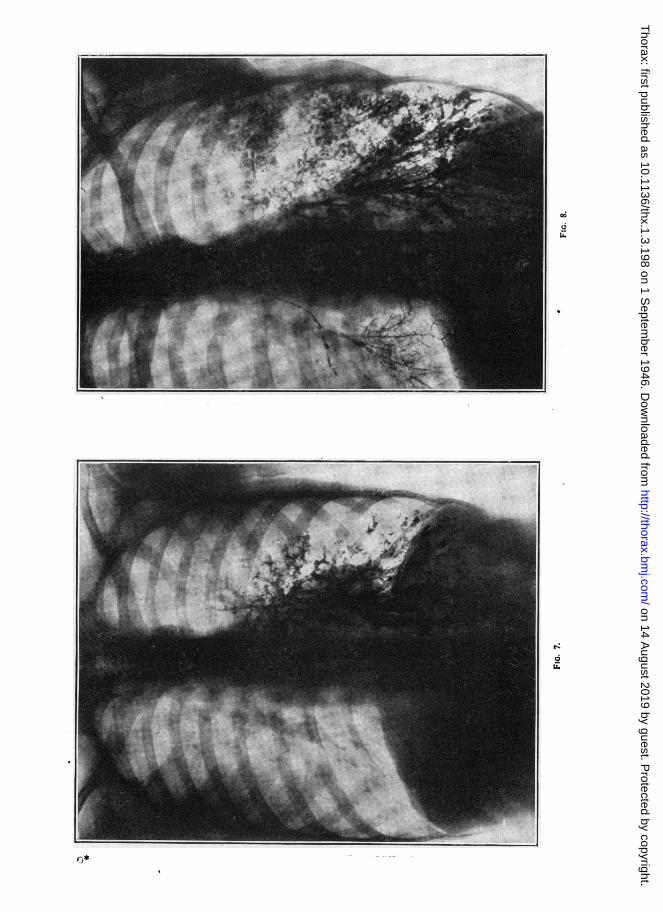
On July 13, 1931, she was admitted to Brompton Hospital. Her weight was 4 st. 5 lb.and the signs in the chest were unaltered. Slight clubbing of the fingers was present. Abronchogram on July 20 (Fig. 7) showed fusiform dilatations of bronchi at the left base.The temperature rose to 99° F. on several occasions in one month during her stay inhospital. Postural drainage and breathing exercises were given. On return from conva-lescence on Jan. 2, 1932, her weight was 5 st. 5 lb. and there were no symptoms and noabnormal signs in the lungs.
In Oct., 1932, the Mantoux reaction was negative up to 1 in 1,000. Radiographs nolonger showed the left lower lobe collapsed. She was seen again in March, 1934, whenshe had had a cough for three weeks. Ralles were still present at the left base. Her weightwas 6 st. 4 lb. A bronchogram at this time was difficult to interpret, but the bronchi ofthe left lower lobe appeared to be normal. After this, she was seen at infrequent intervals.She attended several times after " colds " when she had a cough each time; no r'ales werediscovered at the left base.
In March, 1946, she was seen again; there were no symptoms and she felt perfectlywell. The weight was 9 st. 2 lb. There were no abnormal physical signs, the left basebeing free from rales. Bronchograms showed the bronchi of the left lower lobe withinnormal limits, though they were of great calibre than those of the right lower lobe (Fig. 8).
Comment.-In this case, even an approximate date for the re-expansion ofthe left lower lobe and the return of the bronchi to normal cannot be given. Itcan be stated, however, that the lobe was collapsed for at least four months. Thepatient was afebrile from the start and the evidence for the presence of anyinfection was slight.
DISCUSSIONEarly theories of the pathogenesis of bronchiectasis were many and varied;
most were based on the idea of a weakening and destruction of the elastic tissueof the bronchial wall, and a few only were compatible with a reversible process.One theory postulated the destruction of the bronchial wall and ulceration of thesurrounding lung tissue with a relining of this abscess cavity by epithelium, aprocess clearly incompatible with recovery; this theory still finds adherents(Lisa and Rosenblatt, 1943).
Clinically it has been long recognized that bronchiectasis is accompanied byalveolar dilatation (emphysema) and displacement of the thoracic boundariesand of the thoracic viscera, and that in the majority of cases there is no obstruc-tion of the main bronchus to the bronchiectatic area. Any theory which attemptsto explain the process of bronchial dilatation must also explain these associatedphenomena.
The theory that bronchiectasis is one of the compensatory mechanisms calledinto play by the shrinkage of part of the lung is based on two fundamental
201
on 14 August 2019 by guest. P
rotected by copyright.http://thorax.bm
j.com/
Thorax: first published as 10.1136/thx.1.3.198 on 1 S
eptember 1946. D
ownloaded from
cki&
on 14 August 2019 by guest. P
rotected by copyright.http://thorax.bm
j.com/
Thorax: first published as 10.1136/thx.1.3.198 on 1 S
eptember 1946. D
ownloaded from
IL
0
on 14 August 2019 by guest. P
rotected by copyright.http://thorax.bm
j.com/
Thorax: first published as 10.1136/thx.1.3.198 on 1 S
eptember 1946. D
ownloaded from
Lz~
on 14 August 2019 by guest. P
rotected by copyright.http://thorax.bm
j.com/
Thorax: first published as 10.1136/thx.1.3.198 on 1 S
eptember 1946. D
ownloaded from
(6
6
Lz6
on 14 August 2019 by guest. P
rotected by copyright.http://thorax.bm
j.com/
Thorax: first published as 10.1136/thx.1.3.198 on 1 S
eptember 1946. D
ownloaded from
F. P. LEE LANDER
concepts. Firstly, that the shrinkage of the lung is occurring in a closed cavity,and consequently compensation for this shrinkage has to occur (i) by a decrease involume of the closed cavity and (ii) by an increase in volume of the unaffectedportion of the lung. Secondly, the " shrinking portion " of the lung consists ofall the air-containing portion of the lung (bronchi and alveoli) distal to theobstruction.
Experimental studies on the relation between bronchiectasis and atelectasis
Much experimental work on the aetiology and pathogenesis of bronchiectasishas been done in recent years, some of it vitiated by failure to take into accountthe considerations outlined above. For instance, the demonstration of earlybronchiectasis in experimental animals has been attempted from post-mortemstudies and not by iodized oil injection on the living animal.
In studies on the relation between atelectasis produced by bronchial obstruc-tion and bronchiectasis, the type of obstruction and its position in the bronchialtree has varied in the different experiments. These studies fall into two groups;those in which the main bronchus was permanently obstructed, and those inwhich the obstruction was finally distal to the main bronchus.
Studies in which the main bronchus was permanently obstructed
Adams and Escudero (1938), and Tannenberg and Pinner (1942), obstructedthe main bronchus by means of a lead shot or ligation of the bronchus, bothmethods producing an immobile obstruction.
In an obstructed system, air distal to the obstruction is absorbed by thepulmonary circulation and cannot be replaced from any outside source. As theair is absorbed, the pressure in that system drops. If the system were a per-fectly rigid one, the pressure in it would drop until the same level of gaseouspartial pressures was reached as existed in the pulmonary circulation, and thenno more absorption of air would occur. The broncho-pulmonary system, how-ever, is an elastic one; and as the air in an obstructed bronchus is absorbedand the pressure tends to fall, the elasticity of the bronchial walls allows of theircoming together, and this shrinkage in volume maintains the intrabronchialpressure at a high level. This process goes on until all the air is absorbed andthe bronchial and alveolar walls are in apposition. At the same time as thebronchial walls are collapsing, the mediastinum is moving over to the affectedside, the diaphragm is rising, and the unaffected homolateral lung to a largerextent, and the contralateral lung to a lesser extent, are becoming emphysematous.
If, however, the vanishing air is replaced by bronchial secretion, then threethings can happen, dependent on the volume of the replacing secretion. Eitherthe bronchi will remain their usual size (in the unlikely event of the secretionexactly replacing the air), or the bronchi will b2 larger than normal (more
206
on 14 August 2019 by guest. P
rotected by copyright.http://thorax.bm
j.com/
Thorax: first published as 10.1136/thx.1.3.198 on 1 S
eptember 1946. D
ownloaded from
BRONCHIECTASIS AND ATELECTASIS
replacing secretion than vanishing air), or the bronchi will be slightly smaller(less replacing secretion than vanishing air).
Tannenberg and Pinner demonstrated in their uninfected animals with mainbronchial obstruction that the bronchi distal to the obstruction were completelycollapsed, with their walls in apposition. In infected cases they demonstratedbronchial dilatations filled with bronchial secretion distal to the obstruction.
Studies in which the main bronchus was not permanently obstructed
In peripheral bronchial obstruction, similar arguments apply up to a point,but there is a fundamental difference between this condition and main bronchialobstruction; the air in the larger bronchi is proximal to the obstruction andoutside the atelectatic system, and consequently the bronchi will behavedifferently.
The air distal to the obstruction, i.e., in the bronchioli and alveoli, is absorbed;and with this absorption the alveoli and bronchioli contract until finally they arecompletely or almost completely airless. The bronchi, however, are proximalto the obstruction and are in communication with the outside air; thus they arefree to take part in the compensatory measures. That the uninfected bronchus ishighly elastic and is capable of complete obliteration with its walls in appositionhas been shown by Tannenberg and Pinner's experiment; and, just as thebronchus is capable of complete obliteration, it is also capable of considerableexpansion.
In this state the bronchi are dilated in order to occupy space left by the con-tracting lung. If the thorax is opened and the affected portion of lung or wholelung is removed, then the balance of tensions is upset and the dilated bronchi andthe dilated alveoli, if still elastic, can assume their usual extra-thoracic size. If,however, the collapse has been long-standing or infection has led to destructionand fibrosis and has fixed the size of the bronchi, then the dilatation will still bedemonstrable post-mortem. Likewise, if the dilatation is fixed by the presenceof secretion inside the bronchus behind a permanent main bronchial obstruction,as in Tannenberg and Pinner's experiments, then this dilatation will still persistafter removal from the thorax. Adams and Escudero (1938), by incompletelyobstructing main bronchi and adding infection, produced a large flow of bronchialsecretion. This thick secretion completed the obstruction and could not escape,and with the resultant atelectasis it was sucked down the bronchial tree andat length obstructed the finer bronchi. The state of affairs that then existed wasperipheral bronchial obstruction with a patent main bronchus.
Lander and Davidson (1 938a) introduced a mobile obstruction, in the formn ofviscid gum acacia, into the main bronchi of cats. This plug, as a result of theensuing atelectasis, was sucked down the bronchial tree, split at each bronchialdivision, and obstructed every branch until finally the obstruction came to rest
207
on 14 August 2019 by guest. P
rotected by copyright.http://thorax.bm
j.com/
Thorax: first published as 10.1136/thx.1.3.198 on 1 S
eptember 1946. D
ownloaded from
F. P. LEE LANDER
in the finer bronchi. Iodized oil was then introduced, and radiography showeddilatation of the bronchi in the atelectatic lobes.
Respiratory changes in calibre of bronchiMacklin (1929) reviewed the literature on calibre changes in normal bronchi
which occurred with respiration. The balance of experimental evidence favouredthe view that the bronchi became shorter and narrower with expiration and longerand broader with inspiration. Studies with iodized oil have, since this date,supported this view, though some observers report that the vertically disposedbronchi of the lower lobes in shortening with expiration may, at some stage ofthe expiratory phase, becomes broader (Heinbecker, 1927). All these changes,as pictured by bronchography with iodized oil, are not marked, as they show achange in diameter only; bronchoscopic methods, which record volume changes,show the respiratory changes in size of the bronchi more clearly (Ellis, 1936).
Lander and Davidson (1 938b) showed that similar though more markedchanges occurred with respiration in dilated bronchi. Their cases were selectedfrom subjects whose bronchiectasis, although of long standing, showed few signsof infection. These findings have been challenged. Greenfield (1940) publishedfive cases in which he stated that he was unable to demonstrate changes in calibreof dilated bronchi with respiration. Detailed examination of his publishedradiographs, however, does not bear out his conclusions. Dilatation is in factdemonstrated in the bronchi in which he stated it had not occurred. In threecases the bronchi are larger in inspiration, and in one case in expiration. In theone case in which no calibre change is visible, careful study of the height of thediaphragm, the angles formed by the rib crossings, and the relation of bronchi torib crossings, suggests that there was very little change in lung volume betweenthe " inspiratory " and " expiratory " bronchograms.
Further studies to demonstrate the ek' sticity of dilated bronchi were under-taken by Lander and Davidson (1938a). They showed that if air were introducedinto the pleural cavity on the affected side in cases of atelectasis, thus compensat-ing for the shrinkage of the lung, then bronchi which had been dilated assumeda more normal size. This change in calibre was, however, only temporarybecause, with the absorption of the pneumothorax, the bronchial dilatation willagain be manifest if the atelectasis persists.
Tannenberg and Pinner (1942) denied that pneumothorax prevented theoccurrence of bronchiectasis. This finding was based on their animals in whomcollapse had been produced by main bronchial obstruction. In all their animalsin which bronchiectasis was produced, infection had been introduced and thedilated bronchi were filled with secretion-a state of affairs which would notallow of regression of changes that had already occurred. The final and con-clusive proof that dilated bronchi may retain their elasticity lies in the demon-stration of the return to normal calibre of bronchi previously shown to be dilated.
208
on 14 August 2019 by guest. P
rotected by copyright.http://thorax.bm
j.com/
Thorax: first published as 10.1136/thx.1.3.198 on 1 S
eptember 1946. D
ownloaded from
BRONCHIECTASIS AND ATELECTASIS
This was first shown by Findlay (1935), and further cases have since been addedby Jennings (1937), Lander and Davidson (1938a), Ogilvie (1941), Fleischner(1941), and Blades and Dugan (1944).
That these reversible cases do not represent true bronchiectasis has been sug-gested by many authors. Hinshaw and Schmidt (1944) talk of the " illusion ofbronchiectasis which may result owing to the foreshortening and apparent widen-ing of the larger bronchi in an atelectatic lobe." Blades and Dugan (1944) dis-miss the possibility of reversible bronchial dilatation being true bronchiectasison the grounds that bronchiectasis is a chronic infective process which is pro-gressive and not reversible. Lisa and Rosenblatt (1943) reject the possibility ofreversible bronchiectasis being an early stage of infected bronchiectasis by theiradherence to the thesis that bronchial dilatations are the result of ulceration ofbronchial wall and lung tissue and relining of the resulting cavities with bronchialepithelium. They reject these reversible cases with the statement "that thediagnosis is clinical (bronchogram) and not pathological. Possibly the broncho-gram of a collapsed lobe gives the appearance of bronchiectasis without its actualoccurrence." Kornblum (1944), in discussing reversible bronchiectasis, says:" If it can be established that the condition is definite clinical bronchiectasis, thenour entire concept of the disease must be revised."
CONCLUSIONS
That there is an intimate relationship between bronchiectasis and atelectasishas been long recognized and commented on. Many theories have been formu-lated to explain this relationship, and there has been much argument as to thenature of the actual dilating force. Andrus (1937), in a detailed examination ofall the physical forces, came to the following conclusions: " These forces (arisingas a result of pulmonary atelectasis) provide much the most satisfactory explana-tion of bronchiectasis "; and " both infection and injury to the bronchial walland an abnormal intensity of mechanical dilating stress are customarily necessaryfor the production of bronchial dilatation."
Lander and Davidson (1938a), as the result of experimental work carried outon animals and human subjects, came to the conclusion that infection was notnecessary for the production of bronchial dilatation and that the dilating forcesconsequent on atelectasis were sufficient of themselves. The fact that mosthuman cases had a coincidental infection was not denied. The theory thatbronchial dilatation is one of the compensatory mechanisms consequent onpulmonary collapse was advanced and supported by experimental observation.Bronchial dilatation (bronchiectasis) and alveolar dilatation (emphysema) werethus regarded as part of the same process.
It is suggested that all cases of bronchiectasis are, at an early stage of theircareer, capable -of reversion to normal. In the majority of cases in which
209
on 14 August 2019 by guest. P
rotected by copyright.http://thorax.bm
j.com/
Thorax: first published as 10.1136/thx.1.3.198 on 1 S
eptember 1946. D
ownloaded from
F. P. LEE LANDER
bronchiectasis is permanent, this reversion to normal is prevented by a failure ofthe lung to expand; in a minority of cases the bronchiectasis is maintained, inspite of re-expansion of the lung, by a permanent damage to the bronchial wall.
SUMMARYFour cases of atelectasis with bronchiectasis are described. In all four cases
re-aeration and re-expansion of the collapsed portion of the lung occurred. Inthree. this was accompanied by regression of the bronchiectasis with return tonormal. In the fourth case, the bronchiectasis persisted. The reason for thepersistence of the dilatation in the one case is discussed, and the suggestion ismade that in this case infection had rendered the dilated bronchi incapableof returning to normal calibre; this may supply a link between reversiblebronchiectasis and permanent dilatation. That this is not the usual way in whichpermanent dilatation is brought about is suggested by a detailed study of casesof bronchiectasis. In most of them persistent atelectasis with attendant crowdingtogether of bronchi accompanies the bronchial dilatation. Damage by infection,as suggested in this case, however, may explain the dilatation that occasionallyoccurs in cases in which no crowding together of dilated bronchi is seen.
The experimental work on the aetiology of bronchiectasis is reviewed.It is concluded that bronchiectasis occurs as a compensatory phenomenon
when atelectasis takes place in a semi-rigid compartment, the thorax, and becausein many cases of atelectasis the bronchi are obstructed peripherally and notcentrally.
I wish to acknowledge the kindness of Dr. Geoffrey Marshall and Dr. J. G. Scaddingin permitting publication of two of their cases.
REFERENCESAdams, W .E., and Escudero, L. (1938). Tubercle, 19, 351.Andrus, P. M. (1937). Amer. Rev. Tuberc., 36, 46.Anspach, W. E. (1939). Amer. J. Roentgen, 41, 173.Blades, B., and Dugan, D. J. (1944). J. Thorac. Surg., 13, 40.Ellis, M. (1936). Proc. roy. Soc. Med., 29, 527.Findlay, L. (1935). Arch. Dis. Childh., 10, 61.Fleischner, F. G. (1941). Amer. J. Roentgen, 46, 166.Greenfield, J. (1940). J. Clin. Invest., 19, 723.Heinbecker, P. (1927). J. Clin. Invest., 4, 459.Hinshaw, H. C., and Schmidt, H. W. (1944). Dis. Chest., 10, 115.Jennings, G. H. (1937). Brit. med. J., 2, 963.Kornblum, K. (1944). Amer. J. Roentgen, 51, 292.Lander, F. P. L., and Davidson, M. (1938a). Brit. J. Radiol., 11, 65.
(1938b). Brit. med. J., 1, 1,047.Lisa, J. R., and Rosenblatt, M. B. (1943). Bronchiectasis. Oxford University Press.Macklin, C. C. (1929). Physiol. Rev., 9, 1.Ogilvie, A. G. (1941). Arch. intern. Med., 68, 395.Singer, J. J., and Graham, E. A. (1926). Amer. J. Roentgen, 15, 54.Tannenberg, J.. and Pinner, M. (1942). J. Thorac. Surg., 11, 571.Wall, C., and Hoyle, J. C. (1933). Brit. med. J., 1, 597.Warner, W. P., and Graham, D. (1933). Arch. intern. Med., 52, 888.Warner, W. P. (1934). Quart. J. Med., 3, 401.
210
on 14 August 2019 by guest. P
rotected by copyright.http://thorax.bm
j.com/
Thorax: first published as 10.1136/thx.1.3.198 on 1 S
eptember 1946. D
ownloaded from