Embed Size (px)
Citation preview

Bronchopulmonary Dysplasia
John Salyer RRT-NPS, MBA, FAARC
Director Respiratory Therapy
Seattle Children’s Hospital and Research Institute
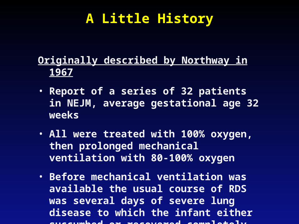
A Little History
Originally described by Northway in 1967
• Report of a series of 32 patients in NEJM, average gestational age 32 weeks
• All were treated with 100% oxygen, then prolonged mechanical ventilation with 80-100% oxygen
• Before mechanical ventilation was available the usual course of RDS was several days of severe lung disease to which the infant either succumbed or recovered completely in 7-10 days
History (cont.)
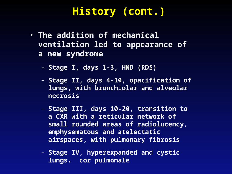
• The addition of mechanical ventilation led to appearance of a new syndrome
– Stage I, days 1-3, HMD (RDS)
– Stage II, days 4-10, opacification of lungs, with bronchiolar and alveolar necrosis
– Stage III, days 10-20, transition to a CXR with a reticular network of small rounded areas of radiolucency, emphysematous and atelectatic airspaces, with pulmonary fibrosis
– Stage IV, hyperexpanded and cystic lungs. cor pulmonale
Definition
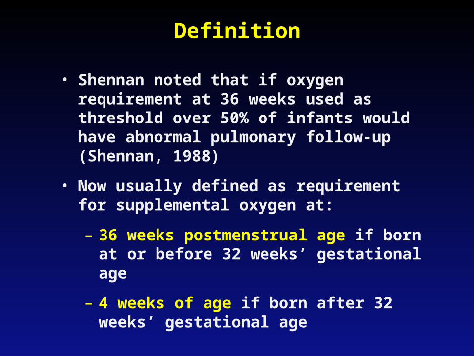
• Shennan noted that if oxygen requirement at 36 weeks used as threshold over 50% of infants would have abnormal pulmonary follow-up (Shennan, 1988)
• Now usually defined as requirement for supplemental oxygen at:
– 36 weeks postmenstrual age if born at or before 32 weeks’ gestational age
– 4 weeks of age if born after 32 weeks’ gestational age
Physiologic Test
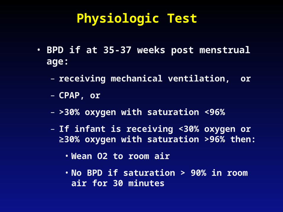
• BPD if at 35-37 weeks post menstrual age:
– receiving mechanical ventilation, or
– CPAP, or
– >30% oxygen with saturation <96%
– If infant is receiving <30% oxygen or ≥30% oxygen with saturation >96% then:
• Wean O2 to room air
• No BPD if saturation > 90% in room air for 30 minutes
Pathology Classic BPD
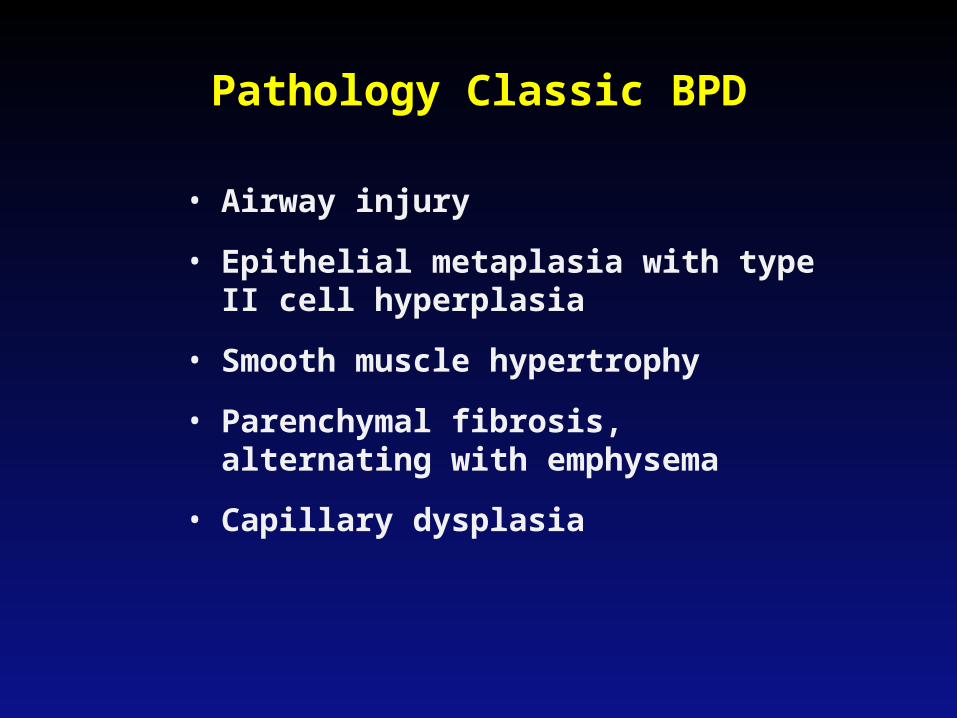
• Airway injury
• Epithelial metaplasia with type II cell hyperplasia
• Smooth muscle hypertrophy
• Parenchymal fibrosis, alternating with emphysema
• Capillary dysplasia
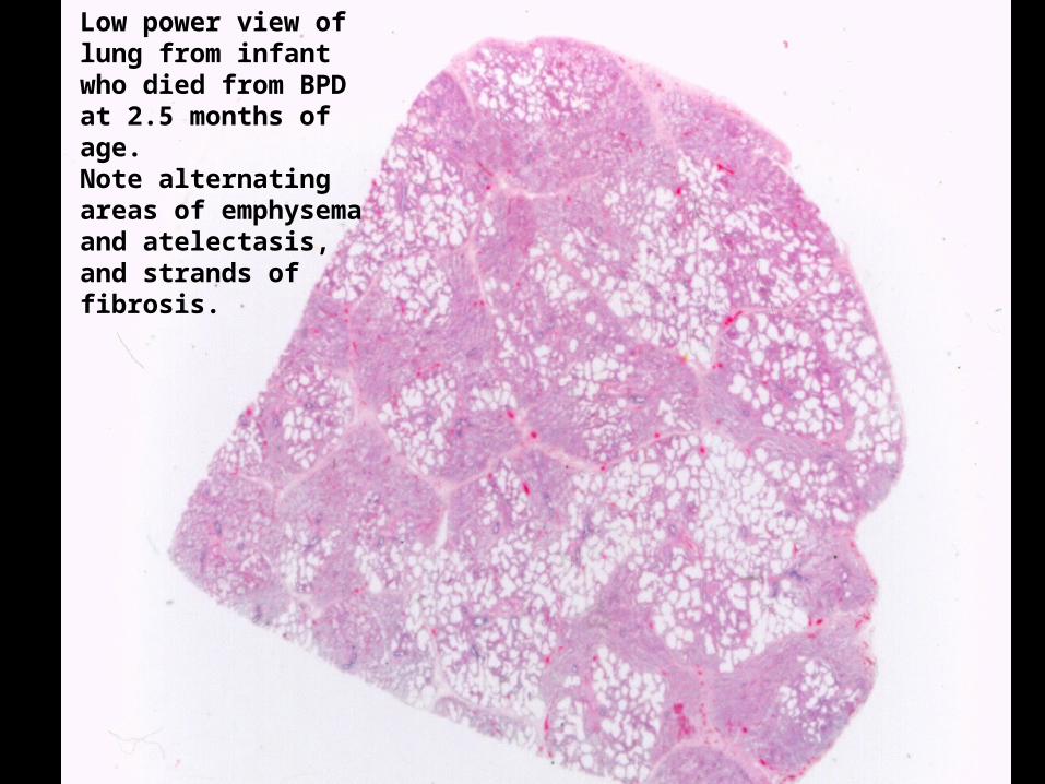
Low power view of lung from infant who died from BPD at 2.5 months of age. Note alternating areas of emphysema and atelectasis, and strands of fibrosis.
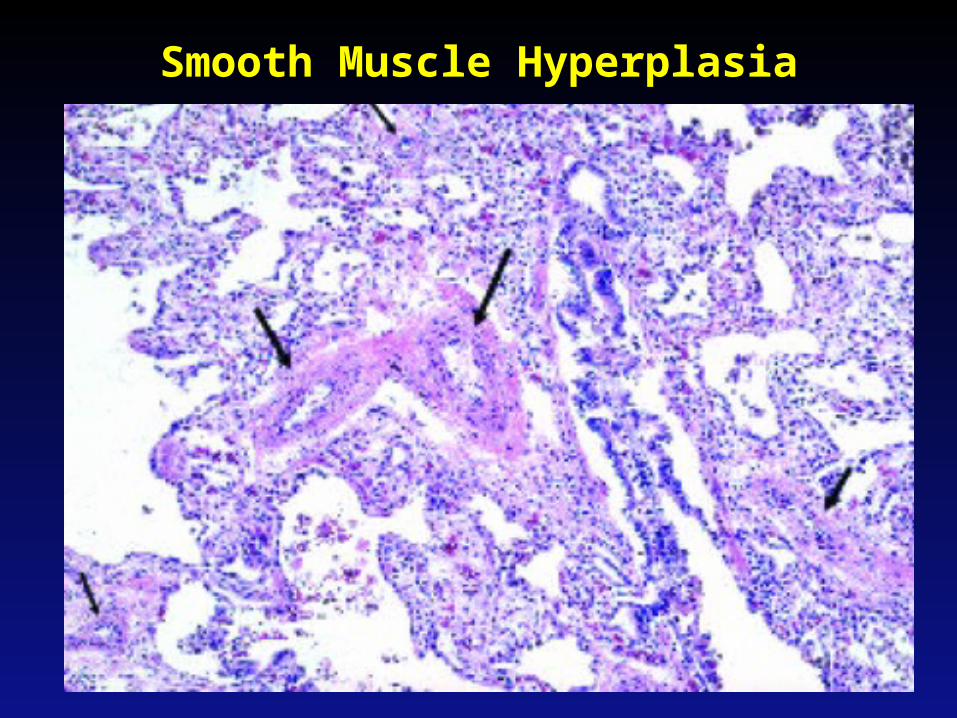
Smooth Muscle Hyperplasia
Albertine et al., Am J Respir Crit Care. 2008
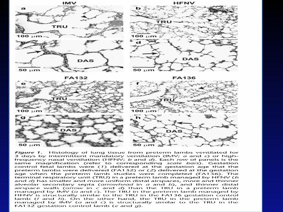
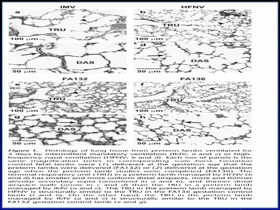
Histology of BPD
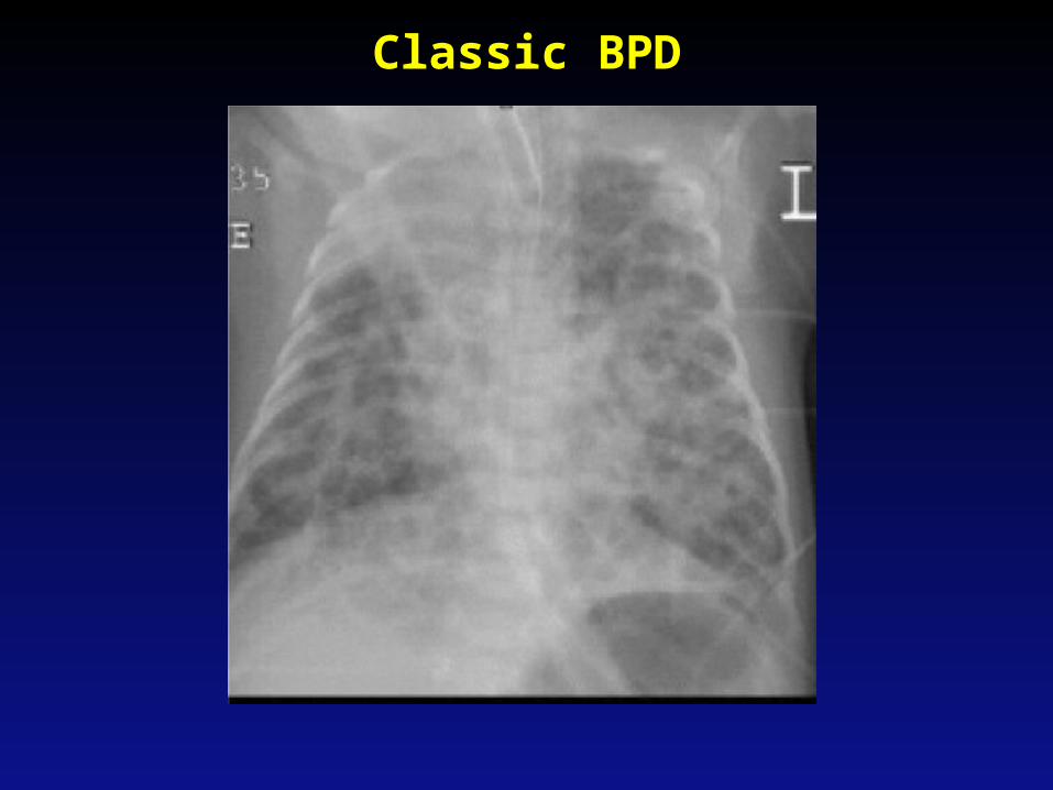
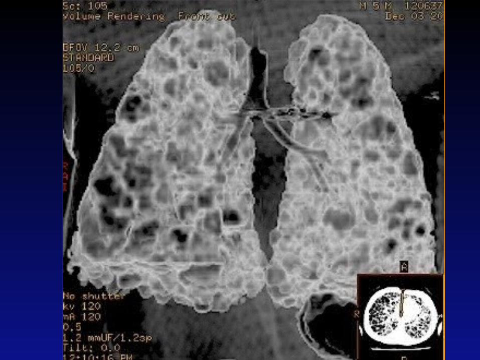
Classic BPD
Classic vs. New BPD• Much of the histologic findings of “old
BPD” seen in animal models with oxygen toxicity
• New epidemiology
– Most common in ELBW infants
– Risk factors: low gestational age, postnatal infection, maternal chorioamnionitis, PDA, mechanical ventilation
– Some ELBW infants do not have RDS, with minimal early oxygen or mechanical ventilation
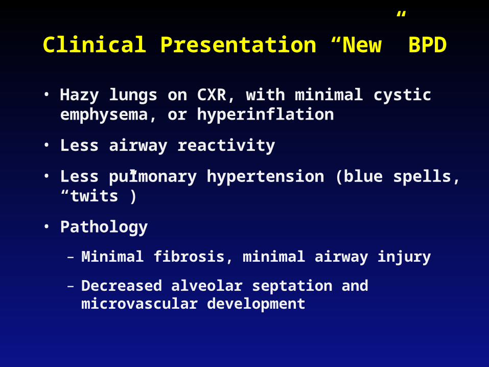
Clinical Presentation “New” BPD
• Hazy lungs on CXR, with minimal cystic emphysema, or hyperinflation
• Less airway reactivity
• Less pulmonary hypertension (blue spells, “twits”)
• Pathology
– Minimal fibrosis, minimal airway injury
– Decreased alveolar septation and microvascular development
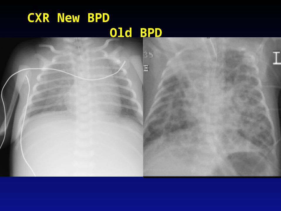
CXR New BPD Old BPD
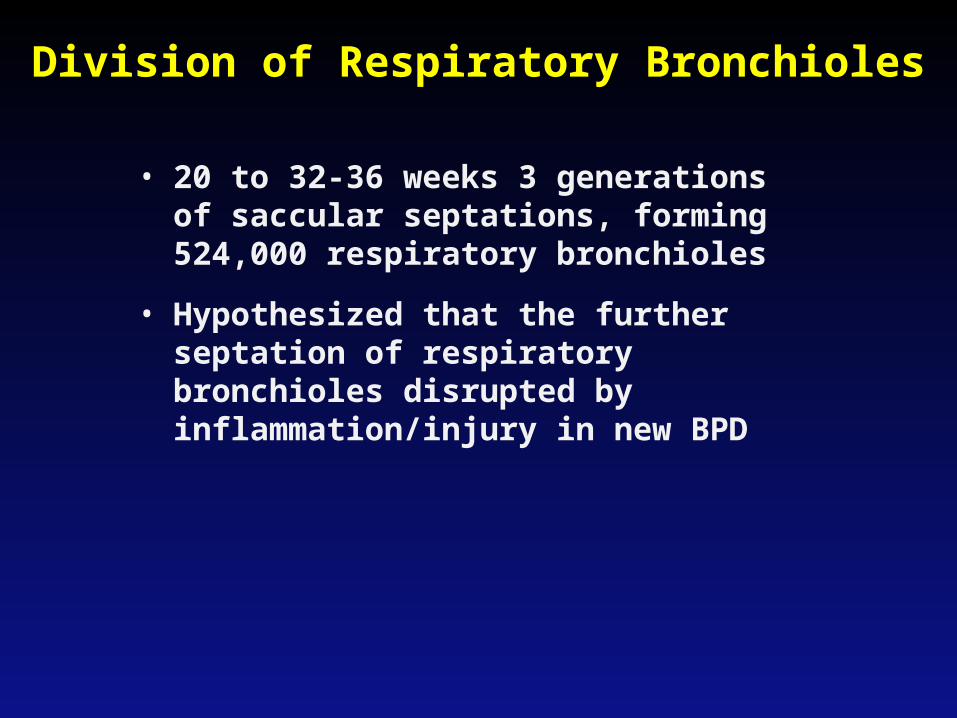
Division of Respiratory Bronchioles
• 20 to 32-36 weeks 3 generations of saccular septations, forming 524,000 respiratory bronchioles
• Hypothesized that the further septation of respiratory bronchioles disrupted by inflammation/injury in new BPD
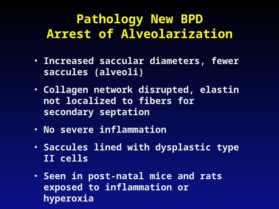
Pathology New BPDArrest of Alveolarization
• Increased saccular diameters, fewer saccules (alveoli)
• Collagen network disrupted, elastin not localized to fibers for secondary septation
• No severe inflammation
• Saccules lined with dysplastic type II cells
• Seen in post-natal mice and rats exposed to inflammation or hyperoxia
Pathogenesis “New” BPD:Overview
• Immaturity of structure and function before about 32 weeks’ gestation
– Poorly developed airway supporting structures
– Surfactant deficiency
– Decreased compliance
– Underdeveloped antioxidant mechanisms
– Inadequate fluid clearance
• Inflammation
– Mechanical ventilation
– Oxygen toxicity
– Infection
Pathogenesis:Mechanical Injury
• Over-distension of airways and airspaces (Volutrauma)– Excessively large tidal volumes, not pressure
(Hernandez, 1989)– Association of decreased PCO2 with increased
risk BPD (Garland, 1995)– Maximum end-inspiratory volume more important
than tidal volume or FRC (Dreyfuss, 1993)– As few as 6 large manual inflations was enough to
increase the lung damage premature lambs (Bjorklund, 1997)
– Positive pressure causes bronchiolar lesions (Nilsson, 1978)
Pathogenesis:Oxygen Toxicity
• Safe FiO2 level and duration are unknown
• Cell damage from reactive oxygen metabolites– Cell enzyme inactivation
• Preterm infants may have inadequate antioxidant defenses– Nutrient deficiencies– Immature enzyme development
Pathogenesis:Infection
• Four-fold increase in BPD incidence with sepsis (Rojas, 1995)
• Due to increased concentration of proinflammatory cytokines in amniotic fluid?
• Association with Ureaplasma urealyticum (Hannaford, 1999)
• Maternal chorioamnionitis associated with decreased RDS, but increased risk of BPD
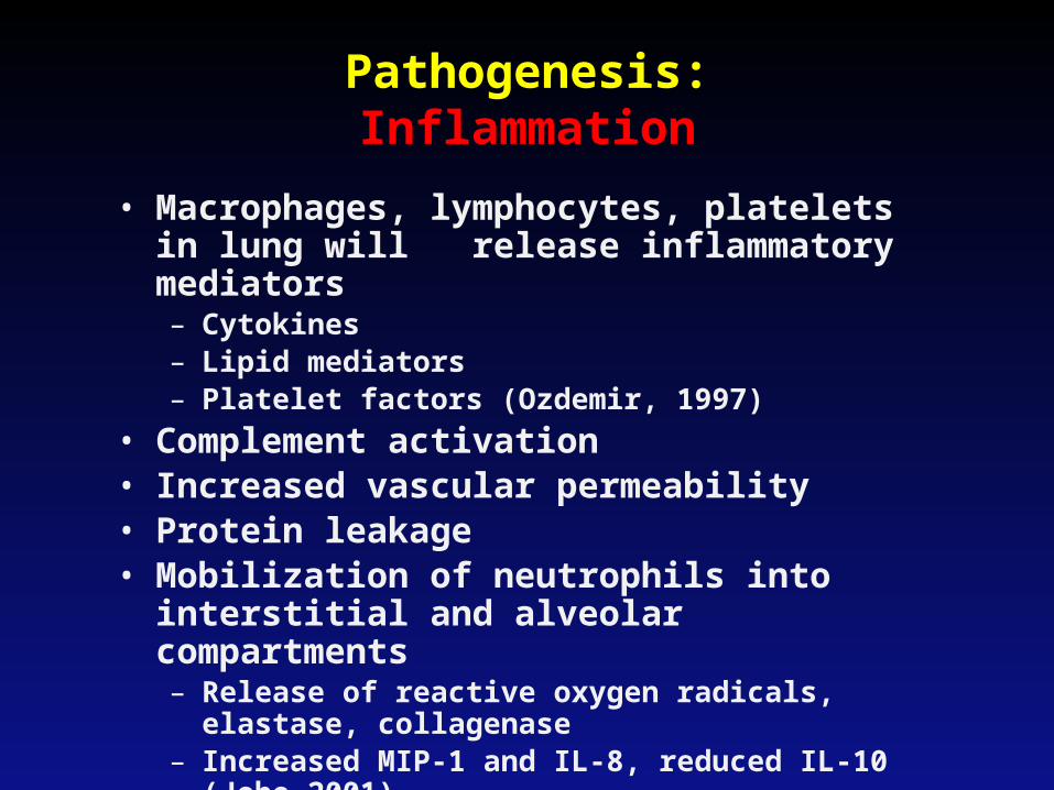
Pathogenesis:Inflammation
• Macrophages, lymphocytes, platelets in lung will release inflammatory mediators– Cytokines– Lipid mediators– Platelet factors (Ozdemir, 1997)
• Complement activation• Increased vascular permeability• Protein leakage• Mobilization of neutrophils into interstitial
and alveolar compartments– Release of reactive oxygen radicals, elastase,
collagenase– Increased MIP-1 and IL-8, reduced IL-10 (Jobe
2001)
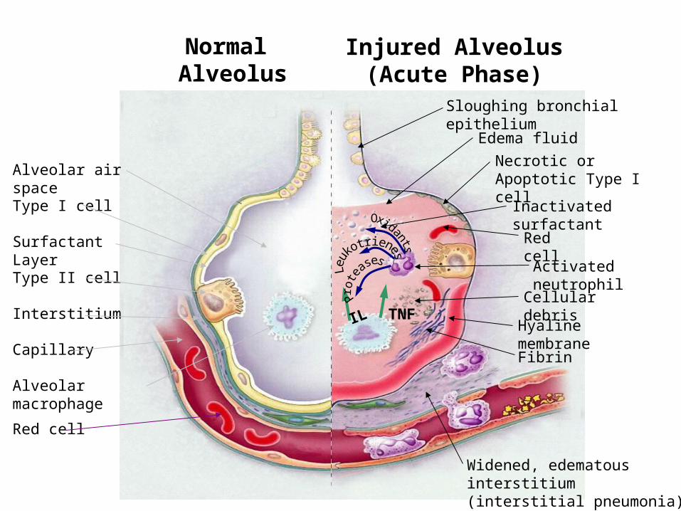
Normal Alveolus
Injured Alveolus (Acute Phase)
Alveolar air spaceType I cell
Surfactant Layer
Type II cell
Interstitium
Capillary
Alveolar macrophage
Sloughing bronchial epithelium
Edema fluid
Necrotic or Apoptotic Type I cell
Red cell
Cellular debris
Hyaline membrane
Fibrin
Widened, edematous interstitium(interstitial pneumonia)
TNFIL
Red cell
Inactivated surfactant
Activated neutrophil
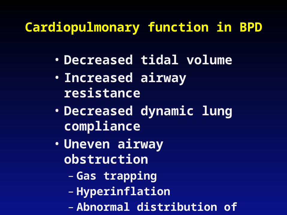
Cardiopulmonary function in BPD
• Decreased tidal volume• Increased airway resistance• Decreased dynamic lung
compliance• Uneven airway obstruction
– Gas trapping– Hyperinflation– Abnormal distribution of
ventilation
• Bronchomalacia
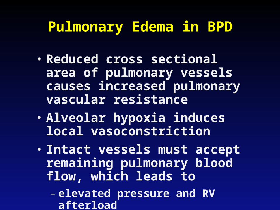
Pulmonary Edema in BPD
• Reduced cross sectional area of pulmonary vessels causes increased pulmonary vascular resistance
• Alveolar hypoxia induces local vasoconstriction
• Intact vessels must accept remaining pulmonary blood flow, which leads to– elevated pressure and RV afterload
– increased fluid filtration in interstitium
Clinical Course of BPD
• Most: gradual improvement over weeks or months
• Some: marked pulmonary instability in first few weeks followed by weeks or months of ventilator dependence
• Few: pulmonary hypertension and cor pulmonale
Prevention of BPD
• Antenatal glucocorticoid steroids - lead to decreased incidence RDS, but only modest decrease in BPD
• Nutrition – although poor growth is associated with BPD, no studies show impact of nutrition in altering risk for BPD
• Vitamin A – Slight decrease in BPD with IM supplementation in ELBW infants (Darlow, 2011), UW study showed increased sepsis with IM vit A (48.5% vs. 12%), but no decrease BPD
• Surfactant – decreases risk of pneumothorax, improves survival, decreases need for oxygen and ventilation, but no decrease in rate of BPD.
Prevention of BPD (cont.)
• Limited oxygen – SUPPORT trial with lower oxygen targets saw decreased incidence of ROP, but increased mortality and no decrease in BPD
• Permissive hypercapnea – Target of pCO2>52 vs. <48 not associated with decreased BPD in ELBW, but decreased ventilation at 36 wks (Carlo, 2002)
• HFOV – not associated with decreased BPD compared to modern conventional ventilation
Prevention of BPD (cont.)
• PDA treatment – incidence of PDA goes up with ligation (Chorne, 2007)
• Inhaled Nitric Oxide – NO-CLD trial showed decreased BPD in treatment group. Survival without BPD 43.9% vs. 36.8% (Ballard, 2006)
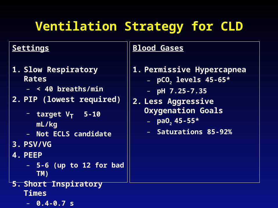
Ventilation Strategy for CLD
Settings
1. Slow Respiratory Rates– < 40 breaths/min
2. PIP (lowest required)
– target VT 5-10 mL/kg
– Not ECLS candidate
3. PSV/VG
4. PEEP – 5-6 (up to 12 for bad TM)
5. Short Inspiratory Times– 0.4-0.7 s
Blood Gases
1. Permissive Hypercapnea– pCO2 levels 45-65*
– pH 7.25-7.35
2. Less Aggressive Oxygenation Goals– paO2 45-55*
– Saturations 85-92%
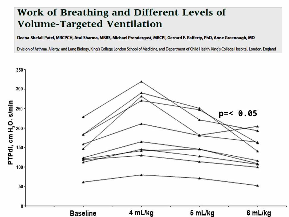
p=< 0.05
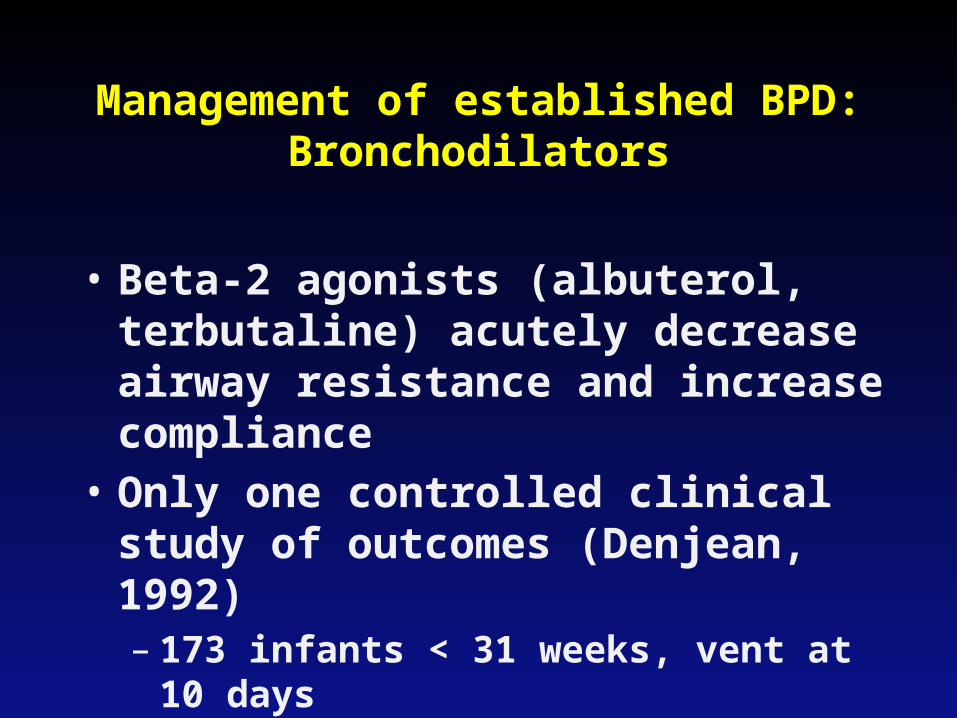
Management of established BPD:Bronchodilators
• Beta-2 agonists (albuterol, terbutaline) acutely decrease airway resistance and increase compliance
• Only one controlled clinical study of outcomes (Denjean, 1992)– 173 infants < 31 weeks, vent at 10 days– No effect on survival, severity of BPD,
duration of vent or oxygen use
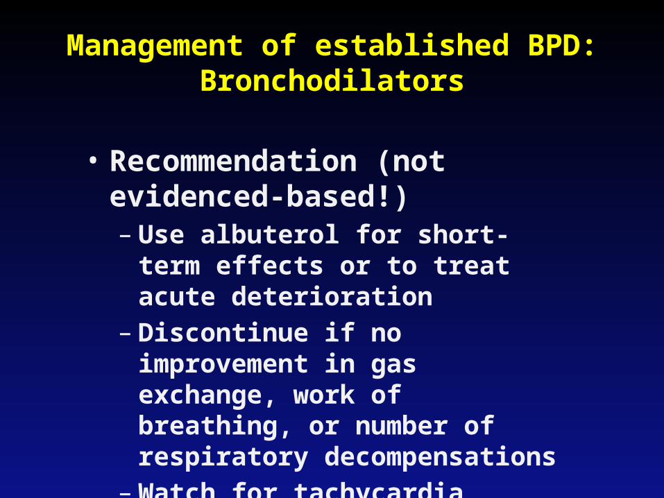
Management of established BPD:Bronchodilators
• Recommendation (not evidenced-based!)– Use albuterol for short-term
effects or to treat acute deterioration
– Discontinue if no improvement in gas exchange, work of breathing, or number of respiratory decompensations
– Watch for tachycardia, arrhythmia, hypokalemia, irritability
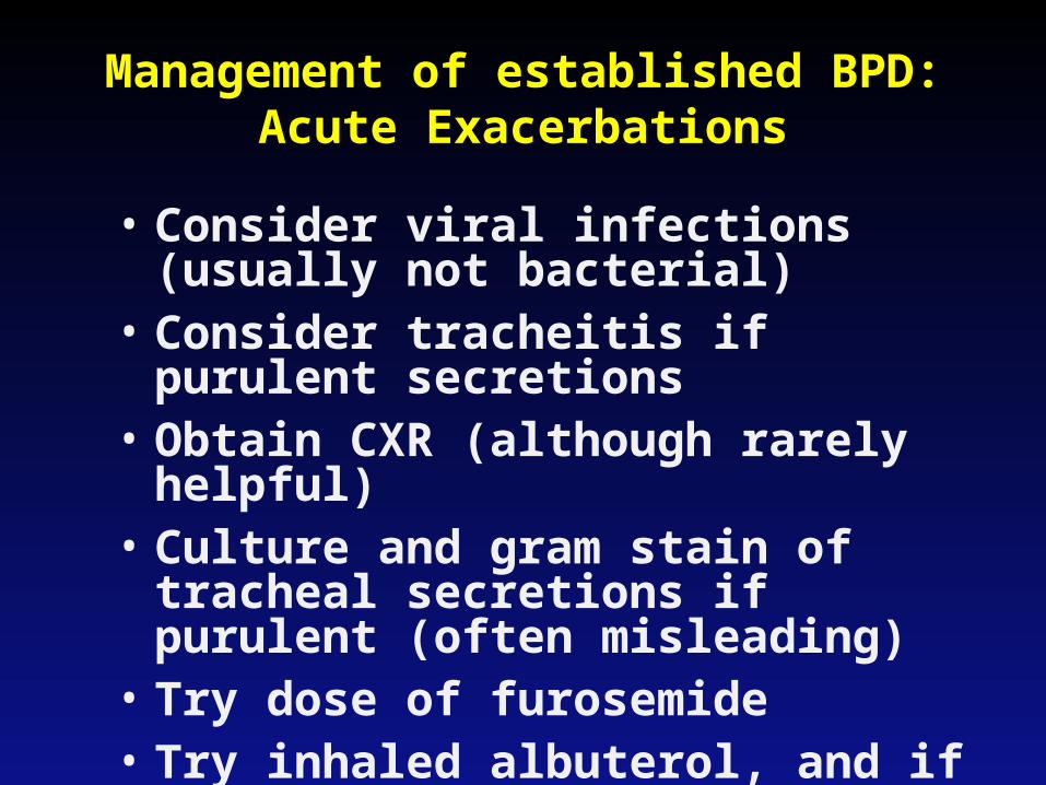
Management of established BPD:Acute Exacerbations
• Consider viral infections (usually not bacterial)
• Consider tracheitis if purulent secretions
• Obtain CXR (although rarely helpful)• Culture and gram stain of tracheal
secretions if purulent (often misleading)
• Try dose of furosemide• Try inhaled albuterol, and if poor
response, try ipratropium• Consider inhaled corticosteroids• Consider systemic corticosteroids
for 5 days, esp. if at term corrected age
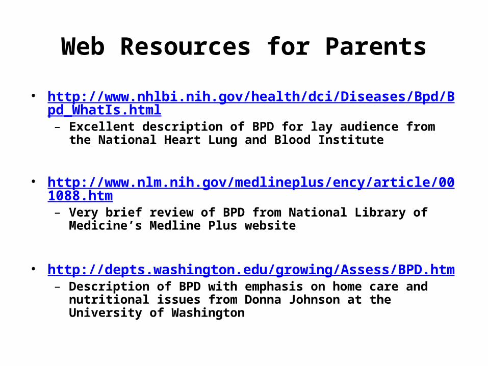
Web Resources for Parents
• http://www.nhlbi.nih.gov/health/dci/Diseases/Bpd/Bpd_WhatIs.html– Excellent description of BPD for lay audience from the National
Heart Lung and Blood Institute
• http://www.nlm.nih.gov/medlineplus/ency/article/001088.htm– Very brief review of BPD from National Library of Medicine’s
Medline Plus website
• http://depts.washington.edu/growing/Assess/BPD.htm– Description of BPD with emphasis on home care and nutritional
issues from Donna Johnson at the University of Washington

The End