Embed Size (px)
Citation preview

METHODOLOGY ARTICLE Open Access
BSSF: a fingerprint based ultrafast binding sitesimilarity search and function analysis serverBing Xiong1*†, Jie Wu2†, David L Burk3, Mengzhu Xue1, Hualiang Jiang1, Jingkang Shen1*
Abstract
Background: Genome sequencing and post-genomics projects such as structural genomics are extending thefrontier of the study of sequence-structure-function relationship of genes and their products. Although manysequence/structure-based methods have been devised with the aim of deciphering this delicate relationship, therestill remain large gaps in this fundamental problem, which continuously drives researchers to develop novelmethods to extract relevant information from sequences and structures and to infer the functions of newlyidentified genes by genomics technology.
Results: Here we present an ultrafast method, named BSSF(Binding Site Similarity & Function), which enablesresearchers to conduct similarity searches in a comprehensive three-dimensional binding site database extractedfrom PDB structures. This method utilizes a fingerprint representation of the binding site and a validated statisticalZ-score function scheme to judge the similarity between the query and database items, even if their similarities areonly constrained in a sub-pocket. This fingerprint based similarity measurement was also validated on a knownbinding site dataset by comparing with geometric hashing, which is a standard 3D similarity method. Thecomparison clearly demonstrated the utility of this ultrafast method. After conducting the database searching, thehit list is further analyzed to provide basic statistical information about the occurrences of Gene Ontology termsand Enzyme Commission numbers, which may benefit researchers by helping them to design further experimentsto study the query proteins.
Conclusions: This ultrafast web-based system will not only help researchers interested in drug design andstructural genomics to identify similar binding sites, but also assist them by providing further analysis of hit listfrom database searching.
BackgroundOver the past decade, a significant proportion of theprotein structures deposited in the Protein Data Bank(PDB) have come from the advanced high-throughputmethods of various structural genomics initiatives[1,2].Although the themes of these funded post-genomic pro-jects differ at many levels, there is a central problemwhich is: how these sequences and structures are relatedto their functions. By increasing the structural reper-toire, it is hoped that we will be able to improve ourunderstanding of fold space and how proteins evolvenew functions[3,4]. This structure-function relationship
is especially essential in the drug design field whereresearchers try to target specific proteins involved indisease mechanisms by using structure-based drugdesign methods [5-7].The number of in silico methods which can infer pro-
tein functions has grown enormously in the recentyears. They can be categorized as sequence-based andstructure-based[8]. Powerful BLAST-like sequence-search methods are able to transfer the function of awell-defined protein family to a protein with a highsequence similarity[9]. For lower sequence similarityinstances, more subtle methods such as “profile” or“hidden Markov models” can be constructed from mul-tiple sequence alignments and applied to find obscurepatterns in the protein sequences, thus assigning a func-tion to them [10-12]. All of these algorithms aboveassume that similar sequences are derived via divergent
* Correspondence: [email protected]; [email protected]† Contributed equally1State Key Laboratory of Drug Research, Shanghai Institute of MateriaMedica, Chinese Academy of Sciences, 555 Zuchongzhi Road, Zhangjiang Hi-Tech Park, Pudong, Shanghai, 201203, PR China
Xiong et al. BMC Bioinformatics 2010, 11:47http://www.biomedcentral.com/1471-2105/11/47
© 2010 Xiong et al; licensee BioMed Central Ltd. This is an Open Access article distributed under the terms of the Creative CommonsAttribution License (http://creativecommons.org/licenses/by/2.0), which permits unrestricted use, distribution, and reproduction inany medium, provided the original work is properly cited.

evolutions. However, this is limiting, as demonstrated bynumerous studies showing that evolution in structurespace is much more conserved than in sequence space[13]. This has spurred researchers to develop methodsto infer functions directly from structural information.Many structure fold/domain classification databaseshave been compiled in efforts to build a basis for suchalgorithms [14-16]. Although they have assistedresearchers in assigning functions to proteins with simi-lar folds, these fold information based methods alsohave their drawbacks, as it is shown that sequence/structure can also dynamically change due to convergentevolution[17]. This is exemplified by many proteinsinvolved in metabolism pathways, which although theydo not have any fold similarity, all process similar meta-bolites. One explanation could be that they have a simi-lar spatial arrangement of key residues in their catalyticbinding sites. Such limitation further encouragesresearchers to develop methods based on key residuesor local structural motifs.Searching for similar local spatial patterns in structure
databases is an especially challenging task, since itinvolves a large searching space and is usually time-con-suming. Given its importance both in basic biologyresearch and drug design, several algorithms have beendevised to tackle this difficult problem of finding similarfunctional sites in structures [18-24]. Among these localspatial similarity detection methods, some rely on thecurated local structure patterns, like in TESS andSPASM systems, and identify the similar local structuresby comparing these curated structure templates with thequery. Others are more flexible and able to take manystructures into account, then build the structure pat-terns during the process and detect the similar localstructures on the fly as exemplified by Hamelryck’s mul-tidimensional index tree method[25]. These methodsnot only give hints about structure evolution and pro-tein functions, but also play a significant roles in pre-dicting drug side-effects caused by ligand cross bindingto similar surface patches on various target structures.At the fundamental level, the elementary searching algo-rithms used in these methods are usually based on geo-metric hashing and graph clique detection. Due to thesignificant computational expenses of these algorithms,they typically rely on previously calculated data sets andit would be nontrivial to apply them within the wholestructure space represented by the PDB. Users can onlyvisualize and analyze results stored in a pre-compileddatabase. While as a consequence of structural genomicsresearch, there is a continuously increasing demand forperforming binding site similarity searches with userinput structures in order to find possible functions forthese binding sites as well as for the proteins in a largescale and fast way.
Here we present a new ultrafast binding site similaritysearch method along with a simple analysis of occur-rences of Gene Ontology (GO) term and Enzyme Com-mission (EC) numbers in hit list[26]. Our method wasinspired by the fingerprint and pharmacophore conceptscommonly used in chemoinformatics[27]. We first sys-tematically extracted possible binding sites from proteinstructures, mapped pharmacophore properties to thebinding sites and calculated fingerprints for later data-base searches. To enable users to identify subtle similar-ity between binding sites, a panel of fingerprintmeasurements was tested to find the optimal solution.Further, a statistics-based score method was developedto evaluate hits with the aim of eliminating false posi-tives. This novel binding site similarity search methodshould enable researchers in the structural biology fieldto examine in detail for possible binding sites in anultrafast large scale way. Also it will benefit researchersin the field of drug development by allowing them topredict and investigate possible side effects due to ligandcross binding events.
MethodsExtract possible binding sites from PDB databaseFirst, a total of 41449 protein structures were gatheredfrom the RCSB PDB (up to 2008.1)[28]. Then, a geome-try-based protein binding site detection method, namedPASS (Putative Active Sites with Spheres)[29], wasadopted to extract possible binding sites from everypolypeptide chain in the database of protein structures.This tool is able to characterize concave regions of aprotein surface and to identify positions likely to repre-sent binding sites based upon the size, shape, and burialextent of these volumes[29]. The output sphericalprobes of PASS were processed with an in-house pro-gram based on a minimum spanning tree algorithm tosplit the probes around the protein surface into clustersat least 5 Å apart. This resulted in 201,233 probe clus-ters, which were further filtered so that only probe clus-ters containing 30 to 200 probes were considered asbinding sites. These feasible binding sites were used toextract protein residues within 6 Å of these probes.These binding site probes along with the identifiedbinding site residues were stored into the database forlater visualization and fingerprint calculation. Two otherdatasets which compiled with known small molecularbinding sites were also constructed for a better evalua-tion of our method. First, the HETATM residues inPDB database were filtered such that only the oneswhich have molecular weights between 100 and 800were retained. Also, if one residue shows up for morethan 10 times in PDB database, then it will be regardedas a common molecule and discarded. Finally, 13227PDB structures were subjected as a database for later
Xiong et al. BMC Bioinformatics 2010, 11:47http://www.biomedcentral.com/1471-2105/11/47
Page 2 of 11
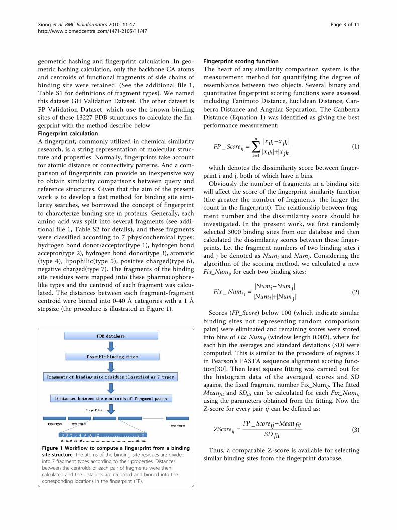
geometric hashing and fingerprint calculation. In geo-metric hashing calculation, only the backbone CA atomsand centroids of functional fragments of side chains ofbinding site were retained. (See the additional file 1,Table S1 for definitions of fragment types). We namedthis dataset GH Validation Dataset. The other dataset isFP Validation Dataset, which use the known bindingsites of these 13227 PDB structures to calculate the fin-gerprint with the method describe below.Fingerprint calculationA fingerprint, commonly utilized in chemical similarityresearch, is a string representation of molecular struc-ture and properties. Normally, fingerprints take accountfor atomic distance or connectivity patterns. And a com-parison of fingerprints can provide an inexpensive wayto obtain similarity comparisons between query andreference structures. Given that the aim of the presentwork is to develop a fast method for binding site simi-larity searches, we borrowed the concept of fingerprintto characterize binding site in proteins. Generally, eachamino acid was split into several fragments (see addi-tional file 1, Table S2 for details), and these fragmentswere classified according to 7 physicochemical types:hydrogen bond donor/acceptor(type 1), hydrogen bondacceptor(type 2), hydrogen bond donor(type 3), aromatic(type 4), lipophilic(type 5), positive charged(type 6),negative charged(type 7). The fragments of the bindingsite residues were mapped into these pharmacophore-like types and the centroid of each fragment was calcu-lated. The distances between each fragment-fragmentcentroid were binned into 0-40 Å categories with a 1 Åstepsize (the procedure is illustrated in Figure 1).
Fingerprint scoring functionThe heart of any similarity comparison system is themeasurement method for quantifying the degree ofresemblance between two objects. Several binary andquantitative fingerprint scoring functions were assessedincluding Tanimoto Distance, Euclidean Distance, Can-berra Distance and Angular Separation. The CanberraDistance (Equation 1) was identified as giving the bestperformance measurement:
FP Scorexik x jkxik x jk
ij
k
n
_| |
| | | |
1
(1)
which denotes the dissimilarity score between finger-print i and j, both of which have n bins.Obviously the number of fragments in a binding site
will affect the score of the fingerprint similarity function(the greater the number of fragments, the larger thecount in the fingerprint). The relationship between frag-ment number and the dissimilarity score should beinvestigated. In the present work, we first randomlyselected 3000 binding sites from our database and thencalculated the dissimilarity scores between these finger-prints. Let the fragment numbers of two binding sites iand j be denoted as Numi and Numj. Considering thealgorithm of the scoring method, we calculated a newFix_Numij for each two binding sites:
Fix NumNumi Num jNumi Num j
i j_| |
| | | |
(2)
Scores (FP_Score) below 100 (which indicate similarbinding sites not representing random comparisonpairs) were eliminated and remaining scores were storedinto bins of Fix_Numij (window length 0.002), where foreach bin the averages and standard deviations (SD) werecomputed. This is similar to the procedure of regress 3in Pearson’s FASTA sequence alignment scoring func-tion[30]. Then least square fitting was carried out forthe histogram data of the averaged scores and SDagainst the fixed fragment number Fix_Numij. The fittedMeanfit and SDfit can be calculated for each Fix_Numij
using the parameters obtained from the fitting. Now theZ-score for every pair ij can be defined as:
ZScoreFP Scoreij Mean fit
SD fitij
_(3)
Thus, a comparable Z-score is available for selectingsimilar binding sites from the fingerprint database.
Figure 1 Workflow to compute a fingerprint from a bindingsite structure. The atoms of the binding site residues are dividedinto 7 fragment types according to their properties. Distancesbetween the centroids of each pair of fragments were thencalculated and the distances are recorded and binned into thecorresponding locations in the fingerprint (FP).
Xiong et al. BMC Bioinformatics 2010, 11:47http://www.biomedcentral.com/1471-2105/11/47
Page 3 of 11

ResultsAnalysis of possible binding sitesThe goal of structural biology is to investigate andunderstand protein functions through three dimensionalstructures. Proteins execute their functions via bindingto other cellular components such as ligands. The con-tact points located on the protein surface are commonlyknown as binding sites. Although it still remains a chal-lenge to identify binding sites solely from a three-dimensional structure, several computational methodshave been developed to detect such spatial motifs. Oneof them, PASS[29], is a binding site detection programbased on an analysis of the geometric features of theprotein surface. Due to the great difficulty of identifyingbiologically meaningful binding sites, we decided togather all possible binding sites for the analysis. At first,a total of 41449 structures were retrieved from the PDB[28] database and filtered to remove all non-standardamino acids. Next, each chain containing more than 100amino acids was saved as a file to be used in bindingsite detection using the PASS program, which resultedin 201233 possible binding sites, or roughly two bindingsites per polypeptide chain. A detailed analysis of thesepossible binding sites shows that the average size of abinding site is 30 amino acids (summarized in additionalfile 1, table S3). Mapping the binding site residues topharmacophore fragments identifies about 88 fragmentsper binding site. By analyzing the pharmacophore typedistribution in the binding sites, it is clearly shown thatboth hydrogen bond donor/acceptor and aromatic frag-ments are enriched in the binding sites compared to thewhole proteins, while not the lipophilicpharmacophores.To further examine the amino acid distributions in
binding sites, we also investigated the amino acid occur-rence both for all the residues in binding sites (within 6Å of the PASS probes) and only solvent accessible resi-dues. As shown in Figure 2, the amino acid distributionpattern of whole binding sites is consistent with thefragment types found in the binding site, while the dis-tribution of solvent-accessible residues are quite differ-ent. In binding sites, the positively/negatively chargedresidues (ARG, LYS; ASP, GLU) occur more frequently,while the amino acids CYS and PRO are rarely found.This is consistent with an analysis of the catalytic resi-dues in enzyme binding sites. Although in enzymes, his-tidine is also a critical player in catalysis[31].Similarity scoring systemDue to the difficulty of obtaining a gold standard bench-mark of similar binding sites in a meaningful scale, weuse simulated datasets described below as a control toidentify the most appropriate measurement for thosefingerprints containing modest similarity. We first
grouped the binding sites in the database according totheir fragment numbers, denoted as Groupn (n is thefragment number in the binding site). For each Group5×i(i = 6, 7, 8, ..., 40), we selected 10 binding sites ran-domly. Two synthesized binding site datasets were cre-ated with the following strategies.Dataset IFor each binding site S (with fragment number Nums),we created 100 new structures denoted as Snew k
(k =1,2,3, ..., 100). In each Snew k
, the previous structure ofS was kept and Num k
s 100 random points wereadded with the follow procedure:
• Fragment types are selected randomly from 1 to 7;• Two farthest points in binding site are located, andthen two spheres of 8 Å radius are created centredby these two points;• Randomly set point coordinates inner the spheresuntil certain number of points have been got, everypoint should be separated by at least 3.5 Å.
Dataset IIThis dataset is derived from Dataset I. The atom typesin Snew k
are re-defined randomly while the coordinatesof binding site structures are retained. We consider thisdataset as a random dataset.Several binary and numerical fingerprint similarity
measurement methods were assessed using these twodatasets with the aim of finding one that could separatethe similar binding sites from the random ones. Theinfluence of bin step size (0.1 Å, 0.5 Å, 1 Å, 1.5 Å, 2 Å)in the fingerprint calculation was checked, and itshowed that a 1 Å bin size was detailed enough to givea good description of binding site shape. After investi-gating the similarity measurement methods, the Can-berra Distance (see Method Section) was found to bethe most appropriate scoring function in our case. Asshown in Figure 3, this similarity measurement method
Figure 2 Binding site residues distribution.
Xiong et al. BMC Bioinformatics 2010, 11:47http://www.biomedcentral.com/1471-2105/11/47
Page 4 of 11

is capable of separating partially similar binding sitesfrom random binding sites when they have similar sizes,especially when the fragment number in the originalbinding site is less than 100. Even when the bindingsites are large, this scoring function still worked if thefragment numbers added do not exceed 50% of the ori-ginal fragment number.Although the Canberra Distance scoring method out-
performs other fingerprint scoring methods in thissimulation, it was clearly not feasible to separate similarbinding sites from random ones if the binding sites werein different groups. This spurred us to devise a Z-scorefunction to correct the similarity score calculated fordifferent binding sites.For the sake of generalization, we randomly selected
3000 binding sites from our binding site fingerprintdatabase and calculated similarities for each pair(9,000,000 pairs). After removing obviously similar pairs(FP_Score < 100.0), the histograms of the mean andstandard deviation versus the Fix_Numij were calculatedwith the procedure of regress 3 in Pearson sequencealignment score function[30]. Only the number of scoredata points in the histogram which are larger than 5000were considered reliable and used in later fitting. Asdepicted in Figure 4, the smooth line for the mean valueis obtained from the cubic polynomial fitting with an R-square value of 0.991. The standard deviation fitting isslightly worse with an R-square value of 0.953.With the parameters obtained above, the mean and
standard deviation of the score at a given Fix_Numij canbe defined as the following:
Mean Fix Num Fix Numfit ij ij 169 09461 98 64539 2328 25499 2. . _ . _ 2201 57396 3. _Fix Numij (4)
SD Fix Num Fix Numfit ij ij 27 9058 4 23179 502 12018 821 62. . _ . _ . 00683 3 Fix Numij_ (5)
The Z-score can then be computed with Equation 3.To evaluate the Z-score performance, Dataset I and II
were used again to recalculate the Z-score for them. Itwas found that the Z-score for the similarity of the ran-dom binding sites were around 2.5, except for the caseof very small binding sites. This should allow users tobetter judge the results from database search.In general, throughout the analysis of the simulated
datasets, the more points added to the original bindingsites, the more difficult it is to detect the subtle similar-ity between them. Also, the Z-score scheme needs acut-off value to indicate cases of reliable similarity whensearching the binding site database. To accomplish thiscritical assessment for the boundary of the Z-score func-tion, the Receiver Operating Characteristic (ROC) curvemethod was adopted[32]. In order to calculate the ROCcurves, we randomly chose 1000 binding sites from thesimulated Dataset I and labelled these binding sites asthe true similar group. Another 1000 binding sites ran-domly chosen from Dataset II were labelled as the falsegroup. We then varied the Z-score cut-off (from -5 to 5with a step size of 0.1) to calculate true positive (TP),
Figure 3 Raw fingerprint scores for two simulated bindingsites data sets. Here only six groups are shown. The black arescores of Dataset I. The red lines are scores of random Dataset II.
Figure 4 Least square fitting of the mean and standarddeviation of the raw fingerprint scores.
Xiong et al. BMC Bioinformatics 2010, 11:47http://www.biomedcentral.com/1471-2105/11/47
Page 5 of 11

false positive (FP), false negative (FN) and true negative(TN) values. After that, the true positive rate (TPR) andfalse positive rate (FPR) were calculated following equa-tion 7,8 and plotted as a ROC curve (Figure 5, stars).
TPR TP TP FN / ( ) (6)
FPR FP FP TN / ( ) (7)
The TPR represented the sensitivity while the FPR isthe 1-specificity.As demonstrated in Figure 5, it is not surprising that
the classification ability is limited in some cases.Although at lower Z-score cut-offs (from -5 to -2) theTPR is dominant, the similarity detection ability falls offas long as the Z-score cut-off rose up. To take accountof the effects of added points on Z-score performance,we also randomly chose 1000 binding sites from DatasetI and converted binding sites with an added point per-centage larger than 80% to the false group. Combinedwith another 1000 randomly chosen binding sites fromDataset II, the ROC curve was calculated again andplotted as the 80% line in Figure 5. Similarly, we calcu-lated 60%, 50%, 40% and 30% lines and plotted them inFigure 5. Clearly and intuitively, the power of the classi-fier increases as the more subtly similar binding sitesare assigned to the false group. From the ROC curve, itwas illustrated that even when we added 50% randomfragments to the binding sites, the Z-score scheme wasstill able to give encouraging results. With a Z-scorecut-off of -1.5, the true positive rate is about 50% andthe false positive rate is only 10%.Assessment with known ligand binding sitesTo validate our fingerprint scoring strategy, a geometrichashing based similarity measurement was implementedfor a comparison. Geometric hashing is a well known
sequence-independent 3D similarity searching method,which has been adopted as a basis by web servers likeSitesBase[33]. 450 PDB entries were randomly selectedfrom known binding site dataset. Then they weresearched by both geometric hashing and our fingerprintscoring method, against GH and FP Validation Datasetrespectively. Totally 431 PDB binding sites were suc-cessfully processed by both methods.(All the validationresults and softwares can be accessed at our web sitehttp://202.127.30.184:8080/bssf/validate_exp.jsp andhttp://202.127.30.184:8080/JChem/li/indexGH.jsp).The output from geometric hashing method was cate-
gorized into three levels of similarities according to thepercentages of matched points with the query (>1/3,>1/2 and > 2/3). In this test, 11121, 4309, 1568 pairs ofsimilar PDB entries were found at 1/3, 1/2 and 2/3levels respectively.We also defined a manner to classify the results from
fingerprint based method. For each query, first, wecounted PDB entries which were found as similar bygeometric hashing method in certain level, this numberwas set as Nsim. Then the searching result of fingerprintmethod was sorted by the Z-scores from lowest to high-est. After we get the numbers, the sorted list of finger-print search result will be truncated so that only thefirst Nsim × Nfold (Nfold = 1,2,3,4,5) entries will be kept.Then the truncated list was investigated by counting thePDB entries(Nfound) which were considered as similar ingeometric hashing measurements. Finally, the numberNfound/Nsim was used as a success ratio to assess the fin-gerprint Z-score strategy.As demonstrated in Figure 6, even when truncated at
1 fold, the fingerprint result is still promising. If we usethe geometric hashing level 2/3 as the positive data, atNfold = 1, the fingerprint method can detect about 87%PDB entries from the geometric hashing similar list. AtNfold = 2, it can detect about 92% PDB entries. If lowersimilarity level in geometric hashing method wasselected as positive data set, the PDB entries detected byfingerprint method also diverge gradually. Through Fig-ure 6, one can found that the results from Nfold = 2 arealmost the same as the five fold, which clearly showsthe nature of rapid convergence of this fingerprintmethod. We also checked the average Z-scores amongthe truncated list. For the truncated list from Nfold = 1fold, they are -4.3, -5.2 and -6.0 at the 1/3, 1/2, 2/3 levelrespectively. While for the fold two truncated list, theaverage Z-score values are slightly up to -3.4, -4.1 and-4.8.Taking a close look at the results from geometric
hashing and fingerprint methods, it is clear that theycan complement each other. For example, According tothe PDB annotation, the entry 3H4A is E.coli 6-Hydro-xymethyl-7,8-dihydropterin pyrophosphokinase (HPPK)
Figure 5 ROC curves calculated from the Dataset I and II. toassess the boundaries of Z score cutoff and fix number.
Xiong et al. BMC Bioinformatics 2010, 11:47http://www.biomedcentral.com/1471-2105/11/47
Page 6 of 11

complexed with AMPCPP[34]. From the geometrichashing searching, total 11 PDB entries were found hav-ing similarities better than 1/3 level (Matched pointsmore than 1/3 of query binding site). Blast searching thesequence of 3H4A against these 11 PDB also shows thatall of them have Blast E-value which are lower than 1E-50. This demonstrated our geometric hashing procedureis capable to find the similar binding sites. Interestingly,after investigating the fold two truncated list from fin-gerprint method, we found this list not only covers the11 entries found above, but also includes some otherentries, like 1CBK and 2BMB (which have low blast E-values 4E-47 and 1E-17 with 3H4A), 1C85, 1C86 and1C88 (all of which are protein-tyrosine phosphatase 1B)[28]. All of these extra entries have low fingerprint rawscores and Z-scores but not show up in either geometrichashing.We also incorporate SCOP classification and blast E-
values to check the sensitivity and robustness of our fin-gerprint Z-score method, since both of topology andsequence similarities can partly imply a possibility ofbinding sites similarity. PDB entries from known bind-ing site data set were grouped based on sequence identi-ties and SCOP family terms. Then for each of the 431PDB entries, we checked the results from our methodwith these reference groups for overlaps. As shown inFigure 7, at low Z-score value, the similar pairs foundindeed have high sequence similarity. This furtherdemonstrated the ability of our fingerprint scoring sys-tem in detecting the obviously similar cases. At Z-scorecutoff -3.0, about 80% predicted similar pairs are eithercontained in the same SCOP family or have thesequence blast E-value lower than 1E-10. However, theother 20% cases, which also have strong similarity Z-scores, can not be simply regarded as false positive,
because two proteins may have similar shape of bindingsites but don’t have much similarity at the overall level.For example, in this study, from the result list of PDBentry 1DJY, 1PCM has a Z-score -3.15. But these twoproteins do not show any relation on either sequencesimilarity (E-value 1E-10) or SCOP classification.Through detailed examination of their active sites, itwas found that both of them contain a metal atom andphosphate sugar-like ligands, which implies that theirbinding sites have certain similarity(Please refer theadditional file 1, Figure S1).Comparison with other servers/softwaresFinding similar binding sites is an important issue in thefield of computational structural biology. It is not onlyuseful in rational drug design, but also can provideinformation on protein functions. To evaluate the per-formance of our fingerprint scoring method, we alsocompared it with several web servers and softwares. ThePocketMatch dataset was used in this comparison[35].As listed in Table 1, the fingerprint Z-score method iseffective in all the cases except the 1ZID_ZID-2CIG_1DG pair, for which PocketMatch has a lowPMCmin Score. Also, the comparison shows that in sev-eral cases, the SuMo and geometric hashing methodused in SitesBase fail to identify the similarity of thepairs. while the fingerprint Z-score method is able tofind the relations. Clearly, the Z-score scheme is neces-sary in fingerprint method. For example, the 1GJC_130and 1V2Q_ANH have a raw fingerprint score 194.09,which is high enough to recognize as similar. But the Z-score method normalizes this raw score and gives the-4.00, a value strongly suggested the similarity relation-ship between two binding sites.
Figure 6 Comparison of the geometric hashing method withthe fingerprint Z-score method.
Figure 7 Validation the fingerprint Z-score method with SCOPand Blast Dataset.
Xiong et al. BMC Bioinformatics 2010, 11:47http://www.biomedcentral.com/1471-2105/11/47
Page 7 of 11

Web serverA supporting website http://202.127.30.184:8080/bssf/was constructed for user-defined calculations and pre-dictions. The web site was built with Java JSP technol-ogy and the analysis and prediction procedure consist offour steps: 1) The user supplies a newly determinedcrystal structure or a PDB entry ID to conduct the ana-lysis. Following submission of the data, the binding sitedetection and fingerprint calculations are initialized. 2)The user can visualize the binding sites in the crystalstructure using the web embedded Jmol[36] program. 3)The user can select the binding site to perform a data-base search with the Z-score scheme. After finishingthis fast database search (normally about 1 minute), theresult will appear in a table on the web page for inspec-tion. The user can download the result for later analysisor further query the NCBI with blast program, checkingthe PDB entry in PDB database. Also, the user can takefurther steps to analyze the hit list to check the occur-rences of some basic information such as GO terms andEC numbers. These metric may benefit researchers todesign new experiments to study the query protein athand.
DiscussionA major goal of structural biology is to understand cel-lular functions in the context of the atomic details ofmolecules. With increasing deposits of three-dimen-sional structures in the RCSB Protein Data Bankthrough structural genomics initiatives, there is a
pressing requirement for experimental or computationalmethods to correlate functions to these structures. Thissequence-structure-function relationship underlies thenumerous investigations aimed at dissecting the biologi-cal properties of proteins.In the present work, we describe a fast method for
detecting similar binding sites in protein structures inthe whole PDB database. This may shed light on proteinfunction and possible drug side-effects due to ligandcross binding to similar sites. Our method is developedfor 3D local structure similarity detection and comple-ments sequence-based or fold-based methods. It canuncover similarities in small spatial surface regions onprotein structures and provide additional evidence forinferring protein functions[37]. In contrast to many 3Dmotif similarity searching methods, we use a fingerprintapproach to represent the binding sites. The fingerprintconcept is heavily implemented in the chemoinformaticsfield for small molecule database searching and hasbeen proved to be fast and useful in ligand similarityresearch. We have extended it to label binding sites inmacromolecules. To simplify the large number of atomsin binding sites and to implicitly add flexibility for fin-gerprint representation, every residue in each bindingsite was fragmented into subgroups and mapped to 7properties similar to the pharmacophore concept. Thisfingerprint representation not only eliminated thesequence order dependence usually encountered insequence/structure similarity measurements, but alsoenabled an ultrafast method of searching a
Table 1 Case studies by comparison with other servers/softwares.
PDB1 PDB2 ProFunc SuMo SitesBase PocketMatch PyMol BSSF
Score Score Score PI PMScoreMin PMScore rmsd FP_Score Z_score
Cases from same SCOP families
1DHJ_MTX 4DFR_MTX 112 100 83.33 0 85.25 85.25 0.231 61.91 -3.87
1A4G_ZMR 1NSC_SIA 112 76 93.67 0 99.91 88.39 0.141 85.58 -2.90
1SDU_MK1 1SDT_MK1 X NA 100 0 98.93 88.39 0.298 40.34 -5.00
1B42_SAH 2VP3_SAH 241 91 76.92 0 99.4 89.29 0.088 77.96 -3.20
1GJC_130 1V2Q_ANH 229 89 29.38 3E-28 93.65 50.17 0.294 194.09 -4.00
1GJC_130 2AYW_ONO 217 40 NA NA 56.9 52.29 1.186 394.22 -1.42
1GJC_130 1O3P_655 X NA 54.80 0 100.0 88.01 0.113 87.31 -2.95
1ADD_1DA 2ADA_HPR 150 45 93.06 0 94.72 83.59 0.149 77.22 -3.21
1KV5_PGA 2JGQ_PO4 104.58 X NA NA 80.48 28.40 3.527 83.63 -2.98
1BZC_TP1 1GFY_12P 174 27 60 2.4E-40 96.87 75.41 0.236 86.52 -2.94
1DJX_13P 1DJY_12P X 58 86.79 2.5E-38 100 69.05 0.160 61.35 -4.16
1AJ6_NOV 1EI1_ANP 102 X 55.55 7.6E009 91.53 21.16 3.019 214.00 -1.78
Cases from different SCOP families
1ECM_TSA 4CSM_TSA X X 54.65 8.6E-27 74.22 55.56 0.640 84.86 -3.02
1M6Z_HEC 1LGA_HEM X X X X 67.58 63.85 5.875 149.16 -0.66
1ZID_ZID 2CIG_1DG X X X X 58.94 56.01 5.691 176.40 0.27
1V07_HEM 1HB1_HEM X X 46.81 2.6E-16 68.94 61.42 0.690 144.04 -0.92
Xiong et al. BMC Bioinformatics 2010, 11:47http://www.biomedcentral.com/1471-2105/11/47
Page 8 of 11

comprehensive binding site database. The time con-sumed to perform a single query with our binding sitedatabase (188959 entries after the binding sites smallerthan 30 points or larger than 200 points being filtered)is approximately 1 minute on an Intel XEON 2.8 G pro-cessor. This gives researchers tremendous opportunitiesto conduct large scale comparison studies to elucidatefunctions for any possible binding site.The method described here differs substantially with
structure-template based methods. In these methods,local structure-template are curated from the proteinstructures and usually only contains very few residues,exemplified in TESS system three residues catalytic traid“O-HIS-O”[24]. Although the method presented herealso needs to extract the binding sites from PDB struc-tures in advance, the binding sites are not limited to afixed number of residues. Based on the fingerprint con-cept, variable binding sites can be represented and com-pared without any difficulty. Such circumstances wouldbe very time-consuming with the graph clique algorithmor geometric hashing algorithm based methods [22,23].Recently, Xie and Bourne, based on the weighted graphmaximum clique detection algorithm, devised a methodSOIPPA, which can find the similar functional sitesthrough sequence order-independent profile-profilealignment[38]. Through implemented several heuristicrules, authors accelerate the functional matching phaseand simultaneously found and aligned the similar bind-ing sites. But due to that the intrinsic algorithm is basedon the graph maximum clique detection, the runningtime still beyond the routine database search for thewhole PDB database with all the possible binding sites,especially in the situation of fast growing of the struc-tures out from the structural genomics project.In comparison to the similar very fast method
pvSOAR[18], which also extracts possible binding siteswith an automatic alpha shape method, our method issequence-order independent and does not take intoaccount the local sequence similarity between the twobinding sites. This represents a more natural way todescribe the shape and properties of a binding site, espe-cially where two binding sites only share sub-pocketsimilarity. WebFeature is another ultrafast binding sitesimilarity comparison and functional annotation system[39]. It uses the calculated biophysical properties ofbinding sites to represent the binding site and utilizes amachine learning method to train the system and pre-dict the function for the query. Compared to theirapproaches, our method is merely based on the originalPDB structure data and does not go through the train-ing phase. Also the WebFeature method is based on thealready determined functional motif stored in PROSITEdatabase, then may not cover all the possible bindingsites represented in the PDB structures.
The binding site database in our method could befurther improved to expand coverage and accuracy. Inthe current implementation, only binding sites thatinvolve single polypeptide chains are taken into account.We do so mainly because it is very difficult to separatetrue multi-chain complexes from artefacts due to crystalpacking interactions in a unit cell. Nevertheless, includ-ing such binding sites in our database will expand itscoverage and enhance function inference. Another draw-back of our method is that the PASS predicted bindingsites may not be the true binding sites on the proteinsurface. This may increase the false positive rate andreduce prediction power. Although there exists suchcomputational methods to identify the true bindingsites, this shows to be a very difficult task due to thelimitation of our knowledge of possible protein-ligandinteractions which exist in nature.A major challenge in analyzing local spatial patterns is
how to assess the significance of the detected similarity.Due to the difficulty to obtain the gold standard of thebinding site data sets, we decided to use the simulatedbinding sites as the representation for later statisticaljudgement. To overcome the limitations of the originalfingerprint Canberra Distance score function, we deviseda Z-score scheme and investigated its boundary in detailby gradually changing two variables, namely Z-scorecut-off number and Fix number. It was found from theROC curve that the performance is promising even at aZ-score cut-off value of -1.5 and with less than 50%added random points. This validation strengthens theutility of our method and provides guidelines for laterdatabase searches. As demonstrated by the ROC curve,our method has the capability to detect sub-pocket simi-larity. It is very important in drug design, to detect suchweak similarity, since a ligand may only interact with afew key residues in a binding site to execute its biologi-cal role. This will help researchers to identify possibletargets similar to known drug targets and to predictside-effects for certain drugs.In future, one important further extension of our
method is to combine it with other sequence-based orstructure-based function inference methods to enhanceaccuracy in assigning functions. Recently Brylinski andSkolnick provided a method named FINDSITE[40],which can locate the binding sites in protein structurethrough a threading alignment of distant homologies.Their method can successfully identify 70.9% bindingsites in the top five predicted binding sites. Althoughthe prediction power dropped down when the sequenceidentities of homologies are below 35%, combinationswith the fold information or sequence information couldimprove the prediction accuracy[41]. Like in the com-prehensive protein functional annotation database Pro-Know[42], Pal etc. integrate information about the query
Xiong et al. BMC Bioinformatics 2010, 11:47http://www.biomedcentral.com/1471-2105/11/47
Page 9 of 11

protein and then weighted the information in a Bayesframework. Their investigation clearly demonstratedthat the multiple sources of information will enhancethe prediction power. Given the ability of our ultrafastbinding site similarity method, it could be assembledwith others sequence and structure similarity measure-ment and improves the prediction for the binding sitefunctions.
ConclusionsIt is well recognized that sequence and structural foldare dynamically changing under evolutionary pressureover long time scale. These diverging and convergingevolutionary phenomena produces a challenging pro-blem of how to infer the functions of newly discoveredgenes from their sequences and structures. Manysequence-based and structure-based methods have beendeveloped to correlate functions to sequences and struc-tures and to extend our ability to understand the funda-mental relationship between sequence, structure andfunction. Although some cases can be easily solved,some more difficult cases often just contain very weaksequence and structure similarity with proteins withknown functions in curated databases. As a conse-quence, there is an ongoing need of novel methods tobroaden our capability to predict function in this post-genomics era. Here we presented a novel and fast bind-ing site similarity detection and function inference sys-tem. By utilizing fingerprint representations of bindingsites, we are able to conduct an economical similaritymeasurement. Furthermore, for the accurate detectionof similar binding sites, especially ones where there isonly weak or sub-pocket similarity, a statistical validatedZ-score scheme was devised to improve sensitivity. Thissystem could be used in the drug design field to identifypromising targets for drugs by using the binding site ofits known target as a query. It could also benefitresearchers in the field of structural biology field byallowing them to find similar structures at binding sitelevel.
Additional file 1: Supporting material. It includes the fragment typesin geometric hashing method and pharmacophore fingerprint. Also itcontains the properties of binding sites.Click here for file[ http://www.biomedcentral.com/content/supplementary/1471-2105-11-47-S1.DOC ]
AcknowledgementsWe thank Dr. Albert M. Berghuis (McGill University, Canada) to providecritical comments and suggestions. This work was financially supported byNational Natural Science Foundation of China (Grant 30600784 to B.X.) andState Key Program of Basic Research of China (Grant 009CB918502 to B.X.).Funding: This work was financially supported by:1, National Natural Science Foundation of China Grant 30600784.
2, State Key Program of Basic Research of China Grant 009CB918502.
Author details1State Key Laboratory of Drug Research, Shanghai Institute of MateriaMedica, Chinese Academy of Sciences, 555 Zuchongzhi Road, Zhangjiang Hi-Tech Park, Pudong, Shanghai, 201203, PR China. 2Department of AppliedMathematics and Statistics, Stony Brook University, 100 Nicolls Rd, StonyBrook, NY, 11794, USA. 3Department of Biochemistry, McGill University, 740Dr. Penfield Avenue, Montreal, Quebec, H3A 1A4, Canada.
Authors’ contributionsBX and JS performed the design of the study. BX and JW programmed thesoftware and constructed web site. JW and MX performed the PDBstructures processed and extracted binding sites. DLB, HJ and JS performedanalysis, test the web site and conducted the case study. BX, JW and DLBwrote the draft. Finally, all the authors read and approved the finalmanuscript.
Received: 16 July 2009Accepted: 25 January 2010 Published: 25 January 2010
References1. Warr WA: Future structural genomics initiatives: an interview with Helen
Berman, director of the Protein Data Bank. Journal of Computer-AidedMolecular Design 2008, 22(10):707-710.
2. Kouranov A, Xie L, De la Cruz J, Chen L, Westbrook J, Bourne PE,Berman HM: The RCSB PDB information portal for structural genomics.Nucleic Acids Research 2006, 34:D302-D305.
3. Godzik A, Jambon M, Friedberg I: Computational protein functionprediction: Are we making progress?. Cellular and Molecular Life Sciences2007, 64(19-20):2505-2511.
4. Redfern OC, Dessailly B, Orengo CA: Exploring the structure and functionparadigm. Current Opinion in Structural Biology 2008, 18(3):394-402.
5. Buchanan SG: Structural genomics: Bridging functional genomics andstructure-based drug design. Current Opinion in Drug Discovery &Development 2002, 5(3):367-381.
6. Lundstrom K: Structural genomics: the ultimate approach for rationaldrug design. Mol Biotechnol 2006, 34(2):205-212.
7. Veber DF, Drake FH, Gowen M: The new partnership of genomics andchemistry for accelerated drug development. Current Opinion in ChemicalBiology 1997, 1(2):151-156.
8. Lee D, Redfern O, Orengo C: Predicting protein function from sequenceand structure. Nature Reviews Molecular Cell Biology 2007, 8(12):995-1005.
9. Altschul SF, Madden TL, Schaffer AA, Zhang JH, Zhang Z, Miller W,Lipman DJ: Gapped BLAST and PSI-BLAST: a new generation of proteindatabase search programs. Nucleic Acids Research 1997, 25(17):3389-3402.
10. Bateman A, Birney E, Cerruti L, Durbin R, Etwiller L, Eddy SR, Griffiths-Jones S, Howe KL, Marshall M, Sonnhammer ELL: The Pfam ProteinFamilies Database. Nucleic Acids Research 2002, 30(1):276-280.
11. Engelhardt BE, Jordan MI, Muratore KE, Brenner SE: Protein molecularfunction prediction by Bayesian phylogenomics. Plos ComputationalBiology 2005, 1(5):432-445.
12. Soding J, Biegert A, Lupas AN: The HHpred interactive server for proteinhomology detection and structure prediction. Nucleic Acids Research 2005,33:W244-W248.
13. Chothia C, Lesk AM: The Relation between the Divergence of Sequenceand Structure in Proteins. Embo Journal 1986, 5(4):823-826.
14. Holm L, Sander C: Mapping the protein universe. Science 1996,273(5275):595-602.
15. Murzin AG, Brenner SE, Hubbard T, Chothia C: Scop - a StructuralClassification of Proteins Database for the Investigation of Sequencesand Structures. Journal of Molecular Biology 1995, 247(4):536-540.
16. Orengo CA, Michie AD, Jones S, Jones DT, Swindells MB, Thornton JM:CATH - a hierarchic classification of protein domain structures. Structure1997, 5(8):1093-1108.
17. Andreeva A, Murzin AG: Evolution of protein fold in the presence offunctional constraints. Current Opinion in Structural Biology 2006,16(3):399-408.
18. Binkowski TA, Adamian L, Liang J: Inferring functional relationships ofproteins from local sequence and spatial surface patterns. Journal ofMolecular Biology 2003, 332(2):505-526.
Xiong et al. BMC Bioinformatics 2010, 11:47http://www.biomedcentral.com/1471-2105/11/47
Page 10 of 11
19. Kleywegt GJ: Recognition of spatial motifs in protein structures. Journalof Molecular Biology 1999, 285(4):1887-1897.
20. Laskowski RA, Watson JD, Thornton JM: Protein function prediction usinglocal 3D templates. Journal of Molecular Biology 2005, 351(3):614-626.
21. Russell RB: Detection of protein three-dimensional side-chain patterns:New examples of convergent evolution. Journal of Molecular Biology 1998,279(5):1211-1227.
22. Schmitt S, Kuhn D, Klebe G: A new method to detect related functionamong proteins independent of sequence and fold homology. Journal ofMolecular Biology 2002, 323(2):387-406.
23. Shulman-Peleg A, Nussinov R, Wolfson HJ: Recognition of functional sitesin protein structures. Journal of Molecular Biology 2004, 339(3):607-633.
24. Wallace AC, Borkakoti N, Thornton JM: TESS: A geometric hashingalgorithm for deriving 3D coordinate templates for searching structuraldatabases. Application to enzyme active sites. Protein Science 1997,6(11):2308-2323.
25. Hamelryck T: Efficient identification of side-chain patterns using amultidimensional index tree. Proteins-Structure Function and Bioinformatics2003, 51(1):96-108.
26. Ashburner M, Ball CA, Blake JA, Botstein D, Butler H, Cherry JM, Davis AP,Dolinski K, Dwight SS, Eppig JT, et al: Gene Ontology: tool for theunification of biology. Nature Genetics 2000, 25(1):25-29.
27. Willett P, Barnard JM, Downs GM: Chemical similarity searching. Journal ofChemical Information and Computer Sciences 1998, 38(6):983-996.
28. Berman HM, Westbrook J, Feng Z, Gilliland G, Bhat TN, Weissig H,Shindyalov IN, Bourne PE: The Protein Data Bank. Nucleic Acids Research2000, 28(1):235-242.
29. Brady GP, Stouten PFW: Fast prediction and visualization of proteinbinding pockets with PASS. Journal of Computer-Aided Molecular Design2000, 14(4):383-401.
30. Pearson WR: Empirical statistical estimates for sequence similaritysearches. Journal of Molecular Biology 1998, 276(1):71-84.
31. Gutteridge A, Thornton JM: Understanding nature’s catalytic toolkit.Trends in Biochemical Sciences 2005, 30(11):622-629.
32. Fawcett T: An introduction to ROC analysis. Pattern Recognition Letters2006, 27(8):861-874.
33. Gold ND, Jackson RM: SitesBase: a database for structure-based protein-ligand binding site comparisons. Nucleic Acids Res 2006, , 34 Database:D231-234.
34. Blaszczyk J, Li Y, Shi G, Yan H, Ji X: Dynamic roles of arginine residues 82and 92 of Escherichia coli 6-hydroxymethyl-7,8-dihydropterinpyrophosphokinase: crystallographic studies. Biochemistry 2003,42(6):1573-1580.
35. Yeturu K, Chandra N: PocketMatch: a new algorithm to compare bindingsites in protein structures. BMC Bioinformatics 2008, 9:543.
36. Jmol: an open-source Java viewer for chemical structures in 3D. http://www.jmol.org/.
37. Laskowski RA, Luscombe NM, Swindells MB, Thornton JM: Protein clefts inmolecular recognition and function. Protein Science 1996, 5(12):2438-2452.
38. Xie L, Bourne PE: Detecting evolutionary relationships across existing foldspace, using sequence order-independent profile-profile alignments.Proceedings of the National Academy of Sciences of the United States ofAmerica 2008, 105(14):5441-5446.
39. Liang MP, Banatao DR, Klein TE, Brutlag DL, Altman RB: WebFEATURE: aninteractive web tool for identifying and visualizing functional sites onmacromolecular structures. Nucleic Acids Research 2003, 31(13):3324-3327.
40. Brylinski M, Skolnick J: A threading-based method (FINDSITE) for ligand-binding site prediction and functional annotation. Proceedings of theNational Academy of Sciences of the United States of America 2008,105(1):129-134.
41. Jiang XY, Nariai N, Steffen M, Kasif S, Kolaczyk ED: Integration of relationaland hierarchical network information for protein function prediction.Bmc Bioinformatics 2008, 9:350-364.
42. Pal D, Eisenberg D: Inference of protein function from protein structure.Structure 2005, 13(1):121-130.
doi:10.1186/1471-2105-11-47Cite this article as: Xiong et al.: BSSF: a fingerprint based ultrafastbinding site similarity search and function analysis server. BMCBioinformatics 2010 11:47.
Submit your next manuscript to BioMed Centraland take full advantage of:
• Convenient online submission
• Thorough peer review
• No space constraints or color figure charges
• Immediate publication on acceptance
• Inclusion in PubMed, CAS, Scopus and Google Scholar
• Research which is freely available for redistribution
Submit your manuscript at www.biomedcentral.com/submit
Xiong et al. BMC Bioinformatics 2010, 11:47http://www.biomedcentral.com/1471-2105/11/47
Page 11 of 11