Embed Size (px)
Citation preview
Burkholderia pseudomallei Triggers Altered Inflammatory Profiles in aWhole-Blood Model of Type 2 Diabetes-Melioidosis Comorbidity
Jodie Morris,a Natasha Williams,a Catherine Rush,a Brenda Govan,a Kunwarjit Sangla,b Robert Norton,c and Natkunam Ketheesana
Infectious Diseases and Immunopathogenesis Research Group, Discipline of Microbiology and Immunology, James Cook University, Townsville, Queensland, Australiaa;Department of Endocrinology, Townsville Hospital, Townsville, Queensland, Australiab; and Pathology Queensland, Townsville Hospital, Townsville, Queensland, Australiac
Melioidosis is a potentially fatal disease caused by the bacterium Burkholderia pseudomallei. Type 2 diabetes (T2D) is the mostcommon comorbidity associated with melioidosis. B. pseudomallei isolates from melioidosis patients with T2D are less virulentin animal models than those from patients with melioidosis and no identifiable risk factors. We developed an ex vivo whole-blood assay as a tool for comparison of early inflammatory profiles generated by T2D and nondiabetic (ND) individuals in re-sponse to a B. pseudomallei strain of low virulence. Peripheral blood from individuals with T2D, with either poorly controlledglycemia (PC-T2D [n � 6]) or well-controlled glycemia (WC-T2D [n � 8]), and healthy ND (n � 13) individuals was stimulatedwith B. pseudomallei. Oxidative burst, myeloperoxidase (MPO) release, expression of pathogen recognition receptors (TLR2,TLR4, and CD14), and activation markers (CD11b and HLA-DR) were measured on polymorphonuclear (PMN) leukocytes andmonocytes. Concentrations of plasma inflammatory cytokine (interleukin-6 [IL-6], IL-12p70, tumor necrosis factor alpha [TNF-�], monocyte chemoattractant protein 1 [MCP-1], IL-8, IL-1�, and IL-10) were also determined. Following stimulation, oxida-tive burst and MPO levels were significantly elevated in blood from PC-T2D subjects compared to controls. Differences were alsoobserved in expression of Toll-like receptor 2 (TLR2), CD14, and CD11b on phagocytes from T2D and ND individuals. Levels ofIL-12p70, MCP-1, and IL-8 were significantly elevated in blood from PC-T2D subjects compared to ND individuals. Notably,differential inflammatory responses of PC-T2D, WC-T2D, and ND individuals to B. pseudomallei occur independently of bacte-rial load and confirm the efficacy of this model of T2D-melioidosis comorbidity as a tool for investigation of dysregulated PMNand monocyte responses to B. pseudomallei underlying susceptibility of T2D individuals to melioidosis.
Melioidosis, caused by Burkholderia pseudomallei is an in-creasingly important disease in the tropics (17, 25). Melioid-
osis is the third most common cause of death from infectiousdisease in northeast Thailand (37). In northern Australia, me-lioidosis pneumonia is an increasingly recognized cause of com-munity-acquired pneumonia (19). Rapid progression to septicshock and death are common complications (19). Type 2 diabetes(T2D) is the most significant risk factor associated with suscepti-bility to infection with B. pseudomallei. Up to 42% of patients withmelioidosis in Australia (16, 19), 60% of patients in Thailand (37),and 76% of patients in India (48) have preexisting T2D. A consid-erable proportion (43%) of nondiabetic patients with melioidosishave other underlying risk factors associated with progression toT2D, including increased body mass index (BMI), hypertension,hypertriglyceridemia, and renal problems (Townsville Hospitalmelioidosis database, R. Norton, unpublished data). There areinconsistencies in reports on the impact of T2D on severity of B.pseudomallei infection (19, 30, 55). Given T2D is the most com-mon risk factor for melioidosis and that individuals with preexist-ing diabetes are more likely to present with acute rather thanchronic infection (18, 19), this comorbid condition undoubtedlyimpacts the ability of the host immune response to control B.pseudomallei infection.
The inflammatory nature of insulin resistance and T2D is welldocumented (52). Defects in cellular immune responses, whichtend to be more pronounced in diabetic individuals with poorglycemic control (40, 41, 53, 57), have been described for otherinfections (33). However, the impact of T2D on immunopatho-genesis of B. pseudomallei infection remains an underresearchedarea. We have previously demonstrated variation in virulence lev-els among clinical isolates of B. pseudomallei in animal models
(58). In BALB/c mice, the virulence of B. pseudomallei isolatesfrom patients with preexisting T2D is significantly lower than thatof isolates recovered from patients with no identifiable risk factors(58), suggesting immunopathological changes associated with di-abetes increase susceptibility to otherwise innocuous strains of B.pseudomallei.
Recent in vitro studies have demonstrated defects in the re-sponse of individual leukocyte types from diabetic hosts toward B.pseudomallei (11, 45, 65). Compared to healthy controls, mono-nuclear leukocytes isolated from peripheral blood of diabetic in-dividuals express lower levels of interleukin-17 (IL-17) followingexposure to B. pseudomallei (45). Recently, we demonstrated thatbone marrow-derived dendritic cells (BMDC) and peritoneal elic-ited macrophages (PEM) isolated from streptozotocin-induceddiabetic mice are impaired in their ability to internalize and kill B.pseudomallei in vitro (65). In addition, polymorphonuclear leuko-cytes (PMN) from patients with melioidosis and comorbid T2Dhave been shown to have impaired phagocytosis and migration inresponse to IL-8 and a reduced ability to delay apoptosis followingexposure to B. pseudomallei (11). Defects in PMN function inT2D, excess alcohol consumption, and renal disease are well de-
Received 29 February 2012 Accepted 26 March 2012
Published ahead of print 2 April 2012
Editor: A. J. Bäumler
Address correspondence to Jodie L. Morris, [email protected].
Supplemental material for this article may be found at http://iai.asm.org/.
Copyright © 2012, American Society for Microbiology. All Rights Reserved.
doi:10.1128/IAI.00212-12
June 2012 Volume 80 Number 6 Infection and Immunity p. 2089–2099 iai.asm.org 2089
on February 10, 2018 by guest
http://iai.asm.org/
Dow
nloaded from
scribed and were the basis for trial therapy with granulocyte col-ony-stimulating factor (G-CSF) in melioidosis (12, 14, 31, 46, 67).However, a disadvantage of functional assays using a single leuko-cyte type in isolation is the inability to consider the modulatingeffects of other cell types and plasma constituents on inflamma-tory responses important in controlling B. pseudomallei infection.
Therefore, in the present study, we developed an ex vivo whole-blood assay and characterized its efficacy as a tool for assessingearly inflammatory responses toward B. pseudomallei infection insusceptible hosts. We compared bacterial loads and inflammatoryprofiles in peripheral blood of individuals with T2D and in healthyindividuals without T2D following exposure to a B. pseudomalleistrain with low virulence in an animal model of melioidosis (5). Inaddition, we sought to determine the influence of glycemic con-trol on inflammatory responses to B. pseudomallei, by comparingprofiles in blood from individuals with either poorly controlled(PC) or well-controlled (WC) T2D. Comparable bacterial num-bers were demonstrated for all groups throughout the experimen-tal period. Importantly, however, we demonstrate contrasting ex-pression of pathogen recognition receptors (PRRs; e.g., Toll-likereceptor 2 [TLR2], TLR4, and CD14) and activation markers(CD11b, HLA-DR) on PMN and monocytes from T2D individu-als, particularly those with poorly controlled glycemia, andhealthy controls in response to stimulation with B. pseudomallei.Similarly, exposure to B. pseudomallei led to differences in thesecretion of inflammatory cytokines between controls and indi-viduals with T2D. Parallels between the findings of the presentstudy with those described from patients (62) and animal models(6, 32, 59) support a role for our whole-blood model of diabetes-melioidosis comorbidity as a valuable tool in investigating im-paired early host cell interactions that underlie susceptibility tomelioidosis.
MATERIALS AND METHODSParticipants. A total of 14 individuals with diabetes were recruitedthrough the outpatient Endocrinology Clinic of the Townsville Hospital,Queensland, Australia. T2D was diagnosed according to the World HealthOrganization criteria (66). Participants with T2D consisted of 6 males and8 females, aged 31 to 78 years (mean, 54.4 years, standard deviation [SD],12.6 years). The mean duration from the first diagnosis of diabetes was11.8 years (SD, 8.9; range, 6 months to 33 years). Participants with T2Dwere administered either sulfonylurea (8 patients), �-glucosidase (2 pa-tients), or biguanide (9 patients) or were administered no medication,being under dietary control only (3). Individuals with diabetes were sub-divided based on their history of glycemic control, as indicated by thepercentage of glycosylated hemoglobin (HbA1c) on monthly clinic visitsover a period of at least 6 months: poorly controlled (PC-T2D; HbA1c �8.5%; n � 6) or well controlled (WC-T2D; HbA1c � 5.5 to 7.5%; n � 8).Healthy, nondiabetic (ND) controls were age and gender matched (meanage, 56 years; SD, 11.7 years; range, 29 to 76 years). All participants—thosewith diabetes or healthy controls— had no history of melioidosis and wereseronegative for antibodies against B. pseudomallei according to an indi-rect hemagglutination assay (3).
Venous blood (40 ml) was obtained from participants in sodium hep-arin Vacutainer tubes (Becton Dickinson). Experiments were performedover a series of runs, where each run consisted of at least one diabeticindividual age and gender matched to a healthy control individual. Thisstudy was approved by the human ethics review committees of the Towns-ville Hospital (71/04) and James Cook University (H3483). All partici-pants gave informed consent.
Biochemical and hematological measurements. Blood cell counts,including numbers and proportions of leukocytes, erythrocyte sedimen-
tation rate (ESR), HbA1c, and C-reactive protein (CRP) were determinedin unstimulated peripheral blood by the routine diagnostic laboratory atthe Townsville Hospital.
Whole-blood stimulation assay. A previously characterized (5) B.pseudomallei clinical strain of low virulence (NCTC13179) was used for invitro stimulation in whole-blood assays. The isolate was grown to thelogarithmic phase, washed twice, and resuspended in phosphate-bufferedsaline (PBS; pH 7.2) to 4 � 108 CFU/ml, as described previously (5). B.pseudomallei was added to whole blood for the assays described below at amultiplicity of infection (MOI) of 1:5 (1 leukocyte to 5 bacteria). Un-stimulated, control samples received an equivalent volume of PBS only.Stimulated and unstimulated whole blood was incubated at 37°C in 5%CO2 with gentle mixing for up to 3.5 h.
B. pseudomallei persistence. Persistence of B. pseudomallei through-out the culture period was monitored in stimulated blood samples bymeasuring total and intracellular bacterial loads at 1 and 4 h of incubation.The total B. pseudomallei concentration (intracellular and extracellular)was determined by culturing serial dilutions of whole blood onto Ash-down agar at the time points indicated. Intracellular bacterial loads weredetermined following lysis of erythrocytes in erythrocyte lysis buffer(Qiagen) and washing of the remaining leukocytes. Leukocytes wereresuspended to the original volume, and serial dilutions were plated ontoAshdown agar. Colonies were enumerated after 48 h of culture, and the B.pseudomallei load was determined. Assays were performed in duplicate ateach time point for all samples. Data for total bacterial load are expressedas log10 CFU/ml of blood. Intracellular bacterial loads are expressed aslog10 CFU/106 leukocytes.
Flow cytometric analysis of PMN and monocyte cell surface activa-tion markers. Using flow cytometry, we compared the levels of activationof blood phagocytes from PC-T2D, WC-T2D, and ND individuals follow-ing exposure to B. pseudomallei. The PRRs TLR2, TLR4, and CD14 havepreviously been demonstrated to play a role in the pathogenesis of me-lioidosis (61, 63, 64). We therefore assessed their expression on PMN andmonocytes prior to and following stimulation with B. pseudomallei. Inaddition, blood phagocyte activation was compared for PC-T2D, WC-T2D, and ND individuals by measuring changes in the expression of cellsurface activation markers on PMN (CD11b) and monocytes (CD11b andHLA-DR) following stimulation with B. pseudomallei. Monoclonal anti-bodies were purchased from BD Biosciences (CD45, CD3, CD16, CD14,HLA-DR, and isotype controls) and Jomar Biosciences (CD11b and iso-type control) conjugated to fluorescein isothiocyanate (FITC), phyco-erythrin (PE), or peridinin-chlorophyll protein (PerCP). Toll-like recep-tor 2 (TLR2)-Alexa Fluor 488 and TLR4-biotinylated (detected withstreptavidin-conjugated PE) antibodies were also purchased from BDBiosciences. All monoclonal antibodies, except TLR4, were employed indirect immunofluorescence tests using whole blood according to themanufacturers’ instructions. Briefly, appropriate monoclonal antibodieswere added to 100 �l of whole blood and incubated on ice for 30 min.Erythrocyte lysing solution (1� FACSlyse; BD Biosciences) was added for10 min at room temperature. Samples were washed twice then resus-pended in 2% paraformaldehyde. Immunofluorescence-positive cellswere determined by flow cytometry (BD FACScan, BD Biosciences). PMNactivation was assessed by CD11b, TLR2, and TLR4 expression after gat-ing on CD16� granulocytes. Monocyte activation was assessed by CD11b,CD14, HLA-DR, TLR2, and TLR4 expression after gating on CD45�
CD3� mononuclear leukocytes. Sample analysis was performed withCellQuestPro software (BD Biosciences). Data are expressed as mean flu-orescence intensity in unstimulated (unstim) and B. pseudomallei-stimu-lated (stim) samples.
MPO activity. The release of myeloperoxidase (MPO) from activatedPMN was measured in plasma from stimulated and unstimulated samplesat 1 and 3.5 h of culture using a sandwich enzyme-linked immunosorbentassay (ELISA) for human MPO according to the manufacturer’s protocol(Hycult Biotech). Samples were analyzed in duplicate in the same run.
Morris et al.
2090 iai.asm.org Infection and Immunity
on February 10, 2018 by guest
http://iai.asm.org/
Dow
nloaded from
Oxidative burst activity. Intracellular oxidative burst activity of PMNand monocytes was determined by flow cytometry using the fluorogenicsubstrate dihydrorhodamine 123 (DHR; Invitrogen). One hundred mi-croliters of whole blood was mixed with B. pseudomallei at an MOI of 1:5(or PBS only for unstimulated control samples) for 30 min. DHR (100�M) was added to the samples at 37°C, and incubation was continued fora further 10 min in order to allow nonfluorescent DHR to convert tofluorescent rhodamine 123 upon the production of reactive oxygen spe-cies. Lysing solution (1� FACSlyse) was added for 10 min at room tem-perature. After washing, CD16-PE was added to samples and incubatedfor 20 min on ice. Samples were washed twice and resuspended in 2%paraformaldehyde for flow cytometric analysis. Granulocyte populationswere gated on forward versus side scatter plots, followed by gating onCD16� granulocytes. Monocytes were gated on forward versus side scat-ter plots. Oxidative burst was monitored by determining the proportionof cells positive for rhodamine 123 and the relative fluorescence intensitiesof the gated PMN or monocytes.
Inflammatory cytokine analysis. At 1 and 3.5 h of incubation, 1-mlaliquots of B. pseudomallei-stimulated and unstimulated blood were cen-trifuged (500 � g, 5 min). Plasma was removed and stored at �70°C untilfurther analysis. Inflammatory cytokines were measured with the BD Bio-sciences CBA kit: IL-8, IL-1�, IL-6, IL-10, tumor necrosis factor alpha(TNF-�), and IL-12p70 (human inflammatory cytokines) using a BDFACScan flow cytometer (BD Biosciences) and a BD Biosciences CBAMCP-1 Flex kit with a BD FACSCalibur flow cytometer (BD Biosciences),according to the manufacturers’ instructions. Assays were performed induplicate at each time point for all samples. Cytokine concentration (pg/ml) was determined with calibration curves separately established usingthe CBA analysis software (BD Biosciences).
Statistical analysis. Differences in inflammatory responses of un-stimulated and B. pseudomallei-stimulated samples were examined usingWilcoxon’s signed rank test. Differences in inflammatory responses be-tween the PC-T2D, WC-T2D, and ND groups were calculated usingKruskal-Wallis test. Simple linear correlations between HbA1c, as amarker of glycemic control, and immunological measures at 3.5 h of cul-ture were determined by Spearman’s rank correlation coefficient. A Pvalue of 0.05 was considered significant. All statistical analyses wereconducted using Graphpad Prism v5 software. Results are expressed as themedian interquartile range (IQR) unless otherwise specified.
RESULTSClinical, biochemical, and hematological characteristics. Com-pared to ND individuals, ESR, CRP, and leukocyte numbers wereelevated in unstimulated blood from PC-T2D individuals (Table1). Increased leukocyte levels corresponded to neutrophilia in PC-T2D individuals.
B. pseudomallei persistence. Growth kinetics of B. pseudomal-lei in whole blood were comparable for PC-T2D, WC-T2D, andND individuals throughout the experimental period (see Fig. S1 inthe supplemental material). Following stimulation, there were nosignificant differences in total or intracellular bacterial loads be-tween the three groups at 1 or 4 h of culture.
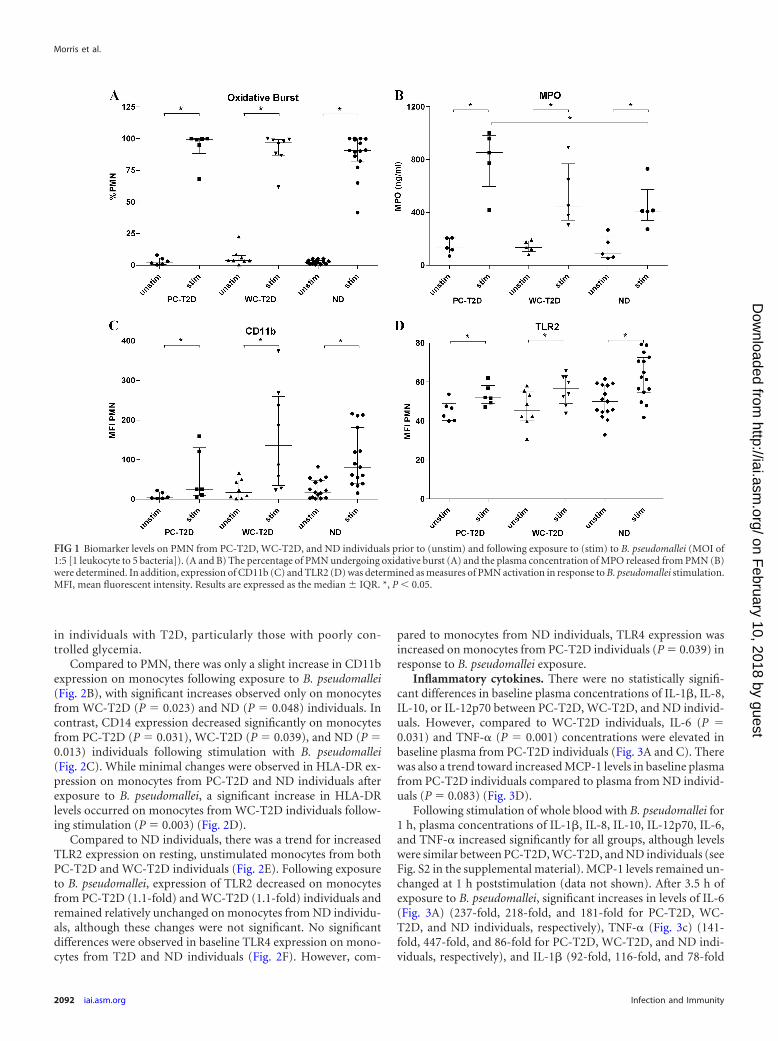
PMN activation. Following B. pseudomallei stimulation, thepercentage of PMN undergoing oxidative burst (Fig. 1A) in-creased significantly for all groups (42-fold for PC-T2D, 25-foldfor WC-T2D, and 36-fold for ND individuals). While there was atrend for greater levels of oxidative burst activity for PMN fromPC-T2D compared to ND and WC-T2D individuals, this did notreach statistical significance. Levels of MPO, an enzyme that isliberated from activated PMN, were similarly increased in B. pseu-domallei-stimulated blood samples (Fig. 1B) and were signifi-cantly higher in blood from PC-T2D individuals compared to ND
individuals (P � 0.05; 6.5-fold versus 4.7-fold increase for PC-T2D and ND individuals, respectively).
We also measured surface expression of the integrin, CD11b, asa marker of PMN activation. CD11b levels increased on PMNfrom all groups following exposure to B. pseudomallei (Fig. 1C).There was a trend toward lower CD11b expression on PMN fromPC-T2D individuals following stimulation, compared to WC-T2D (P � 0.08) and ND (P � 0.094) individuals.
Levels of TLR2 expression were comparable on PMN fromPC-T2D, WC-T2D, and ND individuals at baseline (Fig. 1D). Af-ter exposure to B. pseudomallei, TLR2 expression increased signif-icantly for all groups, with the highest levels observed on PMNfrom ND individuals (Fig. 1D). Minimal TLR4 expression wasdetected on PMN at baseline among the three groups, and theselevels remained relatively unchanged following exposure to B.pseudomallei (data not shown).
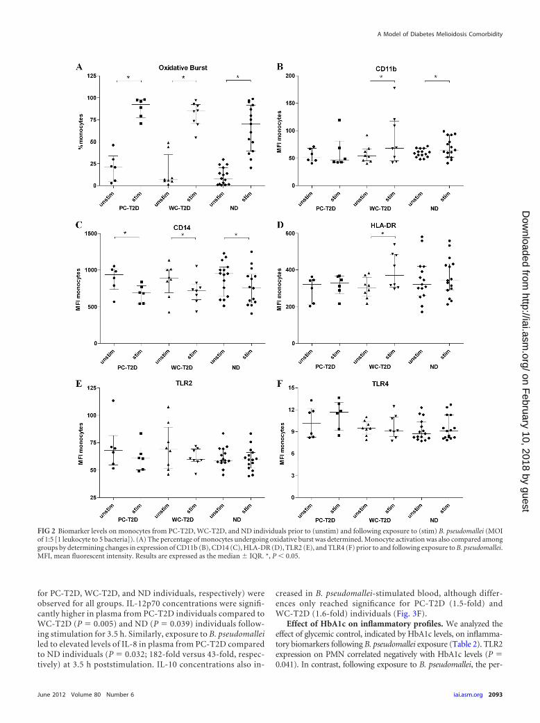
Monocyte activation. Following stimulation, the percentageof DHR� monocytes (Fig. 2A) and the intensity of the oxidativeburst (data not shown) increased significantly for all groups,with the highest fold change observed for WC-T2D individuals(12.8-fold) compared to PC-T2D (4.3-fold) and ND (8.7-fold)individuals. There was a trend toward higher levels of oxidativeburst activity in monocytes from PC-T2D individuals com-pared to monocytes from ND individuals (P � 0.067) follow-ing stimulation. Monocyte oxidative responses to B. pseu-domallei reflected the same trends observed in PMN, wherebyboth the number of cells undergoing oxidative burst activityand the intensity of the oxidative burst response were increased
TABLE 1 Clinical, biochemical, and hematological characteristics of theindividuals in this studya
Characteristic
Result for group:
PC-T2D WC-T2D ND
n 6 8 13No. (males/females) 2/4 4/4 6/7BMI (kg/m2) (mean SD) 40.8 4.5* 34.0 5.9 �Family history of
diabetes (%)100 75 �
TC (mmol/liter) 4.9 0.6 4.5 1.3 �TG (mmol/liter) 2.5 1.4 1.8 0.9 �HbA1c (%) 10.1 1.2** 6.1 0.9 5.1 0.4ESR (mm/hour) 32.8 18.4** 42.1 24† 10.7 7.6CRP (mg/liter) 8.7 7.3** 7.0 5.2 2.0 0.9RBCC (1012/liter) 4.9 0.6 4.6 0.3 4.8 0.5Hemoglobin (g/liter) 136.7 14.9 132.5 13.4 139.4 16.0Hematocrit 0.42 0.03 0.4 0.03 0.42 0.4MCV (femtoliter) 86.3 7.6 88.3 4.8 88.4 7.3Platelets (109/liter) 270.8 91.7 241.8 117.7 236.5 47.7WBCC (109/liter) 8.05 2.5** 6.4 1.4 5.6 1.1Lymphocytes (109/liter) 2.25 0.39 1.75 0.41 1.84 0.48Monocytes (109/liter) 0.59 0.21 0.54 0.14 0.45 0.11Neutrophils (109/liter) 4.86 2.2** 3.84 1.13 3.04 0.72a WC, well-controlled glycemia; PC, poorly controlled glycemia; T2D, type 2 diabetes;ND, nondiabetic control individuals; BMI, body mass index; HbA1c, glycatedhemoglobin A1c; TC, total cholesterol; TG, triglycerides; ESR, erythrocytesedimentation rate; CRP, C-reactive protein; RBCC, red blood cell count; MCV, meancorpuscular volume; WBCC, white blood cell count. �, not determined. Nominalvariables are presented as numbers; continuous variables are presented as the mean standard deviation. Continuous variables were compared between groups by usingMann-Whitney U test. *, PC-T2D compared to WC-T2D, P 0.05; **, PC-T2Dcompared to ND, P 0.05; †, WC-T2D compared to ND, P 0.05.
A Model of Diabetes Melioidosis Comorbidity
June 2012 Volume 80 Number 6 iai.asm.org 2091
on February 10, 2018 by guest
http://iai.asm.org/
Dow
nloaded from
in individuals with T2D, particularly those with poorly con-trolled glycemia.
Compared to PMN, there was only a slight increase in CD11bexpression on monocytes following exposure to B. pseudomallei(Fig. 2B), with significant increases observed only on monocytesfrom WC-T2D (P � 0.023) and ND (P � 0.048) individuals. Incontrast, CD14 expression decreased significantly on monocytesfrom PC-T2D (P � 0.031), WC-T2D (P � 0.039), and ND (P �0.013) individuals following stimulation with B. pseudomallei(Fig. 2C). While minimal changes were observed in HLA-DR ex-pression on monocytes from PC-T2D and ND individuals afterexposure to B. pseudomallei, a significant increase in HLA-DRlevels occurred on monocytes from WC-T2D individuals follow-ing stimulation (P � 0.003) (Fig. 2D).
Compared to ND individuals, there was a trend for increasedTLR2 expression on resting, unstimulated monocytes from bothPC-T2D and WC-T2D individuals (Fig. 2E). Following exposureto B. pseudomallei, expression of TLR2 decreased on monocytesfrom PC-T2D (1.1-fold) and WC-T2D (1.1-fold) individuals andremained relatively unchanged on monocytes from ND individu-als, although these changes were not significant. No significantdifferences were observed in baseline TLR4 expression on mono-cytes from T2D and ND individuals (Fig. 2F). However, com-
pared to monocytes from ND individuals, TLR4 expression wasincreased on monocytes from PC-T2D individuals (P � 0.039) inresponse to B. pseudomallei exposure.
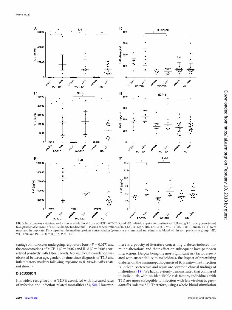
Inflammatory cytokines. There were no statistically signifi-cant differences in baseline plasma concentrations of IL-1�, IL-8,IL-10, or IL-12p70 between PC-T2D, WC-T2D, and ND individ-uals. However, compared to WC-T2D individuals, IL-6 (P �0.031) and TNF-� (P � 0.001) concentrations were elevated inbaseline plasma from PC-T2D individuals (Fig. 3A and C). Therewas also a trend toward increased MCP-1 levels in baseline plasmafrom PC-T2D individuals compared to plasma from ND individ-uals (P � 0.083) (Fig. 3D).
Following stimulation of whole blood with B. pseudomallei for1 h, plasma concentrations of IL-1�, IL-8, IL-10, IL-12p70, IL-6,and TNF-� increased significantly for all groups, although levelswere similar between PC-T2D, WC-T2D, and ND individuals (seeFig. S2 in the supplemental material). MCP-1 levels remained un-changed at 1 h poststimulation (data not shown). After 3.5 h ofexposure to B. pseudomallei, significant increases in levels of IL-6(Fig. 3A) (237-fold, 218-fold, and 181-fold for PC-T2D, WC-T2D, and ND individuals, respectively), TNF-� (Fig. 3c) (141-fold, 447-fold, and 86-fold for PC-T2D, WC-T2D, and ND indi-viduals, respectively), and IL-1� (92-fold, 116-fold, and 78-fold
FIG 1 Biomarker levels on PMN from PC-T2D, WC-T2D, and ND individuals prior to (unstim) and following exposure to (stim) to B. pseudomallei (MOI of1:5 [1 leukocyte to 5 bacteria]). (A and B) The percentage of PMN undergoing oxidative burst (A) and the plasma concentration of MPO released from PMN (B)were determined. In addition, expression of CD11b (C) and TLR2 (D) was determined as measures of PMN activation in response to B. pseudomallei stimulation.MFI, mean fluorescent intensity. Results are expressed as the median IQR. *, P 0.05.
Morris et al.
2092 iai.asm.org Infection and Immunity
on February 10, 2018 by guest
http://iai.asm.org/
Dow
nloaded from
for PC-T2D, WC-T2D, and ND individuals, respectively) wereobserved for all groups. IL-12p70 concentrations were signifi-cantly higher in plasma from PC-T2D individuals compared toWC-T2D (P � 0.005) and ND (P � 0.039) individuals follow-ing stimulation for 3.5 h. Similarly, exposure to B. pseudomalleiled to elevated levels of IL-8 in plasma from PC-T2D comparedto ND individuals (P � 0.032; 182-fold versus 43-fold, respec-tively) at 3.5 h poststimulation. IL-10 concentrations also in-
creased in B. pseudomallei-stimulated blood, although differ-ences only reached significance for PC-T2D (1.5-fold) andWC-T2D (1.6-fold) individuals (Fig. 3F).
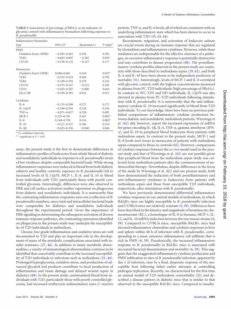
Effect of HbA1c on inflammatory profiles. We analyzed theeffect of glycemic control, indicated by HbA1c levels, on inflamma-tory biomarkers following B. pseudomallei exposure (Table 2). TLR2expression on PMN correlated negatively with HbA1c levels (P �0.041). In contrast, following exposure to B. pseudomallei, the per-
FIG 2 Biomarker levels on monocytes from PC-T2D, WC-T2D, and ND individuals prior to (unstim) and following exposure to (stim) B. pseudomallei (MOIof 1:5 [1 leukocyte to 5 bacteria]). (A) The percentage of monocytes undergoing oxidative burst was determined. Monocyte activation was also compared amonggroups by determining changes in expression of CD11b (B), CD14 (C), HLA-DR (D), TLR2 (E), and TLR4 (F) prior to and following exposure to B. pseudomallei.MFI, mean fluorescent intensity. Results are expressed as the median IQR. *, P 0.05.
A Model of Diabetes Melioidosis Comorbidity
June 2012 Volume 80 Number 6 iai.asm.org 2093
on February 10, 2018 by guest
http://iai.asm.org/
Dow
nloaded from
centage of monocytes undergoing respiratory burst (P � 0.027) andthe concentrations of MCP-1 (P � 0.002) and IL-8 (P � 0.005) cor-related positively with HbA1c levels. No significant correlation wasobserved between age, gender, or time since diagnosis of T2D andinflammatory markers following exposure to B. pseudomallei (datanot shown).
DISCUSSION
It is widely recognized that T2D is associated with increased ratesof infection and infection-related mortalities (33, 50). However,
there is a paucity of literature concerning diabetes-induced im-mune alterations and their effect on subsequent host-pathogeninteractions. Despite being the most significant risk factor associ-ated with susceptibility to melioidosis, the impact of preexistingdiabetes on the immunopathogenesis of B. pseudomallei infectionis unclear. Bacteremia and sepsis are common clinical findings ofmelioidosis (18). We had previously demonstrated that comparedto individuals with no identifiable risk factors, individuals withT2D are more susceptible to infection with less virulent B. pseu-domallei isolates (58). Therefore, using a whole-blood stimulation
FIG 3 Inflammatory cytokine production in whole blood from PC-T2D, WC-T2D, and ND individuals prior to (unstim) and following 3.5 h of exposure (stim)to B. pseudomallei (MOI of 1:5 [1 leukocyte to 5 bacteria]). Plasma concentrations of IL-6 (A), IL-12p70 (B), TNF-� (C), MCP-1 (D), IL-8 (E), and IL-10 (F) weremeasured in duplicate. Data represent the median cytokine concentration (pg/ml) in unstimulated and stimulated blood within each participant group (ND,WC-T2D, and PC-T2D) IQR. *, P 0.05.
Morris et al.
2094 iai.asm.org Infection and Immunity
on February 10, 2018 by guest
http://iai.asm.org/
Dow
nloaded from
assay, the present study is the first to demonstrate differences ininflammatory profiles of leukocytes from whole blood of diabeticand nondiabetic individuals in response to a B. pseudomallei strainof low virulence, despite comparable bacterial loads. While stronginflammatory responses were induced in blood from both diabeticsubjects and healthy controls, exposure to B. pseudomallei led toincreased levels of IL-12p70, MCP-1, IL-8, and IL-10 in bloodfrom individuals with T2D, particularly those with poorly con-trolled glycemia. Interestingly, differences were also observed inPRR and cell surface activation marker expression on phagocytesfrom diabetic and nondiabetic individuals. It is noteworthy thatthe contrasting inflammatory responses were not a reflection of B.pseudomallei numbers, since total and intracellular bacterial loadswere comparable for diabetics and nondiabetic individualsthroughout the experimental period. Given the importance ofPRR signaling in determining the subsequent activation of diverseimmune response pathways, the contrasting expression identifiedon phagocytes in the present study may underpin the susceptibil-ity of T2D individuals to melioidosis.
Chronic low-grade inflammation and oxidative stress are welldocumented in T2D and play an important role in the develop-ment of many of the metabolic complications associated with in-sulin resistance (27, 44). In addition to many metabolic abnor-malities, a variety of immunological abnormalities continue to beidentified that conceivably contribute to the increased susceptibil-ity of T2D individuals to infections such as melioidosis (35, 44).Prolonged hyperglycemia, oxidative stress, and production of ad-vanced glycated end products contribute to local production ofinflammation and tissue damage and delayed wound repair indiabetics (68). In the present study, unstimulated blood from in-dividuals with T2D, particularly those with poorly controlled gly-cemia, had increased erythrocyte sedimentation rates, C-reactive
protein, TNF-�, and IL-6 levels, all of which are consistent with anunderlying inflammatory state which has been shown to occur inassociation with T2D (35, 43, 44).
Recruitment, migration, and activation of leukocyte subsetsare crucial events during an immune response that are regulatedby chemokines and inflammatory cytokines. However, while thesemediators are indispensable for the effective clearance of a patho-gen, an excessive inflammatory response is potentially destructiveand may contribute to disease progression (60). The proinflam-matory cytokine profiles observed in the present study are consis-tent with those described in melioidosis sepsis (29, 62), and bothIL-6 and IL-10 have been shown to be independent predictors ofmortality (51). Interestingly, levels of MCP-1 and IL-8 correlatedwith glycemic control, with the highest concentrations measuredin plasma from PC-T2D individuals (high percentage of HbA1c).In contrast to WC-T2D and ND individuals, IL-12p70 was alsoelevated in plasma from PC-T2D individuals following stimula-tion with B. pseudomallei. It is noteworthy that the anti-inflam-matory cytokine IL-10 increased significantly in blood from T2Dindividuals. To our knowledge, there have been no previous pub-lished comparisons of inflammatory cytokine production be-tween diabetic and nondiabetic melioidosis patients. Wiersinga etal. (62) did, however, report the increased expression of mRNAfor genes encoding IL-1�, IL-6, TNF-�, gamma interferon (IFN-�), and IL-10 in peripheral blood leukocytes from patients withmelioidosis sepsis. In contrast to the present study, MCP-1 andIL-8 levels were lower in monocytes of patients with melioidosissepsis compared to those in controls (62). However, comparisonsof cytokine responses between the ex vivo model used in the pres-ent study and that of Wiersinga et al. (62) are not possible giventhat peripheral blood from the melioidosis sepsis study was col-lected from melioidosis patients after the commencement of an-timicrobial therapy. Nevertheless, despite differences in the focusof the study by Wiersinga et al. (62) and our present study, bothhave demonstrated the induction of both proinflammatory andanti-inflammatory cytokines in leukocytes from patients withmelioidosis sepsis and those from susceptible T2D individuals,respectively, after stimulation with B. pseudomallei.
We have previously demonstrated differential inflammatorycytokine responses in our animal models of melioidosis, in whichBALB/c mice are highly susceptible to B. pseudomallei infectionand C57BL/6 mice are relatively resistant (6, 59). Differences havebeen described in the kinetics and magnitude of keratinocyte che-moattractant (KC), a homologue of IL-8 in humans, MCP-1, IL-12, and IL-10 mRNA induction between the two mouse strains (6,59). Compared to C57BL/6 mice, susceptible BALB/c mice hadelevated inflammatory chemokine and cytokine responses in liverand spleen within 48 h of infection with B. pseudomallei, corre-sponding to a more extensive inflammatory cell infiltrate that isrich in PMN (6, 59). Paradoxically, the increased inflammatoryresponse to B. pseudomallei in BALB/c mice is associated withincreased bacterial dissemination and mortality (6, 59). This sug-gests that the exaggerated inflammatory cytokine production andPMN infiltration to sites of B. pseudomallei infection, apparent byday 2 of infection, may be a final, desperate response of the sus-ceptible host following failed earlier attempts at controllingpathogen replication. Recently, we characterized for the first timean animal model of T2D melioidosis comorbidity (32) and de-scribed a disease pattern in diabetic mice that is similar to thatobserved in the susceptible BALB/c mice. Compared to nondia-
TABLE 2 Association of percentage of HbA1c as an indicator ofglycemic control with inflammatory biomarkers following exposure toB. pseudomallei
Inflammatory biomarkertype 95% CIa Spearman’s r P valueb
PMNOxidative burst (DHR) �0.283–0.462 0.104 0.592TLR2 �0.663–0.007 �0.382 0.041*CD11b �0.578–0.132 �0.257 0.177
MonocytesOxidative burst (DHR) 0.040–0.681 0.410 0.027*TLR2 �0.332–0.419 0.050 0.795TLR4 �0.109–0.593 0.279 0.143CD11b �0.553–0.167 �0.223 0.245CD14 �0.454–0.307 �0.086 0.664HLA-DR �0.358–0.395 0.022 0.911
CytokinesIL-6 �0.133–0.598 0.271 0.171IL-12p70 �0.180–0.556 0.226 0.256TNF-� �0.071–0.637 0.328 0.095MCP-1 0.235–0.781 0.567 0.002*IL-8 0.168–0.759 0.524 0.005*IL-10 �0.221–0.537 0.186 0.354IL-1� �0.423–0.356 �0.040 0.844
a CI, confidence intervals.b *, significant at P 0.05.
A Model of Diabetes Melioidosis Comorbidity
June 2012 Volume 80 Number 6 iai.asm.org 2095
on February 10, 2018 by guest
http://iai.asm.org/
Dow
nloaded from
betic mice, in diabetic mice, hyperproduction of proinflammatorycytokines, including � F-�, IL-1�, and IL-6, and extensive PMNinfiltration and tissue damage at sites of infection preceded mor-tality in the first few days following B. pseudomallei infection (32).Data from the present study demonstrate that the levels of controlof B. pseudomallei persistence within the first few hours of expo-sure are similar for PC-T2D, WC-T2D, and ND individuals. Im-portantly, despite comparable bacterial loads, the inflammatorypathways triggered in leukocytes in response to this bacteriumdiffer significantly for T2D and ND individuals.
In the present study, there was evidence of lower proinflam-matory cytokines in blood from WC-T2D compared to PC-T2Dindividuals. Given the inflammatory nature of diabetes, there hasbeen increasing interest in the identification of compounds withantioxidative and anti-inflammatory properties in the treatmentof infection in individuals with diabetes (10). This is supported byrecent evidence that for patients with melioidosis sepsis, thosewith preexisting T2D that were taking glyburide, a sulfonylureawith anti-inflammatory properties, were more likely to survivethan nondiabetics or T2D individuals receiving other antidiabetictreatments prior to diagnosis with melioidosis (34). T2D individ-uals recruited to the present study were receiving one or more of avariety of antidiabetic medication classes, including sulfonylureas,�-glucosidases, and biguanides. Unfortunately, this meant thatgroup sizes were inadequate for analysis of the potential effects ofantidiabetic medication on inflammatory profiles generated in re-sponse to B. pseudomallei exposure. Nevertheless, the findings inthe present study have clearly demonstrated that glycemic controldoes influence the host inflammatory response to B. pseudomallei.The hyperinflammatory responses observed between 24 and 48 hin susceptible BALB/c mice (6, 59), in diabetic mice (32), and inpatients with melioidosis sepsis (62) reflect the inflammatory pro-files observed in the whole-blood assay used in the present study.Based on our findings, we propose that very early interactionsbetween B. pseudomallei and PMN and monocytes are altered inthe diabetic host, such that a compensatory dysregulated hyper-inflammatory response is triggered independently of bacterialload, resulting in increased local tissue damage and bacterial dis-semination.
Both PMN and monocytes are vulnerable to functional distur-bances due to hyperglycemia-induced stress (33, 35, 68). Oxida-tive stress is well described in diabetes, contributing to endothelialdysfunction and vascular inflammation (20). Trends for increasedoxidative burst activity observed in PMN and monocytes fromPC-T2D individuals both in unstimulated and stimulated bloodcompared to healthy controls are consistent with the findings ofothers (4, 23). MPO, a principle enzyme released by activatedPMN, was significantly elevated in plasma from PC-T2D individ-uals compared to ND individuals following exposure to B. pseu-domallei.
PMN migration from the bloodstream into sites of infectionwithin tissues occurs rapidly through a process that is tightly reg-ulated. Activated PMN display an upregulated expression ofCD11b and other integrins and chemokine receptors, which facil-itate their transmigration into sites of infection within tissue (15).Reduced PMN function has been demonstrated in patients withmelioidosis and comorbid T2D (11). Based on the association ofPMN functional defects with the most common underlying riskfactors for melioidosis, G-CSF, which stimulates the productionand function of PMN, was used in a trial as an adjunctive therapy
for the treatment of septic shock from melioidosis (12, 14, 31, 46,67). Although G-CSF had no effect on mortality for patients withsevere melioidosis sepsis (12), its efficacy in treatment of patientswith less severe forms of melioidosis has not been investigated. Inthe present study, we examined CD11b expression, which medi-ates inflammation by regulating leukocyte adhesion and migra-tion, as additional measures of PMN and monocyte activation inresponse to B. pseudomallei exposure. CD11b was upregulated onPMN from all groups after stimulation with B. pseudomallei, sim-ilar to responses described in sepsis patients (38). However, therewas a trend toward reduced levels of CD11b on PMN from PC-T2D individuals at baseline and following stimulation, supportingdecreased migratory capacity previously described for PMN fromT2D individuals (11, 28). In contrast, CD11b expression was com-parable for monocytes from T2D and ND individuals prior to andfollowing stimulation with B. pseudomallei. In addition to CD11b,we measured changes in expression of HLA-DR as cell surfacemarkers of monocyte activation. HLA-DR, or the major histo-compatibility complex (MHC) class II surface receptor, has a cen-tral role in antigen-specific interactions between monocytes andlymphocytes. Similar to CD11b, HLA-DR levels remained rela-tively unchanged in PC-T2D and ND individuals. However, itremains to be determined whether other integrins or chemokinereceptors involved in monocyte activation are altered in diabeticindividuals following exposure to B. pseudomallei.
PRRs play an important role in the activation and regulation ofthe innate immune response. TLRs are transmembrane proteinsthat mediate host cell signaling in response to a range of extracel-lular and endosomal pathogen-associated molecules. TLR4 per-mits signaling in response to lipopolysaccharide (LPS) and non-infective inflammatory stimuli, such as heat shock protein 60(HSP60) (reviewed in reference 42). TLR2, which can het-erodimerize with other TLRs, responds to a more diverse group ofligands, including lipoproteins, peptidoglycans, and atypical LPS(reviewed in reference 41). B. pseudomallei has been shown tointeract with CD14, TLR2, and TLR4 (61, 63, 64), and our grouprecently identified differences in TLR2 and TLR4 expression onmonocytes following stimulation with B. pseudomallei isolates oflow and high virulence (M. Feterl and N. Ketheesan, unpublisheddata). TLRs have also been implicated in the pathogenesis of in-sulin resistance and its complications, with observations thatTLR2 and TLR4 expression is increased on monocytes from dia-betic individuals (21, 22). Endogenous ligands, such as fatty acids,heat shock proteins, hyaluronan, and high-mobility group B1protein induce the production of proinflammatory cytokinesthrough the stimulation of TLR2 and TLR4 in individuals withdiabetes (21). Given the importance of TLR2, TLR4, and CD14 inmelioidosis, we therefore investigated changes in the expression ofthese pathogen recognition receptors on phagocytes from T2Dand ND individuals in response to B. pseudomallei stimulation.CD14, one of the first-described PRRs, exists in a membrane-bound form on monocytes, where it serves as a ligand-bindingcomponent of the LPS-receptor complex (7). Membrane-boundCD14 can also be shed into a soluble form upon monocyte acti-vation (7). CD14 expression decreased on monocytes exposed toB. pseudomallei, with greater decreases observed for monocytesfrom T2D individuals compared to ND individuals. Changes inCD14 expression may reflect internalization of membrane-boundCD14 (39) or increased shedding of surface CD14 (48). Decreasedmembrane CD14 on monocytes, described as monocyte hypore-
Morris et al.
2096 iai.asm.org Infection and Immunity
on February 10, 2018 by guest
http://iai.asm.org/
Dow
nloaded from
sponsiveness, has been shown to correlate with sepsis severity (9,49). Our findings of decreased surface expression of CD14 onmonocytes from T2D individuals suggest alterations in monocytefunctional capacity following exposure to B. pseudomallei maycontribute to more severe disease progression in T2D individuals.This is consistent with the clinical observation that individualswith diabetes are more likely to present with melioidosis sepsisthan individuals with no identifiable risk factors (19).
TLR4 expression on PMN was low, as previously reported (36).In contrast, TLR2 expression increased on PMN from both T2Dand ND individuals after stimulation, with levels tending to begreatest on PMN from healthy controls. On monocytes, TLR4levels increased marginally following B. pseudomallei exposure,with no significant differences observed between groups. WhileTLR2 expression remained relatively unchanged on monocytesfrom ND individuals in response to B. pseudomallei stimulation,there was a trend for decreased TLR2 expression on monocytesfrom both PC-T2D and WC-T2D individuals. The pattern of de-creased TLR2 and CD14 levels on monocytes from diabetic indi-viduals in the present study is consistent with the profile describedfor patients with severe sepsis, although no comparisons havebeen made between diabetic and nondiabetic patients (9, 49, 62).In addition, peripheral blood samples from the sepsis studies werecollected from patients following commencement of antimicro-bial therapy after hospital admission, making it difficult to drawparallels to the early changes in TLR expression measured in ourwhole-blood stimulation assay.
The MyD88-dependent pathway is activated by TLR2 andTLR4, resulting in the production of proinflammatory cytokinessuch as TNF-�, IL-6, and IL-8 (reviewed in reference 42). TLR4also activates the Toll/IL-1 receptor (TIR) domain-containingadaptor-inducing IFN-� (TRIF)-dependent pathway leading toincreased production of IL-12, IL-10, and IFN-� (reviewed inreference 42). Compared to ND individuals, plasma from PC-T2D individuals had elevated levels of IL-12 and IL-10, in additionto higher concentrations of TNF-�, IL-6, and IL-8, suggesting thatboth MyD88- and TRIF-dependent pathways had been activated.This is supported by the observation of slight increases in TLR4expression and decreased CD14 activation on monocytes fromPC-T2D individuals after exposure to B. pseudomallei. Given thecontrasting TLR and CD14 activation and inflammatory cytokineresponse generated in leukocytes from PC-T2D and ND individ-uals in the present study, it is likely that future kinetic studiesfocusing on PRR activation in response to B. pseudomallei willdemonstrate differences within these groups that may be impor-tant in directing the generation of subsequent inflammatory re-sponses.
TLR activation not only influences PMN survival, but also hasa regulatory effect on chemokine receptor expression on leuko-cytes, thereby influencing their migratory capacity (1, 26, 47).Downregulation of chemokine receptors aids retention of PMN atsites of infection, and bacterial phagocytosis has been shown topromote this downregulation (24). A reduced migratory responseof diabetic PMN toward IL-8 has been demonstrated in vitro (11)and is supported by the differences observed in CD11b expressionon PMN from PC-T2D and ND individuals in the present study. Arole for altered PMN migratory response to B. pseudomallei in thesusceptibility of diabetic individuals to melioidosis is also sup-ported by findings that upregulation of ICAM-1, involved inCD11b/CD18-mediated neutrophil adhesion and migration
through endothelium, is attenuated in endotoxemia in diabeticindividuals compared to healthy controls (2). Murine studies havehighlighted the importance of CD11b/CD18 upregulation onPMN in the early control of bacterial replication following infec-tion (8). Within the first hours of Gram-negative bacterial infec-tion, a rapid PMN influx is evident at sites of inflammation inimmunocompetent mice (69). However, a short delay in veryearly PMN migration provides an opportunity for the bacteriumto replicate and express virulence factors that facilitate its persis-tence in the host, thereby influencing disease progression (69). Wepropose a similar mechanism is associated with susceptibility ofT2D individuals to melioidosis, whereby an initial delay in PMNmigration and/or activation allows B. pseudomallei to multiply,establish an intracellular niche, and therefore render subsequentinflammatory responses insufficient to contain infection. Ourdata are supported by a recent transcriptome analysis of innateimmune responses triggered after infection of B. pseudomallei in achemically induced diabetic mouse model. Despite comparablebacterial loads in the first 24 h of infection, differential expressionprofiles for genes involved in PRR signaling pathways were dem-onstrated for diabetic and nondiabetic mice (13). While 16 hpostinfection was the earliest time point assessed in the microar-ray studies of diabetic mice (13), they provide further support toour data that the presence of very early defects (3.5 h) in theability of phagocytes from diabetic individuals to detect and re-spond appropriately to B. pseudomallei underlies contrasting dis-ease progression in diabetic and nondiabetic hosts. Further stud-ies to investigate interactions between B. pseudomallei, PRRsignaling, and the effects on leukocyte migration to sites of infec-tion in individuals at risk of melioidosis are certainly warranted.
Our study has clear limitations. Specifically, the sample sizewas small and our investigations measured selected markers of theinflammatory response within 4 h of exposure to B. pseudomallei.Nonetheless, we believe that the data presented here identify noveldifferences in early cellular responses to B. pseudomallei betweendiabetic and nondiabetic individuals which are not attributed todifferences in bacterial persistence. In contrast to studies examin-ing functional responses of single leukocyte subsets, the use of awhole-blood approach enabled us to examine the responses ofrelevant leukocytes simultaneously, therefore more closely mod-eling in vivo inflammatory responses generated in the early stagesof B. pseudomallei infection in diabetic and nondiabetic individu-als. In addition, utilization of a whole-blood assay avoids potentialbias from cellular stimulation associated with leukocyte subsetisolation techniques (54). Data from this proof-of-principle studyprovide an immunological basis for the clinical observations ob-served in patients with T2D and melioidosis comorbidity (19, 62),supporting the utility of ex vivo whole-blood assays as a tool forevaluation of the mechanisms underlying susceptibility to B. pseu-domallei in this at-risk population. Such studies are not possibleutilizing blood from patients with diabetes and melioidosis, giventhat by the time they are hospitalized the infection has alreadybeen established. In addition, in most instances, patients recruitedfor such studies would have commenced antimicrobial therapy.Use of a whole-blood model will enable investigation of the veryearly interactions between human leukocytes and B. pseudomalleithat are important in determining the outcome of infection. Giventhe complexity of leukocyte-leukocyte cross talk in innate immu-nity, use of a whole-blood model also permits measurement ofinflammatory changes following B. pseudomallei exposure that
A Model of Diabetes Melioidosis Comorbidity
June 2012 Volume 80 Number 6 iai.asm.org 2097
on February 10, 2018 by guest
http://iai.asm.org/
Dow
nloaded from
more closely reflect in vivo interactions than would be possibleusing specific leukocyte subsets in isolation, in vitro.
In summary, this is the first study to describe and characterizean ex vivo whole-blood model of comorbidity between diabetesand melioidosis. We have shown for the first time that contrastinginflammatory profiles for PC-T2D, WC-T2D, and ND individualsare evident as early as 3.5 h following exposure to a B. pseudomalleistrain of low virulence, despite comparable bacterial loads. Datafrom our study suggest very early interactions between B. pseu-domallei and PMN and monocytes are altered in hosts with dia-betes, contributing to impaired leukocyte activation and thedevelopment of an exaggerated inflammatory response. The avail-ability of a model that reflects clinical parameters observed inpatients with T2D and melioidosis is invaluable since it will facil-itate further studies of the dysregulated interactions betweenphagocytes and B. pseudomallei in the initial stages of infection insusceptible hosts and the effects of therapeutics on these interac-tions. This is significant, given the continuing increase in the prev-alence of T2D, which could potentially place more people at risk ofsevere melioidosis in regions of endemicity.
ACKNOWLEDGMENTS
We are grateful to the volunteers who participated in the present studyand wish to thank Kelly Hodgson and Marshall Feterl for their laboratoryassistance and Lauren Kromoloff for assistance with collation of clinicaldata.
This project was supported by a grant from the Townsville HospitalPrivate Practice Research Fund.
REFERENCES1. Alves-Filho JC, et al. 2009. Regulation of chemokine receptor by toll-like
receptor 2 is critical to neutrophil migration and resistance to polymicro-bial sepsis. Proc. Natl. Acad. Sci. U. S. A. 106:4018 – 4023.
2. Andreasen AS, et al. 2010. Type 2 diabetes mellitus is associated withimpaired cytokine response and adhesion molecule expression in humanendotoxemia. Intensive Care Med. 36:1548 –1555.
3. Ashdown LR. 1987. Indirect hemagglutination test for melioidosis. Med.J. Aust. 147:364 –365.
4. Ayilavarapu S, et al. 2010. Diabetes-induced oxidative stress is mediatedby Ca2�-independent phospholipase A2 in neutrophils. J. Immunol. 184:1507–1515.
5. Barnes J, Ketheesan N. 2005. Melioidosis: routes of infection. Emerg.Infect. Dis. 4:638 – 639.
6. Barnes JL, et al. 2001. Induction of multiple chemokine and colony-stimulating factor genes in experimental Burkholderia pseudomallei infec-tion. Immunol. Cell Biol. 79:490 –501.
7. Bazil V, Strominger JL. 1991. Shedding as a mechanism of down-modulation of CD14 on stimulated human monocytes. J. Immunol. 147:1567–1574.
8. Borjesson DL, Simon SI, Hodzic E, Ballantyne CM, Barthold SW. 2002.Kinetics of CD11b/CD18 upregulation during infection with the agent ofhuman granulocytic ehrlichiosis in mice. Lab. Invest. 82:303–311.
9. Brunialti, MKC, et al. 2006. TLR2, TLR4, CD14, CD11b, and CD11cexpressions on monocytes surface and cytokine production in patientswith sepsis, severe sepsis, and septic shock. Shock 25:351–357.
10. Ceriello A, Testa R. 2009. Antioxidant anti-inflammatory treatment intype 2 diabetes. Diabetes Care 32:S232–236.
11. Chanchamroen S, Kewcharoenwong C, Susaengrat W, Ato M, Lert-memongkolchai G. 2009. Human polymorphonuclear responses to Burk-holderia pseudomallei in healthy and diabetic subjects. Infect. Immun. 77:456 – 463.
12. Cheng A, et al. 2007. A randomized controlled trial of granulocyte colonystimulating factor for the treatment of septic shock due to melioidosis inThailand. Clin. Infect. Dis. 45:308 –314.
13. Chin C-Y, Monack DM, Nathan S. 2012. Delayed activation of hostinnate immune pathways in streptozotocin-induced diabetic hosts leads
to more severe disease during infection with Burkholderia pseudomallei.Immunology 135:312–322.
14. Chonchol M. 2006. Neutrophil dysfunction and infection risk in end-stage renal disease. Semin. Dial. 19:291–296.
15. Cowburn AS, Condliffe AM, Farahi N, Summers C, Chilvers ER. 2008.Advances in neutrophil biology: clinical implications. Chest 134:606 –612.
16. Currie B, et al. 2000. Endemic melioidosis in tropical Northern Australia:a 10-year prospective study and review of the literature. Clin. Infect. Dis.31:981–986.
17. Currie B, et al. 2000. The epidemiology of melioidosis in Australia andPapua New Guinea. Acta Trop. 74:121–127.
18. Currie B, et al. 2004. Melioidosis epidemiology and risk factors from aprospective whole population study in northern Australia. Trop. Med. Int.Health 9:1167–1174.
19. Currie B, Ward L, Cheng A. 2010. The epidemiology and clinical spec-trum of melioidosis: 540 cases from the 20 year Darwin prospective study.PLoS Negl. Trop. Dis. 4:e900.
20. Dandona P, Aljada A, Chaudhuri A, Mohanty P. 2004. Endothelialdysfunction, inflammation and diabetes. Rev. Endocr. Metab. Disord.5:189 –197.
21. Dasu MR, Devaraj S, Park S, Jialal I. 2010. Increased toll-like receptor(TLR) activation and TLR ligands in recently diagnosed type 2 diabeticsubjects. Diabetes Care 33:861– 868.
22. Devaraj S, Jialal I, Yun J-M, Bremer A. 2011. Demonstration of in-creased toll-like receptor 2 and toll-like receptor 4 expression in mono-cytes of type 1 diabetes mellitus patients with microvascular complica-tions. Metabolism 60:256 –259.
23. Ding Y, et al. 2007. Activation of RAGE induces elevated O2-generationby mononuclear phagocytes in diabetes. J. Leukoc. Biol. 81:520 –527.
24. Doroshenko T, et al. 2002. Phagocytosing neutrophils down-regulate theexpression of chemokine receptors CXCR1 and CXCR2. Blood 100:2668 –2671.
25. Douglas M, et al. 2004. Epidemiology of community-acquired and nos-ocomial bloodstream infections in tropical Australia: a 12 month prospec-tive study. Trop. Med. Int. Health 9:795– 804.
26. Dragomir E, Simionescu M. 2006. Monocyte chemoattractant protein-1—a major contributor to the inflammatory process associated with dia-betes. Arch. Physiol. Biochem. 112:239 –244.
27. Fernandez-Real JM, Pickup JC. 2008. Innate immunity, insulin resis-tance and type 2 diabetes. Trends Endocrinol. Metab. 19:10 –16.
28. Fortes ZB, Farsky SP, Oliveira MA, Garcia-Leme J. 1991. Direct vitalmicroscopic study of defective leukocyte-endothelial interactions in dia-betes mellitus. Diabetes 40:1267–1273.
29. Friedland JS, Suputtamongkol Y, Remck DG. 1992. Prolonged elevationof interleukin-8 and interleukin-6 concentrations in plasma and of leuko-cyte interleukin-8 mRNA levels during septicemic and localized Pseu-domonas pseudomallei infection. Infect. Immun. 60:2402–2408.
30. Hassan M, et al. 2010. Incidence, risk factors and clinical epidemiology ofmelioidosis: a complex socio-ecological emerging infectious disease in theAlor Setar region of Kedah, Malaysia. BMC Infect. Dis. 10:302.
31. Heinzelmann M, Mercer-Jones MA, Passmore JC. 1999. Neutrophilsand renal failure. Am. J. Kidney Dis. 34:384 –399.
32. Hodgson K, Morris J, Williams N, Govan B, Ketheesan N. 2010.PP-060-23. Immunological defects associated with type 2 diabetes con-tribute to susceptibility to bacterial infection. Int. Immunol. Meet. Abstr.22(Suppl 1, Pt 3):iii97–iii110.
33. Jacob A, et al. 2008. Sepsis-induced inflammation is exacerbated in ananimal model of type 2 diabetes. Int. J. Clin. Exp. Med. 1:22–31.
34. Koh GCKW, et al. 2011. Glyburide is anti-inflammatory and associatedwith reduced mortality in melioidosis. Clin. Infect. Dis. 52:717–725.
35. Kolb H, Mandrup-Poulsen T. 2005. An immune origin of type 2 diabetes?Diabetologia 48:1038 –1050.
36. Kurt-Jones EA, et al. 2002. Role of Toll-like receptor 2 (TLR2) in neu-trophil activation: GM-CSF enhances TLR2 expression and TLR2-mediated interleukin responses in neutrophils. Blood 100:1860 –1868.
37. Limmathurotsakul D, et al. 2010. Increasing incidence of humanmelioidosis in Northeast Thailand. Am. J. Trop. Med. Hyg. 82:1113–1117.
38. Lin RY, Astiz ME, Saxon JC, Rackow EC. 1993. Altered leukocyteimmunophenotypes in septic shock. Studies of HLA-DR, CD11b, CD14and IL-2R expression. Chest 104:847– 853.
39. Lin SM, et al. 2004. Differential regulation of membrane CD14 expres-
Morris et al.
2098 iai.asm.org Infection and Immunity
on February 10, 2018 by guest
http://iai.asm.org/
Dow
nloaded from
sion and endotoxin-tolerance in alveolar macrophages. Am. J. Respir. CellMol. Biol. 31:162–170.
40. Mustafa AS, El-Shamy AM, Madi NM, Amoudy HA, Al-Attiyah R.2008. Cell-mediated immune responses to complex and single mycobac-terial antigens in tuberculosis patients with diabetes. Med. Princ. Pract.17:325–330.
41. Naguib G, Al-Mashat H, Desta T, Graves ET. 2004. Diabetes prolongsthe inflammatory response to bacterial stimulus through cytokine dys-regulation. J. Invest. Dermatol. 123:87–92.
42. Netea MG, van der Graaf C, Van der Meer JW, Kullberg BJ. 2004.Toll-like receptors and the host defense against microbial pathogens:bringing specificity to the innate-immune system. J. Leukoc. Biol. 75:749 –755.
43. Packiavathy ASC, Ramalingam M. 2010. Evaluation of haematologicaland immunogical status in type 2 diabetes. Chemistry 7:1029 –1032.
44. Pickup JC, Crook MA. 1998. Is type II diabetes mellitus a disease of theinnate immune system? Diabetologia 41:1241–1248.
45. Pongcharoen S, et al. 2008. Reduced interleukin-17 expression of Burk-holderia pseudomallei-infected peripheral blood mononuclear cells of di-abetic patients. Asian Pac. J. Allergy Immunol. 26:63– 69.
46. Powell K, Ulett G, Hirst R, Norton R. 2003. G-CSF immunotherapy fortreatment of acute disseminated murine melioidosis. FEMS Microbiol.Lett. 224:315–318.
47. Sabroe I, Dower SK, Whyte MKB. 2005. The role of toll-like receptors inthe regulation of neutrophil migration, activation, and apoptosis. Clin.Infect. Dis. 41(Suppl 7):S421–S426.
48. Saravu K, et al. 2010. Melioidosis in southern India: epidemiological andclinical profile. Southeast Asian J. Trop. Med. Public Health 41:401– 409.
49. Schaaf B, et al. 2009. Mortality in human sepsis is associated with downregulation of Toll-like receptor 2 and CD14 expression on blood mono-cytes. Diagn. Pathol. 4:12.
50. Shah B, Hux J. 2003. Quantifying the risk of infectious diseases for peoplewith diabetes. Diabetes Care 26:510 –513.
51. Simpson AJ, et al. 2000. Prognostic value of cytokine concentrations(tumor necrosis factor-alpha, interleukin-6, and interleukin-10) and clin-ical parameters in severe melioidosis. J. Infect. Dis. 181:621– 625.
52. Spranger J, et al. 2003. Inflammatory cytokines and the risk to developtype 2 diabetes: results of the prospective population-based EuropeanProspective Investigation into Cancer and Nutrition (EPIC)-PotsdamStudy. Diabetes 52:812– 817.
53. Stentz FB, Kitabchi AE. 2003. Activated T lymphocytes in type 2 diabetes:implications from in vitro studies. Curr. Drug Targets 4:493–503.
54. Stibenz D, Buhrer C. 1994. Down-regulation of L-selectin surface
expression by various leukocyte isolation procedures. Scand. J. Immu-nol. 39:59 – 63.
55. Suputtamongkol Y, et al. 1999. Risk factors for melioidosis and bactere-mic melioidosis. Clin. Infect. Dis. 29:408 – 413.
56. Reference deleted.57. Tennenberg S, Finkenauer R, Dwivedi A. 1999. Absence of lipopolysac-
charide-induced inhibition of neutrophil apoptosis in patients with dia-betes. Arch. Surg. 134:1229 –1233.
58. Ulett G, et al. 2001. Burkholderia pseudomallei virulence: definition, sta-bility and association with clonality. Microb. Infect. 3:621– 663.1.
59. Ulett G, Ketheesan N, Hirst R. 2000. Cytokine gene expression in in-nately susceptible BALB/c mice and relatively resistant C57BL/6 mice dur-ing infection with virulent Burkholderia pseudomallei infection. Infect.Immun. 68:2034 –2042.
60. Walley KR, Lukacs NW, Standiford TJ, Strieter RM, Kunkel SL. 1996.Balance of inflammatory cytokines related to severity and mortality ofmurine sepsis. Infect. Immun. 64:4733– 4738.
61. West TE, Ernst RK, Jansson-Hutson MJ, Skerrett SJ. 2008. Activation oftoll-like receptors by Burkholderia pseudomallei. BMC Immunol. 9:46.
62. Wiersinga WJ, et al. 2007. High-throughput mRNA profiling character-izes the expression of inflammatory molecules in sepsis caused by Burk-holderia pseudomallei. Infect. Immun. 75:3074 –3079.
63. Wiersinga WJ, et al. 2008. CD14 impairs host defense against Gramnegative sepsis caused by Burkholderia pseudomallei in mice. J. Infect. Dis.198:1388 –1397.
64. Wiersinga WJ, et al. 2007. Toll-like receptor 2 impairs host defense inGram-negative sepsis caused by Burkholderia pseudomallei (melioidosis).PLoS Med. 4:e248.
65. Williams NL, Morris JL, Rush C, Govan BL, Ketheesan N. 2011. Impactof streptozotocin-induced diabetes on functional responses of dendriticcells and macrophages toward Burkholderia pseudomallei. FEMS Immu-nol. Med. Microbiol. 61:218 –227.
66. World Health Organization Expert Committee on the Diagnosis andClassification of Diabetes Mellitus. 2003. Report of the Expert Commit-tee on the Diagnosis and Classification of Diabetes Mellitus. Diabetes Care26:S5–S20.
67. Wu D, Cederbaum AI. 2003. Alcohol, oxidative stress, and free radicaldamage. Alcohol Res. Health 27:277–284.
68. Yan SF, Ramasamy R, Naka Y, Schmidt AM. 2003. Glycation, inflam-mation, and RAGE: a scaffold for the macrovascular complications ofdiabetes and beyond. Circ. Res. 93:1159 –1163.
69. Yang KK, et al. 2002. Neutrophil influx in response to a peritoneal infec-tion with Salmonella is delayed in lipopolysaccharide-binding protein orCD14-deficient mice. J. Immunol. 169:4475– 4480.
A Model of Diabetes Melioidosis Comorbidity
June 2012 Volume 80 Number 6 iai.asm.org 2099
on February 10, 2018 by guest
http://iai.asm.org/
Dow
nloaded from