Embed Size (px)
Citation preview

BURNS OCCUPATIONAL THERAPY Diane Makovsky and Lisa Pignatiello

Overview
Disorder Diagnosis and classification Etiology Population Treatment Long term prognosis Occupation and client factors OT Interventions Medical input Sources

Disorder
Burns are “injuries that result from direct contact with or exposure
to any thermal, chemical, electrical, or radiation source…
Burn injuries occur when energy from a heat source is transferred to the tissues of the body.
The depth of injury is a function of temperature or source of energy (e.g., radiation) and duration of exposure” (Goodman & Fuller, 2009, 435).
While burns affect all parts of the body, injury to the face, hand, and feet are most common.
Diagnosis
Burns are diagnosed based on the risk of infection, death, or functional or cosmetic disability.
One of the highest risk factors associated with burns is infection.
The factors that are used to determine the severity of the burn are: Depth of the burn Burn size (TBSA – Total body surface area burned) Burn location Age and health of the injured person Cause of the burn

Diagnosis (2)
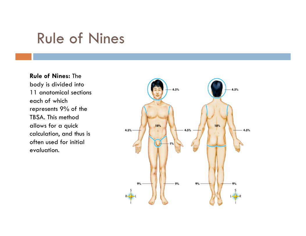
There are two main methods to determine percentage of Total Body Surface Area or TBSA. Rule of Nines:
The body is divided into 11 anatomical sections each of which represents 9% of the TBSA.
Lund-Browder: More accurate and used for both TBSA and fluid replacement. Used more in hospitals. Not for immediate assessments as it is more time
consuming.
“Although TBSA is expressed as a numeric value, it does not mean that a 50% burn is twice as serious as a 25% burn because it does not include information on depth or locus of the burn” (Van Loey and Van Son, 2003, 268).
Each wound may have a different depth, and so each requires assessment and treatment individually.
Rule of Nines
Rule of Nines: The body is divided into 11 anatomical sections each of which represents 9% of the TBSA. This method allows for a quick calculation, and thus is often used for initial evaluation.

Classification
Burns are classified by the cellular level of the injury. 1st degree burns are also known as superficial burns.
First-degree burns are isolated to the epidermis.
2nd degree burns involve the epidermis and the upper layer of the dermis. They are then sub-divided into superficial partial-thickness and
deep partial-thickness.
3rd degree burns are also known as full-thickness burns They involve both the epidermis and dermis.

1st Degree – Superficial Burn
Common Causes: Sunburn Ultraviolet exposure Brief exposure to flame or hot liquids
Appearance: Mild to severe redness Skin blanches with no pressure Dry No blisters Variable amount of edema
Sensation: Painful Hyperesthetic – highly sensitized Tingling Pain eased by cooling
Course: Discomfort lasts about 48 hours Desquamation, or peeling, in three to seven days

2nd Degree – Partial-Thickness Burn
Common Causes: Superficial: Scalding from liquids, semi-liquids, solids Deep: Immersion scald, flame
Appearance: Superficial: Large, thick-walled blisters covering extensive area Deep: Edema that is mottled with a red base or broken epidermis that is wet, shiny
and weeping Sensation:
Painful Sensitive to cool air
Course: Superficial: Heal in 14 to 21 days Deep: Requires 21 to 28 days to heal
Healing depends on depth and absence of infection

3rd Degree – Full Thickness Burn
Common Causes: Prolonged exposure to flame, scalding liquids, steam, chemicals, or electrical voltage
Appearance: Variable (can be deep red, black, white, or brown) Dry surface Edema Exposed fat Disrupted tissue
Sensation: Little or no pain (at immediate burn site) Insensate
Course: Dead skin suppurates and liquefies after 2 to 3 weeks Spontaneous healing without grafting is unusual Requires removal of eschar Often requires grafting Hypertrophic scarring and contractures likely Prolonged recovery period

4th Degree Burn
In most instances, the three levels of burn are all that are used for classification.
Some literature mentions 4th degree burns, and define it as a burn that has penetrated beyond the dermis and involves fat tissue, muscle, and bone.
Fourth degree burns often result from a person not being able to remove him- or herself from a burn source, such as being caught in a burning vehicle or burned by a high-voltage electricity wire (Murphy, Colwell, Pineda, & Bryan, 2009).
It has been noted that the reason people are less aware of this burn is that very few people with this level of injury survive.
Survival is improved if the injury is to a part that can be amputated.

Etiology
75% of burn injuries result from the actions of the injured person. (Goodman, 2009)
Besides classification by cellular damage, burns are also classified by their causes. Thermal, Chemical, Electrical, Radiation
Many burns, particularly 1st and 2nd degree, are often managed without any medical intervention.
Burns have a voluntary reporting procedure, and so the number of actual burns may be under-reported

Population (1)
Burns can happen to anyone, however most burn victims are: Children and Seniors
In the United States, it is estimated 1.2 million people suffer a burn each year. More than 50,000 require hospitalization. Approximately 5,000 die from burn injuries. It is thought that half of all burns involve smoking or
substance abuse. (Murphy et al., 2009) The U.S. population most affected, is:
70% male 17% children under the age of 5 12% age 60 or above (American Burn Association, 2009).

Population (2)
Burns are a global problem, and burn management in developing countries is difficult and costly. India, for instance, with a population of more than 1
billion, has an estimated 700,000-800,000 burn admissions annually (Baghel, Shukla, Mathur, & Randa, (2009).
Globally, burns are a serious public health problem. There are over 300,000 deaths each year from fires
alone, with more deaths from scalds, electrical burns, and other forms of burns, for which global data are not available

Population (3)
Of reported burns 66% are in the home
Symptoms to be Aware of
Disruption of any portion of the skin may result in complications including: Infection Dehydration Shock Inability to maintain correct temperature regulation Loss of sensation Destruction of muscle tissue Respiratory problems Issues with excretion Excruciating pain Contractures Hypertrophic scars

Psychological Symptoms
Psychological symptoms Withdrawal Denial Regression Anxiety Depression Grief Post Traumatic Stress Disorder (PTSD) Acute and long-term neuropsychological problems (most
often with electrical injury) Sleep disturbance
Emergent Phase Treatment
Screen client for immediate needs, varies by burn Reduce edema Position and splint to help prevent contracture, which
will begin within 24 to 48 hours Static splinting in a functional position Monitoring to adjust splints to avoid pressure and skin
damage Wound dressing, removing devitalized tissue, apply
topical agents Antimicrobials include mafenide acetate, silver sulfadiazine
cream, and silver nitrate solution.

Acute Phase Treatment
Obtain better history, including any other medical issues Conduct ADL / IADL assessments
Transfers, ambulation, sitting tolerance (given medical clearance)
Splinting, positioning, ROM, strengthening, environmental modifications, pain remediation, adaptations, client and family education Activity in this phase prevents muscle atrophy, reduce edema, build
endurance, and preserve range of motion Contraindications to exercise in this stage include:
Exposed tendons Grafting takes 3 to 10 days to adhere, so during this phase
immobilization is important Fractures
Video

Rehabilitation Phase Treatment
On emergence from burn unit the goal is to help the client return to pre-injury occupational level. Desensitization, as grafted skin is often hypersensitive Begin working on ADLs and IADLs Circuit training to stretch, improve strength and endurance Scar management – massage and pressure garments Supporting pharmacotherapy and pain management Joining support groups Psychological therapy Educating client and family Coping strategies

Treatment During Rehabilitation
Circuit training to stretch, improve strength and endurance

Long Term Prognosis
Varies enormously and depends on Size and depth of the burn The patient’s history The treatment regimen
“People who have sustained a burn injury are at risk of developing certain psychopathology, in particular Depression and anxiety disorders. Alcohol and substance abuse occur post-burn.
The psychological impact of a burn injury is affected by The experience of the traumatic accident, The prolonged painful treatment during hospitalization, And by integration into society with an altered body” (Van Loey &Van
Son, 2003, 269).

Occupation & Client Factors
Burns can affect almost all occupation and client factors
Occupations: ADLs, IADLs, Rest & Sleep, Education, Work, Play, Leisure,
Social Participation Scars and tissue damage can affect almost all aspects of daily
performance
Client factors Values, Beliefs, and Spirituality
Burn victims often lose self confidence and self esteem Addressing social issues shown to help victim’s deal with
disfigurement

OT Interventions
Stretching and ROM to reduce contracture
Use of games and virtual reality tools to Reduce fear of treatment Extend activity through fun (distraction techniques)
Pain Management and adjustment of activity
Anxiety management Meditation and Yoga
Group socialization activities to deal with embarrassment over scars
Environmental adaptations
Family and patient education

Other Modalities
Honey (Baghel et al., 2009) A study in India found that dressing a wound with honey had a better
outcome in preventing preventive hypertrophic scarring and contracture than treatment with Silver Sulfadiazene (SSD).
Finger-web reconstruction (Emsen, 2008) Surgeons in Turkey have begun performing Cross Incision Plasty. This
surgery uses the skin of the sidewalls of the proximal phalanges to fill in the burned web space on the hand, eliminating skin grafts, and enabling better ROM.
Hypnotherapy Acupuncture Herbs and aloe
Medical & Other Inputs Needed
Medical Skills First Aid Intravenous Fluids Wound treatment Antibiotics
Analgesics Skin Grafting Psychosocial
The full team includes
" First Responders
" Physicians
" Nurses
" Physical Therapists
" Occupational Therapists
" Respirator therapists
" Nutritionists
" Social Workers
" Psychiatrists
" Psychologists
" Speech and Language Pathologists
" Orthotists
" Prosthetists
" Child care
" Recreational therapists
" Pastoral caregivers
" Clergy
" Interpreters
" Cultural support personnel
" Vocational counselors

Medical First Aid – 1st & 2nd Degree
For minor burns, - first-degree burns and second-degree burns limited to 3 inches in diameter: Cool the burn. Cover the burn with a sterile gauze bandage. Take an over-the-counter pain reliever. Rehydration.

Medical First Aid – Major Burns
For major burns, call 911 or emergency medical help.
Until an emergency unit arrives, follow these steps:
Remove from source of burn.
Don't remove burned clothing.
Don't immerse large severe burns in cold water.
Check for signs of circulation (breathing, coughing or movement).
Elevate the burned body part or parts.

Sources American Burn Association (2009). National Burn Repository 2009 Report Dataset Version 5.0. Chicago: American Burn Association.
Baghel, P., Shukla, S., Mathur, R., & Randa, R. (2009). A comparative study to evaluate the effect of honey dressing and silver sulfadiazene dressing on wound healing in burn patients. Indian Journal of Plastic Surgery, 42(2), 176-181. doi:10.4103/0970-0358.59276
Baker, R., Jones, S., Sanders, C., Sadinski, C., Martin-Duffy, K., Berchin, H., Valentine, S. (1996). Degree of burn, location of burn, and length of hospital stay as predictors of psychosocial status and physical functioning. The Journal Of Burn Care & Rehabilitation, 17(4), 327-333.
Emsen, I. (2008). The Cross Incision Plasty (†) for reconstruction of the burned web space: introduction of an alternative technique for the correction of dorsal and volar neosyndactyly. Journal of Burn Care & Research, 29(2), 378-385. doi:10.1097/BCR.0B013e31816673cd
Goodman, C. C., & Fuller, K.S. (2009). Pathology: Implications for the physical therapist (3rd ed.). Philadelphia: Saunders Elsevier.
Haik, J., Tessone, A., Nota, A., Mendes, D., Raz, L., Goldan, O., et al. (2006). The use of video capture virtual reality in burn rehabilitation: the possibilities. Journal Of Burn Care & Research: Official Publication Of The American Burn Association, 27(2), 195-197.
Holaday, M., & McPhearson, R. (1997). Resilience and severe burns. Journal of Counseling & Development, 75(5), 346-356. Retrieved from Academic Search Premier database.
Hill, K., O'Brien, K., & Yurt, R. (2007). Therapeutic efficacy of a therapeutic cooking group from the patients' perspective. Journal Of Burn Care & Research: Official Publication Of The American Burn Association, 28(2), 324-327.
Hwang, Y., Chen-Sea, M., & Chen, C. (2009). Factors related to return to work and job modification after a hand burn. Journal Of Burn Care & Research: Official Publication Of The American Burn Association, 30(4), 661-667.
Kwan, M., & Ha, K. (2002). Splinting programme for patients with burnt hand. Hand Surgery: An International Journal Devoted to Hand and Upper Limb Surgery and Related Research: Journal of the Asia-Pacific Federation of Societies for Surgery of the Hand, 7(2), 231-241. Retrieved from MEDLINE database.

Sources Murphy, P., Colwell, C., Pineda, G., & Bryan, T. (2009). Burning issues: by understanding the pathophysiology of burns, providers can give
patients their best chance at good outcomes. EMS Magazine, 38(10), 83-90.
Pessina, M. A., Orroth, A. C. (2002). Burn injuries. In Radomski, M.V., Latham, C.A.T (Eds.), Occupational therapy for physical dysfunction (6th ed.) (1244-1263). Philadelphia: Lippincott Williams & Wilkins.
Reeves, J., James, F., & McNeill, R. (2009). Providing psychosocial and physical rehabilitation advice for patients with burns. Journal of Advanced Nursing, 65(5), 1039-1043. Retrieved from CINAHL Plus with Full Text database.
Reeves, S.U. (2006). Burns and burn rehabilitation. In H.M. Pendleton & W. Schultz-Krohn (Eds.), Pedretti’s Occupational Therapy: Practice Skills for Physical Dysfunction (6th ed., pp.1056-1094). St. Louis, Missouri: Mosby.
Riley, D., Clark, M., & Wong, T. (2002). World Trade Center terror: explosion trauma -- blast, burns, and crush injury. Topics in Emergency Medicine, 24(2), 47.
Saladin, K. S. (2010). Anatomy & Physiology: The Unity of Form and Function (5th ed.). New York, NY: McGraw-Hill.
Van Loey, N., & Van Son, M. (2003). Psychopathology and psychological problems in patients with burn scars: Epidemiology and management. American Journal of Clinical Dermatology, 4(4), 245-272.
Wiebelhaus, P., Hansen, S. (2001). Managing burn emergencies. Nursing Management, 32(7 part 1), 29-36. Retrieved from CINAHL Plus with Full Text database.
http://ameriburn.org/resources_factsheet.php
Nucleus Medical Media (http://catalog.nucleusinc.com/generateexhibit.php?ID=1871&ExhibitKeywordsRaw=&TL=&A=2).
Daily Telegraph (http://www.telegraph.co.uk/news/uknews/4698866/Schoolgirl-suffers-first-degree-burns-after-19-minutes-in-tanning-salon.html).
1st Degree burn photography and Rule of Nines graphic. (http://www.cpsd.us/CRLS/LC_R/classrooms/AUGUSTINE/Integumentary/index_files/frame.html).