Embed Size (px)
Citation preview
• OLYMPUS CORPORATION has obtained the ISO9001/ISO14001.• OLYMPUS CORPORATION has obtained the MD540624/ISO13485.• Illumination devices for microscope have suggested lifetimes.
Periodic inspections are required. Please visit our web site for details.• Specifications and appearances are subject to change without any notice or obligation on the part of the manufacturer.
Fixed Stage Upright Microscope
BX51WIFixed Stage Upright Microscope with
Motorized Focusing
BX61WI
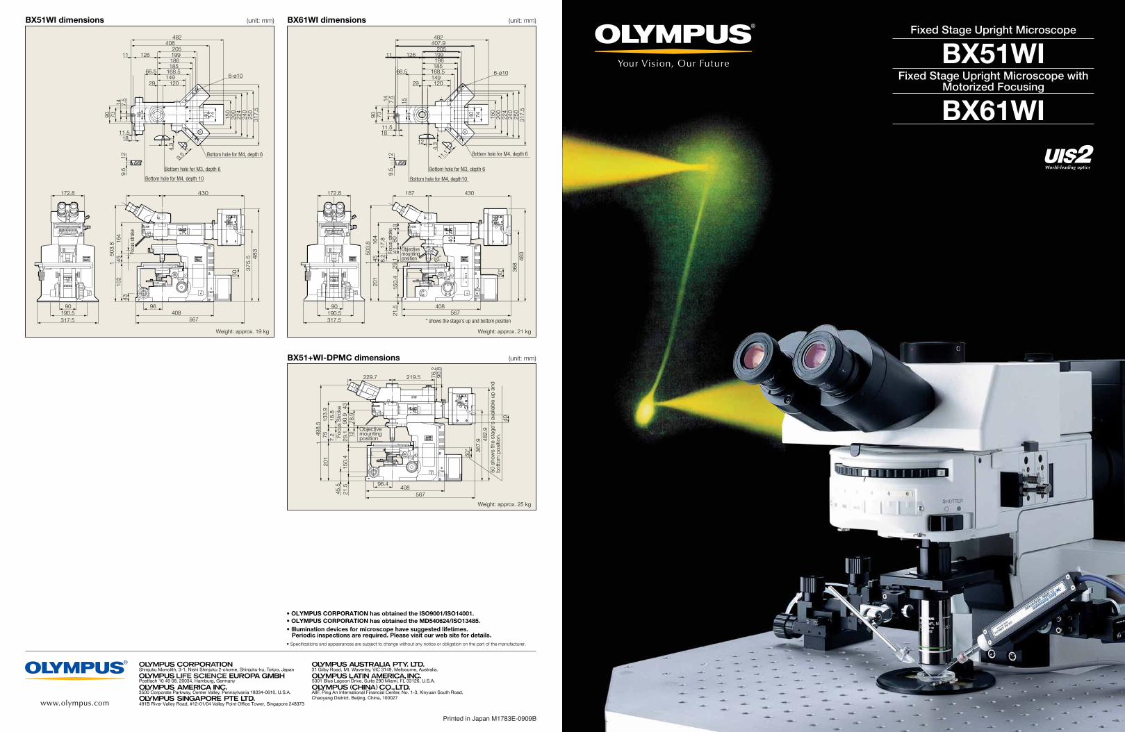
BX51WI dimensions (unit: mm)
Focu
s st
roke
503.
8 164
102
451
43
50 3
75
.5 48
3
430172.8
90190.5317.5
96408
567
12 Bottom hole for M4, depth 6
205
240
200
74
Bottom hole for M3, depth 6
12029
90 73
199186
149
Bottom hole for M4, depth 10
168.5
40
66.5
15 250
224
150
317.
5
1811.5
14 7.5
9.5
6-ø10
185
4.3
9.5
126
408482
11
40
317.5
430
567
483
408
187
503.
820
11 45
172.8
90
164
190.5
50*
Objective mounting position
29.1
150.
421
.5
368
4380
41
62°
* shows the stage's up and bottom position
Focu
s st
roke
8.2
17.8
6-ø10
250
11.1
9.5
7.514
11.5
11
482407.9
40 224
317.
5
126
150
66.5 168.5149
186199
12
29 120
74
Bottom hole for M3, depth 6
200
240
Bottom hole for M4, depth 6
15
7390
18
185
205
12
4.3
Bottom hole for M4, depth10
BX61WI dimensions (unit: mm)
BX51+WI-DPMC dimensions (unit: mm)
Focu
s S
trok
e
50 s
how
s th
e st
age'
s av
aila
ble
up a
nd
bott
om p
ositi
on.
567
482.
9408
201
1
50*
7.2
18.8
Objective mounting position
45.5
29.1
150.
421
.5
40
367.
9
43
96.4
127513
3.9
78.9
90.9
498.
5
76.2
229.7 219.5 90.8
Printed in Japan M1783E-0909B
Weight: approx. 25 kg
Weight: approx. 21 kgWeight: approx. 19 kg
21
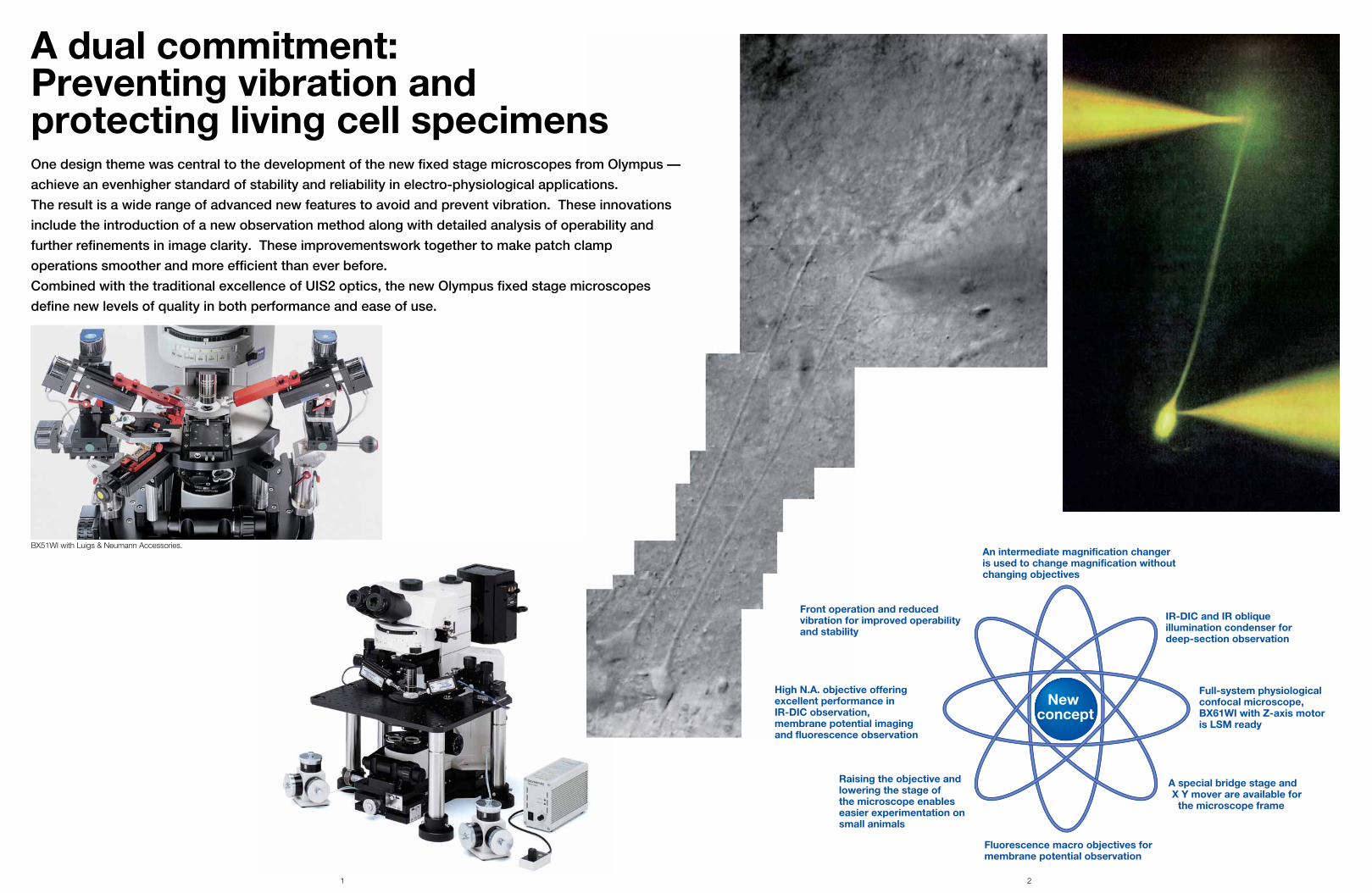
BX51WI with Luigs & Neumann Accessories.
New concept
High N.A. objective offeringexcellent performance inIR-DIC observation, membrane potential imaging and fluorescence observation
Front operation and reduced vibration for improved operability and stability
Raising the objective and lowering the stage ofthe microscope enables easier experimentation onsmall animals
Fluorescence macro objectives formembrane potential observation
A special bridge stage and X Y mover are available for
the microscope frame
Full-system physiologicalconfocal microscope,BX61WI with Z-axis motor is LSM ready
IR-DIC and IR obliqueillumination condenser fordeep-section observation
An intermediate magnification changer is used to change magnification without changing objectives
A dual commitment:Preventing vibration and protecting living cell specimensOne design theme was central to the development of the new fixed stage microscopes from Olympus —
achieve an evenhigher standard of stability and reliability in electro-physiological applications.
The result is a wide range of advanced new features to avoid and prevent vibration. These innovations
include the introduction of a new observation method along with detailed analysis of operability and
further refinements in image clarity. These improvementswork together to make patch clamp
operations smoother and more efficient than ever before.
Combined with the traditional excellence of UIS2 optics, the new Olympus fixed stage microscopes
define new levels of quality in both performance and ease of use.
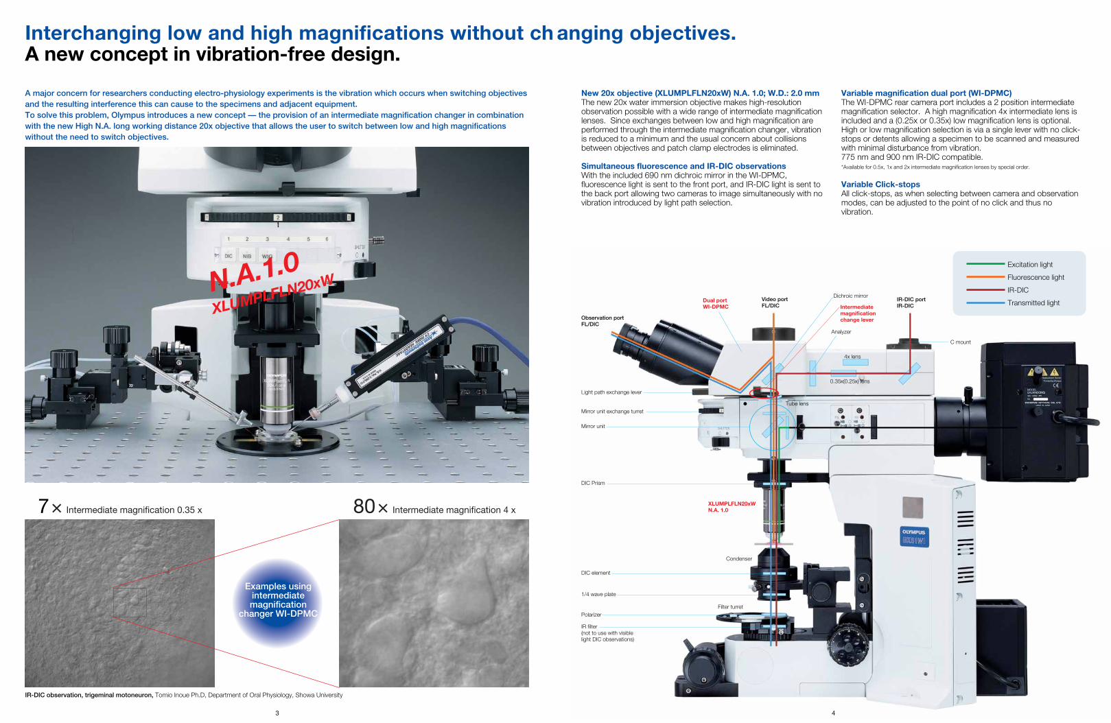
New 20x objective (XLUMPLFLN20xW) N.A. 1.0; W.D.: 2.0 mmThe new 20x water immersion objective makes high-resolutionobservation possible with a wide range of intermediate magnificationlenses. Since exchanges between low and high magnification areperformed through the intermediate magnification changer, vibrationis reduced to a minimum and the usual concern about collisionsbetween objectives and patch clamp electrodes is eliminated.
Simultaneous fluorescence and IR-DIC observationsWith the included 690 nm dichroic mirror in the WI-DPMC,fluorescence light is sent to the front port, and IR-DIC light is sent tothe back port allowing two cameras to image simultaneously with novibration introduced by light path selection.
Variable magnification dual port (WI-DPMC)The WI-DPMC rear camera port includes a 2 position intermediatemagnification selector. A high magnification 4x intermediate lens isincluded and a (0.25x or 0.35x) low magnification lens is optional.High or low magnification selection is via a single lever with no click-stops or detents allowing a specimen to be scanned and measuredwith minimal disturbance from vibration. 775 nm and 900 nm IR-DIC compatible.*Available for 0.5x, 1x and 2x intermediate magnification lenses by special order.
Variable Click-stopsAll click-stops, as when selecting between camera and observationmodes, can be adjusted to the point of no click and thus novibration.
Condenser
Filter turret
7× Intermediate magnification 0.35 x 80× Intermediate magnification 4 x
Examples usingintermediatemagnification
changer WI-DPMC
IR-DIC observation, trigeminal motoneuron, Tomio Inoue Ph.D, Department of Oral Physiology, Showa University
N.A.1.0XLUMPLFLN20xW
Interchanging low and high magnifications without ch anging objectives.A new concept in vibration-free design.
A major concern for researchers conducting electro-physiology experiments is the vibration which occurs when switching objectivesand the resulting interference this can cause to the specimens and adjacent equipment. To solve this problem, Olympus introduces a new concept — the provision of an intermediate magnification changer in combinationwith the new High N.A. long working distance 20x objective that allows the user to switch between low and high magnificationswithout the need to switch objectives.
IR-DIC portIR-DIC
XLUMPLFLN20xWN.A. 1.0
Video portFL/DIC
Light path exchange lever
Intermediatemagnificationchange lever
IR filter(not to use with visiblelight DIC observations)
Observation portFL/DIC
Dual portWI-DPMC
Mirror unit exchange turret
0.35x(0.25x) lens
Tube lens
Mirror unit
DIC Prism
Analyzer
C mount
Dichroic mirror
4x lens
Polarizer
1/4 wave plate
DIC element
3 4 4
Excitation light
Fluorescence light
IR-DIC
Transmitted light
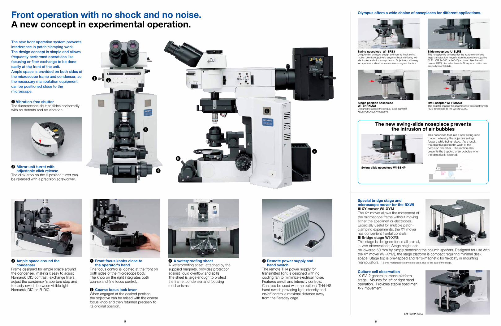
q Vibration-free shutterThe fluorescence shutter slides horizontallywith no detents and no vibration.
Special bridge stage andmicroscope mover for the BXWI■ XY mover WI-XYM The XY mover allows the movement ofthe microscope frame without movingeither the specimen or electrodes.Especially useful for multiple patch-clamping experiments, the XY moverhas convenient frontal controls.■ Bridge stage WI-XYSThis stage is designed for small animal,in-vivo observations; Stage height canbe lowered 50 mm by simply detaching the column spacers. Designed for use withthe XY mover (WI-XYM), the stage platform is compact requiring minimal deskspace. Stage top is pre-tapped and ferro-magnetic for flexibility in mountingmanipulators. * Some manipulators cannot be used, due to the size of the stage.u Remote power supply and
hand switchThe remote TH4 power supply fortransmitted light is designed with no cooling fan to minimize electrical noise.Features on/off and intensity controls. Can also be used with the optional TH4-HShand switch providing light intensity andon/off control a maximal distance awayfrom the Faraday cage.
y A waterproofing sheetA waterproofing sheet, attached by thesupplied magnets, provides protectionagainst liquid overflow and spills. The sheet is large enough to protect the frame, condenser and focusingmechanisms .
e Ample space around the condenser
Frame designed for ample space aroundthe condenser, making it easy to adjustNomarski DIC contrast, exchange filters,adjust the condenser's aperture stop and to easily switch between visible light,Nomarski DIC or IR-DIC.
w Mirror unit turret with adjustable click release
The click-stop on the 6 position turret canbe released with a precision screwdriver.
This nosepiece features a new swing-slidemotion, whereby the objective swingsforward while being raised. As a result, the objective clears the walls of the perfusion chamber. This motion alsoprevents the trapping of air bubbles whenthe objective is lowered.
Olympus offers a wide choice of nosepieces for different applications.
The new swing-slide nosepiece prevents the intrusion of air bubbles
Swing-slide nosepiece WI-SSNP
Single position nosepieceWI-SNPXLU2Designed to accept the unique, large diameterXLUMPLFLN20xW objective.
RMS adapter WI-RMSADThis adapter enables the attachment of an objective withRMS thread size to the WI-SNPXLU2.
Swing nosepiece WI-SRE3Uniquie slim, compact design and front-to-back swingmotion permits objective changes without interfering withelectrodes and micromanipulators. Objective positioningincorporates a vibration-free counterspring mechanism.
Slide nosepiece U-SLREThis nosepiece is designed for the attachment of onelarge diameter, low magnification fluorescence objective(XLFLUOR 2x/340 or 4x/340) and one objective withnormal (RMS) diamerter threads. Nosepiece motion is asimple horizontal slide.
r Front focus knobs close to the operator's hand
Fine focus control is located at the front onboth sides of the microscope body. The knob on the right integrates bothcoarse and fine focus control.
t Coarse focus lock leverWhen engaged at the desired position, the objective can be raised with the coarsefocus knob and then returned precisely toits original position.
Front operation with no shock and no noise.A new concept in experimental operation.
r
e
wq
The new front operation system preventsinterference in patch clamping work. The design concept is simple and allowsfrequently performed operations likefocusing or filter exchange to be doneeasily at the front of the unit. Ample space is provided on both sides ofthe microscope frame and condenser, sothe necessary manipulation equipmentcan be positioned close to themicroscope.
u
5 6
Culture cell observationIX-SVL2 general purpose platformstage. Mounts for left or right handoperation. Provides stabile specimen X-Y movement.
BX51WI+IX-SVL2
y
t
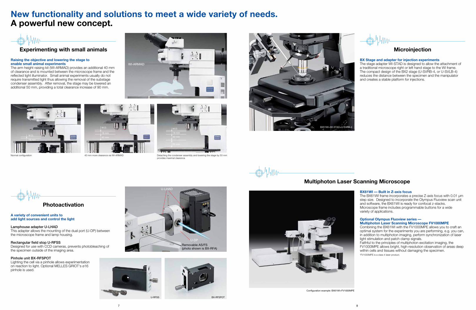
Raising the objective and lowering the stage to enable small animal experimentsThe arm height raising kit (WI-ARMAD) provides an additional 40 mmof clearance and is mounted between the microscope frame and thereflected light illuminator. Small animal experiments usually do notrequire transmitted light thus allowing the removal of the substagecondenser assembly. After removal, the stage may be lowered anadditional 50 mm, providing a total clearance increase of 90 mm.
A variety of convenient units toadd light sources and control the light
Lamphouse adapter U-LHADThis adapter allows the mounting of the dual port (U-DP) between the microscope frame and lamp housing.
Rectangular field stop U-RFSSDesigned for use with CCD cameras, prevents photobleaching of the specimen outside of the imaging area.
Pinhole unit BX-RFSPOTLighting the cell via a pinhole allows experimentation on reaction to light. Optional MELLES GRIOT's ø16pinhole is used.
New functionality and solutions to meet a wide variety of needs.A powerful new concept.
U-DP
U-LHAD
WI-ARMAD
BX-RFSPOT
Removable AS/FS(photo shown is BX-RFA)
Experimenting with small animals
Photoactivation
BX61WI — Built in Z-axis focus The BX61WI frame incorporates a precise Z-axis focus with 0.01 µmstep size. Designed to incorporate the Olympus Fluoview scan unitand software, the BX61WI is ready for confocal z-stacks.Microscope frame includes programmable buttons for a wide variety of applications.
Optional Olympus Fluoview series — Multiphoton Laser Scanning Microscope FV1000MPECombining the BX61WI with the FV1000MPE allows you to craft anoptimal system for the experiments you are performing, e.g. you can,in addition to multiphoton imaging, perform synchronization of laserlight stimulation and patch clamp signals.Faithful to the principles of multiphoton excitation imaging, theFV1000MPE allows bright, high-resolution observation of areas deepwithin cells and tissues without damaging the specimen.*FV1000MPE is a class 4 laser product.
Multiphoton Laser Scanning Microscope
Normal configuration 40 mm more clearance via WI-ARMAD Detaching the condenser assembly and lowering the stage by 50 mmprovides maximal clearance
7 8
BX Stage and adapter for injection experimentsThe stage adapter WI-STAD is designed to allow the attachment of a traditional microscope right or left hand stage to the WI frame. The compact design of the BX2 stage (U-SVRB-4, or U-SVLB-4)reduces the distance between the specimen and the manipulator and creates a stable platform for injections.
Microinjection
BX51WI+WI-STAD+U-SVRB-4
40 mm
50 mm
18 mm
W.D.
18 mm
W.D.18 mm
40 mm
W.D.
U-RFSS
Configuration example: BX61WI+FV1000MPE
9
Ultimate image clarity for electro-physiological experiments.A new concept in live cell observation.
IR-DIC/ Nomarski DIC observation
Oblique illumination observation
Oblique observation optimizes contrast by changing the direction of the specimen shadowOlympus has developed an oblique condenser (WI-OBCD) whoselong working distance enables the angles of shadow to be alteredthrough 360 degrees without moving the specimen. Requiring noadditional accessories, oblique illumination is easy to set up andcontrol. Plastic dishes (normally unsuitable for all types of DIC) areeasy to image with oblique illumination. The oblique illumination slitaperture is variable in size and on a slider allowing quick changeover.
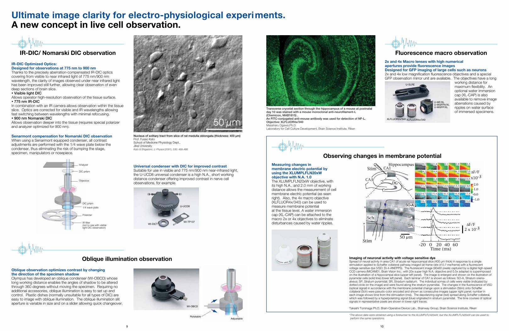
IR-DIC Optimized Optics:Designed for observations at 775 nm to 900 nmThanks to the precisely aberration-compensated IR-DIC opticscovering from visible to near infrared light of 775 nm/900 nmwavelength, the clarity of images observed under near infrared lighthas been improved still further, allowing clear observation of evendeep sections of brain slice.• Visible light DICAllows operator high-resolution observation of the tissue surface.• 775 nm IR-DICIn combination with an IR camera allows observation within the tissueslice. Optics are corrected for visible and IR wavelengths allowingfast switching between wavelengths with minimal refocusing.• 900 nm Nomarski DICAllows observation deeper into the tissue (requires special polarizerand analyzer optimized for 900 nm).
Analyzer
DIC prism
Objective
DIC prism
1/4 wave plate
Polarizer
IR filter(not to use with visiblelight DIC observation)
10
Nucleus of solitary tract from slice of rat medulla oblongata (thickness: 400 µm)Prof. Fusao KatoSchool of Medicine Physiology Dept.,Jikei UniversityKato & Shigetomi, J. Physiol.(2001), 530: 469-486
Transverse cryostat section through the hippocampus of a mouse at postnatalday 10 was stained with a mouse monoclonal anti-neurofilament-L (Chemicon, MAB1615) .An FITC-conjugated anti-mouse antibody was used for detection of NF-L.Objective: XLFLUOR4x/340Masaharu Ogawa,Ph.DLaboratory for Cell Culture Development, Brain Science Institute, Riken
Observing changes in membrane potential
Fluorescence macro observation
Measuring changes in membrane electric potential by using the XLUMPLFLN20xW objective with N.A. 1.0The XLUMPLFLN20xW objective, withits high N.A., and 2.0 mm of workingdistance allows the measurement of cellmembrane electric potential (as seenright). Also, the 4x macro objective(XLFLUOR4x/340) can be used tomeasure membrane potential at the tissue level. A water immersioncap (XL-CAP) can be attached to themacro 2x or 4x objectives to eliminatedisturbances caused by water ripples.
2x and 4x Macro lenses with high numerical apertures provide fluorescence images Designed for GFP imaging of large cells such as neurons2x and 4x low magnification fluorescence objectives and a specialGFP observation mirror unit are available. The objectives have a long
working distance formaximum flexibility. Anoptional water immersioncap (XL-CAP) is alsoavailable to remove imageaberrations caused byripples on water surfaceof immersed specimens.
Imaging of neuronal activity with voltage sensitive dyeSpread of neural activity in area CA1 of acute rat hippocampal slice (400 µm thick) in response to a singlestimulation applied to Schaffer collateral pathway imaged (at frame rate of 0.7 ms/frame) with a fluorescentvoltage sensitive dye (VSD; Di-4-ANEPPS). The fluorescent image (90x60 pixels) captured by a digital high-speedCCD camera (MiCAM01, Brain Vision Inc.; with 20x super high N.A. objective and 0.5x adapter) is superimposedon the illustration of a hippocampal slice (upper left panel). The image is enlarged and shown on the illustration ofpyramidal cells (solid line) (lower left panel). Each laminar of CA1 is shown as follows: SO-A, Stratum oriens-alveus; SP, Stratum pyramidal; SR, Stradum radiatum. The individual somas of cells were visible (indicated bydotted circle on the image) and were found along the stratum pyramidal. The changes in the fluorescence of VSD(optical signal) in accordance with the membrane potential change upon a stimulation (Stim) onto Schaffercollateral (Sch) were pseudo-color encoded and shown as consecutive images (upper right panel; number ineach image shows time from the stimulation (ms)). The depolarizing signal (red) spread along Schaffer collateral,which was followed by a hyperpolarizing signal (blue) originated in stratum pyramidal. The time courses of opticalsignals in representative pixels are shown in lower right traces.
Takashi Tominaga Ph.D, Brain-Operative Device Lab., Brainway Group, Brain Science Institute, Riken
*The above data were obtained using a forerunner to the XLUMPLFLN20xW, but the XLUMPLFLN20xW can be used toperform the same operations.
Universal condenser with DIC for improved contrastSuitable for use in visible and 775 nm/900 nm near-infrared light, the U-UCD8 universal condenser is a high N.A., short workingdistance condenser offering improved contrast in nerve cellobservations, for example.
Adjustable
U-UCD8
WI-TP137WI-DICT
WI-OBCD
U-SLRE
XLFLUOR2x/340 XLFLUOR4x/340
U-MF/XLU-MGFPA/XLU-MGFP/XL
Rotatable
Senarmont compensation for Nomarski DIC observationWhen using a Senarmont equipped condenser, all contrastadjustments are performed with the 1/4 wave plate below thecondenser, thus eliminating the risk of bumping the stage, specimen, manipulators or nosepiece.
WI-XYM dimensions (unit: mm)
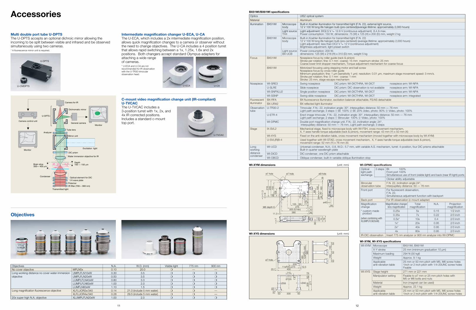
Intermediate magnification changer U-ECA, U-CAThe U-ECA, which includes a 2x intermediate magnification position,allows quick magnification changes to a camera or observer withoutthe need to change objectives. The U-CA includes a 4 position turretthat allows rapid switching between a 1x, 1.25x, 1.6x and 2xpositions. Both changers accept standard Olympus adapters forattaching a wide rangeof cameras.* U-ECA and U-CA are not recommended for IR observationwith the U-TR30 trinocularobservation head.
Accessories
Multi double port tube U-DPTSThe U-DPTS accepts an optional dichroic mirror allowing theincoming to be split between visible and infrared and be observedsimultaneously using two cameras.* A fluorescence mirror unit is required.
Camera control unit
Monitor
Camera for IR
U-DPTS
General camera
Water immersion objective for IR
Optical element for DIC
DIC prism
IR filter (700— 900 nm)
Tube lensAnalyzer
Mirror unit
Excitation light
Transmitted light
Dichromatic mirror
Depth100 — 150 μmBrain slice
300— 400 μm
Condenser
Polarizer1/4 wave plate
Monitor
C-mount video magnification change unit (IR-compliant)U-TVCACThe U-TVCAC includes a 3-position turret with 1x, 2x, and4x IR corrected positions.Includes a standard c-mount top port.
Objectives
Objectives N.A. W.D. (mm) Visible light 775 nm 900 nmNo cover objective MPLN5x 0.10 20.0 ❍ — —Long working distance no cover water immersion UMPLFLN10xW 0.30 3.5 ❍ ❍ ❍
objective UMPLFLN20xW 0.50 3.5 ❍ ❍ ❍
LUMPLFLN40xW 0.80 3.3 ❍ ❍ ❍
LUMPLFLN60xW 1.00 2.0 ❍ ❍ ❍
LUMFLN60xW 1.10 1.5 ❍ ❍ ❍
Long magnification fluorescence objective XLFLUOR2x/340 0.14 21.0 (include 5 mm water) ❍ ❍ —XLFLUOR4x/340 0.28 29.5 (include 5 mm water) ❍ ❍ —
20x super high N.A. objective XLUMPLFLN20xW 1.00 2.0 ❍ ❍ ❍
11 12
BX51WI/BX61WI specifications
WI-XYM, WI-XYS specifications
M6 depth 6
380
ø9 holeø7 holeø7 hole
355.5(14in)275 11
1056
ø40
38
70 78
105
108
150
200
216
33
8
7
11.3
11.3
60 7755
68
58ø40
68
10
160
2830
28
100
10
200
110
30050
∅105
∅58 ∅40
180
209
50
1.8
1227
1
11xP25=275 12.540020.7
ø7 hole
12xP
25=
300
380
100
100
5050
50
5020 50
WI-XYS dimensions (unit: mm)
Optics UIS2 optical systemMaterial Aluminum
Illumination BX51WI Microscope Built-in Koehler illumination for transmitted light (F.N. 22), external light source, body 12 V 100 W long life halogen bulb (pre-centered)(average lifetime: approximately 2,000 hours)Light source Light adjustment: DC2.5 V g 12.6 V (continuous adjustment) 8.4 A max.TH4 Power consumption: 150 W, dimensions: 75 (W) x 125 (H) x 200 (D) mm, weight 2 kg
BX61WI Microscope Built-in Koehler illumination for transmitted light (F.N. 22)body 12 V 100 W long life halogen bulb (pre-centered) (average lifetime: approximately 2,000 hours)
Light adjustment: less than DC2 V g 12 V (continuous adjustment)Brightness adjustment, light preset switch
Light source Power consumption: 200 W,BX-UCB dimensions: 125 (W) x 216 (H) x 310 (D) mm, weight 5 kg
Focus BX51WI Nosepiece focus by roller guide (rack & pinion)Stroke per rotation; fine: 0.1 mm coarse: 15 mm maximum stroke: 25 mmCoarse lower limit stopper mechanism, Torque adjustment mechanism for coarse focus
BX61WI Motorized focusing using stepping motor and ball screwNosepiece focus by cross roller guideMinimum graduation; fine: 1 µm (sensitivity 1 µm) resolution: 0.01 µm, maximum stage movement speed: 3 mm/s.Stroke per rotation; fine: 0.1 mm coarse: 1 mmStroke: 25 mm, stage escape mechanism
Nosepiece WI-SRE3 Swing nosepiece DIC prism: WI-DICTHRA, WI-DICT nosepiece arm: WI-NPA
U-SLRE Slide nosepiece DIC prism: DIC observation is not available nosepiece arm: WI-NPA WI-SNPXLU2 Single position nosepiece DIC prism: WI-DICTHRA, WI-DICT nosepiece arm: WI-NPA
WI-SSNP Swing-slide nosepiece DIC prism: WI-DICTHRA, WI-DICT nosepiece arm: integratedFluorescent BX-RFA BX fluorescence illuminator, excitation balancer attachable, FS/AS detachableilluminator BX-URA2 BX reflected light illuminatorObservation U-TR30-2 Trinocular, F.N.: 22, inclination angle: 30°, interpupillary distance: 50 mm — 76 mmtube Light path exchange; 3 steps q BI: 100% w BI: 20% video, photo: 80% e Video, photo: 100%
U-ETR-4 Erect image trinocular, F.N.: 22, inclination angle: 30°, interpupillary distance: 50 mm — 76 mmLight path exchange; 2 steps q Binocular: 100% w Video, photo: 100%
WI-DPMC Double port magnification change unit, F.N.: 22, inclination angle: 24°,interpupillary distance: 50 mm — 76 mm, Light path exchange; 3 steps
Stage IX-SVL2 Mechanical stage, fixed to microscope body with WI-FSH, cross movement mechanism, X, Y axes handle torque adjustable (rack & pinion), movement range: 43 mm (Y) x 50 mm (X)
WI-XYS Fixed on the anti-vibration table, cross movement mechanism (moved together with microscope body by WI-XYM)
U-SVL(R)B-4 Used together with WI-STAD, cross movement mechanism, X, Y axes handle torque adjustable (rack & pinion), movement range: 52 mm (Y) x 76 mm (X)
Long WI-UCD Universal condenser, N.A.: 0.8, W.D.: 5.7 mm, with variable A.S. mechanism, turret: 4-position, four DIC prisms attachable working Built-in quarter wavelength platedistance WI-DICD DIC condenser, one DIC prism attachable condenser
WI-OBCD Oblique condenser, built-in variable oblique illumination stop
WI-XYM Microscope BX51WI, BX61WIX-Y stroke 25 mm (minimum graduation 10 µm)
Maximum loading 294 N (30 kgf)Weight Approx. 9.1 kg
Applicable 25 mm or 50 mm pitch with M5, M6 screw holesanti-vibration table 1inch or 2 inch pitch with 1/4-20UNC screw holes
FixableWI-XYS Stage height 271 mm or 221 mm
Manipulator setting Fixable to ø7 mm or 25 mm pitch holes with M5 or M6 bolts and nuts
Material Iron (magnet can be used)
Weight Approx. 22.1 kgApplicable 25 mm or 50 mm pitch with M5, M6 screw holesanti-vibration table 1inch or 2 inch pitch with 1/4-20UNC screw holes
WI-DPMC specifications
Optical 3 steps BI: 100%light path Front port 100%exchange Simultaneous use of front (visible light) and back (near IR light) ports
Clicker ability adjustable
Binocular F.N. 22, inclination angle 24°, observation tube Interpupillary distance: 50 — 76 mmFront port For fluorescent observation,
F.N. 22, Simultaneous adjustment function with backport
Back port For IR observation (c mount adapter)Magnification Magnification changer Total N.A. Projectionchange lens magnification magnification magnification
* custom made 0.25x 5x 0.15 1/2 inchproduct 0.35x 7x 0.22 2/3 inch
(when combining with 0.5x* 10x 0.4 2/3 inchXLUMPLFLN20xW)
1x* 20x 0.95 2/3 inch2x* 40x 0.95 2/3 inch
4x 80x 0.95 2/3 inchIR-DIC observation Insert 775 nm analyzer or 900 nm analyzer into WI-DPMC
U-CMDPTS
U-DPTS
U-ECA U-CA
U-TVCAC
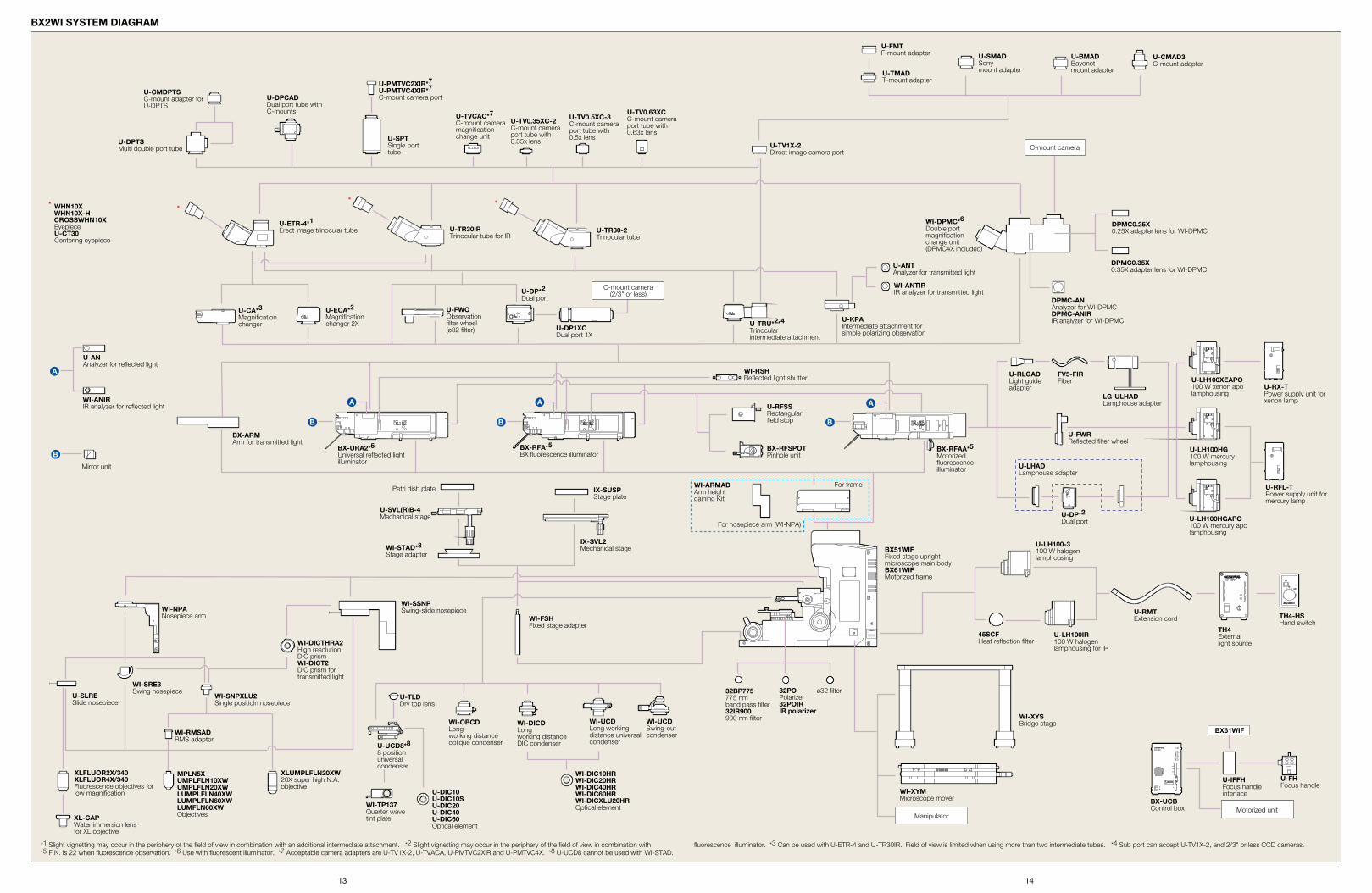
BX2WI SYSTEM DIAGRAM
U-TVCAC*7C-mount camera magnification change unit
U-DPTSMulti double port tube
U-CMDPTSC-mount adapter for U-DPTS
U-TR30-2Trinocular tube
U-TR30IRTrinocular tube for IR
U-ETR-4*1Erect image trinocular tube
U-ECA*3Magnification changer 2X
WI-RSHReflected light shutter
U-CA*3Magnification changer
U-DP*2 Dual port
U-DP1XCDual port 1X
U-TRU*2*4 Trinocularintermediate attachment
U-KPAIntermediate attachment for simple polarizing observation
U-ANTAnalyzer for transmitted light
WI-DPMC*6Double port magnificationchange unit(DPMC4X included)
DPMC0.25X0.25X adapter lens for WI-DPMC
DPMC0.35X0.35X adapter lens for WI-DPMC
DPMC-ANAnalyzer for WI-DPMCDPMC-ANIRIR analyzer for WI-DPMC
WI-ANTIRIR analyzer for transmitted light
U-FWOObservation filter wheel(ø32 filter)
BX-RFAA*5Motorized fluorescence illuminator
BX-URA2*5Universal reflected lightilluminator
U-LH100XEAPO100 W xenon apo lamphousing
U-LH100HGAPO100 W mercury apo lamphousing
U-LH100HG100 W mercury lamphousing
U-LH100IR100 W halogen lamphousing for IR
U-LH100-3100 W halogen lamphousing
U-RMTExtension cord
BX51WIFFixed stage upright microscope main bodyBX61WIFMotorized frame
45SCFHeat reflection filter
TH4-HSHand switch
TH4External light source
WI-XYSBridge stage
WI-XYMMicroscope mover
ø32 filter32POPolarizer32POIRIR polarizer
32BP775775 nm band pass filter32IR900900 nm filterWI-UCD
Long working distance universal condenser
WI-UCDSwing-out condenser
WI-DIC10HRWI-DIC20HRWI-DIC40HRWI-DIC60HRWI-DICXLU20HROptical element
WI-DICDLong working distanceDIC condenser
WI-OBCDLong working distanceoblique condenser
WI-FSHFixed stage adapter
WI-TP137Quarter wave tint plate
U-IFFHFocus handle interface
U-FHFocus handle
U-UCD8*88 positionuniversalcondenser
BX-UCBControl box
XLUMPLFLN20XW20X super high N.A. objective
MPLN5XUMPLFLN10XWUMPLFLN20XWLUMPLFLN40XWLUMPLFLN60XWLUMFLN60XWObjectives
U-SLRESlide nosepiece
WI-SRE3Swing nosepiece
WI-NPANosepiece arm
WI-ARMADArm height gaining Kit
For nosepiece arm (WI-NPA)
For frame
BX-ARMArm for transmitted light
WI-SSNPSwing-slide nosepiece
WI-SNPXLU2Single positioin nosepiece
WI-RMSADRMS adapter
WI-DICTHRA2High resolutionDIC prismWI-DICT2DIC prism for transmitted light
XLFLUOR2X/340XLFLUOR4X/340Fluorescence objectives for low magnification
XL-CAPWater immersion lens for XL objective
U-DIC10U-DIC10SU-DIC20U-DIC40U-DIC60Optical element
C-mount camera
C-mount camera(2/3" or less)
U-RLGADLight guide adapter
FV5-FIRFiber
U-DP*2 Dual port
U-FWRReflected filter wheel
LG-ULHADLamphouse adapter
U-LHADLamphouse adapter
BX61WIF
* WHN10X WHN10X-H CROSSWHN10X
EyepieceU-CT30Centering eyepiece
* * *
IX-SVL2Mechanical stage
IX-SUSPStage plate
WI-STAD*8Stage adapter
Petri dish plate
U-SVL(R)B-4Mechanical stage
U-ANAnalyzer for reflected light
WI-ANIRIR analyzer for reflected light U-RFSS
Rectangular field stop
BX-RFSPOTPinhole unit
Mirror unit
U-TLDDry top lens
U-TV1X-2Direct image camera port
U-SPTSingle port tube
U-PMTVC2XIR*7U-PMTVC4XIR*7C-mount camera port
U-CMAD3C-mount adapter
U-BMADBayonetmount adapter
U-FMTF-mount adapter
U-TMADT-mount adapter
U-SMADSonymount adapter
ManipulatorMotorized unit
*1 Slight vignetting may occur in the periphery of the field of view in combination with an additional intermediate attachment. *2 Slight vignetting may occur in the periphery of the field of view in combination with fluorescence illuminator. *3 Can be used with U-ETR-4 and U-TR30IR. Field of view is limited when using more than two intermediate tubes. *4 Sub port can accept U-TV1X-2, and 2/3" or less CCD cameras. *5 F.N. is 22 when fluorescence observation. *6 Use with fluorescent illuminator. *7 Acceptable camera adapters are U-TV1X-2, U-TVACA, U-PMTVC2XIR and U-PMTVC4X. *8 U-UCD8 cannot be used with WI-STAD.
BX-RFA*5BX fluorescence illuminator
U-TV0.63XCC-mount camera port tube with 0.63x lens
U-TV0.5XC-3C-mount camera port tube with 0.5x lens
U-TV0.35XC-2C-mount camera port tube with 0.35x lens
U-RX-TPower supply unit forxenon lamp
U-RFL-TPower supply unit formercury lamp
U-DPCADDual port tube with C-mounts
13 14